Article

What Is behind the Correlation Analysis of Diarrheagenic E. coli Pathotypes?

by
Mahmoud M. Bendary
1,*,†,
Marwa I. Abd El-Hamid
2,†,
Majid Alhomrani
3,4,
Abdulhakeem S. Alamri
3,4,
Rana Elshimy
5,6,
Rasha A. Mosbah
7,
Mosa M. Bahnass
8,
Nasreen N. Omar
9,
Mohammad M. Al-Sanea
10,*,
Arwa R. Elmanakhly
11,
Nesreen A. Safwat
11 and
Walaa A. Alshareef
12
1
Department of Microbiology and Immunology, Faculty of Pharmacy, Port Said University, Port Said 42511, Egypt
2
Department of Microbiology, Faculty of Veterinary Medicine, Zagazig University, Zagazig 44511, Egypt
3
Department of Clinical Laboratories Sciences, The Faculty of Applied Medical Science, Taif University, Taif 26432, Saudi Arabia
4
Centre of Biomedical Science Research (CBSR), Deanship of Scientific Research, Taif University, Taif 26432, Saudi Arabia
5
Department of Microbiology and Immunology, Egyptian Drug Authority, Giza 12511, Egypt
6
Department of Microbiology and Immunology, Faculty of Pharmacy, Alhram Canadian University, Giza 12511, Egypt
7
Infection Control Unit, Zagazig University Hospital, Zagazig 44511, Egypt
8
Department of Animal Medicine (Infectious Disease), Faculty of Veterinary Medicine, Zagazig University, Zagazig 44511, Egypt
9
Department of Biochemistry, Faculty of Pharmacy, Modern University for Technology and Information, Cairo 19448, Egypt
10
Pharmaceutical Chemistry Department, College of Pharmacy, Jouf University, Sakaka 72314, Saudi Arabia
11
Department of Microbiology and Immunology, Faculty of Pharmacy, Modern University for Technology and Information, Cairo 11559, Egypt
12
Department of Microbiology and Immunology, Faculty of Pharmacy, October 6 University, 6 October 12566, Egypt
*
Authors to whom correspondence should be addressed.
These authors contributed equally to this work.
Biology 2022, 11(7), 1004; https://doi.org/10.3390/biology11071004
Submission received: 19 April 2022 / Revised: 17 June 2022 / Accepted: 22 June 2022 / Published: 2 July 2022
(This article belongs to the Special Issue Microbial Diversity and Microbial Resistance)

Abstract

:

Simple Summary

To date, despite the efforts made to monitor the wide spread of resistant pathogens, especially multidrug-resistant (MDR) diarrheagenic E. coli, there are limitations in the correlation analysis for these pathogens worldwide. Therefore, it seems important and so timely to assess the E. coli pathotypes and their correlations with hosts, antimicrobial resistance, virulence gene profiles, and serotypes. Our promising results gave a clear indication for the epidemic situation of diarrheagenic E. coli (DEC) in Egypt and suggested that restricted recommendations and a search for novel alternative therapies are urgently needed due to the wide spread of MDR and multi-virulent E. coli strains in addition to their heterogeneous nature. This study can be implemented in the infection control guidance with enhanced protocols to hinder the spread of MDR E. coli pathotypes in Egyptian hospitals.

Abstract

The treatment failure recorded among patients and animals infected with diarrheagenic Escherichia coli (DEC) was increased due to the presence of specific virulence markers among these strains. These markers were used to classify DEC into several pathotypes. We analyzed the correlations between DEC pathotypes and antimicrobial resistances, the existence of virulence genes, serotypes, and hosts. The ETEC pathotype was detected with a high prevalence rate (25%). Moreover, the ETEC and EPEC pathotypes were highly associated with human infections in contrast to the EIEC and EAEC phenotypes, which were commonly recognized among animal isolates. Interestingly, the antimicrobial resistance was affected by E. coli pathotypes. With the exception of EIEC and STEC, imipenem represented the most effective antibiotic against the other pathotypes. There were fixed correlations between the DEC pathotypes and the presence of virulence markers and hosts; meanwhile, their correlation with serotypes was variable. Additionally, the vast majority of our isolates were highly diverse, based on both phenotypic and ERIC molecular typing techniques. Our promising results gave a clear indication for the heterogeneity and weak clonality of DEC pathotypes in Egypt, which can be utilized in the evaluation of the current therapeutic protocols and infection control guidelines.

1. Introduction

Escherichia coli (E. coli) strains are normal inhabitants of both animal and human gastrointestinal tracts, and they are among the bacterial species that are most frequently recovered from stool cultures [1]. When E. coli strains obtain precise genetic materials, they become pathogenic; these pathogenic E. coli clones are among the most virulent enteric bacterial pathogens [2]. Taken together, most E. coli strains cause serious health problems for both humans and animals as they can cause serious intestinal as well as extraintestinal infections. Therefore, next-generation therapies, in addition to discovering and developing novel antimicrobial agents, are urgently needed [3,4]. Diarrheagenic E. coli (DEC) are among the most abundant bacterial pathogens that cause gastroenteritis worldwide [5,6].
Recently, several DEC pathotypes were relatively defined. Six categories of DEC were documented based to their serotypes, particular virulence features, and various clinical and epidemiological features. They include enteropathogenic E. coli (EPEC), enteroinvasive E. coli (EIEC), shiga-toxin-producing E. coli (STEC), enterotoxigenic E. coli (ETEC), enterohaemorrhagic E. coli (EHEC), and enteroaggregative E. coli (EAEC) [6]. Numerous virulence-associated factors contribute to the pathogenicity of E. coli pathotypes since they help in the colonization of these microorganisms to the host surfaces, the invasion of host tissues, the avoidance of the host defense mechanisms, and the stimulation of the inflammatory responses with a consequence of causing clinical diseases [7].
In the epidemiological research, determining the genetic relationships between the clinically important E. coli species and tracking their infection sources are essential for assessing the emergent risk to public health and for enabling more targeted approaches to reducing the spread of pathogenic E. coli strains [8]. Additionally, the correlation analysis between DEC pathotypes and antimicrobial resistance as well as virulence profiles may help physicians to avoid treatment failure. The choice of antimicrobial therapies relies on the type of DEC in addition to its virulence and resistance profiles. Therefore, success in DEC treatment depends on the accurate and rapid characterization of DEC pathotypes. For the accurate control of DEC-related diarrhea, especially in children, the pathotyping and phenotypic or genotypic testing should usually be incorporated in medical settings [9,10]. Recently, various genotypic methods have been developed as appropriate tools for the molecular epidemiological investigations [11]. Among the PCR-based methods, enterobacterial repetitive intergenic consensus (ERIC) PCR is considered a quick, sharp, simple, and cost-effective genotyping approach used for characterizing the diversity of E. coli strains.
Therefore, the current study was planned to clarify the occurrence of DEC as a major cause of diarrhea in both humans and animals and to additionally characterize their pathotypes and genetic relatedness. We also employed correlation analyses between pathotypes and host, antimicrobial resistance, serotypes, and virulence profiles as well as the molecular typing of DEC using an ERIC-PCR technique to clarify the epidemic situation of the circulating pathotypes in Egypt.

2. Materials and Methods

2.1. Ethical Statement

The investigated isolates were kindly obtained from the laboratories of microbiology at the Faculty of Veterinary Medicine, Zagazig University, and the Faculty of Pharmacy, Port-Said University. Therefore, the participants’ informed consent and the ethical approval for performing this work were not required.

2.2. Phenotypic and Genotypic Confirmation of DEC Isolates

This is a retrospective study carried out on 140 (80 human and 60 cow and equine) DEC isolates, which were verified via using API 20E strips (BioMérieux, Mary l’Etoile, France) and applying the molecular identification of the 16S rRNA gene [12].

2.3. Molecular Confirmation of DEC Pathotypes

The definition of the molecularly characterized DEC isolates into pathotypes was carried out via PCR assays using particular primer sets for the detection of the appropriate virulence genes of the EPEC, STEC, EAEC, EIEC, EHEC, and ETEC pathotypes. The DEC isolates that harbored three or more virulence genes were categorized as multi-virulent [13]. All PCR amplification assays were performed in a total reaction mixture of 25 μL comprising 12.5 μL of 2X DreamTaqTM Green PCR Master Mix (Fermentas, Hanover, Germany), 0.1 μL of 100 pmoL of each primer (Sigma-Aldrich, St. Louis, MO, USA), 2 μL of DEC genomic DNA, and DNase/RNase-free water (up to 25 μL). All PCR amplifications were carried out on a PTC-100TM thermal cycler (Waltham, MA, USA), adopting the previously mentioned thermal cycling settings [14,15,16,17,18,19,20,21,22,23,24,25]. The used primer sequences and the amplicon sizes are described in Table 1. The amplified PCR products were verified by electrophoresis on 1.5% agarose gel (Sigma-Aldrich, St. Louis, MO, USA) after staining with 0.5 μg/mL ethidium bromide (Sigma-Aldrich, St. Louis, MO, USA) and were then visualized and immediately photographed over a UV transilluminator (Spectroline, Millipore, Sigma-Aldrich, St. Louis, MO, USA). A 100 bp DNA ladder (Fermentas, Hanover, Germany) that was included as a molecular marker was utilized to define the PCR fragment sizes. All PCR assays were repeated three times with appropriate PCR positive controls (DNA templates of previously identified DEC pathotypes) and no template/negative control. All positive DEC control strains were kindly obtained by the Animal Health Research Institute, Egypt.

2.4. Serotyping

The serotyping of the investigated DEC isolates was carried out via agglutination tests with both polyvalent and monovalent O-specific antisera according to the manufacturers’ recommendations (Test Sera Enteroclon, Anti-Coli, Berlin, Germany).

2.5. Antimicrobial Susceptibility Testing

Determining the antimicrobial susceptibility patterns of the tested DEC isolates was carried out via the standardized Kirby–Bauer disc diffusion technique using various antimicrobial discs and Muller Hinton agar following the Clinical and Laboratory Standards Institute (CLSI) guidelines [26]. The antimicrobial agents that were used are listed in Table 2. The disc susceptibility patterns were confirmed using the broth microdilution method [27]. The definition of MDR isolates was proposed as a non-susceptibility to at least one antimicrobial agent in three or more different classes [28].

2.6. Enterobacterial Repetitive Intergenic Consensus Polymerase Chain Reaction (ERIC-PCR) for DNA Fingerprinting of DEC

For achieving the genomic fingerprinting of DEC isolates via ERIC-PCR, one pair of primer sets (ERIC1: 5-ATGTAAGCTCCTGGGGATTCAC-3′ and ERIC2: 5′-AAGTAAGTGACTGGGGTGAGCG-3′) was used. The amplification reactions comprised an initial denaturation step (95 °C, 2 min), then 35 cycles of denaturation (92 °C, 30 s), annealing (50 °C, 1 min), and extension (65 °C, 8 min), and finally an extension step (65 °C, 8 min) [29]. The ERIC-PCR fragments were examined via 1% gel electrophoresis with the 100-base-pair DNA ladder (Fermentas, Litvany) being used as a molecular weight size marker. The banding patterns and the amplicon sizes were used in the dendrogram construction. The 1 and 0 scores were attributed to the presence and absence of ERIC-PCR bands, respectively. The R program was used for constructing the computerized dendrograms.

2.7. Statistical Analysis

A two-way analysis of variance (ANOVA) without replication was used to detect the significant variations in the of resistance levels of the investigated isolates against the antimicrobials that were used. A p value less than 0.05 was a typical statistically significant test result. All discriminatory power (DI-value) and statistical and correlation analyses were conducted via the R packages corrplot, heatmaply, hmisc, and ggpubr and GraphPad Prism, version 6, provided by GraphPad Software Inc., San Diego, CA, USA. The correlation coefficient (r) was determined to estimate the strengths of the associations between the pathotypes and the antimicrobial resistance, virulence profiles, serotypes, and hosts using the mentioned R packages. The r-values of 0:0.5 and 0.5:1 reflect weaker and stronger positive correlations, respectively; meanwhile, weaker and stronger negative correlations were detected when the r-values were equal to 0:−0.5 and 0.5:−1, respectively.

3. Results

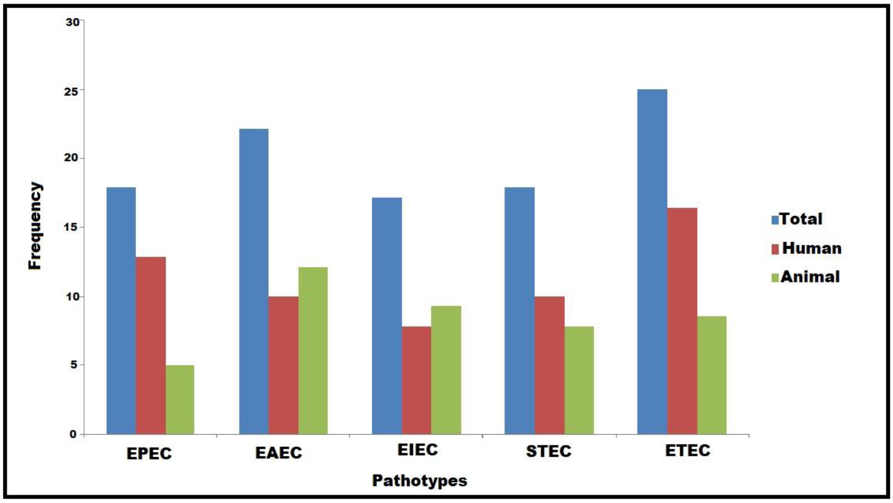
3.1. Characterization and Virulence-Associated Features of DEC Pathotypes

All our DEC isolates (140) were confirmed by both the API 20E identification system and PCR detection of the 16SrRNA gene. Considering the PCR amplification of DEC virulence genes under study, five pathotypes were detected among all human and animal isolates, including EPEC, EAEC, EIEC, STEC, and ETEC (Figure 1). Considering the prevalence rates of all DEC pathotypes, ETEC followed by EAEC represented the most common ones (25% and 22.1%, respectively); meanwhile, EIEC had the lowest prevalence rate (17.1%), as shown in Figure 1. With the exception of EIEC and EAEC, the other pathotypes were the most widespread among human isolates (Figure 1). Regarding the existence of virulence genes, all the examined isolates harbored fimH and ompA genes; meanwhile, none of the DEC pathotypes carried the kpsMTII, hly, or vt2e genes. Interestingly, the astA gene was detected among all pathotypes with a high prevalence rate among EAEC (Figure 2).

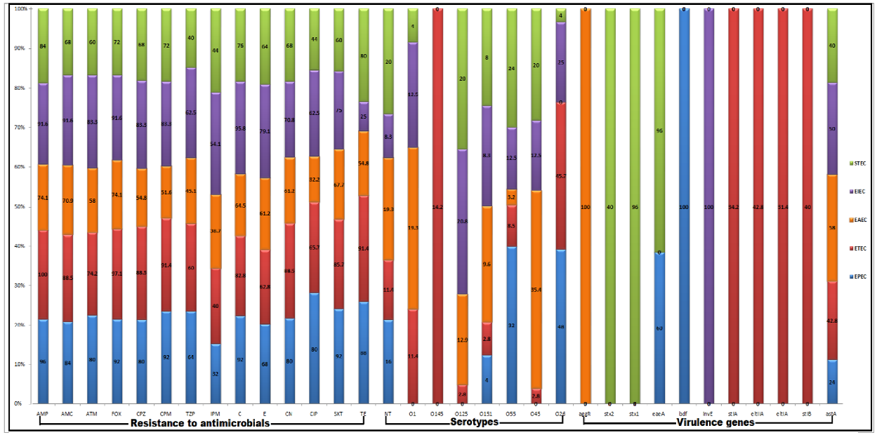
3.2. Serotyping of DEC Pathotypes

A total of 119 DEC isolates were serotyped into seven various serotypes, including O26, O45, O55, O151, O125, O145, and O1; meanwhile, 21 isolates (15%) were untypeable. The distribution of the identified serotypes among the recovered DEC isolates is illustrated in Figure 2. All E. coli pathotypes were detected among the O55 and O151 serotypes. Of note, all isolates belonging to the O145 serotype were ETEC. The EPEC pathotypes could not be detected among the O45, O145, O1, and O125 serotypes. All isolates of the O26 serotype were distributed among all pathotypes except EAEC (Figure 2).

3.3. Antimicrobial Susceptibility Patterns of DEC Pathotypes

Taken together, the analyzed antimicrobial susceptibility data revealed significant variations in the resistance patterns of the investigated DEC isolates against selected antimicrobial agents (p > 0.05). Notably, 90% of the tested isolates exhibited the MDR patterns, with additional evidence of the high occurrence of antimicrobial resistance among human isolates. The resistance patterns of different pathotypes are illustrated in Figure 2. Notably, high resistance rates to sulfamethoxazole/trimethoprim, ciprofloxacin, piperacillin/tazobactam, and cefepime and a maximum overall sensitivity to imipenem were observed among the EPEC pathotype. Moreover, high resistance rates against amoxicillin/clavulanic acid, aztreonam, imipenem, erythromycin, and chloramphenicol as well as a lower resistance percentage to tetracycline were noticed among the EIEC pathotype. Of note, the maximum susceptibility rates to amoxicillin/clavulanic acid, cefoxitin, piperacillin/tazobactam, and sulfamethoxazole/trimethoprim were detected among the STEC pathotype. Moreover, high resistance percentages to ampicillin, cefoxitin, cefoperazone, gentamycin, and tetracycline and lower levels of resistance to ampicillin, aztreonam, cefoperazone, cefepime, chloramphenicol, erythromycin, gentamycin, and ciprofloxacin were noticed among the ETEC and EAEC pathotypes, respectively. With the exception of EIEC and STEC, imipenem was the most effective antimicrobial drug against the other E. coli pathotypes. Meanwhile, tetracycline and piperacillin/tazobactam were highly effective against EIEC and STEC, respectively.

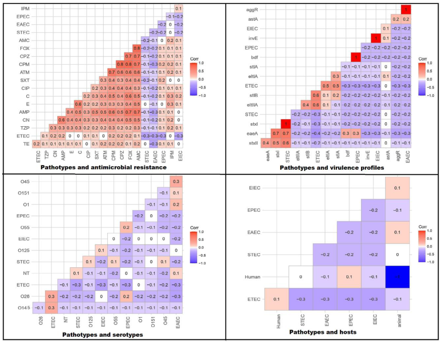
3.4. Correlation between Pathotypes and Antimicrobial Resistance, Serotypes, Virulence Gene Existence, and Host Types

A high variability in the antimicrobial resistance patterns was detected across the DEC pathotypes. In general, the antimicrobial resistance was affected by the DEC pathotypes. The sensitive phenotype was correlated with EAEC and STEC; meanwhile, the resistance phenotypes were correlated with other pathotypes (r-value = 0:0.5), as shown in Figure 3. Notably, the ETEC pathotype was positively correlated with O26 and O145 (r-value = 0.3 for each) but it was negatively correlated with O45 and O125 (r-value = −0.2 and −0.1, respectively). Moreover, there were no correlations between ETEC and O1, O55, and O151. Concerning the EPEC pathotype, it had a positive relationship with O26 and O55 (r-value = 0.2 for each) and negative correlations with other serotypes (r-value = −0.1:−0.3). Positive correlations were announced between EIEC and O125 (r-value = 0.1); EAEC and O45 (r-value = 0.3) and O1 (r-value = 0.2); and STEC and O55, O125, and O45 (r-value = 0.1 for each). On the other hand, there were negative correlations between EAEC and O26, O55, and O145 (r-value = −0.3, −0.2, and −0.1, respectively) and STEC and O26, O145, and O1 (r-value = −0.2, −0.1, and −0.1, respectively). Moreover, there were no correlations between EIEC and O26, O45, O55, O151, and O1 and STEC and O151. As was expected, there were strong positive correlations between the investigated pathotypes and their respective virulence genes (r-value = 1). In another context, the diarrheagenic human infections were always associated with the ETEC and EPEC pathotypes; however, both the EIEC and EAEC pathotypes were commonly identified among the recovered animal isolates. Notably, the STEC pathotype was not correlated with human or animal hosts (Figure 3).

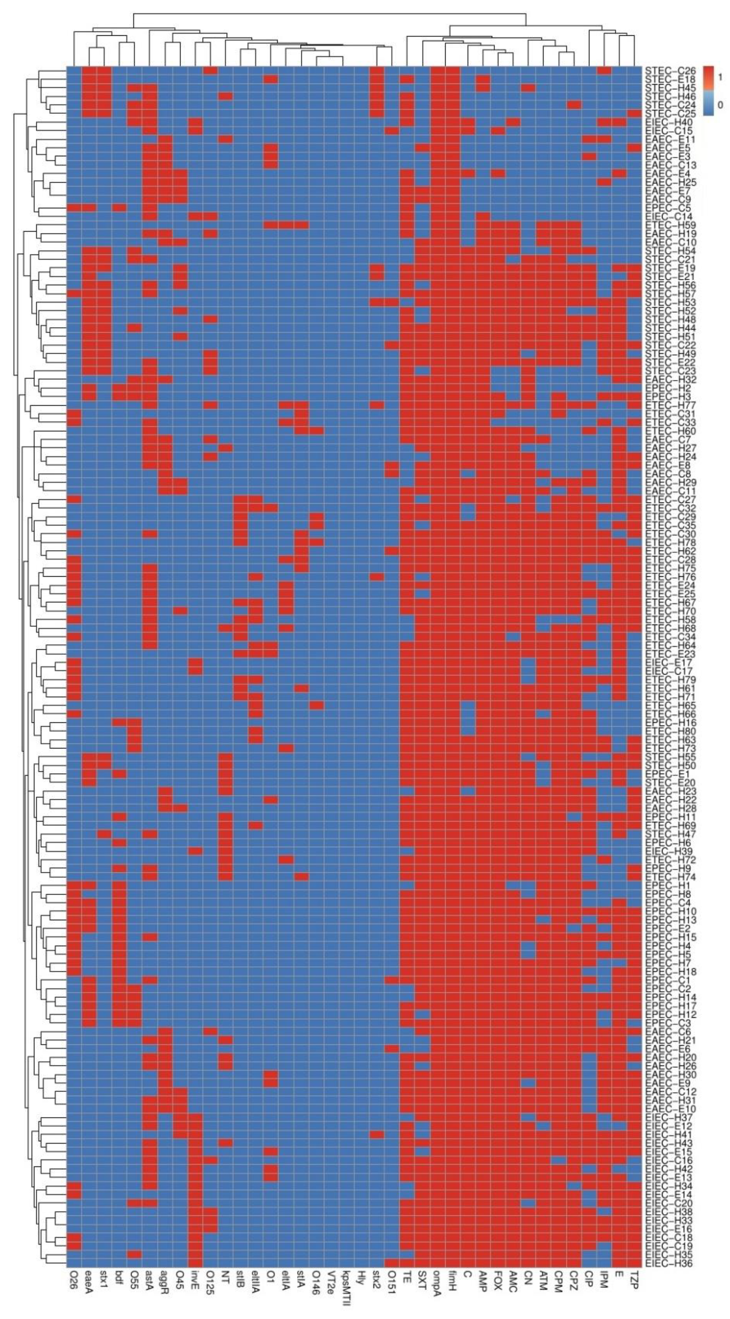
3.5. Phenotyping and Molecular Genotyping of DEC Isolates within and among Different Pathotypes

On the basis of serotyping and antimicrobial resistance and virulence gene patterns, our 140 investigated DEC isolates, except six isolates (two pairs of equine and human isolates and one pair of cow and equine isolates), were typed into diverse lineages (Figure 4). The three pairs of the previously related isolates included two EAEC, two EIEC, and one pair of EAEC with EPEC pathotypes (Figure 4). Interestingly, these typing tools showed high discriminatory power (DI-value = 0.999). Complementary to this, another molecular typing tool used in our manuscript was ERIC-PCR. The relationship between DEC isolates within and among different pathotypes can be distinguished on the basis of ERIC banding patterns (Figure 5). The dendrogram constructed on the basis of different amplicons of the ERIC-PCR assay is shown in Figure 6. As demonstrated in the dendrogram, there were 16 branches of related DEC isolates. They comprised 13 branches of two isolates, 2 branches of three isolates, and 1 branch of four isolates. In spite of the presence of these related isolates, this molecular typing tool showed high discriminatory power (DI-value = 0.997). Fortunately, the related isolates within the 13 branches observed in the ERIC-PCR dendrogram were distinguished according to serotyping, and antimicrobial resistance and virulence gene profiles. Moreover, the related six isolates, grouped into three pairs, as illustrated in Figure 4, gave different ERIC-PCR fingerprints. Therefore, it is noteworthy that all our investigated isolates were typed, confirming the high heterogenicity of all DEC, even within each serotype or pathotype.

4. Discussion

The treatment failure crises has been reported for the resistant pathogens, especially in Egypt [30]. Furthermore, the resistant strains, such as E. coli as well as Staphylococcus aureus and Salmonella sp. [31,32,33], harbor several virulence arrays, which increase the morbidity of the diseases caused by these pathogens. Therefore, epidemiological studies of these strains are urgently needed. Interestingly, this report has focused on the detection of genetic relatedness between DEC pathotypes by the ERIC-PCR technique and additionally investigated the correlations between the DEC pathotypes and the antimicrobial resistances, the existence of virulence genes, serotypes, and the types of hosts to develop efficient strategies for the control and treatment of this pathogen.
Of note, ETEC was the most prevalent pathotype among our DEC isolates and among the isolates investigated in the west of Iran [34]; meanwhile, EPEC and STEC were the most predominant pathotypes in other previous studies [35,36]. The variations in the prevalence of DEC pathotypes may be correlated with environmental contamination and the nutritional types for each geographic area in addition to the reservoir roles of household animals [37]. Regarding the resistance profiles of DEC pathotypes, the prevalence of MDR isolates among the ETEC and EPEC pathotypes, which were mainly associated with the human diseases, was shocking. These results strengthen the hypothesis of the continuous evolution of microbial resistance towards the available antimicrobial drugs. Therefore, the international society strongly advocates the implementation of continuous monitoring strategies for antimicrobial resistance among DEC strains in line with the One Health concept [38].
There are scarce studies investigating the distribution of E. coli pathotypes among human and animal hosts. Although previous studies contradict the hypothesis of host specificity and adaptation of DEC pathotypes due to cross reactivity between human and animal [39,40], we found that human and animal infections were associated with the ETEC and EPEC, and EIEC and EAEC pathotypes, respectively. Concerning the correlation between pathotypes and serotypes, there were variations in the DEC serotypes among previous studies [41,42]. This variation could be accepted depending on the variable correlation between E. coli pathotypes, serotypes, and the clinical forms of their infections.
ERIC-PCR is a cost-effective fingerprint and fast molecular typing technique. The clonal variability of DEC can be assessed using this molecular typing tool. In this research, the ERIC-PCR technique showed high discriminatory power (DI-value = 0.997). In accordance with our findings, a high degree of heterogeneity among E. coli isolates was observed in previous reports on the basis of the analysis of ERIC-PCR results [43,44]. Therefore, the ERIC-PCR approach has been identified as a good tool to differentiate between the closely related strains. Furthermore, there were large genetic variations among the DEC strains within each pathotype, as the strains of single pathotypes belonged to various phenotypic and genotypic lineages, suggesting that there is no definite factor responsible for DEC resistance and virulence and their expression levels may be affected by numerous conditions.

5. Conclusions

Strong positive correlations between DEC pathotypes and the existence of virulence markers and hosts were confirmed; meanwhile, the serotypes could not be correlated well with the DEC pathotypes. Therefore, we suggested that the DEC pathotypes were the powerful factor affecting the degree of morbidity and the choice of treatment protocols. Moreover, our results presented an indication of the heterogeneous nature of DEC pathotypes, as evidenced by the ERIC-PCR results. Our shocking results for the high prevalence of MDR strains and the heterogeneous nature of DEC pathotypes may encourage health care decision makers to introduce more restricted recommendations and surveillance strategies. Additionally, the pathotyping and phenotypic or genotypic testing should be incorporated in medical settings due to the positive correlations between DEC pathotypes and antimicrobial resistance patterns. Therefore, more surveys are required to collect further information about the epidemiological details of DEC isolates to recognize the sources of their infections and implement control measures for avoiding their spread.

Author Contributions

Investigation, N.N.O. and R.A.M.; methodology, M.M.B. (Mahmoud M. Bendary) and M.I.A.E.-H.; software, W.A.A.; resources, M.M.B. (Mosa M. Bahnass); validation, M.A. and A.S.A.; formal analysis, N.N.O., R.E. and A.R.E.; visualization N.N.O.; writing—review and editing, M.M.B. (Mahmoud M. Bendary) and M.I.A.E.-H.; supervision, M.M.A.-S. and N.A.S. All authors have read and agreed to the published version of the manuscript.

Funding

The authors would like to acknowledge Taif University Researchers Supporting Project number (TURSP 2020/288), Taif University, Taif, Saudi.

Institutional Review Board Statement

Not applicable for this study.

Informed Consent Statement

Not applicable, as all isolates were kindly obtained from the microbiology laboratories of the Faculty of Pharmacy, Port-Said University, and the Faculty of Veterinary Medicine, Zagazig University.

Data Availability Statement

All data generated or analyzed during this study are included in the submitted manuscript.

Conflicts of Interest

The authors declare no conflict of interest.

References

  1. Allocati, N.; Masulli, M.; Alexeyev, M.F.; Di-Ilio, C. Escherichia coli in Europe: An overview. Int. J. Environ. Res. Public Health 2013, 10, 6235–6254. [Google Scholar] [CrossRef] [PubMed]
  2. Croxen, M.A.; Law, R.J.; Scholz, R.; Keeney, K.M.; Wlodarska, M.; Finlay, B.B. Recent advances in understanding enteric pathogenic Escherichia coli. Clin. Microbiol. Rev. 2013, 26, 822–880. [Google Scholar] [CrossRef] [PubMed] [Green Version]
  3. Elfaky, M.A.; Abdel-Hamid, M.I.; Khalifa, E.; Alshareef, W.A.; Mosbah, R.A.; Elazab, S.T.; Ghoneim, M.M.; Al-Sanea, M.M.; Bendary, M.M. Innovative next-generation therapies in combating multi-drug-resistant and multi-virulent Escherichia coli isolates: Insights from in vitro, in vivo, and molecular docking studies. Appl. Microbiol. Biotechnol. 2022, 106, 1691–1703. [Google Scholar] [CrossRef]
  4. Mosallam, F.M.; Helmy, E.A.; Bendary, M.M.; El-Batal, I.A. Potency of a novel synthesized Ag- eugenol nanoemulsion for treating some bacterial and fungal pathogens. J. Mater. Res. 2021, 36, 1524–1537. [Google Scholar] [CrossRef]
  5. Jang, J.; Hur, H.G.; Sadowsky, M.J.; Byappanahalli, M.N.; Yan, T.; Ishii, S. Environmental Escherichia coli: Ecology and public health implications—A review. J. Appl. Microbiol. 2017, 123, 570–581. [Google Scholar] [CrossRef] [PubMed] [Green Version]
  6. Robins-Browne, R.M.; Bordun, A.M.; Tauschek, M.; Bennett-Wood, V.R.; Russell, J.; Oppedisano, F.; Lister, N.A.; Bettelheim, K.A.; Fairley, C.K.; Sinclair, M.I.; et al. Escherichia coli and community-acquired gastroenteritis, Melbourne, Australia. Emerg. Infect. Dis. 2004, 10, 1797–1805. [Google Scholar] [CrossRef]
  7. Pakbin, B.; Brück, W.M.; Rossen, J. Virulence Factors of Enteric Pathogenic Escherichia coli: A Review. Int. J. Mol. Sci. 2021, 22, 9922. [Google Scholar] [CrossRef]
  8. Johnson, J.R.; O’Bryan, T.T.; Kuskowski, M.; Maslow, J.N. Ongoing horizontal and vertical transmission of virulence genes and papA alleles among Escherichia coli blood isolates from patients with diverse-source bacteremia. Infect. Immun. 2001, 69, 5363–5374. [Google Scholar] [CrossRef] [Green Version]
  9. Murphy, R.; Palm, M.; Mustonen, V.; Warringer, J.; Farewell, A.; Parts, L.; Moradigaravand, D. Genomic Epidemiology and Evolution of Escherichia coli in Wild Animals in Mexico. mSphere 2021, 6, e00738-20. [Google Scholar] [CrossRef]
  10. Singh, P.; Metgud, S.C.; Roy, S.; Purwar, S. Evolution of diarrheagenic Escherichia coli pathotypes in India. J. Lab. Physicians 2019, 11, 346–351. [Google Scholar] [CrossRef]
  11. Ammar, A.M.; Abd El-Hamid, M.I.; El-Malt, R.M.S.; Azab, D.S.; Albogami, S.; Al-Sanea, M.M.; Soliman, W.E.; Ghoneim, M.M.; Bendary, M.M. Molecular Detection of Fluoroquinolone Resistance among Multidrug-, Extensively Drug-, and Pan-Drug-Resistant Campylobacter Species in Egypt. Antibiotics 2021, 10, 1342. [Google Scholar] [CrossRef]
  12. Mamun, M.M.; Parvej, M.S.; Ahamed, S.; Hassan, J.; Nazir, K.; Nishikawa, Y.; Nishikawa, Y.; Rahman, M.T. Prevalence and characterization of shiga toxigenic Escherichia coli in broiler birds in Mymensingh. Bangl. J. Vet. Med. 2016, 14, 5–8. [Google Scholar] [CrossRef] [Green Version]
  13. Ali, M.M.; Mohamed, Z.K.; Klena, J.D.; Ahmed, S.F.; Moussa, T.A.; Ghenghesh, K.S. Molecular characterization of diarrheagenic Escherichia coli from Libya. Am. J. Trop. Med. Hyg. 2012, 86, 866–871. [Google Scholar] [CrossRef] [PubMed] [Green Version]
  14. Johnson, W.M.; Pollard, D.R.; Lior, H.; Tyler, S.D.; Rozee, K.R. Differentiation of genes coding for Escherichia coli verotoxin 2 and the verotoxin associated with porcine edema disease (VTe) by the polymerase chain reaction. J. Clin. Microbiol. 1990, 28, 2351–2353. [Google Scholar] [CrossRef] [PubMed] [Green Version]
  15. Piva, I.C.; Pereira, A.L.; Ferraz, L.R.; Silva, R.S.; Vieira, A.C.; Blanco, J.E.; Blanco, M.; Blanco, J.; Giugliano, L.G. Virulence Markers of Enteroaggregative Escherichia coli Isolated from Children and Adults with Diarrhea in Brasília. Braz. J. Clin. Microbiol. 2003, 41, 1827–1832. [Google Scholar] [CrossRef] [PubMed] [Green Version]
  16. Ewers, C.; Janßen, T.; Kießling, S.; Philipp, H.C.; Wieler, L.H. Rapid detection of virulence-associated genes in avian pathogenic Escherichia coli by multiplex polymerase chain reaction. Avian Dis. 2005, 49, 269–273. [Google Scholar] [CrossRef] [PubMed]
  17. Dipineto, L.; Santaniello, A.; Fontanella, M.; Lagos, K.; Fioretti, A.; Menna, L.F. Presence of shiga toxin-producing Escherichia coli O157,H7 in living layer hens. Lett. Appl. Microbiol. 2006, 43, 293–295. [Google Scholar] [CrossRef]
  18. Ewers, C.; Li, G.; Wilking, H.; Kiebling, S.; Alt, K.; Antáo, E.M.; Laturnus, C.; Diehl, I.; Glodde, S.; Homeier, T.; et al. Avian pathogenic uropathogenic and newborn meningitis-causing Escherichia coli, How closely related are they? Inter. J. Med. Microbiol. 2007, 297, 163–176. [Google Scholar] [CrossRef]
  19. Müller, D.; Greune, L.; Heusipp, G.; Karch, H.; Fruth, A.; Tschäpe, H.; Schmidt, M.A. Identification of unconventional intestinal pathogenic Escherichia coli isolates expressing intermediate virulence factor profiles by using a novel single-step multiplex PCR. Appl. Environ. Microbiol. 2007, 73, 3380–3390. [Google Scholar] [CrossRef] [Green Version]
  20. Yu, K.X.; Thong, K.L. Multiplex PCR for simultaneous detection of virulence genes in Escherichia coli. Malays. J. Sci. 2009, 28, 1–14. [Google Scholar] [CrossRef] [Green Version]
  21. Ghanbarpour, R.; Salehi, M. Determination of adhesin encoding genes in Escherichia coli isolates from omphalitis of chicks. Amer. J. Ani. Vet. Sci. 2010, 5, 91–96. [Google Scholar] [CrossRef] [Green Version]
  22. Abbasi, P.; Kargar, M.; Doosti, A.; Mardaneh, J.; Dehyadegari, M.A.; Ghorbani-Dalini, S. Multiplex real-time PCR assay for the detection of LT STIa and STIb genes in enterotoxigenic Escherichia coli. Int. J. Entric Pathog. 2014, 2, 16431. [Google Scholar] [CrossRef]
  23. Yun, Z.; Zeng, L.; Huang, W. Detection and categorization of diarrheagenic Escherichia coli with auto-microfluidic thin-film chip Method. Sci. Rep. 2018, 8, 12926. [Google Scholar] [CrossRef] [PubMed]
  24. Omolajaiye, S.A.; Afolabi, K.O.; Iweriebor, B.C. Pathotyping and antibiotic resistance profiling of Escherichia coli isolates from children with acute diarrhea in amatole district municipality of eastern cape South Africa. BioMed Res. Int. 2020, 20, 10. [Google Scholar] [CrossRef]
  25. Guion, C.E.; Ochoa, T.J.; Walker, C.M.; Barletta, F.; Cleary, T.G. Detection of diarrheagenic Escherichia coli by use of melting-curve analysis and real-time multiplex PCR. J. Clin. Microbiol. 2008, 46, 1752–1757. [Google Scholar] [CrossRef] [Green Version]
  26. Clinical and Laboratory Standards Institute. Performance Standards for Antimicrobial Susceptibility Testing; CLSI Document M100-S15; Clinical and Laboratory Standards Institute: Wayne, PA, USA, 2019. [Google Scholar]
  27. M07; Methods for Dilution Antimicrobial Susceptibility Tests for Bacteria That Grow Aerobically. 11th ed. Approved Standard. CLSI: Wayne, PA, USA, 2018.
  28. Magiorakos, A.P. Multidrug-resistant extensively drug-resistant and pandrug-resistant bacteria, an international expert proposal for interim standard definitions for acquired resistance. Clin. Microbiol. Infect. 2012, 18, 268–281. [Google Scholar] [CrossRef] [Green Version]
  29. Ramazanzadeh, R.; Zamani, S.; Zamani, S. Genetic diversity in clinical isolates of Escherichia coli by enterobacterial repetitive intergenic consensus (ERIC)-PCR technique in Sanandaj hospitals. Iran J. Microbiol. 2013, 5, 126–131. [Google Scholar] [PubMed]
  30. Ghaly, M.; Shaheen, A.; Bouhy, A.; Bendary, M. Alternative therapy to manage otitis media caused by multidrug-resistant fungi. Arch. Microbiol. 2020, 202, 1231–1240. [Google Scholar] [CrossRef]
  31. Bendary, M.M.; Solyman, S.M.; Azab, M.M.; Mahmoud, N.F.; Hanora, A.M. Characterization of Methicillin Resistant Staphylococcus aureus isolated from human and animal samples in Egypt. Cell. Mol. Biol. 2016, 29, 94–100. [Google Scholar]
  32. Bendary, M.M.; Solyman, S.M.; Azab, M.M.; Mahmoud, N.F.; Hanora, A.M. Genetic diversity of multidrug resistant Staphylococcus aureus isolated from clinical and non clinical samples in Egypt. Cell. Mol. Biol. 2016, 62, 55. [Google Scholar] [PubMed]
  33. Ghaly, M.F.; Nasr, Z.M.; Abousaty, A.I.; Seadawy, H.G.; Shaheen, M.A.A.; Albogami, S.; Al-Sanea, M.M.; Bendary, M.M. Alternative and Complementary Therapies against Foodborne Salmonella Infections. Antibiotics 2021, 10, 1453. [Google Scholar] [CrossRef] [PubMed]
  34. Derakhshan, S.; Farhadifar, F.; Roshani, D.; Ahmadi, A.; Haghi, F. Study on the presence of resistant diarrheagenic pathotypes in Escherichia coli isolated from patients with urinary tract infection. Gastroenterol. Hepatol. Bed Bench 2019, 12, 348–357. [Google Scholar]
  35. Lamprecht, C.; Romanis, M.; Huisamen, N.; Carinus, A.; Schoeman, N.; Sigge, G.O. Escherichia coli with virulence factors and multidrug resistance in the Plankenburg River. S. Afr. J. Sci. 2014, 110, 1–6. [Google Scholar] [CrossRef] [Green Version]
  36. Fallah, N.; Ghaemi, M.; Ghazvini, K.; Rad, M.; Jamshidi, A. Occurrence pathotypes and antimicrobial resistance profiles of diarrheagenic Escherichia coli strains in animal source food products from public markets in Mashhad Iran. Food Control 2021, 121, 107640. [Google Scholar] [CrossRef]
  37. Askari, A.; Ghanbarpour, R.; Akhtardanesh, B.; Aflatoonian, M.R.; Sharifi, H.; Jajarmi, M.; Molaei, R. Detection of zoonotic diarrheagenic pathotypes of Escherichia coli in healthy household dogs. Iran. J. Microbiol. 2020, 12, 522–530. [Google Scholar] [CrossRef] [PubMed]
  38. Bordier, M.; Binot, A.; Pauchard, Q.; Nguyen, D.T.; Trung, T.N.; Fortané, N.; Goutard, F.L. Antibiotic resistance in Vietnam, moving towards a One Health surveillance system. BMC Public Health 2018, 18, 1–14. [Google Scholar] [CrossRef]
  39. Ferdous, J.; Rashid, R.B.; Sultana, R.; Saima, S.; Jahan Prima, M.; Begum, A.; Mackie Jensen, P.K. Is It Human or Animal? The Origin of Pathogenic, E. coli in the Drinking Water of a Low-Income Urban Community in Bangladesh. Trop. Med. Infect. Dis. 2021, 6, 181. [Google Scholar] [CrossRef]
  40. Iman, S. Escherichia coli pathotypes associated with diarrhea in human and domestic animals. Am. J. Anim. Vet. Sci. 2014, 155, 161. [Google Scholar]
  41. Monaghan, Á.; Byrne, B.; Fanning, S.; Sweeney, T.; McDowell, D.; Bolton, D.J. Serotypes and virulence profiles of non-O157 Shiga toxin-producing Escherichia coli isolates from bovine farms. Appl. Environ. Microbiol. 2011, 77, 8662–8668. [Google Scholar] [CrossRef] [Green Version]
  42. Sharada, R.; Ruban, S.W.; Thiyageeswaran, M. Isolation characterization and antibiotic resistance pattern of E. coli isolated from poultry. Eur. J. Sci. Res. 2010, 5, 18–22. [Google Scholar]
  43. Oltani, M.; Peighambari, S.M.; Askari, M.; Sadrzadeh, A. Molecular typing of avian Escherichia coli isolates by enterobacterial repetitive intergenic consensus sequences-polymerase chain reaction (ERIC-PCR). Iran. J. Vet. Med. 2012, 6, 143–148. [Google Scholar]
  44. Brocchi, M.; Ferreira, A.; Lancellotti, M.; Stehling, E.G.; Campos, T.A.; Nakazato, G. Typing of avian pathogenic Escherichia coli strains by REP-PCR. Pesq. Vet. Bras. 2006, 26, 69–73. [Google Scholar] [CrossRef]
Figure 1. Occurrence and distribution of DEC pathotypes among human and animal hosts. EAEC: enteroaggregative E. coli, ETEC: enterotoxigenic E. coli, EPEC: enteropathogenic E. coli, EIEC: enteroinvasive E. coli, STEC: shiga-toxin-producing E. coli.
Biology 11 01004 g001
Figure 2. Frequency of resistance to antimicrobials, serotypes, and virulence markers of DEC pathotypes. TZP: piperacillin/tazobactam, C: chloramphenicol, TE: tetracycline, AMP: ampicillin, CN: gentamycin, E: erythromycin, ATM: aztreonam, SXT: sulfamethoxazole/trimethoprim, AMC: amoxicillin/clavulanic acid, IPM: imipenem, CIP: ciprofloxacin, FOX: cefoxitin, CPM: cefepime, CPZ: cefoperazone, EAEC: enteroaggregative E. coli, ETEC: enterotoxigenic E. coli, EPEC: enteropathogenic E. coli, EIEC: enteroinvasive E. coli, STEC: shiga-toxin-producing E. coli.
Biology 11 01004 g002
Figure 3. Correlation coefficient (r) between diarrheagenic E. coli pathotypes and antimicrobial resistance, virulence gene existence, serotypes, and host types. Red and blue colors specify positive and negative correlations, respectively. The color key denotes the correlation coefficient (R). The darker red and blue colors indicate stronger positive (R = 0.5:1) and negative (R = −0.5:−1) correlations, respectively. TZP: piperacillin/tazobactam, TE: tetracycline, AMP: ampicillin, IPM: imipenem, CN: gentamycin, E: erythromycin, ATM: aztreonam, CIP: ciprofloxacin, C: chloramphenicol, SXT: sulfamethoxazole/trimethoprim, AMC: amoxicillin/clavulanic acid, FOX: cefoxitin, CPM: cefepime, CPZ: cefoperazone, EAEC: enteroaggregative E. coli, ETEC: enterotoxigenic E. coli, EPEC: enteropathogenic E. coli, EIEC: enteroinvasive E. coli, STEC: shiga-toxin-producing E. coli.
Biology 11 01004 g003
Figure 4. Heat map and hierarchical clustering of diarrheagenic E. coli pathotypes according to the occurrence of serotypes, antimicrobial resistances, and virulence genes. Blue and red colors indicate the sensitivity and resistance to a certain antimicrobial and to the absence and presence of a particular serotype and virulence gene, respectively. The code numbers on the right side of the heat map denote the numbers of diarrheagenic E. coli pathotypes from equine (E), cow (C), and human (H) sources. TZP: piperacillin/tazobactam, C: chloramphenicol, TE: tetracycline, AMP: ampicillin, CN: gentamycin, E: erythromycin, ATM: aztreonam, SXT: sulfamethoxazole/trimethoprim, AMC: amoxicillin/clavulanic acid, IPM: imipenem, CIP: ciprofloxacin, FOX: cefoxitin, CPM: cefepime, CPZ: cefoperazone, EAEC: enteroaggregative E. coli, ETEC: enterotoxigenic E. coli, EPEC: enteropathogenic E. coli, EIEC: enteroinvasive E. coli, STEC: shiga-toxin-producing E. coli.
Biology 11 01004 g004
Figure 5. Dendrogram showing the relatedness of diarrheagenic E. coli pathotypes isolated from equine (E), cow (C), and human (H) origins, as determined by ERIC-PCR fingerprinting. EAEC: enteroaggregative E. coli, ETEC: enterotoxigenic E. coli, EPEC: enteropathogenic E. coli, EIEC: enteroinvasive E. coli, STEC: shiga-toxin-producing E. coli.
Biology 11 01004 g005
Figure 6. Heat map and hierarchical clustering of diarrheagenic E. coli pathotypes, according to the generated ERIC-PCR amplicon size (bp) profiles. Blue and red colors indicate the absence and presence of particular ERIC-PCR bands, respectively. The code numbers on the right side of the heat map denote the numbers of diarrheagenic E. coli pathotypes from equine (E), cow (C), and human (H) sources. TZP: piperacillin/tazobactam, C: chloramphenicol, TE: tetracycline, AMP: ampicillin, CN: gentamycin, E: erythromycin, ATM: aztreonam, SXT: sulfamethoxazole/trimethoprim, AMC: amoxicillin/clavulanic acid, IPM: imipenem, CIP: ciprofloxacin, FOX: cefoxitin, CPM: cefepime, CPZ: cefoperazone, EAEC: enteroaggregative E. coli, ETEC: enterotoxigenic E. coli, EPEC: enteropathogenic E. coli, EIEC: enteroinvasive E. coli, STEC: shiga-toxin-producing E. coli.
Biology 11 01004 g006
Table 1. The primer sequences and the predicted amplicon sizes of the diarrheagenic E. coli genes under study.
Target GeneSpecificityPrimer Sequence (5′-3′)Amplicon Size (bp)Reference
16S rRNARNA component of the 30S ribosomal subunitF: GACCTCGGTTTAGTTCACAGA
R: CACACGCTGACGCTGACCA
585[12]
ompAOuter membrane proteinF: AGCTATCGCGATTGCAGTG
R: GGTGTTGCCAGTAACCGG
919[18]
kpsMTIIAdhesionF: CAGGTAGCGTCGAACTGTA
R: CATCCAGACGATAAGCATGAGCA
280[18]
hlyHemolysinF: AACAAGGATAAGCACTGTTCTGGCT
R: ACCATATAAGCGGTCATTCCCGTCA
117[15]
stx2Shiga toxin 2F: CCATGACAACGGACAGCAGTT
R: CCTGTCAACTGAGCAGCACTTTG
779[17]
stx1Shiga toxin 1F: ACACTGGATGATCTCAGTGG
R: CTGAATCCCCCTCCATTATG
614[17]
fimHAdhesionF: TGCAGAACGGATAAGCCGTGG
R: GCAGTCACCTGCCCTCCGGTA
508[21]
vt2eVero toxinF: CCTTAACTAAAAGGAATATA
R: CTGGTGGTGTATGATTAATA
230[14]
astAEnterotoxinF: TGCCATCAACACAGTATATCC
R: TCAGGTCGCGAGTGACGGC
116[16]
invETranscriptional regulation of invasionF: CGATAGATGGCGAGAAATTATATCCCG
R: CGATCAAGAATCCCTAACAGAAGAATCAC
766[22]
aggRTranscriptional activator of adherence fimbriaeF: ACGCAGAGTTGCCTGATAAAG
R: AATACAGAATCGTCAGCATCAGC
400[19]
eaeAIntiminF: GTAAAGTCCGTTACCCCAACCTG
R: GCACACGGAGCTCCTCAGTCTCC
218[24]
eltIAHeat-labile toxin I F: TTACGGCGTTACTATCCTCTCTA
R: GGTCTCGGTCAGATATGTGATTC
275[20]
eltIIAHeat-labile toxin II F: ATATCATTTTCTGTTTCAGCAAA
R: CAATAAAATCATCTTCGCTCATG
720[20]
stIAHeat-stable toxin AF: TTTCCCCTCTTTTAGTCAGTCAA
R: GCAGGATTACAACACAATTCACAGCAG
159[25]
stIBHeat-stable toxin BF: TGCTAAACCAGTAGAGTCTTCAAAA
R: GCAGGATTACAACACAATTCACAGC
138[25]
bfpBundle-forming pilusF: GACACCTCATTGCTGAAGTCG
R: CCAGAACACCTCCGTTATGC
910[19]
Table 2. Zone diameter and MIC breakpoints for diarrheagenic E. coli isolates.
Antimicrobial
Agent
SymbolConc.
(ug)
Interpretative Categories
Zone Diameter Breakpoints (mm)MIC Breakpoints (μg/mL)
ResistanceSensitiveResistanceSensitive
AmpicillinAMP10≤13≥17≥32 ≤8
Amoxicillin/clavulanic acidAMC20/10≤13≥18≥32/16≤8/4
AztreonamATM30≤17≥21≥16≤4
CefoxitinFOX30≤14≥18≥32 ≤8
CefoperazoneCPZ75≤15≥21≥64≤16
CefepimeCPM30≤18≥25≥16≤2
Piperacillin/tazobactamTZP100/10≤17≥21≥128/4≤16/4
ChloramphenicolC30≤12≥18≥32 ≤8
ImipenemIPM10≤19≥23≥4≤1
ErythromycinE15
GentamycinCN10≤12≥15≥16≤4
CiprofloxacinCIP5≤15≥21≥4≤1
Sulfamethoxazole/trimethoprimSXT23.75/1.25≤10≥16≥4/76≤2/38
TetracyclineTE30≤11≥15≥16≤4
Publisher’s Note: MDPI stays neutral with regard to jurisdictional claims in published maps and institutional affiliations.

