Article

Soil Fungal Diversity and Ecology Assessed Using DNA Metabarcoding along a Deglaciated Chronosequence at Clearwater Mesa, James Ross Island, Antarctic Peninsula

by
Vivian N. Gonçalves
1,
Juan M. Lirio
2,
Silvia H. Coria
2,
Fabyano A. C. Lopes
3,
Peter Convey
4,5,6,7,
Fábio S. de Oliveira
8,
Micheline Carvalho-Silva
9,
Paulo E. A. S. Câmara
8 and
Luiz H. Rosa
1,10,*
1
Departamento de Microbiologia, Universidade Federal de Minas Gerais, Belo Horizonte 31270-901, Brazil
2
Instituto Antártico Argentino, 25 de Mayo 1143, General San Martín, Buenos Aires B1650HMK, Argentina
3
Laboratório de Microbiologia, Universidade Federal Do Tocantins, Porto Nacional 77500-000, Brazil
4
British Antarctic Survey, NERC, High Cross, Madingley Road, Cambridge CB3 0ET, UK
5
Department of Zoology, University of Johannesburg, P.O. Box 524, Auckland Park 2006, South Africa
6
Millennium Institute Biodiversity of Antarctic and Subantarctic Ecosystems, BASE, University Austral of Chile, Valdivia 5090000, Chile
7
Cape Horn International Center (CHIC), O’Higgins 310, Puerto Williams 6350000, Chile
8
Departamento de Geografia, Universidade Federal de Minas Gerais, Belo Horizonte 31270-901, Brazil
9
Departamento de Botânica, Universidade de Brasília, Brasília 70297-400, Brazil
10
Laboratório de Microbiologia Polar e Conexões Tropicais, Instituto de Ciências Biológicas, Universida-de Federal de Minas Gerais, Belo Horizonte 31270-901, Brazil
*
Author to whom correspondence should be addressed.
Biology 2023, 12(2), 275; https://doi.org/10.3390/biology12020275
Submission received: 23 December 2022 / Revised: 6 February 2023 / Accepted: 8 February 2023 / Published: 9 February 2023
(This article belongs to the Special Issue 2nd Edition of Diversity of Soil Fungal Communities)

Abstract

:

Simple Summary

We characterized the fungal organisms present in soils at James Ross Island, north-east Antarctic Peninsula, using DNA metabarcoding. Taxa detected included members of the widespread phyla Ascomycota, Basidiomycota and Mortierellomycota. Additionally, the uncommon phyla Chytridiomycota, Rozellomycota, Monoblepharomycota, Zoopagomycota and Basidiobolomycota were detected. Unknown fungi and taxa identified at generic and specific levels dominated the assemblages. The fungal sequence assemblages displayed high diversity and richness, and moderate dominance, and included taxa known to play saprophytic, pathogenic and symbiotic functions. Soils of Clearwater Mesa contain a complex fungal community, including fungal groups considered rare in Antarctica, dominated by cold-adapted cosmopolitan, endemic, saprotrophic and phytopathogenic taxa.

Abstract

We studied the fungal diversity present in soils sampled along a deglaciated chronosequence from para- to periglacial conditions on James Ross Island, north-east Antarctic Peninsula, using DNA metabarcoding. A total of 88 amplicon sequence variants (ASVs) were detected, dominated by the phyla Ascomycota, Basidiomycota and Mortierellomycota. The uncommon phyla Chytridiomycota, Rozellomycota, Monoblepharomycota, Zoopagomycota and Basidiobolomycota were detected. Unknown fungi identified at higher hierarchical taxonomic levels (Fungal sp. 1, Fungal sp. 2, Spizellomycetales sp. and Rozellomycotina sp.) and taxa identified at generic and specific levels (Mortierella sp., Pseudogymnoascus sp., Mortierella alpina, M. turficola, Neoascochyta paspali, Penicillium sp. and Betamyces sp.) dominated the assemblages. In general, the assemblages displayed high diversity and richness, and moderate dominance. Only 12 of the fungal ASVs were detected in all chronosequence soils sampled. Sequences representing saprophytic, pathogenic and symbiotic fungi were detected. Based on the sequence diversity obtained, Clearwater Mesa soils contain a complex fungal community, including the presence of fungal groups generally considered rare in Antarctica, with dominant taxa recognized as cold-adapted cosmopolitan, endemic, saprotrophic and phytopathogenic fungi. Clearwater Mesa ecosystems are impacted by the effects of regional climatic changes, and may provide a natural observatory to understand climate change effects over time.

1. Introduction

Antarctica hosts largely undisturbed ecosystems, many of which have been the subject of limited biological research. These ecosystems and their biodiversity are exposed to multiple extreme conditions that combine cold, dry and ultra-oligotrophic stresses, offering unique opportunities to discover and study extremophilic organisms [1,2]. Typically, Antarctic soils are dry, and are exposed to large and rapid changes in temperature, and high levels of solar radiation. They are also generally nutrient-poor, typically ranging between moderately and ultra-oligotrophic [3].
Clearwater Mesa, located on James Ross Island, includes approximately 8 km2 of ice-free area of volcanic geology, and lies at up to 250 m a.s.l. on the south-east side of Croft Bay [4]. Clearwater Mesa is part of the James Ross Island Volcanic Group and comprises rocks of the Pliocene age, composed mainly of alkaline basalts and palagonitized hyaloclastite breccias, covered by glacial deposits with basaltic clasts [5]. In ecology, chronosequence soils represent a set of sites formed from the same substrate that differ in age and the levels of geological and biological development since they were formed [6]. Clearwater Mesa has a characteristic chronosequence profile from the seashore to the Haddington Ice Cap, which encompasses a gradient from para- to periglacial conditions.
The soils of Antarctica offer opportunities to investigate the regional-to-global environmental effects of microbiota on community structure and function [7]. The Antarctic Peninsula region has been strongly influenced by the effects of regional climate change since the mid-20th Century [8] and, therefore, provides an attractive field laboratory for the study of different aspects of fungal biology under extreme conditions [2,9]. Most studies to date of Antarctic fungal diversity have focused on soils [3,10]. However, the majority of these studies relied on traditional culturing approaches [11], which, as is typical of microbiota, do not reveal the full microbial diversity present, and particularly, that of basal, cryptic and rare phyla. The recent availability and application of molecular biological culture-independent approaches have highlighted that fungal diversity in Antarctic soils is likely to be considerably greater than previously appreciated [9,11,12]. In the current study, we applied a DNA metabarcoding approach using high throughput sequencing (HTS) to detect and characterize the soil fungal community along a chronosequence from para- to periglacial conditions at Clearwater Mesa, James Ross Island.

2. Methods

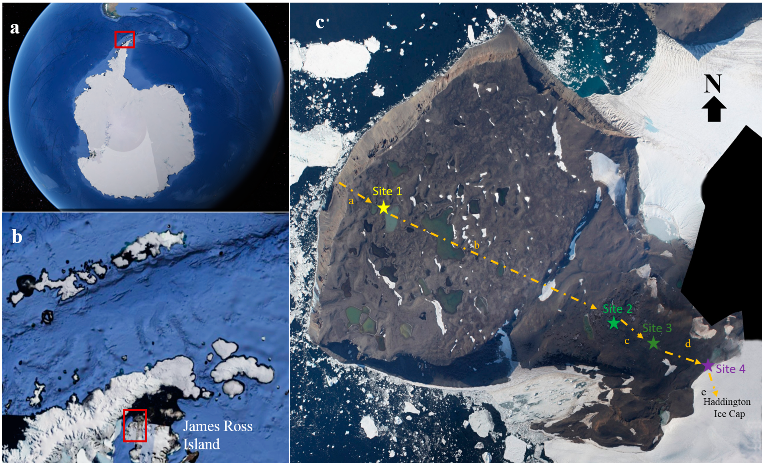
2.1. Study Sites and Sediment Sampling

Four topsoil samples were collected in the Clearwater Mesa region of James Ross Island, north-east Antarctic Peninsula, across an environmental gradient or chronosequence from para- (Site 1) to periglacial (Sites 2, 3 and 4) conditions (Figure 1). The characteristics of each sampling point are presented in Table 1. Topsoils were collected during the austral summer in February 2021 (Supplementary Figure S1). The chronosequence extended 4770 m, from the coast to the edge of the Haddington Ice Cap, with an approximately NW-SE (azimuth 114°) orientation. All samples were collected over the volcanic bedrock of basaltic composition belonging to the James Ross Island Volcanic Group. Sampling locations were selected to be free of lichens and mosses. All soil samples were collected from the center of sorted stone circles, comprising mainly clay material. Soil samples were collected in triplicate and immediately placed in sterile Whirl-pack bags (Nasco, Ft. Atkinson, WI, USA). Approximately 500 g of each topsoil sample was then immediately sub-sampled (in triplicate), sealed, placed in sterile Whirl-pack bags and frozen at −20 °C until processing in the laboratory at the Federal University of Minas Gerais, Brazil. There, the samples were gradually thawed at 4 °C for 24 h before carrying out DNA extraction.

2.2. Soil Physical and Chemical Analyses

The granulometric soil characteristics of sand, silt and clay soil chemical analyses were determined following Teixeira et al. [13]. All analyses were performed in triplicate.

2.3. DNA Extraction, Illumina Library Construction and Sequencing

Three replicate sub-samples were obtained from each soil sample under strict control conditions to avoid external contamination, as described by Rosa et al. [11]. Total DNA was extracted from these using the FastDNA Spin Kit for Soil (MPBIO, Solon, OH, USA). DNA quality was analyzed using agarose gel electrophoresis (1% agarose in 1× Trisborate-EDTA) and then quantified using the Quanti-iT™ Pico Green dsDNA Assay (Invitrogen). The internal transcribed spacer 2 (ITS2) of the rDNA was used as a DNA barcode for fungal identification [14,15], using the primers ITS3 and ITS4 [16]. The Herculase II Fusion DNA Polymerase Nextera XT Index Kit V2 was used for library construction and DNA amplification followed the Illumina 16S Metagenomic Sequencing Library Preparation protocol (Part #15044223, Rev. B). Paired-end sequencing (2 × 300 bp) was performed on a MiSeq platform (Illumina) by Macrogen Inc. (Seoul, South Korea).

2.4. Data Analysis and Fungal Identification

Quality analysis of the libraries was performed using BBDuk v. 38.87 in BBmap software, as described by Bushnell [17], with the following parameters: Illumina adapters removing (Illumina artefacts and the PhiX Control v3 Library); ktrim = l; k = 23; mink = 11; hdist =1; minlen = 50; tpe; tbo; qtrim = rl; trimq = 20; ftm = 5; maq = 20. The remaining sequences were imported to the QIIME2 version 2021.4 (https://qiime2.org/ (accessed on 1 July 2022)) for bioinformatics analyses [18]. The qiime2-dada2 plugin was applied for filtering, dereplication, turn paired-end fastq files into merged and chimera removal using default parameters [19]. Taxonomic assignments were determined for amplicon sequence variants (ASVs) in the steps: (1) ASVs were classified using the qiime2-feature-classifier [20] classify-sklearn against the UNITE Eukaryotes ITS database version 8.3 [21]; (2) Remaining unclassified ASVs were filtered and aligned against the filtered NCBI non-redundant nucleotide sequences (nt) database (October 2021) using BLASTn [22] with default parameters; the nt database was filtered using the following keywords: “ITS1″, “ITS2″, “Internal transcribed spacer″ and “internal transcribed spacer″; and (3) Output files from BLASTn [22] were imported to MEGAN6 [23] and taxonomic assignments were performed using the “megan-nucl-Jan2021.db″ mapping file with default parameters and trained with Naive Bayes classifier and a confidence threshold of 98.5%. Krona [24] was used for generating taxonomic profiles. The heatmap of ASV abundance and clustering analysis were produced using Heatmapper [25] following the parameters: average linkage, spearman rank correlation and Z-score, among samples for each ASV.
According to Medinger et al. [26], different factors, such as extraction, PCR and primer bias can affect the number of DNA reads obtained, and for this reason, lead to the misinterpretation of absolute DNA read abundance [27]. However, according to Giner et al. [28], such biases do not affect the proportionality between reads and cell abundance, implying that more reads are linked with higher abundance [29,30]. For comparative purposes, we used the number of DNA reads as a proxy for relative abundance. For fungal classification we followed Kirk et al. [31], Tedersoo et al. [32] and MycoBank (http://www.mycobank.org (accessed on 1 July 2022)).

2.5. Fungal Diversity and Ecology

The relative abundance (RA) criteria established by Rosa et al. [33] were used, where fungal ASVs > 10% were considered dominant, those between 1% and 10% as intermediate and those with <1% as minor components of the fungal community. Relative abundance values were used to quantify fungal assemblage diversity, richness, dominance and similarities using the Fisher’s α, Margalef’s, Simpson’s, Sorensen, Bray–Curtis and Mao Tao indices. All indices were obtained with 95% confidence, with bootstrap values calculated from 1000 replicates by the PAST program 1.90 [34]. Ecological functional assignments of fungal taxa’s generic levels were obtained using FunGuild [35].

3. Results

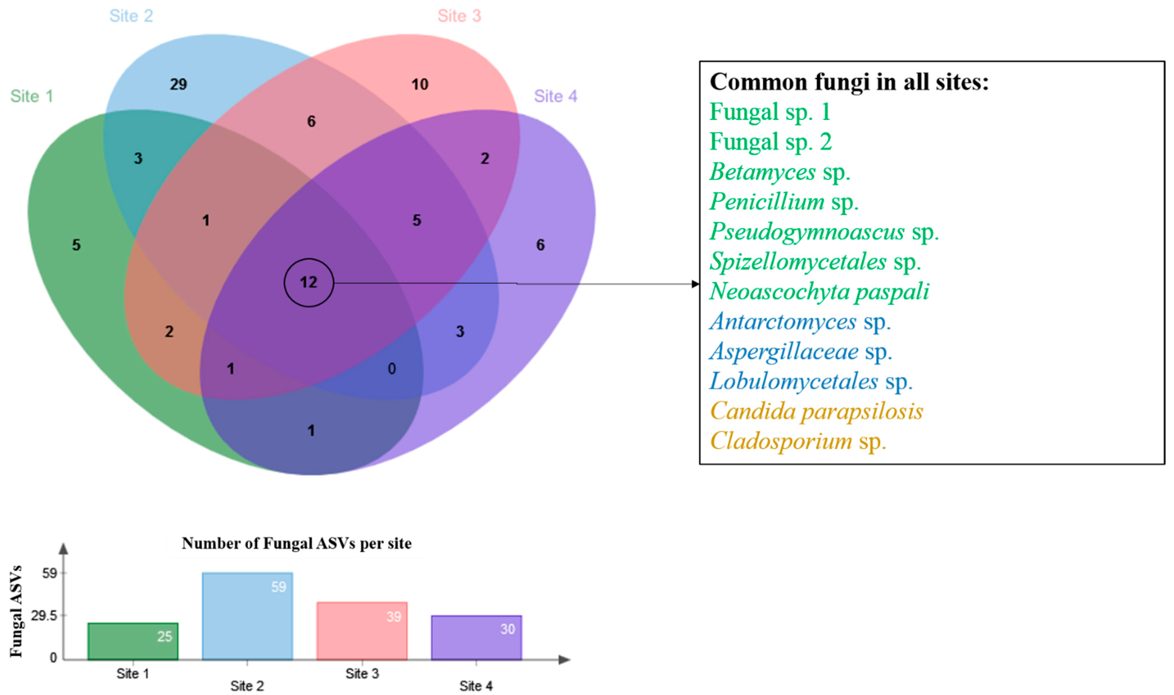
3.1. Fungal Taxonomy

A total of 127,774 DNA reads were detected in the four soils sampled, which represented 88 fungal ASVs (Supplementary Table S1). The Mao Tao curves of the assemblages present in Sites 2 and 4 reached the asymptote. However, those of Sites 1 and 3 did not reach asymptote, indicating that further fungal sequence diversity is likely to be present in these soils (Supplementary Figure S2). The relative abundances of representative fungi at different hierarchical levels varied across the four sites (Supplementary Figures S3 and S4). The metabarcoding approach revealed the presence of taxa of fungal phyla often detected in Antarctica by culturing and eDNA methods (Ascomycota, Basidiomycota and Mortierellomycota). However, metabarcoding also detected the more rarely detected fungal phyla Chytridiomycota, Rozellomycota, Monoblepharomycota, Zoopagomycota and Basidiobolomycota.
Unknown fungi identified at higher hierarchical taxonomic levels (Fungal sp. 1, Fungal sp. 2, Spizellomycetales sp. and Rozellomycotina sp.), as well as taxa identified at generic and specific levels (Mortierella sp., Pseudogymnoascus sp., Mortierella alpina, M. turficola, Neoascochyta paspali, Penicillium sp. and Betamyces sp.) dominated the fungal assemblages. However, the majority of diversity present in the assemblages represented fungal taxa detected at intermediate and rare relative abundance. A total of 35 fungal ASVs (39.7%) could only be assigned to phylum, class, order or family (higher taxonomic levels).

3.2. Geological and Physicochemical Characteristics of Soil Sampling Sites in Relation to Fungal Assemblages Detected

The soils showed homogeneous granulometric composition and a loam texture in the paraglacial zone (Site 1), transitioning to clay-sand-loam in the periglacial zone (Sites 2, 3 and 4). The contents of fine particles (clay and silt) were similar in all soils (approximately 50%), as were the contents of coarse and fine sand. Additionally, there was a homogeneous distribution of fractions, regardless of the parent material of the soils. The soils had alkaline pH, always above 7 and reaching 8.9. All soils were eutrophic, with high base saturation, reaching 100%, with zero potential acidity. Only the soil from Site 2 showed slightly lower values of CEC and sum of bases. The organic carbon content was very low (<1%) and the P content was distinct only in the soil from Site 1.
The diversity indices of the fungal assemblages detected varied across the different sampling sites but, in general, they displayed high diversity and richness, and moderate dominance (Table 1). The soil sampled from Site 2 displayed the highest number of assigned taxa and diversity indices, followed by those of Sites 3, 4 and 1. Geologically, the soils at each site were located over a similar bedrock. Site 4 was located only 50 m from the retreating Haddington Ice Cap front, which we assume represents a very young ice-free area. The location of Site 1 is closest to the coast of Croft Bay and may be more exposed to sea spray, although is still at about 250 m a.s.l. on the mesa. There was no indication of any link between the geological or physicochemical parameters measured and the diversity indices obtained.

3.3. Fungal Assemblage Distribution along the Soil Chronosequence

The Sorensen and Bray–Curtis indices were applied to compare the fungal assemblage distribution along the chronosequence soils (Supplementary Figure S5). Both similarity indices showed the same pattern, with the assemblages detected at Site 2 differing from those of Sites 1, 3 and 4. Of the 88 fungal ASVs, only 12 (13.6%) were detected in all soil samples (Figure 2; Supplementary Table S2). Fungal ASV distribution changed across the sites, with the soils sharing some taxa and also hosting some specific fungal taxa. Site 2 hosted 29 exclusive ASVs. The most abundant fungal ASVs, Fungal sp. 1, Fungal sp. 2, Betamyces sp., Penicillium sp., Pseudogymnoascus sp., Spizellomycetales sp. and Neoascochyta paspali were present at all four sites sampled. Functional ecology assignments of the fungal ASVs at the generic level (Supplementary Table S3) suggested that the assemblages detected in the soils were dominated by saprophytic, plant and animal pathogenic and symbiotic taxa.

4. Discussion

4.1. Fungal Taxonomy

Soils represent perhaps the most studied substrate as habitat for fungi in Antarctica [2]. Until recently, most fungal biodiversity studies have used traditional culture-dependent methods [10]. However, culture-independent methodologies have increasingly become available in recent years, and have started to be applied to Antarctic soils. For instance, Baeza et al. [12] applied a culture-independent approach to characterize fungi in soils from different Antarctic regions, reporting taxa of the phyla Ascomycota, Basidomycota, Chytridiomycota and Neocallimastigomycota. Newsham et al. [9] reported taxa of the phyla Ascomycota, Basidiomycota, Chytridiomycota, Glomeromycota and Mucoromycota in soils sampled from maritime Antarctica. Rosa et al. [11] similarly reported DNA sequences of taxa belonging the phyla Ascomycota, Basidiomycota, Mortierellomycota, Mucoromycota, Chytridiomycota and Rozellomycota in soils subject to and protected from human influence on Deception Island. In common with these studies, we detected taxa of the phyla Ascomycota, Basidiomycota, Mortierellomycota, Chytridiomycota and Rozellomycota. However, we also detected the DNA of taxa belonging to the more rarely reported phyla Monoblepharomycota, Zoopagomycota and Basidiobolomycota. In agreement with the studies of Newsham et al. [9], Rosa et al. [11] and Baeza et al. [12] we detected 35 ASVs (39.7% of the total assigned) in the sampled soils that could only be classified to higher taxonomic levels, suggesting that it is likely that Antarctica hosts many as yet unrecognized fungal taxa or taxa not currently included in the consulted sequence databases. Fungal sp. 1, Fungal sp. 2, Spizellomycetales sp., Rozellomycotina sp., Mortierella sp., Pseudogymnoascus sp., Mortierella alpina, M. turficola, Neoascochyta paspali, Penicillium sp. and Betamyces sp. were the most abundant fungal taxa detected in the Clearwater Mesa soils. However, we recognize that, as our study focused on fungal diversity assigned from DNA sequences, further specific studies are necessary to elucidate whether these fungi are present in viable form.
The phyla Chytridiomycota and Rozellomycota include freshwater zoosporic fungi rarely reported in Antarctic environments [11,33]. Members of both phyla have been reported from ice-covered lakes in the McMurdo Dry Valleys [36]. Some representatives of these phyla are parasites of other taxa, including fungi, algae and aquatic invertebrates [37,38]. Spizellomycetales (an order of Chytridiomycota) includes 27 species, including cosmopolitan and frequently found saprophytes and/or parasites in freshwater and soil [31]. Spizellomycetales species, such as members of the genera Spizellomyces and Gaertneriomyces, are considered important plant pathogens [39]. The genus Betamyces (Chytridiomycota) currently includes only a single recognized species, Betamyces americae-meridionalis, reported from pollen baits at the Paraná River, Buenos Aires, Argentina [40]. Betamyces spp. have also been reported from soil in Costa Rica [40]. Recently, using the same metabarcoding approach as in the current study, Gonçalves et al. [4] reported Spizellomycetales sp., Rozellomycota sp. and Betamyces sp. as dominant fungal taxa present in lake sediments that were also sampled from Clearwater Mesa.
Pseudogymnoascus and Mortierella are genera that include various psychrophilic species, with many commonly present in different Antarctic environments. Pseudogymnoascus (syn. Geomyces) and Mortierella are commonly reported from cold habitats of Arctic, alpine, Antarctic and temperate ecosystems [2]. In Antarctica, both genera are widely distributed in terrestrial and marine ecosystems, including soils, mosses, lichens, plants, macroalgae, lake and marine sediments and soils [41]. Members of the genus Penicillium occur globally, including some truly cosmopolitan species, and have been reported from multiple substrates in Antarctica, including soils [10,42,43] and permafrost [44,45], and are associated with macroalgae [3]. Pseudogymnoascus, Mortierella and Penicillium taxa have been detected in metabarcoding studies of soils and other Antarctic substrates such as rocks [46], marine sediments [47], mosses [41], snow [48] and lake sediments [49,50]. The genus Neoascochyta includes 18 recognized species [51], including the phytopathogenic N. paspali [52]. In Antarctica, N. paspali (syn. Phoma paspali) was detected in a study based on culture-dependent methods from a freshwater lake on King George Island [53]. DNA sequences of N. paspali have also been reported in air and snow samples from Livingston Island [54] and in ice [46].

4.2. Fungal Distribution along the Chronosequence

Our sequence data revealed the potential presence of moderate-to-rich fungal diversity in all of the soils collected, but did not indicate any relationship between the diversity values obtained and the position of the soils within the chronosequence. The fungal sequence assemblage detected in soil sampled at Site 2 displayed the highest diversity indices and appeared distinct from those of Site 1, 3 and 4. However, no differences were identified in the physical (granulometry) and chemical parameters of the four sites. Only Site 2 soil showed lower exchangeable Ca values although, even so, the exchange complex remained saturated with bases and the soils were eutrophic. Further studies are necessary to clarify whether the lower Ca values at Site 2 have any influence on nutrient utilization in these soils, or a relationship with the greater fungal diversity recorded at this site.
Despite the lack of difference in soil properties between the four sites sampled here, there were some interesting features of the fungal ASV distribution along the chronosequence. Pseudogymnoascus sp., Thelebolus balaustiformis and Antarctomyces sp., all considered Antarctic endemics, displayed high or moderate abundance at Sites 2, 3 and 4, which have been more recently exposed by glacial retreat; in contrast, these taxa displayed their lowest abundance at Site 1, which may represent a more developed soil. In addition, Site 2, relatively distant from both the coast and glacier margin, generated the most DNA reads, which might indicate a warmer microclimate in comparison with the other sampling sites.
Comparing the fungal sequence diversity detected in these soils with those detected in lake sediments from Clearwater Mesa reported by Gonçalves et al. [4] and de Souza et al. [49] (Supplementary Table S4; Supplementary Figure S6), the diversity and richness indices of the lake sediments were generally higher than those of the soils. This may be a result of the sediments accumulating different inputs over time (soil, organic matter and biological propagules) and, consequently, accumulating DNA of different organisms. Around 36% of fungal ASVs were shared between the soils and lake sediments of Clearwater Mesa, also consistent with a portion of the fungal DNA present in lake sediments originating from the surrounding soils.

4.3. Ecology

The fungal genera identified in the soils of Clearwater Mesa are characterized by different ecological functions, including saprophytes, mutualists, symbionts and parasites, as also reported in a study of soil from Deception Island [11], and from lake sediments on Clearwater Mesa [4,49]. Among the genera reported in Supplementary Table S3, saprophytes dominated the assemblages in all four sampling sites, followed by plant and animal pathogens and symbionts, a pattern consistent with multiple recent fungal metabarcoding studies in different Antarctic habitats [11,33,46,48,49,50,54,55,56]. Fungi are able to colonize polar environments and degrade organic matter at low temperatures, generating breakdown products containing carbon, nitrogen and other elements, which become available to other organisms [57]. Taken together, this body of research provides support for the hypothesis that Antarctic ecosystems host complex fungal communities that play vital roles in the decomposition of organic matter under extreme conditions.

5. Conclusions

The fungal sequence assemblages detected in this DNA metabarcoding study support the presence of a complex soil fungal community, including the presence of fungal groups generally considered rare in Antarctica (Chytridiomycota, Rozellomycota, Monoblepharomycota, Zoopagomycota and Basidiobolomycota). Soils collected along a chronosequence from para- to periglacial conditions hosted a fungal community dominated by cold-adapted cosmopolitan (Penicillium) and some endemic (Mortierella, Pseudogymnoascus, Thelebolus and Antarctomyces) taxa, including saprophytic (Penicillium and Betamyces) and phytopathogenic (Neoascochyta) representatives. The community was dominated, overall, by saprophytes, which are likely to contribute to organic matter decomposition in soils, thereby supplying other elements of the soil food web. Clearwater Mesa is in a region of the Antarctic Peninsula that is impacted by the effects of regional climatic changes and may provide a natural observatory for the study of climate change effects over time.

Supplementary Materials

The following supporting information can be downloaded at: https://www.mdpi.com/article/10.3390/biology12020275/s1, Figure S1: General and detailed images of the soils sampled at (a, b) site 1, (c, d) site 2, (e, f) site 3 and (g, h) site 4 on Clearwater Mesa, James Ross Island, Antarctic Peninsula. Photos taken by Juan Lirio; Figure S2: Rarefaction curves, with 95% confidence limits, of fungal amplicon sequence variants (ASVs) obtained from the four soils sampled at Clearwater Mesa, James Ross Island, Antarctic Peninsula. Figure S3: Krona chart showing the abundances of different fungal taxonomic levels detected in soil samples from (a) site 1, (b) site 2, (c) site 3 and (d) site 4 at Clearwater Mesa, James Ross Island, Antarctic Peninsula. Figure S4: Heat map of fungal assemblage relative abundances detected in the four soils sampled. The color intensities range from red (highest relative abundance) to green (lowest relative abundance). These values represent percentages of DNA fungal reads from sites 1, 2, 3 and 4 at Clearwater Mesa, James Ross Island, Antarctic Peninsula. The heatmap of ASV abundance was created using the following parameters: Average Linkage, Spearman Rank Correlation, and Z-score among samples for each ASV Figure S5: Dendrograms showing the Sorensen (a) and (b) Bray-Curtis similarity measures for the fungal assemblages detected in soils sampled from the four sites on Clearwater Mesa, James Ross Island, Antarctic Peninsula. The results were obtained with 95% confidence and bootstrap values calculated from 1000 iterations Figure S6: Venn diagram showing the overlap in distribution of fungal amplicon sequence variants (ASVs) among the soils and lake sediments analyzed in the current study and by de Souza et al. (2022) and Gonçalves et al. (2022) at Clearwater Mesa, James Ross Island, Antarctic Pensinsula Table S1: Relative abundances of the fungal amplicon sequence variants (ASVs) detected in four soils sampled from Clearwater Mesa, James Ross Island, Antarctic Peninsula. Table S2: Shared fungal amplicon sequence variants (ASVs) detected in soil samples collected in Clearwater Mesa, James Ross Island, Antarctica. Table S3: Ecological profiles obtained from the FUNGuild database and specific citations at generic level of the fungi detected in soil samples obtained from Clearwater Mesa, James Ross Island, Antarctic Peninsula. Table S4: Comparison of fungal sequence diversity present in soils and lake sediments on Clearwater Mesa, James Ross Island. Ref [4,49,58,59,60,61,62,63,64,65,66] are cited in Supplementary Materials.

Author Contributions

V.N.G., J.M.L., S.H.C. and L.H.R. conceived the study. V.N.G., P.E.A.S.C. and L.H.R. performed DNA extraction from soil samples. F.A.C.L. performed the metabarcoding analysis. F.S.d.O. performed the soil physicochemical analysis. V.N.G., L.H.R., J.M.L., S.H.C., P.E.A.S.C., F.A.C.L., P.C., F.S.d.O. and M.C.-S. analyzed the results and wrote the manuscript. All authors have read and agreed to the published version of the manuscript.

Funding

This study received financial support from CNPq, CAPES, FNDCT, FAPEMIG, INCT Criosfera, PROANTAR. P. Convey is supported by NERC core funding of the British Antarctic Survey’s ‘Biodiversity, Evolution and Adaptation’ Team. We thank Instituto Antartico Argentino for logistical and financial support for the field campaign in Antarctica during the 2020/21 austral summer and Lagos Group Team for their help in the field.

Institutional Review Board Statement

Not applicable as the study did not involve human or animal subjects. The collections and studies performed were authorized by PROANTAR.

Informed Consent Statement

Not applicable.

Data Availability Statement

All raw sequences have been deposited in the NCBI database under the codes SAMN32942657, SAMN32942658, SAMN32942659, SAMN32942660, SAMN32942661, SAMN32942662, SAMN32942663, SAMN32942664, SAMN32942665, SAMN32942667 and SAMN32942668.

Conflicts of Interest

The authors declare that they have no conflict of interest.

References

  1. Ruisi, S.; Barreca, D.; Selbmann, L.; Zucconi, L.; Onofri, S. Fungi in Antarctica. Rev. Environ. Sci. Biotechnol. 2007, 6, 127–141. [Google Scholar] [CrossRef]
  2. Rosa, L.H.; Zani, C.L.; Cantrell, C.L.; Duke, S.O.; Dijck, P.V.; Desideri, A.; Rosa, C.A. Fungi in Antarctica: Diversity, ecology, effects of climate change, and bioprospection for bioactive compounds. In Fungi of Antarctica: Diversity, Ecology and Biotechnological Applications; Rosa, L.H., Ed.; Springer: Berlin/Heidelberg, Germany, 2019; pp. 1–18. [Google Scholar]
  3. Godinho, V.M.; Furbino, L.E.; Santiago, I.F.; Pellizzari, F.M.; Yokoya, N.S.; Pupo, D.; Alves, T.M.A.; Junior, P.A.S.; Romanha, A.J.; Zani, C.L.; et al. Diversity and bioprospecting of fungal communities associated with endemic and cold-adapted macroalgae in Antarctica. ISME J. 2013, 7, 1434–1451. [Google Scholar] [CrossRef] [PubMed]
  4. Gonçalves, V.N.; de Souza, L.M.D.; Lirio, J.M.; Coria, S.H.; Lopes, F.A.C.; Convey, P.; Carvalho-Silva, M.; de Oliveira, F.S.; Câmara, P.E.A.S.; Rosa, L.H. Diversity and ecology of fungal assemblages present in lake sediments at Clearwater Mesa, James Ross Island, Antarctica, assessed using metabarcoding of environmental DNA. Fungal Biol. 2022, 126, 640–647. [Google Scholar] [CrossRef] [PubMed]
  5. Smellie, J.L.; Johnson, J.S.; Nelson, A.E. Geological Map of James Ross Island. I. James Ross Island Volcanic Group. (1:125,000 scale). BAS GEOMAP 2013, 2, 5. [Google Scholar]
  6. Tscherko, D.; Hammesfahr, U.; Zeltner, G.; Kandeler, E.; Böcker, R. Plant succession and rhizosphere microbial communities in a recently deglaciated alpine terrain. Basic Appl. Ecol. 2005, 6, 367–383. [Google Scholar] [CrossRef]
  7. Fell, J.W.; Scorzetti, G.; Connell, L.; Craig, S. Biodiversity of microeukaryotes in Antarctic Dry Valley soils with >5% soil moisture. Soil Biol. Biochem. 2006, 38, 3107–3119. [Google Scholar] [CrossRef]
  8. Siegert, M.; Atkinson, A.; Banwell, A.; Brandon, M.; Convey, P.; Davies, B.; Downie, R.; Edwards, T.; Hubbard, B.; Marshall, G.; et al. The Antarctic Peninsula under a 1.5 °C global warming scenario. Front. Environ. Sci. 2019, 7, 102. [Google Scholar] [CrossRef]
  9. Newsham, K.K.; Davey, M.L.; Hopkins, D.W.; Dennis, P.G. Regional diversity of maritime Antarctic soil fungi and predicted responses of guilds and growth forms to climate change. Front. Microbiol. 2021, 11, 615659. [Google Scholar] [CrossRef]
  10. Gomes, E.C.Q.; Godinho, V.M.; Silva, D.A.S.; de Paula, M.T.R.; Vitoreli, G.A.; Zani, C.L.; Alves, T.M.A.; Junior, P.A.S.; Murta, S.M.F.; Barbosa, E.C.; et al. Cultivable fungi present in Antarctic soils: Taxonomy, phylogeny, diversity, and bioprospecting of antiparasitic and herbicidal metabolites. Extremophiles 2018, 22, 381–393. [Google Scholar] [CrossRef]
  11. Rosa, L.H.; da Silva, T.H.; Ogaki, M.B.; Pinto, O.H.B.; Stech, M.; Convey, P.; Carvalho-Silva, M.; Rosa, C.A.; Câmara, P.E.A.S. DNA metabarcoding uncovers fungal diversity in soils of protected and non-protected areas on Deception Island, Antarctica. Sci. Rep. 2020, 10, 21986. [Google Scholar] [CrossRef]
  12. Baeza, M.; Alcaíno, J.; Cifuntes, V. Amplicon-metagenomic analysis of fungi from Antarctic terrestrial habitats. Front. Microbiol. 2017, 8, 2235. [Google Scholar] [CrossRef] [Green Version]
  13. Teixeira, P.C.; Donagemma, G.K.; Fontana, A.; Teixeira, W.G. Manual de Métodos de Análise de Solo, 3rd ed.; Centro Nacional de Pesquisa de Solos: Rio de Janeiro, Brazil, 2017. [Google Scholar]
  14. Chen, S.; Yao, H.; Han, J.; Liu, C.; Song, J.; Shi, L.; Leon, C. Validation of the ITS2 region as a novel DNA barcode for identifying medicinal plant species. PLoS ONE 2010, 5, e8613. [Google Scholar] [CrossRef]
  15. Richardson, R.T.; Lin, C.H.; Sponsler, D.B.; Quijia, J.O.; Goodell, K.; Johnson, R.M. Application of ITS2 metabarcoding to determine the provenance of pollen collected by honey bees in an agroecosystem. Appl. Plant Sci. 2015, 3, 1400066. [Google Scholar] [CrossRef]
  16. White, T.J.; Bruns, T.; Lee, S.J.W.T.; Taylor, J. Amplification and direct sequencing of fungal ribosomal RNA genes for phylogenetics. In PCR Protocols: A Guide to Methods and Applications; Innis, M.A., Gelfand, D.H., Sninsky, J.J., White, T.J., Eds.; Academic Press: New York, NY, USA, 1990; pp. 315–322. [Google Scholar]
  17. Bushnell, B. BBMap: A Fast, Accurate, Splice-Aware Aligner (No. LBNL-7065E); Lawrence Berkeley National Lab. (LBNL): Berkeley, CA, USA, 2014. [Google Scholar]
  18. Bolyen, E.; Rideout, J.R.; Dillon, M.R.; Bokulich, N.A.; Abnet, C.C.; Al-Ghalith, G.A.; Alexander, H.; Alm, E.J.; Arumugam, M.; Asnicar, F.; et al. Reproducible, interactive, scalable and extensible microbiome data science using QIIME 2. Nat. Biotechnol. 2019, 37, 852–857. [Google Scholar] [CrossRef]
  19. Callahan, B.J.; McMurdie, P.J.; Rosen, M.J.; Han, A.W.; Johnson, A.J.A.; Holmes, S.P. DADA2: High-resolution sample inference from Illumina amplicon data. Nat. Methods 2016, 13, 581–583. [Google Scholar] [CrossRef]
  20. Bokulich, N.A.; Kaehler, B.D.; Rideout, J.R.; Dillon, M.; Bolyen, E.; Knight, R.; Huttley, G.A.; Caporaso, J. Optimizing taxonomic classification of marker-gene amplicon sequences with QIIME 2′s q2-feature-classifier plugin. Microbiome 2018, 6, 1–17. [Google Scholar] [CrossRef]
  21. Abarenkov, K.; Allan, Z.; Timo, P.; Raivo, P.; Filipp, I.; Nilsson, H.R.; Urmas, K. UNITE QIIME Release for Eukaryotes. Version 04.02.2020; UNITE Community: Salt Lake City, UT, USA, 2020. [Google Scholar]
  22. Camacho, C.; Coulouris, G.; Avagyan, V.; Ma, N.; Papadopoulos, J.; Bealer, K.; Madden, T.L. BLAST+: Architecture and applications. BMC Bioinform. 2009, 10, 421. [Google Scholar] [CrossRef]
  23. Huson, D.H.; Beier, S.; Flade, I.; Górska, A.; El-Hadidi, M.; Mitra, S.; Ruscheweyh, H.J.; Tappu, R. MEGAN community edition-interactive exploration and analysis of large-scale microbiome sequencing data. PLoS Comput. Biol. 2016, 12, e1004957. [Google Scholar] [CrossRef]
  24. Ondov, B.D.; Bergman, N.H.; Phillippy, A.M. Interactive metagenomic visualization in a web browser. BMC Bioinform. 2011, 12, 385. [Google Scholar] [CrossRef]
  25. Babicki, S.; Arndt, D.; Marcu, A.; Liang, Y.; Grant, J.; Maciejewski, A.; Wishart, D.S. Heatmapper: Web-Enabled Heat Mapping for All. Nucleic Acids Res. 2016, 44, 147–153. [Google Scholar] [CrossRef]
  26. Medinger, R.; Nolte, V.; Pandey, R.V.; Jost, S.; Ottenwälder, B.; Schlötterer, C.; Boenigk, J. Diversity in a hidden world: Potential and limitation of next-generation sequencing for surveys of molecular diversity of eukaryotic microorganisms. Mol. Ecol. 2010, 19, 32–40. [Google Scholar] [CrossRef] [PubMed] [Green Version]
  27. Weber, A.A.; Pawlowski, J. Can abundance of protists be inferred from sequence data: A case study of Foraminifera. PLoS ONE 2013, 8, e56739. [Google Scholar] [CrossRef]
  28. Giner, C.R.; Forn, I.; Romac, S.; Logares, R.; de Vargas, C.; Massana, R. Environmental sequencing provides reasonable estimates of the relative abundance of specific picoeukaryotes. Appl. Environ. Microbiol. 2016, 82, 4757–4766. [Google Scholar] [CrossRef] [PubMed]
  29. Deiner, K.; Bik, H.M.; Mächler, E.; Seymour, M.; Lacoursière-Roussel, A.; Altermatt, F.; Creer, S.; Bista, I.; Lodge, D.M.; de Vere, N.; et al. Environmental DNA metabarcoding: Transforming how we survey animal and plant communities. Mol. Ecol. 2017, 26, 5872–5895. [Google Scholar] [CrossRef]
  30. Hering, D.; Borja, A.; Jones, J.I.; Pont, D.; Boets, P.; Bouchez, A.; Bruce, K.; Drakare, S.; Hänfling, B.; Kahlert, M.; et al. Implementation options for DNA-based identification into ecological status assessment under the European Water Framework Directive. Water Res. 2018, 138, 192–205. [Google Scholar] [CrossRef] [PubMed]
  31. Kirk, P.M.; Cannon, P.F.; Minter, D.W.; Stalpers, J.A. Dictionary of the Fungi, 10th ed.; CAB International: Wallingford, UK, 2011. [Google Scholar]
  32. Tedersoo, L.; Sánchez-Ramírez, S.; Koljalg, U.; Bahram, M.; Döring, M.; Schigel, D.; May, T.; Ryberg, M.; Abarenkov, K. High-level classification of the Fungi and a tool for evolutionary ecological analyses. Fungal Divers. 2018, 90, 135–159. [Google Scholar] [CrossRef]
  33. Rosa, L.H.; Pinto, O.H.B.; Convey, P.; Carvalho-Silva, M.; Rosa, C.A.; Câmara, P.E.A.S. DNA metabarcoding to assess the diversity of airborne fungi present over Keller Peninsula, King George Island, Antarctica. Microb. Ecol. 2021, 82, 165–172. [Google Scholar] [CrossRef]
  34. Hammer, Ø.; Harper, D.A.T.; Ryan, P.D. PAST: Paleontological statistics software package for education and data analysis. Palaeontol. Electron. 2001, 4, 9. [Google Scholar]
  35. Nguyen, N.H.; Song, Z.; Bates, S.T.; Branco, S.; Tedersoo, L.; Menke, J.; Kennedy, P.G. FUNGuild: An open annotation tool for parsing fungal community datasets by ecological guild. Fungal Ecol. 2016, 20, 241–248. [Google Scholar] [CrossRef]
  36. Rojas-Jimenez, K.; Wurzbacher, C.; Bourne, E.C.; Chiuchiolo, A.; Priscu, J.C.; Grossart, H.P. Early diverging lineages within Cryptomycota and Chytridiomycota dominate the fungal communities in ice-covered lakes of the McMurdo Dry Valleys, Antarctica. Sci. Rep. 2017, 7, 15348. [Google Scholar] [CrossRef]
  37. Kagami, M.; Miki, T.; Takimoto, G. Mycoloop: Chytrids in aquatic food webs. Front. Microbiol. 2014, 5, 166. [Google Scholar] [CrossRef]
  38. Letcher, P.M.; Powell, M.J. A taxonomic summary and revision of Rozella (Cryptomycota). IMA Fungus 2018, 9, 383–399. [Google Scholar] [CrossRef] [Green Version]
  39. Hajek, A.E.; Longcore, J.E.; Simmons, D.R.; Peters, K.; Humber, R.A. Chytrid mycoparasitism of entomophthoralean azygospores. J. Invertebr. Pathol. 2013, 114, 333–336. [Google Scholar] [CrossRef]
  40. Letcher, P.M.; Velez, C.G.; Schultz, S.; Powell, M.J. New taxa are delineated in Alphamycetaceae (Rhizophydiales, Chytridiomycota). Nova Hedwig. 2012, 94, 9–29. [Google Scholar] [CrossRef]
  41. Rosa, L.H.; Coelho, L.C.; Pinto, O.H.B.; Carvalho-Silva, M.; Convey, P.; Rosa, C.A.; Câmara, P.E.A.S. Ecological succession of fungal and bacterial communities in Antarctic mosses affected by a fairy ring disease. Extremophiles 2021, 25, 471–481. [Google Scholar] [CrossRef]
  42. McRae, C.F.; Hocking, A.D.; Seppelt, R.D. Penicillium species from terrestrial habitats in the Windmill Islands, east Antarctica, including a new species, Penicillium antarcticum. Polar Biol. 1999, 21, 97–111. [Google Scholar] [CrossRef]
  43. Godinho, V.M.; Gonçalves, V.N.; Santiago, I.F.; Figueredo, H.M.; Vitoreli, G.A.; Schaefer, C.E.G.R.; Barbosa, E.C.; Oliveira, J.G.; Alves, T.M.A.; Zani, C.L.; et al. Diversity and bioprospection of fungal community present in oligotrophic soil of continental Antarctica. Extremophiles 2015, 19, 585–596. [Google Scholar] [CrossRef]
  44. Zucconi, L.; Selbmann, L.; Buzzini, P.; Turchetti, B.; Guglielmin, M.; Frisvad, J.C.; Onofri, S. Searching for eukaryotic life preserved in Antarctic permafrost. Polar Biol. 2012, 35, 749–757. [Google Scholar] [CrossRef]
  45. da Silva, T.H.; Silva, D.A.S.; de Oliveira, F.S.; Schaefer, C.E.G.R.; Rosa, C.A.; Rosa, L.H. Diversity, distribution, and ecology of viable fungi in permafrost and active layer of Maritime Antarctica. Extremophiles 2020, 24, 565–576. [Google Scholar] [CrossRef]
  46. de Menezes, G.C.A.; Câmara, P.E.A.S.; Pinto, O.H.B.; Carvalho-Silva, M.; Oliveira, F.S.; Schaefer, C.D.R.; Convey, P.; Rosa, C.A.; Rosa, L.H. (Fungal diversity present on rocks from a polar desert in continental Antarctica assessed using DNA metabarcoding. Extremophiles 2021, 25, 193–202. [Google Scholar] [CrossRef]
  47. Ogaki, M.B.; Pinto, O.H.B.; Vieira, R.; Neto, A.A.; Convey, P.; Carvalho-Silva, M.; Rosa, C.A.; Câmara, P.E.A.S.; Rosa, L.H. Fungi present in Antarctic deep-sea sediments assessed using DNA metabarcoding. Microb. Ecol. 2021, 82, 157–164. [Google Scholar] [CrossRef]
  48. Rosa, L.H.; de Menezes, G.C.A.; Pinto, O.H.B.; Convey, P.; Carvalho-Silva, M.; Simões, J.C.; Rosa, C.A.; Câmara, P.E.A.S. Fungal diversity in seasonal snow of Martel Inlet, King George Island, South Shetland Islands, assessed using DNA metabarcoding. Polar Biol. 2022, 45, 627–636. [Google Scholar] [CrossRef]
  49. de Souza, L.M.D.; Lirio, J.M.; Coria, S.H.; Lopes, F.A.C.; Convey, P.; Carvalho-Silva, M.; Oliveira, F.S.; Rosa, C.A.; Câmara, P.E.A.S.; Rosa, L.H. Diversity, distribution and ecology of fungal communities present in Antarctic lake sediments uncovered by DNA metabarcoding. Sci. Rep. 2022, 12, 8407. [Google Scholar] [CrossRef] [PubMed]
  50. Rosa, L.H.; Ogaki, M.B.; Lirio, J.M.; Vieira, R.; Coria, S.H.; Pinto, O.H.B.; Carvalho-Silva, M.; Convey, P.; Rosa, C.A.; Câmara, P.E.A.S. Fungal diversity in a sediment core from climate change impacted Boeckella Lake, Hope Bay, north-eastern Antarctic Peninsula assessed using metabarcoding. Extremophiles 2022, 26, 16. [Google Scholar] [CrossRef]
  51. Chen, Q.; Jiang, J.R.; Zhang, G.Z.; Cai, L.; Crous, P.W. Resolving the Phoma enigma. Stud. Mycol. 2015, 82, 137–217. [Google Scholar] [CrossRef] [PubMed]
  52. Golzar, H.; Thomas, G.; Jayasena, K.W. Neoascochyta species cause leaf scorch on wheat in Australia. Australas. Plant Dis. Notes 2019, 914, 1. [Google Scholar] [CrossRef]
  53. Gonçalves, V.N.; Vaz, A.B.; Rosa, C.A.; Rosa, L.H. Diversity and distribution of fungal communities in lakes of Antarctica. FEMS Microbiol. Ecol. 2012, 82, 459–471. [Google Scholar] [CrossRef]
  54. Rosa, L.H.; Pinto, O.H.B.; Šantl-Temkiv, T.; Convey, P.; Carvalho-Silva, M.; Rosa, C.A.; Câmara, P.E.A.S. DNA metabarcoding of fungal diversity in air and snow of Livingston Island, South Shetland Islands, Antarctica. Sci. Rep. 2020, 10, 21793. [Google Scholar] [CrossRef]
  55. de Souza, L.D.M.; Ogaki, M.B.; Câmara, P.E.A.S.; Pinto, O.H.; Convey, P.; Carvalho-Silva, M.; Rosa, C.A.; Rosa, L.H. Assessment of fungal diversity present in lakes of Maritime Antarctica using DNA metabarcoding: A temporal microcosm experiment. Extremophiles 2021, 25, 77–84. [Google Scholar] [CrossRef]
  56. da Silva, T.H.; Câmara, P.E.A.S.; Pinto, O.H.B.; Carvalho-Silva, M.; Oliveira, F.S.; Convey, P.; Rosa, C.A.; Rosa, L.H. Diversity of fungi present in permafrost in the South Shetland Islands, maritime Antarctic. Microb. Ecol. 2022, 83, 58–67. [Google Scholar] [CrossRef]
  57. Schütte, U.M.; Henning, J.A.; Ye, Y.; Bowling, A.; Ford, J.; Genet, H.; Waldrop, M.P.; Turetsky, M.R.; White, J.R.; Bever, J.D. Effect of permafrost thaw on plant and soil fungal community in a boreal forest: Does fungal community change mediate plant productivity response? J. Ecol. 2019, 107, 1737–1752. [Google Scholar] [CrossRef]
  58. Al-Tohamy, R.; Kenawy, E.R.; Sun, J.; Ali, S.S. Performance of a newly isolated salt-tolerant yeast strain Sterigmatomyces halophilus SSA-1575 for azo dye decolorization and detoxification. Front. Microbiol. 2020, 11, 1163. [Google Scholar] [CrossRef]
  59. Fleet, G.; Prakitchaiwattana, C.; Beh, A.; Heard, G. The yeast ecology of wine grapes. In Biodiversity and Biotechnology of Wine Yeasts; Ciani, M., Ed.; Research Signpost: Kerala, India, 2022; pp. 1–17. [Google Scholar]
  60. Karpov, S.A.F.L.; Alacid, E. Dinomyces arenysensis gen. et sp. nov. (Rhizophydiales, Dinomycetaceae fam. nov.), a chytrid infecting marine dinoflagellates. Protist 2014, 165, 230–244. [Google Scholar]
  61. Li, A.H.; Yan, F.X.; Groenewald, M. Diversity and phylogeny of basidiomycetous yeasts from plant leaves and soil: Proposal of two new orders, three new families, eight new genera and one hundred and seven new species. Stud. Mycol. 2020, 96, 17–140. [Google Scholar] [CrossRef]
  62. Ming, C.; Huang, J.; Wang, Y. Revision of the medically relevant species of the yeast genus Diutina. Med. Mycol. 2019, 57, 226–233. [Google Scholar] [CrossRef]
  63. Pykälä, J.; Kantelinen, A.; Myllys, L. Taxonomy of Verrucaria species characterized by large spores, perithecia leaving pits in the rock and a pale thin thallus in Finland. MycoKeys 2020, 72, 43. [Google Scholar] [CrossRef]
  64. Sampaio, J.P. Leucosporidium Fell, Statzell, IL Hunter & Phaff (1969). In The Yeasts; Elsevier: Amsterdam, The Netherlands, 2011; pp. 1485–1494. [Google Scholar]
  65. Saubin, M.; Devillers, H.; Proust, L. Investigation of genetic relationships between Hanseniaspora species found in grape musts revealed interspecific hybrids with dynamic genome structures. Front. Microbiol. 2020, 10, 2960. [Google Scholar] [CrossRef]
  66. Sun, X.; Guo, L.D. Endophytic fungi VI. Ciliophora quercus sp. nov. from China. Nova Hedwigia 2007, 85, 403–406. [Google Scholar] [CrossRef]
Figure 1. Satellite images (a,b) (obtained in Google Earth Pro, 2019). (a) Antarctica with the northern Antarctic Peninsula inside the red rectangle; (b) The western part of James Ross Island inside the red rectangle; (c) Clearwater Mesa with colored stars representing Site 1 (64°01′16.5″ S; 57°43′49.6″ W), site 2 (64°01′53.5″ S; 57°40′47.0″ W), Site 3 (64°02′03.6″ S; 57°40′05.9″ W) and Site 4 (64°02′09.5″ S; 57°39′22.1″ W) on James Ross Island. Photo c taken by Natacha Lopez. Yellow line represents the distances between the sites sampled: a = 650 m, b = 2700 m, c = 640 m, d = 730 m and e = 50 m.
Biology 12 00275 g001
Figure 2. Venn diagram showing the distribution of fungal amplicon sequence variants (ASVs) across the four soil samples obtained at Clearwater Mesa, James Ross Island, north-east Antarctic Peninsula. In the key: green represents the dominant taxa, blue those of intermediate abundance and orange the rare taxa.
Biology 12 00275 g002
Table 1. Locations, characteristics, soils physicochemical data and diversity indices of the fungal assemblages present in soil samples obtained at Clearwater Mesa, James Ross Island, north-east Antarctic Peninsula.
Clearwater Mesa Soils
ParametersSite 1Site 2Site 3Site 4
Geology
Location64°01′16.5″ S; 57°43′49.6″ W64°01′53.5″ S; 57°40′47.0″ W64°02′03.6″ S; 57°40′05.9″ W64°02′09.5″ S; 57°39′22.1″ W
Altitude (meters above sea level)247182186201
Distance to coastline (m)650335039904720
Sampling site characteristicsLocated in patterned ground over basaltic lava flowLocated in patterned ground over basaltic hyaloclastite brecciasLocated in elongated patterned ground, very rich in clay sediment, formed over basaltic hyaloclastite brecciasLocated in basal till deposit composed mainly by basaltic angular clast, with incipient formation of patterned ground, over basaltic hyaloclastite breccias
Sediment physical parameters
Clay (%)25211424
Silt26313720
Coarse sand25272128
Fine sand24202822
Textural classClay-sand-loamLoamLoamLoam
Sediment chemical parameters
pH in H2O8.88.38.97.6
Exchangeable P—mg dm3103.213.66.710.7
Sum of exchangeable bases Ca+K+Mg (SB)—cmolc dm38.814.546.548.75
Percentage of base saturation (PBS)-%100.0100.0100.089.8
H+Al—potential acidity—cmolc dm30.000.000.000.99
Cation exchange capacity at pH 7 (CEC)—cmolc dm38.814.546.549.74
Total organic carbon (TOC)—dag kg−10.70.70.930.54
Micronutrient Fe—mg dm3-1214.214.341.014.8
Micronutrient Mn—mg dm3-172.127.458.724.1
Fungal diversity indices
Number of DNA reads15,63656,47338,57517,090
Number of taxa assigned25593931
Fisher’s-α (diversity)10.760.4323.5115.39
Margalef (richness)5.2112.68.256.51
Simpson’s (dominance)0.810.830.840.86
Disclaimer/Publisher’s Note: The statements, opinions and data contained in all publications are solely those of the individual author(s) and contributor(s) and not of MDPI and/or the editor(s). MDPI and/or the editor(s) disclaim responsibility for any injury to people or property resulting from any ideas, methods, instructions or products referred to in the content.

