Review

Proline Isomerization: From the Chemistry and Biology to Therapeutic Opportunities

1
Department of Medicine, University of Toledo College of Medicine, Toledo, OH 43614, USA
2
Department of Cell and Cancer Biology, University of Toledo College of Medicine, Toledo, OH 43614, USA
3
Department of Bioengineering, University of Toledo College of Engineering, Toledo, OH 43606, USA
*
Author to whom correspondence should be addressed.
Biology 2023, 12(7), 1008; https://doi.org/10.3390/biology12071008
Submission received: 2 May 2023 / Revised: 27 June 2023 / Accepted: 7 July 2023 / Published: 14 July 2023

Abstract

:

Simple Summary

Proline isomerization influences protein folding and function tremendously and serves as a unique type of post-translational modification that regulates multiple biological pathways. Although impactful, the importance and prevalence of proline isomerization as a regulation mechanism in biological systems has not been fully understood or recognized. Aiming to fill gaps and bring new awareness, in this wholistic review, we attempt to connect various aspects of proline isomerization from its chemistry, historic discovery, and biological function, to related diseases. Therapeutic opportunities opened up by this unique behavior of proline and future urgent needs pertinent to the topic are clearly communicated.

Abstract

Proline isomerization, the process of interconversion between the cis- and trans-forms of proline, is an important and unique post-translational modification that can affect protein folding and conformations, and ultimately regulate protein functions and biological pathways. Although impactful, the importance and prevalence of proline isomerization as a regulation mechanism in biological systems have not been fully understood or recognized. Aiming to fill gaps and bring new awareness, we attempt to provide a wholistic review on proline isomerization that firstly covers what proline isomerization is and the basic chemistry behind it. In this section, we vividly show that the cause of the unique ability of proline to adopt both cis- and trans-conformations in significant abundance is rooted from the steric hindrance of these two forms being similar, which is different from that in linear residues. We then discuss how proline isomerization was discovered historically followed by an introduction to all three types of proline isomerases and how proline isomerization plays a role in various cellular responses, such as cell cycle regulation, DNA damage repair, T-cell activation, and ion channel gating. We then explore various human diseases that have been linked to the dysregulation of proline isomerization. Finally, we wrap up with the current stage of various inhibitors developed to target proline isomerases as a strategy for therapeutic development.

1. Introduction

Isomers are a group of molecules that have the same composition of atoms but differ in the 3-dimensional arrangement of these atoms. Isomerization is a process in which one isomer undergoes structural changes to produce a different isomer. Isomerization can occur through bond rotations or rearrangement reactions. Cis/trans isomerization, also known as geometric isomerization, is a type of isomerization in which a molecule changes its spatial arrangement around a double bond or a ring structure. In a cis-isomer, the functional groups of interest are on the same side of a double bond or a ring, while in a trans-isomer, they are on opposite sides. Cis/trans isomerization switches a cis-isomer into a trans-isomer and vice versa.
Amino acids in a polypeptide chain can adopt either cis- or trans-conformation around the peptidyl partial double-bond. This can be denoted by an omega angle of 0° or 180°, respectively (Figure 1). The cis-conformation refers to a configuration where the two Cα atoms of the two connected adjacent amino acids are located on the same side of the peptidyl bond, whereas the trans-conformation refers to a configuration where they are on opposite sides of the bond. The two Cα atoms and their connected R groups are usually much bulkier than the hydrogen and oxygen atom around the partial peptidyl double bond and, as a consequence, cis-conformation is generally less energetically favorable due to steric hindrance leading to a strong trans-conformation preference by almost all other standard amino acids except for proline (Figure 2A,B), due to its special ring structure which is composed of a nitrogen atom bonded to the α- and delta-carbon (Figure 1B). Compared with the other 19 standard amino acids, additional phi angle constraints are exerted on proline due to this ring structure (Figure 1B), leading to a distinctive kink in the polypeptide chain that can be disruptive to regular α-helical or β-sheet secondary structures. Consequently, proline has a profound impact on protein folding, stability, and function. In addition, the unique ring structure also leads to another remarkable property of proline, that is its weaker preference to the trans-conformation around the peptidyl bond connecting it to its preceding amino acid residue. This is because the cyclic structure of proline loops the side chain atoms back to the backbone and brings either its Cα or Cδ to the same side of the Cα atom of the preceding residue in cis- or trans-conformation, respectively (Figure 2C,D), leading to a much smaller difference in steric crowdedness between these two conformations compared with the difference seen in linear residues (Figure 2A,B). As a result, proline residues can adopt a significant abundance of both cis- and trans-conformations.

2. The Discovery and Study of Proline Isomerization in History: From Initial Observations to Modern Insights

The study of proline has its roots in a landmark discovery by Richard Willstatter in 1900 [1]. He synthesized proline from sodium malonic ester and trimethylene bromide. A year later in 1901, Emil Fischer isolated proline from hydrolyzed casein [2]. Since then, the characterization of the peptide bond and depiction of atomic structures of amino acids including proline have revealed the existence of cis- and trans-conformers. It was not until several decades later, in 1975, that Brandts and his colleagues showed the role of proline isomerization as a rate-limiting step in the unfolding and refolding of bovine ribonuclease A [3]. In their research, they conducted double-jump experiments to investigate the behavior of the enzyme. Initially, the enzyme solution in its native state was exposed to a denaturing condition by lowering the pH to 2.0 at 51 °C. In a subsequent experiment, the enzyme was exposed to a refolding condition by raising the pH to 4.5 at 0 °C. By being able to observe kinetic phases during the transition period, they found that proline isomerization was a major contributing factor to the gradual step in the denaturation reaction. This was probably the first work showing the biological relevance of proline cis/trans isomerization. Additional in-depth investigations have further supported the significant impact of proline isomerization on the kinetics of protein folding over the years [4,5,6].
In the mid-1900s, X-ray crystallography was used to determine the structure of myoglobin [7] and lysozyme [8] and then became an important tool to study the structures of proteins in the following years. X-ray crystallography, being a static snapshot of the protein crystal structure, was not initially able to capture the dynamic process of isomerization or conformational variations in proteins. However, as a greater quantity of crystal structures became available, it was clear that one protein can adopt multiple conformations. Smith et al. initially examined the crystal structures of the proteins crambin, erabutoxin, myohemerythrin, and lamprey hemoglobin deposited in the Protein Data Bank (PDB) and discussed their intrinsic conformational variability [9]. They suggested that structures determined at a high resolution (≤2 Å ) more readily detect structural variations. Multiple and distinct conformations for a few amino acid side chains including proline were revealed in protein crystals in their native state. This significant observation paved the way for researchers to understand protein flexibility at the level of amino acid side chains. Breakthrough work on characterizing cis/trans peptide bonds in protein structures was carried out by Stewart et al. in 1990 [10]. They studied about 150 protein crystalline models and about 33,000 peptide bonds from the PDB and observed an increased prevalence of cis-peptide bonds, particularly at Xaa-Pro located in bends and turns. This implies proline may potentially play a role in structural heterogeneity. In addition, the group also postulated a possible bias toward the trans-conformation in the molecular-structure-modeling step of X-ray crystallography. In 1999, Pal and Chakrabarti compared cis-peptide bonds containing proline and non-proline residues in 147 protein crystal structures and showed the prevalence of cis-proline nearby aromatic residues due to their stabilization effect via CH---pi interactions [11]. In 2002, Reimer and Fisher reported that two distinct isomers were identified in nearly 4% of the ~800 non-homologous protein crystal structures due to proline cis/trans isomerization [12]. They showed local structural rearrangement extending through the protein backbone from proline cis/trans isomerization site to its neighboring region. Interestingly, they also found that protein-protein interactions involving proline-containing motifs are conformer-specific, rendering specific biological functionality to one of the conformers. Finally, Pahlke et al. found that cis-prolines occur more frequently in surface-accessible areas [13].
Compared with X-ray crystallography, nuclear magnetic resonance (NMR) spectroscopy has proven to be more successful in the study of proline isomerization, although it works only for small-sized proteins. The sample being in the solution state increases its physiological relevance and facilitates the unbiased observation of the cis/trans populations. Additionally, NMR is a rate-sensitive method as the rate of peptidyl-prolyl cis/trans isomerization is slower than the NMR chemical shift integration time scale, thus, the shift of the two resonance frequencies of isomers can be seen in NMR spectra when triggering factors are introduced [14]. In 1981, Grathwohl and Wuthrich used 13C NMR to study the cis/trans conformers in simple polypeptides containing only alanine and proline in different lengths [15]. They showed that 13C NMR can experimentally determine the ratio of trans/cis-isomers. Using their method, they observed that this ratio can change when the pH of the solution changes for some peptides. Their experiments also confirmed that the dominant form of either cis- or trans-isomer is sensitive to the nature of the preceding residues, namely, the type of Xaa in peptides containing Xaa-Pro. In the following decade, consecutive studies using 1H and 13C NMR spectroscopy on collagen [16], staphylococcal nuclease [17], insulin [18], and calbindin D9k [19,20] revealed the existence of multiple conformations of these proteins due to cis/trans isomerization. Advancements in 2D and 3D NMR techniques have led to a deeper understanding of the role cis- and trans-proline populations play in structural changes of proteins. Recently, Sebak et al. devised a 1H NMR technique that boasts enhanced sensitivity, enabling the detection of even the rarest conformers in 2022 [21]. To this day, NMR remains the most effective and widely used direct approach to detect cis/trans isomerization in proteins. In addition, researchers have combined NMR and X-ray crystallography to validate the findings from each individual method. For example, in the study of staphylococcal nuclease [22], and calbindin D9k [23], high resolution crystal structures were found to be agreeable with their previously reported NMR data [16,18,19,20,24].
In addition to NMR and X-ray crystallography, which provide direct and hard evidence, there are several other indirect methods that can be used to detect possible cis/trans isomerization in proteins. For example, proline isomerization events that cause cis/trans population shift can be inferred by their biological consequences. These can be detected by molecular biology methods such as Western blot, pull-down assay, co-immunoprecipitation, etc. Recently, surface plasmon resonance combined with molecular dynamics simulations [25] and mass spectrometry [26,27,28] have been shown to hold promising results. Furthermore, although challenges remain, multiple attempts using bioinformatics and machine learning algorithms to predict cis/trans isomerization in proteins have been made [29,30,31,32,33].
Today, proline isomerization is recognized as a crucial mechanism in regulating cellular processes and is an area of active research within the fields of biochemistry and molecular biology. Despite many advances, there is still much to be learned about proline isomerization, making it an exciting and rapidly evolving area of scientific inquiry. The continued exploration of proline isomerization holds the promise of unlocking new insights into the fundamental processes that govern life.

3. Proline Isomerization Catalyzers: Peptidyl-Prolyl Isomerases

Proline isomerization is a constant interconversion process that goes from cis to trans and vice versa. Over time, the concentrations of cis- and trans-conformers become constant and the rates of proline isomerization in both directions become equal and the system reaches dynamic equilibrium. The ratio of the concentrations of the trans- to cis-isomer at equilibrium is called the equilibrium constant (Keq) and can be given by the following equation: Keq = [trans]/[cis]. The value of Keq quantifies the relative preference of either conformer under a given condition. Keq > 1 indicates the trans-conformer is preferred, while Keq < 1 indicates the cis-conformer is preferred. A Keq = 1 indicates both conformers are equally favorable. At a given condition at equilibrium, Keq is a constant. However, Keq is a condition-dependent parameter and varies when the environment or the protein structure are changed. These changes upset the equilibrium leading to unequal interconversion rates between cis- and trans-conformers until a new equilibrium constant, Keq’, is reached. Due to the small energy difference between the proline cis- and trans-conformers, it is common to see that at one condition, the trans-conformer is dominant (Keq > 1) while at a different condition, cis is more favorable (Keq’ < 1). When cis/trans conformational changes are mentioned in this paper, this refers to an inversion of the dominant conformer in a new equilibrium state.
Proline isomerization is a very slow interconversion process at a scale of 2.5 × 10⁻³ s−1 at 25 °C [15]. This rate-limiting step prevents a quick shift from cis to trans or vice versa when a triggering factor is present. However, this process can be accelerated by a group of enzymes called peptidyl-proline isomerases (PPIases), which catalyze the conversion by a factor of 103–106 fold [34]. It is important to note that PPIases do not change the equilibrium constant at a given condition but accelerate the speed of reaching a new equilibrium state when a triggering factor disrupts the original equilibrium. Based on substrate specificity, PPIases have been categorized into three groups: cyclophilins, FKBPs and parvulins. Each type of PPIase shows sequence-specificity for Xaa-Pro motifs and catalyzes proline isomerization in its protein substrates; however, there is no sequence homology among these types of PPIases. Furthermore, the active sites of these PPIases, and the structure of the substrates/ligands they bind to, are also different in structure. All PPIases act like a switch by accelerating the conversion to the dominant isomer form of proline when conditions are changed, and are involved in the regulation of protein folding, activation, and/or degradation [14]. Below, we briefly introduce each group of PPIases.

3.1. Cyclophilins

Cyclophilin A (CypA) was the first PPIase discovered during the search for the protein target of the immunosuppressive drug cyclosporin A (CsA), in 1984 [35]. Its peptidyl-prolyl catalytic activity was described in the same year and was shown to accelerate protein refolding [35,36] and the crystal structure of CypA bound with CsA was solved after about a decade [37]. Cyclophilins are members of the immunophilin family, and they have been highly conserved throughout their evolution from bacteria to human [38]. The human genome encodes 18 different cyclophilin genes, and 7 of them (CypA, CypB, CypC, CypD, CypE, Cyp40, CypNK) have been well characterized [39]. Each cyclophilin consists of at least one cyclosporin-binding domain (CLD) that is 109 amino acids long and contains the PPIase activity.
CypA is one of the most dominant cyclophilin isoforms in human tissues and is abundant in the cytosolic fraction of T-cells [40]. It is also the smallest cyclophilin that contains a single CLD. The three-dimensional structure of CypA reveals that it folds into an eight-stranded anti-parallel β barrel with one α-helix on both sides (Figure 3A). It has been demonstrated that critical residues in the active site include Phe60 which correctly positions the prolyl ring of the substrate and Arg55 that may stabilize the sp3 hybridization of the proline nitrogen atom during the transition state and thereby facilitate the turning of the omega angle [41]. In addition to CLD, some cyclophilins have unique domains that determine their sub-cellular localization and functions. For instance, CypD is localized in mitochondria because of its mitochondrial localization signal domain in the N-terminal [42]. Larger cyclophilins, such as Cyp40, have a C-terminal tetratricopeptide repeats (TPR) domain that binds to heat-shock proteins and other protein chaperones in response to cellular stress [43]. Cyclophilins are ubiquitously expressed in human tissues and are involved in the regulation of protein folding, protein complex assembly [44] and trafficking [45], signal transduction [46,47,48], T-cell activation [49,50], and mitochondrial permeability transition [51]. Detailed information about the structure and function of cyclophilins can be found in other reviews, such as those by Galat 2004 [39], Wang et al. 2005 [38], and Davis et al. 2010 [52].

3.2. FK506 Binding Proteins (FKBPs)

During the search to identify the protein target and elucidate the mode of action of the immunosuppressive drug FK506 (tacrolimus), researchers discovered a 12-kDa protein in T-lymphocytes. This protein binds to FK506 and was subsequently named FK506 binding protein (FKBP) [57,58]. In addition to FK506, this FKBP, later referred to as FKBP12, was also found to bind to another immunosuppressive drug, rapamycin (sirolimus) [59]. FKBP12-FK506 and FKBP12-rapamycin complex binds to calcineurin and mammalian target of rapamycin (mTOR), respectively [59,60]. Wilson et al. determined the crystal structure of FKBP12 in complexed with FK506 and rapamycin and confirmed the earlier observations in 1995 [54]. The three-dimensional structure of FKBP12 reveals an overall structure of five-stranded antiparallel β-sheets with a short α-helix (Figure 3B).
Like cyclophilins, FKBPs are a group of highly conserved proteins belonging to the immunophilin family. They are present in all eukaryotes from yeast to human. In human, there are several FKBP homologs, but only a few of them (FKBP12, FKBP13, FKBP25, FKBP51, FKBP52) have been well characterized to date [61]. They are named based on their molecular weight and are subdivided into four groups; cytoplasmic (FKBP12, FKBP12.6, FKBP51, FKBP52), endoplasmic reticulum (FKBP13, FKBP19, FKBP22, FKBP63, FKBP65), nuclear (FKBP25, AIPL1) and TPR-containing FKBPs (FKBP36, FKBP37, FKBP38, FKBP44, FKBP51, FKBP52) [39,61,62]. One of the differences between FKBP12 and larger FKBPs such as FKBP51 and FKBP52 is that the larger FKBPs consist of two or more consecutive FK506 binding domains. All FKBPs possess at least one FK506 binding domain in their N-terminal region, which serves for both PPIase activity and FK506 binding. The PPIase domains of most FKBPs exhibit high sequence homology and similarities in their tertiary structures. Apart from the PPIase domain, each FKBP contains unique domains that determine their specific function and subcellular localization such as the TPR domain (FKBP51, FKBP52) which mediates protein-protein interactions and the calmodulin-binding domain or EF-hand motif (FKBP22, FKBP23, FKBP65, FKBP66) that binds to multiple calcium ions. In mammals, FKBPs are expressed in most tissues and therefore play diverse roles in several biochemical processes including protein folding and trafficking, receptor signaling, transcription control, apoptosis, and T-cell activation [63,64]. More detailed information regarding the structure and function of each FKBP can be found in the reviews by Tong and Jiang [65] and Kolos et al. [61].

3.3. Parvulins

The first parvulin was discovered in E. coli, and is only 92 amino acids long, which makes it the smallest PPIase known so far [66,67]. Its PPIase activity was shown by monitoring the time course of the difference in absorbance at 390 nm and 520 nm using a protease-coupled enzymatic assay. The tetrapeptide succinyl-Ala-Leu-Pro-Phe-4-nitroanilinde was used as the substrate which releases 4-nitroanilinde from its C-terminal by α-chymotrypsin digestion only if the tetrapeptide is in the trans-conformation. The appearance of 4-nitroanilinde was detected by its absorbance at 390 nm. The absorbance at 520 nm was also monitored as a reference because the absorbance at this wavelength is constant. α-chymotrypsin’s high specificity for the trans-substrate requires that any increase in the kinetic rate is directly proportional to the conversion from cis- to trans-conformation. It was determined that the tetrapeptide substrate exists at a cis:trans ratio of around 1:5 at pH 7.8. In 1997, Scholz et al. showed that parvulin is involved in the folding of ribonuclease T1 protein of Aspergillus oryzae by catalyzing proline isomerization on Pro39 and Pro55 to their cis- form from the unfolded trans-intermediate of the protein.
The ortholog of parvulin in humans is called Pin1 (Protein Interactor with NIMA1) or Par18, which was discovered by Lu et al. [68] while investigating the regulatory mechanisms of cell division and cell cycle in 1996. They employed a two-hybrid yeast assay to show that Pin1 interacts with NIMA (Never in Mitosis A) and is involved in cell cycle control. Other than Pin1, Par14 and Par17 are two other members in this family. Par14 and Par17 are different splicing isoforms both encoded by Pin4 gene. Par14 is a 13 kDa protein discovered by Uchida et al. and Rulten et al. in 1999 [69,70]. Par17 differs from Par14 by the presence of an extra 25 residues at the N-terminal [71,72]. Unlike FKBPs and cyclophilins, parvulins are not associated with immunosuppression, but are also highly conserved from prokaryotes to human along with their conserved PPIase domain, which is similar to the other two types of PPIases.
Among all parvulins in human, Pin1 is the most well-studied. Pin1 binds to a wide range of substrates, influencing protein folding, stability, and subcellular localization, and is involved in a variety of cellular processes such as cell cycle progression, transcription response to DNA damage and stress, and immune responses by regulating multiple cellular signaling pathways. It consists of two domains: a WW domain in the N-terminal region that binds to phosphorylated serine or threonine followed by a proline motif, and a PPIase domain in the C-terminal region that possesses peptidyl-prolyl isomerase activity (Figure 3C). The crystal structure of the PPIase domain of Pin1 is composed of four antiparallel strands of β-sheets surrounded by α-helices [55]. Pin1 is unique in its ability to specifically bind to and isomerize phosphoproteins at the pSer/pThr-Pro site. The substrate-binding pocket of Pin1 is therefore unique due to the presence of highly conserved basic residues (Lys63, Arg68, and Arg69) that enable the PPIase domain to recognize substrates in a phosphorylation-dependent manner [73,74].
Par14 and Par17 are distinct from Pin1 in that the WW domain is replaced by an N-terminus DNA-binding domain [75]. Additionally, in contrast with Pin1, Par14 and Par17 prefer arginine or leucine residues preceding proline [76]. Burgardt et al. have shown that Par17 is involved in calcium ion and calmodulin-dependent tubulin polymerization in 2015 [77]. Very recently, Kim showed that Par14/17 catalyzes isomerization of the Arg133-Pro134 motif of Hepatitis B core protein and upregulates hepatitis B virus (HBV) replication in 2023 [78]. The cellular function of Par14 and Par17 is still being explored. Further details regarding these parvulins can be found in other reviews, such as those by Lu et al. [79], El Boustrani et al. [80], Matena et al. [72] and Chen et al. [81]. A comparison of all three families of PPIases is summarized in Table 1.

4. Tying It Altogether: Showcase of Proline Isomerization as a Regulatory Mechanism in Cellular Response and Function

Many studies have shown that proline isomerization is involved in regulating signal transduction and thus cellular functions. Proline isomerization is widely accepted as a non-covalent modification of peptide backbone that is different from covalent post-translational modifications that happen on the side chains of a specific amino acid residue such as phosphorylation or acylation. Proline isomerization is unique in that the structural changes happen on the main chain, and it involves multiple players beside the protein being switched between cis- and trans-conformation around a specific proline. Collectively, these factors impact cellular functions via regulation of cellular processes such as DNA repair, cell cycle, apoptosis, and immune response. Below, some proteins are showcased to exhibit proline isomerization being utilized as a regulatory mechanism in cellular response and function.

4.1. ATR Is Regulated by Proline Isomerization to Switch between Its Dual Role in Modulating Cell Death and DNA Damage Checkpoint Signaling

Ataxia telangiectasia and Rad3-related protein (ATR) is a member of the serine/threonine phosphoinositide-kinase-related-3 kinases that phosphorylates various substrate proteins, including checkpoint kinase 1 (Chk1) and p53, in response to DNA damage agents. ATR plays a crucial role in DNA damage response (DDR) as a master regulator, and it is involved in checkpoint signaling pathways to maintain the stability and integrity of genomes [82]. Structurally, ATR consists of several domains, including the N-terminal domain Huntington, Elongation Factor 3, PR65/A, TOR (HEAT) repeats (N-HEAT) and middle HEAT (M-HEAT), BH3 domain, the FRAP-ATM-TRRAP (FAT) domain, the kinase domain, and the C-terminal domain with FATC, with the latter three domains bearing catalytic activities.
ATR has been reported to have two distinct conformations due to proline isomerization at the Ser428-Pro429 motif [83,84,85]. The roles of these two conformations are different: cis-ATR is located in mitochondria at outer membrane of the cytoplasm and is anti-apoptotic and probably supports oncogenesis in certain circumstances, whereas trans-ATR is located in the nucleus and is the critical kinase involved in modulating DNA damage checkpoint signaling and maintaining genomic integrity (Figure 4A). ATR is naturally more stable in the cis-form in the cytoplasm, and it carries an exposed/activated BH3 domain that can bind to tBid and prevent the activation of pro-apoptotic molecules such as Bax or Bak, and therefore cis-ATR is anti-apoptotic. The shift from cis- to trans-ATR is triggered by the phosphorylation on Ser428. Pin1 recognizes the pSer428-Pro429 motif and accelerates the conversion to trans-ATR [86]. Once being converted, trans-ATR is transported to the nucleus and binds to ATRIP. In response to DNA damage, ATR auto-phosphorylates on its own Thr1989 in the FAT domain in the nucleus and recruits TopBP1 to become further activated, triggering a cascade of events involving various critical downstream proteins such as p53 and checkpoint kinases such as Chk1. This eventually leads to cell cycle arrest in the S-phase, resulting in either DNA damage repair or apoptosis depending on the severity of the DNA damage [87,88]. The cryo-EM structure of the ATR-ATRIP complex reveals that ATRIP interacts with ATR monomers through the N-HEAT on the N-terminus [86]. It has been shown that upon DNA damage, such as UV, protein phosphatase 2A (PP2A) can dephosphorylate the Ser428 site leading to an increased level of cytoplasmic cis-ATR and subsequently enhance anti-apoptotic activities. The distinct functions of cis- and trans-ATR require a delicate balance of the levels of these two forms to ensure cellular homeostasis that prevents cis-ATR being exploited by cancer cells for their survival while maintaining genome integrity and stability in normal cells.

4.2. p53 Is Regulated by Proline Isomerization at Multiple Sites in Response to DNA Repair and Cellular Stress, Respectively

p53 is a transcription factor and a tumor suppressor that controls cell fate by regulating critical cellular processes such as DNA repair, cell cycle, and apoptosis through various downstream signaling pathways. p53 exists as a homo-tetramer with each subunit consisting of a transactivation domain (TAD 1/2) in the N-terminal region, followed by a proline-rich region (PRR) located between positions 61 and 94, a DNA binding core domain, a tetramerization domain in the middle, and a carboxy terminus domain (CTD) in the C-terminal region [90]. The protein is tightly regulated in healthy cells, with proline isomerization identified as an important mechanism that regulates the protein in multiple folds.
Firstly, proline isomerization is a regulation mechanism for p53 stability. In response to DNA damage, p53 undergoes phosphorylation at its different serine/threonine sites, resulting in increased amount and transcriptional activity that leads to cell cycle arrest. Zheng et al. first described how p53 activation depends on Pin1 in response to genotoxic insult [91]. They showed that DNA-damage-mediated phosphorylation of Ser/Thr-Pro motifs promotes the interaction of Pin1 with p53. Numerous studies have since sought to clarify how Pin1 regulates p53 with the two main effects of Pin1 on phosphorylated p53 noted: (i) Pin1 increases the half-life of p53 and (ii) Pin1 enhances DNA binding activity and transcriptional activity of p53 with its target genes (such as p21, Bax). More recently, the exact molecular basis of regulation of p53 via proline isomerization was elucidated. Under normal and unstressed conditions, the p53 level is at minimum. Under DNA damage, Pin1 catalyzes the cis/trans isomerization at several phosphorylated Ser/Thr-Pro sites (Ser33, Ser46, Thr81 and Ser315) on p53 resulting in a weaker binding affinity to Mouse double minute 2 homolog (MDM2), a E3 ubiquitin ligase which signals to degrade p53, and thereby rescues p53 from degradation [92,93]. The theoretical difference of affinity between the cis- and trans-p53 with MDM2 has been estimated to be around 2 kcal/mol [94]. Additionally, Zheng et al. showed that phosphorylation on p53 at all three sites, pSer33, pThr81 and pSer315, is required for Pin1 to bind and to enhance the transcriptional activity of phosphorylated p53 via increasing its affinity to the DNA element, in particular, the p21 promoter region, resulting in p53-induced G1 cell cycle arrest in response to DNA-damage [91]. This observation is in agreement with the discovery of another group who observed that Pin1 binds to phosphorylated p53 at pSer33 and pSer46 in response to radiation DNA damage in mouse embryo fibroblasts, and cells co-transfected with Pin1 and p53 showed enhanced p21 transactivation compared to cells transfected with Pin1 alone [92].
In addition, proline isomerization has also been identified as a mechanism for p53 to exert its regulatory effects on apoptosis under cell stress via direct interaction with Bax, which is an apoptotic effector known to be traditionally activated by BH3 domain-containing proteins such as BIM and BID. However, p53 activates Bax by a different mechanism [95]. Under cell stress, Ser46 of p53 is phosphorylated, and Pin1 subsequently recognizes the pSer46-Pro47 motif and catalyzes cis- to trans-proline isomerization at the Pro47 site of p53. The conformational change in p53 results in the release of α helices 6 through 9 of Bax, a characteristic structural displacement required for Bax activation [95].
Finally, apart from Pin1, research has revealed that certain cyclophilins, namely CypD and Cyp18, are involved in phosphorylation-independent proline isomerization of p53 in response to DNA damage or cellular stress. Specifically, molecular dynamics simulations predicted that CypD binds to p53 at Pro151 in the DNA binding domain [96], while another study suggests that it binds to the N-terminal domain (NTD) of p53 [97]. The interaction between CypD and p53 is believed to facilitate p53-mediated opening of mitochondria permeability transition pores and programmed necrosis, particularly in response to oxidative stress [98], although the detailed mechanism is still elusive. In addition to CypD, it has been reported that Cyp18 may bind to Pro72 in p53, reducing its DNA-binding activity and inhibits its apoptotic activity in T-cells [99]. The contrasting effects of Pin1 and Cyp18 on p53 can be attributed to the fact that DNA damage was not induced in the latter case. This suggests that the influence of Pin1 and Cyp18 on p53 may be context-dependent and could vary depending on the presence or absence of DNA damage.

4.3. Itk Is Activated by Proline Isomerization in T-Cells

Interleukin-2 inducible T-cell kinase (Itk) is a non-receptor (Tec family) tyrosine kinase that plays a critical role in the development and differentiation of T-cells. Itk is highly expressed in immune cells including T-cells, mast cells, and Natural Killer cells. Itk is a cytosolic enzyme composed of five distinct domains: the N-terminal pleckstrin homology (PH), Tec homology (TH), Src homology 3 (SH3), Src homology 2 (SH2) and the C-terminal kinase catalytic domain. Itk has been recognized as a positive mediator in T-cell receptor (TCR) signaling [14]. Itk is activated upon T-cell receptor crosslinking, which occurs when the TCR binds to its specific antigen on the surface of an antigen-presenting cell or an infected cell. This interaction triggers a signaling cascade that leads to the activation of Itk, as well as other proteins involved in T-cell activation. After being activated, Itk triggers the phosphorylation and activation of PLCg1, which then regulates TCR stimulation by activating the Mitogen-activated Protein Kinase (MAPK) pathway and/or the Protein Kinase C (PKC) pathway, resulting in the production of interleukin 2 (IL-2) [100]. Research has indicated that dysregulation of Itk can lead to various T-cell/B-cell lymphoma, leukemia, and autoimmune diseases [14].
The regulation of Itk catalytic activity in T-cells is critical for proper T-cell signaling, and the SH2 domain of Itk plays a significant role in this process. Inactive Itk is self-inhibitory by forming a closed dimeric structure via self-association between the SH2 domain of one subunit and the SH3 domain of the opposing subunit [101]. When TCR is activated, signaling proteins such as SLP-76 are phosphorylated on certain tyrosine residues and the phosphotyrosines can compete to bind with the SH2 domain of Itk. This leads to the disruption of Itk homodimerization and activation of the Itk catalytic domain [102]. CypA is involved in the above regulatory process and the affinity of the SH2 domain to phosphorylated proteins or the SH3 domain has been shown to be greatly affected by proline isomerization. CypA can also interact with Itk through its SH2 domain and inhibits its kinase activity in the absence of TCR signals [101]. Under the CypA-bound state, Pro287 in the SH2 domain of Itk is in cis-conformation as suggested by NMR studies and the cis-SH2 domain favors homodimerization with the SH3 domain from the other Itk subunit. Upon TCR stimulation, CypA catalyzes the proline isomerization from cis to trans at the Asn286-Pro287 site within the SH2 domain (Figure 4B). In contrast with the cis-SH2 domain, the trans-SH2 domain preferentially binds to phosphotyrosine proteins [103]. As a result, the cis- to trans-isomerization on Itk in the SH2 domain by CypA disrupts Itk homodimerization and promotes the association of Itk to phosphotyrosine proteins [89,101,104]. Furthermore, binding of the phosphoprotein results in the release of CypA, causing structural perturbations in the catalytic domain of Itk, which activates its kinase activity and initiates the signaling cascade [101,105].

4.4. The Gate Function of 5-HT3 Receptor Is Regulated by Proline Isomerization

5-hydroxytryptamine type 3 (5-HT3) receptor is a membrane protein that belongs to the superfamily called Cys-loop ligand-gated ion channel and is activated by binding of 5-hydroxytryptamine, more commonly known as serotonin. Cys-loop ion channels consist of a characteristic loop formed by a varying number and sequence identity of amino acids bridged between two cysteine residues (Cys-Xaan-Cys) near the N-terminal domain. 5-HT3 receptor is cation-selective and selectively permeable to sodium, potassium, and calcium ions [106]. The receptor consists of five subunits that are arranged around a central pore, which transports cations. Each subunit has an extracellular N-terminal domain that contains the neurotransmitter binding site, a transmembrane domain made up of four α-helices (M1, M2, M3, and M4) that form the ion channel pore, and an intracellular domain. The receptor opens the ion pore when an agonist, such as 5-hydroxytryptamine, binds to the extracellular domain and converts this chemical signal into an electric signal throughout the nervous system [107].
In an earlier investigation in 2001, Deane and Lummis identified several conserved proline residues within the 5-HT3 receptor with a small proportion of these prolines adopting a cis-conformation [108]. A different study by Lummis et al. in 2005 revealed that mouse 5-HT3 channels open upon ligand binding and are regulated by the cis/trans isomerization of a conserved proline residue, Pro308 (or Pro8* as noted in the paper) [109]. This proline residue is located at the interface between the extracellular and transmembrane domain (M2-M3 loop), which is believed to have a significant impact on the structure of the protein and its ability to gate the channel. The function of the receptor was found to be impaired when amino acids other than proline were introduced at this position through site-directed mutagenesis. In an additional mutagenesis study, Pro308 was substituted by cyclic proline analogues that bear varying degrees of preference for maintaining cis- or trans-conformations. The mutations with proline analogues that strongly favored the trans-conformation resulted in non-functional channels. A relationship between the free energy calculation difference of cis- and trans-isomers of the proline analogues and their activity of functional 5-HT3 receptors, as determined by the EC50 value for receptor activation, showed that those proline analogues which favored cis-conformation resulted in increased speed of 5-HT3 activation [109]. The role of Pro308 in the opening and closing of the channel was reinforced by 1H NMR experiments using peptides containing an M2-M3 loop [109]. In the closed state of the ion channel, Pro308 is in the trans-conformation. Upon binding of the ligand, Pro308 undergoes isomerization from trans- to cis-conformation, resulting in a structural change that reorients the M2 transmembrane helix and opens the channel. A molecular dynamics simulation study by Crnjar et al. in 2019 showed that isomerization of Pro 308 affects not only the structure of the receptor but also its electrostatic properties which decrease the electro potential of Asp271 that pulls Na+ ions towards the channel [110]. Even with this evidence, the role of proline isomerization in channel gating of the human 5-HT3 receptor homologue is still controversial. Paulsen et al. performed a mutagenesis electrophysiology study in 2009 on the human 5-HT3 receptor expressed in Xenopus oocytes [111]. This study showed that substitution to His or Trp of Pro303, which is the equivalent proline of Pro308 in the mouse homologue, did not substantially impair the channel gating process induced by serotonin, suggesting that cis/trans isomerization of this residue does not regulate channel gating. Pro303His and Pro303Trp resulted in the receptor currents being more sensitive to changes in the concentration of external Ca2+ compared to the wild-type receptor. Additionally, the mutations also caused the currents induced by 5-HT3 to desensitize more rapidly than those in the wild-type receptor, suggesting that Pro303 participates in controlling the sensitivity of the 5-HT3 receptor to its agonist through the rate of receptor desensitization [111]. However, it has also been suggested that the rates of activation of the 5-HT3 receptor are too fast to be driven by the conformational change of Pro303 and isomerization may not be the primary mechanism for channel gating [112]. This discrepancy can be solved if the isomerization process is catalyzed by some unidentified PPIases, which may sufficiently increase conformational change rate to the point required for ion gating regulation. Therefore, more studies are urgently needed to elucidate this mechanism to reach a definitive conclusion.

5. Role of Proline Isomerization in Human Disease

We have shown that proline isomerization can have a significant impact on protein structure and function and is employed as a mechanism in multiple biochemical processes and cellular responses. Therefore, the expression, enzymatic activity, post-translational modification, and localization of all PPIases are strictly regulated under normal physiological conditions. Malfunction or dysregulation of these enzymes can lead to various diseases. In this section, we discuss how proline isomerization and PPIases are linked to autoimmune diseases, cancers, viral infections, and neurodegenerative diseases.

5.1. Autoimmune Disease

The recognition of the link between proline isomerization and immunity has a long-standing history as evidenced by the fact that the first PPIase, Cyclophilin A, was discovered during the search for the protein target of the immunosuppressive drug cyclosporin. It is therefore no surprise that research groups discovered that proline isomerization plays critical roles in modulating immune cell activation and immune tolerance, concluding that dysregulation of PPIase activity may contribute to autoimmunity. In this section, we focus on how PPIases are involved in the pathogenesis of some autoimmune diseases, such as multiple sclerosis, systemic lupus erythematosus, and rheumatoid arthritis.
Multiple sclerosis (MS) is a chronic and progressive autoimmune disorder characterized by the immune system targeting the myelin sheath in the central nervous system. The hallmark features of MS include axonal injury, neuronal loss, and central nervous system atrophy [113,114], which are thought to result from the accumulation of reactive oxygen species, neuronal Ca2+ overload, and the activation of Ca2+-dependent cysteine proteases called calpains [115,116]. CypD is a key regulator of the mitochondrial permeability transition pore (mPTP), which is a non-specific channel in mitochondrial inner membrane that facilitates the entry of small molecules (<1.5 kDa) into mitochondria. The binding of CypD to mPTP leads to the formation of a channel that allows the influx of solutes up to 1.5 kDa, which can trigger mitochondrial swelling and rupture, and subsequent cytochrome C release and cell death (Figure 5A). In studies using an animal model for MS, CypD knockout mice showed better recovery from MS compared to control mice [117]. Additionally, selective inhibition of CypD in neuronal cells by a CypD inhibitor, JW47, showed significant protection of axons in the experimental model. This neuroprotective effect of JW47 was due to its direct inhibition of Ca2+-mediated mPTP formation [118]. Therefore, inhibition of CypD–mPTP interaction can protect against axonal injury and neurodegeneration, highlighting the potential of this pathway as a target for MS treatment.
Recent research by Ge et al. in 2021 has shown that inhibition of Pin1 by juglone, a Pin1 inhibitor, can also significantly reduce inflammation and demyelination in a mouse model of MS called experimental autoimmune encephalomyelitis [119]. This effect was achieved through the suppression of CD4+ T-cells that produce IFN-β and IL-2, specifically Th1 and Th17 cells. It is well-established that CD4+ T-cells primed against myelin oligodendrocyte glycoprotein antigen are involved in MS pathogenesis [120]. Furthermore, Juglone treatment also resulted in the suppression of the costimulatory molecule CD38 in dendritic cells, suggesting a pathogenic role for Pin1 in MS [119]. In addition, myelin basic protein (MBP), a critical component of the myelin sheath in the central nervous system, contains proline-rich motifs and has been identified as a ligand for Fyn-SH3 (Src homology 3) domain [121]. This has led to the speculation that proline isomerization, specifically through Pin1, could potentially modulate MBP function and contribute to MS [122,123,124]. Evidently, future studies to elucidate the detailed mechanism and to explore Pin1 inhibition as a potential therapeutic strategy are essential to develop novel methods for MS treatment.
Systemic lupus erythematosus (SLE) is a systemic autoimmune disease that can affect multiple organs such as skin, lungs, heart, kidneys, muscles, and joints. One of the prominent molecular hallmarks in SLE patients is increased Type 1 IFN in their serum, which is produced primarily by myeloid cells [124,125]. The elevated production of Type 1 IFN leads to hypergammaglobulinemia in the following steps: (i) breakdown of immune tolerance, which normally prevents the immune system from attacking self-tissues, (ii) activation of autoreactive B cells, leading to the production of autoantibodies against self-antigens, (iii) autoantibodies then form immune complexes, which deposit in various tissues and organs, causing inflammation and tissue damage. In addition, type 1 IFN can also enhance the survival and activation of B cells, which are responsible for producing autoantibodies [124,126,127,128]. Tun-Kyi et al. reported the role of Pin1 in Toll-like receptor (TLR) signaling and type 1 IFN activation using bone marrow-derived myeloid dendritic cells from mice in 2011 [129]. Pin1 is activated by the activation of TLR7 and TLR9 in response to foreign DNA signatures. Pin1 then binds to IL-1 receptor-associated kinase-1 (IRAK1), inducing conformational changes that promote dissociation from the Myddosome receptor complex and its activation. This facilitates the activation of the transcription factor Interferon Regulatory Factor 7 (IRF7) and induces the production of type 1 IFN. Wei et al. connected the link between Pin1-mediated production of type 1 IFN via TLR-7/TLR-9/IRAK-1/IRF-7 signaling and the development of SLE in 2016 [130]. They observed that Pin1 activity is elevated in monocytes from SLE patients compared to control groups. Furthermore, in vitro Pin1 knockdown, or treatment of cells with Pin1 inhibitor All-trans retinoic acid (ATRA), suppresses Pin1/IRAK-1/IRK-7. In addition, ATRA treatment of a lupus-prone mice model significantly reduced SLE-related phenotypes. Therefore, targeting Pin1-mediated production of type 1 IFN via TLR-7/TLR-9/IRAK-1/IRF-7 signaling may provide a promising novel therapeutic approach for SLE treatment and further studies are clearly warranted.
In addition, recent research has identified Pin1 as an important regulator of IL-6 expression in SLE [131]. IL-6 is a pro-inflammatory cytokine that plays a key role in the differentiation of B cells into antibody-producing plasma cells, differentiation of CD4 naïve T-cells into Th17 cells, and differentiation of CD8+ T-cells into cytotoxic T-cells [132]. Dysregulation of IL-6 production can result in autoantibody production, contributing to SLE by promoting the survival and differentiation of autoreactive B cells, which produce antibodies that target self-tissues. IL-6 expression blockade greatly improved the phenotype in SLE [133]. IL-6 signaling is mediated by the IL-6 receptor (IL-6R) and the signal transducer and activator of transcription 3 (STAT3), and dysregulation of this pathway has been implicated in the pathogenesis of several diseases, including cancer and autoimmune disorders. Pin1 interacts with STAT3, promoting its phosphorylation and nuclear localization [134]. Pin1 inhibition in Juglone-treated NZB/W F1 lupus mice resulted in suppression of B-cell differentiation and T-cell activation compared to non-treated mice. Furthermore, decreased STAT3 phosphorylation was observed in T-cells of Juglone-treated mice [131]. These findings suggest that Pin1 plays a critical role in regulating IL-6 signaling and may contribute to the pathogenesis of SLE.
As well as multiple sclerosis and systemic lupus erythematosus, CypA has been found to be associated with rheumatoid arthritis (RA), which is characterized by chronic inflammation of joints that leads to severe pain, swelling, and stiffness of joints. The chronic inflammation can gradually erode the cartilage and bone within the joints, causing deformity, and eventually loss of function of the affected joints. The secreted form of CypA is found to be elevated in the synovial fluids of rheumatoid arthritis patients and macrophages in the synovial lining layer are the major source of this CypA [135,136]. CypA contributes to the destruction of cartilage and bone by upregulating MMP-9 (Matrix Metalloproteinase-9) expression and adhesion of monocytes/macrophages to extracellular matrix [137]. CypA also affects IL-8-directed chemotaxis in neutrophils of RA patients and is responsible for CypA-mediated neutrophil migration into the joints, elevated MMPs secretion, and cell invasion of synoviocytes, which are key pathological mechanisms of RA [138,139,140,141].

5.2. Cancer

Cancer is a highly intricate and constantly evolving disease that arises as a result of often multifactorial interactions between genomic, epigenomic, proteomic, and environmental factors. It represents a complex and heterogeneous class of disorders characterized by a diverse array of molecular and cellular aberrations, including dysregulated proliferation, impaired cell death, genomic instability, and immune evasion. Since proline isomerization mediated by prolyl isomerases is a critical post-translational modification that regulates protein folding, stability, and function, emerging evidence suggests that aberrant expression of prolyl isomerases can contribute to the pathogenesis of cancer by affecting a wide range of oncogenic signaling pathways, including DNA repair, cell cycle regulation, apoptosis, and immune surveillance. A large-scale human tissue microarray study compared normal and cancerous tissues to reveal that the protein expression level of Pin1 is significantly overexpressed in most common cancers including prostate, breast, lung, and colon cancer [142]. Studies revealed overexpression of Pin1 in both mRNA and protein levels for oral squamous cell carcinoma [143,144], gastric cancer [145], and acute myeloid leukemia [146], and only at the protein level in colorectal cancer [147,148], osteosarcoma [149], and pancreatic cancer [150]. Recently in 2023, Naito et al. reported that Par14 is overexpressed in prostate cancer, and it promotes proliferation by directly binding to the androgen receptor and regulating androgen receptor signaling pathways [151]. On the other hand, FKBPs (mainly FKBP51 and FKBP52) and Cyp40 are overexpressed in steroid hormone-dependent cancers such as breast and prostate cancer [152,153,154]. Recently, a few groups have reported overexpression of FKBP10, FKBP11, FKBP5 in renal cancer and hepatocellular carcinoma [155,156,157].
Overexpression of Pin1 has been correlated with the poor clinical outcome of cancer patients by affecting multiple oncogenic signaling pathways [158,159,160]. Pin1 was first described as a mitotic regulator of cyclin D1 in HeLa cells [161]; overexpression of Pin1 increases the cyclin D1 level by multiple signaling pathways including C-Jun/Fos, β-catenin, and NF-Kβ [161,162,163]. Moreover, Pin1 directly binds and activates 56 oncogenic proteins and inactivates 26 tumor suppressors [164]. The cis/trans isomerization also stabilizes and increases the half-life of oncogenic proteins by preventing their nuclear localization and degradation via the ubiquitination–proteasome pathway [79]. Pin1 knockout and/or knockdown induces apoptosis, and suppression of oncogenesis both in vitro and in vivo [165,166,167,168,169]. The rs2233678 polymorphism in the Pin1 promoter region (-842 G > C) has been shown to decrease the risk of cancers including breast cancer [170,171]. There are 32 somatic mutations reported in the Pin1 gene among different types of cancer suggesting a correlation between Pin1 mutation and the increased risk of cancer [80]. Additionally, Pin1 also plays a role in centrosome amplification and genomic stability [172]. Pin1 mediates tamoxifen resistance in the MCF-7 breast cancer cell line by increasing the mRNA and protein expression of LC-3, a marker for autophagy, which is believed to be utilized by cancer cells as a coping scheme for survival at the point of drug selection [173]. Pin1-dependent vascular endothelial growth factor (VEGF) production in MCF-7 cells was observed in tamoxifen resistant cells [174]. Wang et al. showed that Pin1 mediated cisplatin resistance in cervical cancer via the FOXM1/WNT-β-catenin signaling pathway in 2016 [175]. In addition, some studies revealed new insights regarding Pin1 as a potential regulator for induction and maintenance of pluripotent stem cells via the regulation of Oct4, which is an imperative transcription factor for embryonic stem cells [176]. Despite most studies suggesting Pin1 is pro-cancerous, it has also been observed that Pin1’s high expression correlates with a better prognosis in melanoma, prostate, and testis cancer patients, as revealed from a bioinformatics analysis of tumors in the Human Protein Atlas [87]. They suggested that the relationship between p53 and Pin1 expression in cancer cells may explain this phenotype. Pin1-mediated isomerization of wild-type p53 can potentially activate its tumor-suppressive functions, whereas mutant p53, which is more prevalent in cancer, may activate its oncogenic function. Taken together, the above findings suggest that Pin1’s function in cancer cells can be complicated and whether Pin1 is pro or against cancer cell growth depends on the tissue and the existence and status of other protein factors. Therefore, future research delineating Pin1’s function in different cell types and genetic background is greatly needed and would be important to determine Pin1 as a target for cancer therapy, which is also highly relevant to precision medicine.
FKBPs, such as FKBP5, FKBP51, and FKBP52, are important components of steroid hormone receptor-Hsp90 complex [152,177]. They directly bind to Hsp90, via their TPR domains, and serve as co-chaperones [178]. FKBP52 binds to androgen receptor (AR) [179], estrogen receptor α (ERα) [154], glucocorticoid receptor (GR) [180], and progesterone receptor (PR) [181] in the nucleus. Maeda et al. reported that FKBP52 regulates dimerization of AR in 2022 [182]. Similar to Pin1, these interactions lead to increased transcriptional activity of these receptors, which leads to cell proliferation in breast and prostate cancer (Figure 5B). FKBP51 also mediates radio-resistance in malignant melanomas via regulation of the NF-Kβ pathway [183].
Cyp40 can also bind to the Hsp90-ERα complex and increase the transcriptional activity of the receptor [184]. Additionally, Cyp40 siRNA experiments in ALK+ ALCL cell lines directed the potential role of Cyp40 in oncogenesis in lymphoma. The role of PPIases in cancer has been reviewed by several other authors and it is recommended to refer to them for more in-depth information [81,178,185,186].

5.3. Infectious Disease

Prolyl isomerases have been known to play a significant role in various stages of viral propagation. HIV-1 (Human Immunodeficiency Virus type 1) provided the most prominent example of proline isomerization in viral pathogenesis for the first time. Two distinct PPIases, human CypA and Pin1, have been found to play an important role in the life cycle of HIV and induce viral pathological effects in humans. The mature HIV-1 capsid is a fullerene cone-shaped structure composed of 125 hexametric and 12 pentameric units. Hexametric units consist of 6 p24 molecules arranged in a circular ring, stacked on top of each other to form a lattice structure that makes up the bulk of the capsid, while pentameric units consist of 5 p24 molecules arranged in a pentagonal ring, and they are located at the narrow end of the cone and stabilize the overall structure [187]. The capsid core is responsible for aiding viral infection by interacting with host cells, transporting the viral genome and the reverse transcriptase machinery to the host nucleus [188]. Since the first evidence of interaction between CypA and HIV-1 was found [189], several independent groups have shown that the incorporation of CypA into HIV particles facilitates HIV replication, maturation, and infectivity [190,191,192]. The underlying mechanism has been deciphered by the collective works using both crystallography and NMR spectroscopy [45,189,193]. When the virus is transported to the host cell, CypA binds to the capsid and catalyzes proline isomerization at the Gly89-Pro90 site in the capsid protein, leading to conformational changes that increase the affinity between CypA and capsid and the formation of a stable complex that is important for unpacking the capsid (Figure 5C). The N-terminal domain of the HIV-1 capsid (residues 1-151) has been captured complexed with CypA by X-ray crystallography [45] (PDB: 1AK4). Specifically, the active site of CypA was found to bind to a flexible and exposed loop region (residues 85-93) of the capsid protein located between helix 4 and 5 on the N-terminal domain. Additionally, it was found that the CypA-capsid interaction also prevents the binding of and replication restriction from human TRIM5α. Genetic tools and functional assays have shown that disrupting the CypA–capsid interaction can render HIV-1 susceptible to endogenous TRIM5α [194].
In addition to CypA, Pin1 also plays a significant role in facilitating the life cycle of HIV-1 and promoting infection. Pin1 is involved in several key processes including capsid core uncoating, reverse transcription, and integration. In 2010, Misumi et al. reported that Pin1 interacts with the HIV capsid and the proline isomerization is essential for capsid–capsid protein dissociation and disassembly of the HIV-1 core into the host cells at the early stage of the HIV-1 life cycle [195]. Using Pin1 pull-down assay, they showed that Pin1 binds preferentially at the pSer16-Pro17 motif in HIV-1 capsid proteins. The results indicate that the capsid core, which has been phosphorylated within the viral particle following its release from infected cells, binds directly to Pin1 upon entering host cells and then undergoes an uncoating process. Furthermore, restrictions in reverse transcription and HIV-1 replication were observed when a double mutation S16A/P17A was introduced to the capsid. Additionally, suppressing Pin1 expression led to the attenuation of HIV-1 replication and resulted in the accumulation of capsid core particulates in the cytosol of the host cells.
In addition to its role in promoting HIV uncoating and replication, Pin1 also binds and inhibits APOBEC3G, a cytidine deaminase protein that has antiviral activity, by restricting HIV-1 replication, leading to its degradation via proteasome [196]. This degradation mechanism was originally thought to be mediated solely by HIV-1’s viral infectivity factor (Vif), a viral protein [197]. Therefore, Pin1-mediated depletion of APOBEC3G represents a novel mechanism of its degradation. Pin1 has been shown to promote viral genome integration into the host cell genome. This is mediated by the JNK-mediated phosphorylation of the Ser57-Pro58 motif of HIV-integrase via the NF-Kβ signaling pathway [46,198].
In comparison to cyclophilins and Pin1, the role of FKBPs in HIV infection is not as clear. However, recently, CRISPR-Cas9 screening and co-immunoprecipitation assay showed that FKBP3 indirectly binds with HIV-1-long terminal repeats (HIV-LTR) and through YY1 and HDAC 1/2 proteins [199]. These interactions allow histone deacetylation of HIV-LTR which is shown to be associated with inhibition of HIV replication and promotion of HIV-1 latency.
Apart from HIV, hepatitis C virus (HCV) is the most widely studied viral model system to understand the mechanism of viral replication. The insight into the role of cyclophilins in HCV replication came from in vitro cyclosporin inhibition of HCV replication [200,201]. It was demonstrated that CypA and CypB bind with HCV NS5A/B, an RNA-dependent RNA polymerase, promoting its RNA-binding activity and thereby HCV replication [202,203]. Although the crystal structure of CypA/B with NSB5A/B has not yet been solved, Li et al. predicted their complex structure using protein–protein docking and molecular dynamics simulation and suggested in 2022 that the hydrophobic pocket (Ile57, Phe60, Trp105, Trp121) of CypA interacts with the proline-rich D2 domain of NS5A. The exact mechanism underlying the isomerization and hard evidence depicting the complex structure of CypA-NS5A has yet to be elucidated. Interestingly, Pin1 has also been shown to bind directly to both NS5A and NS5B and to modulate HCV replication [204]. In addition to cyclophilins and Pin1, FKBP, especially FKBP8, has also been shown to bind with NS5A of HCV via its TPR domain by immunoprecipitation assay, which promotes HCV replication [205].
As well as HIV and HCV, there is evidence to show PPIases also impact on hepatitis B virus (HBV) replication and virion secretion. Pin1 stabilizes the hepatitis B virus by directly binding to two separate proteins, HBV core protein and HBV X protein. These proteins are vital for maintaining HBV genome integrity and replication. Dephosphorylation at the sites Thr160 and Ser162 of HBV core protein results in the loss of Pin1 binding and degradation of HBV core protein by lysosome [206]. Pin1 can also bind to the highly conserved Ser41-Pro42 sequence of HBV X and stabilize the protein, which contributes to viral replication and viral entry into the host cells [207,208]. Interestingly, this interaction has also been indicated to play a role in virus-induced tumorigenesis, i.e., hepatocarcinogenesis [206]. Furthermore, Par14 and Par17 can also promote HBV virus replication through formation of a ternary complex with covalently closed circular DNA and HBX protein [209]. As well as Pin1, there is limited information on FKBPs in relation to HBV, but there is some indirect evidence suggesting the involvement of cyclophilins. Using cyclophilin inhibitors and RNA interference experiments, several groups have shown that CypA directly binds to HBX [210], and promotes HBV replication, production and secretion [211,212,213]. Finally, to conclude this section, the connection between PPIases and viral infections covered here is far from being complete and one can refer to additional review articles by Yamamoto et al. [214], Wu et al. [215] and Kanna et al. [216] to access more comprehensive details regarding other viral infections, including SARS-CoV.

5.4. Neurodegenerative Disease

Neurodegenerative diseases, such as Alzheimer’s disease (AD) and Parkinson’s disease (PD), are primarily characterized by the progressive loss of neurons. Prolyl isomerases are expressed at higher levels in neurons than in other differentiated cells. Numerous studies have demonstrated the protective role of proline isomerization during neuronal differentiation. Consequently, prolyl isomerase dysregulation has been associated with abnormal aggregation of neuronal substrates, including tau, amyloid precursor proteins (APP), and α-synuclein in age-dependent neurodegenerative diseases.
In Alzheimer’s disease, the accumulation of proteins such as tau and APP into plaques is believed to be partly caused by changes in proline isomerization. The cis/trans isomerization of the pThr231-Pro232 motif in tau mediated by Pin1 plays a crucial role in the development and progression of AD and tauopathies [217] (Figure 5D). Trans-tau promotes the health of neurons, while the cis-isomer of tau has been linked to AD-like symptoms, including decreased microtubule formation, increased tau phosphorylation, aggregation, and tangle formation which led to neuronal apoptosis, and neurodegeneration [218]. Pin1 regulates tau by converting cis-tau to trans-tau, which then binds to microtubules and maintains their dynamics. Additionally, Pin1-deficient mice have been found to display tau pathology and neuronal loss. In addition to tau, Pin1-mediated isomerization of the APP protein has also been linked to AD. Pin1 binds and catalyzes the pThr668-Pro669 motif in APP [219]. Isomerization from cis- to trans-APP results in the stabilization of APP in plasma membrane and decreased protein turnover of APP, which leads to a decrease in the activity of Glycogen Synthase Kinase 3 Beta (GSK3β), a detrimental factor in neuronal death, and inhibition of APP binding to Fe65, which modulates trafficking and proteolytic processing and production of β-amyloid peptides that lead to neuronal apoptosis when binding to the intracellular domain of APP [220]. In contrast, the cis-isomer of APP promotes an increase in β-amyloid peptide production, resulting in plaque formation and the amyloidogenic processing pathway [221]. This effect is also observed when Pin1 levels are low in neurons. Altogether, this evidence indicates the protective role of Pin1 in AD.
Parkinson’s disease (PD) has been linked to the pathogenesis of α-synuclein, as indicated by the presence of amyloid-like aggregates of α-synuclein in the brains of PD patients. In addition to its role in AD, Pin1 has also been found to accumulate and co-localize with α-synuclein inclusions in the Lewy bodies of human PD brains [222] (Figure 5D). Pin1 promotes the formation of α-synuclein inclusions by stabilizing α-synuclein and enhancing its binding with synphilin-1. In addition to Pin1, it has been shown that CypA binds and catalyzes the proline isomerization of Pro128 located at the C-terminus of α-synuclein, resulting in misfolding and aggregation [223]. Moreover, FKBP12 has also been associated with α-synuclein aggregation [224]. More detailed information on the role of Pin1 in neuronal signaling can be found in the review by Fagiani et al. in 2021 [225].

6. Prolyl Isomerase Inhibitors

As discussed above, PPIases and their mediated proline isomerization are involved in the pathogenesis of many diseases. It is therefore no surprise that PPIases have been recognized as potential therapeutic targets for disease treatments. The mode of action of different PPIase inhibitors varies depending on the type of PPIase and the specific compound. In this section, we provide a concise overview of various inhibitors currently characterized for each type of PPIase using numerous methods such as structure-based drug design, natural product screening, and hit-to-lead optimization. Table 2 summarizes the inhibitors discussed for quick reference. Achievements of the use of PPIase inhibitors in preclinical and clinical settings to explore their anti-inflammatory, anti-viral, and anti-cancer properties will be discussed.

6.1. Cyclophilin Inhibitors

Human CypA is the most well studied cyclophilin, and cyclosporine is a well-characterized inhibitor that binds to CypA with high affinity as shown by cell-free competitive Enzyme-linked immunoassay (ELISA) experiments (Kd = 6 nM, IC50 = 420 ± 56 nM) [245]. Cyclosporine is a cyclic undecapeptide, originally isolated from ascomycete fungi, Tolypocladium inflatum, by Borel and colleagues in 1976 [246] (Figure 6A). It binds to the hydrophobic core in the active site of the PPIase domain of cyclophilins, and the resulting complex ends up binding and inhibiting calcineurin. Cyclosporine, therefore, is a calcineurin inhibitor and is commonly used as an immunosuppressant agent that suppresses T-cell activity by inhibiting calcium-dependent IL-2 production [247].
Interestingly, although high doses of cyclosporine (≥4–5 mg/kg per day) are recommended for organ transplantation and this dose is required for immune suppression, several independent groups have shown that low doses of cyclosporine (≤3 mg/kg per day) stimulate pro-inflammatory cytokines such as IL-12, IFN-gamma, and TNF-α and promote cell death in mouse models [248,249,250]. The phase I/II clinical trial conducted by Ross et al. in 1997 also suggests increased survival in advanced non-small cell lung cancer patients who were given low dose of cyclosporine (1–2 mg/kg per day) [251]. The contrasting role of low doses of cyclosporine is favorable for its use as anticancer therapeutics, although our understanding of this phenomena is still limited.
In principle, cyclosporine holds promise as an antiviral drug by targeting host cyclophilin, which is required for the replication of some viruses, including HIV and hepatitis B and C viruses. Indeed, in vitro experiments showed that cyclosporine inhibits HIV replication and initial clinical trials of cyclosporine in HIV patients showed a decrease in the viral load. However, using cyclosporine for the treatment of HIV is challenged by its immunosuppressive activity that worsened the clinical condition of the immunocompromised HIV patients. As a result, cyclosporine is not primarily used for the treatment of HIV as of today but is used in combination therapy with antiretroviral drugs in low doses (0.3–0.6 mg/kg), which limits its prospects as shown in a pilot clinical trial with 20 HIV-infected patients [252]. To improve the clinical outcome, several analogues of cyclosporine were developed (Figure 6). Debio-025 or alisporivir is one of them that does not have immunosuppressive activity but inhibits cyclophilin more potently and selectively than cyclosporine. They showed promising results in an antiviral assay against HIV [227] and HCV [228]. An in vitro experiment showed clearance of the HCV replicon when Debio-025 was used in combination with ribavirin or HCV NS3 protease or NS5B polymerase inhibitors [253]. Subsequently, it became the first cyclosporine analog entered for clinical trial, which showed a decreased viral load with 23 HIV-HCV co-infected patients [254]. The drug then moved to phase II trial with lower doses and in combination with pegylated interferon on 90 chronic HCV-infected patients which also significantly reduced the viral load [255]. However, in both clinical trials, reversible hyperbilirubinemia was reported. In addition, a clinical trial on 290 chronic HCV genotype 1 patients was carried out using Debio-025 with pegylated interferon and ribavirin; however, the result has yet to be released (Debiopharm International SA 2009–2016, NCT00854802). SCY-635 is another non-immunosuppressive cyclosporine analogue that possesses potent inhibitory effects on HCV RNA replication. SCY-635 exhibited particularly increased potency when used in combination with α-interferon and ribavirin, the standard-of-care treatment for HCV infections [231]. SCY-635 has been tested in a phase I trial and no toxicity effects were observed. Although no further report on its clinical use is available, SCY-635 is a promising molecule to enter the next stage of clinical trial. NIM811 is also a non-immunosuppressive analog with increased antiviral activity against both HIV and HCV infection [256,257]. It has entered Phase II clinical trials and showed synergistic antiviral activity against HCV when used in combination with α interferon [229]. Additionally, along with another cyclosporine analog, UNIL025, it exhibited neuroprotection effects because these drugs inhibit mitochondrial permeability transition and prevent calcium-induced brain mitochondria swellings [258]. CRV431 (formerly known as CPI-431-32) is probably the most potent non-immunosuppressive cyclosporine analog with broad-spectrum antiviral activity against HIV, HCV, and HBV. Using an in vitro co-culture model for HCV and HIV co-infection, CRV431 inhibited replication of HCV (IC50 = 0.18 ± 0.03 nM) and HIV-1 (IC50 = 0.23 ± 0.01 nM). It prevented the CypA–capsid and CypA–NS5A interactions in HIV-1 and HCV, respectively, rendering the virus non-infectious due to premature uncoating [259]. Gallay et al. tested the efficacy of CRV431 in transgenic mice and observed that the drug inhibited liver HBV DNA and cleared HBsAg serum level with no known toxicities in 2019 [212]. Interestingly, CRV431 decreased fibrosis and showed antitumor activity in a chronic liver disease mouse model. Due to its antisteatotic effects, a phase II clinical trial on 47 participants with non-alcoholic fatty disease has recently been completed, although the result is not yet available (Hepion Pharmaceuticals, Inc).
In addition to cyclosporine, sanglifehrins are a different class of naturally occurring cyclophilin inhibitors which were isolated and discovered by Sanglier et al. from actinomycetes, Streptomyces sp. A92-308110, in 1999 [260] (Figure 6B). There have been about 20 sanglifehrins isolated so far and Sanglifehrin A (SFA) is the most abundant compound [226]. SFA exhibits strong binding to CypA (IC50 = 6.9 ± 0.9 nM) with immunosuppressive activity. In contrast to cyclosporine, SFA blocks IL-2 mediated T-cell proliferation at the G1 phase of the cell cycle in a calcium-independent manner without interfering with IL-2 transcription [245,261]. Furthermore, Pua et al. revealed that SFA achieves this by binding to and inhibiting the activity of inosine 5′-monophosphate dehydrogenase 2 (IMPDH2), a known regulator of cell growth in 2017 [262]. Sanglifehrins A, B, C and D have been reported to inhibit HCV replication in vitro and in vivo more potently than cyclosporine [263,264]. In addition to antiviral activity, SFA has also been characterized as an anti-fibrotic agent and a dendritic cell chemokine and migration inhibitor [265,266]. With the objective of enhancing the binding affinity and specificity of the original sanglifehrin compounds for CypA, several groups have been working to develop sanglifehrin analogs. In a study by Bobardt et al. [267], it was demonstrated that NV556, an analog of sanglifehrin, exhibited antiviral effects against HCV infection in a humanized liver mice model. They found that a single oral dose of NV556 effectively suppressed viral load and prevented viral relapse for up to nine months. Additionally, NV556 displayed the ability to reduce liver fibrosis and hepatocellular carcinoma in mouse models of non-alcoholic steatohepatitis [230,233]. NV651 is another sanglifehrin analog which has demonstrated efficacy in reducing cancer cell proliferation in both in vitro and in vivo studies. Furthermore, it is more potent compared to Sorafenib, an FDA approved drug and the current first-line treatment for late-stage hepatocellular carcinoma, in HEPG2 cells (IC50NV651 = 6.3 nM, IC50Sorafenib = 2331 nM) [232]. NV651 shows promising potential as an anti-HCV therapy, particularly as patients’ resistance to Sorafenib is developed. An in vitro experiment using a combination of NV651, and cisplatin inhibited cell proliferation and increased apoptosis. Additionally, impaired cell cycle progression and DNA repair was observed in HEPG2 cells with combination therapy compared to cisplatin alone [268]. At the time of writing this review, clinical trials for sanglifehrins and its analogues have not yet been initiated.

6.2. FKBP Inhibitors

FK506 and rapamycin are two FDA approved natural ligands for immunosuppression. Although the targets of these drugs were unknown when they were first discovered, they were found to bind to FKBPs, especially FKBP12 [269,270,271,272]. These macrolides are obtained from actinomycetes, Streptomyces tsukubaensis, and Streptomyces hygroscopicus, respectively (Figure 7). The functional group of both FK506 and rapamycin is the pipecolic acid group. Studies showed that the pipecolate (piperidine-2-carboxylic acid) moiety of these drugs binds to the active site of FKBPs, as revealed by the crystal structure of FK506 and rapamycin complexed with FKBP12, respectively [273,274,275]. The complexes inhibit calcineurin and mTOR, respectively, leading to the inhibition of their downstream effectors. Further studies showed that they also have neuro-regenerative and neurotrophic effects, suggesting their potential use for the treatment of neurodegenerative diseases such as Alzheimer’s disease and Parkinson’s disease. One major challenge in using these drugs for these diseases was their immunosuppressive effects. As a matter of course, tremendous efforts to develop non-immunosuppressive analogs of FK506 and rapamycin were made. Several research groups developed strategies to modify the chemical groups of these ligands. One strategy is to keep the pipecolate group while modifying the moieties that interact with calcineurin and FKBP-rapamycin-binding (FRB) domain of mTOR to some other aliphatic chain including the occasional bulky phenyl group. Alternatively, the esters of the pipecolate group are modified into amidyl, sulfonyl, or carbonyl oxygens [61]. This approach guided by structure-activity relationship studies ultimately led to new small molecule inhibitors for FKBPs. GPI-1485 was one of the first synthetic FKBP inhibitors to demonstrate a successful design in its development (Figure 7). It is an inhibitor for FKBP12 that entered Phase II clinical trials in the treatment of Parkinson’s disease where it showed marginal effects in compared to a placebo (NINDS NET-PD Investigators, 2007). Furthermore, several groups refuted the application of GPI-derivative compounds as neurotrophic and neuroprotective agents for the treatment of Parkinson’s disease because these compounds bind poorly to FKBP12, opposing the earlier observations [276,277]. They reported that GPI compounds exhibited lower levels of inhibition on FKBP12 compared to FK506 and rapamycin. Additionally, these compounds were observed to undergo ester hydrolysis, which impacted their activity, half-life, and bioavailability in the neurons of the dorsal root ganglion. As well as neurodegenerative diseases, FKBP inhibitors have also shown good potential in treating microbial infections [278,279,280,281,282], and cancer [283,284,285] and new research is greatly needed to back up repurposing these drugs or inhibitors for other diseases.
While the investigation of currently available drugs began with FKBP12, there is increasing interest in studying the roles of FKBP51 and FKBP52 in the pathogenesis of various diseases and in targeting them for drug discovery. Designing inhibitors that specifically target one of the FKBP homologs has proven to be challenging, given the high degree of similarity in the catalytic domains of proteins such as FKBP51 and FKBP52. Recent developments of SaFit1 and SaFit2 (Selective Antagonist of FKBP51 by induced fit) have shown increased potency (Ki = 4 nM and 8 nM, respectively) and selectivity to the FKBP51 isoform [234] (Figure 7). Meanwhile, more research is crucial to characterize the PPIase activity and biological function of these FKBP homologs. For a more thorough review of FKBP inhibitors, reading the review by Kolos et al. [61] is highly recommended.

6.3. Pin1 Inhibitors

Among all Parvulins, Pin1 is the only extensively studied one, and the development of its inhibitors has been intensely pursued due to its involvement in diseases. In particular, overexpression of Pin1 has been linked with tumorigenesis in several types of cancer. Therefore, most of the currently available inhibitors have been characterized as anticancer agents in various cancer cell lines and mouse models, with the primary aim of suppressing tumor growth and/or preventing tumor recurrence, as well as enhancing the understanding of Pin1 biology. Pin1 inhibitors can be divided into covalent (Figure 8) and noncovalent inhibitors (Figure 9), both of which have pros and cons. Covalent inhibitors form covalent bonds with side chain residues in Pin1 and permanently modify it, and they can have high potency and longer duration of action. The noncovalent inhibitors bind reversibly with Pin1 through binding, unbinding, and rebinding, on the other hand, allowing more precise control over the inhibitory effects [238].
The first Pin1 covalent inhibitor, Juglone, also known as 5-hydroxy-1,4-napthoquinone, was isolated from walnuts and characterized more than two decades ago [286]. Two Juglone molecules covalently bind to the side chains of Cys41 and Cys69 in the Pin1 catalytic domain simultaneously, resulting in its partial unfolding, loss of activity, and further degradation by proteases [286,287]. Since its discovery, Juglone and its derivatives have demonstrated significant anticancer properties, including antiproliferative [288], anti-angiogenic [289], pro-apoptotic [290], and inhibitory effects on migration and invasion [291,292]. These compounds have been extensively investigated as potential cancer therapeutics in various types of cancer, including breast cancer [293,294], cervical cancer [291,295], lung cancer [235], prostate cancer [296,297], glioma [288,289,298], pancreatic cancer [292,299] and bladder cancer [300,301]. However, clinical studies of Juglone have been unsuccessful due to its cytotoxicity, poor solubility, and stability in biological systems. The cytotoxicity could come from off-target effects of Juglone due to its lack of structural complexity and therefore limited specificity, although Juglone has been shown to be selective towards Pin1 without affecting other PPIases such as FKBPs and cyclophilins. Nevertheless, the action mechanism and drug properties of these derivatives are yet to be understood. KPT-6566, a promising covalent inhibitor with added functional groups to the naphthyl backbone, exhibits greater specificity than Juglone (Figure 8). Molecular docking suggests that the electrophilic sulfanyl–acetate group may form a disulfide bond with Cys113, a highly conserved residue in the catalytic active site in Pin1, leading to its degradation and the production of reactive oxygen species (ROS) and DNA damage. Furthermore, KPT-6566 demonstrates efficient cell permeability and inhibitory effects on Pin1 function, both in vitro and in vivo, as evidenced by its ability to suppress lung metastasis in a breast cancer in mouse model [236]. KPT-6566 holds promise in the treatment of various cancers such as breast, pancreas, prostate, and lung cancers, due to its ability to exhibit antiproliferative and proapoptotic effects [164]. A recent study showed the therapeutic importance of Juglone and KPT-6566 as a potent anticancer drug for CD44+ CD133+ colorectal tumor-initiating cancer cells [302]. Recently, some other Juglone derivatives with triazolyl group and electron withdrawing groups such as –NO2 and -CN have been shown to be more selective to cancer cells than Juglone and can be potential anticancer agents [303]. Nevertheless, the action mechanism and drug properties of these derivatives are yet to be understood.
In addition to KPT-6566, (S)-2 is another covalent inhibitor that has been shown to inhibit Pin1 by covalently binding to Cys113, although they do not share structural similarity. An in vitro protease-coupled assay on the prostate cancer cell line PC-3 showed that (S)-2 inhibited Pin1 and exhibited moderate cytotoxicity via suppression of CyclinD1 expression [237]. BJP-06-005-3, a compound derived from the non-covalent inhibitor D-PEPTIDE, utilizes a chloroacetamide group to act as an electrophile and forms a covalent bond with Cys113 in Pin1. BJP-06-005-3 promotes Pin1 degradation, leading to a downstream suppression of Pin1-mediated Myc-Ras signaling pathways and reduced cell viability in pancreatic ductal adenocarcinoma [238]. Recently, a more potent covalent inhibitor of Pin1 called Sulfopin, which also acts on Cys113, has been discovered by Dubiella et al. in 2021 [240]. Two independent chemoproteomics methods showed that Sulfopin is highly selective for Pin1. Furthermore, they also showed that inhibition of Pin1 activity suppressed the downstream c-Myc target genes and reduced tumor progression in both in vitro and in vivo neuroblastoma and pancreatic cancer models. In addition, more recently, through structure-based optimization, Liu et al. revealed another novel covalent inhibitor of Pin1, ZL-Pin13 (IC50 = 0.067 ± 0.03 uM) in 2022 [239]. It covalently binds with Cys113 of Pin1 and selectively inhibits Pin1 as determined by X-ray crystallography. In vitro studies with MDA-MD-231 cells showed that ZL-Pin13 inhibited proliferation of TNBC cells as well as downregulated Pin1-substrates c-Myc, MCL-1 and Cyclin D1 [239].
Regarding non-covalent inhibitors for Pin1, D-PEPTIDE (Ac-Phe-D-PhosThr-Pip-Nal-Gln-NH2) is a non-natural peptide-based non-covalent inhibitor of Pin1 that binds to the active site of Pin1 with high specificity (Figure 9). X-ray crystallography demonstrated that it binds at the active site of Pin1 and interacts with Lys63 and Arg69 with its phosphate moiety [55]. The non-natural proline analogue Pipecolic acid (Pip), with its six-membered ring, is responsible for the inhibitor’s high specificity for Pin1. An in vitro isothermal titration calorimetry experiment showed an improved potency and specificity of D-PEPTIDE (Ki = 20.4 ± 4.3 nM). Despite the challenge of cell permeability due to the phosphate group, this discovery paved the way for the idea that incorporating a cyclic peptide polyketide bridge resembling FK506 and rapamycin into Pin1 inhibitors’ structure could significantly enhance their binding affinity [55].
In addition to D-PEPTIDE, there are other non-covalent inhibitors primarily targeting the prolyl isomerase domain of Pin1, including peptidyl [304], and small molecules such as aryl indanyl ketones [305], selenium derivatives [306], non-heterocyclic derivatives [307], pyrimidine derivatives [308], and thiazole derivatives [309]. Pfizer made a significant effort to produce a non-covalent inhibitor by developing three small molecule inhibitors named Compound 21b, Compound 23b, and Compound 22c (racemic), using a combination of structure-based drug designs and combinatorial methods [241,242,243]. While compound 21b contained a phosphate group and showed high potency, it was not active in cells. Replacing the phosphate group with a carboxylic acid in compound 23b led to a decrease in both potency and activity in cells. However, through structure-based optimization and by exploring H-bond interactions in Pin1’s active site, they developed Compound 22c (racemic), a more potent drug with increased cell permeability. In addition, recent studies utilizing in silico receptor-based pharmacophore modeling, consensus docking, and molecular dynamics simulations coupled with in vitro cell-based assays discovered two non-covalent Pin1 inhibitors, VS1 and VS2. They both inhibit Pin1 activity, and VS2 in particular showed antiproliferative activity with IC50 to be 19 ± 310 μM, 66 ± 6 μM, and 29 ± 4 μM in three ovarian cancer cells, SKOV3, OVCAR5 and OVCAR3, respectively, highlighting the role of powerful computational screening algorithms in the development of new potent Pin1 inhibitors [244].
Despite all these efforts, there has been no compound targeting Pin1 to enter clinical trials, as of writing this paper. However, in a drug-repurposing effort, ATRA, an FDA-approved drug for the treatment of acute promyelocytic leukemia (APL), has also been shown to be a non-covalent Pin1 inhibitor. ATRA inhibits Pin1 by binding to its active site and disrupts the interactions between Pin1 and its substrate proteins and induces its degradation. ATRA has been tested in clinical settings for various types of cancers, including metastatic breast cancer in phase II trials [310], non-small cell lung cancer in phase II trials in combination with paclitaxel and cisplatin [311], pancreatic cancer in phase I trials in combination with gemcitabine-nab-paclitaxel [312], and adenoid cystic carcinoma in phase II trials [313]. ATRA demonstrated enhanced responses and survival rates in all clinical trials, while also being well-tolerated and safe. Therefore, ATRA has been the only Pin1 inhibitor that has moved to clinical trials so far and is ready to progress to subsequent phases of clinical trials.

7. Conclusions

As evidenced above, the discovery and study of proline isomerization in protein folding and regulation has opened up exciting avenues in understanding the fundamental disease mechanisms and significant progress has been made in applying these findings to yield potential therapeutics that can have antiviral, anticancer, or neurotrophic/neuroprotective properties. Although much is known about the biological functions of CypA, FKBP12, FKBP51/52, and Pin1, other isoforms and closely related proteins with structural and functional similarities are still poorly understood and require further study. These studies are not only crucial for establishing attractive protein targets for drug discovery and development but will also help to reach higher selectivity and specificity in the inhibitors to achieve the required efficacy and safety profiles for clinical testing.
Among all these PPIase inhibitors, cyclophilin inhibitors appear to be the most successful, with a few compounds having moved into clinical testing. Comparatively, inhibitors targeting FKBP and Pin1 are relatively less developed. The reason could be that targeting FKBP and Pin1 is more complicated due to their essential and heterogenous functions in different tissues such as in neurons and cancer cells, complex network of interacting proteins and pathways being affected, and inhibitors not being specific to the isoform. Tissue-specific drug delivery strategies, such as prodrug or soft-drug formulations, can be a good approach to tackle the problems. Alternatively, drugs can be cleverly designed to inhibit PPIases in specific target cells as a drug–antibody conjugate. Despite all the difficulties, targeting PPIases remains attractive for researchers in the drug discovery field. With the advent of new technology and methods, we believe these ongoing studies are likely to lead to new medications for patients with various disorders in which PPIase plays a role.

Author Contributions

Conceptualization: J.-Y.L.; Writing—original draft preparation: J.-Y.L., D.G., J.A.D., A.T. and Y.Z.; Writing—review and editing: J.-Y.L., D.G., J.A.D., J.-T.Z. and Y.Z. Supervision, J.-Y.L.; Funding acquisition, J.-Y.L. and Y.Z. All authors have read and agreed to the published version of the manuscript.

Funding

This work is supported in part by NIH grant R01-GM127656 and R01-CA219342.

Institutional Review Board Statement

Not applicable.

Informed Consent Statement

Not applicable.

Data Availability Statement

Not applicable.

Acknowledgments

The authors wish to thank Laura Price for proofreading the article. Molecular graphics were prepared using UCSF Chimera, developed by the Resource for Biocomputing, Visualization, and Informatics at the University of California, San Francisco, with support from NIH P41-GM103311.

Conflicts of Interest

The authors declare no conflict of interest.

References

  1. Willstatter, R. Synthesis of hygric acid. Berichte Dtsch. Chem. Ges. 1900, 33, 1160. [Google Scholar] [CrossRef]
  2. Fischer, E. Hydrolysis of casein by means of hydrochloric acid. J. Chem. Soc. 1901, 33, 151–176. [Google Scholar]
  3. Brandts, J.F.; Halvorson, H.R.; Brennan, M. Consideration of the Possibility that the slow step in protein denaturation reactions is due to cis-trans isomerism of proline residues. Biochemistry 1975, 14, 4953–4963. [Google Scholar] [CrossRef] [PubMed]
  4. Schmid, F.X.; Baldwin, R.L. Acid catalysis of the formation of the slow-folding species of RNase A: Evidence that the reaction is proline isomerization. Proc. Natl. Acad. Sci. USA 1978, 75, 4764–4768. [Google Scholar] [CrossRef]
  5. Lin, L.N.; Brandts, J.F. Role of cis-trans isomerism of the peptide bond in protease specificity. Kinetic studies on small proline-containing peptides and on polyproline. Biochemistry 1979, 18, 5037–5042. [Google Scholar] [CrossRef] [PubMed]
  6. Lin, L.N.; Brandts, J.F. Kinetic mechanism for conformational transitions between poly-L-prolines I and II: A study utilizing the cis-trans specificity of a proline-specific protease. Biochemistry 1980, 19, 3055–3059. [Google Scholar] [CrossRef] [PubMed]
  7. Kendrew, J.C.; Bodo, G.; Dintzis, H.M.; Parrish, R.G.; Wyckoff, H.; Phillips, D.C. A three-dimensional model of the myoglobin molecule obtained by X-ray analysis. Nature 1958, 181, 662–666. [Google Scholar] [CrossRef] [PubMed]
  8. Blake, C.C.; Koenig, D.F.; Mair, G.A.; North, A.C.; Phillips, D.C.; Sarma, V.R. Structure of hen egg-white lysozyme. A three-dimensional Fourier synthesis at 2 Angstrom resolution. Nature 1965, 206, 757–761. [Google Scholar] [CrossRef]
  9. Smith, J.L.; Hendrickson, W.A.; Honzatko, R.B.; Sheriff, S. Structural Heterogeneity in Protein Crystals. Biochemistry 1986, 25, 5018–5027. [Google Scholar] [CrossRef]
  10. Stewart, D.E.; Sarkar, A.; Wampler, J.E. Occurrence and role of cis peptide bonds in protein structures. J. Mol. Biol. 1990, 214, 253–260. [Google Scholar] [CrossRef]
  11. Pal, D.; Chakrabarti, P. Cis peptide bonds in proteins: Residues involved, their conformations, interactions and locations. J. Mol. Biol. 1999, 294, 271–288. [Google Scholar] [CrossRef] [PubMed]
  12. Reimer, U.; Fischer, G. Local structural changes caused by peptidyl-prolyl cis/trans isomerization in the native state of proteins. Biophys. Chem. 2002, 96, 203–212. [Google Scholar] [CrossRef]
  13. Pahlke, D.; Leitner, D.; Wiedemann, U.; Labudde, D. COPS—Cis/trans peptide bond conformation prediction of amino acids on the basis of secondary structure information. Bioinformatics 2005, 21, 685–686. [Google Scholar] [CrossRef] [PubMed]
  14. Andreotti, A.H. Native state proline isomerization: An intrinsic molecular switch. Biochemistry 2003, 42, 9515–9524. [Google Scholar] [CrossRef] [PubMed]
  15. Grathwohl, C.; Wuthrich, K. Nmr-Studies of the Rates of Proline Cis-Trans Isomerization in Oligopeptides. Biopolymers 1981, 20, 2623–2633. [Google Scholar] [CrossRef]
  16. Sarkar, S.K.; Young, P.E.; Sullivan, C.E.; Torchia, D.A. Detection of cis and trans X-Pro peptide bonds in proteins by 13C NMR: Application to collagen. Proc. Natl. Acad. Sci. USA 1984, 81, 4800–4803. [Google Scholar] [CrossRef] [PubMed]
  17. Hodel, A.; Rice, L.M.; Simonson, T.; Fox, R.O.; Brunger, A.T. Proline Cis-Trans Isomerization in Staphylococcal Nuclease—Multi-Substate Free-Energy Perturbation Calculations. Protein Sci. 1995, 4, 636–654. [Google Scholar] [CrossRef] [PubMed]
  18. Higgins, K.A.; Craik, D.J.; Hall, J.G.; Andrews, P.R. Cis-trans isomerization of the proline residue in insulin studied by 13C NMR spectroscopy. Drug Des. Deliv. 1988, 3, 159–170. [Google Scholar] [PubMed]
  19. Chazin, W.J.; Kordel, J.; Drakenberg, T.; Thulin, E.; Brodin, P.; Grundstrom, T.; Forsen, S. Proline isomerism leads to multiple folded conformations of calbindin D9k: Direct evidence from two-dimensional 1H NMR spectroscopy. Proc. Natl. Acad. Sci. USA 1989, 86, 2195–2198. [Google Scholar] [CrossRef] [PubMed]
  20. Kordel, J.; Skelton, N.J.; Akke, M.; Chazin, W.J. High-resolution structure of calcium-loaded calbindin D9k. J. Mol. Biol. 1993, 231, 711–734. [Google Scholar] [CrossRef]
  21. Sebak, F.; Ecsedi, P.; Bermel, W.; Luy, B.; Nyitray, L.; Bodor, A. Selective (1) H(alpha) NMR Methods Reveal Functionally Relevant Proline cis/trans Isomers in Intrinsically Disordered Proteins: Characterization of Minor Forms, Effects of Phosphorylation, and Occurrence in Proteome. Angew. Chem. Int. Ed. Engl. 2022, 61, e202108361. [Google Scholar] [CrossRef] [PubMed]
  22. Hynes, T.R.; Fox, R.O. The Crystal-Structure of Staphylococcal Nuclease Refined at 1.7 a Resolution. Proteins-Struct. Funct. Genet. 1991, 10, 92–105. [Google Scholar] [CrossRef] [PubMed]
  23. Svensson, L.A.; Thulin, E.; Forsen, S. Proline Cis-Trans Isomers in Calbindin D9k Observed by X-ray Crystallography. J. Mol. Biol. 1992, 223, 601–606. [Google Scholar] [CrossRef] [PubMed]
  24. Evans, P.A.; Dobson, C.M.; Kautz, R.A.; Hatfull, G.; Fox, R.O. Proline isomerism in staphylococcal nuclease characterized by NMR and site-directed mutagenesis. Nature 1987, 329, 266–268. [Google Scholar] [CrossRef]
  25. Shinoda, K.; Fujitani, H. Initiation of prolyl cis-trans isomerisation in the CDR-H3 loop of an antibody in response to antigen binding. Sci. Rep. 2017, 7, 16964. [Google Scholar] [CrossRef]
  26. Lietz, C.B.; Chen, Z.; Yun Son, C.; Pang, X.; Cui, Q.; Li, L. Multiple gas-phase conformations of proline-containing peptides: Is it always cis/trans isomerization? Analyst 2016, 141, 4863–4869. [Google Scholar] [CrossRef]
  27. Silzel, J.W.; Murphree, T.A.; Paranji, R.K.; Guttman, M.M.; Julian, R.R. Probing the Stability of Proline Cis/Trans Isomers in the Gas Phase with Ultraviolet Photodissociation. J. Am. Soc. Mass Spectrom. 2020, 31, 1974–1980. [Google Scholar] [CrossRef]
  28. Guttman, M.; Padte, N.N.; Huang, Y.; Yu, J.; Rocklin, G.J.; Weitzner, B.D.; Scian, M.; Ho, D.D.; Lee, K.K. The influence of proline isomerization on potency and stability of anti-HIV antibody 108. Sci. Rep. 2020, 10, 14313. [Google Scholar] [CrossRef]
  29. Wang, M.L.; Li, W.J.; Wang, M.L.; Xu, W.B. Support vector machines for prediction of peptidyl prolyl cis/trans isomerization. J. Pept. Res. 2004, 63, 23–28. [Google Scholar] [CrossRef]
  30. Song, J.; Burrage, K.; Yuan, Z.; Huber, T. Prediction of cis/trans isomerization in proteins using PSI-BLAST profiles and secondary structure information. BMC Bioinform. 2006, 7, 124. [Google Scholar] [CrossRef] [PubMed]
  31. Exarchos, K.P.; Exarchos, T.P.; Papaloukas, C.; Troganis, A.N.; Fotiadis, D.I. PBOND: Web server for the prediction of proline and non-proline cis/trans isomerization. Genom. Proteom. Bioinform. 2009, 7, 138–142. [Google Scholar] [CrossRef] [PubMed]
  32. Al-Jarrah, O.Y.; Yoo, P.D.; Taha, K.; Muhaidat, S.; Shami, A.; Zaki, N. Randomized Subspace Learning for Proline Cis-Trans Isomerization Prediction. IEEE/ACM Trans. Comput. Biol. Bioinform. 2015, 12, 763–769. [Google Scholar] [CrossRef] [PubMed]
  33. Singh, J.; Hanson, J.; Heffernan, R.; Paliwal, K.; Yang, Y.; Zhou, Y. Detecting Proline and Non-Proline Cis Isomers in Protein Structures from Sequences Using Deep Residual Ensemble Learning. J. Chem. Inf. Model. 2018, 58, 2033–2042. [Google Scholar] [CrossRef] [PubMed]
  34. Hanes, S.D. Prolyl isomerases in gene transcription. Biochim. Biophys. Acta-Gen. Subj. 2015, 1850, 2017–2034. [Google Scholar] [CrossRef] [PubMed]
  35. Handschumacher, R.E.; Harding, M.W.; Rice, J.; Drugge, R.J. Cyclophilin—A Specific Cytosolic Binding-Protein for Cyclosporin-A. Science 1984, 226, 544–547. [Google Scholar] [CrossRef]
  36. Fischer, G.; Wittmann-Liebold, B.; Lang, K.; Kiefhaber, T.; Schmid, F.X. Cyclophilin and peptidyl-prolyl cis-trans isomerase are probably identical proteins. Nature 1989, 337, 476–478. [Google Scholar] [CrossRef] [PubMed]
  37. Mikol, V.; Kallen, J.; Pflugl, G.; Walkinshaw, M.D. X-ray structure of a monomeric cyclophilin A-cyclosporin A crystal complex at 2.1 A resolution. J. Mol. Biol. 1993, 234, 1119–1130. [Google Scholar] [CrossRef] [PubMed]
  38. Wang, P.; Heitman, J. The cyclophilins. Genome Biol. 2005, 6, 226. [Google Scholar] [CrossRef] [PubMed]
  39. Galat, A. Function-dependent clustering of orthologues and paralogues of cyclophilins. Proteins 2004, 56, 808–820. [Google Scholar] [CrossRef]
  40. Koletsky, A.J.; Harding, M.W.; Handschumacher, R.E. Cyclophilin: Distribution and variant properties in normal and neoplastic tissues. J. Immunol. 1986, 137, 1054–1059. [Google Scholar] [CrossRef]
  41. Schiene-Fischer, C.; Fischer, G.; Braun, M. Non-Immunosuppressive Cyclophilin Inhibitors. Angew. Chem.-Int. Ed. 2022, 61, e202201597. [Google Scholar] [CrossRef] [PubMed]
  42. Bergsma, D.J.; Eder, C.; Gross, M.; Kersten, H.; Sylvester, D.; Appelbaum, E.; Cusimano, D.; Livi, G.P.; McLaughlin, M.M.; Kasyan, K.; et al. The cyclophilin multigene family of peptidyl-prolyl isomerases. Characterization of three separate human isoforms. J. Biol. Chem. 1991, 266, 23204–23214. [Google Scholar] [CrossRef]
  43. Kieffer, L.J.; Seng, T.W.; Li, W.; Osterman, D.G.; Handschumacher, R.E.; Bayney, R.M. Cyclophilin-40, a protein with homology to the P59 component of the steroid receptor complex. Cloning of the cDNA and further characterization. J. Biol. Chem. 1993, 268, 12303–12310. [Google Scholar] [CrossRef] [PubMed]
  44. Schonbrunner, E.R.; Mayer, S.; Tropschug, M.; Fischer, G.; Takahashi, N.; Schmid, F.X. Catalysis of protein folding by cyclophilins from different species. J. Biol. Chem. 1991, 266, 3630–3635. [Google Scholar] [CrossRef] [PubMed]
  45. Gamble, T.R.; Vajdos, F.F.; Yoo, S.; Worthylake, D.K.; Houseweart, M.; Sundquist, W.I.; Hill, C.P. Crystal structure of human cyclophilin A bound to the amino-terminal domain of HIV-1 capsid. Cell 1996, 87, 1285–1294. [Google Scholar] [CrossRef] [PubMed]
  46. Saleh, T.; Jankowski, W.; Sriram, G.; Rossi, P.; Shah, S.; Lee, K.B.; Cruz, L.A.; Rodriguez, A.J.; Birge, R.B.; Kalodimos, C.G. Cyclophilin A promotes cell migration via the Abl-Crk signaling pathway. Nat. Chem. Biol. 2016, 12, 117–123. [Google Scholar] [CrossRef] [PubMed]
  47. Xue, C.; Sowden, M.P.; Berk, B.C. Extracellular and Intracellular Cyclophilin A, Native and Post-Translationally Modified, Show Diverse and Specific Pathological Roles in Diseases. Arterioscler. Thromb. Vasc. Biol. 2018, 38, 986–993. [Google Scholar] [CrossRef] [PubMed]
  48. Luan, X.H.; Yang, W.X.; Bai, X.Y.; Li, H.Q.; Li, H.Z.; Fan, W.H.; Zhang, H.; Liu, W.J.; Sun, L. Cyclophilin A is a key positive and negative feedback regulator within interleukin-6 trans-signaling pathway. Faseb J. 2021, 35, e21958. [Google Scholar] [CrossRef] [PubMed]
  49. Colgan, J.; Asmal, M.; Neagu, M.; Yu, B.; Schneidkraut, J.; Lee, Y.; Sokolskaja, E.; Andreotti, A.; Luban, J. Cyclophilin A regulates TCR signal strength in CD4+ T cells via a proline-directed conformational switch in Itk. Immunity 2004, 21, 189–201. [Google Scholar] [CrossRef] [PubMed]
  50. Gegunde, S.; Alfonso, A.; Alvarino, R.; Alonso, E.; Gonzalez-Juanatey, C.; Botana, L.M. Crosstalk between cyclophilins and T lymphocytes in coronary artery disease. Exp. Cell Res. 2021, 400, 112514. [Google Scholar] [CrossRef]
  51. Nakagawa, T.; Shimizu, S.; Watanabe, T.; Yamaguchi, O.; Otsu, K.; Yamagata, H.; Inohara, H.; Kubo, T.; Tsujimoto, Y. Cyclophilin D-dependent mitochondrial permeability transition regulates some necrotic but not apoptotic cell death. Nature 2005, 434, 652–658. [Google Scholar] [CrossRef]
  52. Davis, T.L.; Walker, J.R.; Campagna-Slater, V.; Finerty, P.J.; Paramanathan, R.; Bernstein, G.; MacKenzie, F.; Tempel, W.; Hui, O.Y.; Lee, W.H.; et al. Structural and Biochemical Characterization of the Human Cyclophilin Family of Peptidyl-Prolyl Isomerases. PLoS Biol. 2010, 8, e1000439. [Google Scholar] [CrossRef] [PubMed]
  53. Jin, L.; Harrison, S.C. Crystal structure of human calcineurin complexed with cyclosporin A and human cyclophilin. Proc. Natl. Acad. Sci. USA 2002, 99, 13522–13526. [Google Scholar] [CrossRef] [PubMed]
  54. Wilson, K.P.; Yamashita, M.M.; Sintchak, M.D.; Rotstein, S.H.; Murcko, M.A.; Boger, J.; Thomson, J.A.; Fitzgibbon, M.J.; Black, J.R.; Navia, M.A. Comparative X-Ray Structures of the Major Binding-Protein for the Immunosuppressant Fk506 (Tacrolimus) in Unliganded Form and in Complex with Fk506 and Rapamycin. Acta Crystallogr. Sect. D-Biol. Crystallogr. 1995, 51, 511–521. [Google Scholar] [CrossRef] [PubMed]
  55. Zhang, Y.; Daum, S.; Wildemann, D.; Zhou, X.Z.; Verdecia, M.A.; Bowman, M.E.; Lucke, C.; Hunter, T.; Lu, K.P.; Fischer, G.; et al. Structural basis for high-affinity peptide inhibition of human Pin1. ACS Chem. Biol. 2007, 2, 320–328. [Google Scholar] [CrossRef] [PubMed]
  56. Pettersen, E.F.; Goddard, T.D.; Huang, C.C.; Couch, G.S.; Greenblatt, D.M.; Meng, E.C.; Ferrin, T.E. UCSF Chimera--a visualization system for exploratory research and analysis. J. Comput. Chem. 2004, 25, 1605–1612. [Google Scholar] [CrossRef] [PubMed]
  57. Siekierka, J.J.; Hung, S.H.; Poe, M.; Lin, C.S.; Sigal, N.H. A cytosolic binding protein for the immunosuppressant FK506 has peptidyl-prolyl isomerase activity but is distinct from cyclophilin. Nature 1989, 341, 755–757. [Google Scholar] [CrossRef] [PubMed]
  58. Harding, M.W.; Galat, A.; Uehling, D.E.; Schreiber, S.L. A receptor for the immunosuppressant FK506 is a cis-trans peptidyl-prolyl isomerase. Nature 1989, 341, 758–760. [Google Scholar] [CrossRef] [PubMed]
  59. Liang, J.; Choi, J.; Clardy, J. Refined structure of the FKBP12-rapamycin-FRB ternary complex at 2.2 angstrom resolution. Acta Crystallogr. Sect. D-Struct. Biol. 1999, 55, 736–744. [Google Scholar] [CrossRef] [PubMed]
  60. Griffith, J.P.; Kim, J.L.; Kim, E.E.; Sintchak, M.D.; Thomson, J.A.; Fitzgibbon, M.J.; Fleming, M.A.; Caron, P.R.; Hsiao, K.; Navia, M.A. X-ray Structure of Calcineurin Inhibited by the Immunophilin Immunosuppressant Fkbp12-Fk506 Complex. Cell 1995, 82, 507–522. [Google Scholar] [CrossRef]
  61. Kolos, J.M.; Voll, A.M.; Bauder, M.; Hausch, F. FKBP Ligands—Where We Are and Where to Go? Front. Pharmacol. 2018, 9, 1425. [Google Scholar] [CrossRef] [PubMed]
  62. Fischer, G.; Aumuller, T. Regulation of peptide bond cis/trans isomerization by enzyme catalysis and its implication in physiological processes. Rev. Physiol. Biochem. Pharmacol. 2003, 148, 105–150. [Google Scholar] [PubMed]
  63. Kang, C.B.; Hong, Y.; Dhe-Paganon, S.; Yoon, H.S. FKBP family proteins: Immunophilins with versatile biological functions. Neurosignals 2008, 16, 318–325. [Google Scholar] [CrossRef] [PubMed]
  64. Ghartey-Kwansah, G.; Li, Z.; Feng, R.; Wang, L.; Zhou, X.; Chen, F.Z.; Xu, M.M.; Jones, O.; Mu, Y.; Chen, S.; et al. Comparative analysis of FKBP family protein: Evaluation, structure, and function in mammals and Drosophila melanogaster. BMC Dev. Biol. 2018, 18, 7. [Google Scholar] [CrossRef] [PubMed]
  65. Tong, M.; Jiang, Y. FK506-Binding Proteins and Their Diverse Functions. Curr. Mol. Pharmacol. 2015, 9, 48–65. [Google Scholar] [CrossRef] [PubMed]
  66. Rahfeld, J.U.; Rucknagel, K.P.; Schelbert, B.; Ludwig, B.; Hacker, J.; Mann, K.; Fischer, G. Confirmation of The Existence of A 3rd Family Among Peptidyl-Prolyl Cis/Trans Isomerases—Amino-Acid-Sequence And Recombinant Production of Parvulin. FEBS Lett. 1994, 352, 180–184. [Google Scholar] [CrossRef]
  67. Rahfeld, J.U.; Schierhorn, A.; Mann, K.; Fischer, G. A Novel Peptidyl-Prolyl Cis/Trans Isomerase from Escherichia coli. FEBS Lett. 1994, 343, 65–69. [Google Scholar] [CrossRef] [PubMed]
  68. Lu, K.P.; Hanes, S.D.; Hunter, T. A human peptidyl-prolyl isomerase essential for regulation of mitosis. Nature 1996, 380, 544–547. [Google Scholar] [PubMed]
  69. Uchida, T.; Fujimori, F.; Tradler, T.; Fischer, G.; Rahfeld, J.U. Identification and characterization of a 14 kDa human protein as a novel parvulin-like peptidyl prolyl cis/trans isomerase. FEBS Lett. 1999, 446, 278–282. [Google Scholar] [CrossRef]
  70. Rulten, S.; Thorpe, J.; Kay, J. Identification of eukaryotic parvulin homologues: A new subfamily of peptidylprolyl cis-trans isomerases. Biochem. Biophys. Res. Commun. 1999, 259, 557–562. [Google Scholar] [CrossRef]
  71. Mueller, J.W.; Kessler, D.; Neumann, D.; Stratmann, T.; Papatheodorou, P.; Hartmann-Fatu, C.; Bayer, P. Characterization of novel elongated Parvulin isoforms that are ubiquitously expressed in human tissues and originate from alternative transcription initiation. BMC Mol. Biol. 2006, 7, 9. [Google Scholar] [CrossRef] [PubMed]
  72. Matena, A.; Rehic, E.; Honig, D.; Kamba, B.; Bayer, P. Structure and function of the human parvulins Pin1 and Par14/17. Biol. Chem. 2018, 399, 101–125. [Google Scholar] [CrossRef] [PubMed]
  73. Ranganathan, R.; Lu, K.P.; Hunter, T.; Noel, J.P. Structural and functional analysis of the mitotic rotamase Pin1 suggests substrate recognition is phosphorylation dependent. Cell 1997, 89, 875–886. [Google Scholar] [CrossRef] [PubMed]
  74. Yaffe, M.B.; Schutkowski, M.; Shen, M.; Zhou, X.Z.; Stukenberg, P.T.; Rahfeld, J.U.; Xu, J.; Kuang, J.; Kirschner, M.W.; Fischer, G.; et al. Sequence-specific and phosphorylation-dependent proline isomerization: A potential mitotic regulatory mechanism. Science 1997, 278, 1957–1960. [Google Scholar] [CrossRef] [PubMed]
  75. Mueller, J.W.; Bayer, P. Small family with key contacts: par14 and par17 parvulin proteins, relatives of pin1, now emerge in biomedical research. Perspect. Med. Chem. 2008, 2, 11–20. [Google Scholar] [CrossRef] [PubMed]
  76. Uchida, T.; Takamiya, M.; Takahashi, M.; Miyashita, H.; Ikeda, H.; Terada, T.; Matsuo, Y.; Shirouzu, M.; Yokoyama, S.; Fujimori, F.; et al. Pin1 and Par14 peptidyl prolyl isomerase inhibitors block cell proliferation. Chem. Biol. 2003, 10, 15–24. [Google Scholar] [CrossRef]
  77. Burgardt, N.I.; Schmidt, A.; Manns, A.; Schutkowski, A.; Jahreis, G.; Lin, Y.J.; Schulze, B.; Masch, A.; Lucke, C.; Weiwad, M. Parvulin 17-catalyzed Tubulin Polymerization Is Regulated by Calmodulin in a Calcium-dependent Manner. J. Biol. Chem. 2015, 290, 16708–16722. [Google Scholar] [CrossRef]
  78. Kim, K. PPIases Par14/Par17 Affect HBV Replication in Multiple Ways. Viruses 2023, 15, 457. [Google Scholar] [CrossRef] [PubMed]
  79. Lu, K.P.; Zhou, X.Z. The prolyl isomerase PIN1: A pivotal new twist in phosphorylation signalling and disease. Nat. Rev. Mol. Cell Biol. 2007, 8, 904–916. [Google Scholar] [CrossRef]
  80. El Boustani, M.; De Stefano, L.; Caligiuri, I.; Mouawad, N.; Granchi, C.; Canzonieri, V.; Tuccinardi, T.; Giordano, A.; Rizzolio, F. A Guide to PIN1 Function and Mutations across Cancers. Front. Pharmacol. 2019, 9, 1477. [Google Scholar] [CrossRef] [PubMed]
  81. Chen, Y.; Wu, Y.R.; Yang, H.Y.; Li, X.Z.; Jie, M.M.; Hu, C.J.; Wu, Y.Y.; Yang, S.M.; Yang, Y.B. Prolyl isomerase Pin1: A promoter of cancer and a target for therapy. Cell Death Dis. 2018, 9, 883. [Google Scholar] [CrossRef] [PubMed]
  82. Brown, E.J.; Baltimore, D. Essential and dispensable roles of ATR in cell cycle arrest and genome maintenance. Genes Dev. 2003, 17, 615–628. [Google Scholar] [CrossRef] [PubMed]
  83. Hilton, B.A.; Li, Z.; Musich, P.R.; Wang, H.; Cartwright, B.M.; Serrano, M.; Zhou, X.Z.; Lu, K.P.; Zou, Y. ATR Plays a Direct Antiapoptotic Role at Mitochondria, which Is Regulated by Prolyl Isomerase Pin1. Mol. Cell 2015, 60, 35–46. [Google Scholar] [CrossRef] [PubMed]
  84. Makinwa, Y.; Cartwright, B.M.; Musich, P.R.; Li, Z.K.; Biswas, H.; Zou, Y. PP2A Regulates Phosphorylation-Dependent Isomerization of Cytoplasmic and Mitochondrial-Associated ATR by Pin1 in DNA Damage Responses. Front. Cell Dev. Biol. 2020, 8, 813. [Google Scholar] [CrossRef] [PubMed]
  85. Biswas, H.; Zhao, S.J.; Makinwa, Y.; Bassett, J.S.; Musich, P.R.; Liu, J.Y.; Zou, Y. Prolyl Isomerization-Mediated Conformational Changes Define ATR Subcellular Compartment-Specific Functions. Front. Cell Dev. Biol. 2022, 10, 826576. [Google Scholar] [CrossRef] [PubMed]
  86. Rao, Q.H.; Liu, M.J.; Tian, Y.; Wu, Z.H.; Hao, Y.H.; Song, L.; Qin, Z.Y.; Ding, C.; Wang, H.W.; Wang, J.W.; et al. Cryo-EM structure of human ATR-ATRIP complex. Cell Res. 2018, 28, 143–156. [Google Scholar] [CrossRef] [PubMed]
  87. Makinwa, Y.; Musich, P.R.; Zou, Y. Phosphorylation-Dependent Pin1 Isomerization of ATR: Its Role in Regulating ATR’s Anti-apoptotic Function at Mitochondria, and the Implications in Cancer. Front. Cell Dev. Biol. 2020, 8, 281. [Google Scholar] [CrossRef]
  88. Liu, M.D.; Zeng, T.L.; Zhang, X.; Liu, C.Y.; Wu, Z.H.; Yao, L.M.; Xie, C.C.; Xia, H.; Lin, Q.; Xie, L.P.; et al. ATR/Chk1 signaling induces autophagy through sumoylated RhoB-mediated lysosomal translocation of TSC2 after DNA damage. Nat. Commun. 2018, 9, 4139. [Google Scholar] [CrossRef]
  89. Mallis, R.J.; Brazin, K.N.; Fulton, D.B.; Andreotti, A.H. Structural characterization of a proline-driven conformational switch within the Itk SH2 domain. Nat. Struct. Biol. 2002, 9, 900–905. [Google Scholar] [CrossRef]
  90. Joerger, A.C.; Fersht, A.R. The tumor suppressor p53: From structures to drug discovery. Cold Spring Harb. Perspect. Biol. 2010, 2, a000919. [Google Scholar] [CrossRef]
  91. Zheng, H.W.; You, H.; Zhou, X.Z.; Murray, S.A.; Uchida, T.; Wult, G.; Gu, L.; Tang, X.R.; Lu, K.P.; Xiao, Z.X.J. The prolyl isomerase Pin1 is a regulator of p53 in genotoxic response. Nature 2002, 419, 849–853. [Google Scholar] [CrossRef] [PubMed]
  92. Wulf, G.M.; Liou, Y.C.; Ryo, A.; Lee, S.W.; Lu, K.P. Role of Pin1 in the regulation of p53 stability and p21 transactivation, and cell cycle checkpoints in response to DNA damage. J. Biol. Chem. 2002, 277, 47976–47979. [Google Scholar] [CrossRef] [PubMed]
  93. Zacchi, P.; Gostissa, M.; Uchida, T.; Salvagno, C.; Avolio, F.; Volinia, S.; Ronai, Z.; Blandino, G.; Schneider, C.; Del Sal, G. The prolyl isomerase Pin1 reveals a mechanism to control p53 functions after genotoxic insults. Nature 2002, 419, 853–857. [Google Scholar] [CrossRef]
  94. Zhan, Y.A.; Ytreberg, F.M. The cis conformation of proline leads to weaker binding of a p53 peptide to MDM2 compared to trans. Arch. Biochem. Biophys. 2015, 575, 22–29. [Google Scholar] [CrossRef] [PubMed]
  95. Follis, A.V.; Llambi, F.; Merritt, P.; Chipuk, J.E.; Green, D.R.; Kriwacki, R.W. Pin1-Induced Proline Isomerization in Cytosolic p53 Mediates BAX Activation and Apoptosis. Mol. Cell 2015, 59, 677–684. [Google Scholar] [CrossRef] [PubMed]
  96. Kumutima, J.; Yao, X.Q.; Hamelberg, D. p53 Is Potentially Regulated by Cyclophilin D in the Triple-Proline Loop of the DNA Binding Domain. Biochemistry 2021, 60, 597–606. [Google Scholar] [CrossRef] [PubMed]
  97. Zhao, J.; Liu, X.Y.; Blayney, A.; Zhang, Y.M.; Gandy, L.; Mirsky, P.O.; Smith, N.; Zhang, F.M.; Linhardt, R.J.; Chen, J.H.; et al. Intrinsically Disordered N-terminal Domain (NTD) of p53 Interacts with Mitochondrial PTP Regulator Cyclophilin D. J. Mol. Biol. 2022, 434, 167552. [Google Scholar] [CrossRef]
  98. Karch, J.; Molkentin, J.D. Is p53 the Long-Sought Molecular Trigger for Cyclophilin D-Regulated Mitochondrial Permeability Transition Pore Formation and Necrosis? Circ. Res. 2012, 111, 1258–1260. [Google Scholar] [CrossRef] [PubMed]
  99. Baum, N.; Schiene-Fischer, C.; Frost, M.; Schumann, M.; Sabapathy, K.; Ohlenschlager, O.; Grosse, F.; Schlott, B. The prolyl cis/trans isomerase cyclophilin 18 interacts with the tumor suppressor p53 and modifies its functions in cell cycle regulation and apoptosis. Oncogene 2009, 28, 3915–3925. [Google Scholar] [CrossRef] [PubMed]
  100. Andreotti, A.H.; Schwartzberg, P.L.; Joseph, R.E.; Berg, L.J. T-cell signaling regulated by the Tec family kinase, Itk. Cold Spring Harb. Perspect. Biol. 2010, 2, a002287. [Google Scholar] [CrossRef]
  101. Brazin, K.N.; Mallis, R.J.; Fulton, D.B.; Andreotti, A.H. Regulation of the tyrosine kinase Itk by the peptidyl-prolyl isomerase cyclophilin, A. Proc. Natl. Acad. Sci. USA 2002, 99, 1899–1904. [Google Scholar] [CrossRef] [PubMed]
  102. Bunnell, S.C.; Diehn, M.; Yaffe, M.B.; Findell, P.R.; Cantley, L.C.; Berg, L.J. Biochemical interactions integrating Itk with the T cell receptor-initiated signaling cascade. J. Biol. Chem. 2000, 275, 2219–2230. [Google Scholar] [CrossRef]
  103. Pletneva, E.V.; Sundd, M.; Fulton, D.B.; Andreotti, A.H. Molecular details of Itk activation by prolyl isomerization and phospholigand binding: The NMR structure of the Itk SH2 domain bound to a phosphopeptide. J. Mol. Biol. 2006, 357, 550–561. [Google Scholar] [CrossRef] [PubMed]
  104. Severin, A.; Joseph, R.E.; Boyken, S.; Fulton, D.B.; Andreotti, A.H. Proline Isomerization Preorganizes the Itk SH2 Domain for Binding to the Itk SH3 Domain. J. Mol. Biol. 2009, 387, 726–743. [Google Scholar] [CrossRef] [PubMed]
  105. Breheny, P.J.; Laederach, A.; Fulton, D.B.; Andreotti, A.H. Ligand specificity modulated by prolyl imide bond cis/trans isomerization in the Itk SH2 domain: A quantitative NMR study. J. Am. Chem. Soc. 2003, 125, 15706–15707. [Google Scholar] [CrossRef] [PubMed]
  106. Lynagh, T.; Lynch, J.W. Ivermectin binding sites in human and invertebrate Cys-loop receptors. Trends Pharmacol. Sci. 2012, 33, 432–441. [Google Scholar] [CrossRef]
  107. Thompson, A.J.; Lummis, S.C. 5-HT3 receptors. Curr. Pharm. Des. 2006, 12, 3615–3630. [Google Scholar] [CrossRef] [PubMed]
  108. Deane, C.M.; Lummis, S.C.R. The role and predicted propensity of conserved proline residues in the 5-HT3 receptor. J. Biol. Chem. 2001, 276, 37962–37966. [Google Scholar] [CrossRef] [PubMed]
  109. Lummis, S.C.; Beene, D.L.; Lee, L.W.; Lester, H.A.; Broadhurst, R.W.; Dougherty, D.A. Cis-trans isomerization at a proline opens the pore of a neurotransmitter-gated ion channel. Nature 2005, 438, 248–252. [Google Scholar] [CrossRef]
  110. Crnjar, A.; Comitani, F.; Hester, W.; Molteni, C. Trans-Cis Proline Switches in a Pentameric Ligand-Gated Ion Channel: How They Are Affected by and How They Affect the Biomolecular Environment. J. Phys. Chem. Lett. 2019, 10, 694–700. [Google Scholar] [CrossRef]
  111. Paulsen, I.M.; Martin, I.L.; Dunn, S.M.J. Isomerization of the proline in the M2-M3 linker is not required for activation of the human 5-HT(3)A receptor. J. Neurochem. 2009, 110, 870–878. [Google Scholar] [CrossRef] [PubMed]
  112. Mosesso, R.; Dougherty, D.A.; Lummis, S.C.R. Proline Residues in the Transmembrane/Extracellular Domain Interface Loops Have Different Behaviors in 5-HT3 and nACh Receptors. ACS Chem. Neurosci. 2019, 10, 3327–3333. [Google Scholar] [CrossRef] [PubMed]
  113. Trapp, B.D.; Peterson, J.; Ransohoff, R.M.; Rudick, R.; Mork, S.; Bo, L. Axonal transection in the lesions of multiple sclerosis. N. Engl. J. Med. 1998, 338, 278–285. [Google Scholar] [CrossRef] [PubMed]
  114. Bjartmar, C.; Trapp, B.D. Axonal and neuronal degeneration in multiple sclerosis: Mechanisms and functional consequences. Curr. Opin. Neurol. 2001, 14, 271–278. [Google Scholar] [CrossRef] [PubMed]
  115. Dutta, R.; McDonough, J.; Yin, X.; Peterson, J.; Chang, A.; Torres, T.; Gudz, T.; Macklin, W.B.; Lewis, D.A.; Fox, R.J.; et al. Mitochondrial dysfunction as a cause of axonal degeneration in multiple sclerosis patients. Ann. Neurol. 2006, 59, 478–489. [Google Scholar] [CrossRef] [PubMed]
  116. Metwally, E.; Al-Abbadi, H.A.; Hashem, M.A.; Mahmoud, Y.K.; Ahmed, E.A.; Maaty, A.I.; Helal, I.E.; Ahmed, M.F. Selective Calpain Inhibition Improves Functional and Histopathological Outcomes in a Canine Spinal Cord Injury Model. Int. J. Mol. Sci. 2022, 23, 11772. [Google Scholar] [CrossRef] [PubMed]
  117. Forte, M.; Gold, B.G.; Marracci, G.; Chaudhary, P.; Basso, E.; Johnsen, D.; Yu, X.; Fowlkes, J.; Rahder, M.; Stem, K.; et al. Cyclophilin D inactivation protects axons in experimental autoimmune encephalomyelitis, an animal model of multiple sclerosis. Proc. Natl. Acad. Sci. USA 2007, 104, 7558–7563. [Google Scholar] [CrossRef] [PubMed]
  118. Warne, J.; Pryce, G.; Hill, J.M.; Shi, X.; Lenneras, F.; Puentes, F.; Kip, M.; Hilditch, L.; Walker, P.; Simone, M.I.; et al. Selective Inhibition of the Mitochondrial Permeability Transition Pore Protects against Neurodegeneration in Experimental Multiple Sclerosis. J. Biol. Chem. 2016, 291, 4356–4373. [Google Scholar] [CrossRef] [PubMed]
  119. Ge, Z.Z.; Wu, Y.B.; Xue, Z.Y.; Zhang, K.; Zhang, R.X. The therapeutic effects of the peptidyl-prolyl cis/trans isomerase Pin1 inhibitor juglone on animal-model experimental autoimmune encephalomyelitis. J. Physiol. Pharmacol. 2021, 72, 195–202. [Google Scholar]
  120. Goverman, J.; Woods, A.; Larson, L.; Weiner, L.P.; Hood, L.; Zaller, D.M. Transgenic mice that express a myelin basic protein-specific T cell receptor develop spontaneous autoimmunity. Cell 1993, 72, 551–560. [Google Scholar] [CrossRef] [PubMed]
  121. Polverini, E.; Rangaraj, G.; Libich, D.S.; Boggs, J.M.; Harauz, G. Binding of the proline-rich segment of myelin basic protein to SH3 domains: Spectroscopic, microarray, and modeling studies of ligand conformation and effects of posttranslational modifications. Biochemistry 2008, 47, 267–282. [Google Scholar] [CrossRef] [PubMed]
  122. Vassall, K.A.; Jenkins, A.D.; Bamm, V.V.; Harauz, G. Thermodynamic analysis of the disorder-to-alpha-helical transition of 18.5-kDa myelin basic protein reveals an equilibrium intermediate representing the most compact conformation. J. Mol. Biol. 2015, 427, 1977–1992. [Google Scholar] [CrossRef] [PubMed]
  123. Vakilian, M. A review on the effect of prolyl isomerization on immune response aberration and hypersensitivity reactions: A unifying hypothesis. Clin. Immunol. 2022, 234, 108896. [Google Scholar] [CrossRef] [PubMed]
  124. Lang, K.S.; Burow, A.; Kurrer, M.; Lang, P.A.; Recher, M. The role of the innate immune response in autoimmune disease. J. Autoimmun. 2007, 29, 206–212. [Google Scholar] [CrossRef]
  125. Sim, T.M.; Ong, S.J.; Mak, A.; Tay, S.H. Type I Interferons in Systemic Lupus Erythematosus: A Journey from Bench to Bedside. Int. J. Mol. Sci. 2022, 23, 2505. [Google Scholar] [CrossRef]
  126. Preble, O.T.; Black, R.J.; Friedman, R.M.; Klippel, J.H.; Vilcek, J. Systemic lupus erythematosus: Presence in human serum of an unusual acid-labile leukocyte interferon. Science 1982, 216, 429–431. [Google Scholar] [CrossRef]
  127. Lipsky, P.E. Systemic lupus erythematosus: An autoimmune disease of B cell hyperactivity. Nat. Immunol. 2001, 2, 764–766. [Google Scholar] [CrossRef]
  128. Bennett, L.; Palucka, A.K.; Arce, E.; Cantrell, V.; Borvak, J.; Banchereau, J.; Pascual, V. Interferon and granulopoiesis signatures in systemic lupus erythematosus blood. J. Exp. Med. 2003, 197, 711–723. [Google Scholar] [CrossRef]
  129. Tun-Kyi, A.; Finn, G.; Greenwood, A.; Nowak, M.; Lee, T.H.; Asara, J.M.; Tsokos, G.C.; Fitzgerald, K.; Israel, E.; Li, X.; et al. Essential role for the prolyl isomerase Pin1 in Toll-like receptor signaling and type I interferon-mediated immunity. Nat. Immunol. 2011, 12, 733–741. [Google Scholar] [CrossRef]
  130. Wei, S.; Yoshida, N.; Finn, G.; Kozono, S.; Nechama, M.; Kyttaris, V.C.; Zhen Zhou, X.; Tsokos, G.C.; Ping Lu, K. Pin1-Targeted Therapy for Systemic Lupus Erythematosus. Arthritis Rheumatol. 2016, 68, 2503–2513. [Google Scholar] [CrossRef] [PubMed]
  131. Takeno, M.; Gunn, J. A novel role of peptidyl-prolyl isomerase-1 as inducer of IL-6 expression in systemic lupus erythematosus. Am. J. Biomed. 2015, 3, 439–450. [Google Scholar] [CrossRef]
  132. Tanaka, T.; Narazaki, M.; Kishimoto, T. IL-6 in inflammation, immunity, and disease. Cold Spring Harb. Perspect. Biol. 2014, 6, a016295. [Google Scholar] [CrossRef] [PubMed]
  133. Tackey, E.; Lipsky, P.E.; Illei, G.G. Rationale for interleukin-6 blockade in systemic lupus erythematosus. Lupus 2004, 13, 339–343. [Google Scholar] [CrossRef]
  134. Lufei, C.; Koh, T.H.; Uchida, T.; Cao, X. Pin1 is required for the Ser727 phosphorylation-dependent Stat3 activity. Oncogene 2007, 26, 7656–7664. [Google Scholar] [CrossRef] [PubMed]
  135. Billich, A.; Winkler, G.; Aschauer, H.; Rot, A.; Peichl, P. Presence of cyclophilin A in synovial fluids of patients with rheumatoid arthritis. J. Exp. Med. 1997, 185, 975–980. [Google Scholar] [CrossRef] [PubMed]
  136. Kim, H.; Kim, W.J.; Jeon, S.T.; Koh, E.M.; Cha, H.S.; Ahn, K.S.; Lee, W.H. Cyclophilin A may contribute to the inflammatory processes in rheumatoid arthritis through induction of matrix degrading enzymes and inflammatory cytokines from macrophages. Clin. Immunol. 2005, 116, 217–224. [Google Scholar] [CrossRef]
  137. Yang, Y.; Lu, N.; Zhou, J.; Chen, Z.N.; Zhu, P. Cyclophilin A up-regulates MMP-9 expression and adhesion of monocytes/macrophages via CD147 signalling pathway in rheumatoid arthritis. Rheumatology 2008, 47, 1299–1310. [Google Scholar] [CrossRef] [PubMed]
  138. Zhang, B.; Wang, C.H.; Wang, Y.H.; Fan, C.M.; Zhu, P. The role of CyPA in chemotaxis of neutrophil in rheumatoid arthritis and secretion of interleukin-8. Xi Bao Yu Fen Zi Mian Yi Xue Za Zhi 2009, 25, 423–425. [Google Scholar]
  139. Wang, L.; Wang, C.H.; Jia, J.F.; Ma, X.K.; Li, Y.; Zhu, H.B.; Tang, H.; Chen, Z.N.; Zhu, P. Contribution of cyclophilin A to the regulation of inflammatory processes in rheumatoid arthritis. J. Clin. Immunol. 2010, 30, 24–33. [Google Scholar] [CrossRef]
  140. Wang, C.H.; Dai, J.Y.; Wang, L.; Jia, J.F.; Zheng, Z.H.; Ding, J.; Chen, Z.N.; Zhu, P. Expression of CD147 (EMMPRIN) on neutrophils in rheumatoid arthritis enhances chemotaxis, matrix metalloproteinase production and invasiveness of synoviocytes. J. Cell Mol. Med. 2011, 15, 850–860. [Google Scholar] [CrossRef]
  141. Nigro, P.; Pompilio, G.; Capogrossi, M.C. Cyclophilin A: A key player for human disease. Cell Death Dis. 2013, 4, e888. [Google Scholar] [CrossRef]
  142. Bao, L.; Kimzey, A.; Sauter, G.; Sowadski, J.M.; Lu, K.P.; Wang, D.G. Prevalent overexpression of prolyl isomerase Pin1 in human cancers. Am. J. Pathol. 2004, 164, 1727–1737. [Google Scholar] [CrossRef]
  143. Miyashita, H.; Uchida, T.; Mori, S.; Echigo, S.; Motegi, K. Expression status of Pin1 and cyclins in oral squamous cell carcinoma: Pin1 correlates with Cyclin D1 mRNA expression and clinical significance of cyclins. Oncol. Rep. 2003, 10, 1045–1048. [Google Scholar] [CrossRef] [PubMed]
  144. Leung, K.W.; Tsai, C.H.; Hsiao, M.; Tseng, C.J.; Ger, L.P.; Lee, K.H.; Lu, P.J. Pin1 overexpression is associated with poor differentiation and survival in oral squamous cell carcinoma. Oncol. Rep. 2009, 21, 1097–1104. [Google Scholar] [PubMed]
  145. Zhang, Z.; Yu, W.; Zheng, M.; Liao, X.; Wang, J.; Yang, D.; Lu, W.; Wang, L.; Zhang, S.; Liu, H.; et al. Pin1 inhibition potently suppresses gastric cancer growth and blocks PI3K/AKT and Wnt/beta-catenin oncogenic pathways. Mol. Carcinog. 2019, 58, 1450–1464. [Google Scholar] [CrossRef] [PubMed]
  146. Lian, X.; Lin, Y.M.; Kozono, S.; Herbert, M.K.; Li, X.; Yuan, X.; Guo, J.; Guo, Y.; Tang, M.; Lin, J.; et al. Pin1 inhibition exerts potent activity against acute myeloid leukemia through blocking multiple cancer-driving pathways. J. Hematol. Oncol. 2018, 11, 73. [Google Scholar] [CrossRef] [PubMed]
  147. Kim, C.J.; Cho, Y.G.; Park, Y.G.; Nam, S.W.; Kim, S.Y.; Lee, S.H.; Yoo, N.J.; Lee, J.Y.; Park, W.S. Pin1 overexpression in colorectal cancer and its correlation with aberrant beta-catenin expression. World J. Gastroenterol. 2005, 11, 5006–5009. [Google Scholar] [CrossRef] [PubMed]
  148. Kuramochi, J.; Arai, T.; Ikeda, S.; Kumagai, J.; Uetake, H.; Sugihara, K. High Pin1 expression is associated with tumor progression in colorectal cancer. J. Surg. Oncol. 2006, 94, 155–160. [Google Scholar] [CrossRef] [PubMed]
  149. Zhou, L.; Park, B.H.; Park, J.H.; Jang, K.Y.; Park, H.S.; Wagle, S.; Lee, K.B.; Kim, J.R. Overexpression of the prolyl isomerase PIN1 promotes cell growth in osteosarcoma cells. Oncol. Rep. 2013, 29, 193–198. [Google Scholar] [CrossRef] [PubMed]
  150. Koikawa, K.; Kibe, S.; Suizu, F.; Sekino, N.; Kim, N.; Manz, T.D.; Pinch, B.J.; Akshinthala, D.; Verma, A.; Gaglia, G.; et al. Targeting Pin1 renders pancreatic cancer eradicable by synergizing with immunochemotherapy. Cell 2021, 184, 4753–4771.e27. [Google Scholar] [CrossRef]
  151. Naito, M.; Ikeda, K.; Aoyama, S.; Kanamoto, M.; Akasaka, Y.; Kido, Y.; Nakanishi, M.; Kanna, M.; Yamamotoya, T.; Matsubara, A.; et al. Par14 interacts with the androgen receptor, augmenting both its transcriptional activity and prostate cancer proliferation. Cancer Med. 2023, 12, 8464–8475. [Google Scholar] [CrossRef] [PubMed]
  152. Ratajczak, T.; Cluning, C.; Ward, B.K. Steroid Receptor-Associated Immunophilins: A Gateway to Steroid Signalling. Clin. Biochem. Rev. 2015, 36, 31–52. [Google Scholar] [PubMed]
  153. Periyasamy, S.; Warrier, M.; Tillekeratne, M.P.; Shou, W.; Sanchez, E.R. The immunophilin ligands cyclosporin A and FK506 suppress prostate cancer cell growth by androgen receptor-dependent and -independent mechanisms. Endocrinology 2007, 148, 4716–4726. [Google Scholar] [CrossRef]
  154. Habara, M.; Sato, Y.; Goshima, T.; Sakurai, M.; Imai, H.; Shimizu, H.; Katayama, Y.; Hanaki, S.; Masaki, T.; Morimoto, M.; et al. FKBP52 and FKBP51 differentially regulate the stability of estrogen receptor in breast cancer. Proc. Natl. Acad. Sci. USA 2022, 119, e2110256119. [Google Scholar] [CrossRef] [PubMed]
  155. Sun, Z.; Qin, X.; Fang, J.; Tang, Y.; Fan, Y. Multi-Omics Analysis of the Expression and Prognosis for FKBP Gene Family in Renal Cancer. Front. Oncol. 2021, 11, 697534. [Google Scholar] [CrossRef] [PubMed]
  156. Zhang, C.; Cui, X.; Feng, L.; Han, Z.; Peng, D.; Fu, W.; Xing, Y. The deficiency of FKBP-5 inhibited hepatocellular progression by increasing the infiltration of distinct immune cells and inhibiting obesity-associated gut microbial metabolite. J. Gastrointest. Oncol. 2021, 12, 711–721. [Google Scholar] [CrossRef]
  157. Xiao, Y.; Li, S.; Zhang, M.; Liu, X.; Ju, G.; Hou, J. A Novel Biomarker, FKBP10, for Poor Prognosis Prediction in Patients with Clear Cell Renal Cell Carcinoma. Evid.-Based Complement. Altern. Med. 2022, 2022, 5490644. [Google Scholar] [CrossRef] [PubMed]
  158. Wulf, G.; Finn, G.; Suizu, F.; Lu, K.P. Phosphorylation-specific prolyl isomerization: Is there an underlying theme? Nat. Cell Biol. 2005, 7, 435–441. [Google Scholar] [CrossRef]
  159. Zhou, X.Z.; Lu, K.P. The isomerase PIN1 controls numerous cancer-driving pathways and is a unique drug target. Nat. Rev. Cancer 2016, 16, 463–478. [Google Scholar] [CrossRef]
  160. Zannini, A.; Rustighi, A.; Campaner, E.; Del Sal, G. Oncogenic Hijacking of the PIN1 Signaling Network. Front. Oncol. 2019, 9, 94. [Google Scholar] [CrossRef]
  161. Wulf, G.M.; Ryo, A.; Wulf, G.G.; Lee, S.W.; Niu, T.H.; Petkova, V.; Lu, K.P. Pin1 is overexpressed in breast cancer and cooperates with Ras signaling in increasing the transcriptional activity of c-Jun towards cyclin D1. EMBO J. 2001, 20, 3459–3472. [Google Scholar] [CrossRef]
  162. Ryo, A.; Nakamura, M.; Wulf, G.; Liou, Y.C.; Lu, K.P. Pin1 regulates turnover and subcellular localization of beta-catenin by inhibiting its interaction with APC. Nat. Cell Biol. 2001, 3, 793–801. [Google Scholar] [CrossRef] [PubMed]
  163. Ryo, A.; Suizu, F.; Yoshida, Y.; Perrem, K.; Liou, Y.C.; Wulf, G.; Rottapel, R.; Yamaoka, S.; Lu, K.P. Regulation of NF-kappaB signaling by Pin1-dependent prolyl isomerization and ubiquitin-mediated proteolysis of p65/RelA. Mol. Cell 2003, 12, 1413–1426. [Google Scholar] [CrossRef] [PubMed]
  164. Yu, J.H.; Im, C.Y.; Min, S.H. Function of PIN1 in Cancer Development and Its Inhibitors as Cancer Therapeutics. Front. Cell Dev. Biol. 2020, 8, 120. [Google Scholar] [CrossRef] [PubMed]
  165. Wulf, G.; Garg, P.; Liou, Y.C.; Iglehart, D.; Lu, K.P. Modeling breast cancer in vivo and ex vivo reveals an essential role of Pin1 in tumorigenesis. EMBO J. 2004, 23, 3397–3407. [Google Scholar] [CrossRef] [PubMed]
  166. Tan, X.; Zhou, F.; Wan, J.; Hang, J.; Chen, Z.; Li, B.; Zhang, C.; Shao, K.; Jiang, P.; Shi, S.; et al. Pin1 expression contributes to lung cancer: Prognosis and carcinogenesis. Cancer Biol. Ther. 2010, 9, 111–119. [Google Scholar] [CrossRef]
  167. Franciosa, G.; Diluvio, G.; Gaudio, F.D.; Giuli, M.V.; Palermo, R.; Grazioli, P.; Campese, A.F.; Talora, C.; Bellavia, D.; D’Amati, G.; et al. Prolyl-isomerase Pin1 controls Notch3 protein expression and regulates T-ALL progression. Oncogene 2016, 35, 4741–4751. [Google Scholar] [CrossRef]
  168. D’Artista, L.; Bisso, A.; Piontini, A.; Doni, M.; Verrecchia, A.; Kress, T.R.; Morelli, M.J.; Del Sal, G.; Amati, B.; Campaner, S. Pin1 is required for sustained B cell proliferation upon oncogenic activation of Myc. Oncotarget 2016, 7, 21786–21798. [Google Scholar] [CrossRef]
  169. Karna, S.K.L.; Ahmad, F.; Lone, B.A.; Pokharel, Y.R. Knockdown of PTOV1 and PIN1 exhibit common phenotypic anti-cancer effects in MDA-MB-231 cells. PLoS ONE 2019, 14, e0211658. [Google Scholar] [CrossRef]
  170. Han, C.H.; Lu, J.; Wei, Q.; Bondy, M.L.; Brewster, A.M.; Yu, T.K.; Buchholz, T.A.; Arun, B.K.; Wang, L.E. The functional promoter polymorphism (−842G>C) in the PIN1 gene is associated with decreased risk of breast cancer in non-Hispanic white women 55 years and younger. Breast Cancer Res. Treat. 2010, 122, 243–249. [Google Scholar] [CrossRef]
  171. Li, Q.; Dong, Z.; Lin, Y.; Jia, X.; Li, Q.; Jiang, H.; Wang, L.; Gao, Y. The rs2233678 polymorphism in PIN1 promoter region reduced cancer risk: A meta-analysis. PLoS ONE 2013, 8, e68148. [Google Scholar] [CrossRef] [PubMed]
  172. Suizu, F.; Ryo, A.; Wulf, G.; Lim, J.; Lu, K.P. Pin1 regulates centrosome duplication, and its overexpression induces centrosome amplification, chromosome instability, and oncogenesis. Mol. Cell Biol. 2006, 26, 1463–1479. [Google Scholar] [CrossRef] [PubMed]
  173. Namgoong, G.M.; Khanal, P.; Cho, H.G.; Lim, S.C.; Oh, Y.K.; Kang, B.S.; Shim, J.H.; Yoo, J.C.; Choi, H.S. The prolyl isomerase Pin1 induces LC-3 expression and mediates tamoxifen resistance in breast cancer. J. Biol. Chem. 2010, 285, 23829–23841. [Google Scholar] [CrossRef] [PubMed]
  174. Kim, M.R.; Choi, H.S.; Yang, J.W.; Park, B.C.; Kim, J.A.; Kang, K.W. Enhancement of vascular endothelial growth factor-mediated angiogenesis in tamoxifen-resistant breast cancer cells: Role of Pin1 overexpression. Mol. Cancer Ther. 2009, 8, 2163–2171. [Google Scholar] [CrossRef] [PubMed]
  175. Wang, T.; Liu, Z.; Shi, F.; Wang, J. Pin1 modulates chemo-resistance by up-regulating FoxM1 and the involvements of Wnt/beta-catenin signaling pathway in cervical cancer. Mol. Cell Biochem. 2016, 413, 179–187. [Google Scholar] [CrossRef]
  176. Nishi, M.; Akutsu, H.; Masui, S.; Kondo, A.; Nagashima, Y.; Kimura, H.; Perrem, K.; Shigeri, Y.; Toyoda, M.; Okayama, A.; et al. A distinct role for Pin1 in the induction and maintenance of pluripotency. J. Biol. Chem. 2011, 286, 11593–11603. [Google Scholar] [CrossRef]
  177. Sinars, C.R.; Cheung-Flynn, J.; Rimerman, R.A.; Scammell, J.G.; Smith, D.F.; Clardy, J. Structure of the large FK506-binding protein FKBP51, an Hsp90-binding protein and a component of steroid receptor complexes. Proc. Natl. Acad. Sci. USA 2003, 100, 868–873. [Google Scholar] [CrossRef]
  178. Solassol, J.; Mange, A.; Maudelonde, T. FKBP family proteins as promising new biomarkers for cancer. Curr. Opin. Pharmacol. 2011, 11, 320–325. [Google Scholar] [CrossRef] [PubMed]
  179. Cheung-Flynn, J.; Prapapanich, V.; Cox, M.B.; Riggs, D.L.; Suarez-Quian, C.; Smith, D.F. Physiological role for the cochaperone FKBP52 in androgen receptor signaling. Mol. Endocrinol. 2005, 19, 1654–1666. [Google Scholar] [CrossRef] [PubMed]
  180. Riggs, D.L.; Roberts, P.J.; Chirillo, S.C.; Cheung-Flynn, J.; Prapapanich, V.; Ratajczak, T.; Gaber, R.; Picard, D.; Smith, D.F. The Hsp90-binding peptidylprolyl isomerase FKBP52 potentiates glucocorticoid signaling in vivo. EMBO J. 2003, 22, 1158–1167. [Google Scholar] [CrossRef] [PubMed]
  181. Yang, Z.; Wolf, I.M.; Chen, H.; Periyasamy, S.; Chen, Z.; Yong, W.; Shi, S.; Zhao, W.; Xu, J.; Srivastava, A.; et al. FK506-binding protein 52 is essential to uterine reproductive physiology controlled by the progesterone receptor A isoform. Mol. Endocrinol. 2006, 20, 2682–2694. [Google Scholar] [CrossRef] [PubMed]
  182. Maeda, K.; Habara, M.; Kawaguchi, M.; Matsumoto, H.; Hanaki, S.; Masaki, T.; Sato, Y.; Matsuyama, H.; Kunieda, K.; Nakagawa, H.; et al. FKBP51 and FKBP52 regulate androgen receptor dimerization and proliferation in prostate cancer cells. Mol. Oncol. 2022, 16, 940–956. [Google Scholar] [CrossRef] [PubMed]
  183. Romano, S.; D’Angelillo, A.; Pacelli, R.; Staibano, S.; De Luna, E.; Bisogni, R.; Eskelinen, E.L.; Mascolo, M.; Cali, G.; Arra, C.; et al. Role of FK506-binding protein 51 in the control of apoptosis of irradiated melanoma cells. Cell Death Differ. 2010, 17, 145–157. [Google Scholar] [CrossRef] [PubMed]
  184. Pearson, J.D.; Mohammed, Z.; Bacani, J.T.; Lai, R.; Ingham, R.J. The heat shock protein-90 co-chaperone, Cyclophilin 40, promotes ALK-positive, anaplastic large cell lymphoma viability and its expression is regulated by the NPM-ALK oncoprotein. BMC Cancer 2012, 12, 229. [Google Scholar] [CrossRef] [PubMed]
  185. Lee, J.; Kim, S.S. An overview of cyclophilins in human cancers. J. Int. Med. Res. 2010, 38, 1561–1574. [Google Scholar] [CrossRef]
  186. Lu, Z.; Hunter, T. Prolyl isomerase Pin1 in cancer. Cell Res. 2014, 24, 1033–1049. [Google Scholar] [CrossRef]
  187. Pornillos, O.; Ganser-Pornillos, B.K.; Yeager, M. Atomic-level modelling of the HIV capsid. Nature 2011, 469, 424–427. [Google Scholar] [CrossRef]
  188. Toccafondi, E.; Lener, D.; Negroni, M. HIV-1 Capsid Core: A Bullet to the Heart of the Target Cell. Front. Microbiol. 2021, 12, 652486. [Google Scholar] [CrossRef]
  189. Luban, J.; Bossolt, K.L.; Franke, E.K.; Kalpana, G.V.; Goff, S.P. Human immunodeficiency virus type 1 Gag protein binds to cyclophilins A and B. Cell 1993, 73, 1067–1078. [Google Scholar] [CrossRef] [PubMed]
  190. Franke, E.K.; Yuan, H.E.; Luban, J. Specific incorporation of cyclophilin A into HIV-1 virions. Nature 1994, 372, 359–362. [Google Scholar] [CrossRef] [PubMed]
  191. Sokolskaja, E.; Sayah, D.M.; Luban, J. Target cell cyclophilin A modulates human immunodeficiency virus type 1 infectivity. J. Virol. 2004, 78, 12800–12808. [Google Scholar] [CrossRef] [PubMed]
  192. Thali, M.; Bukovsky, A.; Kondo, E.; Rosenwirth, B.; Walsh, C.T.; Sodroski, J.; Gottlinger, H.G. Functional association of cyclophilin A with HIV-1 virions. Nature 1994, 372, 363–365. [Google Scholar] [CrossRef] [PubMed]
  193. Yoo, S.; Myszka, D.G.; Yeh, C.; McMurray, M.; Hill, C.P.; Sundquist, W.I. Molecular recognition in the HIV-1 capsid/cyclophilin A complex. J. Mol. Biol. 1997, 269, 780–795. [Google Scholar] [CrossRef] [PubMed]
  194. Kim, K.; Dauphin, A.; Komurlu, S.; McCauley, S.M.; Yurkovetskiy, L.; Carbone, C.; Diehl, W.E.; Strambio-De-Castillia, C.; Campbell, E.M.; Luban, J. Cyclophilin A protects HIV-1 from restriction by human TRIM5alpha. Nat. Microbiol. 2019, 4, 2044–2051. [Google Scholar] [CrossRef]
  195. Misumi, S.; Inoue, M.; Dochi, T.; Kishimoto, N.; Hasegawa, N.; Takamune, N.; Shoji, S. Uncoating of human immunodeficiency virus type 1 requires prolyl isomerase Pin1. J. Biol. Chem. 2010, 285, 25185–25195. [Google Scholar] [CrossRef] [PubMed]
  196. Watashi, K.; Khan, M.; Yedavalli, V.R.; Yeung, M.L.; Strebel, K.; Jeang, K.T. Human immunodeficiency virus type 1 replication and regulation of APOBEC3G by peptidyl prolyl isomerase Pin1. J. Virol. 2008, 82, 9928–9936. [Google Scholar] [CrossRef] [PubMed]
  197. Stopak, K.; de Noronha, C.; Yonemoto, W.; Greene, W.C. HIV-1 Vif blocks the antiviral activity of APOBEC3G by impairing both its translation and intracellular stability. Mol. Cell 2003, 12, 591–601. [Google Scholar] [CrossRef]
  198. Manganaro, L.; Lusic, M.; Gutierrez, M.I.; Cereseto, A.; Del Sal, G.; Giacca, M. Concerted action of cellular JNK and Pin1 restricts HIV-1 genome integration to activated CD4+ T lymphocytes. Nat. Med. 2010, 16, 329–333. [Google Scholar] [CrossRef] [PubMed]
  199. Yang, X.; Zhao, X.; Zhu, Y.; Shen, Y.; Wang, Y.; Lu, P.; Jiang, Z.; Pan, H.; Yang, J.; Xun, J.; et al. FKBP3 Induces Human Immunodeficiency Virus Type 1 Latency by Recruiting Histone Deacetylase 1/2 to the Viral Long Terminal Repeat. mBio 2021, 12, e0079521. [Google Scholar] [CrossRef] [PubMed]
  200. Watashi, K.; Shimotohno, K. The roles of hepatitis C virus proteins in modulation of cellular functions: A novel action mechanism of the HCV core protein on gene regulation by nuclear hormone receptors. Cancer Sci. 2003, 94, 937–943. [Google Scholar] [CrossRef]
  201. Nakagawa, M.; Sakamoto, N.; Enomoto, N.; Tanabe, Y.; Kanazawa, N.; Koyama, T.; Kurosaki, M.; Maekawa, S.; Yamashiro, T.; Chen, C.H.; et al. Specific inhibition of hepatitis C virus replication by cyclosporin A. Biochem. Biophys. Res. Commun. 2004, 313, 42–47. [Google Scholar] [CrossRef] [PubMed]
  202. Watashi, K.; Ishii, N.; Hijikata, M.; Inoue, D.; Murata, T.; Miyanari, Y.; Shimotohno, K. Cyclophilin B is a functional regulator of hepatitis C virus RNA polymerase. Mol. Cell. 2005, 19, 111–122. [Google Scholar] [CrossRef] [PubMed]
  203. Chatterji, U.; Bobardt, M.D.; Lim, P.; Gallay, P.A. Cyclophilin A-independent recruitment of NS5A and NS5B into hepatitis C virus replication complexes. J. Gen. Virol. 2010, 91 Pt 5, 1189–1193. [Google Scholar] [CrossRef] [PubMed]
  204. Lim, Y.S.; Hwang, S.B. Hepatitis C virus NS5A protein interacts with phosphatidylinositol 4-kinase type III alpha and regulates viral propagation. J. Biol. Chem. 2011, 286, 11290–11298. [Google Scholar] [CrossRef] [PubMed]
  205. Okamoto, M.; Utsunomiya, T.; Wakiyama, S.; Hashimoto, M.; Fukuzawa, K.; Ezaki, T.; Hanai, T.; Inoue, H.; Mori, M. Specific gene-expression profiles of noncancerous liver tissue predict the risk for multicentric occurrence of hepatocellular carcinoma in hepatitis C virus-positive patients. Ann. Surg. Oncol. 2006, 13, 947–954. [Google Scholar] [CrossRef] [PubMed]
  206. Nishi, M.; Miyakawa, K.; Matsunaga, S.; Khatun, H.; Yamaoka, Y.; Watashi, K.; Sugiyama, M.; Kimura, H.; Wakita, T.; Ryo, A. Prolyl Isomerase Pin1 Regulates the Stability of Hepatitis B Virus Core Protein. Front. Cell Dev. Biol. 2020, 8, 26. [Google Scholar] [CrossRef]
  207. Pang, R.; Lee, T.K.; Poon, R.T.; Fan, S.T.; Wong, K.B.; Kwong, Y.L.; Tse, E. Pin1 interacts with a specific serine-proline motif of hepatitis B virus X-protein to enhance hepatocarcinogenesis. Gastroenterology 2007, 132, 1088–1103. [Google Scholar] [CrossRef] [PubMed]
  208. Datta, S.; Banerjee, A.; Chandra, P.K.; Biswas, A.; Panigrahi, R.; Mahapatra, P.K.; Panda, C.K.; Chakrabarti, S.; Bhattacharya, S.K.; Chakravarty, R. Analysis of hepatitis B virus X gene phylogeny, genetic variability and its impact on pathogenesis: Implications in Eastern Indian HBV carriers. Virology 2008, 382, 190–198. [Google Scholar] [CrossRef]
  209. Saeed, U.; Piracha, Z.Z.; Kwon, H.; Kim, J.; Kalsoom, F.; Chwae, Y.J.; Park, S.; Shin, H.J.; Lee, H.W.; Lim, J.H.; et al. The HBV Core Protein and Core Particle Both Bind to the PPiase Par14 and Par17 to Enhance Their Stabilities and HBV Replication. Front. Microbiol. 2021, 12, 795047. [Google Scholar] [CrossRef]
  210. Foster, R.T.; Ure, D.R.; Trepanier, D.J.; Gallay, P. The cyclophilin inhibitor CRV431 prevents both cyclophilin A-HBx complex formation and HBV replication. J. Hepatol. 2017, 66, S699. [Google Scholar] [CrossRef]
  211. Tian, X.; Zhao, C.; Zhu, H.; She, W.; Zhang, J.; Liu, J.; Li, L.; Zheng, S.; Wen, Y.M.; Xie, Y. Hepatitis B virus (HBV) surface antigen interacts with and promotes cyclophilin a secretion: Possible link to pathogenesis of HBV infection. J. Virol. 2010, 84, 3373–3381. [Google Scholar] [CrossRef]
  212. Gallay, P.; Ure, D.; Bobardt, M.; Chatterji, U.; Ou, J.; Trepanier, D.; Foster, R. The cyclophilin inhibitor CRV431 inhibits liver HBV DNA and HBsAg in transgenic mice. PLoS ONE 2019, 14, e0217433. [Google Scholar] [CrossRef] [PubMed]
  213. Phillips, S.; Chokshi, S.; Chatterji, U.; Riva, A.; Bobardt, M.; Williams, R.; Gallay, P.; Naoumov, N.V. Alisporivir inhibition of hepatocyte cyclophilins reduces HBV replication and hepatitis B surface antigen production. Gastroenterology 2015, 148, 403–414.e7. [Google Scholar] [CrossRef] [PubMed]
  214. Yamamoto, N.; Ariumi, Y.; Nishida, N.; Yamamoto, R.; Bauer, G.; Gojobori, T.; Shimotohno, K.; Mizokami, M. SARS-CoV-2 infections and COVID-19 mortalities strongly correlate with ACE1 I/D genotype. Gene 2020, 758, 144944. [Google Scholar] [CrossRef] [PubMed]
  215. Wu, Y.; Ma, Z.; Zhang, Y.; Zhang, M.; Shi, X.; Zhang, M.; Zhang, W.; Liu, W. The role of cyclophilins in viral infection and the immune response. J. Infect. 2022, 85, 365–373. [Google Scholar] [CrossRef] [PubMed]
  216. Kanna, M.; Nakatsu, Y.; Yamamotoya, T.; Encinas, J.; Ito, H.; Okabe, T.; Asano, T.; Sakaguchi, T. Roles of peptidyl prolyl isomerase Pin1 in viral propagation. Front. Cell Dev. Biol. 2022, 10, 1005325. [Google Scholar] [CrossRef] [PubMed]
  217. Lu, P.J.; Wulf, G.; Zhou, X.Z.; Davies, P.; Lu, K.P. The prolyl isomerase Pin1 restores the function of Alzheimer-associated phosphorylated tau protein. Nature 1999, 399, 784–788. [Google Scholar] [CrossRef] [PubMed]
  218. Nakamura, K.; Greenwood, A.; Binder, L.; Bigio, E.H.; Denial, S.; Nicholson, L.; Zhou, X.Z.; Lu, K.P. Proline isomer-specific antibodies reveal the early pathogenic tau conformation in Alzheimer’s disease. Cell 2012, 149, 232–244. [Google Scholar] [CrossRef] [PubMed]
  219. Ma, S.L.; Pastorino, L.; Zhou, X.Z.; Lu, K.P. Prolyl isomerase Pin1 promotes amyloid precursor protein (APP) turnover by inhibiting glycogen synthase kinase-3beta (GSK3beta) activity: Novel mechanism for Pin1 to protect against Alzheimer disease. J. Biol. Chem. 2012, 287, 6969–6973. [Google Scholar] [CrossRef]
  220. Lucia, P.; Asami, K.; Xiao Zhen, Z.; Kun Ping, L. Pin1 Protects Against Alzheimer’s Disease: One Goal, Multiple Mechanisms. In Understanding Alzheimer’s Disease; Inga, Z., Ed.; IntechOpen: Rijeka, Croatia, 2013; p. 6. [Google Scholar]
  221. Lee, T.H.; Pastorino, L.; Lu, K.P. Peptidyl-prolyl cis-trans isomerase Pin1 in ageing, cancer and Alzheimer disease. Expert Rev. Mol. Med. 2011, 13, e21. [Google Scholar] [CrossRef]
  222. Ryo, A.; Togo, T.; Nakai, T.; Hirai, A.; Nishi, M.; Yamaguchi, A.; Suzuki, K.; Hirayasu, Y.; Kobayashi, H.; Perrem, K.; et al. Prolyl-isomerase Pin1 accumulates in lewy bodies of parkinson disease and facilitates formation of alpha-synuclein inclusions. J. Biol. Chem. 2006, 281, 4117–4125. [Google Scholar] [CrossRef] [PubMed]
  223. Favretto, F.; Baker, J.D.; Strohaker, T.; Andreas, L.B.; Blair, L.J.; Becker, S.; Zweckstetter, M. The Molecular Basis of the Interaction of Cyclophilin A with alpha-Synuclein. Angew. Chem. Int. Ed. Engl. 2020, 59, 5643–5646. [Google Scholar] [CrossRef] [PubMed]
  224. Caminati, G.; Martina, M.R.; Menichetti, S.; Procacci, P. Blocking the FKBP12 induced dendrimeric burst in aberrant aggregation of alpha-synuclein by using the ElteN378 synthetic inhibitor. J. Enzym. Inhib. Med. Chem. 2019, 34, 1711–1715. [Google Scholar] [CrossRef] [PubMed]
  225. Fagiani, F.; Govoni, S.; Racchi, M.; Lanni, C. The Peptidyl-prolyl Isomerase Pin1 in Neuronal Signaling: From Neurodevelopment to Neurodegeneration. Mol. Neurobiol. 2021, 58, 1062–1073. [Google Scholar] [CrossRef] [PubMed]
  226. Sedrani, R.; Kallen, J.; Martin Cabrejas, L.M.; Papageorgiou, C.D.; Senia, F.; Rohrbach, S.; Wagner, D.; Thai, B.; Jutzi Eme, A.M.; France, J.; et al. Sanglifehrin-cyclophilin interaction: Degradation work, synthetic macrocyclic analogues, X-ray crystal structure, and binding data. J. Am. Chem. Soc. 2003, 125, 3849–3859. [Google Scholar] [CrossRef]
  227. Ptak, R.G.; Gallay, P.A.; Jochmans, D.; Halestrap, A.P.; Ruegg, U.T.; Pallansch, L.A.; Bobardt, M.D.; de Bethune, M.P.; Neyts, J.; De Clercq, E.; et al. Inhibition of human immunodeficiency virus type 1 replication in human cells by Debio-025, a novel cyclophilin binding agent. Antimicrob. Agents Chemother. 2008, 52, 1302–1317. [Google Scholar] [CrossRef]
  228. Paeshuyse, J.; Kaul, A.; De Clercq, E.; Rosenwirth, B.; Dumont, J.M.; Scalfaro, P.; Bartenschlager, R.; Neyts, J. The non-immunosuppressive cyclosporin DEBIO-025 is a potent inhibitor of hepatitis C virus replication in vitro. Hepatology 2006, 43, 761–770. [Google Scholar] [CrossRef]
  229. Ma, S.; Boerner, J.E.; TiongYip, C.; Weidmann, B.; Ryder, N.S.; Cooreman, M.P.; Lin, K. NIM811, a cyclophilin inhibitor, exhibits potent in vitro activity against hepatitis C virus alone or in combination with alpha interferon. Antimicrob. Agents Chemother. 2006, 50, 2976–2982. [Google Scholar] [CrossRef] [PubMed]
  230. Kuo, J.; Serrano, S.S.; Gronberg, A.; Massoumi, R.; Hansson, M.J.; Gallay, P. Cyclophilin Inhibitor NV556 Reduces Fibrosis and Hepatocellular Carcinoma Development in Mice With Non-Alcoholic Steatohepatitis. Front. Pharmacol. 2019, 10, 1129. [Google Scholar] [CrossRef] [PubMed]
  231. Hopkins, S.; Scorneaux, B.; Huang, Z.; Murray, M.G.; Wring, S.; Smitley, C.; Harris, R.; Erdmann, F.; Fischer, G.; Ribeill, Y. SCY-635, a novel nonimmunosuppressive analog of cyclosporine that exhibits potent inhibition of hepatitis C virus RNA replication in vitro. Antimicrob. Agents Chemother. 2010, 54, 660–672. [Google Scholar] [CrossRef]
  232. Serrano, S.S.; Tavecchio, M.; Gronberg, A.; Sime, W.; Jemaa, M.; Moss, S.; Gregory, M.A.; Gallay, P.; Elmer, E.; Hansson, M.J.; et al. Novel Cyclophilin Inhibitor Decreases Cell Proliferation and Tumor Growth in Models of Hepatocellular Carcinoma. Cancers 2021, 13, 3041. [Google Scholar] [CrossRef] [PubMed]
  233. Serrano, S.S.; Gronberg, A.; Longato, L.; Rombouts, K.; Kuo, J.; Gregory, M.; Moss, S.; Elmer, E.; Mazza, G.; Gallay, P.; et al. Evaluation of NV556, a Novel Cyclophilin Inhibitor, as a Potential Antifibrotic Compound for Liver Fibrosis. Cells 2019, 8, 1409. [Google Scholar] [CrossRef] [PubMed]
  234. Gaali, S.; Kirschner, A.; Cuboni, S.; Hartmann, J.; Kozany, C.; Balsevich, G.; Namendorf, C.; Fernandez-Vizarra, P.; Sippel, C.; Zannas, A.S.; et al. Selective inhibitors of the FK506-binding protein 51 by induced fit. Nat. Chem. Biol. 2015, 11, 33–37. [Google Scholar] [CrossRef] [PubMed]
  235. Zhang, X.B.; Zou, C.L.; Duan, Y.X.; Wu, F.; Li, G. Activity guided isolation and modification of juglone from Juglans regia as potent cytotoxic agent against lung cancer cell lines. BMC Complement. Altern. Med. 2015, 15, 396. [Google Scholar] [CrossRef]
  236. Campaner, E.; Rustighi, A.; Zannini, A.; Cristiani, A.; Piazza, S.; Ciani, Y.; Kalid, O.; Golan, G.; Baloglu, E.; Shacham, S.; et al. A covalent PIN1 inhibitor selectively targets cancer cells by a dual mechanism of action. Nat. Commun. 2017, 8, 15772. [Google Scholar] [CrossRef]
  237. Ieda, M.; Fu, J.D.; Delgado-Olguin, P.; Vedantham, V.; Hayashi, Y.; Bruneau, B.G.; Srivastava, D. Direct reprogramming of fibroblasts into functional cardiomyocytes by defined factors. Cell 2010, 142, 375–386. [Google Scholar] [CrossRef]
  238. Pinch, B.J.; Doctor, Z.M.; Nabet, B.; Browne, C.M.; Seo, H.S.; Mohardt, M.L.; Kozono, S.; Lian, X.; Manz, T.D.; Chun, Y.; et al. Identification of a potent and selective covalent Pin1 inhibitor. Nat. Chem. Biol. 2020, 16, 979–987. [Google Scholar] [CrossRef]
  239. Liu, L.; Zhu, R.; Li, J.; Pei, Y.; Wang, S.; Xu, P.; Wang, M.; Wen, Y.; Zhang, H.; Du, D.; et al. Computational and Structure-Based Development of High Potent Cell-Active Covalent Inhibitor Targeting the Peptidyl-Prolyl Isomerase NIMA-Interacting-1 (Pin1). J. Med. Chem. 2022, 65, 2174–2190. [Google Scholar] [CrossRef]
  240. Dubiella, C.; Pinch, B.J.; Koikawa, K.; Zaidman, D.; Poon, E.; Manz, T.D.; Nabet, B.; He, S.; Resnick, E.; Rogel, A.; et al. Sulfopin is a covalent inhibitor of Pin1 that blocks Myc-driven tumors in vivo. Nat. Chem. Biol. 2021, 17, 954–963. [Google Scholar] [CrossRef]
  241. Guo, C.; Hou, X.; Dong, L.; Dagostino, E.; Greasley, S.; Ferre, R.; Marakovits, J.; Johnson, M.C.; Matthews, D.; Mroczkowski, B.; et al. Structure-based design of novel human Pin1 inhibitors (I). Bioorg. Med. Chem. Lett. 2009, 19, 5613–5616. [Google Scholar] [CrossRef] [PubMed]
  242. Dong, L.; Marakovits, J.; Hou, X.; Guo, C.; Greasley, S.; Dagostino, E.; Ferre, R.; Johnson, M.C.; Kraynov, E.; Thomson, J.; et al. Structure-based design of novel human Pin1 inhibitors (II). Bioorg. Med. Chem. Lett. 2010, 20, 2210–2214. [Google Scholar] [CrossRef] [PubMed]
  243. Guo, C.; Hou, X.; Dong, L.; Marakovits, J.; Greasley, S.; Dagostino, E.; Ferre, R.; Johnson, M.C.; Humphries, P.S.; Li, H.; et al. Structure-based design of novel human Pin1 inhibitors (III): Optimizing affinity beyond the phosphate recognition pocket. Bioorg. Med. Chem. Lett. 2014, 24, 4187–4191. [Google Scholar] [CrossRef] [PubMed]
  244. Poli, G.; Di Stefano, M.; Estevez, J.A.; Minutolo, F.; Granchi, C.; Giordano, A.; Parisi, S.; Mauceri, M.; Canzonieri, V.; Macchia, M.; et al. New PIN1 inhibitors identified through a pharmacophore-driven, hierarchical consensus docking strategy. J. Enzym. Inhib. Med. Chem. 2022, 37, 145–150. [Google Scholar] [CrossRef] [PubMed]
  245. Zenke, G.; Strittmatter, U.; Fuchs, S.; Quesniaux, V.F.; Brinkmann, V.; Schuler, W.; Zurini, M.; Enz, A.; Billich, A.; Sanglier, J.J.; et al. Sanglifehrin A, a novel cyclophilin-binding compound showing immunosuppressive activity with a new mechanism of action. J. Immunol. 2001, 166, 7165–7171. [Google Scholar] [CrossRef]
  246. Borel, J.F.; Feurer, C.; Gubler, H.U.; Stähelin, H. Biological effects of cyclosporin A: A new antilymphocytic agent. Agents Actions 1976, 6, 468–475. [Google Scholar] [CrossRef] [PubMed]
  247. Pereira, G.M.; Miller, J.F.; Shevach, E.M. Mechanism of action of cyclosporine A in vivo. II. T cell priming in vivo to alloantigen can be mediated by an IL-2-independent cyclosporine A-resistant pathway. J. Immunol. 1990, 144, 2109–2116. [Google Scholar] [CrossRef] [PubMed]
  248. Olsson, R.; Remberger, M.; Hassan, Z.; Omazic, B.; Mattsson, J.; Ringden, O. GVHD prophylaxis using low-dose cyclosporine improves survival in leukaemic recipients of HLA-identical sibling transplants. Eur. J. Haematol. 2010, 84, 323–331. [Google Scholar] [CrossRef] [PubMed]
  249. Lopez-Flores, R.; Bojalil, R.; Benitez, J.C.; Ledesma-Soto, Y.; Terrazas, C.A.; Rodriguez-Sosa, M.; Terrazas, L.I. Consecutive low doses of cyclosporine A induce pro-inflammatory cytokines and accelerate allograft skin rejection. Molecules 2011, 16, 3969–3984. [Google Scholar] [CrossRef]
  250. Flores, C.; Fouquet, G.; Moura, I.C.; Maciel, T.T.; Hermine, O. Lessons to Learn From Low-Dose Cyclosporin-A: A New Approach for Unexpected Clinical Applications. Front. Immunol. 2019, 10, 588. [Google Scholar] [CrossRef]
  251. Ross, H.J.; Cho, J.; Osann, K.; Wong, S.F.; Ramsinghani, N.; Williams, J.; Downey-Hurtado, N.; Slater, L.M. Phase I/II trial of low dose cyclosporin A with EP for advanced non-small cell lung cancer. Lung Cancer 1997, 18, 189–198. [Google Scholar] [CrossRef]
  252. Nicolas, D.; Ambrosioni, J.; Sued, O.; Brunet, M.; Lopez-Dieguez, M.; Manzardo, C.; Aguero, F.; Tuset, M.; Plana, M.; Guardo, A.C.; et al. Cyclosporine A in addition to standard ART during primary HIV-1 infection: Pilot randomized clinical trial. J. Antimicrob. Chemother. 2017, 72, 829–836. [Google Scholar] [CrossRef] [PubMed]
  253. Coelmont, L.; Kaptein, S.; Paeshuyse, J.; Vliegen, I.; Dumont, J.M.; Vuagniaux, G.; Neyts, J. Debio 025, a cyclophilin binding molecule, is highly efficient in clearing hepatitis C virus (HCV) replicon-containing cells when used alone or in combination with specifically targeted antiviral therapy for HCV (STAT-C) inhibitors. Antimicrob. Agents Chemother. 2009, 53, 967–976. [Google Scholar] [CrossRef] [PubMed]
  254. Flisiak, R.; Horban, A.; Gallay, P.; Bobardt, M.; Selvarajah, S.; Wiercinska-Drapalo, A.; Siwak, E.; Cielniak, I.; Higersberger, J.; Kierkus, J.; et al. The cyclophilin inhibitor Debio-025 shows potent anti-hepatitis C effect in patients coinfected with hepatitis C and human immunodeficiency virus. Hepatology 2008, 47, 817–826. [Google Scholar] [CrossRef] [PubMed]
  255. Flisiak, R.; Feinman, S.V.; Jablkowski, M.; Horban, A.; Kryczka, W.; Pawlowska, M.; Heathcote, J.E.; Mazzella, G.; Vandelli, C.; Nicolas-Metral, V.; et al. The cyclophilin inhibitor Debio 025 combined with PEG IFN alpha 2a significantly reduces viral load in treatment-naive hepatitis C patients. Hepatology 2009, 49, 1460–1468. [Google Scholar] [CrossRef] [PubMed]
  256. Rosenwirth, B.; Billich, A.; Datema, R.; Donatsch, P.; Hammerschmid, F.; Harrison, R.; Hiestand, P.; Jaksche, H.; Mayer, P.; Peichl, P.; et al. Inhibition of human immunodeficiency virus type 1 replication by SDZ NIM 811, a nonimmunosuppressive cyclosporine analog. Antimicrob. Agents Chemother. 1994, 38, 1763–1772. [Google Scholar] [CrossRef]
  257. Lawitz, E.; Godofsky, E.; Rouzier, R.; Marbury, T.; Nguyen, T.; Ke, J.; Huang, M.; Praestgaard, J.; Serra, D.; Evans, T.G. Safety, pharmacokinetics, and antiviral activity of the cyclophilin inhibitor NIM811 alone or in combination with pegylated interferon in HCV-infected patients receiving 14 days of therapy. Antivir. Res. 2011, 89, 238–245. [Google Scholar] [CrossRef] [PubMed]
  258. Hansson, M.J.; Mattiasson, G.; Mansson, R.; Karlsson, J.; Keep, M.F.; Waldmeier, P.; Ruegg, U.T.; Dumont, J.M.; Besseghir, K.; Elmer, E. The nonimmunosuppressive cyclosporin analogs NIM811 and UNIL025 display nanomolar potencies on permeability transition in brain-derived mitochondria. J. Bioenerg. Biomembr. 2004, 36, 407–413. [Google Scholar] [CrossRef]
  259. Gallay, P.A.; Bobardt, M.D.; Chatterji, U.; Trepanier, D.J.; Ure, D.; Ordonez, C.; Foster, R. The Novel Cyclophilin Inhibitor CPI-431-32 Concurrently Blocks HCV and HIV-1 Infections via a Similar Mechanism of Action. PLoS ONE 2015, 10, e0134707. [Google Scholar] [CrossRef] [PubMed]
  260. Sanglier, J.J.; Quesniaux, V.; Fehr, T.; Hofmann, H.; Mahnke, M.; Memmert, K.; Schuler, W.; Zenke, G.; Gschwind, L.; Maurer, C.; et al. Sanglifehrins A, B, C and D, novel cyclophilin-binding compounds isolated from Streptomyces sp. A92-308110 I. Taxonomy, fermentation, isolation and biological activity. J. Antibiot. 1999, 52, 466–473. [Google Scholar] [CrossRef]
  261. Zhang, L.H.; Liu, J.O. Sanglifehrin A, a novel cyclophilin-binding immunosuppressant, inhibits IL-2-dependent T cell proliferation at the G1 phase of the cell cycle. J. Immunol. 2001, 166, 5611–5618. [Google Scholar] [CrossRef]
  262. Pua, K.H.; Stiles, D.T.; Sowa, M.E.; Verdine, G.L. IMPDH2 Is an Intracellular Target of the Cyclophilin A and Sanglifehrin A Complex. Cell Rep. 2017, 18, 432–442. [Google Scholar] [CrossRef] [PubMed]
  263. Gregory, M.A.; Bobardt, M.; Obeid, S.; Chatterji, U.; Coates, N.J.; Foster, T.; Gallay, P.; Leyssen, P.; Moss, S.J.; Neyts, J.; et al. Preclinical characterization of naturally occurring polyketide cyclophilin inhibitors from the sanglifehrin family. Antimicrob. Agents Chemother. 2011, 55, 1975–1981. [Google Scholar] [CrossRef]
  264. Moss, S.J.; Bobardt, M.; Leyssen, P.; Coates, N.; Chatterji, U.; Dejian, X.; Foster, T.; Liu, J.; Nur-e-Alam, M.; Suthar, D.; et al. Sangamides, a new class of cyclophilin-inhibiting host-targeted antivirals for treatment of HCV infection. MedChemComm 2012, 3, 944–949. [Google Scholar] [CrossRef]
  265. Immecke, S.N.; Baal, N.; Wilhelm, J.; Bechtel, J.; Knoche, A.; Bein, G.; Hackstein, H. The cyclophilin-binding agent Sanglifehrin A is a dendritic cell chemokine and migration inhibitor. PLoS ONE 2011, 6, e18406. [Google Scholar] [CrossRef] [PubMed]
  266. Flaxman, H.A.; Chrysovergi, M.A.; Han, H.; Kabir, F.; Lister, R.T.; Chang, C.F.; Black, K.E.; Lagares, D.; Woo, C.M. Sanglifehrin A mitigates multi-organ fibrosis in vivo by inducing secretion of the collagen chaperone cyclophilin B. bioRxiv 2023. [Google Scholar] [CrossRef]
  267. Bobardt, M.; Hansson, M.J.; Mayo, P.; Ure, D.; Foster, R.; Gallay, P. Structurally distinct cyclosporin and sanglifehrin analogs CRV431 and NV556 suppress established HCV infection in humanized-liver mice. PLoS ONE 2020, 15, e0237236. [Google Scholar] [CrossRef] [PubMed]
  268. Serrano, S.S.; Tavecchio, M.; Mallik, J.; Gronberg, A.; Elmer, E.; Kifagi, C.; Gallay, P.; Hansson, M.J.; Massoumi, R. Synergistic Effects of Sanglifehrin-Based Cyclophilin Inhibitor NV651 with Cisplatin in Hepatocellular Carcinoma. Cancers 2022, 14, 4553. [Google Scholar] [CrossRef] [PubMed]
  269. Kino, T.; Hatanaka, H.; Hashimoto, M.; Nishiyama, M.; Goto, T.; Okuhara, M.; Kohsaka, M.; Aoki, H.; Imanaka, H. FK-506, a novel immunosuppressant isolated from a Streptomyces. I. Fermentation, isolation, and physico-chemical and biological characteristics. J. Antibiot. 1987, 40, 1249–1255. [Google Scholar] [CrossRef] [PubMed]
  270. Kino, T.; Hatanaka, H.; Miyata, S.; Inamura, N.; Nishiyama, M.; Yajima, T.; Goto, T.; Okuhara, M.; Kohsaka, M.; Aoki, H.; et al. FK-506, a novel immunosuppressant isolated from a Streptomyces. II. Immunosuppressive effect of FK-506 in vitro. J. Antibiot. 1987, 40, 1256–1265. [Google Scholar] [CrossRef]
  271. Vezina, C.; Kudelski, A.; Sehgal, S.N. Rapamycin (AY-22,989), a new antifungal antibiotic. I. Taxonomy of the producing streptomycete and isolation of the active principle. J. Antibiot. 1975, 28, 721–726. [Google Scholar] [CrossRef]
  272. Sehgal, S.N.; Baker, H.; Vezina, C. Rapamycin (AY-22,989), a new antifungal antibiotic. II. Fermentation, isolation and characterization. J. Antibiot. 1975, 28, 727–732. [Google Scholar] [CrossRef] [PubMed]
  273. Liu, J.; Farmer, J.D., Jr.; Lane, W.S.; Friedman, J.; Weissman, I.; Schreiber, S.L. Calcineurin is a common target of cyclophilin-cyclosporin A and FKBP-FK506 complexes. Cell 1991, 66, 807–815. [Google Scholar] [CrossRef] [PubMed]
  274. Vanduyne, G.D.; Standaert, R.F.; Schreiber, S.L.; Clardy, J. Atomic-Structure of the Rapamycin Human Immunophilin Fkbp-12 Complex. J. Am. Chem. Soc. 1991, 113, 7433–7434. [Google Scholar] [CrossRef]
  275. Vanduyne, G.D.; Standaert, R.F.; Karplus, P.A.; Schreiber, S.L.; Clardy, J. Atomic Structures of the Human Immunophilin Fkbp-12 Complexes with Fk506 and Rapamycin. J. Mol. Biol. 1993, 229, 105–124. [Google Scholar] [CrossRef]
  276. Bocquet, A.; Lorent, G.; Fuks, B.; Grimee, R.; Talaga, P.; Daliers, J.; Klitgaard, H. Failure of GPI compounds to display neurotrophic activity in vitro and in vivo. Eur. J. Pharmacol. 2001, 415, 173–180. [Google Scholar] [CrossRef]
  277. Eberling, J.L.; Pivirotto, P.; Bringas, J.; Steiner, J.P.; Kordower, J.H.; Chu, Y.; Emborg, M.E.; Bankiewicz, K.S. The immunophilin ligand GPI-1046 does not have neuroregenerative effects in MPTP-treated monkeys. Exp. Neurol. 2002, 178, 236–242. [Google Scholar] [CrossRef]
  278. Odom, A.; Del Poeta, M.; Perfect, J.; Heitman, J. The immunosuppressant FK506 and its nonimmunosuppressive analog L-685,818 are toxic to Cryptococcus neoformans by inhibition of a common target protein. Antimicrob. Agents Chemother. 1997, 41, 156–161. [Google Scholar] [CrossRef]
  279. Cruz, M.C.; Del Poeta, M.; Wang, P.; Wenger, R.; Zenke, G.; Quesniaux, V.F.; Movva, N.R.; Perfect, J.R.; Cardenas, M.E.; Heitman, J. Immunosuppressive and nonimmunosuppressive cyclosporine analogs are toxic to the opportunistic fungal pathogen Cryptococcus neoformans via cyclophilin-dependent inhibition of calcineurin. Antimicrob. Agents Chemother. 2000, 44, 143–149. [Google Scholar] [CrossRef]
  280. Steinbach, W.J.; Cramer, R.A., Jr.; Perfect, B.Z.; Asfaw, Y.G.; Sauer, T.C.; Najvar, L.K.; Kirkpatrick, W.R.; Patterson, T.F.; Benjamin, D.K., Jr.; Heitman, J.; et al. Calcineurin controls growth, morphology, and pathogenicity in Aspergillus fumigatus. Eukaryot. Cell 2006, 5, 1091–1103. [Google Scholar] [CrossRef] [PubMed]
  281. Norville, I.H.; O’Shea, K.; Sarkar-Tyson, M.; Zheng, S.; Titball, R.W.; Varani, G.; Harmer, N.J. The structure of a Burkholderia pseudomallei immunophilin-inhibitor complex reveals new approaches to antimicrobial development. Biochem. J. 2011, 437, 413–422. [Google Scholar] [CrossRef]
  282. Rasch, J.; Theuerkorn, M.; Unal, C.; Heinsohn, N.; Tran, S.; Fischer, G.; Weiwad, M.; Steinert, M. Novel Cycloheximide Derivatives Targeting the Moonlighting Protein Mip Exhibit Specific Antimicrobial Activity Against Legionella pneumophila. Front. Bioeng. Biotechnol. 2015, 3, 41. [Google Scholar] [CrossRef] [PubMed]
  283. De Leon, J.T.; Iwai, A.; Feau, C.; Garcia, Y.; Balsiger, H.A.; Storer, C.L.; Suro, R.M.; Garza, K.M.; Lee, S.; Kim, Y.S.; et al. Targeting the regulation of androgen receptor signaling by the heat shock protein 90 cochaperone FKBP52 in prostate cancer cells. Proc. Natl. Acad. Sci. USA 2011, 108, 11878–11883. [Google Scholar] [CrossRef] [PubMed]
  284. D’Arrigo, P.; Russo, M.; Rea, A.; Tufano, M.; Guadagno, E.; Del Basso De Caro, M.L.; Pacelli, R.; Hausch, F.; Staibano, S.; Ilardi, G.; et al. A regulatory role for the co-chaperone FKBP51s in PD-L1 expression in glioma. Oncotarget 2017, 8, 68291–68304. [Google Scholar] [CrossRef] [PubMed]
  285. Annett, S.; Moore, G.; Short, A.; Marshall, A.; McCrudden, C.; Yakkundi, A.; Das, S.; McCluggage, W.G.; Nelson, L.; Harley, I.; et al. FKBPL-based peptide, ALM201, targets angiogenesis and cancer stem cells in ovarian cancer. Br. J. Cancer. 2020, 122, 361–371. [Google Scholar] [CrossRef]
  286. Hennig, L.; Christner, C.; Kipping, M.; Schelbert, B.; Rucknagel, K.P.; Grabley, S.; Kullertz, G.; Fischer, G. Selective inactivation of parvulin-like peptidyl-prolyl cis/trans isomerases by juglone. Biochemistry 1998, 37, 5953–5960. [Google Scholar] [CrossRef]
  287. Chao, S.H.; Greenleaf, A.L.; Price, D.H. Juglone, an inhibitor of the peptidyl-prolyl isomerase Pin1, also directly blocks transcription. Nucleic Acids Res. 2001, 29, 767–773. [Google Scholar] [CrossRef] [PubMed]
  288. Pavan, V.; Ribaudo, G.; Zorzan, M.; Redaelli, M.; Pezzani, R.; Mucignat-Caretta, C.; Zagotto, G. Antiproliferative activity of Juglone derivatives on rat glioma. Nat. Prod. Res. 2017, 31, 632–638. [Google Scholar] [CrossRef]
  289. Wang, J.; Liu, K.; Wang, X.F.; Sun, D.J. Juglone reduces growth and migration of U251 glioblastoma cells and disrupts angiogenesis. Oncol. Rep. 2017, 38, 1959–1966. [Google Scholar] [CrossRef]
  290. Wu, J.; Zhang, H.; Xu, Y.; Zhang, J.; Zhu, W.; Zhang, Y.; Chen, L.; Hua, W.; Mao, Y. Juglone induces apoptosis of tumor stem-like cells through ROS-p38 pathway in glioblastoma. BMC Neurol. 2017, 17, 70. [Google Scholar] [CrossRef] [PubMed]
  291. Zhao, X.; Zhu, W.; Zhang, R.; Zhang, M.; Zhao, J.; Hou, J.; Zhang, W. Targeted juglone blocks the invasion and metastasis of HPV-positive cervical cancer cells. J. Pharmacol. Sci. 2019, 140, 211–217. [Google Scholar] [CrossRef]
  292. Avci, E.; Arikoglu, H.; Erkoc Kaya, D. Investigation of juglone effects on metastasis and angiogenesis in pancreatic cancer cells. Gene 2016, 588, 74–78. [Google Scholar] [CrossRef] [PubMed]
  293. Jin, X.; Zhang, Y.; Zhang, Z.; Che, D.; Lv, H. Juglone loaded poloxamer 188/phospholipid mixed micelles evaluated in vitro and in vivo in breast cancer. Int. J. Pharm. 2016, 515, 359–366. [Google Scholar] [CrossRef] [PubMed]
  294. Sajadimajd, S.; Yazdanparast, R. Sensitizing effect of juglone is mediated by down regulation of Notch1 signaling pathway in trastuzumab-resistant SKBR3 cells. Apoptosis 2017, 22, 135–144. [Google Scholar] [CrossRef] [PubMed]
  295. Zhao, X.; Song, X.; Zhao, J.; Zhu, W.; Hou, J.; Wang, Y.; Zhang, W. Juglone Inhibits Proliferation of HPV-Positive Cervical Cancer Cells Specifically. Biol. Pharm. Bull. 2019, 42, 475–480. [Google Scholar] [CrossRef] [PubMed]
  296. Kanaoka, R.; Kushiyama, A.; Seno, Y.; Nakatsu, Y.; Matsunaga, Y.; Fukushima, T.; Tsuchiya, Y.; Sakoda, H.; Fujishiro, M.; Yamamotoya, T.; et al. Pin1 Inhibitor Juglone Exerts Anti-Oncogenic Effects on LNCaP and DU145 Cells despite the Patterns of Gene Regulation by Pin1 Differing between These Cell Lines. PLoS ONE 2015, 10, e0127467. [Google Scholar] [CrossRef]
  297. Fang, F.; Chen, S.; Ma, J.; Cui, J.; Li, Q.; Meng, G.; Wang, L. Juglone suppresses epithelial-mesenchymal transition in prostate cancer cells via the protein kinase B/glycogen synthase kinase-3beta/Snail signaling pathway. Oncol. Lett. 2018, 16, 2579–2584. [Google Scholar]
  298. Meskelevicius, D.; Sidlauskas, K.; Bagdonaviciute, R.; Liobikas, J.; Majiene, D. Juglone Exerts Cytotoxic, Anti-proliferative and Anti-invasive Effects on Glioblastoma Multiforme in a Cell Culture Model. Anticancer Agents Med. Chem. 2016, 16, 1190–1197. [Google Scholar] [CrossRef] [PubMed]
  299. Gokturk, F.; Erkoc-Kaya, D.; Arikoglu, H. Juglone can inhibit angiogenesis and metastasis in pancreatic cancer cells by targeting Wnt/beta-catenin signaling. Bratisl. Med. J. Bratisl. Lek. Listy 2021, 122, 132–137. [Google Scholar] [CrossRef] [PubMed]
  300. Kviecinski, M.R.; Pedrosa, R.C.; Felipe, K.B.; Farias, M.S.; Glorieux, C.; Valenzuela, M.; Sid, B.; Benites, J.; Valderrama, J.A.; Verrax, J.; et al. Inhibition of cell proliferation and migration by oxidative stress from ascorbate-driven juglone redox cycling in human bladder-derived T24 cells. Biochem. Biophys. Res. Commun. 2012, 421, 268–273. [Google Scholar] [CrossRef] [PubMed]
  301. Bayram, D.; Armagan, I.; Ozgocmen, M.; Senol, N.; Calapoglu, M. Determination of Apoptotic Effect of Juglone on Human Bladder Cancer TCC-SUP and RT-4 Cells: An In Vitro Study. J. Environ. Pathol. Toxicol. Oncol. 2018, 37, 173–181. [Google Scholar] [CrossRef] [PubMed]
  302. Kim, J.; Lee, S.; Sun, R.; Kim, J. Juglone and KPT6566 Suppress the Tumorigenic Potential of CD44(+)CD133(+) Tumor-Initiating Caco-2 Cells In Vitro and In Vivo. Front. Cell Dev. Biol. 2022, 10, 861045. [Google Scholar] [CrossRef] [PubMed]
  303. Tang, Y.T.; Li, Y.; Chu, P.; Ma, X.D.; Tang, Z.Y.; Sun, Z.L. Molecular biological mechanism of action in cancer therapies: Juglone and its derivatives, the future of development. Biomed. Pharmacother. 2022, 148, 112785. [Google Scholar] [CrossRef]
  304. Wildemann, D.; Erdmann, F.; Alvarez, B.H.; Stoller, G.; Zhou, X.Z.; Fanghanel, J.; Schutkowski, M.; Lu, K.P.; Fischer, G. Nanomolar inhibitors of the peptidyl prolyl cis/trans isomerase Pin1 from combinatorial peptide libraries. J. Med. Chem. 2006, 49, 2147–2150. [Google Scholar] [CrossRef] [PubMed]
  305. Daum, S.; Erdmann, F.; Fischer, G.; Feaux de Lacroix, B.; Hessamian-Alinejad, A.; Houben, S.; Frank, W.; Braun, M. Aryl indanyl ketones: Efficient inhibitors of the human peptidyl prolyl cis/trans isomerase Pin1. Angew. Chem. Int. Ed. Engl. 2006, 45, 7454–7458. [Google Scholar] [CrossRef] [PubMed]
  306. Subedi, A.; Shimizu, T.; Ryo, A.; Sanada, E.; Watanabe, N.; Osada, H. Discovery of novel selenium derivatives as Pin1 inhibitors by high-throughput screening. Biochem. Biophys. Res. Commun. 2016, 474, 528–533. [Google Scholar] [CrossRef] [PubMed]
  307. Li, K.; Li, L.; Wang, S.; Li, X.; Ma, T.; Liu, D.; Jing, Y.; Zhao, L. Design and synthesis of novel 2-substituted 11-keto-boswellic acid heterocyclic derivatives as anti-prostate cancer agents with Pin1 inhibition ability. Eur. J. Med. Chem. 2017, 126, 910–919. [Google Scholar] [CrossRef] [PubMed]
  308. Cui, G.; Jin, J.; Chen, H.; Cao, R.; Chen, X.; Xu, B. Synthesis and biological evaluation of pyrimidine derivatives as novel human Pin1 inhibitors. Bioorg. Med. Chem. 2018, 26, 2186–2197. [Google Scholar] [CrossRef] [PubMed]
  309. Du, L.; Wang, X.; Cui, G.; Xu, B. Design, synthesis and biological evaluation of novel thiazole-based derivatives as human Pin1 inhibitors. Bioorg. Med. Chem. 2021, 29, 115878. [Google Scholar] [CrossRef] [PubMed]
  310. Sutton, L.M.; Warmuth, M.A.; Petros, W.P.; Winer, E.P. Pharmacokinetics and clinical impact of all-trans retinoic acid in metastatic breast cancer: A phase II trial. Cancer Chemother. Pharmacol. 1997, 40, 335–341. [Google Scholar] [CrossRef] [PubMed]
  311. Arrieta, O.; Gonzalez-De la Rosa, C.H.; Arechaga-Ocampo, E.; Villanueva-Rodriguez, G.; Ceron-Lizarraga, T.L.; Martinez-Barrera, L.; Vazquez-Manriquez, M.E.; Rios-Trejo, M.A.; Alvarez-Avitia, M.A.; Hernandez-Pedro, N.; et al. Randomized phase II trial of All-trans-retinoic acid with chemotherapy based on paclitaxel and cisplatin as first-line treatment in patients with advanced non-small-cell lung cancer. J. Clin. Oncol. 2010, 28, 3463–3471. [Google Scholar] [CrossRef]
  312. Kocher, H.M.; Basu, B.; Froeling, F.E.M.; Sarker, D.; Slater, S.; Carlin, D.; de Souza, N.M.; De Paepe, K.N.; Goulart, M.R.; Hughes, C.; et al. Phase I clinical trial repurposing all-trans retinoic acid as a stromal targeting agent for pancreatic cancer. Nat. Commun. 2020, 11, 4841. [Google Scholar] [CrossRef] [PubMed]
  313. Hanna, G.J.; ONeill, A.; Cutler, J.M.; Flynn, M.; Vijaykumar, T.; Clark, J.R.; Wirth, L.J.; Lorch, J.H.; Park, J.C.; Mito, J.K.; et al. A phase II trial of all-trans retinoic acid (ATRA) in advanced adenoid cystic carcinoma. Oral Oncol. 2021, 119, 105366. [Google Scholar] [CrossRef] [PubMed]
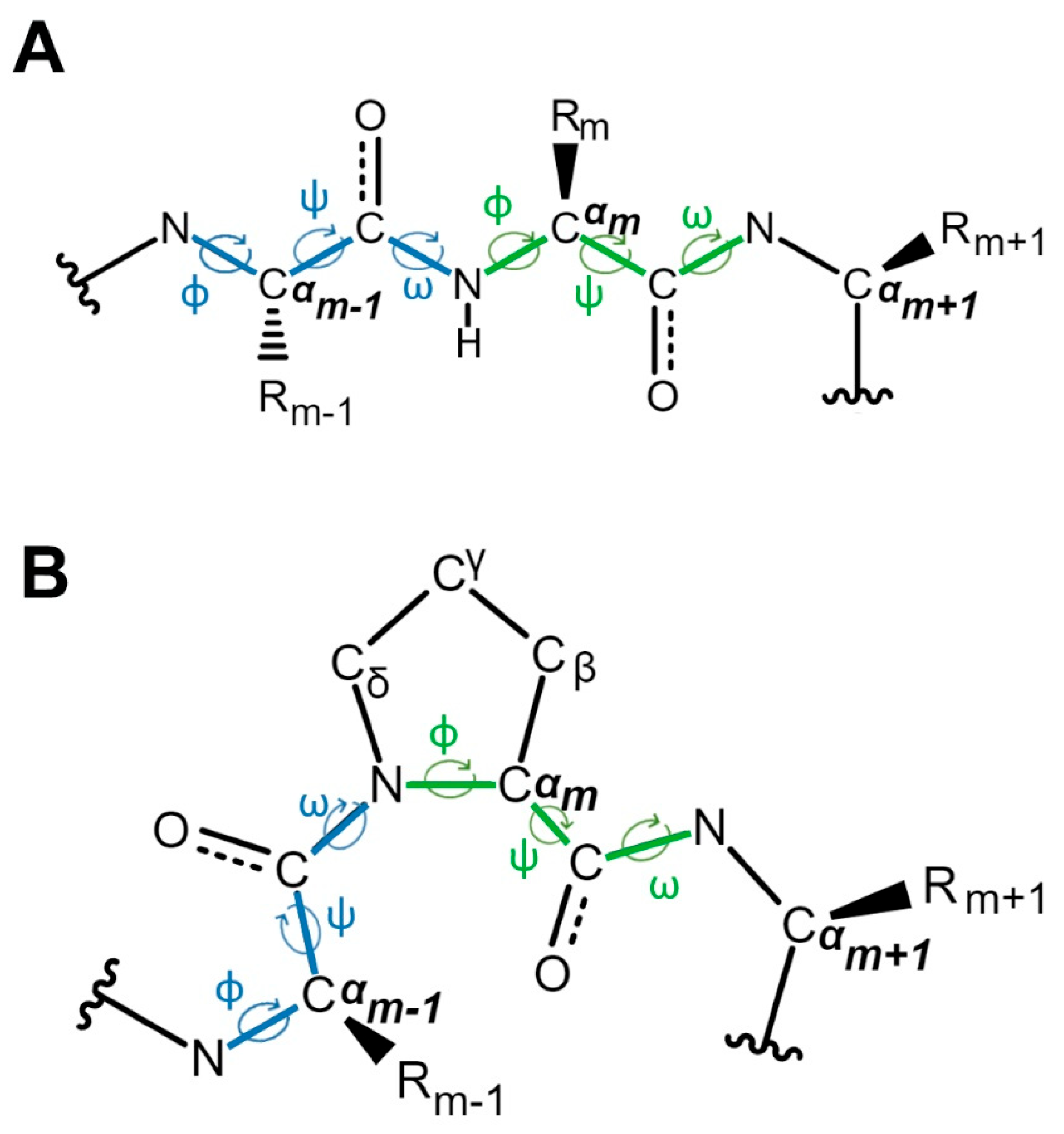
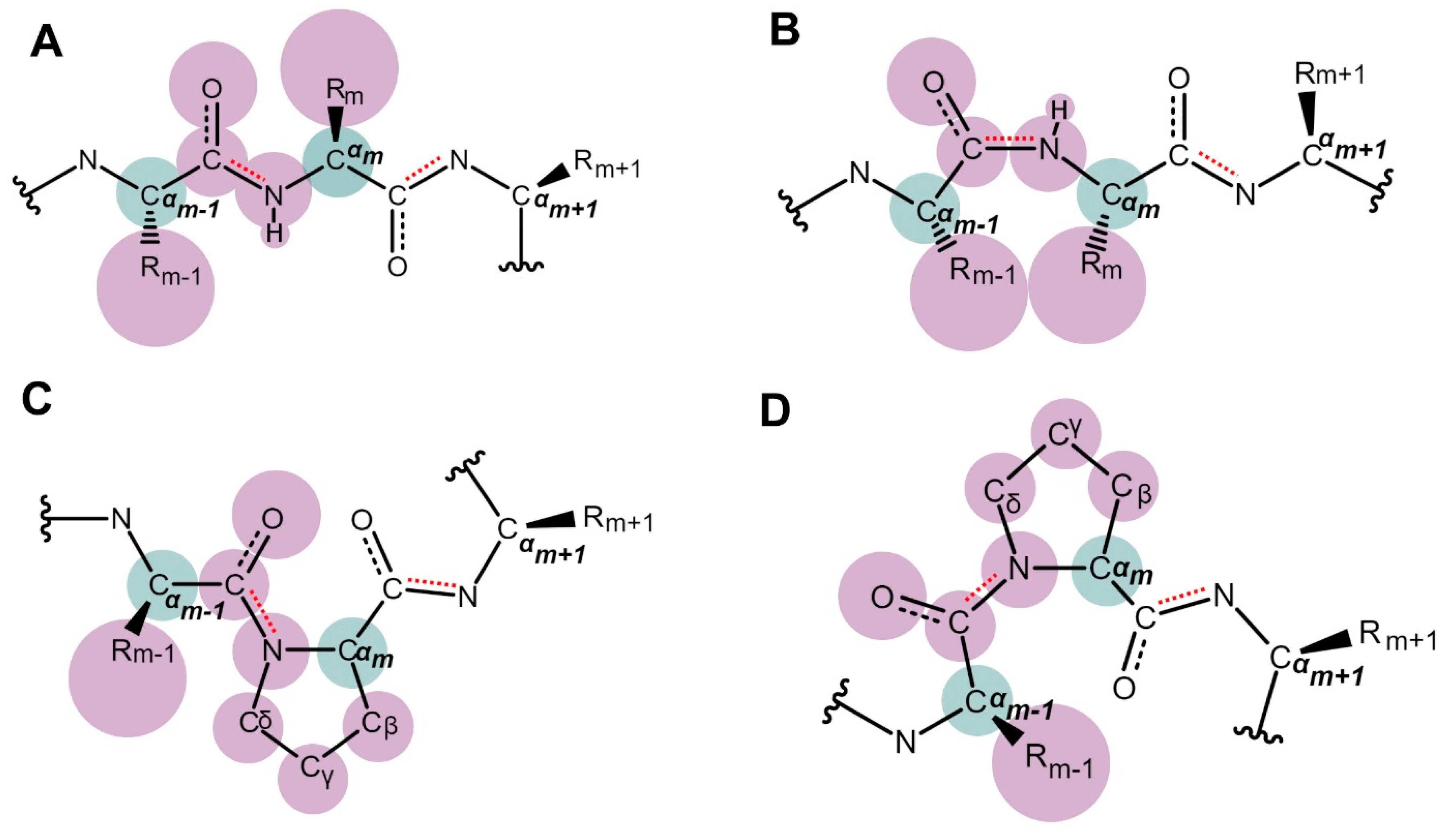
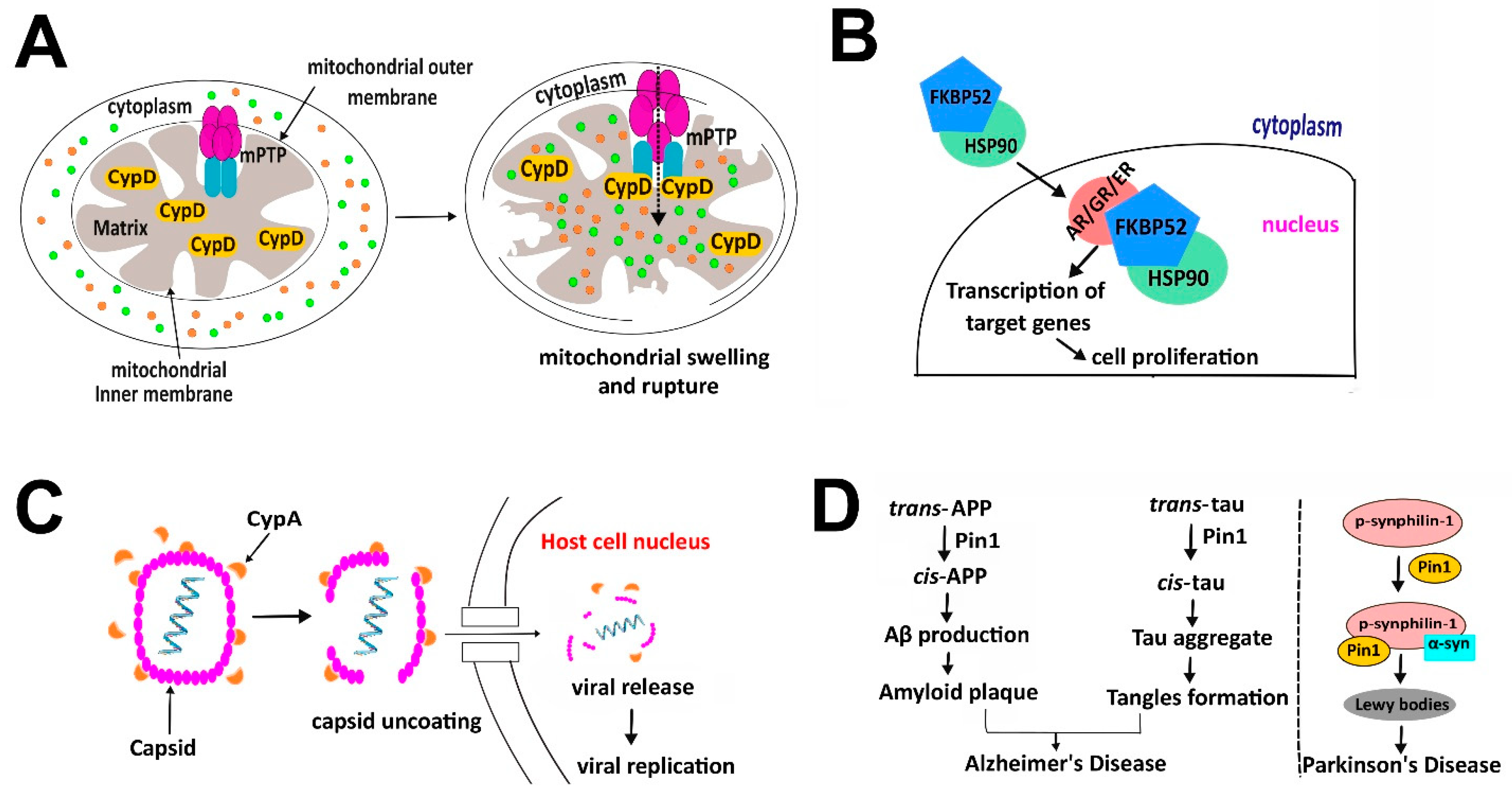
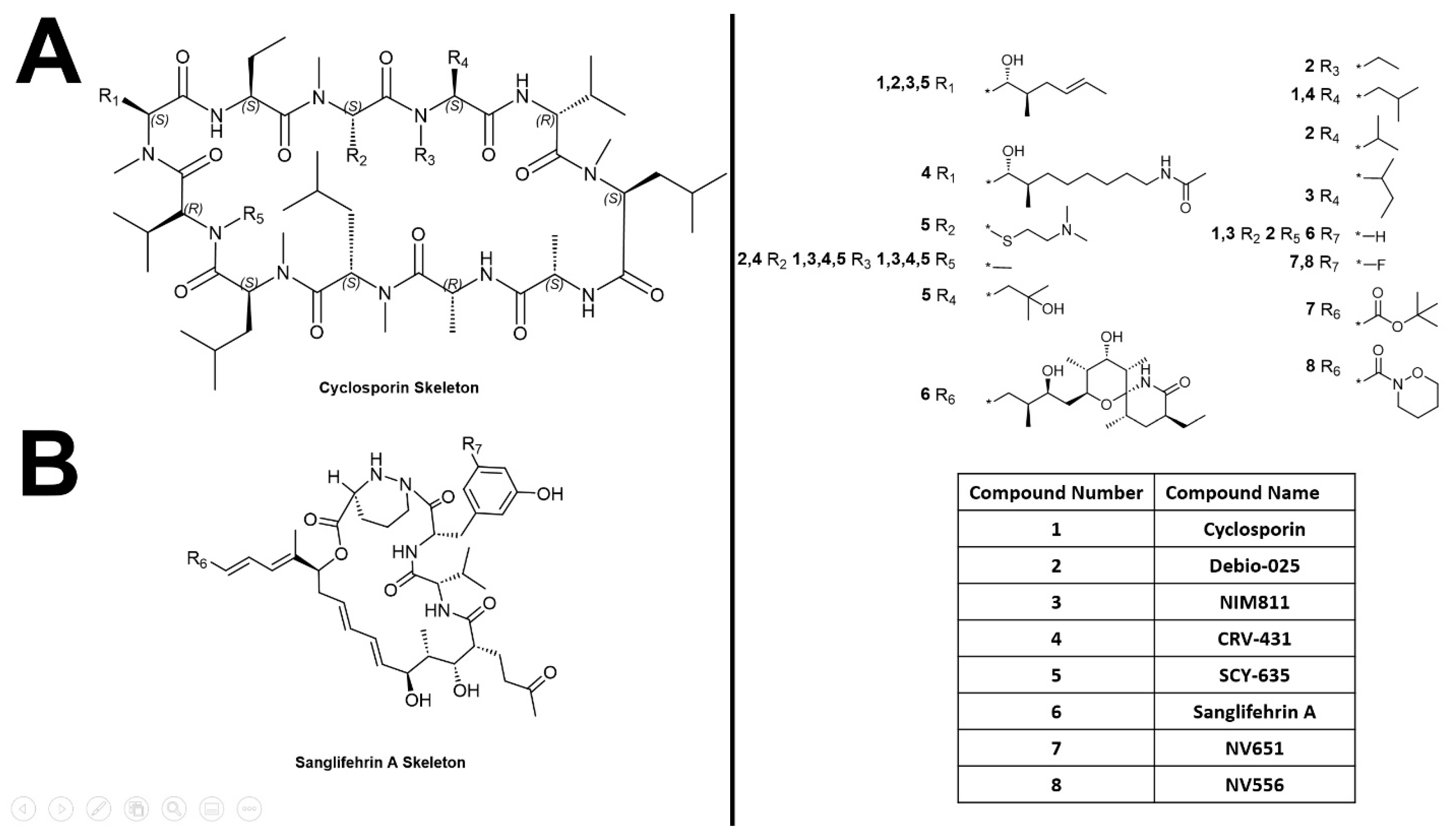
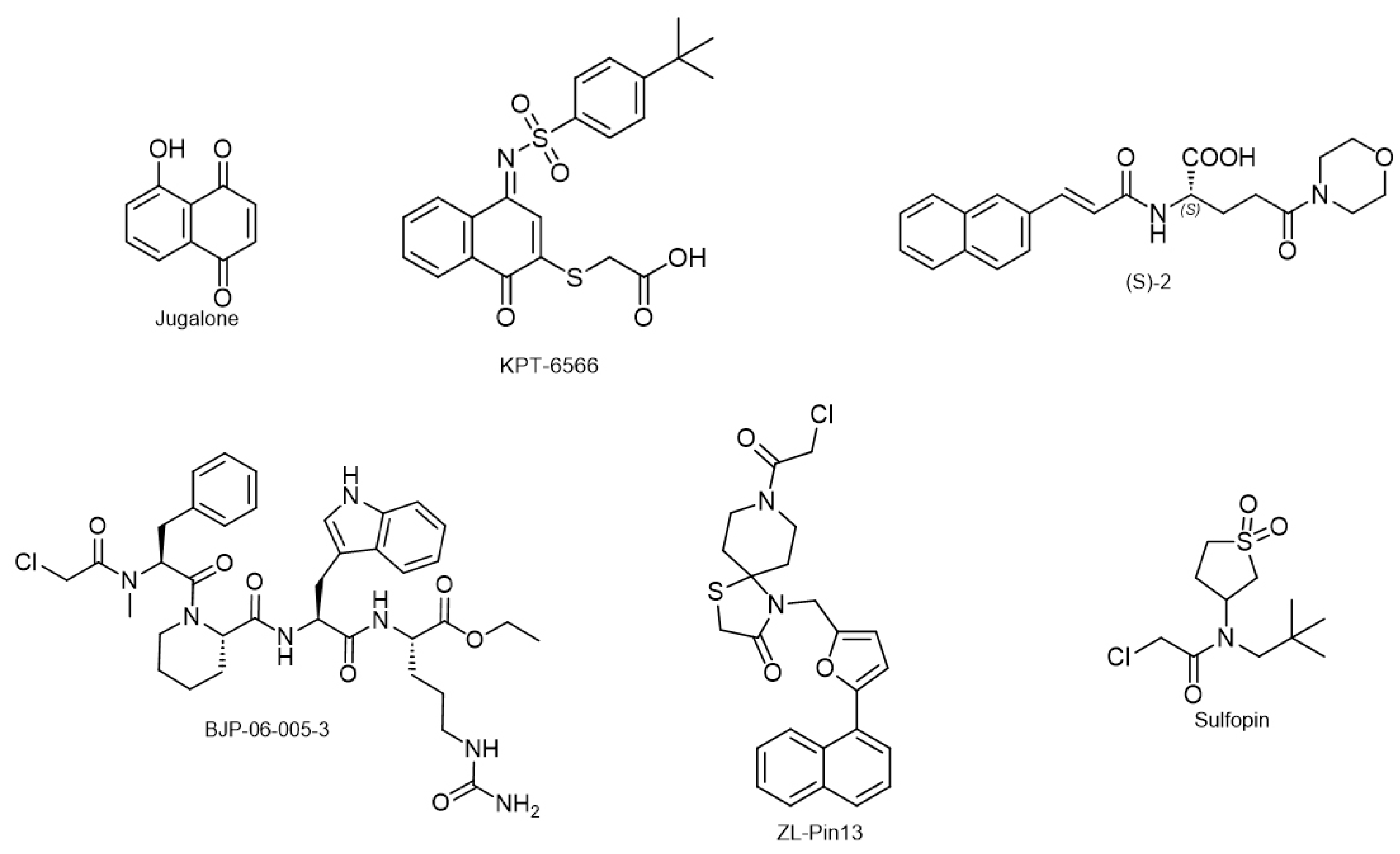
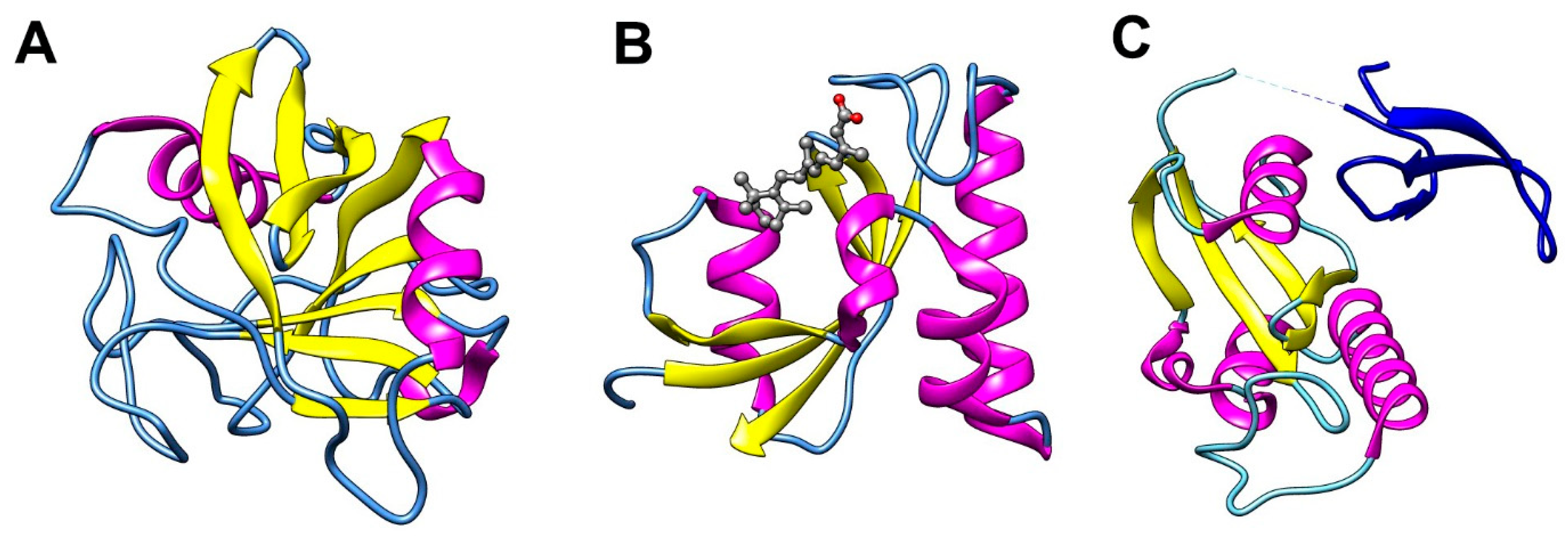
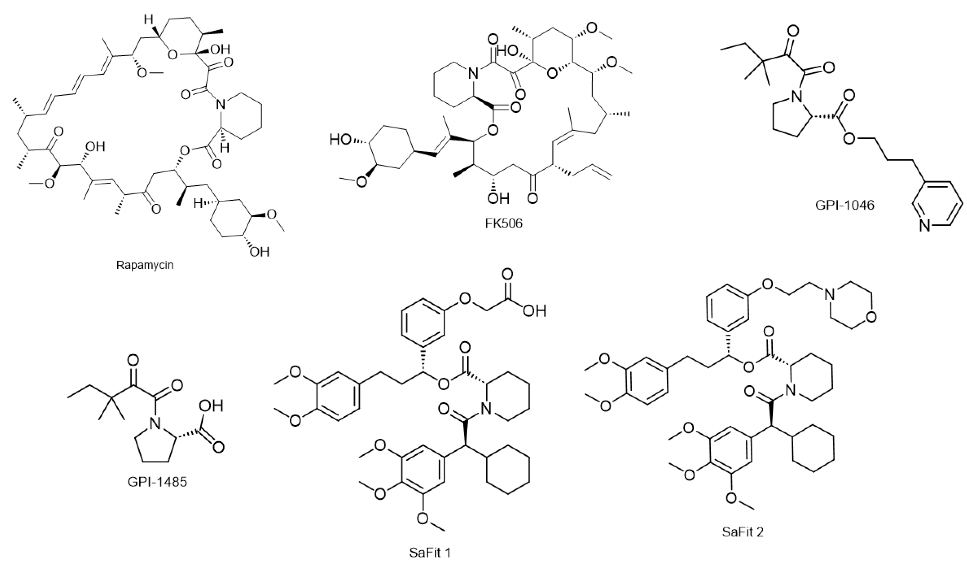
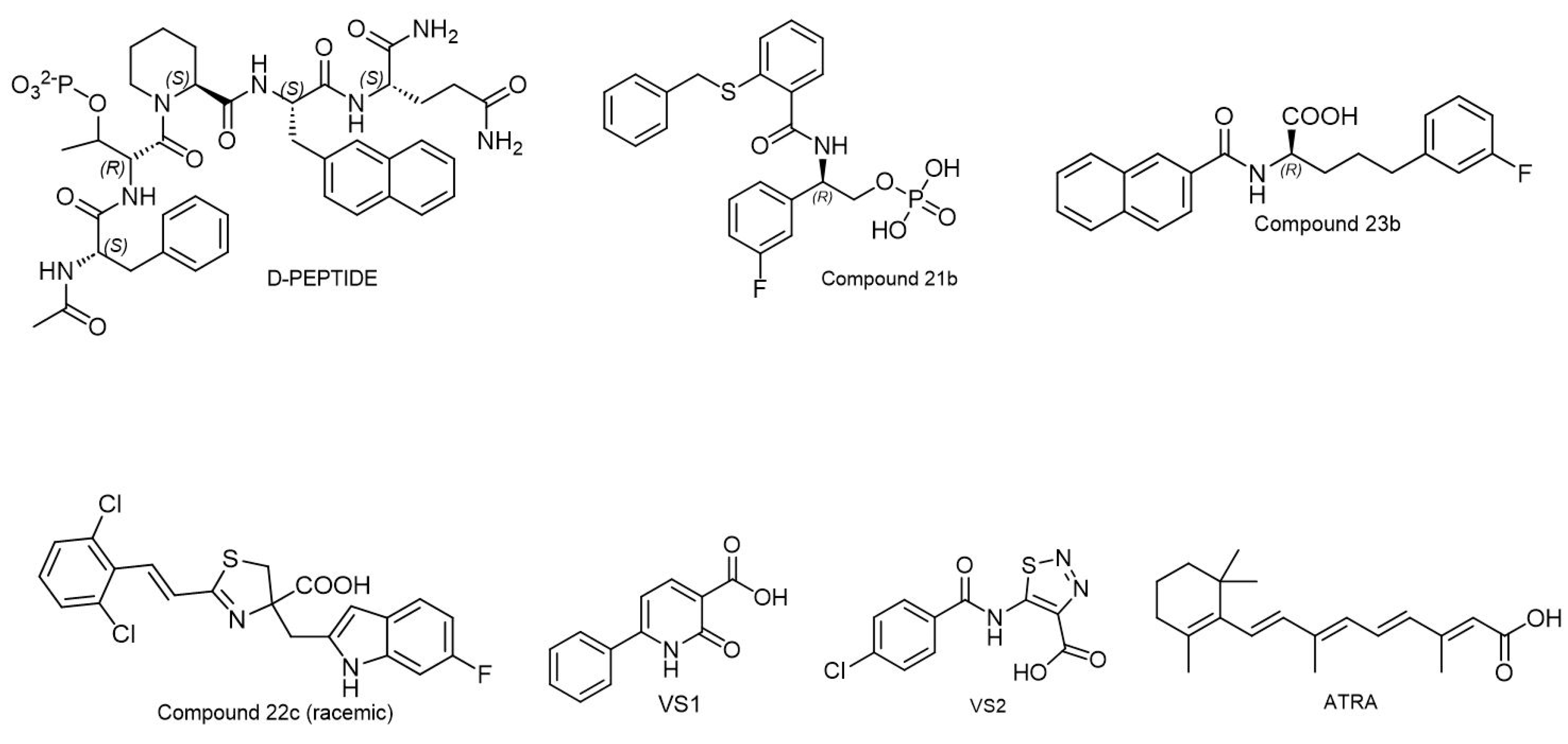
Figure 1. Schematic diagram of protein peptide and the three torsion angles phi (Φ), psi (φ) and omega (ω) that define the conformation of protein backbone. The phi angle is around the -N-Cα- bond; the psi angle is around the -Cα-C- bond and the omega angle is around the -C-N- bond, which is also referred to as the peptide bond. Two sets of phi, psi and omega angles are labeled for residue at position m-1 and m in blue and green, respectively. (A). Peptide that has linear residues at position m-1, m and m+1. (B). Peptide that has a proline at position m. The freedom of the phi angle of proline is restricted due to the ring structure of proline.
Biology 12 01008 g001
Figure 2. Schematic diagram of trans- and cis-conformations around the peptidyl bond that connects residues m and m-1. To aid visualization, atoms around this peptidyl bond are depicted by spheres according to their sizes although not strictly proportionally. All spheres are in pink except for the two Cα atoms that are painted in green for easy identification. (A). Trans-conformation around the peptidyl bond connecting two linear residues. In this conformation, Cαm-1 and Cαm along with their connected linear R groups are dispersed on two sides of the peptidyl bond and therefore this conformation is energetically favorable. (B). Cis-conformation around the peptidyl bond connecting two linear residues. In this conformation, Cαm-1 and Cαm along with their connected linear R groups are concentrated on one side of the peptidyl bond, leading to crowdedness, or even clashing between atoms. Therefore, this conformation is energetically unfavorable. (C). Trans-conformation around the peptidyl bond connecting a linear residue with a proline. In this conformation, although Cαm-1 and Cαm are located on different sides of the peptidyl bond, due to the cyclic arrangement of atoms on proline, the Cδ atom replaces the hydrogen atom and wraps back to the backbone to be close to Cαm-1 and its connected Rm-1 group. This leads to the atoms being not as dispersed as in scenario (A). (D). Cis-conformation around the peptidyl bond connecting a linear residue with a proline. In this conformation, although Cαm-1 and Cαm are located on the same side of the peptidyl bond, because the cyclic arrangement of proline loops the side chain atoms back to the backbone, atoms are not as concentrated on one side as seen in scenario (B). As a result, the energy difference between the trans- and cis-conformation around a peptidyl-prolyl bond is much smaller than that of two linear residues.
Biology 12 01008 g002
Figure 3. Overall structures of all three types of PPIases. Ribbon representation is colored by secondary structure with yellow, magenta, and blue for helices, sheets, and loops, respectively. (A). Overall structure of cyclophilin (PDB code: 1MF8 [53]). (B). Overall structure of FKBP12 (PDB code: 1FKJ [54]). The ligand, FK506, is also displayed in ball-and-stick representation. (C). Overall structure of Pin1 (PDB code: 2ITK [55]). The PPIase domain is colored by secondary structure while the WW domain is shown by dark blue. The dotted line indicates some missing residues connecting the two domains. All three images are generated by UCSF Chimera [56].
Biology 12 01008 g003
Figure 4. (A). Schematic diagram to show how ATR is regulated by proline isomerization to switch between its dual roles in modulating cell death and DNA damage checkpoint signaling. (B). NMR structure of Cis- and trans-proline at position 287 switches the conformation of the Itk SH2 domain. Cis- and trans-conformations are colored in pink (PDB code: 1LUK [89]) and blue (PDB code: 1LUN [89]), respectively. Proline 287 and its preceding residue Asn286 are shown in ball and stick by red and dark blue in cis- and trans-conformation, respectively. The image is generated by using UCSF Chimera [56].
Biology 12 01008 g004
Figure 5. An illustration showing the impact of PPIase in four prominent human diseases. (A). Autoimmune disease. In multiple sclerosis, CypD plays a crucial role by binding and regulating mPTP. This interaction leads to the influx of small molecules depicted by green or orange spheres into the cells, causing mitochondrial swelling and rupture, cytochrome C release, and ultimately cell death. (B). Cancer. FKBP52 serves as the co-chaperone of HSP90 and interacts with steroid hormone receptors, including the androgen receptor (AR), glucocorticoid receptor (GR), and estrogen receptor (ER). This interaction enhances the transcriptional activity of the respective receptors, leading to increased cell proliferation, as observed in breast and prostate cancer. (C). Infectious disease. CypA plays a pivotal role in the life cycle of the HIV-1 virus by catalyzing proline isomerization at the Gly89-Pro90 site of the viral capsid, resulting in its conformational transition that is vital for capsid uncoating, viral genome release, and ultimately promoting viral replication. (D). Neurodegenerative disease. Pin1 plays a significant role in the pathogenesis of Alzheimer’s disease and Parkinson’s disease. In the case of Alzheimer’s disease, Pin1 catalyzes the isomerization of APP and tau from trans to cis, leading to the formation of amyloid plaques and neurofibrillary tangles, respectively, which are distinct hallmarks of Alzheimer’s disease. In the case of Parkinson’s disease, Pin1 interacts with phosphorylated synphilin-1, forming a stable complex with α-synuclein protein and promoting the formation of Lewy bodies, a characteristic feature of Parkinson’s disease. This figure is created using Inkscape (Version 1.2). The RNA icon is created by Servier (Source: Servier, https://smart.servier.com/ (accessed on 1 May 2023)) and is licensed under CC-BY 3.0 Unported (License: CC-BY 3.0 Unported, https://creativecommons.org/licenses/by/3.0/ (accessed on 1 May 2023)).
Biology 12 01008 g005
Figure 6. Chemical structures of cyclophilin inhibitors. (A). Molecular skeleton of cyclosporin. (B) Molecular skeleton of sanglifehrin A. Reference for cyclophilin inhibitors and their corresponding substitutes are shown on the right. Compounds are numbered and shown in bold. Structures of substitutes at different R positions for different compound number are given at the top. Compound names corresponding to compound number are shown in the table at the bottom. The structures are drawn using ChemDraw (Version 22.2.0).
Biology 12 01008 g006
Figure 7. Chemical structures of FKBP inhibitors. Rapamycin and FK506, the two FDA-approved natural ligands for immunosuppression, carry the pipecolate moiety critical for FKBP binding. The two GPI-derivatives carry a similar chemical group to the pipecolate moiety. SaFit1 and SaFit2 are the two inhibitors found to be selective to the FKBP51 isoform. The structures are drawn using ChemDraw (Version 22.2.0).
Biology 12 01008 g007
Figure 8. Chemical structures of Pin1 covalent inhibitors. Juglone, the first Pin1 covalent inhibitor, is a natural and simple compound with limited specificity to Pin1. KPT-6566, a derivative of Juglone with added structural complexity, shows higher specificity to Pin1 than Juglone. (S)-2 and BJP-06-005-3 are covalent inhibitors that do not share structural similarity with Juglone. ZL-Pin13 and Sulfopin, also distinct from Juglone, are two newly discovered covalent inhibitors with high selectivity and potency against Pin1. The structures are drawn using ChemDraw (Version 22.2.0).
Biology 12 01008 g008
Figure 9. Chemical structures of Pin1 non-covalent inhibitors. D-PEPTIDE is a non-natural peptide. Compound 21b, Compound 23b, and Compound 22c (racemic) developed by Pfizer have diverse structures with high potency inhibiting Pin1 activity in vitro. VS1 and VS2 are two newly discovered inhibitors with smaller molecular sizes. ATRA has been the only Pin1 inhibitor that has moved to clinical trials so far. The structures are drawn using ChemDraw (Version 22.2.0).
Biology 12 01008 g009
Table 1. Compare and contrast of three families of PPIases.
Prolyl Isomerase Protein Size Preferred Proline Site TriggersSubstrate ProteinsSubcellular Localization
FK506 binding proteins12–52 kDaXaa-ProLigand binding, change in local environment of the protein such as pHTau APP, ryanodine receptor, Inositol 1,4,5-triphosphate receptor, TGF-β, heat-shock proteins, nuclear transport receptor including androgen receptorCytoplasm (primary), endoplasm reticulum (ER), and nucleus
Cyclophilins 18–45 kDaXaa-ProLigand binding, change in local environment of the protein such as pH HIV1 capsid, NS5A of hepatitis C virus, heat -shock proteins (HSP90), α-synucleinCytosol, ER, mitochondria and nucleus
Parvulins 14, 17, and 18 kDapSer/pThr-Pro (Pin1)
Arg/Leu-Pro (Par14 and Par17)
Phosphorylation on Ser or Thr proceeding to Pro residue (Pin1)
Arginine or leucine residues (Par14 and Par17)
ATR, p53, CDC25, α-synuclein, survivin, β-catenin, NF-kβ, Cyclin D1, C-Jun, RNA pol II Nucleus (Pin1)
Nucleus and cytosol (Par14)
Mitochondria (Par17)
Note: Xaa = any standard amino acid.
Table 2. Summary of inhibitors for all three types of PPIases.
InhibitorsInhibitor Target IC50 or Ki Inhibitor TypeReference
Cyclosporine Cyclophilins420 ± 56 nMImmunosuppressive Sedrani et al., 2003 [226]
Debio-025 0.18 ± 0.03 nMNon-Immunosuppressive
Cyclosporine analog
Ptak et al., 2008 [227], Paeshuyse et al., 2006 [228]
NIM8110.66 μM Non-Immunosuppressive
Cyclosporine analog
Ma et al., 2006 [229]
CRV4312.5 nM
(CypA)
Non-Immunosuppressive
Cyclosporine analog
Kuo et al., 2019 [230]
SCY-6351.84 μM Non-Immunosuppressive Cyclosporine analogHopkins et al., 2010 [231]
Sanglifehrin A 6.9 ± 0.9 nMImmunosuppressive
Sanglifehrin
Sedrani et al., 2003 [226]
NV6516.3 nMNon-Immunosuppressive
Sanglifehrin analog
Serrano et al., 2021 [232]
NV556 Non-Immunosuppressive
Sanglifehrin analog
Kuo et al., 2019 [230], Serrano et al., 2019 [233]
Rapamycin FKBPs0.1 nMImmunosuppressiveKolos et al., 2018 [61]
FK5061 nMImmunosuppressiveKolos et al., 2018 [61]
GPI-1485 ImmunosuppressiveKolos et al., 2018 [61]
SaFit14 nM Non-ImmunosuppressiveGaali et al., 2015 [234]
SaFit28 nM Non-ImmunosuppressiveGaali et al., 2015 [234]
Juglone Pin1 7.68 μMCovalent inhibitor Zhang et al., 2015 [235]
KPT-65660.64 μM Covalent inhibitorCampaner et al., 2017 [236]
(S)-23.2 μMCovalent inhibitorIeda et al., 2010 [237]
BJP-06-005-348 nMNon-covalent inhibitorPinch et al., 2020 [238]
ZL-Pin130.067 ± 0.03 μMCovalent inhibitorLiu et al., 2022 [239]
Sulfopin 38 nM Covalent inhibitorDubiella et al., 2021 [240]
D-PEPTIDE20.4 ± 4.3 nM Non-covalent inhibitorZhang et al., 2007 [55]
Compound 21b6 nM Non-covalent inhibitorGuo et al., 2009 [241]
Compound 23b890 nM Non-covalent inhibitorDong et al., 2010 [242]
Compound 22C196 nM Non-covalent inhibitorGuo et al., 2014 [243]
VS1>100 μMNon-covalent inhibitorPoli et al., 2022 [244]
VS219 ± 3 μMNon-covalent inhibitorPoli et al., 2022 [244]
ATRA112 ± 10 μMNon-covalent inhibitorPoli et al., 2022 [244]
Disclaimer/Publisher’s Note: The statements, opinions and data contained in all publications are solely those of the individual author(s) and contributor(s) and not of MDPI and/or the editor(s). MDPI and/or the editor(s) disclaim responsibility for any injury to people or property resulting from any ideas, methods, instructions or products referred to in the content.

