Review

The Unique Roles of Ion Channels in Pluripotent Stem Cells in Response to Biological Stimuli

by
Taku Kaitsuka
School of Pharmacy at Fukuoka, International University of Health and Welfare, Enokizu 137-1, Okawa 831-8501, Fukuoka, Japan
Biology 2024, 13(12), 1043; https://doi.org/10.3390/biology13121043
Submission received: 17 November 2024 / Revised: 8 December 2024 / Accepted: 11 December 2024 / Published: 13 December 2024

Simple Summary

Pluripotent stem cells like embryonic stem and induced pluripotent stem cells have unique properties of pluripotency and self-renewal. The major role of membrane-bound receptors and their downstream signaling after binding their ligands (leukemia inhibitory factor and growth factors) in maintaining their properties is well documented. So far, the roles of ion and ion channels in pluripotent stem cells have not been much focused on or investigated. Cellular ion homeostasis is essential for all types of cells, and Ca2+ and Mg2+-dependent signaling is also important for cellular events such as metabolism, stress response, cell cycle and survival. In this review, presently restricted insights about ion channels are introduced, and other candidates for ion channels that could regulate pluripotency and self-renewal are extracted for further understanding of pluripotent stem cells.

Abstract

Ion channels are essential for mineral ion homeostasis in mammalian cells, and these are activated or inhibited by environmental stimuli such as heat, cold, mechanical, acidic, or basic stresses. These expressions and functions are quite diverse between cell types. The function and importance of ion channels are well-studied in neurons and cardiac cells, while those functions in pluripotent stem cells (PSCs) were not fully understood. Some sodium, potassium, chloride, calcium, transient receptor potential channels and mechanosensitive Piezo channels are found to be expressed and implicated in pluripotency and self-renewal capacity in PSCs. This review summarizes present and previous reports about ion channels and their response to environmental stimuli in PSCs. Furthermore, we compare the expressions and roles between PSCs and their differentiated embryoid bodies. We then discuss those contributions to pluripotency and differentiation.

1. Introduction

Pluripotent stem cells (PSCs) are quite unique cells that have self-renewal and pluripotency abilities. A significant molecular event for pluripotency is transcriptional regulation mediated by pluripotency factors, OCT4 and NANOG. Epigenetic regulation of chromatins in pluripotency and differentiation genes is also an essential factor for the abilities of PSCs. Negative regulation of differentiation genes is mediated by histone H3 lysine 27 methylation by polycomb groups. Those main events generally occur in the nucleus of cells.
Fundamentally, environmental signals from culture medium to PSCs regulate their pluripotency. In naive PSCs of mouse embryonic stem cells (ESCs), the leukemia inhibitory factor (LIF)-JAK-STAT pathway regulates their self-renewal and pluripotency [1]. LIF binds to the LIF receptor, which forms a complex with the receptor glycoprotein 130 [2]. In primed PSCs of human ESCs, the transforming growth factor-β (TGF-β)/Activin A/Nodal pathway regulates their pluripotency by activating Smad2/3 [3]. In vitro, the basic fibroblast growth factor (bFGF) is essential for human PSC stemness and self-renewal maintenance [4,5,6]. bFGF activates the phosphatidylinositol 3-kinase (PI3K)/AKT pathway, inhibiting ERK activity, WNT signaling and dephosphorylation of glycogen synthase kinase-3β (GSK-3β) [7,8]. Other signals stimulating membrane proteins are involved in the unique function of PSCs, mainly proliferation ability and regulation of the pluripotent factors. For example, disruption of transient receptor potential channel 7 (TRPM7) causes proliferation arrest in mouse embryonic stem cells [9]. The voltage-gated potassium channel (Kv) and membrane potential affect self-renewal in mouse ESCs, and the Kv blocker attenuates its cell cycle [10].
To date, there are few reviews in which the overall argument about the functions and roles of ion channels in PSCs are included. In this review, insights about the functions of ion channels in PSCs are summarized, and their roles are discussed. Additionally, the candidate channels that regulate the property of PSCs are extracted from previous reports using undifferentiated and differentiated cells.

2. Ion Channels

Ion channels are proteins that facilitate the movement of ions across cell membranes, playing a crucial role in cellular functions. These channels are found in both the plasma membrane and intracellular membranes and are expressed in organisms ranging from bacteria to humans, where they are essential for life. In general, excitable cells, including neurons, muscle cells and touch receptor cells, express these channels and use them to generate electrical signals. In non-excitable cells, ion channels regulate intracellular ion levels and affect the activities of intracellular proteins and their functions. Opening of ion channels is mediated by the stimulation from external or internal ligands such as second messengers, voltage, temperature and mechanical stresses.
Cellular ions include sodium (Na+), potassium (K+), calcium (Ca2+), magnesium (Mg2+) and chloride (Cl). Basically, Na+ and K+ regulate the membrane potential of the plasma membrane. Ca2+ acts as a second messenger and activates Ca2+-dependent enzymes. Mg2+ is essential for Mg2+-dependent molecules and proteins like adenosine triphosphate (ATP) and Na-K-ATPase. Cl is the main anion outside the cell and maintains osmotic pressure similar to the above ions.

3. Ion Channels Expressed in Pluripotent Stem Cells

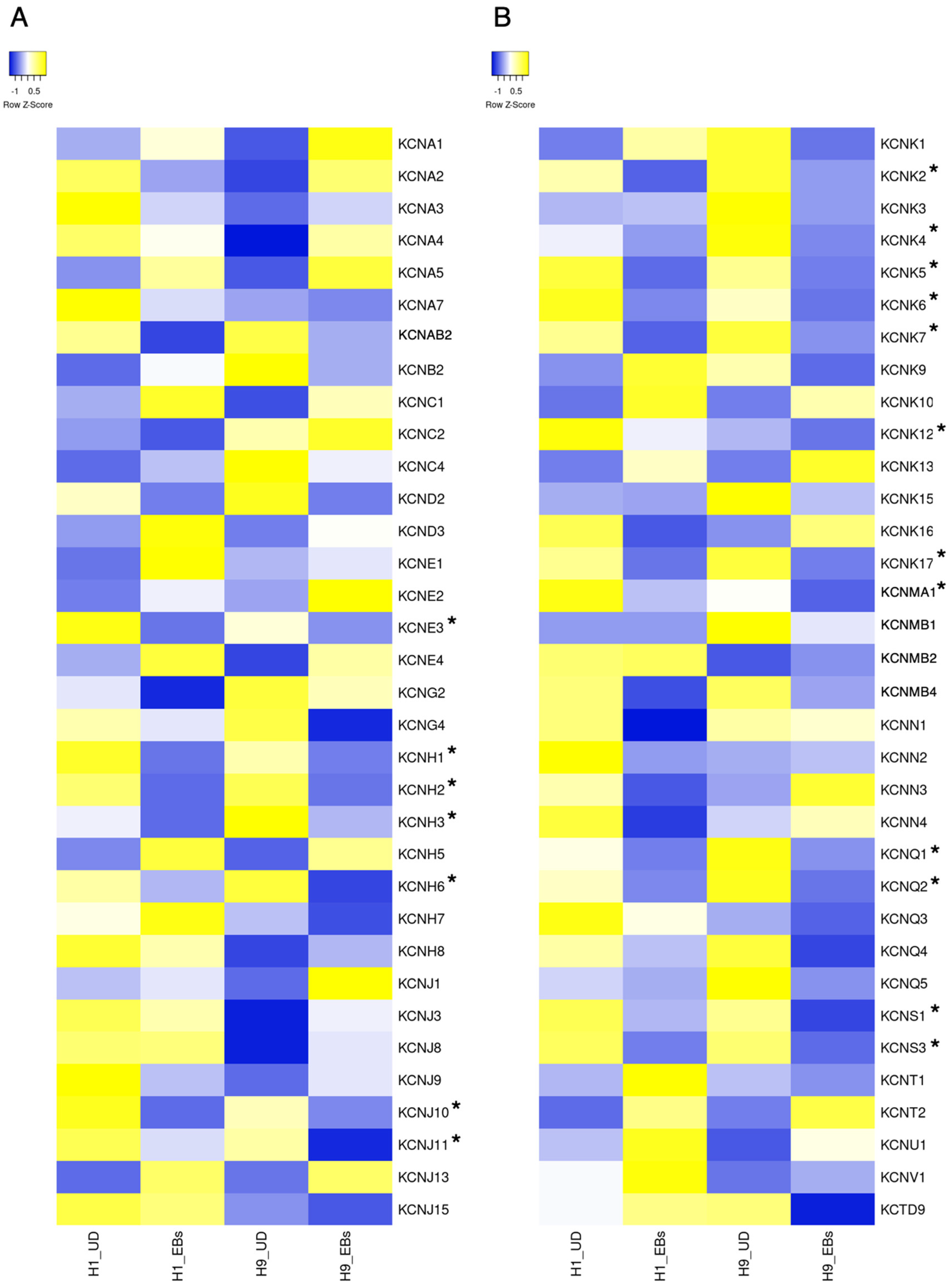
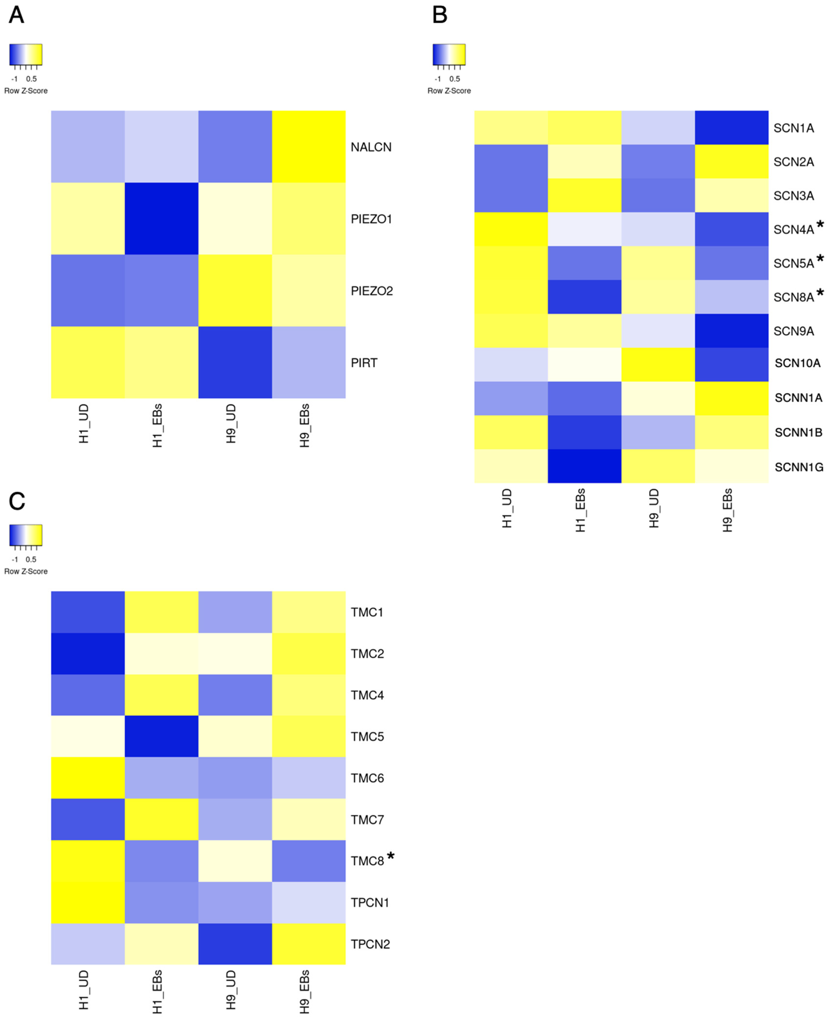
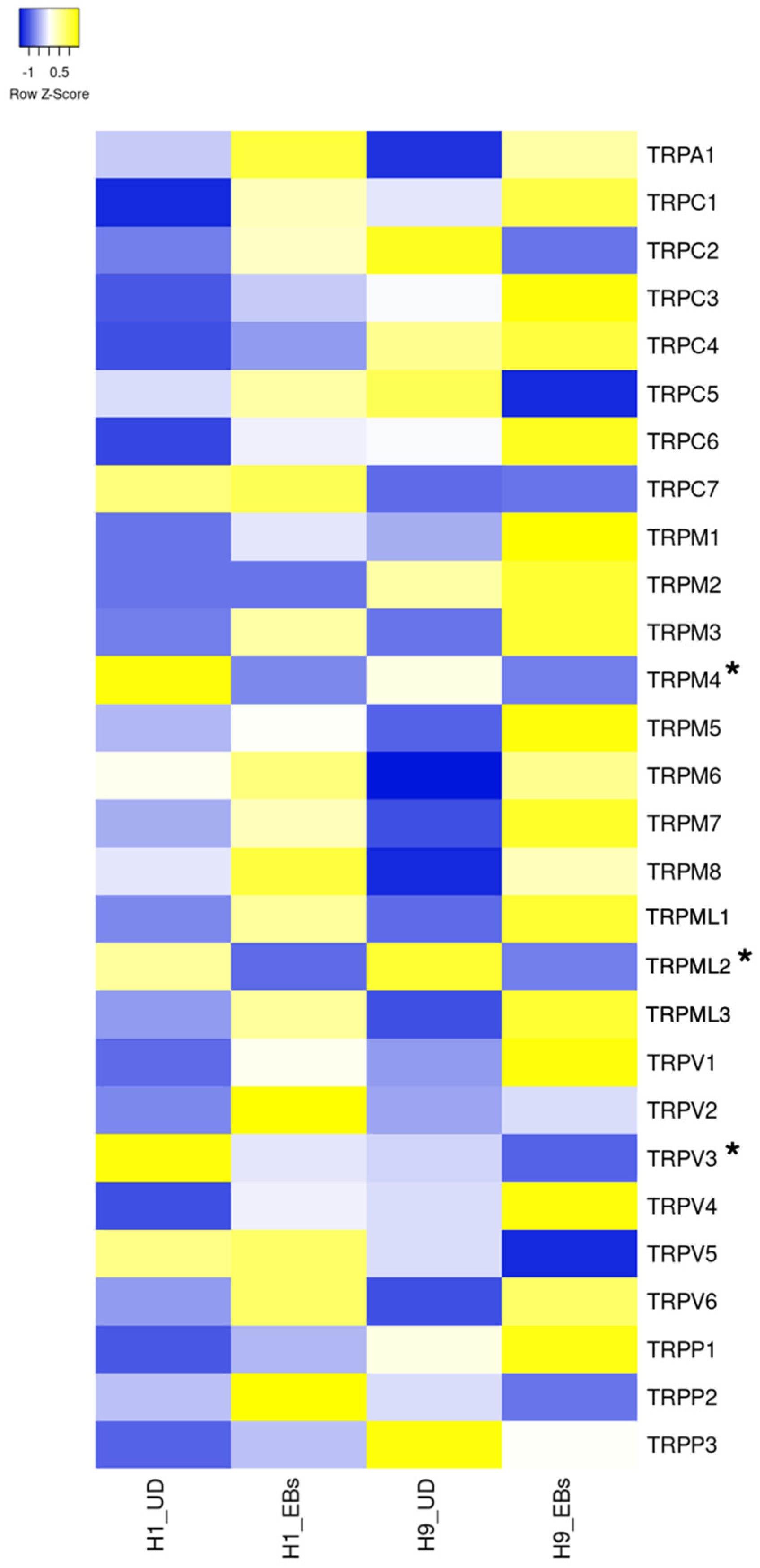
ESCs express a small part of ion channels compared to tissue stem cells and somatic excitable/non-excitable cells [11]. Heatmaps in Figure 1, Figure 2, Figure 3, Figure 4 and Figure 5 show the gene expression levels between human ESCs and their differentiated embryoid bodies (EBs) from the dataset GDS5408 of the National Center for Biotechnology Information (NCBI) website [12]. In this dataset, pluripotency gene NANOG in human ESCs is confirmed to be higher than their EBs, while the differentiation gene SOX17 (endoderm), MESP1 (mesoderm) and SOX1 (ectoderm) are higher in EBs than in ESCs (Figure 1). In the case of ion channels, levels of almost all channel genes are higher in EBs than in ESCs, while some of them become lower after differentiation to EBs (Figure 2, Figure 3, Figure 4 and Figure 5). In this review, we focus on the genes in both cell lines (H1 and H9) of ESCs, which decrease less than 0.8-fold compared to their EBs. Specifically, the expression levels of CLIC4 and CLIC5 decreased in differentiated EBs of both cell lines (Figure 2). Each gene encodes chloride intracellular channel proteins 4 and 5, respectively. HCN4, encoding the hyperpolarization-activated cyclic nucleotide-gated potassium channel 4, also decreased in EBs (Figure 2). Among potassium channels, 19 genes decreased in EBs (Figure 3). These genes and corresponding product proteins are KCNE3 (voltage-gated potassium channel MiRP2), KCNH1 (Kv10.1), KCNH2 (Kv11.1), KCNH3 (Kv12.2), KCNH6 (Kv11.2), KCNJ10 (ATP-sensitive inwardly rectifying potassium channel Kri4.1), KCNJ11 (Kir6.2), KCNK2 (two-pore domain potassium channel TREK-1), KCNK4 (TWIK-related arachidonic acid-stimulated potassium channel TRAAK), KCNK5 (TWIK-related acid-sensitive potassium channel TASK-2), KCNK6 (inward rectifying potassium channel protein TWIK-2), KCNK7 (double-pore potassium channel DP3), KCNK12 (tandem pore domain halothane-inhibited potassium channel THIK-2), KCNK17 (TWIK-related acid-sensitive potassium channel TASK-4), KCNMA1 (calcium-activated potassium channel Kca1.1), KCNQ1 (voltage-gated potassium channel Kv7.1), KCNQ2 (Kv7.2), KCNS1 (Kv9.1) and KCNS3 (Kv9.3). Among sodium channels, SCN4A, SCN5A and SCN8A, which code voltage-gated sodium channels Nav1.4, Nav1.5 and Nav1.6, are decreased genes in EBs (Figure 4B). TMC8, encoding transmembrane channels like 8 and transient receptor potential channels TRPM4, TRPML2 and TRPV3 also decreased in EBs (Figure 4C and Figure 5). Based on the data on changes in expression levels during differentiation, those channels are suggested to have some roles in the unique function of human ESCs. Supporting this speculation, Zhang et al. reviewed that many types of potassium channels are detected and function in stem cells, including human ESCs [11].
There are several studies about the existence of membrane proteins, including ion channels in human and mouse PSCs [14]. Previously, Jiang et al. analyzed the functional importance of ion channels in human and mouse ESCs [15]. In this report, microarray analysis was performed on human-induced pluripotent stem cells (iPSCs), ESCs and MSCs. In iPSCs, CACNA1H, CACNA2D1, CACNA2D2, CACNG4, CACNG7 coding voltage-gated calcium channel Cav3.2, Cavα-2/δ-1, Cavα-2/δ-2, TARPγ-4, TARPγ-7, SCN8A coding voltage-gated sodium channel Nav1.6, KCNC4, KCNK1, KCNK12, KCNK5, KCNK6, KCNQ2, KCNS3, KCNMB4, KCNN2 coding voltage-gated potassium channel Kv3.4, inward rectifying potassium channel TWIK-1, tandem pore domain halothane-inhibited potassium channel THIK-2, TASK-2, TWIK-2, Kv7.2, Kv9.3, BKβ4, small conductance calcium-activated potassium channel SK2 and HCN4 were significantly expressed. The ion channel transcriptome of iPSCs could not be distinguished from that of ESC lines. In Figure 4B, SCN8A is detected as it decreased during differentiation to EBs. Similarly, KCNK5, KCNK6, KCNK12, KCNQ2 and KCNS3 gens decreased in EBs (Figure 3A,B).
On the other hand, studies on specific channels in human PSCs have been performed, as described in the following sections.

4. Sodium Channels

Voltage-gated sodium channel subunits are encoded by SCN1A (Nav1.1), SCN2A (Nav1.2), SCN3A (Nav1.3), SCN4A (Nav1.4), SCN5A (Nav1.5), SCN8A (Nav1.6), SCN9A (Nav1.7) and SCN10A (Nav1.8). The properties and importance of these subunits are well-reviewed by Meisler et al. [16]. Generally, the knockout of a single gene does not cause embryonic lethality [16], showing that other genes compensate for each other in embryonic development. Based on composed data from microarray analysis [12] and previous reports about the expression of ion channels [15], SCN8A seems to have the main role in the transmission of Na2+ in PSCs.

The Role of Sodium Channels in Pluripotent Stem Cells

As mentioned above, Jiang et al. performed transcriptomic analysis of ion channel genes in human ESCs, iPSCs and mesenchymal stem cells (MSCs) [15]. A total of 158 voltage-dependent ion channels were found to be expressed in iPSCs. Among them, only SCN8A was a sodium channel detected in human iPSCs by RT-PCR analysis. However, in the experiment with patch-clamp analysis, the current of the voltage-gated sodium channel could not be detected in human ESCs or iPSCs [15,17]. It is assumed that voltage-gated sodium channels, including SCN8A, are not essential in maintaining the properties of human PSCs.

5. Potassium Channels

Potassium channels consist of numerous families and genes as voltage-gated (KCNA, KCNB, KCNC, KCND, KCNE, KCNF, KCNG, KCNH, KCNI, KCNQ, KCNS and KCNV), inwardly rectifying (KCNJ), two-pore domain (KCNK), calcium-activated (KCNM, KCNN, KCNU) and sodium-activated (KCNT) types. The structure and function of potassium channels have been well-reviewed in several papers [18,19]. Potassium channels mainly function in excitable cells like neurons and cardiomyocytes, and their dysfunction causes psychiatric disorders and cardiovascular diseases in humans. In PSCs, many genes of KCNE3, KCNH1, KCNH2, KCNH3, KCNH6, KCNJ10, KCNJ11, KCNK2, KCNK4, KCNK5, KCNK6, KCNK7, KCNK12, KCNK17, KCNMA1, KCNQ1, KCNQ2, KCNS1 and KCNS3 seem to exist in the pluripotent state of human ESCs via microarray analysis and have some physiological roles (Figure 3).

The Role of Potassium Channels in Pluripotent Stem Cells

Jiang et al. found that KCNC4, KCNK1, KCNK12, KCNK5, KCNK6, KCNQ2, KCNS3, KCNMB4 and KCNN2 genes are expressed in human iPSCs by microarray analysis, and these are confirmed by RT-PCR, except KCNK12 [15]. Non-selective potassium channel blocker tetraethylammonium (TEA)-sensitive delayed rectifier K+ currents (IKDR) are observed in human ESCs and iPSCs, whereas voltage-gated sodium channel and voltage-gated calcium channel currents cannot be measured in both ESCs or iPSCs [15,17]. As a contribution of potassium channel to PSCs properties, it is shown that TEA and a known blocker of several potassium channel subtypes, 4-aminopyridine, inhibits human ESCs and iPSCs proliferation [15,17]. From this work, potassium channels seem to have an important role in human PSCs.
Recently, Sempou et al. showed that membrane voltage (Vm) via a member of potassium voltage-gated channel Kv11.2 (also called human ether-a-go-go 2 protein) encoded by KCNH6 is essential for the exit from pluripotency [20]. Membrane depolarization causes elevation of intracellular Ca2+ via the opening of voltage-gated calcium channels and maintains the pluripotency in Xenopus oocytes and human ESCs. Thus, the depolarization of Vm via inhibition of Kv11.2 leads to the loss of ectodermal and mesodermal cell fates due to the persistence of pluripotency with elevated expression of pluripotency genes. In this depolarization-induced effect, the mTOR pathway is found to be downstream signaling. Namely, the polarization of membrane potential via the Kv11.2 channel promotes the exit from pluripotency and the activation of differentiated cell fates.

6. Calcium Channels

Voltage-gated calcium channel subunits are encoded by CACNA1A (Cav2.1), CACNA1C (Cav1.2), CACNA1H (Cav3.2), CACNA2D1 (Cavα-2/δ-1) and CACNB3 (Cavβ-3). The importance of calcium channels and Ca2+ signaling in ESCs is reviewed in some papers [21,22,23,24].

The Role of Calcium Channels in Pluripotent Stem Cells

In previous reports, voltage-gated calcium channel currents are not detected in human PSCs [15,17]. However, Ca2+ signaling is shown to be important for the properties of PSCs. Specifically, Ca2+ is an important second messenger involved in the maintenance of the pluripotency and self-renewal of human ESCs, as shown by several reports [21,25,26], while these are mainly mediated by store-operated Ca2+ entry via store-operated calcium channels as TRPC and ORAI channels [25].

7. Transient Receptor Potential Channels

In mammals, the TRP channel family is composed of transient receptor potential ankyrin (TRPA), canonical (TRPC), melastatin (TRPM), mucolipin (TRPML), polycystin (TRPP) and vanilloid (TRPV) channels encoded by TRPA1, TRPC1, TRPC2, TRPC3, TRPC4, TRPC5, TRPC6, TRPC7, TRPM1, TRPM2, TRPM3, TRPM4, TRPM5, TRPM6, TRPM7, TRPM8, TRPML1, TRPML2, TRPML3, TRPP1 (PKD2), TRPP2 (PKD2L1), TRPP3 (PKD2L2), TRPV1, TRPV2, TRPV3, TRPV4, TRPV5 and TRPV6. The basic and latest knowledge about TRP channels is reviewed by Cox et al. [27].

7.1. The Role of TRPC Channels in Pluripotent Stem Cells

TRPC3 knockout (KO) reduces the level of pluripotency marker Oct4 in mouse ESCs [28]. Also, TRPC3 KO induces apoptosis and the disruption of the mitochondrial membrane potential in an undifferentiated state and inhibits neural differentiation. TRPC3 is a non-selective cation channel which is permeable to both Na+ and Ca2+. Therefore, Hao et al. suggested that TRPC3 activity might be required for the survival and maintenance of pluripotency in mouse ESCs via the modulation of cellular Ca2+ [28].

7.2. The Role of TRPM Channels in Pluripotent Stem Cells

Among TRPM channels, TRPM4 and TRPM8 KO cell lines in human iPSCs or ESCs exhibit normal morphology, pluripotency and karyotype, showing both channels are not essential for general properties of human PSCs [29,30]. Based on previous reports, TRPM7 seems to be an important TRPM channel in PSCs. TRPM7 is a unique channel that contains an atypical kinase domain at its C-terminus [31,32]. Inhibition of TRPM7 affects the pluripotency and self-renewal of mouse ESCs [33]. They suggested that this effect is caused by decreased phosphorylation of mTOR and subsequent activation of ERK by TRPM7 inhibition. Furthermore, the lack of TRPM7 kinase domain leads to proliferation arrest in mouse ESCs [9]. They suggested that these effects are caused by the disruption of magnesium homeostasis because supplementation of Mg2+ rescues the proliferation arrest of those cells. Therefore, TRPM7 is crucial for the maintenance of pluripotency in mouse PSCs and may also play a role in human PSCs.

7.3. The Role of TRPV Channels in Pluripotent Stem Cells

TRPV1 KO cell line in human ESCs exhibits normal pluripotency and karyotype, showing this channel is not essential for human PSCs [34]. However, Matsuura et al. reported that TRPV1 activation via heat shock and treatment with its agonist eliminates undifferentiated human iPSCs in differentiated cardiac cell sheet tissues [35]. This is based on the difference in tolerance to TRPV1 activation between iPSCs and differentiated cardiac cells. This result suggests that excess activation of TRPV1 causes a deleterious effect on PSCs. Therefore, moderate activity of this channel is required for the maintenance of those cells. In mouse ESCs, TRPV3 is reported to be present in the endoplasmic reticulum [36]. They showed that TRPV3 activation leads to decreased proliferation via G2/M arrest, showing that TRPV3 is required for self-renewal capacity in mouse ESCs.

8. Piezo Channels

Piezo-type mechanosensitive channels are mechanically activated ion channels and regulate cellular mechanotransduction [37]. Two subtypes of the Piezo channel are encoded by PIEZO1 and PIEZO2, and these are important in somatosensation, red blood cell volume regulation and vascular physiology. In PSCs, the expression of these channels and the current via them are shown to be present [38].

The Role of Piezo Channels in Pluripotent Stem Cells

It was found that mechanosensitive current exists in mouse ESCs, and Piezo1 expression is detected by transcriptome analysis [38]. Furthermore, KO of Piezo1 in mouse ESCs significantly reduces the proliferation rate without any changes in pluripotency markers [38].

9. Cyclic Nucleotide-Gated and Hyperpolarization-Activated Channels

CNG, cyclic nucleotide-gated channels are ion channels that are activated by the binding of cGMP or cAMP [39]. CNG channels are formed by four subunits, either of type A (CNG channel α-1 to 4 encoded by CNGA1, CNGA2, CNGA3 and CNGA4) or type B (CNG channel β-1 and 3 encoded by CNGB1 and CNGB3), and are crucial in many physiological processes such as vision and pacemaking in the heart [40,41]. HCN channels consist of four members: brain cyclic nucleotide-gated channel (BCNG)-1 and -2 and hyperpolarization-activated cyclic nucleotide-gated potassium channels (HCN)-3 and -4, encoded by HCN1, HCN2, HCN3 and HCN4, respectively [42]. HCN channels are activated by membrane hyperpolarization, and Na+ and K+ are permeabilized into cells. Activation of this channel is facilitated by direct interaction with cyclic nucleotides. Among them, only HCN3 is found to be expressed on a protein level in mouse ESCs [43].

The Role of HCN Channels in Pluripotent Stem Cells

Hyperpolarization-activated inward currents are present in mouse ESCs, and the application of HCN channel blockers, cesium or ZD7288 reduces the proliferation rate via changes in the cell cycle, suggesting that cell cycle progression and proliferation capacity of mouse ESCs could be regulated by HCN channels [43]. Omelyanenko et al. also reported that HCN channel blocker ZD7288 reduces the proliferation rate of mouse ESCs while pluripotency marker expressions are maintained [44].

10. Acid-Sensing Ion Channels

Acid-sensing ion channels (ASICs) are mainly proton-gated cation channels that sense pH changes and are activated by acidic pH and nonproton ligands [45]. ASICs are predominantly expressed in the central nervous system and have a role in synaptic functions. There are few reports about their roles in PSCs. However, mRNA of ASIC2 and ASIC5 could be expressed in H1 human ESCs (Figure 2A).

11. Chloride Channels

There are five classes of chloride channels: cystic fibrosis transmembrane conductance regulator (CFTR), which is activated by cyclic AMP-dependent phosphorylation; calcium-activated chloride channels (CLCAs); voltage-gated chloride channels (CLCNs); ligand-gated chloride channels; and volume-regulated chloride channels [46]. These channels are involved in key cellular events, including cell volume regulation, transepithelial fluid transport, muscle contraction and neuroexcitation [46]. There are few reports about their roles in PSCs. However, mRNA of CLIC4 and CLIC5 could be expressed in human ESCs, and their levels seem to be downregulated during differentiation (Figure 2C).

12. Transmembrane Channel-like Channels

Transmembrane channel-like proteins 1 (TMC1) and 2 (TMC2) are mechanosensitive ion channels which have a specific function in mechanosensory transduction machinery in hair cells of the inner ear [47]. There are few reports about their roles in PSCs.

13. Two-Pore Channels

Two-pore channels (TPCs) are Ca2+-permeable endo-lysosomal ion channels, which mediate Ca2+ release from acidic organelles in response to cues such as the second messenger, nicotinic acid adenine dinucleotide phosphate (NAADP) [48]. TPCs regulate endosomal function. Therefore, they have many functions in cellular processes. They especially play a role in various infectious diseases [49].

The Role of TPC Channels in Pluripotent Stem Cells

TPC2 expression is detected in mouse ESCs by RT-PCR, and this expression decreases during neural differentiation [50]. TPC2 knockdown promotes mouse ESC differentiation into neural progenitors but inhibits the conversion from neural progenitors to neurons [50]. This report suggests that TPC2 maintains a pluripotent state in PSCs from differentiation cues for neural fate.

14. Summary of Channels Expressed in PSCs

In PSCs, the major ion channels of sodium, potassium and calcium are confirmed to be expressed, especially SCN8A, KCNC4, KCNK1, KCNK5, KCNK6, KCNQ2, KCNS3, KCNMB4, KCNN2 and KCNH6 by previous research reports and SCN4A, SCN5A, SCN8A, KCNE3, KCNH1, KCNH2, KCNH3, KCNH6, KCNJ10, KCNJ11, KCNK2, KCNK4, KCNK5, KCNK6, KCNK7, KCNK12, KCNK17, KCNMA1, KCNQ1, KCNQ2, KCNS1 and KCNS3 by microarray analysis compared with EBs. Previous research suggests that potassium channels are more important for the unique properties of PSCs than sodium channels.
Among other channels, the expressions of TRPC3, TRPM7, TRPV1 and PIEZO1 in PSCs have been confirmed by previous reports and their roles in proliferation and/or pluripotency are shown using human or mouse PSCs. Also, the expression and function of HCN channels in mouse ESCs are shown by previous reports. HCN channels could have a role in the proliferation capacity of PSCs. Furthermore, TPC2 is confirmed to be expressed in mouse ESCs, and its inhibition promotes neural differentiation, showing that the TPC2 channel could have a role in maintaining pluripotency in PSCs. These functional roles of ion channels revealed by previous reports are summarized in Table 1.

15. Candidates of Channels Which Have Some Roles in PSCs

The involvement of other channels encoded by CLIC4, CLIC5, TMC8, TRPM4, TRPML2 and TRPV3, which are detected by composed data from microarray analysis, is still unknown. As mentioned above, TRPM4 KO human ESCs and iPSCs are normally proliferative and have intact pluripotency [29], showing that TRPM4 is expressed but does not have critical functions in human PSCs.
CLIC is a chloride intracellular channel. There are six members in the CLIC family (CLIC1, CLIC2, CLIC3, CLIC4, CLIC5 and CLIC6) [51]. So far, the CLIC4 protein (253 amino acids) is the well-studied family member. It is ubiquitously expressed and has been reported to localize to various subcellular compartments, including organelles, plasma membranes, vesicles and centrosomes [52]. CLICs have been extensively studied in cancer and tumor growth and display differential expression and localization in cancer cells during metastasis [53]. Recently, Sanchez et al. have shown that CLIC4 expression is higher in breast cancers from younger women and those with early-stage metastatic disease [54]. At the cellular level, CLIC4 participates in enhancing TGF-β activity by preventing dephosphorylation of phosphor-SMADs in the nucleus [55]. TGF-β signaling is known to be required to maintain the pluripotent state of human ESCs [56]. In these cells, the TGF-β/SMAD pathway interacts with the master transcription factors, OCT4 and NANOG, which regulate the pluripotent state and differentiation of human ESCs. Then, both OCT4 and NANOG can form a protein complex with SMAD2, and likely SMAD3 (SMAD2/3) [3,57], and these transcription factors tend to co-occupy the genome with OCT4, NANOG and SOX2 in human ESCs [58,59]. Thus, it seems that CLIC4 has some roles in their pluripotency via activation of TGF-β.
TRPML2 encodes Mucolipin-2, also known as MCOLN2, and it is a member of the transient receptor potential mucolipin (TRPML) family consists of three members: TRPML1 (Mucolipin-1), TRPML2 (Mucolipin-2) and TRPML3 (Mucolipin-3) [60]. They are intracellular ion channels of the main Ca2+-permeable channels in endo-lysosomal membranes [61]. TRPMLs contain a six transmembrane domain with cytosolic amino- and carboxyl termini, as well as a channel pore located between transmembrane domains (TM) 5 and TM6 [62]. These channels are known to play a role in endosome–lysosome fusion, scission of endo-lysosomal hybrid organelles, autophagy, vacuolar pH regulation, exocytosis and metal homeostasis [62]. Recent studies have shown that Mucolipin-2 may play an important role in the immune system, while some reports suggest an implication of this channel in cancer progression. TRPML2 expression is elevated in prostate cancer tissues and associated with poor prognosis [63]. Then, overexpression of Mucolipin-2 promoted the proliferation, migration and invasion of prostate cancer cells by regulating the interleukin-1β (IL-1β)/NF-κB pathway. There are some reports about PSCs on the role of NF-κB in maintaining their pluripotency. Undifferentiated human iPSCs show an augmentation of NF-κB activity compared to spontaneously differentiated cells [64]. Inhibition of NF-κB signaling reduces the expression of OCT4 and NANOG and upregulates the differentiation marker WT1 and PAX2. Also, Armstrong et al. showed that inhibition of NF-κB reduces the expression of OCT4, NANOG and the cell surface marker SSEA4 and induces differentiation of human ESCs [65]. These reports suggest a possibility that Mucolipin-2 has some roles in maintaining the pluripotency of human PSCs via regulating the NF-κB pathway. Future studies are required to elucidate this hypothesis.
TMC8 encodes a member of transmembrane channel-like proteins. TMC1 and TMC2, the main members of TMC, are known to be pore-forming subunits of mechanosensitive ion channels [66], while the function of TMC8 protein is not well understood. Recently, TMC proteins have been thought to be critical in the carcinogenesis, proliferation and cell cycle of human cancers [67]. The relationship between the function of TMC8 and human cancers has not been studied. However, dysregulation of TMC8 could affect cancer progression, suggesting that this protein regulates the cell cycle in infinitely proliferative cells like PSCs.

16. Conclusions

Basically, the representative role of ion channels is signal transduction in response to extracellular stimulation and conduction of excitation in excitable cells like neurons, muscles and cardiac cells. In addition to such a basic function, some of these channel proteins regulate proliferative capacity in infinitely progressive cells like cancer cells and PSCs via modulating the cell cycle. Furthermore, maintaining pluripotency or exit to a differentiated state could be regulated by a few channel proteins via affecting pluripotency factor expression. In this review, other candidates that could regulate such properties of PSCs are proposed based on data from previous reports. It could be important to elucidate their involvement in the unique properties of PSCs to fully understand their physiology.

Funding

This research received no external funding.

Institutional Review Board Statement

Not applicable.

Informed Consent Statement

Not applicable.

Data Availability Statement

The data are available from the corresponding author.

Conflicts of Interest

The author declares no conflicts of interest.

References

  1. Niwa, H.; Ogawa, K.; Shimosato, D.; Adachi, K. A parallel circuit of LIF signalling pathways maintains pluripotency of mouse ES cells. Nature 2009, 460, 118–122. [Google Scholar] [CrossRef] [PubMed]
  2. Ohtsuka, S.; Nakai-Futatsugi, Y.; Niwa, H. LIF signal in mouse embryonic stem cells. JAK-STAT 2015, 4, e1086520. [Google Scholar] [CrossRef] [PubMed] [PubMed Central]
  3. Vallier, L.; Mendjan, S.; Brown, S.; Chng, Z.; Teo, A.; Smithers, L.E.; Trotter, M.W.; Cho, C.H.; Martinez, A.; Rugg-Gunn, P.; et al. Activin/Nodal signalling maintains pluripotency by controlling Nanog expression. Development 2009, 136, 1339–1349. [Google Scholar] [CrossRef] [PubMed] [PubMed Central]
  4. Romorini, L.; Garate, X.; Neiman, G.; Luzzani, C.; Furmento, V.A.; Guberman, A.S.; Sevlever, G.E.; Scassa, M.E.; Miriuka, S.G. AKT/GSK3β signaling pathway is critically involved in human pluripotent stem cell survival. Sci. Rep. 2016, 6, 35660. [Google Scholar] [CrossRef] [PubMed] [PubMed Central]
  5. Xu, C.; Rosler, E.; Jiang, J.; Lebkowski, J.S.; Gold, J.D.; O’Sullivan, C.; Delavan-Boorsma, K.; Mok, M.; Bronstein, A.; Carpenter, M.K. Basic fibroblast growth factor supports undifferentiated human embryonic stem cell growth without conditioned medium. Stem. Cells 2005, 23, 315–323. [Google Scholar] [CrossRef] [PubMed]
  6. Levenstein, M.E.; Ludwig, T.E.; Xu, R.H.; Llanas, R.A.; VanDenHeuvel-Kramer, K.; Manning, D.; Thomson, J.A. Basic fibroblast growth factor support of human embryonic stem cell self-renewal. Stem Cells 2006, 24, 568–574. [Google Scholar] [CrossRef] [PubMed] [PubMed Central]
  7. Varzideh, F.; Gambardella, J.; Kansakar, U.; Jankauskas, S.S.; Santulli, G. Molecular Mechanisms Underlying Pluripotency and Self-Renewal of Embryonic Stem Cells. Int. J. Mol. Sci. 2023, 24, 8386. [Google Scholar] [CrossRef] [PubMed] [PubMed Central]
  8. Greber, B.; Lehrach, H.; Adjaye, J. Fibroblast growth factor 2 modulates transforming growth factor beta signaling in mouse embryonic fibroblasts and human ESCs (hESCs) to support hESC self-renewal. Stem Cells 2007, 25, 455–464. [Google Scholar] [CrossRef] [PubMed]
  9. Ryazanova, L.V.; Rondon, L.J.; Zierler, S.; Hu, Z.; Galli, J.; Yamaguchi, T.P.; Mazur, A.; Fleig, A.; Ryazanov, A.G. TRPM7 is essential for Mg2+ homeostasis in mammals. Nat. Commun. 2010, 1, 109. [Google Scholar] [CrossRef] [PubMed] [PubMed Central]
  10. Ng, S.Y.; Chin, C.H.; Lau, Y.T.; Luo, J.; Wong, C.K.; Bian, Z.X.; Tsang, S.Y. Role of voltage-gated potassium channels in the fate determination of embryonic stem cells. J. Cell. Physiol. 2010, 224, 165–177. [Google Scholar] [CrossRef] [PubMed]
  11. Zhang, M.; Che, C.; Cheng, J.; Li, P.; Yang, Y. Ion channels in stem cells and their roles in stem cell biology and vascular diseases. J. Mol. Cell Cardiol. 2022, 166, 63–73. [Google Scholar] [CrossRef] [PubMed]
  12. Kim, J.J.; Khalid, O.; Namazi, A.; Tu, T.G.; Elie, O.; Lee, C.; Kim, Y. Discovery of consensus gene signature and intermodular connectivity defining self-renewal of human embryonic stem cells. Stem Cells 2014, 32, 1468–1479. [Google Scholar] [CrossRef] [PubMed] [PubMed Central]
  13. Babicki, S.; Arndt, D.; Marcu, A.; Liang, Y.; Grant, J.R.; Maciejewski, A.; Wishart, D.S. Heatmapper: Web-enabled heat mapping for all. Nucleic. Acids Res. 2016, 44, W147–W153. [Google Scholar] [CrossRef] [PubMed] [PubMed Central]
  14. Dormeyer, W.; van Hoof, D.; Braam, S.R.; Heck, A.J.; Mummery, C.L.; Krijgsveld, J. Plasma membrane proteomics of human embryonic stem cells and human embryonal carcinoma cells. J. Proteome. Res. 2008, 7, 2936–2951. [Google Scholar] [CrossRef] [PubMed]
  15. Jiang, P.; Rushing, S.N.; Kong, C.W.; Fu, J.; Lieu, D.K.; Chan, C.W.; Deng, W.; Li, R.A. Electrophysiological properties of human induced pluripotent stem cells. Am. J. Physiol. Cell Physiol. 2010, 298, C486–C495. [Google Scholar] [CrossRef] [PubMed] [PubMed Central]
  16. Meisler, M.H.; O’Brien, J.E.; Sharkey, L.M. Sodium channel gene family: Epilepsy mutations, gene interactions and modifier effects. J. Physiol. 2010, 588, 1841–1848. [Google Scholar] [CrossRef] [PubMed] [PubMed Central]
  17. Wang, K.; Xue, T.; Tsang, S.Y.; Van Huizen, R.; Wong, C.W.; Lai, K.W.; Ye, Z.; Cheng, L.; Au, K.W.; Zhang, J.; et al. Electrophysiological properties of pluripotent human and mouse embryonic stem cells. Stem Cells 2005, 23, 1526–1534. [Google Scholar] [CrossRef] [PubMed]
  18. MacKinnon, R. Potassium channels. FEBS Lett. 2003, 555, 62–65. [Google Scholar] [CrossRef] [PubMed]
  19. Kuang, Q.; Purhonen, P.; Hebert, H. Structure of potassium channels. Cell Mol. Life Sci. 2015, 72, 3677–3693. [Google Scholar] [CrossRef] [PubMed] [PubMed Central]
  20. Sempou, E.; Kostiuk, V.; Zhu, J.; Cecilia Guerra, M.; Tyan, L.; Hwang, W.; Camacho-Aguilar, E.; Caplan, M.J.; Zenisek, D.; Warmflash, A.; et al. Membrane potential drives the exit from pluripotency and cell fate commitment via calcium and mTOR. Nat. Commun. 2022, 13, 6681, Erratum in Nat. Commun. 2023, 14, 3264. [Google Scholar] [CrossRef] [PubMed] [PubMed Central]
  21. Apáti, Á.; Berecz, T.; Sarkadi, B. Calcium signaling in human pluripotent stem cells. Cell Calcium. 2016, 59, 117–123. [Google Scholar] [CrossRef] [PubMed]
  22. Hao, B.; Webb, S.E.; Miller, A.L.; Yue, J. The role of Ca2+ signaling on the self-renewal and neural differentiation of embryonic stem cells (ESCs). Cell Calcium. 2016, 59, 67–74. [Google Scholar] [CrossRef] [PubMed]
  23. Forostyak, O.; Forostyak, S.; Kortus, S.; Sykova, E.; Verkhratsky, A.; Dayanithi, G. Physiology of Ca2+ signalling in stem cells of different origins and differentiation stages. Cell Calcium. 2016, 59, 57–66. [Google Scholar] [CrossRef] [PubMed]
  24. Ermakov, A.; Daks, A.; Fedorova, O.; Shuvalov, O.; Barlev, N.A. Ca2+-depended signaling pathways regulate self-renewal and pluripotency of stem cells. Cell Biol. Int. 2018, 42, 1086–1096. [Google Scholar] [CrossRef] [PubMed]
  25. Huang, J.J.; Wang, Y.J.; Zhang, M.; Zhang, P.; Liang, H.; Bai, H.J.; Yu, X.J.; Yang, H.T. Functional expression of the Ca2+ signaling machinery in human embryonic stem cells. Acta Pharmacol. Sin. 2017, 38, 1663–1672. [Google Scholar] [CrossRef] [PubMed] [PubMed Central]
  26. Ermakov, A.; Pells, S.; Freile, P.; Ganeva, V.V.; Wildenhain, J.; Bradley, M.; Pawson, A.; Millar, R.; De Sousa, P.A. A role for intracellular calcium downstream of G-protein signaling in undifferentiated human embryonic stem cell culture. Stem Cell Res. 2012, 9, 171–184. [Google Scholar] [CrossRef] [PubMed]
  27. Cox, C.D.; Poole, K.; Martinac, B. Re-evaluating TRP channel mechanosensitivity. Trends Biochem. Sci. 2024, 49, 693–702. [Google Scholar] [CrossRef] [PubMed]
  28. Hao, H.B.; Webb, S.E.; Yue, J.; Moreau, M.; Leclerc, C.; Miller, A.L. TRPC3 is required for the survival, pluripotency and neural differentiation of mouse embryonic stem cells (mESCs). Sci. China Life Sci. 2018, 61, 253–265. [Google Scholar] [CrossRef] [PubMed]
  29. Zhang, F.; Meier, A.B.; Lipp, P.; Laugwitz, K.L.; Dorn, T.; Moretti, A. Generation of heterozygous (MRli003-A-3) and homozygous (MRli003-A-4) TRPM4 knockout human iPSC lines. Stem Cell Res. 2022, 60, 102731. [Google Scholar] [CrossRef] [PubMed]
  30. Bai, J.; Zhang, J.; Fu, W.; Li, S.; Tian, X.; Li, X.; Zhao, X.; Dong, J. Generation of a TRPM8 knockout hESC line (WAe009-A-A) derived from H9 using CRISPR/Cas9. Stem Cell Res. 2023, 67, 103040. [Google Scholar] [CrossRef] [PubMed]
  31. Runnels, L.W.; Yue, L.; Clapham, D.E. TRP-PLIK a bifunctional protein with kinase and ion channel activities. Science 2001, 291, 1043–1047. [Google Scholar] [CrossRef] [PubMed]
  32. Yamaguchi, H.; Matsushita, M.; Nairn, A.C.; Kuriyan, J. Crystal structure of the atypical protein kinase domain of a TRP channel with phosphotransferase activity. Mol. Cell 2001, 7, 1047–1057. [Google Scholar] [CrossRef] [PubMed]
  33. Kim, W.; Park, S.; Kwon, W.; Kim, D.; Park, J.K.; Han, J.E.; Cho, G.J.; Han, S.H.; Sung, Y.; Yi, J.K.; et al. Suppression of transient receptor potential melastatin 7 regulates pluripotency, proliferation, and differentiation of mouse embryonic stem cells via mechanistic target of rapamycin-extracellular signal-regulated kinase activation. J. Cell Biochem. 2022, 123, 547–567. [Google Scholar] [CrossRef] [PubMed]
  34. Cao, Y.; Tang, L.; Su, J.; Wang, H.; Liang, P.; Rong, K.; Gong, T. Generation of a TRPV1 knockout human pluripotent stem cell line (WAe009-A-U) using CRISPR/Cas9. Stem Cell Res. 2023, 72, 103202. [Google Scholar] [CrossRef] [PubMed]
  35. Matsuura, K.; Seta, H.; Haraguchi, Y.; Alsayegh, K.; Sekine, H.; Shimizu, T.; Hagiwara, N.; Yamazaki, K.; Okano, T. TRPV-1-mediated elimination of residual iPS cells in bioengineered cardiac cell sheet tissues. Sci. Rep. 2016, 6, 21747. [Google Scholar] [CrossRef] [PubMed] [PubMed Central]
  36. Lo, I.C.; Chan, H.C.; Qi, Z.; Ng, K.L.; So, C.; Tsang, S.Y. TRPV3 Channel Negatively Regulates Cell Cycle Progression and Safeguards the Pluripotency of Embryonic Stem Cells. J. Cell Physiol. 2016, 231, 403–413. [Google Scholar] [CrossRef] [PubMed]
  37. Murthy, S.E.; Dubin, A.E.; Patapoutian, A. Piezos thrive under pressure: Mechanically activated ion channels in health and disease. Nat. Rev. Mol. Cell Biol. 2017, 18, 771–783. [Google Scholar] [CrossRef] [PubMed]
  38. Del Mármol, J.I.; Touhara, K.K.; Croft, G.; MacKinnon, R. Piezo1 forms a slowly-inactivating mechanosensory channel in mouse embryonic stem cells. Elife 2018, 7, e33149. [Google Scholar] [CrossRef] [PubMed] [PubMed Central]
  39. Biel, M.; Michalakis, S. Cyclic nucleotide-gated channels. Handb. Exp. Pharmacol. 2009, 191, 111–136. [Google Scholar] [CrossRef] [PubMed]
  40. Mazzolini, M.; Arcangeletti, M.; Marchesi, A.; Napolitano, L.M.R.; Grosa, D.; Maity, S.; Anselmi, C.; Torre, V. The gating mechanism in cyclic nucleotide-gated ion channels. Sci. Rep. 2018, 8, 45. [Google Scholar] [CrossRef] [PubMed] [PubMed Central]
  41. Pan, Y.; Pohjolainen, E.; Schmidpeter, P.A.M.; Vaiana, A.C.; Nimigean, C.M.; Grubmüller, H.; Scheuring, S. Discrimination between cyclic nucleotides in a cyclic nucleotide-gated ion channel. Nat. Struct. Mol. Biol. 2023, 30, 512–520. [Google Scholar] [CrossRef] [PubMed] [PubMed Central]
  42. Benarroch, E.E. HCN channels: Function and clinical implications. Neurology 2013, 80, 304–310. [Google Scholar] [CrossRef] [PubMed]
  43. Lau, Y.T.; Wong, C.K.; Luo, J.; Leung, L.H.; Tsang, P.F.; Bian, Z.X.; Tsang, S.Y. Effects of hyperpolarization-activated cyclic nucleotide-gated (HCN) channel blockers on the proliferation and cell cycle progression of embryonic stem cells. Pflug. Arch. 2011, 461, 191–202. [Google Scholar] [CrossRef] [PubMed]
  44. Omelyanenko, A.; Sekyrova, P.; Andäng, M. ZD7288, a blocker of the HCN channel family, increases doubling time of mouse embryonic stem cells and modulates differentiation outcomes in a context-dependent manner. Springerplus 2016, 5, 41. [Google Scholar] [CrossRef] [PubMed] [PubMed Central]
  45. Ruan, N.; Tribble, J.; Peterson, A.M.; Jiang, Q.; Wang, J.Q.; Chu, X.P. Acid-Sensing Ion Channels and Mechanosensation. Int. J. Mol. Sci. 2021, 22, 4810. [Google Scholar] [CrossRef] [PubMed] [PubMed Central]
  46. Verkman, A.S.; Galietta, L.J. Chloride channels as drug targets. Nat. Rev. Drug Discov. 2009, 8, 153–171. [Google Scholar] [CrossRef] [PubMed] [PubMed Central]
  47. Zheng, W.; Holt, J.R. The Mechanosensory Transduction Machinery in Inner Ear Hair Cells. Annu. Rev. Biophys. 2021, 50, 31–51. [Google Scholar] [CrossRef] [PubMed] [PubMed Central]
  48. Patel, S.; Kilpatrick, B.S. Two-pore channels and disease. Biochim. Biophys. Acta Mol. Cell Res. 2018, 1865, 1678–1686. [Google Scholar] [CrossRef] [PubMed] [PubMed Central]
  49. Gerndt, S.; Krogsaeter, E.; Patel, S.; Bracher, F.; Grimm, C. Discovery of lipophilic two-pore channel agonists. FEBS J. 2020, 287, 5284–5293. [Google Scholar] [CrossRef] [PubMed]
  50. Zhang, Z.H.; Lu, Y.Y.; Yue, J. Two pore channel 2 differentially modulates neural differentiation of mouse embryonic stem cells. PLoS ONE 2013, 8, e66077. [Google Scholar] [CrossRef] [PubMed] [PubMed Central]
  51. Gururaja, R.S.; Patel, N.J.; Singh, H. Intracellular Chloride Channels: Novel Biomarkers in Diseases. Front. Physiol. 2020, 11, 96. [Google Scholar] [CrossRef] [PubMed] [PubMed Central]
  52. Ponsioen, B.; van Zeijl, L.; Langeslag, M.; Berryman, M.; Littler, D.; Jalink, K.; Moolenaar, W.H. Spatiotemporal regulation of chloride intracellular channel protein CLIC4 by RhoA. Mol. Biol. Cell 2009, 20, 4664–4672. [Google Scholar] [CrossRef] [PubMed] [PubMed Central]
  53. Suh, K.S.; Yuspa, S.H. Intracellular chloride channels: Critical mediators of cell viability and potential targets for cancer therapy. Curr. Pharm. Des. 2005, 11, 2753–2764. [Google Scholar] [CrossRef] [PubMed]
  54. Sanchez, V.C.; Yang, H.H.; Craig-Lucas, A.; Dubois, W.; Carofino, B.L.; Lack, J.; Dwyer, J.E.; Simpson, R.M.; Cataisson, C.; Lee, M.P.; et al. Host CLIC4 expression in the tumor microenvironment is essential for breast cancer metastatic competence. PLoS Genet. 2022, 18, e1010271. [Google Scholar] [CrossRef] [PubMed] [PubMed Central]
  55. Shukla, A.; Malik, M.; Cataisson, C.; Ho, Y.; Friesen, T.; Suh, K.S.; Yuspa, S.H. TGF-beta signalling is regulated by Schnurri-2-dependent nuclear translocation of CLIC4 and consequent stabilization of phospho-Smad2 and 3. Nat. Cell Biol. 2009, 11, 777–784. [Google Scholar] [CrossRef] [PubMed] [PubMed Central]
  56. Mullen, A.C.; Wrana, J.L. TGF-β Family Signaling in Embryonic and Somatic Stem-Cell Renewal and Differentiation. Cold Spring Harb. Perspect. Biol. 2017, 9, a022186. [Google Scholar] [CrossRef] [PubMed] [PubMed Central]
  57. Beyer, T.A.; Weiss, A.; Khomchuk, Y.; Huang, K.; Ogunjimi, A.A.; Varelas, X.; Wrana, J.L. Switch enhancers interpret TGF-β and Hippo signaling to control cell fate in human embryonic stem cells. Cell Rep. 2013, 5, 1611–1624. [Google Scholar] [CrossRef] [PubMed]
  58. Brown, S.; Teo, A.; Pauklin, S.; Hannan, N.; Cho, C.H.; Lim, B.; Vardy, L.; Dunn, N.R.; Trotter, M.; Pedersen, R.; et al. Activin/Nodal signaling controls divergent transcriptional networks in human embryonic stem cells and in endoderm progenitors. Stem Cells 2011, 29, 1176–1185. [Google Scholar] [CrossRef] [PubMed]
  59. Mullen, A.C.; Orlando, D.A.; Newman, J.J.; Lovén, J.; Kumar, R.M.; Bilodeau, S.; Reddy, J.; Guenther, M.G.; DeKoter, R.P.; Young, R.A. Master transcription factors determine cell-type-specific responses to TGF-β signaling. Cell 2011, 147, 565–576. [Google Scholar] [CrossRef] [PubMed] [PubMed Central]
  60. Chen, C.C.; Krogsaeter, E.; Kuo, C.Y.; Huang, M.C.; Chang, S.Y.; Biel, M. Endolysosomal cation channels point the way towards precision medicine of cancer and infectious diseases. Biomed. Pharmacother. 2022, 148, 112751. [Google Scholar] [CrossRef] [PubMed]
  61. Morgan, A.J.; Davis, L.C.; Galione, A. Choreographing endo-lysosomal Ca2+ throughout the life of a phagosome. Biochim. Biophys. Acta Mol. Cell Res. 2021, 1868, 119040. [Google Scholar] [CrossRef] [PubMed]
  62. Cuajungco, M.P.; Silva, J.; Habibi, A.; Valadez, J.A. The mucolipin-2 (TRPML2) ion channel: A tissue-specific protein crucial to normal cell function. Pflug. Arch. 2016, 468, 177–192. [Google Scholar] [CrossRef] [PubMed] [PubMed Central]
  63. Yu, H.; Xie, M.; Meng, Z.; Lo, C.Y.; Chan, F.L.; Jiang, L.; Meng, X.; Yao, X. Endolysosomal ion channel MCOLN2 (Mucolipin-2) promotes prostate cancer progression via IL-1β/NF-κB pathway. Br. J. Cancer 2021, 125, 1420–1431. [Google Scholar] [CrossRef] [PubMed] [PubMed Central]
  64. Takase, O.; Yoshikawa, M.; Idei, M.; Hirahashi, J.; Fujita, T.; Takato, T.; Isagawa, T.; Nagae, G.; Suemori, H.; Aburatani, H.; et al. The role of NF-κB signaling in the maintenance of pluripotency of human induced pluripotent stem cells. PLoS ONE 2013, 8, e56399. [Google Scholar] [CrossRef] [PubMed] [PubMed Central]
  65. Armstrong, L.; Hughes, O.; Yung, S.; Hyslop, L.; Stewart, R.; Wappler, I.; Peters, H.; Walter, T.; Stojkovic, P.; Evans, J.; et al. The role of PI3K/AKT MAPK/ERK and NFkappabeta signalling in the maintenance of human embryonic stem cell pluripotency and viability highlighted by transcriptional profiling and functional analysis. Hum. Mol. Genet. 2006, 15, 1894–1913. [Google Scholar] [CrossRef] [PubMed]
  66. Jia, Y.; Zhao, Y.; Kusakizako, T.; Wang, Y.; Pan, C.; Zhang, Y.; Nureki, O.; Hattori, M.; Yan, Z. TMC1 and TMC2 Proteins Are Pore-Forming Subunits of Mechanosensitive Ion Channels. Neuron 2020, 105, 310–321. [Google Scholar] [CrossRef] [PubMed]
  67. Song, J.; Tang, Y.; Luo, X.; Shi, X.; Song, F.; Ran, L. Pan-Cancer Analysis Reveals the Signature of TMC Family of Genes as a Promising Biomarker for Prognosis and Immunotherapeutic Response. Front. Immunol. 2021, 12, 715508. [Google Scholar] [CrossRef] [PubMed] [PubMed Central]
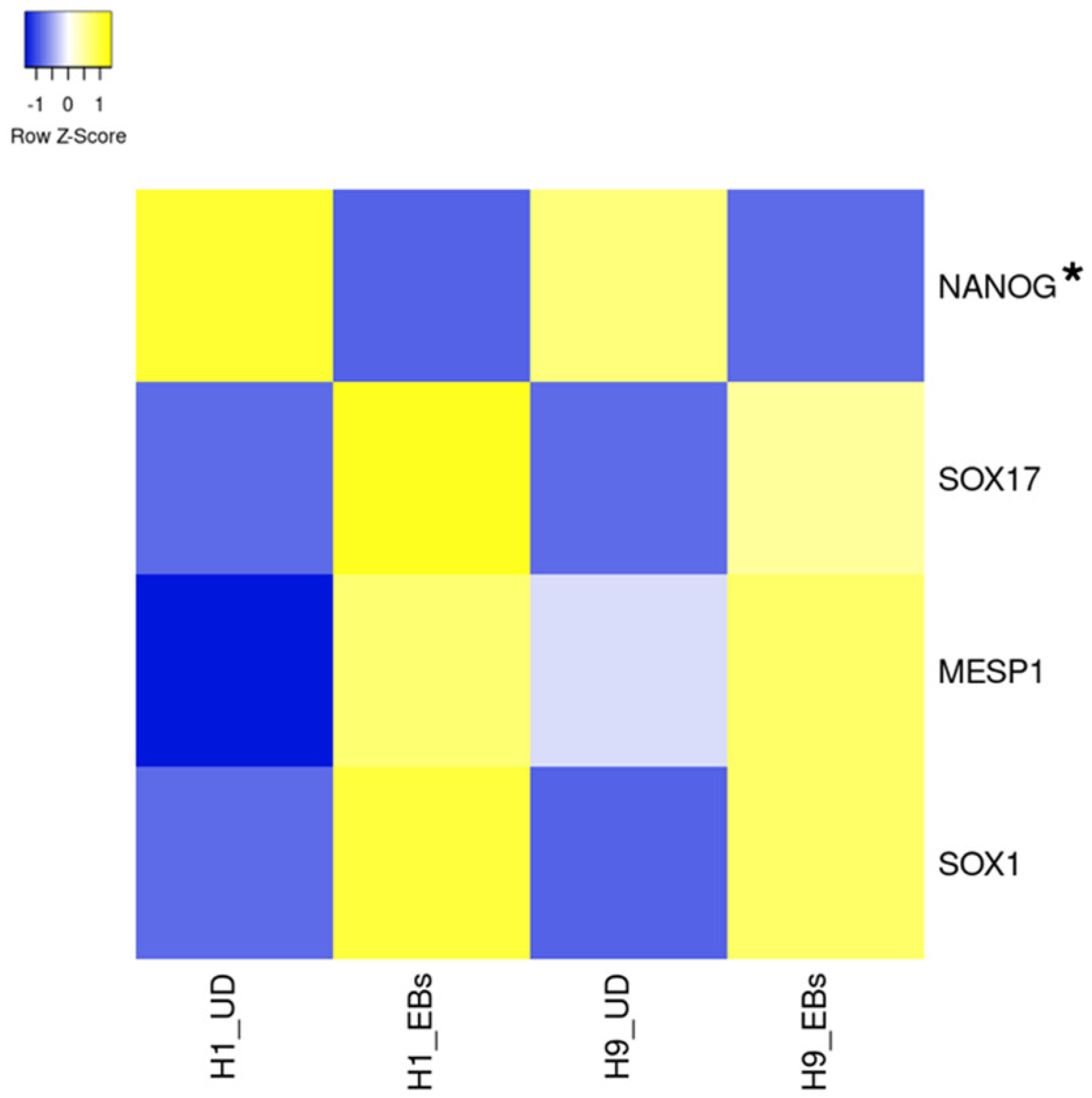
Figure 1. Heatmap of expression levels of pluripotency and differentiation markers in undifferentiated and differentiated human ESCs. The data of mRNA levels in undifferentiated H1 and H9 human ESCs and their differentiated EBs were obtained from the dataset GDS5408 of the NCBI website. Then, a heatmap of pluripotency and differentiation marker levels was created using the Heatmapper website accessed on 7 December 2024 (http://www.heatmapper.ca) [13]. The blue shows downregulation, and the yellow shows upregulation. The asterisk refers to a downregulated gene with a ratio of differentiated EBs to undifferentiated ESCs of less than 0.8-fold in both human ESC lines. UD, undifferentiated.
Biology 13 01043 g001
Figure 2. Heatmap of expression levels of ion channel genes in undifferentiated and differentiated human ESCs. The data of mRNA levels in undifferentiated H1 and H9 human ESCs and their differentiated EBs were obtained from the dataset GDS5408 of the NCBI website. Then, heatmaps of ASICs (A), CACNAs, CACNB, ORAIs (B), CFTR, CLCAs, CLCNs, CLICs (C), CNGA, CNGBs, HCN and HVCN (D) gene levels were created using the Heatmapper website accessed on 7 December 2024 (http://www.heatmapper.ca) [13]. The blue shows downregulation, and the yellow shows upregulation. Asterisks refer to downregulated genes with a ratio of differentiated EBs to undifferentiated ESCs of less than 0.8-fold in both human ESC lines. UD, undifferentiated.
Biology 13 01043 g002
Figure 3. Heatmap of expression levels of ion channel genes in undifferentiated and differentiated human ESCs. The data of mRNA levels in undifferentiated H1 and H9 human ESCs and their differentiated EBs were obtained from the dataset GDS5408 of the NCBI website. Then, heatmaps of KCNAs, KCNBs, KCNCs, KCNDs, KCNEs, KCNGs, KCNHs, KCNJs (A), KCNKs, KCNMs, KCNNs, KCNQs, KCNSs, KCNTs, KCNU, KCNV and KCTD (B) gene levels were created using the Heatmapper website accessed on 7 December 2024 (http://www.heatmapper.ca) [13]. The blue shows downregulation, and the yellow shows upregulation. Asterisks refer to downregulated genes with a ratio of differentiated EBs to undifferentiated ESCs of less than 0.8-fold in both human ESC lines. UD, undifferentiated.
Biology 13 01043 g003
Figure 4. Heatmap of expression levels of ion channel genes in undifferentiated and differentiated human ESCs. The data of mRNA levels in undifferentiated H1 and H9 human ESCs and their differentiated EBs were obtained from the dataset GDS5408 of the NCBI website. Then, heatmaps of NALCN, PIEZOs, PIRT (A), SCNs, SCNNs (B) TMCs and TPCNs (C) gene levels were created using the Heatmapper website accessed on 7 December 2024 (http://www.heatmapper.ca) [13]. The blue shows downregulation, and the yellow shows upregulation. Asterisks refer to downregulated genes with a ratio of differentiated EBs to undifferentiated ESCs of less than 0.8-fold in both human ESC lines. UD, undifferentiated.
Biology 13 01043 g004
Figure 5. Heatmap of expression levels of ion channel genes in undifferentiated and differentiated human ESCs. The data of mRNA levels in undifferentiated H1 and H9 human ESCs and their differentiated EBs were obtained from the dataset GDS5408 of the NCBI website. Then, heatmaps of TRPA, TRPCs, TRPMs, TRPMLs, TRPVs and TRPPs gene levels were created using the Heatmapper website accessed on 7 December 2024 (http://www.heatmapper.ca) [13]. The blue shows downregulation, and the yellow shows upregulation. Asterisks refer to downregulated genes with a ratio of differentiated EBs to undifferentiated ESCs of less than 0.8-fold in both human ESC lines. UD, undifferentiated.
Biology 13 01043 g005
Table 1. The summary of ion channels and their known functional roles in pluripotent stem cells.
Ion ChannelsGeneCell TypeInvolved SignalingFunctional RoleRefs.
K+ channelsKCNsHuman ESCs and iPSCsCell cycleProliferation[15,17]
Kv11.2KCNH6Human ESCsmTOR pathwayDifferentiation[20]
TRPC3Trpc3Mouse ESCsCa2+ signalingPluripotency and survival[28]
TRPM7Trpm7Mouse ESCsmTOR and ERK pathwayPluripotency and self-renewal[33]
TRPM7Trpm7Mouse ESCsMg2+ homeostasisProliferation[9]
Piezo1Piezo1Mouse ESCsMechanotransductionProliferation[38]
HCN channelsHCNsMouse ESCsCell cycleProliferation[43,44]
TPC2Tpc2Mouse ESCsCa2+ signalingPluripotency[50]
Disclaimer/Publisher’s Note: The statements, opinions and data contained in all publications are solely those of the individual author(s) and contributor(s) and not of MDPI and/or the editor(s). MDPI and/or the editor(s) disclaim responsibility for any injury to people or property resulting from any ideas, methods, instructions or products referred to in the content.