Brief Report

Gait Asymmetry and Post-Traumatic Osteoarthritis Following Anterior Cruciate Ligament Rupture: A Preliminary Study

by
Samuel Pringle
and
Kristiaan D’Août
*
Department of Musculoskeletal & Ageing Science, Institute of Life Course and Medical Sciences, University of Liverpool, Liverpool L7 8TX, UK
*
Author to whom correspondence should be addressed.
Biology 2025, 14(2), 208; https://doi.org/10.3390/biology14020208
Submission received: 28 November 2024 / Revised: 5 February 2025 / Accepted: 14 February 2025 / Published: 16 February 2025
(This article belongs to the Special Issue Musculoskeletal Biology: Impact of Ageing and Disease)

Simple Summary

Knee post-traumatic osteoarthritis (PTOA) is a type of osteoarthritis (OA), typically occurring in younger adults following serious joint injuries such as anterior cruciate ligament (ACL) tears. Walking gait asymmetry, whereby a person favours one leg over the other, leads to abnormal joint loading, which can drive the development of OA and PTOA and can cause chronic joint degeneration. Gait asymmetry-induced joint loading worsens late-stage OA severity; however, early-stage involvement and treatments, particularly in PTOA, are poorly defined. This research explored the involvement of gait asymmetry in early-stage PTOA following ACL tears in younger adults. Participants with historical ACL tears (ACL+) and participants with no previous joint trauma (ACL−) underwent gait analysis, determining gait asymmetry and abnormal joint loading severity. Our work found that gait asymmetry and joint load were greater in participants with previous ACL injuries, and gait asymmetry was seen to potentially cause increased knee flexion moments, a common joint load metric. Therefore, these preliminary data imply that gait asymmetry-induced joint loading may contribute to early-stage PTOA in subjects with ACL tears. This study was exploratory, and more research is required to validate and reinforce these findings before prospective treatments can be developed.

Abstract

Knee post-traumatic osteoarthritis (PTOA) often develops in younger populations following anterior cruciate ligament (ACL) rupture, accounting for 12% of all symptomatic osteoarthritis (OA). The current literature implicates gait asymmetry in late-stage knee OA progression; however, early-knee PTOA development involvement is ill defined. This study explored gait asymmetry involvement in early-stage knee PTOA following ACL ruptures. Gait asymmetry, measured as asymmetry in duty factor (relative contact time), and joint loading data were collected, using infrared-camera motion capture and Kistler force plates for participants exhibiting either historical ACL ruptures (ACL+; n = 4) or no previous joint trauma (ACL−; n = 11). Joint loading measures included external knee adduction moment (EKAM) and external knee flexion moment (KFM), early (peak 1; EKAMp1 and KFMp1) and late (peak 2; EKAMp2 and KFMp2), stance peaks (Nm/kg), and respective time integrals (Nm·ms/kg; iEKAMp1, iEKAMp2, iKFMp1, and iKFMp2). ACL+ exhibited greater asymmetrical duty factor (78% difference) and greater joint load differences: EKAMp1 (26%), EKAMp2 (49%), KFMp1 (37%), iKFMp1 (44%), and iKFMp2 (60%). Significant relationships were found between duty factor asymmetry and both KFMp2 (R2 = 0.665) and iKFMp2 (R2 = 0.504). These preliminary data suggest gait asymmetry-induced joint loading may contribute to knee PTOA progression, but further research with increased sample sizes and the quantitative assessment of cartilage status is required.

1. Introduction

Knee post-traumatic osteoarthritis (PTOA) is a common knee disease, causing approximately 12% of all symptomatic osteoarthritis (OA) [1,2] and 25% of knee OA cases [3], and results in significant pain and biomechanical dysfunction [4]. Function can be regained after surgical ACL reconstruction [5] but, unfortunately, the odds of PTOA development increase nearly 7-fold [6], and this occurs disproportionately in younger populations due to greater participation in high-intensity, dynamic activities [7,8]. Unfortunately, limited clinical interventions are available to prevent early PTOA development, with care pathways following major knee joint trauma drastically varying [9,10]. Further, the heterogeneity of OA and differing outcome measures and standards hinder the development of research and treatments [11,12,13]. With the global population expected to increase from 7.7 billion to 9.7 billion by 2050 [14], the impact of PTOA will only worsen. Therefore, understanding underlying factors that drive disease progression is paramount in developing preventative interventions.
Following ACL injury, patients often develop an asymmetrical walking gait, causing increased joint loading on the contralateral limb [15,16]. Notably, a 12-year study investigated late-stage disease severity and found that 80% unilateral knee OA patients developed bilateral knee OA through shifting joint load contralaterally to minimise stress on the ipsilateral joint [17]. This highlights the importance of walking gait asymmetry in joint loading. Conclusively, research investigating gait asymmetry-induced joint loading in early-stage PTOA progression is prudent to assist in the development of preventative care [9,10].
Duty factors represent the proportion of foot–ground contact time during gait, with duty factor asymmetry between legs quantifying greater ground contact time favouring one side to the other, providing a reliable gait asymmetry measure [18,19,20,21]. External knee adduction moment (EKAM) and knee flexion moment (KFM) are regarded as robust predictors of joint loading during the gait stance phase [22,23,24,25]. Importantly, EKAM and KFM are associated with knee OA progression in non-traumatic OA, particularly clinical outcomes such as bone marrow lesions and increased articular cartilage degeneration severity [22,24,26,27,28]. Further, joint moment–time integrals (i.e., angular impulse) provide increased accuracy as they consider the absolute magnitude of joint load during stance phase [22,23,24,29] and are predictive of articular cartilage loss over 12 months [30,31]. Importantly, one study linked KFM with increased medial and lateral cartilage degeneration following ACL rupture [32], with another identifying EKAM as a predictor of increased medial compartment contact forces in ALC rupture patients [33].
The purpose of this study was to investigate whether gait asymmetry is involved in early-knee PTOA progression following ACL rupture. We hypothesised that increased gait asymmetry could be implicated in the increased risk associated with knee PTOA progression in younger populations with ACL ruptures.

2. Materials and Methods
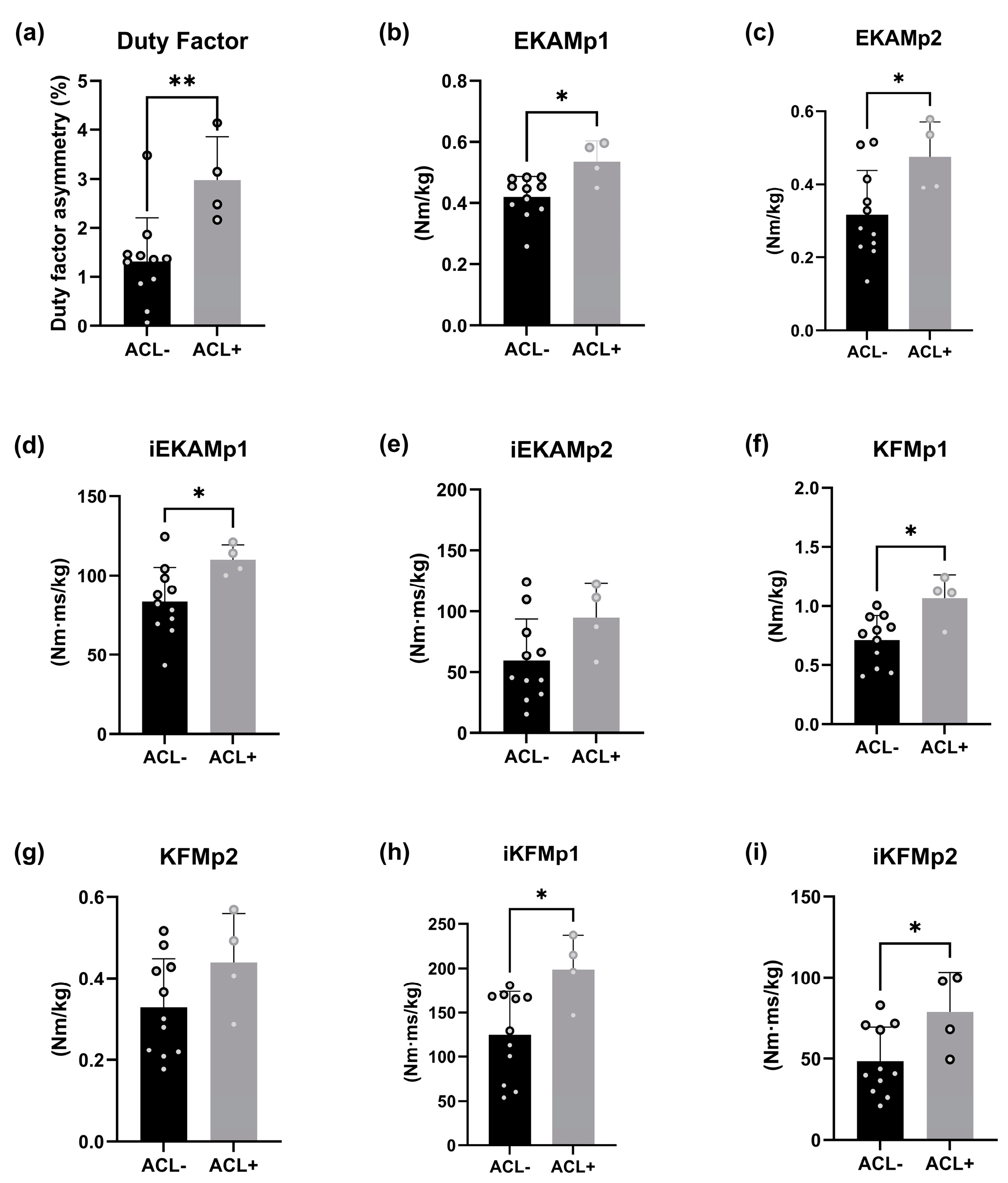
Participants constituted two groups, ACL− (n = 11) and ACL+ (n = 4), which evidenced no historical traumatic joint injuries or a complete ACL rupture (>12 months) with successful rehab completion, respectively (Table 1). Other inclusion criteria included being any sex and aged 25–50 years old. Exclusion criteria included a body mass index > 35, pregnancy, overt OA symptoms [34], and recent injuries or medical conditions affecting normal biomechanics.
Gait biomechanics assessment: Participants walked barefoot using their preferred pace and gait data were reported per leg for each participant across five trial runs. Sixty-seven infrared markers were attached to participants according to University of Liverpool lab standards. A 12 Oqus-7 infrared-camera motion capture system captured kinematics data (Qualisys, Gothenburg, Sweden; sampling rate—200 Hz) and Kistler force plates recorded ground reaction forces (GRF) (Kistler Inc., Winterthur, Switzerland; sampling rate—1000 Hz) with Qualisys Track Manager (QTM; Qualisys, Gothenburg, Sweden), collecting and synchronising datasets. The system was dynamically calibrated at the start of every experimental day, yielding standard errors relating to the spatial position of the markers below 1.5 mm. Force plates were reset between experiments. External joint moments were calculated based on inverse dynamics using Visual3D (C-Motion, Inc., Germantown, MD, USA) [35,36]. The measures included EKAM and KFM early (peak 1; EKAMp1 and KFMp1) and late (peak 2; EKAMp2 and KFMp2) stance peaks (Nm/kg) and their respective time integrals. The latter reflected the overall magnitude of the first and second halves of the stance phase (Nm·ms/kg; iEKAMp1, iEKAMp2, iKFMp1 and iKFMp2) (Figure 1). All external joint moment data were normalised in relation to body mass (kg). All stance and swing times for each limb were extracted using Visual 3D to establish mean duty factor values (stance time/(stance time + swing time)) for each leg [18]. Duty factor asymmetry was calculated using the following formula:
DF asymmetry = 100 (1 − (mean duty factor right/mean duty factor left))
This provided a percentage deviation from zero, whereby zero percent is absolute symmetry, and positive values indicate a longer DF for the right limb.
Statistical analysis: Independent unpaired t-tests were used to compare differences in all functional variable means between ACL+ and ACL− groups and Shapiro–Wilk tests confirmed whether data were normally distributed. Linear regression modelling was applied to analyse predictive relationships between independent and dependent variables, with each data point constituting one trial metric. All statistical analyses were conducted using Prism v9.4.1.

3. Results

Fifteen participants completed the experiments and we observed a 78% difference in duty factor asymmetry between group sample means (p = 0.0067). Additionally, the following functional variables each demonstrated significant mean differences: EKAMp1 (26%; p = 0.0119, ACL+: 0.54 Nm/kg, ACL−: 0.41 Nm/kg), EKAMp2 (49%; p = 0.0361, ACL+: 0.49 Nm/kg, ACL−: 0.30 Nm/kg), iEKAMp1 (30%; p = 0.0366, ACL+: 109.39 Nm·ms/kg, ACL−: 80.84 Nm·ms/kg), KFMp1 (37%; p = 0.0117, ACL+: 1.02 Nm/kg, ACL−: 0.70 Nm/kg), iKFMp1 (44%; p = 0.0178, ACL+: 190.4 Nm·ms/kg, ACL−: 121.00 Nm·ms/kg), and iKFMp2 (60%; p = 0.0329, ACL+: 83.47 Nm·ms/kg, ACL−: 44.92 Nm·ms/kg). However, no significant mean differences were found between iEKAMp2 (58%; p = 0.0892, ACL+: 99.03 Nm·ms/kg, ACL−: 54.75 Nm·ms/kg) and KFMp2 (39%; p = 0.1392, ACL+: 0.47 Nm/kg, ACL−: 0.31 Nm/kg) (Figure 2). When assessing the linear relationship between duty factor asymmetry (independent variable) and each functional variable (dependent variable), KFMp2 (R2 = 0.665; p = < 0.001) and iKFMp2 (R2 = 0.505); p = < 0.001) registered moderate associations with duty factor asymmetry in the ACL+ group. All other variables, across both ACL+ and ACL− groups, demonstrated either nominal R2 values or no significant results (Figure 3).

4. Discussion

This exploratory study provided supporting evidence that increased gait asymmetry-induced joint loading may be involved in knee PTOA progression in subjects with ACL ruptures. ACL+ participants demonstrated greater gait asymmetry relative to ACL− participants and displayed heightened joint loading across several knee PTOA risk-factors (Figure 2). Further, the ACL+ cohort displayed moderate associations between gait asymmetry and KFMp2 and iKFMp2 in the second half of the stance phase (Figure 3).
Gait asymmetry: The study revealed that participants with historical ACL ruptures ( x ¯ = 3%) exhibited a 78% increase in duty factor asymmetry compared to ACL− participants ( x ¯ = 1.3%; Figure 2). Supporting this, a study spanning 24 months (n = 40) demonstrated increased gait asymmetry prevalence in ACL patients following reconstruction [37]. Further, another project illustrated 1.6% and 1% gait asymmetry sample mean reductions associated with reduced knee pain symptoms in knee OA patients [38], validating the clinical relevance of the group sample mean differences.
External knee adduction moment: The study highlighted significantly heightened differences in both EKAM peaks that favoured the ACL+ group, with EKAMp1 (26%) and EKAMp2 (49%). Additionally, the time integral iEKAMp1 exhibited a significant 30% increase in the ACL+ group, with iEKAMp2 also showing an increase at 58%, but this did not reach significance (Figure 2). These data augment current knowledge illustrating greater EKAM magnitude in ACL patients [39] or subjects at high risk of ACL rupture [40,41]. Additionally, Miyazaki et al. (2002) [42] established an association of ~25% increase in EKAM peak magnitude with 6.6 times heightened risk of radiographic knee OA progression over 6 years, highlighting the relevance of the group mean differences in this study. Further, a greater EKAM presence increases compressive forces through the patellofemoral and tibiofemoral joint compartments [22,43], while a 12-year study established increased joint load as a major OA progression contributor [17]. Therefore, ACL rupture subjects may exhibit increased joint moment magnitude, increasing susceptibility to PTOA development.
Knee flexion moment: The evidence indicated KFM peaks and time integrals display greater prevalence of joint load in ACL+ participants. The significant differences were as follows: KFMp1—37%; iKFMp1—44%; iKFMp2—60% (Figure 2). Supporting this, evidence has been cited of increased KFM peaks and time integrals in earlier disease stages (30–55 years old), contributing to increased progressive cartilage loss [24,27,32]. These heightened knee flexion moments increase knee extensor force, generating a greater patellofemoral and tibiofemoral joint reaction force [24,25,44]. This suggests that ACL+ participants experience higher patellofemoral and tibiofemoral joint loading over longer periods, a major risk factor in PTOA progression [26].
Gait asymmetry in post-traumatic osteoarthritis: The finding of the relationship between duty factor asymmetry, as a potential predictor, and OA markers (KFMp2 (R2 = 0.665) and iKFMp2 (R2 = 0.505)) is to our knowledge a novel finding (Figure 3). As such, no direct supporting evidence exists; however, indirect insight is available. Notably, gait asymmetry is significantly increased in younger patients with ACL ruptures compared to uninjured controls [45,46], which can ultimately increase joint load, resulting in greater risk of cartilage matrix degradation [17]. Further, two studies demonstrated increasing OA severity as KFMp2 and iKFMp2 worsened [24,47]. Therefore, the findings associating duty factor asymmetry with KFMp2 and iKFMp2 hold relevance. Combined with the increased prevalence of KFM and EKAM in the ACL+ participants, there exists additional support for the hypothesis: increased gait asymmetry-induced joint loading may be involved in the increased risk associated with early-knee PTOA progression in younger populations with ACL ruptures. Rehabilitation programmes after ACL reconstruction can help to restore symmetry [48,49].
Limitations and future directions: Caution should be exercised when drawing definitive interpretations. We used small sample sizes. To estimate the impact of this, we performed a posteriori power analyses (two-tailed, α = 0.05) in G*Power 3.1.9.7 for two main outcomes: duty factor asymmetry (=ACL−: 0313 ± 0.00892, n = 11; ACL+: 0.0298 ± 0.00875, n = 4) and EKAMp1 (ACL− 0.714 ± 0.198, I = 11; ACL 1.066 ± 0.199, I = 4). The effects sizes were 1.89 and 1.77, respectively. The calculated powers were 0.849 and 0.801, respectively. This means that our power for these important outcomes is good, despite the low numbers. However, the lack of females in the ACL+ group means it may not accurately represent the overall population, and we propose increasing samples sizes [50] and using proportional representation [11] in future studies, especially when ultimately aiming to make clinical recommendations. Women are at a 3–5 times higher risk of ACL injuries, 50–90% of which progress to OA [51], and so any such recommendations can be especially beneficial for women. Using G*Power, the recommendation would be to expand the research to approximately 40–50 participants (allowing a 20% drop-out rate) for a power of 0.95 at α = 0.05 [52]. Additionally, study designs including radiographic imaging (X-Ray, MRI, CT) or detailed physical examination [53] to quantify cartilage degradation and gait asymmetry rectification plans with follow-ups, would provide tangible insights on the effectiveness of managing gait asymmetry as a preventative treatment. Further, another deliberation is whether the ACL rupture is the main cause of PTOA [51,54,55], or whether the secondary trauma of the reconstruction itself is a significant contributor [56,57]. Equally, how, and when does gait asymmetry begin impacting PTOA? Is it simply a by-product of the ACL injury and PTOA development? Or does it have greater implications prior to PTOA in the causation of the ACL rupture, ultimately being the underlying driving factor? These are important considerations whose investigation would greatly help the broader view around the topic in terms of understanding the root causes of the problem in PTOA onset and progression.

5. Conclusions

The findings of this study indicate that gait asymmetry is increased in participants with historical ACL ruptures. Mechanical loading (KFM and EKAM) is heightened in ACL participants relative to participants with no historical knee joint trauma. Further, gait asymmetry may increase knee flexion moment magnitude, suggesting the involvement of gait asymmetry in knee PTOA progression in ACL participants. Future studies should expand the study design to validate and build on these preliminary findings.

Author Contributions

Conceptualization, S.P.; methodology, S.P. and K.D.; software, S.P.; validation, S.P. and K.D.; formal analysis, S.P.; investigation, S.P.; resources, S.P. and K.D.; data curation, S.P.; writing—original draft preparation, S.P.; writing—review and editing, S.P. and K.D.; visualisation, S.P.; supervision, K.D.; project administration, S.P.; funding acquisition, K.D. All authors have read and agreed to the published version of the manuscript.

Funding

This research is an MRes project funded by the MRC/Versus Arthritis Centre for Integrated Research into Musculoskeletal Ageing (CIMA) [MR/R502182/1].

Institutional Review Board Statement

This protocol was approved by the University of Liverpool’s Central University Research Ethics Committee (reference number 5559).

Informed Consent Statement

Informed consent was obtained from all subjects involved in the study. Written informed consent has been obtained from the patient(s) to publish this paper.

Data Availability Statement

Data can be made available upon reasonable request.

Acknowledgments

Special thanks to all study participants.

Conflicts of Interest

The authors declare no conflicts of interest. Further, the project funders had no role in the design of the study; in the collection, analyses, or interpretation of data; in the writing of the manuscript; or in the decision to publish the results.

References

  1. Thomas, A.C.; Hubbard-Turner, T.; Wikstrom, E.A.; Palmieri-Smith, R.M. Epidemiology of Posttraumatic Osteoarthritis. J. Athl. Train. 2017, 52, 491–496. [Google Scholar] [CrossRef] [PubMed]
  2. Sharma, L. Osteoarthritis of the Knee. N. Engl. J. Med. 2021, 384, 51–59. [Google Scholar] [CrossRef]
  3. Hunter, D.J. Osteoarthritis. Best Pract. Res. Clin. Rheumatol. 2011, 25, 801–814. [Google Scholar] [CrossRef]
  4. Chang, J.C.; Sebastian, A.; Murugesh, D.K.; Hatsell, S.; Economides, A.N.; Christiansen, B.A.; Loots, G.G. Global molecular changes in a tibial compression induced ACL rupture model of post-traumatic osteoarthritis. J. Orthop. Res. 2017, 35, 474–485. [Google Scholar] [CrossRef] [PubMed]
  5. Mercurio, M.; Cerciello, S.; Corona, K.; Guerra, G.; Simonetta, R.; Familiari, F.; Galasso, O.; Gasparini, G. Factors Associated With a Successful Return to Performance After Anterior Cruciate Ligament Reconstruction: A Multiparametric Evaluation in Soccer Players. Orthop. J. Sports Med. 2024, 12, 23259671241275663. [Google Scholar] [CrossRef] [PubMed]
  6. Webster, K.E.; Hewett, T.E. Anterior Cruciate Ligament Injury and Knee Osteoarthritis: An Umbrella Systematic Review and Meta-analysis. Clin. J. Sport. Med. 2022, 32, 145–152. [Google Scholar] [CrossRef]
  7. Menendez, M.I.; Hettlich, B.; Wei, L.; Knopp, M.V. Feasibility of Na(18)F PET/CT and MRI for Noninvasive In Vivo Quantification of Knee Pathophysiological Bone Metabolism in a Canine Model of Post-traumatic Osteoarthritis. Mol. Imaging 2017, 16, 1536012117714575. [Google Scholar] [CrossRef] [PubMed]
  8. Riordan, E.A.; Little, C.; Hunter, D. Pathogenesis of post-traumatic OA with a view to intervention. Best Pract. Res. Clin. Rheumatol. 2014, 28, 17–30. [Google Scholar] [CrossRef]
  9. Lau, B.H.; Lafave, M.R.; Mohtadi, N.G.; Butterwick, D.J. Utilization and cost of a new model of care for managing acute knee injuries: The Calgary Acute Knee Injury Clinic. BMC Health Serv. Res. 2012, 12, 445. [Google Scholar] [CrossRef]
  10. Whittaker, J.L.; Culvenor, A.G.; Juhl, C.B.; Berg, B.; Bricca, A.; Filbay, S.R.; Holm, P.; Macri, E.; Urhausen, A.P.; Ardern, C.L.; et al. OPTIKNEE 2022: Consensus recommendations to optimise knee health after traumatic knee injury to prevent osteoarthritis. Br. J. Sports Med. 2022, 56, 1393–1405. [Google Scholar] [CrossRef]
  11. Zaki, S.; Smith, M.M.; Little, C.B. Pathology-pain relationships in different osteoarthritis animal model phenotypes: It matters what you measure, when you measure, and how you got there. Osteoarthr. Cartil. 2021, 29, 1448–1461. [Google Scholar] [CrossRef]
  12. Watt, F.E.; Corp, N.; Kingsbury, S.R.; Frobell, R.; Englund, M.; Felson, D.T.; Levesque, M.; Majumdar, S.; Wilson, C.; Beard, D.J.; et al. Towards prevention of post-traumatic osteoarthritis: Report from an international expert working group on considerations for the design and conduct of interventional studies following acute knee injury. Osteoarthr. Cartil. 2019, 27, 23–33. [Google Scholar] [CrossRef]
  13. Whittaker, J.L.; Roos, E.M. A pragmatic approach to prevent post-traumatic osteoarthritis after sport or exercise-related joint injury. Best Pract. Res. Clin. Rheumatol. 2019, 33, 158–171. [Google Scholar] [CrossRef] [PubMed]
  14. Nations, U. World Population Prospects; United Nations Publications: New York, NY, USA, 2019. [Google Scholar]
  15. Eddo, O.O.; Lindsey, B.W.; Caswell, S.V.; Prebble, M.; Cortes, N. Unintended Changes in Contralateral Limb as a Result of Acute Gait Modification. J. Appl. Biomech. 2020, 36, 13–19. [Google Scholar] [CrossRef]
  16. Iijima, H.; Eguchi, R.; Aoyama, T.; Takahashi, M. Trunk movement asymmetry associated with pain, disability, and quadriceps strength asymmetry in individuals with knee osteoarthritis: A cross-sectional study. Osteoarthr. Cartil. 2019, 27, 248–256. [Google Scholar] [CrossRef]
  17. Metcalfe, A.J.; Andersson, M.L.; Goodfellow, R.; Thorstensson, C.A. Is knee osteoarthritis a symmetrical disease? Analysis of a 12 year prospective cohort study. BMC Musculoskelet. Disord. 2012, 13, 153. [Google Scholar] [CrossRef]
  18. Bonnaerens, S.; Fiers, P.; Galle, S.; Derie, R.; Aerts, P.; Frederick, E.; Kaneko, Y.; Derave, W.; De Clercq, D.; Segers, V. Relationship between duty factor and external forces in slow recreational runners. BMJ Open Sport Exerc. Med. 2021, 7, e000996. [Google Scholar] [CrossRef]
  19. Meder, K.G.; LoJacono, C.T.; Rhea, C.K. A systematic review of non-pharmacological interventions to improve gait asymmetries in neurological populations. Symmetry 2022, 14, 281. [Google Scholar] [CrossRef]
  20. Patoz, A.; Gindre, C.; Thouvenot, A.; Mourot, L.; Hébert-Losier, K.; Lussiana, T. Duty Factor Is a Viable Measure to Classify Spontaneous Running Forms. Sports 2019, 7, 233. [Google Scholar] [CrossRef] [PubMed]
  21. Patoz, A.; Lussiana, T.; Breine, B.; Gindre, C.; Malatesta, D. Accurate estimation of peak vertical ground reaction force using the duty factor in level treadmill running. Scand. J. Med. Sci. Sports 2023, 33, 169–177. [Google Scholar] [CrossRef] [PubMed]
  22. Chang, A.H.; Moisio, K.C.; Chmiel, J.S.; Eckstein, F.; Guermazi, A.; Prasad, P.V.; Zhang, Y.; Almagor, O.; Belisle, L.; Hayes, K.; et al. External knee adduction and flexion moments during gait and medial tibiofemoral disease progression in knee osteoarthritis. Osteoarthr. Cartil. 2015, 23, 1099–1106. [Google Scholar] [CrossRef]
  23. Gerbrands, T.A.; Pisters, M.F.; Theeven, P.J.R.; Verschueren, S.; Vanwanseele, B. Lateral trunk lean and medializing the knee as gait strategies for knee osteoarthritis. Gait Posture 2017, 51, 247–253. [Google Scholar] [CrossRef]
  24. Teng, H.L.; MacLeod, T.D.; Link, T.M.; Majumdar, S.; Souza, R.B. Higher Knee Flexion Moment During the Second Half of the Stance Phase of Gait Is Associated With the Progression of Osteoarthritis of the Patellofemoral Joint on Magnetic Resonance Imaging. J. Orthop. Sports Phys. Ther. 2015, 45, 656–664. [Google Scholar] [CrossRef] [PubMed]
  25. Teng, H.L.; Powers, C.M. Sagittal plane trunk posture influences patellofemoral joint stress during running. J. Orthop. Sports Phys. Ther. 2014, 44, 785–792. [Google Scholar] [CrossRef]
  26. Boswell, M.A.; Uhlrich, S.D.; Kidziński, Ł.; Thomas, K.; Kolesar, J.A.; Gold, G.E.; Beaupre, G.S.; Delp, S.L. A neural network to predict the knee adduction moment in patients with osteoarthritis using anatomical landmarks obtainable from 2D video analysis. Osteoarthr. Cartil. 2021, 29, 346–356. [Google Scholar] [CrossRef]
  27. Chehab, E.F.; Favre, J.; Erhart-Hledik, J.C.; Andriacchi, T.P. Baseline knee adduction and flexion moments during walking are both associated with 5 year cartilage changes in patients with medial knee osteoarthritis. Osteoarthr. Cartil. 2014, 22, 1833–1839. [Google Scholar] [CrossRef] [PubMed]
  28. Hall, M.; Bennell, K.L.; Wrigley, T.V.; Metcalf, B.R.; Campbell, P.K.; Kasza, J.; Paterson, K.L.; Hunter, D.J.; Hinman, R.S. The knee adduction moment and knee osteoarthritis symptoms: Relationships according to radiographic disease severity. Osteoarthr. Cartil. 2017, 25, 34–41. [Google Scholar] [CrossRef]
  29. Creaby, M.W. It’s not all about the knee adduction moment: The role of the knee flexion moment in medial knee joint loading. Osteoarthr. Cartil. 2015, 23, 1038–1040. [Google Scholar] [CrossRef] [PubMed]
  30. Bennell, K.L.; Bowles, K.A.; Wang, Y.; Cicuttini, F.; Davies-Tuck, M.; Hinman, R.S. Higher dynamic medial knee load predicts greater cartilage loss over 12 months in medial knee osteoarthritis. Ann. Rheum. Dis. 2011, 70, 1770–1774. [Google Scholar] [CrossRef]
  31. Teng, H.L.; Calixto, N.E.; MacLeod, T.D.; Nardo, L.; Link, T.M.; Majumdar, S.; Souza, R.B. Associations between patellofemoral joint cartilage T1ρ and T2 and knee flexion moment and impulse during gait in individuals with and without patellofemoral joint osteoarthritis. Osteoarthr. Cartil. 2016, 24, 1554–1564. [Google Scholar] [CrossRef]
  32. Erhart-Hledik, J.; Chu, C.; Asay, J.; Favre, J.; Andriacchi, T. Longitudinal Changes in the Total Knee Joint Moment After Anterior Cruciate Ligament Reconstruction Correlate With Cartilage Thickness Changes. J. Orthop. Res. 2019, 37, 1546–1554. [Google Scholar] [CrossRef]
  33. Wellsandt, E.; Khandha, A.; Manal, K.; Axe, M.J.; Buchanan, T.S.; Snyder-Mackler, L. Predictors of knee joint loading after anterior cruciate ligament reconstruction. J. Orthop. Res. 2017, 35, 651–656. [Google Scholar] [CrossRef]
  34. Favero, M.; Ramonda, R.; Goldring, M.B.; Goldring, S.R.; Punzi, L. Early knee osteoarthritis. RMD Open 2015, 1 (Suppl. S1), e000062. [Google Scholar] [CrossRef]
  35. Nagano, H.; Tatsumi, I.; Sarashina, E.; Sparrow, W.A.; Begg, R.K. Modelling knee flexion effects on joint power absorption and adduction moment. Knee 2015, 22, 490–493. [Google Scholar] [CrossRef] [PubMed]
  36. Whittlesey, S.N.; Hamill, J.; Caldwell, G.E.; Robertson, D.G.E. Research Methods in Biomechanics; Human Kinetics: Champaign, IL, USA, 2014. [Google Scholar]
  37. Capin, J.J.; Zarzycki, R.; Arundale, A.; Cummer, K.; Snyder-Mackler, L. Report of the Primary Outcomes for Gait Mechanics in Men of the ACL-SPORTS Trial: Secondary Prevention With and Without Perturbation Training Does Not Restore Gait Symmetry in Men 1 or 2 Years After ACL Reconstruction. Clin. Orthop. Relat. Res. 2017, 475, 2513–2522. [Google Scholar] [CrossRef] [PubMed]
  38. Kato, N.; Fukusaki, C.; Leetawesup, K.; Kadokura, Y.; Ishii, N. Improvement in gait asymmetry during Nordic walking in patients with lower extremity osteoarthritis. J. Phys. Fit. Sports Med. 2020, 9, 65–73. [Google Scholar] [CrossRef]
  39. Butler, R.J.; Minick, K.I.; Ferber, R.; Underwood, F. Gait mechanics after ACL reconstruction: Implications for the early onset of knee osteoarthritis. Br. J. Sports Med. 2009, 43, 366–370. [Google Scholar] [CrossRef] [PubMed]
  40. Bates, N.A.; Schilaty, N.D.; Nagelli, C.V.; Krych, A.J.; Hewett, T.E. Multiplanar Loading of the Knee and Its Influence on Anterior Cruciate Ligament and Medial Collateral Ligament Strain During Simulated Landings and Noncontact Tears. Am. J. Sports Med. 2019, 47, 1844–1853. [Google Scholar] [CrossRef]
  41. Hewett, T.E.; Ford, K.R.; Xu, Y.Y.; Khoury, J.; Myer, G.D. Utilization of ACL Injury Biomechanical and Neuromuscular Risk Profile Analysis to Determine the Effectiveness of Neuromuscular Training. Am. J. Sports Med. 2016, 44, 3146–3151. [Google Scholar] [CrossRef] [PubMed]
  42. Miyazaki, T.; Wada, M.; Kawahara, H.; Sato, M.; Baba, H.; Shimada, S. Dynamic load at baseline can predict radiographic disease progression in medial compartment knee osteoarthritis. Ann. Rheum. Dis. 2002, 61, 617–622. [Google Scholar] [CrossRef] [PubMed]
  43. Farrokhi, S.; Voycheck, C.A.; Tashman, S.; Fitzgerald, G.K. A biomechanical perspective on physical therapy management of knee osteoarthritis. J. Orthop. Sports Phys. Ther. 2013, 43, 600–619. [Google Scholar] [CrossRef] [PubMed]
  44. Ho, K.Y.; Blanchette, M.G.; Powers, C.M. The influence of heel height on patellofemoral joint kinetics during walking. Gait Posture 2012, 36, 271–275. [Google Scholar] [CrossRef]
  45. Garcia, S.A.; Brown, S.R.; Koje, M.; Krishnan, C.; Palmieri-Smith, R.M. Gait asymmetries are exacerbated at faster walking speeds in individuals with acute anterior cruciate ligament reconstruction. J. Orthop. Res. 2022, 40, 219–230. [Google Scholar] [CrossRef]
  46. Williams, J.; Neal, K.; Alfayyadh, A.; Capin, J.; Khandha, A.; Manal, K.; Potter, H.; Snyder-Mackler, L.; Buchanan, T. Early knee loading asymmetries may be at the root of eventual osteoarthritis development after anterior cruciate ligament reconstruction. Osteoarthr. Cartil. 2020, 28, S226–S227. [Google Scholar] [CrossRef]
  47. Teng, H.L.; Wu, D.; Su, F.; Pedoia, V.; Souza, R.B.; Ma, C.B.; Li, X. Gait Characteristics Associated With a Greater Increase in Medial Knee Cartilage T(1ρ) and T(2) Relaxation Times in Patients Undergoing Anterior Cruciate Ligament Reconstruction. Am. J. Sports Med. 2017, 45, 3262–3271. [Google Scholar] [CrossRef] [PubMed]
  48. Ebert, J.R.; Edwards, P.; Yi, L.; Joss, B.; Ackland, T.; Carey-Smith, R.; Buelow, J.-U.; Hewitt, B. Strength and functional symmetry is associated with post-operative rehabilitation in patients following anterior cruciate ligament reconstruction. Knee Surg. Sports Traumatol. Arthrosc. 2018, 26, 2353–2361. [Google Scholar] [CrossRef] [PubMed]
  49. Hadizadeh, M.; Amri, S.; Roohi, S.A.; Mohafez, H. Assessment of Gait Symmetry Improvements in National Athletes after Anterior Cruciate Ligament Reconstruction during Rehabilitation. Int. J. Sports Med. 2016, 37, 997–1002. [Google Scholar] [CrossRef]
  50. Brunner, J.; Schimmack, U. Estimating population mean power under conditions of heterogeneity and selection for significance. Meta-Psychology 2020, 4. [Google Scholar] [CrossRef]
  51. Wang, L.J.; Zeng, N.; Yan, Z.P.; Li, J.T.; Ni, G.X. Post-traumatic osteoarthritis following ACL injury. Arthritis Res. Ther. 2020, 22, 57. [Google Scholar] [CrossRef] [PubMed]
  52. Kang, H. Sample size determination and power analysis using the G*Power software. J. Educ. Eval. Health Prof. 2021, 18, 17. [Google Scholar] [CrossRef]
  53. Krakowski, P.; Nogalski, A.; Jurkiewicz, A.; Karpiński, R.; Maciejewski, R.; Jonak, J. Comparison of Diagnostic Accuracy of Physical Examination and MRI in the Most Common Knee Injuries. Appl. Sci. 2019, 9, 4102. [Google Scholar] [CrossRef]
  54. Dare, D.; Rodeo, S. Mechanisms of post-traumatic osteoarthritis after ACL injury. Curr. Rheumatol. Rep. 2014, 16, 448. [Google Scholar] [CrossRef] [PubMed]
  55. Racine, J.; Aaron, R.K. Post-traumatic osteoarthritis after ACL injury. RI Med. J. 2014, 97, 25–28. [Google Scholar]
  56. Han, P.F.; Wei, L.; Duan, Z.Q.; Zhang, Z.L.; Chen, T.Y.; Lu, J.G.; Zhao, R.P.; Cao, X.M.; Li, P.C.; Lv, Z.; et al. Contribution of IL-1β, 6 and TNF-α to the form of post-traumatic osteoarthritis induced by “idealized” anterior cruciate ligament reconstruction in a porcine model. Int. Immunopharmacol. 2018, 65, 212–220. [Google Scholar] [CrossRef] [PubMed]
  57. Heard, B.J.; Barton, K.I.; Chung, M.; Achari, Y.; Shrive, N.G.; Frank, C.B.; Hart, D.A. Single intra-articular dexamethasone injection immediately post-surgery in a rabbit model mitigates early inflammatory responses and post-traumatic osteoarthritis-like alterations. J. Orthop. Res. 2015, 33, 1826–1834. [Google Scholar] [CrossRef] [PubMed]
Figure 1. Distribution curves for (a) EKAM (frontal plane) and (b) KFM (sagittal plane) knee joint moments (Nm/kg) amid the walking stance phase, taken from a single-sample trial. Positive and negative values signify (a) adduction and abduction and (b) knee flexion and extension moments, respectively. Each moment curve presents two calculated peaks, which are signified by crosses. Peak 1 (blue) and Peak 2 (green) represent the first and second halve of the stance phase, respectively. The time integral for each phase (1 and 2) of the respective curves (EKAM and KFM) were computed relative to time (Nm·ms/kg) when the knee moments (Nm/kg) were positive. Abbreviations: HS, heel-strike; FF, foot-flat; MS, mid-stance; HO, heel-off; TO, toe-off.
Biology 14 00208 g001
Figure 2. The sample mean distribution of ACL+ and ACL− groups for (a) gait asymmetry as a product of duty factor asymmetry (stance time/(stance time + swing time)), (b) EKAMp1 and (c) EKAMp2, (d) iEKAMp1 and (e) iEKAMp2, (f) KFMp1 and (g) KFMp2, and (h) iKFMp1 and (i) iKFMp2.* = p < 0.05, ** = p < 0.01 observed following am independent unpaired two-sample t-test.
Biology 14 00208 g002
Figure 3. A linear regression model displaying predictive relationships between the independent variable (x), gait asymmetry as a product of duty factor asymmetry (stance time/(stance time + swing time)) and independent variables (y) for (a) KFMp2 (R2 = 0.665; p = < 0.001) and (b) iKFMp2 (R2 = 0.505); p = < 0.001) in the ACL+ group.
Biology 14 00208 g003
Table 1. Participant demographic variables, with group 1 consisting of participants with no articular cartilage ligament rupture (ACL−) and group 2 (ACL+) consists of participants with historical ACL ruptures.
Demographic VariableGroup 1. ACL−Group 2. ACL+p Value
Sex (male/female) (n)5/64/0
Age (y) *35 ± 632 ± 10.1158
Mass (kg) *71.3 ± 12.586.6 ± 7.90.0998
Height (cm) *174 ± 11.3185 ± 3.9<0.001
Body mass index (kg/m2) *23 ± 2.325.2 ± 2.00.1442
Years post-surgery-6.0 ± 3.2 -
* values are mean ± SD; significant differences observed following two-sample t-test between ACL− and ACL+ groups at α = 0.05 and confidence intervals = 95%.
Disclaimer/Publisher’s Note: The statements, opinions and data contained in all publications are solely those of the individual author(s) and contributor(s) and not of MDPI and/or the editor(s). MDPI and/or the editor(s) disclaim responsibility for any injury to people or property resulting from any ideas, methods, instructions or products referred to in the content.

