Article

Characterization and Efficacy of Essential Oil-Based Cosmetic Formulations for Acne-Prone Skin

by
Victor Hugo Pacagnelli Infante
1,
Maxim E. Darvin
2,* and
Patrícia M. B. G. Maia Campos
1,*
1
School of Pharmaceutical Sciences of Ribeirão Preto, University of São Paulo, Av. do Café, s/n—Vila Monte Alegre, Ribeirão Preto 14040-900, SP, Brazil
2
Independent Researcher, 10178 Berlin, Germany
*
Authors to whom correspondence should be addressed.
Cosmetics 2023, 10(6), 158; https://doi.org/10.3390/cosmetics10060158
Submission received: 11 October 2023 / Revised: 10 November 2023 / Accepted: 14 November 2023 / Published: 16 November 2023
(This article belongs to the Special Issue Feature Papers in Cosmetics in 2023)

Abstract

:
This randomized, placebo-controlled, double-blind clinical in vivo study aimed to evaluate the clinical efficacy of a cosmetic formulation for non-inflammatory acne using essential oils. Fifty-three male participants were divided into four groups: a formulation containing a mixture of four essential oils (4EO), Melaleuca alternifolia (M.a.), nanoemulsion of M. alternifolia (Nanoem.), and a placebo group. The participants applied the formulation daily for 90 days and non-invasive skin imaging techniques were employed to assess the outcomes. Skin microrelief images and reflectance confocal microscopy images were captured in the malar region, and Raman spectroscopy was used to analyze the terpene composition of the essential oils, oil mixture, and nanoemulsion. The results indicated that the nanoemulsion, M.a. essential oil, and 4EO formulation effectively reduced the overall number of comedone and improved follicular hyperkeratinization. The nanoemulsion of M.a. demonstrated the most promising outcomes in reducing comedone areas, especially in the infundibular region. This effect could be attributed to the presence of terpinene-4-ol in the essential oil and the enhanced penetration provided by the nanoemulsion formulation. These findings suggest that cosmetic formulations containing essential oils, particularly in nanoemulsion form, have potential against mild acne. This study contributes to our understanding of the relationship between terpene composition and clinical activity, highlighting the importance of innovative delivery systems.

1. Introduction

Acne vulgaris is a prevalent condition affecting the pilosebaceous unit, primarily observed in adolescents and men. However, it is essential to highlight other significant demographic groups, including adult women [1,2,3]. Various factors contribute to the formation of acne lesions, such as abnormal keratinization resulting in comedones, increased and altered sebum production regulated by androgens, and the colonization of hair follicles by Cutibacterium acnes and the release of inflammatory mediators into the skin. Insulin resistance, genetic predisposition, and dietary habits also can play a role in exacerbating acne [4,5,6].
The initial phase of acne, known as comedogenesis, is crucial for disease characterization and progression. Altered pilosebaceous units have been identified as the primary pathological substrate of acne [4]. This understanding is particularly relevant to the cosmetic industry as the development of topical formulations targeting this stage could potentially impede progression to inflammatory lesions, especially for cosmetic acne in acne-prone skin.
In this context, essential oils offer a plausible alternative for the development of innovative cosmetic formulations, but there is a lack of evidence, which should be addressed [7]. Essential oils are complex, volatile compounds known for their distinct aromas. They are primarily composed of terpenes, which are secondary metabolites with various biological properties, including bactericidal, virucidal, fungicidal, and insecticidal activities [7,8,9,10,11,12,13]. Due to their lipophilic nature and small, non-polar molecular structure, essential oils can easily penetrate the upper layers of the skin [14,15]. Recent research has summarized the positive dermatological effects of approximately 90 topical essential oils, including their ability to improve skin barrier function, hydration, sebum levels, and acne conditions [16].
However, it is important to note that essential oils also carry potential risks as they can serve as sources of allergens. The International Fragrance Association (IFRA) has defined essential oils and their components that pose potential allergy risks. IFRA has also established maximum concentrations and usage conditions to ensure the safety of cosmetic products [17]. The terpene composition and appropriate dilution of essential oils are crucial factors in assessing their risk potential. Some oils exhibit dose-dependent toxicity and allergenicity, underscoring the need for careful evaluation of their composition [18]. Additionally, quality control measures must be implemented, considering botanical sources, suppliers, and extraction methods [19].
One of the most extensively studied essential oils for acne treatment is derived from Melaleuca alternifolia leaves, commonly known as tea tree essential oil [20]. Clinical studies involving tea tree oil products have demonstrated their efficacy in treating various superficial conditions, including acne, oral candidiasis, and tinea, and even in improving the photoaging process [20,21,22,23]. Numerous studies indicate the anti-inflammatory and anti-acne properties of melaleuca essential oil. However, it should be noted that topical treatment should be avoided for severe acne as systemic therapy is typically recommended in such cases [20,23,24,25]. On the other hand, the utilization of melaleuca essential oil in non-inflammatory acne treatment and its effect on reducing the hyperkeratinization process are not yet thoroughly studied, making this particularly intriguing for the cosmetic field.
In addition to melaleuca, lavender (Lavandula angustifolia) and oils from the eucalyptus and citrus genus are commonly used as additives in cosmetic, medicinal, and food industry products [26,27,28,29]. These oils are incorporated into cosmetic formulations for their biological activities, as preservatives, and/or as fragrances in semi-solid formulations. Therefore, evaluating the terpene composition of pure essential oils and their mixtures is essential for ensuring product quality control, and Raman spectroscopy provides a suitable non-invasive alternative for this purpose.
The clinical application of essential oils is limited due to challenges in incorporating them into pharmaceutical products, primarily because of their low water solubility and high volatility. These limitations must be considered during the development of pharmaceutical formulations. To overcome these issues, the use of nanoemulsions may present a viable alternative [30]. Nanoemulsions are thermodynamically unstable isotropic and kinetically stable colloidal dispersions in which two immiscible liquids are dispersed and stabilized by a surfactant [30,31]. They have droplet sizes ranging from 20 to 200 nm in diameter and possess significant potential for industrial applications in pharmaceuticals, cosmetics, and personal care products. Nanoemulsions are particularly intriguing for essential oils as they can encapsulate, protect, and facilitate controlled release, thereby enhancing stability and improving efficacy [32,33]. Although there are no studies in the literature on the clinical efficacy of melaleuca nanoemulsion in non-inflammatory acne treatment, it is already known that they can permeate the stratum corneum (SC) [18].
Raman spectroscopy has been demonstrated to be effective in measuring the penetration and effect of cosmetic oils on the SC [34] and shows promise in evaluating essential oil components. Among the various methods, Raman spectroscopy offers several advantages as an analytical non-destructive optical method that is sensitive to the chemical composition of the samples under investigation. It requires minimal or no sample preparation and only a small sample volume. This technique can also be useful to identify mixtures of different essential oils; however, more data are required to understand the strengths and limitations of Raman spectroscopy for the characterization of an essential oil mixture, for example. In this study, we apply this technique to evaluate the essential oil composition and compare it with the results from the clinical studies.
In this context, it is important to choose the appropriate resolution and magnification for each methodology to be more specific in clinical studies. Reflectance confocal microscopy (RCM) is an innovative non-invasive technique used to evaluate the morphological and structural characteristics of the skin, particularly the SC, viable epidermis, and upper dermis layers [4]. This equipment enables the determination of skin quality at a histological level; however, its applications can be extended to gain a better understanding of the performance of cosmetic formulations on the skin and/or overall skin health. In this study, we applied the RCM technique to investigate the efficacy of cosmetic formulations in reducing hyperkeratinization in the follicular area.
Recent studies have already indicated the potential clinical efficacy of a combination of four essential oils (lavender, eucalyptus, tangerine, and melaleuca) as well as pure melaleuca essential oil or melaleuca essential oil in nanoemulsion form in reducing superficial sebum levels and improving collagen density [18,22]. Therefore, the objective of the present work is to assess the clinical efficacy of cosmetic formulations containing these essential oils for acne-prone skin using RCM. Additionally, we have evaluated the synergistic effects of different essential oil combinations and the effects of melaleuca essential oil nanoemulsion.

2. Materials and Methods

2.1. Formulations

The studied formulation was based on emulsions stabilized with a well-defined combination of hydrophilic polymers from natural sources designed with statical methods. Corn (Maizena®, Sao Paulo, SP, Brazil) and tapioca (DaTerrinha®, Sao Paulo, SP, Brazil) starches were obtained at a local market (Ribeirao Preto, SP, Brazil). Vegetable glycerin, butylene glycol, argan oil, disodium EDTA, Carragena Iota and phenoxyethanol, methylparaben, ethylparaben, propylparaben, butylparaben, and isobutylparaben were provided by Mapric (Sao Paulo, SP, Brazil) and the emulsifier Polyglyceryl-6-Distearate was from Gattefosé, France.
The essential oils utilized and the terpene composition according to the provider were Melaleuca alternifolia (teatree) (41% terpin-4-ol; 21% γ-Terpinene; 9% α-Terpinene; 1% 1,8-cineole), Lavandula angustifolia (lavender) (34% linalool; 38% linalyl acetate), Eucalyptus globulus (83% 1,8-cineole; 9% d-limonene), and Citrus reticulata (tangerine) (86% d-limonene; 7% γ-Terpinene), which were dispersed directly in the emulsion under study at a final concentration of 2% (w/w) (Ferquimica, 2021). Four formulations were evaluated: vehicle/placebo group (without essential oils and stabilized with starches); vehicle with lavender, eucalyptus, tangerine, and tea-tree added (4EO concentrations 1:1:1:1, total 2%); vehicle with 2% of pure tea tree essential oil; and vehicle with 2% of tea tree nanoemulsion, which was already characterized (PharmaSpecial, Itapevi, SP, Brazil) [19]. The hydrophilic/lipophilic balance for the nanoemulsion was also characterized by the provider.

2.2. Raman Spectroscopy

To excite probes in the fingerprint region (400–2200 cm−1), excitation at 785 nm was used. The exposure time to record one Raman spectrum was 5 s, which lay within the safety standard and was completely non-destructive for the samples. A total of 10 µL of each essential oil and their mixture was applied directly onto the quartz glass covering the objective of the confocal Raman microscope (Model 3510 SCA, RiverD International B.V., Rotterdam, The Netherlands). The principal scheme of the utilized confocal Raman microscope is presented elsewhere [34]. Twenty-one spectra were recorded for each oil or mixture sample, and ten of them were used for the construction of the models. For correct representation, the spectra containing a strong contribution of quartz glass (Raman band around 400–450 cm−1) and cosmic spikes were deleted from evaluation. We evaluated four pure essential oils and their mixtures in the same proportions to explore the application of the Raman technique in the characterization of essential oil mixtures. We also analyzed the nanoemulsion and compared it with the pure essential oil.

2.3. Formulation Characterization

After 24 h of preparation, the formulations were submitted to accelerated stability studies [19]. Each formulation was prepared and packaged in glass containers with a lid (20 g for each formulation), which were stored at room temperature (25 °C) and challenged by thermal stress at temperatures of 37 °C and 45 °C. Samples were evaluated weekly for visual changes in color, texture, and aroma to observe possible visual alterations and instabilities. Centrifuge tests were performed in the first week with 3 g of each formulation (Excelsa Baby II centrifuge Fanem Ltd., Sao Paulo, SP, Brazil).
The centrifugation parameters were 30 min at 3000 rpm, 3 times. For the organoleptic alterations, the formulations were evaluated at 24 h and 7, 14, 21, and 28 days after the development. The pH was measured on the same days.
The formulations had their physical stability assessed by rheological determinations performed in a model DV-III RV Brookfield rotational rheometer (Stoughton, MA, USA) with a cone-plate configuration connected to a Brookfield software program, RHEOCALC Version V 1.2.19, with a CP52 spindle. To obtain the ascendant curve, rotation speeds were progressively higher (1–20 rpm) and the procedure was repeated in reverse with gradually decreasing speeds (20–1 rpm) for the descendant segment 7 times for 5 s each.

2.4. Casuistic

After receiving approval from the Committee of Ethics in Research Involving Human Subjects (CEP 58368416.6.0000.5403), a total of 53 male participants aged between 18 and 28 years with non-inflammatory acne were randomly assigned to four groups. This study was conducted in accordance with the ethical standards outlined in the Declaration of Helsinki of 1975, as revised in 2013. The exclusion criteria for participation in the study were as follows: smoking, use of oral isotretinoin, presence of endocrine and skin diseases, history of hypersensitivity to cosmetic products, and recent surgeries or aesthetic procedures within the past year. For the RCM study, 24 participants (6 per group) were selected. All 53 participants were assessed for microrelief. The selection of participants was based on a prior clinical evaluation conducted by a dermatologist, ensuring that they did not have prevalent inflammatory acne and mostly exhibited a low level of non-inflammatory acne.
This study was classified as a pilot, randomized, placebo-controlled, double-blind study. The randomization process was performed using the online software RANDOM free version (www.random.org, accessed on 10 July 2017). Prior to the commencement of the study, all participants received a sunscreen formulation with SPF 50 that did not contain any essential oils or other functional cosmetic ingredients and were instructed to use it daily for 15 days. They were also instructed to refrain from using any other skincare cosmetic products during the study period. After 15 days, the participants returned for initial measurements and received the assigned formulation (the objective of this study) based on the randomization. The formulations were packaged in airless pump containers and a standardized application of 1 mL per day was used. The cosmetic formulation was applied topically to the facial region for 90 days before bedtime and the participants’ morphological and structural skin characteristics were evaluated after 90 days. Throughout the entire study period, participants continued to use the sunscreen formulation, and all measurements were conducted in the malar facial region. The study was conducted between May and August, during the autumn and winter seasons in Brazil.
Prior to participating in the study, the participants were informed about the objectives and methods of the research, and they provided their informed consent. Before measurements were taken, the volunteers were acclimatized in a controlled environment with air temperature and relative humidity maintained at 20–22 °C and 45–55%, respectively, for 15 min. The measurements were then performed on the frontal region of the face.

2.5. Clinical Scoring of Acne

To provide complete characterization, the skin was scored, using a five-point scale for the number of visible comedones in the image, where one signified absent and five signified a huge number of comedones. For microrelief, macroscopic images, and RCM images, the malar region was studied. Objective measurements were performed using skin imaging analysis. The volunteers were asked not to use any cosmetics for 24 h and to avoid washing the skin for 2 h before measurements [35].

2.6. Determination of Comedone Amount (Visioscan® VC 98)

The Visioscan® VC 98 device (Courage & Khazaka, Cologne, Germany) was used to evaluate the skin microrelief, which evaluates skin surface characteristics using optical profilometry techniques with a camera scanning process. The images were used for the comedone count, which was defined by characteristic black or white spots in the study regions and, for that, images were obtained in triplicate. The counting was performed using ImageJ bundled with 64-bit Java 8 software and a clinical score was developed: 1—no comedones, 2—between one and five comedones, 3—up to ten small comedones, 4—between five and ten big comedones, 5—more than 10 comedones.

2.7. Evaluation of the Size and Cellular Features of Pores and Pilosebaceous Units (Vivascope® 1500 and VivaCam®)

The evaluation of the cellular and tissue characteristics of pores and comedones was conducted using the Vivascope® 1500 RCM (Lucid, Newark, CA, USA), which enables non-invasive, in vivo visualization of the skin at a higher resolution compared to conventional histology. Comedones were categorized based on their size, shape, presence of hyperkeratinization, and presence of amorphous material and/or inflammatory infiltration in the follicular area. A total of 300 pilosebaceous units (60 per group) were analyzed at each time point and the results were presented as percentages in relation to the total number of pilosebaceous units [4].
The evaluation of the morphological and structural characteristics in different skin layers was conducted using RCM (VivaScopeTM 1500), which utilizes an 830 nm laser source and an immersion objective capable of capturing twenty images per second. Microscopic images were obtained using Vivastack, which generates multiple images at predetermined locations within the tissue, with image sizes of 3 µm × 3 µm up to a depth of 150 µm. The interpretation of the images was conducted based on recent literature and with the assistance of ImageJ® software [36,37].
To assess the size of comedones, we measured the area in three different regions of pilosebaceous units: the SC, at a depth of 15 µm (stratum granulosum, SG), and between 90–100 µm in the papillary dermis, also known as the upper dermis (UD). The visual features were evaluated in the granular and/or spinous layers as the RCM images provided information about shape and other characteristics such as hyperkeratinization and/or the presence of mites.

2.8. Statistics

Statistical analysis was performed utilizing the software Graph Pad Prism 8.0. The data were determined to be normally distributed by the Shapiro–Wilk test of normality and were correlated using the two-way ANOVA test to compare the four groups at the basal and after treatments measurements by RCM and biophysical techniques. We defined p < 0.05 to be statistically significant. When the p-value was close to 0.05, a tendency was indicated. For the results regarding the morphological characteristics evaluated using RCM, we utilized a relative frequency analysis comparing the initial (T0) with the final time (T90).

3. Results

3.1. Characterization of Essential Oils by Raman Spectroscopy

Tangerine essential oil basically contains d-limonene and γ-terpinene according to the technical informs. Raman bands at around 760, 1434, 1646, and 1678 cm−1 are the main indicators of the presence of limonene (Figure 1a). The intense Raman band at 760 cm−1 indicates a high presence of d-limonene.
The composition of the eucalyptus essential oil depends on the species from which it is obtained; the technical report provided by the distributor pointed especially to the presence of 1,8-cineol, d-limonene, and the principal terpenes in its composition. The Raman spectrum of eucalyptus essential oil recorded for this work (Figure 1b) shows a strong band at 650 cm−1, among other weak bands.
The Raman spectrum of lavender essential oil recorded in this study presents strong bands for the C=C stretching modes (1676 and 1644 cm−1) and the CH3/CH2 bending modes (1420 to 1450 cm−1), as well as the weaker bands at 1384, 1342, 1298, 1098, 1020, 932, 832, 800, and 762 cm−1, which can be attributed to linalool (Figure 1c). In addition, we observed the possible presence of 1,8-cineole since the 650 cm−1 band has considerable intensity. This terpene was not mentioned by the producer.
The technical report for melaleuca essential oil reported that the main terpenes were terpinene-4-ol, γ-terpinene, α-terpinene, and 1,8-cineole. The principal band at 730 cm−1 is observed for tea-tree essential oil and is important for its characterization. The Raman spectrum of tea tree oil also shows intense bands at 1612 and 756 cm−1, which can be attributed along the C=C of α-terpinene and the deformation of the γ-terpinene ring, respectively (Figure 1d).
In the oil mixture (1:1:1:1 proportion), it was possible to observe the presence of bands specific to each essential oil, according to the results mentioned before (Figure 1e).

3.2. Nanoemulsion Comparison with Pure Essential Oil

The nanoemulsion presented similar bands for the melaleuca if compared with the pure essential oil but in lower intensities (Figure 2). We also calculated the area under the curve (AUC) for the band centered at 730 cm−1, the principal marker for melaleuca essential oil. For the nanoemulsion, the AUC was 342, and for melaleuca essential oil, the AUC was 5932 Arb. unit, i.e., ≈7 times higher for pure melaleuca.

3.3. Formulation Characteristics

The formulations did vary the range from 4.8 to 5.1 in the pH for 30 days. The only organoleptic alteration was the odor intensity, which was reduced a little more for the formulations with pure essential oils than for the nanoemulsion, which was already weaker in the initial steps of the study. This was observed after 28 days. All the formulations were stable under rotational stress. The addition of essential oils in the placebo formulation reduced more the shear stress than the addition of nanoemulsion, but the low hysteresis area was observed for all the formulations, being the shape from the rheological curve (Figure 3).

3.4. Comedone Count

For this evaluation, we utilized the scores of images from the skin microrelief (Figure 4A). It was possible to observe a reduction in the number of comedones for the groups 4EO (p = 0.03) and nanoemulsion (p < 0.001) after 90 days of treatment (Figure 4B).

3.5. Morphological and Structural Analyses of Skin Acne Using RCM Images

To estimate the morphological features related to the pilosebaceous units in skin acne, we evaluated the presence, shape, and edge of the comedones; the presence of internal amorphous; and the presence of inflammatory infiltrate in the RCM images. The illustrative images are presented in Figure 5.
After the application of the formulation, we observed that all groups had the same proportions of pilosebaceous units with regular and dysmorphic shapes at the initial time (T0). However, after 90 days, the 4EO and nanoemulsion groups showed the most favorable results as they reduced the number of pilosebaceous units with dysmorphic shape and decreased the number of altered pilosebaceous units (Table 1).
A reduction in hyperkeratinization was observed in all groups, with a decrease in the number of pilosebaceous units with thick edges and an increase in the number of units with thinner edges. Additionally, all groups exhibited a reduction in the number of units with internal amorphous material and an increase in the absence of internal amorphous material. However, the results for the placebo group were not statistically significant compared to those for the other groups (Table 1). Initially, all images with dysmorphic pilosebaceous units showed inflammatory infiltrates in the UD region. After 90 days, only the placebo group displayed infiltrated lesions in some images. No spongiosis was observed.

3.6. Areas of Pilosebaceous Units

Regarding the area in the SC for the pilosebaceous units, we could observe that all the formulations reduced the area after 90 days of usage; however, only 4OE and nanoemulsion were significant; the nanoemulsion was also significant in comparison with the placebo (Figure 6).
In the SG, we could also observe that all the formulations reduced the pilosebaceous unit area after 90 days of treatment. A tendency toward significant reduction was observed for the formulation with nanoemulsion after 90 days (p = 0.056) (Figure 7).
In the UD, we could also observe that all the formulations resulted in the reduction of the pilosebaceous unit area after 90 days of treatment; however, only the formulation with nanoemulsion presented a significant reduction (p < 0.001) (Figure 8).

4. Discussion

This study provides relevant data regarding the composition and use of essential oils in formulations for non-inflammatory acne. Additionally, we aimed to understand the effect of essential oil mixtures and terpene composition on the skin in terms of clinical efficacy. The utilization of imaging RCM in this study provided us with insights to better evaluate how essential oils can reduce the formation of comedones, being suitable for cosmetic purposes.

4.1. Formulation Stability—Raman Spectroscopy of Essential Oils and Nanoemulsion

The addition of essential oils in the test formulations decreased the shear stress, resulting in a viscosity reduction when compared with a placebo. The nanoemulsion, on the other hand, was responsible for a lower reduction in shear stress, altering less the formulation stability. These results are important to understand the importance of delivery systems in cosmetic formulations that provide not just better delivery but also better stability. Thus, the organoleptic characteristics were more preserved for the formulation without essential oils, reinforcing the importance of the nanoemulsion. This may be one of the reasons for the best performance of the nanoemulsion in our study regarding its lower volatility.
The strong Raman band at 760 cm−1 indicated a high presence of d-limonene in the tangerine essential oil. There were other less intense bands that also indicated the presence of d-limonene, such as 1076, 1158, and 1290 cm−1. The presence of γ-terpinene can be attributed to the shoulder at 1702 cm−1, which is related to the C=C stretching mode of this compound. However, the quantity of γ-terpinene is likely lower compared to limonene [37]. Tea tree essential oil exhibits a better configuration and higher quantity to characterize this terpene using Raman spectroscopy.
The composition of eucalyptus essential oil depends on the species from which it is obtained. The technical report emphasized the presence of 1,8-cineol and d-limonene as the principal terpenes. The Raman spectrum of eucalyptus essential oil (Figure 1b) displayed a strong band at 650 cm−1, along with other weak bands. The 650 cm−1 band can be attributed to the 1,8-cineole ring deformation mode. The weak bands at 1270, 1163, 1078, 925, and 814 cm−1 are also attributed to 1,8-cineol, as previously reported [19]. The weak Raman bands at 760 and 1018, and the shoulder at 1435 cm−1 may indicate the presence of d-limonene. Among the terpenes present in this essential oil, 1,8-cineole was the most prominent, as evident from the band intensity at 630 cm−1, which aligns with the data provided by the distributor.
The main components of the lavender essential oil according to the technical report are linalool and linalyl acetate. The Raman spectrum of the lavender essential oil recorded in this study (Figure 1c) is in accordance with that previously published by Daferera and collaborators [38] and Vargas Jenrzsch and Ciobotã [19], where the strong bands for the C=C stretching modes (1676 and 1644 cm−1) and the CH3/CH2 bending modes (1420 to 1450 cm−1), as well as the weaker bands at 1384, 1342, 1298, 1098, 1020, 932, 832, 800, and 762 cm−1, can be attributed to linalool. It seems that most of the bands belonging to linalyl acetate are overlapped by those of linalool, making it difficult to observe; however, a weak band at 1744 cm−1 (stretching mode C=O) suggested the presence of esters in the essential oil, being indicative of that terpene.
In addition, we observed the possible presence of 1,8-cineole since the 650 cm−1 band had considerable intensity. This terpene was not mentioned by the producer, but in the literature, it is possible to observe the presence of 1,8-cineole terpene in Lavandula angustifolia essential oils according to the region, production feature, and extraction method. According to Hajhashemi and collaborators [39], when leaves are extracted with the flowers, some 1,8-cineole can be present in the final essential oil.
The technical report on melaleuca essential oil indicated that the main terpenes present were terpinene-4-ol, γ-terpinene, α-terpinene, and 1,8-cineole. Based on previously published data on the terpenes of this oil [19], the Raman bands centered at 1678 and 730 cm−1 can be attributed to terpinen-4-ol (Figure 1d). These bands correspond to the elongation of the C=C double bond and the deformation of the terpinen-4-ol ring, respectively. It is worth noting that the 730 cm−1 band is the principal band observed in tea tree essential oil and is important for its characterization. We also utilized it for the calculation of the melaleuca amount in the nanoemulsion. We observed that the nanoemulsion may contain approximately 10 to 14% of essential oil, which aligns with the information provided by the supplier stating that 8 to 12% of essential oil was used in the nanoemulsion [18].
The Raman spectrum of tea tree oil also exhibited intense bands at 1612 and 756 cm−1, which can be attributed to the C=C bonds in α-terpinene and the ring deformation of γ-terpinene, respectively [40]. The presence of γ-terpinene was further supported by the shoulder at 1702 cm−1, which is attributed to the C=C stretching mode of this compound. Additionally, two medium-intensity Raman bands at 1378 and 1304 cm−1, previously observed in the Raman spectrum of terpinen-4-ol [40], can be attributed to the CH3 and CH2 curvature modes of terpinen-4-ol. The lower-intensity Raman bands at 1016, 954, and 800 cm−1 may also be associated with terpinen-4-ol [19,40]. A weak band at 1160 cm−1 can be attributed to γ-terpinene. At 650 cm−1, a low-intensity band related to the presence of 1,8-cineole could be observed, although with low intensity, indicating its presence in the essential oil.
In the oil mixture, it was possible to observe the principal Raman bands of the melaleuca, eucalyptus, and tangerine essential oils with a reduction in the corresponding Raman band intensities since they were combined (Figure 1e). The Raman spectrum of the eucalyptus essential oil recorded in this study (Figure 1b) showed a strong band at 650 cm−1 due to the presence of 1,8-cineole, which was also present in the mixture. For the tangerine oil, the most prominent band at 760 cm−1 corresponded to d-limonene, which was also observed in the oil mixture. This band was slightly shifted towards lower wave numbers in the mixture due to the presence of a band in the region specific to the melaleuca essential oil. In the case of the melaleuca essential oil, the principal band at 730 cm−1 was well-defined in the oil mixture spectra. However, it is important to highlight the shoulder at 1612 cm−1, which was not present in the Raman spectra of other oils, only in melaleuca.
Based on the current results, we have observed that essential oils are mixtures of various compounds. Therefore, the advantages arising from the combination of these substances can be explored in clinical studies utilizing high-resolution skin imaging techniques. Such studies are essential for the cosmetic, dermatological, and pharmaceutical industries.

4.2. Clinical Study

Non-inflammatory acne is characterized by the presence of open and/or closed comedones. Closed comedones occur when sebum, protein, and skin cells block the follicle opening, resulting in small whitish protrusions beneath the skin surface, as observed in microrelief images. On the other hand, open comedones are filled with excessive oil, protein, and dead skin cells. The surface of open comedones is exposed, giving them a dark appearance with black or brown coloration due to sebum oxidation caused by sunlight exposure [41,42]. It is important to note that comedones can exhibit various shapes depending on the keratinization process, sebum accumulation, and individual cosmetic habits.
Although microrelief images do not provide direct insight into the hyperkeratinization process, they offer excellent visibility of the skin surface, which is useful for analyzing the quantity of comedones. The 4EO and nanoemulsion groups demonstrated the most promising results in reducing the number of visible comedones on the skin surface.
In this study, the group using pure melaleuca essential oil alone did not yield the best results in terms of reducing non-inflammatory lesions. However, it showed a remarkable reduction in hyperkeratinization (Table 1). This reduction is particularly significant as it helped alleviate sebum accumulation in the follicular area. Such accumulation can contribute to increased comedone formation and potentially correlate with the development of subsequent inflammatory lesions. Melaleuca essential oils have been extensively studied in the literature for their effects on inflammatory acne [20] as they are known to reduce levels of inflammatory cytokines in the skin, effectively reducing acne in mild-to-moderate cases.
On the other hand, the nanoemulsion yielded the best results in reducing the number of comedones. In a separate study, we characterized this nanoemulsion [18] and observed significant improvements in various morphological skin properties. Similarly, in this study, the nanoemulsion was effective in reducing the hyperkeratinization process and improving lesion morphology.
The reduction observed in the comedone area in the UD using the nanoemulsion indicated a decrease in sebum and protein accumulation. The UD is the initial region of the infundibular area where sebum produced by the sebaceous glands is stored [42]. The nanoemulsion offers advantages due to the enhanced stability of essential oils and improved skin penetration, as evaluated in previous studies [18].
This pharmaceutical formulation allows for easier handling and administration and can be incorporated into secondary pharmaceutical formulations such as gels, lotions, or creams, as demonstrated in this study. Additionally, the nanoencapsulation of essential oils can enhance their bioavailability by increasing water solubility and facilitating site-specific and controlled delivery, while also protecting them from environmental degradation and premature evaporation [30].
In terms of essential oil combinations, in another study, we observed that a mixture of four essential oils improved the SC barrier [18,22]. In the current study, we found that this mixture was more effective in reducing hyperkeratinization in the upper part of the pilosebaceous unit and the number of comedones. However, there is a lack of literature regarding the limitations and applications of essential oil mixtures for non-inflammatory acne.
According to the results presented in this study, it appears to be important for an anti-acne cosmetic formulation to contain a melaleuca essential oil, which provides terpinen-4-ol. All the results indicate that this compound may play a significant role in reducing hyperkeratinization. It is widely accepted that certain terpenes can enhance the penetration of substances into the skin by affecting the intercellular lipids of the SC, either by fluidizing them or by modifying the conformation of keratinized protein [43]. In line with this notion, essential oils containing terpinen-4-ol in their composition seem to be ideal for reducing non-inflammatory acne. However, further studies are needed to provide more evidence in support of this statement.
An important additional observation in this study was that all the groups showed some improvement in non-inflammatory acne. This observation could be attributed to the incorporation of certain cosmetic habits by some participants during the study. They applied a sunscreen formulation during the day and the formulation with essential oils before bedtime. It is well-known that unprotected sun exposure can exacerbate the process of hyperkeratinization [44]. Additionally, men tend to apply sunscreen less frequently than women [45,46,47]. Therefore, regular application of sunscreen can have a positive impact on skin health in acne, likely by reducing hyperkeratinization. On the other hand, the combination of sun protection with the use of skin care products containing nanoemulsion or a mixture of four essential oils appears to further enhance the analyzed skin parameters.

5. Conclusions

In the present study, we employed innovative non-invasive skin imaging techniques to assess the effectiveness of essential oils in reducing non-inflammatory acne manifestations, being suitable for cosmetic formulations. Furthermore, we successfully conducted a detailed analysis of the terpene composition of each essential oil and oil mixture using Raman spectroscopy. Our findings revealed a significant reduction in comedones, as observed in the microrelief images, in the group treated with melaleuca nanoemulsion and the group using a mixture of four essential oils. Additionally, we observed notable improvements in the morphological and structural characteristics associated with comedones within the pilosebaceous units across all study groups, with particularly remarkable results observed in the three groups incorporating the melaleuca essential oil. These outcomes suggest that the presence of terpinen-4-ol may have been responsible for the reduction in hyperkeratinization, consequently leading to an overall enhancement of skin parameters. Notably, the utilization of the nanoemulsion demonstrated superior outcomes in reducing pilosebaceous areas, particularly in the deeper layers of the skin. This phenomenon could be attributed to the increased penetration and improved vehiculation of terpinen-4-ol into the deeper regions of the pilosebaceous unit.

Author Contributions

V.H.P.I. conducted the study, recruited participants, obtained the skin images, conducted the analysis for all methods, analyzed the statistics, participated in the discussions, and wrote the paper. M.E.D. and P.M.B.G.M.C. were responsible for the formal analyses, supervision, discussions, and revision of the paper. All authors have read and agreed to the published version of the manuscript.

Funding

This work was supported by Fundação de Amparo à Pesquisa do Estado de São Paulo—FAPESP (grant numbers: 2016/13705-0 and 2019/12452-0) and Coordenação de Aperfeiçoamento de Pessoal de Nível Superior Coordenação—CAPES (grant 001).

Institutional Review Board Statement

This study was approved by the Committee of Ethics in Research Involving Human Subjects (CEP 58368416.6.0000.5403 approved on 23 June 2017) and performed in accordance with the ethical standards of the Declaration of Helsinki, as revised in 2013.

Informed Consent Statement

All the volunteers gave their consent to participate in this research. Written informed consent has been obtained from the patients to publish this paper.

Data Availability Statement

The data that support the findings of this study are available upon request from the corresponding author. The data are not publicly available due to privacy or ethical restrictions.

Conflicts of Interest

The authors declare no conflict of interest.

Abbreviations

This section contains a list of all abbreviations used in the main text.
4EOfour essential oils
AUCarea under the curve
IFRAInternational Fragrance Association
M.a.Formulation with Melaleuca alternifolia
Nanoem.nanoemulsion of M.a.
RCMreflectance confocal microscopy
SCstratum corneum
SGstratum granulosum
UDupper dermis

References

  1. McCarty, M. Evaluation and management of refractory acne vulgaris in adolescent and adult men. Dermatol. Clin. 2016, 34, 203–206. [Google Scholar] [CrossRef] [PubMed]
  2. Dagnelie, M.A.; Poinas, A.; Dréno, B. What is new in adult acne for the last 2 years: Focus on acne pathophysiology and treatments. Int. J. Dermatol. 2022, 61, 1205–1212. [Google Scholar] [CrossRef] [PubMed]
  3. Bagatin, E.; Freitas, T.H.P.D.; Rivitti-Machado, M.C.; Ribeiro, B.M.; Nunes, S.; Rocha, M.A.D.D. Adult female acne: A guide to clinical practice. An. Bras. Dermatol. 2019, 94, 62–75. [Google Scholar] [CrossRef] [PubMed]
  4. Manfredini, M.; Mazzaglia, G.; Ciardo, S.; Farnetani, F.; Mandel, V.D.; Longo, C.; Zauli, S.; Betolli, V.; Virgili, A.; Pellacani, G. Acne: In vivo morphologic study of lesions and surrounding skin by means of reflectance confocal microscopy. J. Eur. Acad. Dermatol. Venereol. 2015, 29, 933–939. [Google Scholar] [CrossRef]
  5. Çerman, A.A.; Aktaş, E.; Altunay, İ.K.; Arıcı, J.E.; Tulunay, A.; Ozturk, F.Y. Dietary glycemic factors, insulin resistance, and adiponectin levels in acne vulgaris. J. Am. Acad. Dermatol. 2016, 75, 155–162. [Google Scholar] [CrossRef] [PubMed]
  6. Dréno, B.; Pécastaings, S.; Corvec, S.; Veraldi, S.; Khammari, A.; Roques, C. Cutibacterium acnes (Propionibacterium acnes) and acne vulgaris: A brief look at the latest updates. J. Eur. Acad. Dermatol. Venereol. 2018, 32, 5–14. [Google Scholar] [CrossRef]
  7. Dapkevicius, I.; Romualdo, V.; Marques, A.C.; Lopes, C.M.; Amaral, M.H. Acne Vulgaris Topical Therapies: Application of Probiotics as a New Prevention Strategy. Cosmetics 2023, 10, 77. [Google Scholar] [CrossRef]
  8. Dhifi, W.; Bellili, S.; Jazi, S.; Bahloul, N.; Mnif, W. Essential oils’ chemical characterization and investigation of some biological activities: A critical review. Medicines 2016, 3, 25. [Google Scholar] [CrossRef]
  9. de Andrade, S.F.; Rijo, P.; Rocha, C.; Zhu, L.; Rodrigues, L.M. Characterizing the Mechanism of Action of Essential Oils on Skin Homeostasis—Data from Sonographic Imaging, Epidermal Water Dynamics, and Skin Biomechanics. Cosmetics 2021, 8, 36. [Google Scholar] [CrossRef]
  10. Lee, S.H.; Chow, P.S.; Yagnik, C.K. Developing Eco-Friendly Skin Care Formulations with Microemulsions of Essential Oil. Cosmetics 2022, 9, 30. [Google Scholar] [CrossRef]
  11. Guzmán, E.; Lucia, A. Essential oils and their individual components in cosmetic products. Cosmetics 2021, 8, 114. [Google Scholar] [CrossRef]
  12. Macwan, S.R.; Dabhi, B.K.; Aparnathi, K.D.; Prajapati, J.B. Essential oils of herbs and spices: Their antimicrobial activity and application in preservation of foods. Int. J. Curr. Microbiol. Appl. Sci. 2016, 5, 885–901. [Google Scholar] [CrossRef]
  13. Nazzaro, F.; Fratianni, F.; Coppola, R.; Feo, V.D. Essential oils and antifungal activity. Pharmaceuticals 2017, 10, 86. [Google Scholar] [CrossRef] [PubMed]
  14. Pinto, E.P.; Menezes, R.P.; Tavares, W.D.S.; Ferreira, A.M.; de Sousa, F.F.O.; da Silva, G.A.; Zamorra, R.R.M.; Araújo, R.S.; de Souza, T.M. Copaiba essential oil loaded-nanocapsules film as a potential candidate for treating skin disorders: Preparation, characterization, and antibacterial properties. Int. J. Pharm. 2023, 2023, 122608. [Google Scholar] [CrossRef]
  15. Abelan, U.S.; de Oliveira, A.C.; Cacoci, É.S.P.; Martins, T.E.A.; Giacon, V.M.; Velasco, M.V.R.; Lima, C.R.R.D.C. Potential use of essential oils in cosmetic and dermatological hair products: A review. J. Cosmet. Dermatol. 2022, 21, 1407–1418. [Google Scholar] [CrossRef]
  16. Sharmeen, J.B.; Mahomoodally, F.M.; Zengin, G.; Maggi, F. Essential oils as natural sources of fragrance compounds for cosmetics and cosmeceuticals. Molecules 2021, 26, 666. [Google Scholar] [CrossRef]
  17. de Andrade, S.F.; Rocha, C.; Pinheiro, E.J.; Pereira-Leite, C.; Costa, M.D.C.; Monteiro Rodrigues, L. Revealing the protective effect of topically applied cymbopogon citratus essential oil in human skin through a contact model. Cosmetics 2023, 10, 29. [Google Scholar] [CrossRef]
  18. Sarkic, A.; Stappen, I. Essential oils and their single compounds in cosmetics—A critical review. Cosmetics 2018, 5, 11. [Google Scholar] [CrossRef]
  19. Infante, V.H.; Maia Campos, P.M.; Gaspar, L.R.; Darvin, M.E.; Schleusener, J.; Rangel, K.C.; Meinke, M.C.; Lademann, J. Safety and efficacy of combined essential oils for the skin barrier properties: In vitro, ex vivo and clinical studies. Int. J. Cosmet. Sci. 2022, 44, 118–130. [Google Scholar] [CrossRef]
  20. Vargas Jentzsch, P.; Ciobotă, V. Raman spectroscopy as an analytical tool for analysis of vegetable and essential oils. Flav. Fragr. J. 2014, 29, 287–295. [Google Scholar] [CrossRef]
  21. Hammer, K.A. Treatment of acne with tea tree oil (melaleuca) products: A review of efficacy, tolerability, and potential modes of action. Int. J. Antimicrob. Ag. 2015, 45, 106–110. [Google Scholar] [CrossRef] [PubMed]
  22. Carson, C.F.; Hammer, K.A.; Riley, T.V. Melaleuca alternifolia (tea tree) oil: A review of antimicrobial and other medicinal properties. Clin. Microbiol. Rev. 2006, 19, 50–62. [Google Scholar] [CrossRef] [PubMed]
  23. Infante, V.; Darvin, M.; Silke, L.; Schleusener, J.; Schanzer, S.; Lademann, J.; Meinke, M. Cosmetic Formulations with Melaleuca alternifolia Essential Oil for the Improvement of Photoaged Skin: A Double-Blind, Randomized, Placebo-Controlled Clinical Study. Photochem. Photobiol. 2022, 99, 176–183. [Google Scholar] [CrossRef] [PubMed]
  24. Bassett, I.B.; Barnetson, R.S.C.; Pannowitz, D.L. A comparative study of tea-tree oil versus benzoylperoxide in the treatment of acne. Med. J. Aust. 1990, 153, 455–458. [Google Scholar] [CrossRef]
  25. Enshaieh, S.; Jooya, A.; Siadat, A.H.; Iraji, F. The efficacy of 5% topical tea tree oil gel in mild to moderate acne vulgaris: A randomized, double-blind placebo-controlled study. Ind. J. Dermatol. Venereol. Leprol. 2007, 73, 22. [Google Scholar]
  26. Pazyar, N.; Yaghoobi, R.; Bagherani, N.; Kazerouni, A. A review of applications of tea tree oil in dermatology. Int. J. Dermatol. 2013, 52, 784–790. [Google Scholar] [CrossRef] [PubMed]
  27. Ademosun, A.O.; Oboh, G.; Olasehinde, T.A.; Adeoyo, O.O. From folk medicine to functional food: A review on the bioactive components and pharmacological properties of citrus peels. Orient. Pharm. Exp. Med. 2018, 18, 9–20. [Google Scholar] [CrossRef]
  28. Dhakad, A.K.; Pandey, V.V.; Beg, S.; Rawat, J.M.; Singh, A. Biological, medicinal and toxicological significance of Eucalyptus leaf essential oil: A review. J. Sci. Food Agric. 2018, 98, 833–848. [Google Scholar] [CrossRef] [PubMed]
  29. Kajjari, S.; Joshi, R.S.; Hugar, S.M.; Gokhale, N.; Meharwade, P.; Uppin, C. The Effects of Lavender Essential Oil and Its Clinical Implications in Dentistry: A Review. Int. J. Clin. Ped. Dent. 2022, 15, 385. [Google Scholar] [CrossRef]
  30. Kairey, L.; Agnew, T.; Bowles, E.J.; Barkla, B.J.; Wardle, J.; Lauche, R. Efficacy and safety of Melaleuca alternifolia (tea tree) oil for human health—A systematic review of randomized controlled trials. Front. Pharmacol. 2023, 14, 1116077. [Google Scholar] [CrossRef]
  31. Barradas, T.N.; Silva, K.G.D.H. Nanoemulsions of essential oils to improve solubility, stability, and permeability: A review. Environ. Chem. Lett. 2020, 19, 1153–1171. [Google Scholar] [CrossRef]
  32. Solans, C.; Izquierdo, P.; Nolla, J.; Azemar, N.; Garcia-Celma, M.J. Nano-emulsions. Curr. Opin. Colloid Interface Sci. 2005, 10, 102–110. [Google Scholar] [CrossRef]
  33. Barradas, T.N.; Senna, J.P.; Cardoso, S.A.; Nicoli, S.; Padula, C.; Santi, P.; Rossi, F.; Holanda e Silva, K.G.; Mansur, C.R.E. Hydrogel-thickened nanoemulsions based on essential oils for topical delivery of psoralen: Permeation and stability studies. Eur. J. Pharm. Biopharm. 2017, 116, 38–50. [Google Scholar] [CrossRef] [PubMed]
  34. Choe, C.; Schleusener, J.; Lademann, J.; Darvin, M.E. In vivo confocal Raman microscopic determination of depth profiles of the stratum corneum lipid organization influenced by application of various oils. J. Dermatol. Sci. 2017, 87, 183–191. [Google Scholar] [CrossRef] [PubMed]
  35. Darvin, M.E.; Meinke, M.C.; Sterry, W.; Lademann, J. Optical methods for noninvasive determination of carotenoids in human and animal skin. J. Biomed. Opt. 2013, 18, 061230. [Google Scholar] [CrossRef]
  36. Mercurio, D.G.; Segura, J.H.; Demets, M.B.A.; Maia Campos, P.M.B.G. Clinical scoring and instrumental analysis to evaluate skin types. Clin. Exp. Dermatol. 2013, 38, 302–309. [Google Scholar] [CrossRef]
  37. Schulz, H.; Özkan, G.; Baranska, M.; Krüger, H.; Özcan, M. Characterisation of essential oil plants from Turkey by IR and Raman spectroscopy. V. Spectrosc. 2005, 39, 249–256. [Google Scholar] [CrossRef]
  38. Manfredini, M.; Greco, M.; Farnetani, F.; Mazzaglia, G.; Ciardo, S.; Bettoli, V.; Virgilli, A.; Pellacani, G. In vivo monitoring of topical therapy for acne with reflectance confocal microscopy. Skin Res. Technol. 2017, 23, 36–40. [Google Scholar] [CrossRef]
  39. Daferera, D.J.; Tarantilis, P.A.; Polissiou, M.G. Characterization of essential oils from Lamiaceae species by Fourier transform Raman spectroscopy. J. Agric. Food Chem. 2002, 50, 5503–5507. [Google Scholar] [CrossRef]
  40. Hajhashemi, V.; Ghannadi, A.; Sharif, B. Anti-inflammatory and analgesic properties of the leaf extracts and essential oil of Lavandula angustifolia Mill. J. Ethnopharmacol. 2003, 89, 67–71. [Google Scholar] [CrossRef]
  41. Baranska, M.; Schulz, H.; Reitzenstein, S.; Uhlemann, U.; Strehle, M.A.; Krüger, H.; Quilitzsch, W.; Foley, J.; Popp, J. Vibrational spectroscopic studies to acquire a quality control method of Eucalyptus essential oils. Biopolym. Orig. Res. Biomol. 2005, 78, 237–248. [Google Scholar] [CrossRef] [PubMed]
  42. Tsuchida, K.; Sakiyama, N. Blue light-induced lipid oxidation and the antioxidant property of hypotaurine: Evaluation via measuring ultraweak photon emission. Photochem. Photobiol. Sci. 2023, 22, 345–356. [Google Scholar] [CrossRef] [PubMed]
  43. Maia Campos, P.M.; Melo, M.O.; Mercurio, D. Use of advanced imaging techniques for the characterization of oily skin. Front. Physiol. 2019, 10, 254. [Google Scholar] [CrossRef] [PubMed]
  44. Chen, J.; Jiang, Q.D.; Chai, Y.P.; Zhang, H.; Peng, P.; Yang, X.X. Natural terpenes as penetration enhancers for transdermal drug delivery. Molecules 2016, 21, 1709. [Google Scholar] [CrossRef] [PubMed]
  45. Roberts, C.; Goldstein, E.; Goldstein, B.; Jarman, K.; Paci, K.; Goldstein, A. Men’s attitudes and behaviors about skincare and sunscreen use behaviors. J. Drugs Dermatol. 2020, 20, 88–93. [Google Scholar] [CrossRef]
  46. Infante, V.H.P.; Bagatin, E.; Maia Campos, P.M. Skin photoaging in young men: A clinical study by skin imaging techniques. Int. J. Cosmet. Sci. 2021, 43, 341–351. [Google Scholar] [CrossRef]
  47. McKenzie, C.; Rademaker, A.W.; Kundu, R.V. Masculine norms and sunscreen use among adult men in the United States: A cross-sectional study. J. Am. Acad. Dermatol. 2019, 81, 243–249. [Google Scholar] [CrossRef]
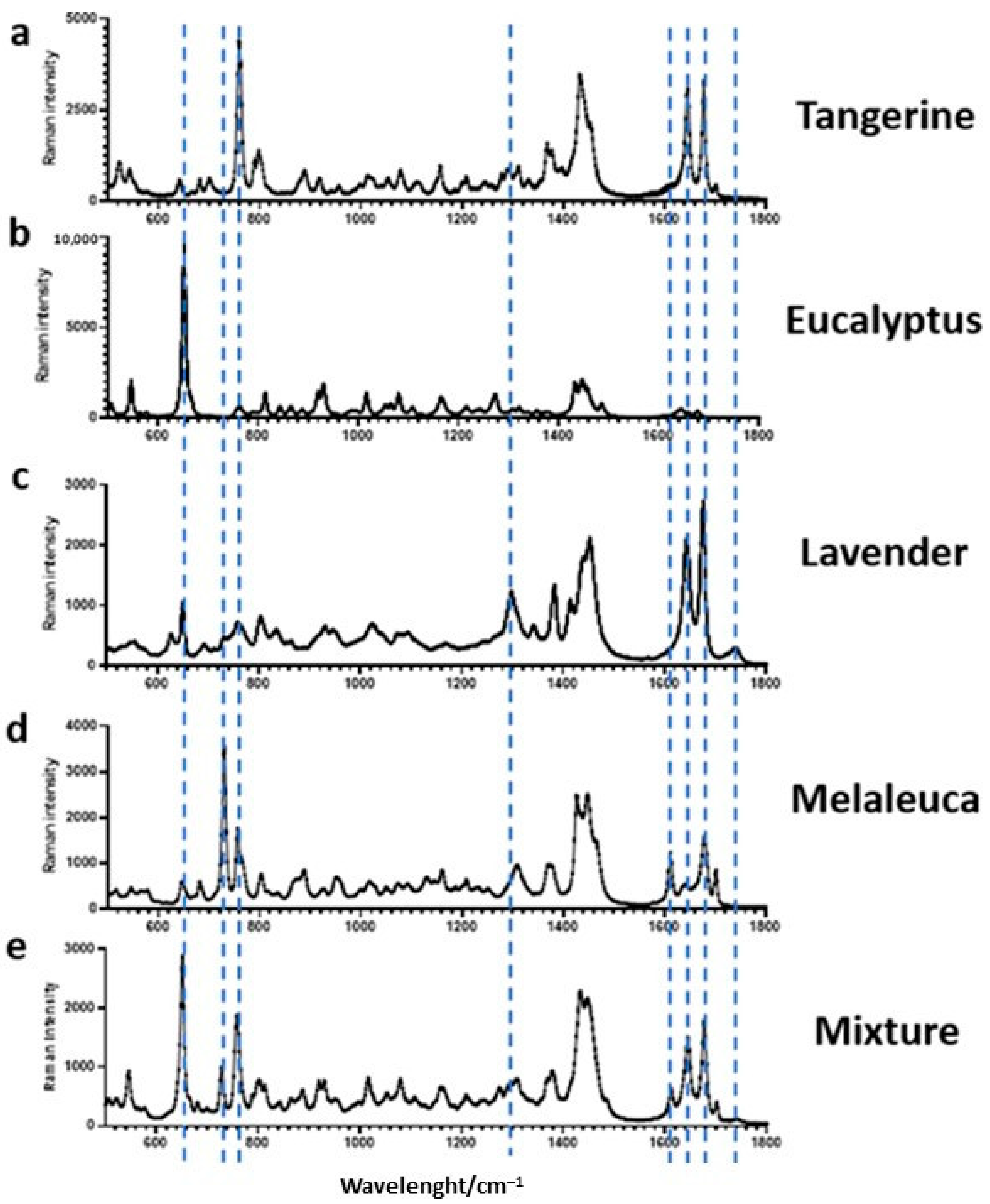
Figure 1. Fingerprint Raman spectra of the four essential oils and their mixture. The characteristic Raman bands for tangerine essential oil at ≈760, 1646, and 1678 cm−1 (a); for eucalyptus essential oil at ≈650 cm−1 (b); for lavender essential oil at ≈650, 1298, 1644, 1676, and 1744 cm−1 (c); and for melaleuca essential oil at ≈730 and 756 cm−1 (d); the mixture of these four essential oils in the 1:1:1:1 proportion (e) are shown with dotted lines.
Cosmetics 10 00158 g001
Figure 2. Raman spectrum of pure melaleuca essential oil (green line) and nanoemulsion (purple line).
Cosmetics 10 00158 g002
Figure 3. Rheological behavior of the placebo (light green circles), Melaleuca alternifolia (dark green squares), Melaleuca alternifolia nanoemulsion (blue triangles), and a mixture of four essential oils (purple diamonds) containing formulations. Formulations were evaluated 24 h after the development.
Cosmetics 10 00158 g003
Figure 4. Scores of comedone number per image using the microrelief images (A) and results before (T0, dark grey) and after (T90, light grey) 90 days (B). (*) means p < 0.05 and (***) means p < 0.001.
Cosmetics 10 00158 g004
Figure 5. Pilosebaceous unit RCM images from participants showing the SC (left), SG (middle), and UD (right), respectively. The images in (A) represent the dysmorphic alterations in the pilosebaceous units. The thick edge (red arrow) and the presence of inflammatory infiltrate in the UD (yellow circle) are presented. The image in (B) is a representation of a comedone with a regular shape, which can also present a thick edge (red arrow); however, the shape is circular and the smaller size is well-delimited, without inflammatory infiltrate observed. Scale bar is 100 µm.
Cosmetics 10 00158 g005
Figure 6. Pilosebaceous unit area in the SC before (T0, grey) and after (T90, white) 90 days of treatment, measured using the RCM images. * means p < 0.05.
Cosmetics 10 00158 g006
Figure 7. The pilosebaceous unit area in the SG before (T0, grey) and after (T90, white) 90 days of treatment, measured using the RCM images. (#) means p = 0.056.
Cosmetics 10 00158 g007
Figure 8. Pilosebaceous unit area in the UD before (T0, grey) and after (T90, white) 90 days of treatment, measured using the RCM images. (***) means p < 0.001.
Cosmetics 10 00158 g008
Table 1. Evaluation of the morphological and structural characteristics of pilosebaceous units according to treatment groups and time. The results are presented in values relative to the total (percentage).
ShapeAbsence of Comedones
(%)
EdgeInternal Amorphous
GroupsRegular (%)Dysmorphic (%)Thin Edge (%)Thick Edge (%)Absent (%)Present
(%)
T0T90T0T90T0T90T0T90T0T90T0T90T0T90
4EO616739--3311448923-2810050
Nanoem.617839--2233566722-1710061
M.a.6172306-22285672226229456
Placebo67563333-111717837218188278
Disclaimer/Publisher’s Note: The statements, opinions and data contained in all publications are solely those of the individual author(s) and contributor(s) and not of MDPI and/or the editor(s). MDPI and/or the editor(s) disclaim responsibility for any injury to people or property resulting from any ideas, methods, instructions or products referred to in the content.