Article

Antioxidative Potentials of Eleutherine bulbosa Bulb and Its Utilization in Topical Cosmetic Emulsion

by
Nattakan Panyachariwat
1,2,
Ampa Jimtaisong
1,2,* and
Nisakorn Saewan
1,2
1
School of Cosmetic Science, Mae Fah Luang University, Chiang Rai 57100, Thailand
2
Cosmetic and Beauty Innovations for Sustainable Development (CBIS) Research Group, Mae Fah Luang University, Chiang Rai 57100, Thailand
*
Author to whom correspondence should be addressed.
Cosmetics 2024, 11(4), 111; https://doi.org/10.3390/cosmetics11040111 (registering DOI)
Submission received: 21 June 2024 / Revised: 1 July 2024 / Accepted: 4 July 2024 / Published: 6 July 2024

Abstract

:
The Eleutherine bulbosa bulb has been reported as a potent antioxidant in food. This work aims to extract the E. bulbosa bulb for use as an antioxidative agent in cosmetics. Water, 95% ethanol (EtOH), and propylene glycol (PG), which are normally used in cosmetic formulation, were employed as green and sustainable extraction solvents. EtOH and PG displayed better candidacy to extract active components from E. bulbosa bulbs than using water, and the mixture of EtOH and PG (EtOH/PG) resulted in the extract with higher bioactive compounds and biological activities compared with using EtOH or PG. The total phenolic content of the EtOH/PG extract was 87.60 ± 2.00 mgGAE/mL which was about an 18–23% increase from when using single EtOH or PG (70.91 ± 2.30, 74.05 ± 0.67 mgGAE/mL). UHPLC-ESI-QTOF-MS/MS analysis showed that the E. bulbosa bulb extracted in EtOH/PG was composed of naphthalenes, naphthoquinones, anthraquinones, myricetin, quercetin, epicatechin, catechin, epigallocatechin, and their derivatives. The ethanolic crude extract exhibited anti-elastase and anti-collagenase activity with the IC50 of 7.76 ± 0.35 and 0.53 ± 0.23 mg/mL, respectively, and was non-cytotoxic to human dermal fibroblast cells at 0.0001–1 mg/mL. The emulsion cream containing 2%(w/w) E. bulbosa bulb concentrated extract was found cosmetically stable after a one-month stability test under 4 °C, ambient temperature (30–35 °C), 45 °C, fluorescent light, and daylight. However, exposure to sunlight during daytime caused changes in the emulsion’s color with ΔE* of 3.85 ± 0.08, and at 45 °C caused the 12% decrease in DPPH activity of emulsion. The finding of this work heightens the antioxidative and safety potentials of the E. bulbosa bulb in cosmetic preparations.

1. Introduction

Eleutherine bulbosa (Synonym: Eleutherine palmifolia, Eleutherine americana Merr.) is a herbal plant belonging to the Iridaceae family [1]. E. bulbosa is a herbaceous, rhizomatous, and perennial flowering plant with a predominantly red bulb color similar to red onion with a leaf in a pleated linear-lanceolate shape [1,2]. It is traditionally used to treat coronary disorders, and used as diuretic, emetic, purgative, prothrombin decreasing, antifertility, antihypertension, wound-healing activity [3]. There are three big groups of compounds which have been isolated from E. bulbosa, i.e., naphthalene, anthraquinone, and naphthoquinone [4]. It was reported that E. bulbosa bulbs extracted in 95% ethanol at room temperature for 7 days exhibited DPPH activity at IC50 of 0.0084 ± 0.0007 mg/mL which is about three time higher than that of Vitamin E (0.023 ± 0.001 mg/mL) [5]. The ethanolic crude extract from E. bulbosa was also found to delay lipid oxidation in pork and give red-colored samples. Moreover, extracts of E. bulbosa have been patented as skin lighteners in Japan on account of the melanogenesis-suppressing and tyrosinase-inhibiting activities of its naphthopyranes [6]. Some experiments have been conducted in the past for validating pharmacological uses especially for antimicrobial, antihypertensive, antidiabetic, anti-inflammatory, and antiviral activities of E. bulbosa [7,8]. Cream and loose powder prepared with E. bulbosa ethanolic extract exhibited anti-acne properties [9,10]. Recently, a study by Kamarudin et al. [11] revealed that the major bioactive compounds from E. bulbosa bulbs are gallic acid, epicatechin gallate, quercetin, rutin, and eleutherine when being extracted using 90% ethanol. Some components, like rutin and quercetin, are useful as sunscreen agent in cosmetics [12]. Comparably, these compounds are well known for their antioxidative effects [13,14,15]. However, there is no report on the development of E. bulbosa as an antioxidative agent in cosmetics. Thus, this work aims to prepare the E. bulbosa bulb extract and study the utilization of the extract as an ingredient in cosmetic preparations. The E. bulbosa bulb was extracted using an ultrasonication technique, which has simple, mild, and environmentally friendly conditions. Water and ethanol, which are normally used in cosmetic formulation, were selected as solvents. Moreover, propylene glycol, which has been used as a moisturizer in cosmetic products, was also employed as an extraction solvent. The active components were determined as phenolics, flavonoids, and anthraquinone content. Antioxidant activities were measured using DPPH and FRAP assay. Anti-elastase and anti-collagenase activities were also reported. In vitro cytotoxicity of the extract was also presented. Utilization of the extract as an active ingredient in cosmetic emulsion dosage form was studied in term of physical effect, color, and antioxidant activities’ changes over time.

2. Materials and Methods

2.1. Materials

DPPH (2, 2′-diphenyl-1-picrylhydrazyl), Folin–Ciocalteu reagent, gallic acid, L-ascorbic acid, and Trolox (6-hydroxy-2,5,7,8-tetramethylchroman-2-carboxylic acid) were purchased from Sigma-Aldrich, Burlington, MA, USA. Anthraquinone (97%) was of Sigma Aldrich®, Praha 4, Czech Republic. Solvents for extraction (95% Ethanol, propylene glycol, and deionized water) are of cosmetic grade. Ingredients for emulsion preparation (Caprylic/capric triglyceride, BASF, Monheim, Germany; Jojoba oil, Statfold, Tamworth, UK; Glyceryl stearate, BASF, Germany; Sorbitan stearate, Eigenmann & Veronelli, Hertfordshire, Italy; Theobroma cacao (Cocoa) seed butter, Statfold, UK; Beeswax, Fumeipharm, Henan Province, China; Stearic acid, Edenor, Selangor, Malaysia; Polyacrylate crosspolymer-11, Clariant, Alava, Spain; Disodium EDTA, BASF, Germany; Butylene glycol, KH Neochem, Chuo-ku, Japan; Glycerin, Emery Oleochemicals, Selangor, Malaysia; Sodium PCA, Ajinomoto, Chuo-ku, Japan; Polysorbate 60, Croda, Bangkok, Thailand; Diazolidinyl urea (and) Iodopropynyl butylcarbamate (and) Propylene glycol, Ashland, Covington, KY, USA) are of cosmetic grade. All chemicals and ingredients were used as received.

2.2. Preparation of Eleutherine bulbosa Extract

E. bulbosa was collected from a private garden in Chiangrai province, Thailand, and the dry samples were prepared by cutting fresh bulbs into small pieces and drying at 60 °C in a hot air oven for 20 h. Effects of extraction solvents on fresh and dry sample were studied. The dry and fresh samples were then subject to extraction in different solvents. A total of 20 g of fresh E. bulbosa bulb was extracted in DI water, ethanol 95%, or propylene glycol 60 mL (sample to solvent ratio of 1:3 w/v). Meanwhile, 9.09 g (equivalent to 20 g fresh sample) of the dry E. bulbosa was extracted in 60 mL solvents. The mixture was sonicated (Ultrasonic Jeken TUC-100, 40 KHz) for 2 h at ambient temperatures and filtered with filter paper. The extraction was performed twice, and the solution extract was then collected for antioxidant activity study. Crude ethanolic extract of fresh E. bulbosa was prepared using the same extraction method. The ethanol was evaporated using a rotary evaporator to obtain crude extract.

2.3. Determination of Total Phenolic Content

The total phenolic content of the extracts was determined by Folin–Ciocalteu total phenolic assay [16,17]. Gallic acid (GA) was used as a standard and a range of concentrations was used to create a standard curve. Deionized water (1.58 mL) and Folin–Ciocalteu reagent (100 µL) were added to the extract sample, the standard, or blank (20 µL). The reaction mixture was incubated at room temperature for 5 min. Sodium carbonate solution (10% w/v, 300 µL) was then mixed with it and incubated for 90 min. The absorbance of the mixture was measured spectrophotometrically at 765 nm (Biochrom Libra S22, Biochrom Ltd., Cambridge, UK). The total phenolic content was calculated from a standard curve and expressed as a gallic acid equivalent (GAE) (mg GAE/mL extract). Determinations were made in triplicate.

2.4. Determination of Total Flavonoid Content

The aluminum chloride colorimetric method was used for the determination of the total flavonoid content of the sample following the reported method [18] with modifications. Quercetin was used to make the standard calibration curve. An amount of 1.2 mL standard quercetin solutions or extracts was separately mixed with 1.2 mL of 2% aluminum chloride. After mixing, the solution was incubated for 60 min at room temperature. The absorbance of the reaction mixtures was measured against blank at 420 nm with a UV-Vis spectrophotometer (Biochrom Libra S22, Biochrom Ltd., Cambridge, UK). The concentration of total flavonoid content in the test samples was calculated from the calibration plot and expressed as µg quercetin equivalent (QE/mL extract). All the determinations were carried out in triplicate.

2.5. Determination of Anthraquinone

Quantitative analysis of anthraquinone was performed by the spectrophotometric method following the previous reported method [19] with modifications. Anthraquinone (97%) was used as the standard and the calibration curve was made from 6 concentrations. All concentrations were measured by the spectrophotometric method at 325 nm (Biochrom Libra S22, Biochrom Ltd., Cambridge, UK). The measurement was taken in triplicate. The relation between concentration and absorbance was plotted and a correlation coefficient (R2) was calculated and expressed as µg anthraquinone/mL extract.

2.6. DPPH Radicals Scavenging Assay

The scavenging activities of the extracts were measured on DPPH radicals [17] with modifications. Ascorbic acid was used as a standard and the range of concentrations were used to create a standard curve. Then, 3 mL of DPPH radicals in absolute ethanol (0.1 mM) was added to the sample (1 mL) or standards (1 mL). The reaction mixture was mixed at ambient temperature and immediately incubated at 37 °C for 30 min, and then the absorbance (Abs) was determined at 517 nm using a spectrophotometer (Biochrom Libra S22, Biochrom Ltd., Cambridge, UK). The DPPH result was reported as the ascorbic equivalent (AAE/mL extract). The concentration giving 50% inhibition (IC50) was also reported.

2.7. Determination of Reducing Power

The reducing power was determined following the previous methods [17,20] with modifications. The extract (1 mL) was mixed with 2.5 mL of phosphate buffer (0.2M, pH 6.66) and 2.5 mL of 1% potassium ferricyanide. The mixtures were incubated at 50 °C for 20 min, after which 2.5 mL of 10% trichloroacetic acid was added followed by centrifuging at 5000 rpm for 10 min. The upper layer (2.5 mL) was mixed with 2.5 mL of deionized water and 0.5 mL of 0.1% ferric chloride. The absorbance of the mixture was measured at 700 nm (Biochrom Libra S22, Biochrom Ltd., Cambridge, UK). Ascorbic acid was used as the reference standard. The result was reported as the ascorbic acid equivalent (AAE/mL extract).

2.8. Ferric Ion Reducing Antioxidant Power (FRAP) Assay

Ferric ion reducing antioxidant power (FRAP) assay was performed following the reported methods [21,22] with modifications. The FRAP reagent was prepared by mixing 38 mM sodium acetate (anhydrous) in distilled water pH 3.6, 20 mM FeCl3·6H2O in distilled water, and 10 mM 2,4,6-tri(2-pyridyl)-s-triazine (TPTZ) in 40 mM HCl in proportions of 10:1:1. This reagent was freshly prepared before each experiment. To each sample, 300 µL of appropriately diluted sample extract and 2700 µL of FRAP reagent were added and the mixture was incubated at 37 °C for 40 min in the dark. In the case of the blank, 300 µL of ethanol was added to 2700 µL of FRAP reagent. The absorbance of the resulting solution was measured at 593 nm (Biochroms Libra S22, Biochrom Ltd., Cambridge, UK). Trolox (6-hydroxy-2,5,7,8-tetramethylchroman-2-carboxylic acid) was used as a reference antioxidant standard. FRAP values were expressed as the Trolox equivalent (TE/mL extract).

2.9. Anti-Elastase Activity

Crude ethanolic extract of fresh E. bulbosa bulb was prepared using the same extraction method. The ethanol was evaporated using a rotary evaporator to obtain crude extract. The elastase assay of the crude extract was evaluated according to the previously reported methods [23,24] with modifications. To evaluate the inhibition of elastase activity, the amount of released p-nitroaniline, which was hydrolyzed from the substrate, N-succinyl-Ala-Ala-Ala-p-nitroanilide, by elastase, was assayed by measuring absorbance at 410 nm. The method used epigallocatechin gallate as standard. The sample was dissolved in 10% DMSO to gain 0.001, 0.01, 1, and 10 mg/mL. The standard epigallocatechin gallate was dissolved in Tris-HCL buffer to 0.0001, 0.001, 0.01, 0.1, and 1 mg/mL concentration. A solution of N-succinyl-Ala-Ala-Ala-p-nitroanilide (1.015 mM) was prepared in a 0.1 M Tris-Cl buffer (pH 8.0) and this solution (130 μL) was added to the sample or standard (10 μL). The mixture was pre-incubated for 5 min at 25 °C before an elastase (0.0375 unit/mL) stock solution (10 μL) was added. After enzyme addition, the mixture was kept at 25 °C for 30 min, and the absorbance was measured at 410 nm. The elastase enzyme inhibition was expressed as 50% inhibitory concentration (IC50).

2.10. Anti-Collagenase Activity

Anti-collagenase activity of the ethanolic crude extract was measured using the previously described procedure [25] with slight modifications. The method used ascorbic acid at a 0.0001–1 mg/mL concentration as standard and used Pz-peptide as a precursor. The sample was dissolved in 10% DMSO to gain 0.001, 0.01, 1, and 10 mg/mL. The mixture tube contained collagenase (5 ug) and Pz-peptide (0.5 mg) in 0.1 MTris buffer (pH 7.1) containing 20 mM CaCl2, in the presence or absence of test compounds (total volume of 1.7 mL). The tube was incubated at 37 °C for 30 min, and 25 mM citric acid solution (1 mL) was added to end the reaction. After mixing with ethyl acetate (5 mL), the absorbance of the organic layer was measured at 320 nm. The collagenase inhibition was expressed as 50% inhibitory concentration (IC50).

2.11. Cytotoxicity Test

The cytotoxic effects of E. bulbosa crude ethanolic extract were evaluated using Sulforhodamine B (SRB) colorimetric assay [26]. The extract was dissolved with 10% DMSO in cell culture. After that, the sample was filtrated with a 0.2 microns membrane filter and diluted to a suitable concentration (0.0001–1 mg/mL) with 10% DMSO in the sterile cell culture. Sodium lauryl sulfate (SLS) was also tested as a positive control. Human skin fibroblasts (passages 59) were cultured in Dulbecco’s modified eagle medium (DMEM) supplemented with 10% fetal bovine serum (FBS) and 1% penicillin/streptomycin solution in a humidified incubator at 37 °C with 5% carbon dioxide. Cells were seeded and incubated for 24 h and were then treated with different concentrations of crude extract. After 72 h, cells were fixed and stained with SRB. The excess dye was washed away, and the bound dye was solubilized in tris buffer to measure absorbance at 510 nm. Cell viability was expressed as a percentage of the control values.

2.12. Qualification of Active Compounds by UHPLC-QTOF-MS

An optimized LC-QTOF method using an UHPLC Agilent 1290 Infinity II System coupled to an Agilent 6545 LC-QTOF/MS (Agilent Technologies, Santa Clara, CA, USA) was developed for the qualification of active compounds. This method involved optimizing various chromatographic parameters, including the mobile phase composition, column type, flow rate, and solvent ratio. A Waters XBridge C18 (100 mm × 2.1 mm, 2.5 μm) column was used for the separation. The extract was diluted with ethanol at a ratio of 1:25 and filtered using the 0.22 µm syringe filters (Labfil, ALWSCI, Zhejiang, China) and transferred into HPLC vials. The elution was achieved using a binary gradient system, with 0.1% formic acid in Water LCMS grade (J.T.baker) as eluent A and 0.1% formic acid in acetonitrile (ACN) as eluent B. The gradient steps were 5–17% B at 0–13 min, followed by 17–100% B at 13–20 min, 100% B at 20–25 min, and 100–5% B at 25–27 min, with a flow rate of 0.2 mL/min. A postrun was set to equilibrate the column for 6 min. Dual AJS (Agilent Jet Strem) ESI was used as an ion source with a sheath gas flow of 12 L/min, sheath gas temperature of 250 °C, drying gas temperature of 350 °C, drying gas flow of 10 L/min, and capillary voltage of 3500 V.
A complete mass scan ranging from m/z 50 to 1100 was used; MS/MS analyses were carried out in automatic mode with collision energy (10, 20 and 40 eV) for fragmentation. Peak identification was performed in both positive and negative modes while the instrument control, data acquisition, and processing were performed using MassHunter workstation software (Qualitative Analysis, version B.8.00) (Agilent Technologies, USA).

2.13. Utilization of E. bulbosa Bulb Extract in Cosmetic Preparation

The cosmetic product was prepared in the form of an oil-in-water emulsion cream. The cosmetic product was formulated by addition of varying amounts of E. bulbosa bulb extract based on its antioxidant activity. An emulsion (Table 1) was prepared by separately weighing the oil and water phase and these were separately heated at 75 ± 2 °C. The oil phase was then added into the water phase and an emulsion was formed by homogenization at 3000 rpm for 5 min at 75 ± 2 °C. The emulsion was then cooled to 45–50 °C and the remaining ingredients were then added and mixed well.

2.14. Accelerated Stability Test of Cosmetic Emulsion

The test was conducted to determine the changes in physical properties of the prepared cosmetic emulsion. The samples were stored in various conditions, which are 4 °C, 45 °C, ambient temperature (30–35 °C), fluorescent light (36 watt, 8 h/day), and daylight (8 h/day), and during the stability test, the physicochemical properties of products, including any changes in color as the measurement of L*a*b* values (Colorimeter, Konica Minolta, Chiyoda-ku, Tokyo, Japan), odor, pH, and phase separation were collected every two weeks [27]. The DPPH radical scavenging activity of emulsion was measured at week 0 and week 4. The E. bulbosa was extracted from emulsion by using ethanol extraction [28]. Briefly, emulsion and ethanol were mixed in the ratio 1:2 w/v with an ultrasonicator for 20 min. Then the mixture was centrifuged at 8000 rpm for 10 min at 25 °C. After centrifugation, the supernatant was collected. The radical scavenging activity of products was assayed by using the DPPH method.

3. Results

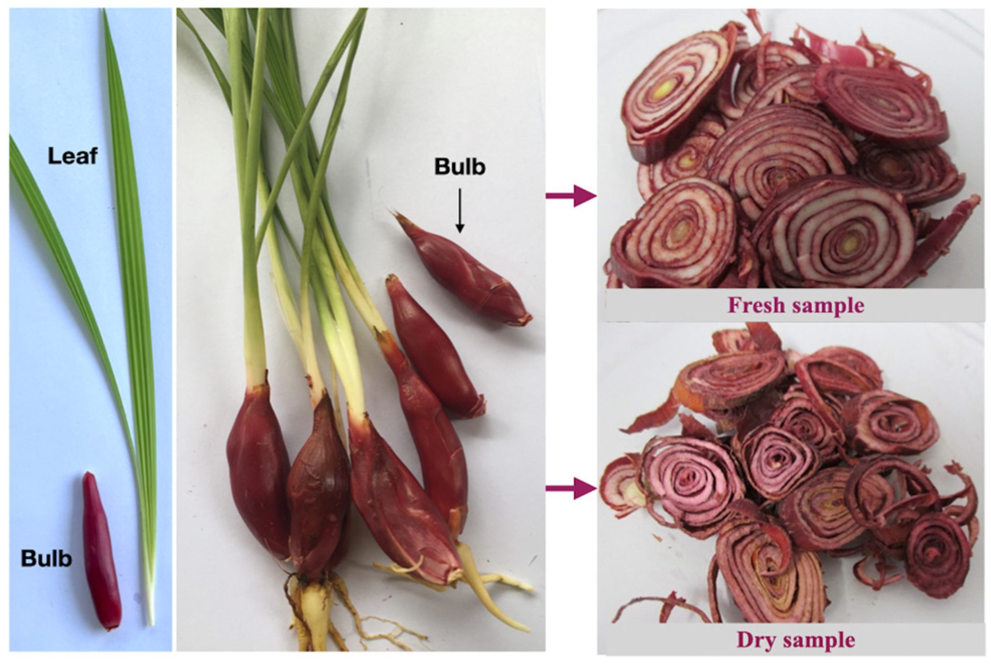
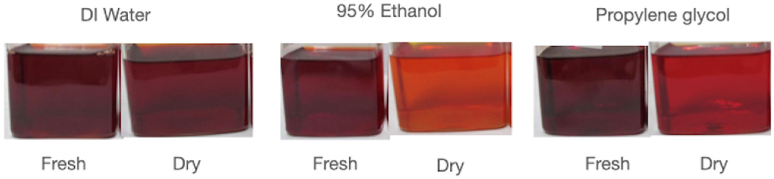
3.1. Preparation of Eleutherine bulbosa Extract

The E. bulbosa sample collected for this study is shown in Figure 1. The plant sample was washed clean, and bulbs were separated for extraction. The dry sample of E. bulbosa bulb was prepared by cutting fresh bulb into small pieces and drying at 60 °C in a hot air oven for 20 h. The obtained dry E. bulbosa yield was 45.46% compared to fresh weight.
The dry and fresh samples were then subject to extraction in different solvents, i.e., DI water, 95% ethanol (EtOH), and propylene glycol (PG). The extracts exhibited a deep red color except for those dry samples in ethanol and propylene glycol, which have clear orange–red color as can be seen in Figure 2.
The bioactive compounds and biological activities of the extract were measured and presented in Table 2. The fresh sample exhibited higher activities than the dry sample in all solvents except for the DPPH and reducing power of those extracted in water that showed relatively similar activities. Considering the extraction solvent, PG and EtOH give extracts with much higher biological activities than that of water. The DPPH of the fresh sample extracted in water is 0.178 ± 0.003 mgAAE/mL extract which is significantly lower than those of EtOH and PG extracts (0.432 ± 0.002, 0.423 ± 0.005 mgAAE/mL extract). The reducing power and FRAP assay of water extract were also significantly lower than those of EtOH and PG extracts (p < 0.05), Table 2.
The EtOH extract possessed relatively higher DPPH activity than that of the PG extract but was not statistically different (p > 0.05). However, the reducing power and FRAP assay of the EtOH extract were significantly higher than the PG extract and these may be related with the significantly higher in total flavonoids and anthraquinone (p < 0.05), while the total phenolic content of EtOH extract is lower than that of PG but not statistically different (p > 0.05). Thus, in terms of solvent for extraction, the mixture of EtOH and PG (EtOH/PG) was selected for further study. Also, fresh E. bulbosa was used. The extract was then preliminarily prepared by using a solvent mixture of EtOH to PG at a 70:30 v/v ratio. The use of mixture of EtOH/PG resulted in an extract that was higher in bioactive compounds and biological activities compared with using single ethanol or propylene glycol. The total phenolic content of EtOH and PG extract was found at 70.91 ± 2.30 and 74.05 ± 0.67 mgGAE/mL, respectively, and it was found at 87.60 ± 2.00 mgGAE/mL (Table 2), which is an increase of about 18–23% compared with using a single solvent. Total flavonoids and anthraquinone content were also found to be increasing. The DPPH, reducing power, and FRAP actives also significantly increased when being extracted using EtOH/PG mixture.

3.2. Qualification of Active Compounds by UHPLC-QTOF-MS

The identities of the phytochemical compounds in E. bulbosa bulbs extracted using EtOH/PG mixture, which exhbited the highest biological activities, were obtained by matching the molecular m/z values from the UHPLC-ESI-QTOF-MS/MS. Naphthalenes, naphthoquinones, and anthraquinones were detected in the EtOH/PG extract, along with a glycoside: eleutherinoside A (Table 3). Moreover, three new proposed compounds, namely emodin, rutaretin, and 2-hydroxy-3,4,6-trimethoxydihydrochalcone, were also found in the extract. The analysis also revealed the presence of one myricetin and two quercetin derivatives. Additionally, epicatechin and its three derivative compounds were also detected. Catechin, epigallocatechin, and their derivatives were also discovered in the E. bulbosa bulbs extracted using EtOH/PG mixture solvent.

3.3. Anti-Elastase and Anti-Collagenase Activity

Elastase is a proteinase enzyme that can reduce elastin by dividing specific peptide bonds. Therefore, the inhibition of elastase activity in the dermis layer can be used to maintain skin elasticity. E. bulbosa crude ethanolic extract showed the 50% inhibitory concentration (IC50) of 7.76 ± 0.35 mg/mL which is lower activity than the standard epigallocatechin gallate (IC50 0.03 ± 0.01 mg/mL). The crude extract also possessed an anti-collagenase activity as its concentration requirement to inhibit 50% (IC50) of the collagenase enzyme was at 0.53 ± 0.23 mg/mL and this is lower in activity than the standard ascorbic acid (IC50, 0.025 ± 0.003 mg/mL). Collagen is the fundamental and the major molecular unit involved in the construction of human skin. It is a protein that is commonly found in the connective tissues of the human body. The inhibition of elastase and collagenase of E. bulbosa bulbs indicates that it can be candidate for anti-aging function.

3.4. Cytotoxicity Test

The cytotoxic effects of E. bulbosa ethanolic crude extract on the viability of the human skin fibroblasts were evaluated using Sulforhodamine B colorimetric assay. The result showed E. bulbosa crude extract at 0.0001 to 1 mg/mL concentrations were non-cytotoxic to human dermal fibroblast cells which have shown cell survival at 103.40 ± 0.56 to 107.25 ± 2.76% (Figure 3). The sodium lauryl sulfate (SLS) which was used as a positive control showed a cytotoxic effect to human dermal fibroblast cells which have shown the cell survival of 19.61 ± 1.32% at 1 mg/mL which decreased from 103.99 ± 1.57% at 0.0001 mg/mL. The finding of this work presents the first report on the cytotoxicity effects of E. bulbosa bulb ethanolic crude extract against human skin fibroblast cell lines.

3.5. Utilization of E. bulbosa Extract in Cosmetic Formulation

3.5.1. Preparation of the Extract

In this work, a mixture of ethanol and propylene glycol (EtOH/PG) at ratio of 70:30 v/v was used to extract the E. bulbosa bulb for cosmetic preparation, and for more suitability in cosmetic formulation, the ethanol from the extract was further completely removed using a rotary evaporator, leaving the PG to obtain the concentrated extract. The characteristics of the concentrated extract were tabulated in Table 4. The parameters are ideal for quality control of the E. bulbosa bulb extract for industrial application. The concentrated extract was then used to study the potential of its use as an active ingredient in cosmetic preparations.

3.5.2. Use of E. bulbosa Bulb Concentrated Extract in Emulsion Formulation

The IC50 of E. bulbosa bulb concentrated extract is 0.0097 ± 0.0002 g/mL. Therefore, the product should contain E. bulbosa extract at 0.97% w/w to exhibit 50% DPPH inhibition, and at 1.94% to theoretically obtain 100% inhibition. Accordingly, the extract was added in the formula at 1.0 and 2.0 w/w to study the effect of extract on physicochemical properties of the cosmetic product. The base of the emulsion was smooth white opaque cream with pH 5.23 ± 0.01. After the extract was added, the pH value of the emulsion at 1.0 and 2.0% (w/w) E. bulbosa bulb concentrated extract was 5.20 ± 0.01 and 5.21 ± 0.01, respectively. The viscosity of the emulsion base was 10,883 ± 35 cPs (RV#05, 30 rpm, Brookfield, Middleboro, MA, USA) and those of the emulsions with 1.0 and 2.0% (w/w) extract were 11,283 ± 80 and 11,377 ± 42 cPs, respectively. The color of the product changed to a pale orange–pink and gradually became darker when increasing the amount of extract (Figure 4). The product has a characteristic odor of the extract.

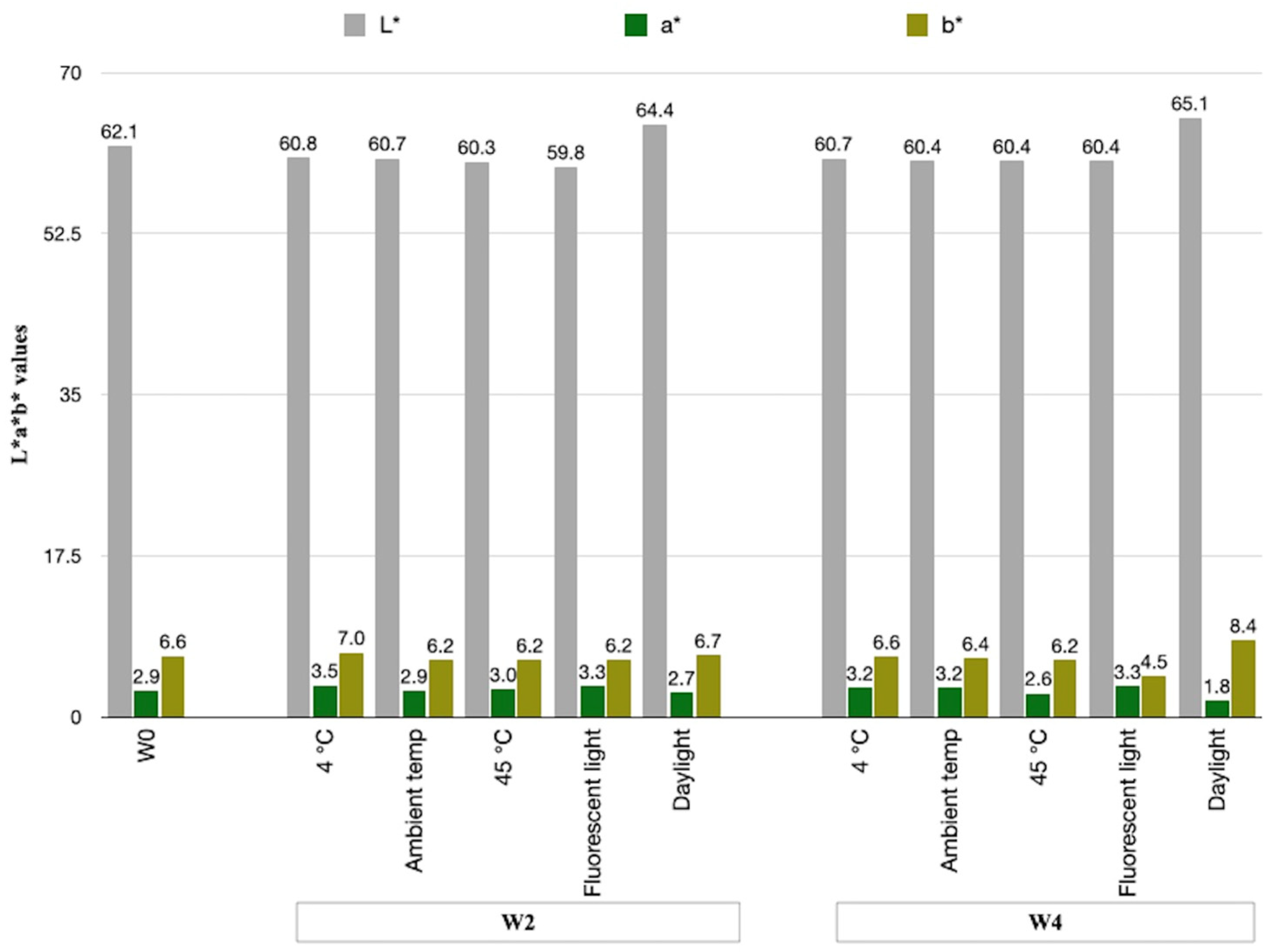
3.5.3. Physical Stability Study of the E. bulbosa Emulsion Cream

The emulsion with 2.0% w/w E. bulbosa extract was subject to an accelerated stability test at different storage conditions, i.e., 4 °C, ambient temperature (30–35 °C), 45 °C, fluorescent light, and daylight. It showed that there was no separation of an emulsion product and the emulsion’s texture remained relatively consistent after testing in all conditions. The pH value of the cream changed slightly after one month of study, from 5.21 ± 0.01 (week 0) to the values in the range of 5.15 ± 0.01 to 5.38 ± 0.01.
The color stability of the E. bulbosa emulsion cream was evaluated by Colorimeter (CM-700d—Konica Minolta, Japan) identifying L*, a*, and b* values, where L* is a lightness value determining white or black, a* is a value determining the color of green (−) or red (+), and b* is a value determining the color of blue (−) or yellow (+). During the study period, there were slight changes in color measurements of the emulsion cream (Figure 5). The L* values decreased slightly overtime under 4 °C, ambient temperature (30–35 °C), 45 °C, and fluorescent-light conditions, while the value increased from 62.06 ± 0.86 to 65.20 ± 0.17 in the daylight condition, indicating a higher lightness. The a* values showed only slight changes in all conditions except for that under daylight which decreased from 2.86 ± 0.01 to 1.80 ± 0.02, indicating declining redness in the product. Also, there was an increase in the b* values from 6.64 ± 0.37 to 8.36 ± 0.06 under the daylight condition, which indicates a more yellow tone of the product.
Finally, the total color difference or ΔE* was calculated, and the values were found to be less than 2 (Table 5) for the products stored under 4 °C, ambient temperature (30–35 °C), and 45 °C, implying that the difference is only noticeable by an experienced observer. While the emulsion kept exposure to fluorescent light and daylight (8 h/day) for one month exhibited the ΔE* of 2.78 ± 0.16 and 3.85 ± 0.08 which indicates that an unexperienced observer can notice the difference.

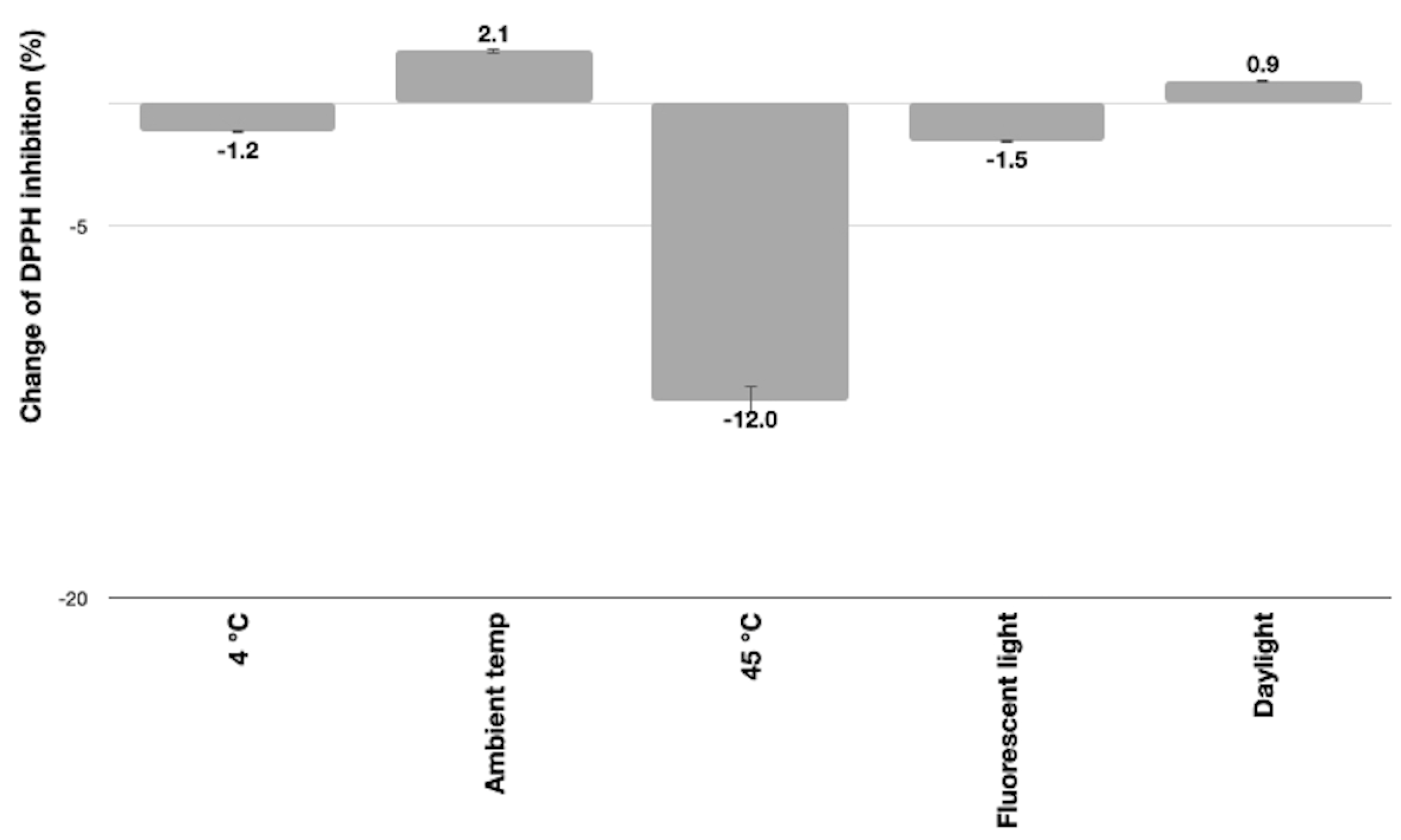
3.5.4. Antioxidant Activity of the E. bulbosa Emulsion Cream

The base formulation without E. bulbosa extract was first tested to identify the antioxidant activity that might influence the activity of the product. The base formulation exhibited an antioxidant capacity of 3.94 ± 0.21% DPPH inhibition, indicating that some ingredients in the formula influenced the cream’s antioxidant activity. The 2% E. bulbosa cream (50% concentration) initially showed a DPPH inhibition of 65.26% and after being tested in accelerated condition for one month it exhibited changes of DPPH inhibition activity as shown in Figure 6. The DPPH inhibition activities of cream exhibited relatively small changes as it showed a decrease under 4 °C and fluorescent light while increasing under ambient temperature (30–35 °C) and daylight conditions. However, the cream at 45 °C exhibited a significant decrease in DPPH of about 12.01 ± 0.56%, thus the high temperatures may damage some active compounds and consequently lower the antioxidant activity.

4. Discussion

4.1. Antioxidative and Cytotoxicity Activity of E. bulbosa Bulb

The E. bulbosa bulb has been used in traditional medicines in wide regions worldwide, and its main bioactive compounds were identified as naphthalene, anthraquinone, and naphthoquinone [1,2]. In Thailand, it is used as a traditional carminative in its pure form, and together with galangal, to treat cold and nasal congestion in children [29]. E. bulbosa bulb was reported to possess strong antimicrobial activities [8,30]. Also, it was reported to exhibit DPPH activity at about three time higher than that of Vitamin E which makes it a good candidate for delay oxidation in food, and gives red-colored samples [5]. The red pigment from E. bulbosa bulbs has been used to color glycerin soap [31]. However, application of E. bulbosa bulbs as an antioxidative agent in cosmetics has never been studied before; thus, the objective of this work is to extract and quantify the phenolic, flavonoid, and anthraquinone contents in E. bulbosa bulbs for use in cosmetic preparations. In this study, water, 95% ethanol (EtOH), and propylene glycol (PG), which were commonly used in cosmetic formulation, were considered as mild and environmentally friendly solvents for the extraction process [32]. The results showed that fresh samples exhibited higher activities than the dry samples in all solvents except for the DPPH, and reducing power of those extracted in water which showed relatively similar activities. The PG and EtOH give extracts with much higher biological activities than that of water. The results obtained were in the same direction as a study carried out by Morabandza et al. [33] that the ethanolic bulb extract of E. bulbosa displayed higher total phenolic and flavonoid content compared to aqueous extract.
The EtOH extract possessed relatively higher bioactive compounds and biological activities than that of PG extract. It was previously reported that naphthalene, anthraquinone, and naphthoquinone are the three dominant constituents of E. bulbosa bulb [1,3,34,35,36,37]. Moreover, the recent study also reported bioactive compounds of gallic acid, epicatechin gallate, quercetin, eleutherin, rutin, chlorogenic acid, kaempferol, and myricetin [11]. Ethanol and propylene glycol can dissolve both polar and non-polar bioactive compounds, and according to the results they exhibit good candidacy to extract active components from E. bulbosa bulbs. Moreover, the use of an EtOH/PG mixture resulted in an extract that was significantly higher in bioactive compounds and biological activities compared with using single ethanol or propylene glycol. This may be due to the relatively small ethanol molecules which could swell the E. bulbosa by infiltrating the tissue and then polyol can easily extract the anthraquinone, polyphenols, and flavonoids. This result is similar to that of the extraction of Camellia oleifera seed dregs [32]. Recently, polyol solvents as well as a mixture polyol solvent were increasingly applied in the recovery of phenolic compounds from plants. They are considered a green non-conventional solvent that is abundant, inexpensive, nontoxic, and inflammable, with a high boiling point [38,39]. Furthermore, propylene glycol was also found to be efficient in extracting cosmetic bioactive compounds from Croton thorelii Gagnep [40].
The active components in E. bulbosa bulbs extracted using EtOH/PG mixture were obtained by using the UHPLC-ESI-QTOF-MS/MS analysis. Naphthalenes, naphthoquinones, and anthraquinones detected were consistent with the data in previous reports [1,34,35,41,42,43,44]. Moreover, three new proposed compounds, namely, emodin, rutaretin, and 2-hydroxy-3,4,6-trimethoxydihydrochalcone, were also found in the extract. Emodin (1,3,8-trihydroxy-6-methyl-anthraquinone) is a natural anthraquinone aglycone, present mainly in herbaceous species of the families Fabaceae, Polygonaceae, and Rhamnaceae, with a physiological role in protection against abiotic stress in vegetative tissues. It has been reported to possess potential therapeutic applications such as anticancer, neuroprotective, antidiabetic, antioxidant, and anti-inflammatory [45]. Anthraquinones are a group of polyphenolic secondary metabolites, and in higher plants, anthraquinones can be found as aglycones or as glycosylated forms. Other common natural anthraquinone aglycones are rhein, aloe-emodin, chrysophanol, and physcion, and the latter two are derived from emodin [46]. Rutaretin is dihydrofuranocoumarin, one of the six types of coumarins [47]. While direct studies on rutaretin’s health effects are limited, related research on flavonoids and rutaretin has shown promising results in areas of anti-inflammatory and potential antidepressant activity [48,49]. Rutaretin was also reported to possess antimicrobial properties [47]. Finally, 2-Hydroxy-3,4,6-trimethoxydihydrochalcone is a chalcone which belongs to the flavonoid class of phenolic compounds. Chalcones were reported to exhibit potential anticancer, anti-inflammatory, antimicrobial, antioxidant, and antiparasitic properties [50]. To confirm the existence of these compounds in E. bulbosa bulbs, quantitative analysis using standard compound is recommended. Myricetin and quercetin derivatives were also present in the extract. Additionally, epicatechin and its three derivative compounds, namely, 4-methyl-epicatechin, epicatechin 5,3′-dimethyl ether, and 3,4-Methylenedioxy epicatechin 5,7-dimethyl ether, were detected. The detection of myricetin, quercetin, and epicatechin is consistent with work reported by Kamarudin, et al. [11]. Besides, catechin and epigallocatechin and their eight derivatives were also discovered in the E. bulbosa bulb extract. The presence of these phytochemical compounds thus supports the effective antioxidative properties of E. bulbosa bulb extract. Therefore, it was theorized that the E. bulbosa bulb extracted using an EtOH/PG mixture may have beneficial skin effects by functioning as a natural antioxidant in anti-aging cosmetic preparations.
E. bulbosa bulbs have also inhibited the elastase and collagenase enzymes in which the increase in elastase and collagenase activities associated with wrinkling, inflammatory, sagging, and laxity of aged skin [51]. Materials that can inhibit elastase and collagenase activity, therefore, can be a cosmetic ingredient in dealing with skin aging. In a study by Thring et al. [52], twenty-three plant extracts were tested for their ability to inhibit elastase and collagenase, nine of which exhibited inhibitory activities. Among these, white tea (Camellia sinensis Kuntze) was found to have the highest inhibition in elastase and collagenase which was thought to be due to its very high phenolic content, along with high antioxidant activities. In another study, 150 plant extracts were tested for their ability to inhibit elastase. Six of these plant extracts showed high activity: Areca catechu (IC50, 42.4 μg/mL), Cinnamonum cassia (IC50, 208.7 μg/mL), Myristica fragrans (IC50, 284.1 μg/mL), Curcuma longa (IC50, 398.4 μg/mL), Alpinia katsumadai (IC50, 465.7 μg/mL), and Dryopteris cassirrhizoma (IC50, 714.4 μg/mL) [53]. Plant extracts which have shown activity in anti-elastase and anti-collagenase activity assays represent a wide variety of the types of phenolic compounds found in the plants. The inhibition of elastase and collagenase of E. bulbosa bulbs may be, therefore, related with its phenolics and flavonoids content which indicate its use as an antioxidant in anti-wrinkle cosmetics [54].
E. bulbosa crude extracts at 0.0001 to 1 mg/mL concentrations were non-cytotoxic to human dermal fibroblast cells. There is very little on the cytotoxicity study of E. bulbosa ethanolic crude extract. Most studies reported that E. bulbosa bulb extracts exhibited potent cytotoxic properties on several cancer cells in vitro. Rani [55] reported that the hexane and ethyl acetate extracts of the E. bulbosa bulb displayed cytotoxic effects on Dalton’s Ascites Lymphoma or DLA cell line, with a half lethal concentration (LC50) of 67.97 μg/mL and 41.02 μg/mL, respectively. E. bulbosa bulb extract using ethanol solvent was reported to have a cytotoxic effect on adenocarcinoma colon cancer cells with an IC50 value of 364.103 μg/mL [56]. Additionally, a combination of the E. bulbosa bulb extract and doxorubicin was found to display a potent cytotoxic effect towards cervical cancer cells (HeLa) [57]. It was also reported that in an in vivo cutaneous wound-healing study conducted on Wistar rats, a high dose of Eleutherine indica methanolic bulb extract showed no symptoms of irritation and inflammation on the rats which revealed that the extract was safe for topical application [58]. Moreover, Ardhany et al. [59] have carried out a primary irritation test of E. bulbosa loose powder using the patch test method on rabbits and humans to determine its safety before it is marketed. The results showed that the primary irritation index of E.bulbosa loose powder on rabbits was 0.125, which was classified in the negligible category, and there were no signs of erythema or edema in humans. The human patch test of anti-acne cream of Eleutherine bulbosa (Mill.) Urb.) with the concentration of 5% and 20% has also been reported to cause no adverse skin reaction [60].
The finding of this work thus presents the first report on the cytotoxicity effects of E. bulbosa bulb ethanolic crude extract against human skin fibroblast cell lines. It should be noted that the EtOH/PG extract was not cytotoxicity tested since the removal of the propylene glycol is not possible due to its high boiling point. However, propylene glycol is allowed to be safely incorporated into cosmetic formulation at a concentration of up to 50% [40]. Thus, the results of this study still give a good indication of the safety potential of E. bulbosa bulb extract for topical application.

4.2. Utilization of E. bulbosa Extract in Cosmetic Formulation

Propylene glycol (PG) is moisture-retaining and nontoxic; the extract can be directly added to the formulation after simple filtration. However, PG has a high boiling point (188.2 °C), so its removal to obtain a concentrated extract is not possible, which may result in an extract with relatively low activity for compounds with a low bioactivity. In this work, an EtOH/PG mixture at ratio of 70:30 v/v was used for preparing the extract, and for more suitability in cosmetic formulation, the ethanol from the extract was removed to obtain the concentrated extract in PG solvent. The E. bulbosa bulb concentrated extract (1.0 and 2.0% (w/w)) was added to emulsion cream, and it rarely affected the pH and viscosity of the prepared emulsion. The color of the product changed from white to a pale orange–pink tone. The product has the characteristic odor of the extract. After a 1-month stability test at different storage conditions (4 °C, ambient temperature (30–35 °C), 45 °C, fluorescent light, and daylight), the products were consistent in texture, and the pH values were in the range of 5.15 ± 0.01 to 5.38 ± 0.01, and these values are considered cosmetically acceptable as the human skin’s pH typically ranges from 4.5 to 6.0 [61,62].
The color of the products stored under 4 °C, ambient temperature (30–35 °C), and 45 °C are considered stable as the change is very small and can only be noticeable by an experienced observer. While the products under fluorescent light and daylight (8 h/day) clearly lost their redness and appeared lighter in color. The total color difference or ΔE* is 2.78 ± 0.16 and 3.85 ± 0.08, respectively, which indicates that an unexperienced observer can notice the difference (2 < ∆E* < 3.5) and clear difference in color is noticed (3.5 < ∆E* < 5), respectively [63]. In term of cosmetic preparation and stability, the E. bulbosa cream is considered to have a good color stability as in general the products are normally stored under control temperatures (20–25 °C) by the manufacturer and distributer and then kept at ambient temperatures (30–35 °C) by the user. It may sometimes be exposed to fluorescent light but rarely to direct sunlight during daytimes. Still, the color change under daylight is considered better and a good possibility for use in cosmetic preparation when compared with some other natural extracts like butterfly pea or purple rice extract, which are well known for their low color stability.
The DPPH inhibition activities of E. bulbosa cream exhibited only relatively small changes under 4 °C, ambient temperature (30–35 °C), fluorescent light, and daylight conditions. Nonetheless, the extraction method may have contributed to the small variation in obtaining antioxidants from the formulation, thereby affecting the difference in antioxidant capacity. However, the cream at 45 °C exhibited a 12.01 ± 0.56% decrease in DPPH, thus the high temperatures may damage the active compounds and consequently lower the antioxidant activity. It suggests storing the product away from direct exposure to high temperatures to retain the product’s efficacy. Besides, encapsulation techniques may be applied to help improve stability of the extract, which consequently will improve the stability of the product as well. E. bulbosa extract can be entrapped into delivery systems, like liposomes, niosomes, or nanocapsules, to overcome some stability problems associated with antioxidants [64]. There has been a report on reductions in radical scavenging activities of plant extracts and cosmetic products during short-term stability investigations, where the extract and products were considered promising for cosmetic applications [65]. It is noteworthy that the one-month stability test performed in this work only indicates the primarily potential stability of the cosmetic product; a 3 to 6 month storage test should be performed to ensure product’s stability before it is marketed.

5. Conclusions

Eleutherine bulbosa bulb was extracted using green and sustainable extraction solvents, i.e., water, 95% ethanol (EtOH), and propylene glycol (PG) which are normally used in cosmetic preparations. The EtOH/PG mixture resulted in the extract with the highest bioactive compounds and biological activities compared with using only PG or EtOH. The extract was composed of naphthalenes, naphthoquinones, anthraquinones, myricetin, quercetin, epicatechin, catechin, epigallocatechin, and their derivatives as active components which support its antioxidative efficacy. E. bulbosa bulb ethanolic crude extract exhibited anti-elastase and anti-collagenase activity and was non-cytotoxic to human dermal fibroblast cells, therefore supporting its safety and anti-aging potentials in cosmetics. Emulsion cream containing 2% (w/w) extract was found physically and cosmetically acceptable after a one-month stability test under 4 °C, ambient temperature, 45 °C, fluorescent light, and daylight. The E. bulbosa cream also has relatively stable DPPH inhibition activities under all storage tests except for the condition at 45 °C, in which the activity decreased by about 12.01 ± 0.56%; thus, it is recommended to avoid exposure to higher temperatures to maintain the antioxidative properties of the product. In conclusion, this study provides a simple and easy method for preparation of E. bulbosa bulb extract using green and sustainable solvents with respectable potentials in antioxidative and safety properties for the further development. Besides, a long-stability and clinical efficacy study may be considered to ensure the potency in cosmetic products.

6. Patents

Jimtaisong A, Thailand Petty Patent Filing No. 2203001861 (pending). Assigned to Mae Fah Luang University. 2022.

Author Contributions

Conceptualization, A.J.; methodology, N.P. and N.S.; validation, A.J.; formal analysis, A.J.; investigation, N.P.; resources, A.J., N.P. and N.S.; writing—original draft preparation, A.J.; writing—review and editing, A.J., N.P. and N.S.; supervision, A.J.; project administration, A.J.; funding acquisition, A.J. All authors have read and agreed to the published version of the manuscript.

Funding

The research on ‘Antioxidative Potentials of Eleutherine bulbosa Bulb and Its Utilization in Topical Cosmetic Emulsion’ by Mae Fah Luang University has received funding support from the National Science, Research, and Innovation Fund (NSRF) (grant no. 652A02024).

Institutional Review Board Statement

Not applicable.

Informed Consent Statement

Not applicable.

Data Availability Statement

Data are contained within the article.

Acknowledgments

Mae Fah Luang University is acknowledged for providing space and facilities.

Conflicts of Interest

The authors declare no conflicts of interest.

References

  1. Kamarudin, A.A.; Sayuti, N.H.; Saad, N.; Razak, N.A.A.; Esa, N.M. Eleutherine bulbosa (Mill.) Urb. Bulb: Review of the Pharmacological Activities and Its Prospects for Application. Int. J. Mol. Sci. 2021, 22, 6747. [Google Scholar] [CrossRef] [PubMed]
  2. Couto, C.L.; Moraes, D.F.; Cartagenes, M.; Do Amaral, F.M.; Guerra, R.N. Eleutherine bulbous (Mill.) Urb.: A Review Study. J. Med. Plants Res. 2016, 10, 286–297. [Google Scholar]
  3. Insanu, M.; Kusmardiyani, S.; Hartati, R. Recent Studies on Phytochemicals and Pharmacological Effects of Eleutherine americana Merr. Procedia Chem. 2014, 13, 221–228. [Google Scholar] [CrossRef]
  4. Wang, X.Y.; Wang, Q.H.; He, Y.; Wang, H. Advances in Studies on Chemical Constituents and Pharmacological Activities of Eleutherine americana. Asia-Pac. Trad. Med. 2015, 11, 39–42. [Google Scholar]
  5. Ifesan, B.O.; Siripongvutikorn, S.; Hutadilok-Towatana, N.; Voravuthikunchai, S.P. Evaluation of The Ability of Eleutherine americana Crude Extract As Natural Food Additive in Cooked Pork. J. Food Sci. 2009, 74, M352–M357. [Google Scholar] [CrossRef] [PubMed]
  6. Gallo, F.R.; Palazzino, G.; Federici, E.; Iurilli, R.; Galeffi, C.; Chifundera, K.; Nicoletti, M. Polyketides from Eleuthe-rine bulbosa. Nat. Prod. Res. 2010, 24, 1578–1586. [Google Scholar] [CrossRef] [PubMed]
  7. Kusuma, I.W.; Arung, E.T.; Rosamah, E.; Purwatiningsih, S.; Kuspradini, H.; Astuti, J.; Kim, Y.U.; Shimizu, K. Antidermatophyte and Antimelanogenesis Compound from Eleutherine americana Grown in Indonesia. J. Nat. Med. 2010, 64, 223–226. [Google Scholar] [CrossRef]
  8. Harlita, T.D.; Oedjijono; Asnani, A. The Antibacterial Activity of Dayak Onion (Eleutherine palmifolia (L.) Merr) towards Pathogenic Bacteria. Trop. Life Sci. Res. 2018, 29, 39–52. [Google Scholar] [CrossRef] [PubMed]
  9. Ardhany, S.D.; Putra, C.D.; Novaryatiin, S. Modification of Anti-acne Bawang Dayak (Eleutherine bulbosa (Mill.) Urb.) Cream to Propionibacterium acnes. J. Adv. Pharm. Technol. Res. 2021, 12, 94–98. [Google Scholar] [CrossRef]
  10. Novaryatiin, S.; Amalia, N.R.; Ardhany, S.D. Formulation of Anti Acne Loose Powder of Bawang Dayak (Eleutherine bulbosa (Mill.) Urb.) Ethanol Extract. Borneo J. Pharm. 2022, 5, 153–160. [Google Scholar] [CrossRef]
  11. Kamarudin, A.A.; Esa, N.M.; Saad, N.; Sayuti, N.H.; Razak, N.A.A. Heat Assisted Extraction of Phenolic Compounds from Eleutherine bulbosa (Mill.) bulb and Its Bioactive Profiles Using Response Surface Methodology. Ind. Crop Prod. 2020, 144, 112064. [Google Scholar] [CrossRef]
  12. He, H.; Li, A.; Li, S.; Tang, J.; Li, L.; Xiong, L. Natural Components in Sunscreens: Topical Formulations with Sun Protection Factor (SPF). Biomed. Pharmacother. 2021, 134, 111161. [Google Scholar] [CrossRef] [PubMed]
  13. Shahidi, F.; Ambigaipalan, P. Phenolics and Polyphenolics in Foods, Beverages and Spices: Antioxidant Activity and Health Effects—A Review. J. Funct. Foods 2015, 18, 820–897. [Google Scholar]
  14. Shen, N.; Wang, T.; Gan, Q.; Liu, S.; Wang, L.; Jin, B. Plant Flavonoids: Classification, Distribution, Biosynthesis, and Antioxidant Activity. Food Chem. 2022, 383, 132531. [Google Scholar] [CrossRef] [PubMed]
  15. Saparbekova, A.A.; Kantureyeva, G.O.; Kudasova, D.E.; Konarbayeva, Z.K.; Latif, A.S. Potential of Phenolic compounds From Pomegranate (Punica granatum L.) By-product with Significant Antioxidant and Therapeutic Effects: A Narrative Review. Saudi J. Biol. Sci. 2023, 30, 103553. [Google Scholar] [CrossRef] [PubMed]
  16. Singleton, V.L.; Rossi, J.A. Colorimetry of Total Phenolics with Phosphomolybdic-phosphotungstic Acid Reagents. Am. J. Enol. Vitic. 1965, 16, 144–148. [Google Scholar] [CrossRef]
  17. Jimtaisong, A.; Krisdaphong, P. Antioxidant Activity of Pandanus amaryllifolius Leaf and Root Extract and its Application in Topical Emulsion. Trop. J. Pharm. Res. 2013, 12, 425–431. [Google Scholar] [CrossRef]
  18. Chandra, S.; Khan, S.; Avula, B.; Lata, H.; Yang, M.H.; Elsohly, M.A.; Khan, I.A. Assessment of Total Phenolic and Flavonoid Content, Antioxidant Properties, and Yield of Aeroponically and Conventionally Grown Leafy Vegetables and Fruit Crops: A Comparative Study. Evid.-Based Complement. Altern. Med. 2014, 2014, 253875. [Google Scholar] [CrossRef] [PubMed]
  19. Khoomsab, R.; Khoomsab, K. Extraction and Determination of Anthraquinone from Herbal Plant as Bird Repellent. Sci. Technol. Asia 2019, 24, 14–20. [Google Scholar]
  20. Bhalodia, N.R.; Nariya, P.B.; Acharya, R.N.; Shukla, V.J. In vitro Antioxidant Activity of Hydro Alcoholic Extract from the Fruit Pulp of Cassia fistula Linn. Ayu 2013, 34, 209–214. [Google Scholar] [CrossRef]
  21. Benzie, I.F.; Strain, J.J. The Ferric Reducing Ability of Plasma (FRAP) As a Measure of “Antioxidant Power”: The FRAP Assay. Anal. Biochem. 1996, 239, 70–76. [Google Scholar] [CrossRef] [PubMed]
  22. Stratil, P.; Klejdus, B.; Kubán, V. Determination of Total Content of Phenolic Compounds and Their Antioxidant Activity in Vegetables—Evaluation of Spectrophotometric Methods. J. Agric. Food Chem. 2006, 54, 607–616. [Google Scholar] [CrossRef]
  23. Lee, S.H.; Sancheti, S.; Sancheti, S.; Seo, S.-Y. Potent Antielastase and Antltyrosinase Activities of Astilbe chenesis. Am. J. Pharm. Toxicol. 2009, 4, 127–129. [Google Scholar]
  24. Kim, S.J.; Sancheti, S.A.; Sancheti, S.S.; Um, B.H.; Yu, S.M.; Seo, S.Y. Effect of 1,2,3,4,6-penta-O-Galloyl-beta-D-Glucose on Elastase and Hyaluronidase Activities and Its Type II Collagen Expression. Acta Pol. Pharm. 2010, 67, 145–150. [Google Scholar] [PubMed]
  25. Park, H.; Sin, B.Y.; Kim, H.P. Inhibition of Collagenase by Anti-inflammatory Synthetic Flavones. J. Appl. Pharmacol. 2006, 14, 36–39. [Google Scholar]
  26. Vichai, V.; Kirtikara, K. Sulforhodamine B Colorimetric Assay for Cytotoxicity Screening. Nat. Protoc. 2006, 1, 1112–1116. [Google Scholar] [CrossRef] [PubMed]
  27. Whangsomnuek, N.; Mungmai, L.; Mengamphan, K.; Amornlerdpison, D. Efficiency of Skin Whitening Cream Containing Etlingera elatior Flower and Leaf Extracts in Volunteers. Cosmetics 2019, 6, 39. [Google Scholar] [CrossRef]
  28. Singh, M.; Seth, P.; Poddar, S. Comparative Analysis of Four Facial Foundation Lotions with Reference to Its Antioxidant Richness and Bio-Safety. Cosmetics 2017, 4, 12. [Google Scholar] [CrossRef]
  29. Saralamp, P.; Chuakul, W.; Temsiririrkkul, R.; Clayton, T. Medicinal Plants in Thailand; Amarin Printing and Publishing: Bangkok, Thailand, 1996; Volume 1. [Google Scholar]
  30. Mahmudah, S.; Muntaha, A.; Muhlisin, A. Effectiveness of Dayak (Eleutherine palmifollia (L) Merr) Extracts Against Escherichia coli In Vitro. Trop. Health Med. Res. 2019, 1, 44–48. [Google Scholar] [CrossRef]
  31. Herlina; Asnani, A.; Diastuti, H. The application of red pigments from Streptomyces K-4B and Dayak onions (Eleutherine palmifolia (L.) Merr.) in colouring glycerine soap. In IOP Conference Series: Materials Science and Engineering; IOP Publishing: Bristol, UK, 2017; Volume 172, p. 012023. [Google Scholar]
  32. Tsai, C.E.; Lin, L.H. DPPH Scavenging Capacity of Extracts from Camellia Seed Dregs Using Polyol Compounds as Solvents. Heliyon 2019, 5, e02315. [Google Scholar] [CrossRef]
  33. Morabandza, C.J.; Okiemy-Akieli, M.G.; Okiemy, E.; Andzi-Barhé, T.; Ongoka, P.R. Total Phenols, Total Flavonoids Content; Antioxidant and Anti-fungal Activities of Ethanolic and Aqueous Extracts of Eleutherine bulbosa (Iridaceae). World J. Pharm. Sci. 2016, 4, 252–255. [Google Scholar]
  34. Komura, H.; Mizukawa, K.; Minakata, H.; Huan, H.; Qin, G.; Xu, R. New Anthraquinones from Eleutherine americana. Chem. Pharm. Bull. 1983, 31, 4206–4208. [Google Scholar] [CrossRef]
  35. Chen, Z.; Huang, H.; Wang, C.; Li, Y.; Ding, J.; Ushio, S.; Hiroshi, N.; Yoichi, I. Hongconin, A new Naphthalene Derivative from Hong-Cong the Rhizome of Eleutherine americana Merr and Heyne (Iridaceae). Chem. Pharm. Bull. 1986, 34, 2743–2746. [Google Scholar]
  36. Hara, H.; Maruyama, N.; Yamashita, S.; Hayashi, Y.; Lee, K.H.; Bastow, F.K.; Chirul; Marumoto, N.; Imakura, Y. Elecanacin, a Novel New Naphthoquinone from the Bulb of Eleutherine americana. Chem. Pharm. Bull. 1997, 45, 1714–1716. [Google Scholar] [CrossRef]
  37. Mahabusarakam, W.; Hemtasin, C.; Chakthong, S.; Voravuthikunchai, S.P.; Olawumi, I.B. Naphthoquinones, Anthraquinones and Naphthalene Derivatives from the Bulbs of Eleutherine americana. Planta Medica 2010, 76, 345–349. [Google Scholar] [CrossRef] [PubMed]
  38. Vieira, V.; Calhelha, R.C.; Barros, L.; Coutinho, J.A.P.; Ferreira, I.C.F.R.; Ferreira, O. Insights on the Extraction Performance of Alkanediols and Glycerol: Using Juglans regia L. Leaves as a Source of Bioactive Compounds. Molecules 2020, 25, 2497. [Google Scholar] [CrossRef] [PubMed]
  39. Myo, H.; Yaowiwat, N.; Pongkorpsakol, P.; Aonbangkhen, C.; Khat-Udomkiri, N. Butylene Glycol Used as a Sustainable Solvent for Extracting Bioactive Compounds from Camellia sinensis Flowers with Ultrasound-Assisted Extraction. ACS Omega 2023, 8, 4976–4987. [Google Scholar] [CrossRef] [PubMed]
  40. Jimtaisong, A.; Saewan, N. Efficiency Evaluation of Topical Emulsion of Croton thorelii Gagnep. Extract and Its Related Properties. Maejo Int. J. Sci. Technol. 2022, 16, 124–134. [Google Scholar]
  41. Bianchi, C.; Ceriotti, G. Chemical and Pharmacological Investigations of Constituents of Eleutherine bulbosa(Miller) Urb. (Iridaceae). J. Pharm. Sci. 1975, 64, 1305–1308. [Google Scholar] [CrossRef]
  42. Xu, J.; Qiu, F.; Qu, G.; Wang, N.; Yao, X. Studies on Antifungal Constituents Isolated from Eleutherine americana. Zhongguo Yaowu Huaxue Zazhi 2005, 15, 157–161. [Google Scholar]
  43. Hong, J.H.; Yu, E.S.; Han, A.R.; Nam, J.W.; Seo, E.K.; Hwang, E.S. Isoeleutherin and Eleutherinol, Naturally Occurring Selective Modulators of Th Cell-mediated Immune Responses. Biochem. Biophys. Res. Commun. 2008, 371, 278–282. [Google Scholar] [CrossRef] [PubMed]
  44. Paramapojn, S.; Ganzera, M.; Gritsanapan, W.; Stuppner, H. Analysis of Naphthoquinone Derivatives in the Asian Medicinal Plant Eleutherine americana by RP-HPLC and LC–MS. J. Pharm. Biomed. Anal. 2008, 47, 990–993. [Google Scholar] [CrossRef] [PubMed]
  45. Sharifi-Rad, J.; Herrera-Bravo, J.; Kamiloglu, S.; Petroni, K.; Mishra, A.P.; Monserrat-Mesquida, M.; Sureda, A.; Martorell, M.; Aidarbekovna, D.S.; Yessimsiitova, Z.; et al. Recent Advances in the Therapeutic Potential of Emodin for Human Health. Biomed. Pharmacother. 2022, 154, 113555. [Google Scholar] [CrossRef] [PubMed]
  46. Li, R.R.; Liu, X.F.; Feng, S.X.; Shu, S.N.; Wang, P.Y.; Zhang, N.; Li, J.S.; Qu, L.B. Pharmacodynamics of Five Anthraquinones (Aloe-emodin, Emodin, Rhein, Chysophanol, and Physcion) and Reciprocal Pharmacokinetic Interaction in Rats with Cerebral Ischemia. Molecules 2019, 24, 1898. [Google Scholar] [CrossRef] [PubMed]
  47. Flores-Morales, V.; Villasana-Ruíz, A.P.; Garza-Veloz, I.; González-Delgado, S.; Martinez-Fierro, M.L. Therapeutic Effects of Coumarins with Different Substitution Patterns. Molecules 2023, 28, 2413. [Google Scholar] [CrossRef] [PubMed]
  48. Rotelli, A.E.; Guardia, T.; Juárez, A.O.; de la Rocha, N.E.; Pelzer, L.E. Comparative Study of Flavonoids in Experimental Models of Inflammation. Pharmacol. Res. 2003, 48, 601–606. [Google Scholar] [CrossRef] [PubMed]
  49. Kabir, A.; Yusha’u, Y.; Adam, U.; Ibrahim, S.; Muhammad, M. Ameliorative Effect of Rutin Supplement on Chronic Unpredictable Mild Stress-Induced Depressive Phenotypes in Mice. Nig. J. Neurosci. 2022, 13, 139–146. [Google Scholar] [CrossRef] [PubMed]
  50. Jasim, H.A.; Nahar, L.; Jasim, M.A.; Moore, S.A.; Ritchie, K.J.; Sarker, S.D. Chalcones: Synthetic Chemistry Follows Where Nature Leads. Biomolecules 2021, 11, 1203. [Google Scholar] [CrossRef]
  51. Lee, J.H.; Park, J.; Shin, D.W. The Molecular Mechanism of Polyphenols with Anti-Aging Activity in Aged Human Dermal Fibroblasts. Molecules 2022, 27, 4351. [Google Scholar] [CrossRef]
  52. Thring, T.S.; Hili, P.; Naughton, D.P. Anti-collagenase, Anti-elastase and Anti-oxidant Activities of Extracts from 21 Plants. BMC Complement. Altern. Med. 2009, 9, 27. [Google Scholar] [CrossRef]
  53. Lee, K.K.; Kim, J.H.; Cho, J.J.; Choi, J.D. Inhibitory Effects of 150 Plant Extracts on Elastase Activity, and Their Anti-inflammatory Effects. Int. J. Cosmet. Sci. 1999, 21, 71–82. [Google Scholar] [CrossRef] [PubMed]
  54. Pimple, B.P.; Badole, S.L. Chapter 67—Polyphenols: A Remedy for Skin Wrinkles; Watson, R.R., Victor, R., Preedy Zibadi, S., Eds.; Polyphenols in Human Health and Disease; Academic Press: Cambridge, MA, USA, 2014; pp. 861–869. ISBN 9780123984562. Available online: https://www.sciencedirect.com/science/article/pii/B9780123984562000670 (accessed on 1 May 2024). [CrossRef]
  55. Rani, V.S. In vitro Cytotoxic Activity and Preliminary Phytochemical Analysis of the Crude Extracts of Eleutherine bulbosa (Miller), Urban. World J. Pharm. Res. 2017, 7, 1022–1029. [Google Scholar]
  56. Lubis, I.A.; Ichwan, M.F.; Mustofa, M.; Satria, D. Anticancer Activity of Eleutherine bulbosa (Mill.) Urb. Extract on WiDr cell line in vitro. In Proceedings of the 2nd Public Health International Conference (PHICo 2017), Medan, Indonesia, 18–19 December 2017; Atlantis Press: Dordrecht, The Netherlands; pp. 123–127. [Google Scholar]
  57. Mutiah, R.; Choiroh, F.; Annisa, R.; Listiyana, A. Combinational Effect of Eleutherine palmifolia (L.) Merr Extract and Doxorubicin Chemotherapy on HeLa Cervical Cancer Cells. In AIP Conference Proceedings July 2019; AIP Publishing LLC.: New York, NY, USA, 2019; Volume 2120, No. 1; p. 070001. [Google Scholar]
  58. Ardhany, S.D.; Novaryatiin, S.; Pratomo, G.S. Irritation Test of Bawang Dayak (Eleutherine bulbosa (Mill.) Urb.) Loose Powder for Acne Vulgaris. Biomed. Pharmacol. J. 2022, 15, 2209. [Google Scholar] [CrossRef]
  59. Ardhany, S.D.; Novaryatiin, S.; Pratama, M.R.F.; Utar, Z. Irritation Test of Bawang Dayak (Eleutherine bulbosa (Mill.) Urb.) Extract Cream with Human Patch Test Method. J. Farm. Sains Praktis. 2021, 7, 74–80. [Google Scholar] [CrossRef]
  60. Upadhyay, A.; Chattopadhyay, P.; Goyary, D.; Mazumder, P.M.; Veer, V. Eleutherine indica L. Accelerates In vivo Cutaneous Wound Healing by Stimulating Smad-mediated Collagen Production. J. Ethnopharmacol. 2013, 146, 490–494. [Google Scholar] [CrossRef] [PubMed]
  61. Jagtap, A.A.; Badhe, Y.S.; Farde, P.D.; Hegde, M.V.; Zanwar, A.A. Long-term Storage Stability Assessment of Omega-3-Fatty Acid Emulsified Formulation Containing Micronutrients. J. Pharm. Innov. 2022, 17, 1126–1135. [Google Scholar] [CrossRef]
  62. Smaoui, S.; ben Hlima, H.; ben Chobba, I.; Kadri, A. Development and Stability Studies of Sunscreen Cream Formulations Containing Three Photo-protective Filters. Arab. J. Chem. 2017, 10, S1216–S1222. [Google Scholar] [CrossRef]
  63. Mokrzycki, W.; Tatol, M. Colour Difference ∆E—A survey. Mach. Graph. Vis. 2011, 20, 383–411. [Google Scholar]
  64. Costa, R.; Santos, L. Delivery Systems for Cosmetics—From Manufacturing to the Skin of Natural Antioxidants. Powder Technol. 2017, 322, 402–416. [Google Scholar] [CrossRef]
  65. Soleimani, S.; Yousefzadi, M.; Babaei Mahani Nezhad, S.; Pozharitskaya, O.N.; Shikov, A.N. Potential of the Ethyl Acetate Fraction of Padina boergesenii as a Natural UV filter in Sunscreen Cream Formulation. Life 2023, 13, 239. [Google Scholar] [CrossRef]
Figure 1. The morphological characteristics of Eleutherine bulbosa and fresh and dry sample of bulb for extraction.
Cosmetics 11 00111 g001
Figure 2. Eleutherine bulbosa bulb extracted by using different solvents.
Cosmetics 11 00111 g002
Figure 3. Cell viability of human dermal fibroblast cells after exposure to the positive control (SLS) and E. bulbosa crude extract.
Cosmetics 11 00111 g003
Figure 4. Appearance of the emulsion base and emulsion containing E. bulbosa bulb extract.
Cosmetics 11 00111 g004
Figure 5. L*a*b* values of E. bulbosa emulsion cream under different storage conditions.
Cosmetics 11 00111 g005
Figure 6. Change in DPPH inhibition of E. bulbosa emulsion cream after one month storage under different conditions.
Cosmetics 11 00111 g006
Table 1. Composition of emulsion product.
Ingredients (INCI Name)% w/wFunction
Oil phase
Caprylic/capric triglyceride4.0Skin conditioning—Occlusive
Jojoba oil4.0Skin conditioning—Emollient
Glyceryl stearate1.5Emulsifier
Sorbitan stearate 1.8Emulsifier
Theobroma cacao (Cocoa) seed butter3.0Emollient
Beeswax6.0Viscosity controlling wax
Stearic acid0.4Emulsion stabilizer
Water phase
Deionized waterqs. to 100Solvent
Polyacrylate crosspolymer-111.7Thickening agent
Disodium EDTA0.2Chelating agent
Butylene glycol3.0Humectant
Glycerin2.0Humectant
Sodium PCA4.0Humectant
Polysorbate 602.2Emulsifier
Diazolidinyl urea (and) Iodopropynyl butylcarbamate (and) Propylene glycol0.3Preservative
E. bulbosa bulb extract1–2Natural antioxidant active
Table 2. Bioactive compounds and biological activities of E. bulbosa solution extracts.
Bioactive Compounds and Biological ActivitiesDI WaterEtOHPGEtOH/PG
FreshDryFreshDryFreshDryFresh
Total phenolic content (mgGAE/mL extract)41.58 ± 1.85 a32.51 ± 0.23 70.91 ± 2.30 b24.58 ± 1.7174.05 ± 0.67 b22.11 ± 0.7587.60 ± 2.00 c
Total Flavonoids (µgQE/mL extract)4.25 ± 0.58 a1.22 ± 0.44 25.97 ± 0.66 b12.01 ± 1.2212.20 ± 0.58 c5.07 ± 0.7726.22 ± 1.23 d
Anthraquinone (mg anthraquinone/mL extract)0.55 ± 0.01 a0.34 ± 0.011.50 ± 0.01 b0.65 ± 0.001.34 ± 0.01 c0.41 ± 0.001.531 ± 0.008 d
DPPH (mgAAE/mL extract)0.178 ± 0.003 a0.180 ± 0.005 a0.432 ± 0.002 b0.157 ± 0.0050.423 ± 0.005 b0.114 ± 0.0020.493 ± 0.002 c
Reducing power (mgAAE/mL extract)0.195 ± 0.009 a0.211 ± 0.042 a0.442 ± 0.033 b0.185 ± 0.019 0.410 ± 0.040 c0.159 ± 0.015 0.509 ± 0.002 d
FRAP assay (mgTE/mL extract)1.09 ± 0.05 a1.03± 0.02 a3.73 ± 0.18 b 1.30± 0.073.10 ± 0.06 c0.89 ± 0.014.46 ± 0.01 d
Values are mean ± SD from triplicate determinations, different superscripts in the same row are significantly different (p< 0.05).
Table 3. Tentatively identified compounds in the E. bulbosa bulbs extracted using EtOH/PG mixture obtained from LC-MS/MS analysis.
Type Proposed Compounds Molecular FormulaRetention Time (min) Adduct Ions (ESI−/ESI+) Molecular Weight Theoretical m/zObserved
m/z
Error (ppm)
NaphthaleneEleutherolC14H12O419.866[M + H]+ + [-H2O]244.0737227.0703227.07020.24
HongconinC16H16O518.532[M + H]+288.1002289.1071289.1076−1.89
Dihydroeleutherinol C15H14O419.071[M − H]258.0892257.0819257.082−0.09
EleutherinolC15H12O417.821[M − H]256.073255.0663255.06630.01
NaphthoquinoneEleutherin C16H16O419.594[M + H]+272.1051273.1121273.1123−0.72
AnthraquinoneChrysophanol
(1,8-dihydroxy-3-methyl-anthraquinone)
C15H10O420.410[M − H]254.0581253.0506253.0507−0.33
Emodin C15H10O519.295[M − H]270.0529269.0455269.0458−0.9
4,8-dihydroxy-3-methoxy-1-methyl antraquinone-2-carboxylic acid methyl ester C18H14O720.336[M − H]342.0743341.0667341.0671−1.3
CoumarinRutaretinC14H14O518.795[M + H]+ + [-H2O]262.0844245.0808245.0812−1.65
Chalcone2-Hydroxy-3,4,6 trimethoxydihydro-chalcone C18H20O520.190[M + H]+316.1315317.1384317.1388−1.53
GlycosideEleutherinoside A C21H22O915.907[M − H]418.1269417.1191417.1198−1.64
Myricetin
derivative
Myricetin 3,7,3′,4′-tetramethyl etherC19H18O821.218[M + H]+ + [-H2O]374.1003357.0969357.0970−0.34
Quercetin
derivative
Quercetin 3-isobutyrateC19H16O821.138[M − H]372.085371.0772371.0778−1.54
Quercetin 4′-isobutyrateC19H16O819.413[M + H]+ + [-H2O]372.084355.0812355.08042.39
Epicatechin and its derivativeEpicatechinC15H14O611.936[M − H]290.079289.0718289.07170.29
4-Methyl-epicatechinC16H16O616.629[M + Na]+304.946327.0839327.08390.02
Epicatechin 5,3′-dimethyl etherC17H18O618.091[M + Na]+318.1104341.0996341.0997−0.28
3,4-Methylenedioxy epicatechin
5,7-dimethyl ether
C18H18O619.758[M + H]+ + [-H2O]330.1106313.1071313.1078−1.38
Catechin and its derivativeCatechinC15H14O69.227[M − H]290.0791289.0718289.0719
Catechin 7-O-apiofuranosideC20H22O1012.136[M − H]422.1215421.1140421.1142−0.48
Catechin-3′-methyl etherC16H16O616.931[M − H]304.0951303.0874303.0879−1.58
Catechin 3-O-alpha-L-rhamnosideC21H24O1015.854[M + H]+ + [-H2O]436.1369419.1337419.13360.12
4′-O-MethylcatechinC16H16O617.696[M + Na]+304.944327.0839327.08370.52
Catechin 3-O-rutinosideC27H34O1518.114[M + H]+598.1819599.1885599.1893−1.3
Catechin 5,7,3′-trimethyl etherC18H20O618.511[M + Na]+332.1266355.1152355.1159−1.88
Catechin 3′,4′-diglucosideC27H34O1620.478[M + H]+ + [-H2O]614.1765597.1728597.1735−1.16
Epigallocatechin and its derivativeEpigallocatechinC15H14O618.607[M + Na]+ + [-H2O]306.0742322.0526322.0526−0.06
Epigallocatechin 3-O-(3,5-di-O- methylgallate)C24H22O1115.955[M − H]486.1146485.1089485.10723.54
Table 4. Characteristics of E. bulbosa bulb concentrated extract for cosmetic preparation.
PropertiesCharacteristics
AppearanceDeep red liquid with characteristic odor
pH6.07 ± 0.01
Total phenolic content (mgGAE/mL extract)136.65 ± 3.62
Total Flavonoids (µg QE/mL extract)28.12 ± 0.87
Anthraquinone (mg anthraquinone/mL extract)2.39 ± 0.03
DPPH (mgAAE/mL extract)1.034 ± 0.002
IC50 (DPPH, g/mL)0.0097 ± 0.0002
Reducing power (mgAAE/mL extract)0.649 ± 0.008
FRAP assay (mgTE/mL extract)6.765 ± 0.042
Table 5. The total color difference (∆E*) values of the E. bulbosa emulsion cream.
Storage ConditionΔE* (W2)ΔE* (W4)
4 °C1.48 ± 0.341.40 ± 0.16
Ambient temperature (30–35 °C)1.41 ± 0.291.71 ± 0.35
45 °C1.79 ± 0.141.72 ± 0.10
Fluorescent light (8 h/day)2.37 ± 0.312.78 ± 0.16
Daylight (8 h/day)2.34 ± 0.203.85 ± 0.08
Disclaimer/Publisher’s Note: The statements, opinions and data contained in all publications are solely those of the individual author(s) and contributor(s) and not of MDPI and/or the editor(s). MDPI and/or the editor(s) disclaim responsibility for any injury to people or property resulting from any ideas, methods, instructions or products referred to in the content.

