Article

Bioactive Triple-Helical Recombinant Collagen Gels for Improved Healing of Sunburned Skin

1
State Key Laboratory of Applied Organic Chemistry, College of Chemistry and Chemical Engineering, Lanzhou University, Lanzhou 730000, China
2
Gansu Engineering Research Center of Medical Collagen, Lanzhou 730000, China
3
Joint Research Center of Collagen of Lanzhou University, China National Biotec Group, Lanzhou Biotechnology Development Co., Lanzhou 730000, China
*
Authors to whom correspondence should be addressed.
Cosmetics 2024, 11(4), 138; https://doi.org/10.3390/cosmetics11040138
Submission received: 10 July 2024 / Revised: 2 August 2024 / Accepted: 7 August 2024 / Published: 13 August 2024

Abstract

:
Excessive ultraviolet (UV) exposure can lead to sunburn, characterized by skin barrier damage, inflammation, pain, and an increased risk of skin cancer. Recombinant collagens have gradually attracted attention due to their high purity, low immunogenicity, batch-to-batch consistency, and excellent solubility. Additionally, the type of dressing significantly affects wound repair. Gels are ideal for sunburn treatment because they maintain a moist environment, adhere firmly, and do not need to be removed. Herein, we have created bioactive triple-helical recombinant collagen (THRC) gels for improved healing of sunburned skin. The THRC gels remained stable after a three-month stability test, displaying a rheological behavior characteristic of non-Newtonian pseudoplastic fluids. In vivo skin irritation tests conducted on New Zealand rabbits demonstrated that THRC gels were safe for use. A sunburned mice model was established to study the biological effects of THRC gels. Non-invasive combo evaluations indicated that THRC gels exhibited an exceptional capability of recovering dermis density, erythema index (EI), hydration, and transepidermal water loss (TEWL) of sunburned skin to a healthy state. Histological observations revealed that THRC gels significantly enhanced the repair of damaged skin by accelerating the recovery process, promoting collagen deposition and regeneration. Molecular biological characterizations further demonstrated their remarkable antioxidant properties, including the inhibition of lipoperoxidation and the enhancement in superoxide dismutase (SOD) and glutathione (GSH) activities. These safe and bioactive recombinant collagen gels provide a novel approach for sunburn repair and show potential for long-term cosmetic benefits.

1. Introduction

Exposure to UV radiation is a common threat to skin health, causing acute sunburn symptoms such as erythema, edema, and dryness [1,2]. Within the UV spectrum, UVA (wavelength: 320–400 nm) penetrates deeper into the skin, reaching the dermis, while UVB (wavelength: 280–320 nm) primarily affects the epidermis [3,4]. About 50% of the UVA can penetrate to the dermis, eliciting apoptosis in cells [5]. UVB is a minor component of UV radiation; however, it is more detrimental to the skin than UVA [6]. UVB not only directly causes DNA damage, but also triggers a cascade of pro-inflammatory and intracellular signals, resulting in inflammatory cell infiltration, epidermal thickening, and flattening of the dermo-epidermal junction [7]. Excessive UV exposure can lead to acute photodamage and, with long-term accumulation, significantly increases the risk of skin cancer, particularly in individuals residing in high-altitude and coastal areas [8,9,10]. Therefore, it will be of great significance to develop effective strategies for the treatment of sunburned skin.
Natural polymers with high bioactivity and biocompatibility have been extensively developed to treat sunburned skin [11,12,13]. Hyaluronic acid (HA), a macromolecule of the extracellular matrix, has been utilized to repair UV-damaged skin due to its efficacy in tissue remodeling, anti-inflammation, and antioxidant properties [14,15]. As the main dermal component in human skin, the damage of collagen has been widely known as a key feature of acute sunburn [16]. Recent research indicated that yak collagen, with its triple-helical structure, offers effective treatment for sunburn [17]. However, the animal-derived collagen suffers from allergic reactions and risks of disease transfer. Our previous study developed a type of triple-helical recombinant collagen that features high purity, low immunogenicity, consistent batch-to-batch quality, and excellent solubility [18]. This recombinant collagen has shown great promise in wound healing due to its high bioactivity and stability, attributes closely related to its triple-helical structure [19]. Consequently, THRC-based wound healing biocomposites remain a significant area of research.
Dressings have been designed to address skin damage promptly, providing effective care and promoting faster healing [20]. Based on the physical form, dressings are available in the types of ointment, film, foam, and gel, which can be applied to wounds with different characteristics [21]. A suitable type of wound dressing can provide a desirable environment for wound healing [22]. Gels offer several advantages over other dressings for treating sunburn, including protection from environmental contaminants, enhanced air exchange, antibacterial properties to combat microorganisms and infections, reduction in dead cells at the wound site, ease of replacement, and non-toxic and non-allergenic characteristics [21,23]. Additionally, gels are characterized by excellent sensory and rheological properties, such as a pleasing sensation when applied to the skin, and a low degree of viscosity [24]. Overall, gels are preferred by people for repairing sunburn wounds.
In this study, we developed a novel THRC gel and evaluated its stability, rheological properties, safety, efficacy, and antioxidant capabilities for repairing sunburned skin. The THRC gels, demonstrating favorable stability, excellent rheological behavior, and high safety, provide strong evidence supporting their potential for commercialization. We further established a sunburn animal model via UV irradiation and compared the healing effects of daily treatment over a 7-day period across different treatment groups. A non-invasive combo was applied to quantify changes in skin condition. A histological analysis and molecular biological methods were utilized to compare the effects of THRC gels and commercialized collagen dressings on sunburned skin. The THRC gels demonstrated superior safety and efficacy, highlighting their potential for applications in dermatology and skincare.

2. Materials and Methods

2.1. Materials

Glycerol, phenoxyethanol, methylparaben, and hydroxyphenylpropyl were purchased form Guangzhou Zhongguang Biotechnology Co., Ltd. (Guangzhou, China); HA with 120–160 w was purchased from Bloomage Biotechnology Co., Ltd. (Beijing, China); H&E, Masson, MDA, SOD, and GSH kits were all purchased from Beijing Solarbio Science & Technology Co., Ltd. (Beijing, China); Hydroxyproline (HYP) (CRM), Chloramine-T (AR), citric acid (AR), isopropyl alcohol (AR), para dimethylaminobenzaldehyde (PDAB) (AR), and 70% perchloric acid (AR) were purchased from Shanghai Aladdin Biochemical Technology Co., Ltd. (Shanghai, China).

2.2. Preparation of THRC Gel

THRC was expressed through the Escherichia coli (E. coli) BL21 strain, employing plasmid pColdIII-THRC in accordance with previous protocols [18,25]. The cells were cultured in 50 mL of a Luria–Bertani (LB) medium supplemented with 100 mg/L ampicillin overnight at 37 °C. Subsequently, the cells were transferred to 1 L of the LB medium and incubated at 37 °C. When the optical density at 600 nm (OD600) reached a range of 0.5–2.0, the temperature was reduced from 37 °C to 25 °C. Then, 1 mM isopropyl β-d-thiogalactopyranoside (IPTG) was introduced to induce protein expression. After 8–36 h of incubation, the cells were harvested and resuspended in a binding buffer (20 mM sodium phosphate buffer, pH 7.4; 500 mM NaCl; and 20 mM imidazole). The cells were lysed using a homogenizer, and the resulting supernatant fraction was collected. Raw proteins were purified through a Ni-NTA Sepharose column using an elution buffer (20 mM sodium phosphate buffer, pH 7.4; 500 mM NaCl; 500 mM imidazole). THRC was obtained through the trypsin digestion of the purified protein, enabling the removal of the folding domain as outlined in previous descriptions. The degraded fragments were eliminated through dialysis using 50 mM phosphate-buffered saline (PBS) at pH 7.4. The purified THRC was lyophilized for future applications and confirmed by SDS-PAGE.
The formula of the THRC gel was as follows: 0.35% THRC, 1.45% HA, 8% glycerol, and a few medical preservatives (phenoxyethanol, etc.). The phase A mixture (water, glycerol, HA, and preservatives) was heated to 85 °C and stirred until complete dissolution. The phase A mixture was then homogenized for 3 min and incubated for 15 min. When the temperature decreased to 30 °C, phase B (THRC solution) was added. Finally, the mixture kept being stirred to reach a transparent solution. The THRC gels were packaged into plastic bottles that had been pre-sterilized with Co-60 irradiation. Each plastic bottle of a 30 mL gel mixture was sealed and stored at 4 °C.

2.3. Stability Assessment of THRC Gel

The formulations had to pass the centrifugation test, the heat test, and the freeze test to evaluate stability [26]. The centrifugation test was performed with 5 mL of the formulation in a plastic tube and subjected to centrifugation at 3000 rpm for 3 cycles of 30 min each. The formulations were stored in glass containers with lids (20 mL for each formulation) at temperatures of 60 °C and −4 °C for 12 h. The parameters evaluated were pH values and organoleptic characteristics (color, odor, and appearance). The determination of the pH values was performed with the formulations diluted in distilled water in a 1:10 ratio, according to the methods in QB/T 2872-2017 [27] and GB/T 13531.1-2008 [28]. The evaluations were performed at the initial time (0 weeks, 0 w) and subsequently at 28 days (4 weeks, 4 w), 56 days (8 weeks, 8 w), and 84 days (12 weeks, 12 w).

2.4. Rheological Behavior of THRC Gel

The rheological behaviors of the THRC gel were evaluated. Rheological experiments were conducted at a constant temperature of 25 °C using an Anton Paar rheometer (MCR 302, Anton Paar, Germany). The fresh gel samples were positioned on the sample stage of a rheometer, and changes in viscosity were evaluated across shear rates ranging from 1 to 100 s−1. Then, we plotted a double logarithmic curve of shear stress versus shear rate. The flow and consistency index were obtained using the Ostwald–deWaele model, represented using the following equation where τ = shear stress (Pa); κ = consistency index (Pa.sn); γ = shear rate (s−1); and n = flow index (dimensionless) [29].
τ = κγn

2.5. Skin Irritation Test of THRC Gels on Rabbits

All animal experiments were conducted according to protocols approved by the ethics committee of the College of Chemistry and Chemical Engineering at Lanzhou University (No. G09, 20220711). All animals were fed with standard laboratory diets referring to the Code of Practice for the Housing and Care of Animals Used in Scientific Procedures. All animal research complied with the commonly accepted “3Rs”.
A skin irritation test was performed to evaluate the tolerance of rabbit skin to the THRC gels based on GB/T 16886.10-2017 (China) [30]. All rabbits weighed 2.0 ± 0.2 kg and were between 3 and 4 months of age. Three New Zealand white male rabbits with healthy skin were used. Three rabbits were fed in a single cage and adapted for 3 days. Then, 24 hrs before the experiment, fur from the backs of the three rabbits was removed and clipped into 10 cm × 15 cm areas with no damage to the epidermis. The shaved area was divided into four sections: control, normal (normal skin plastered with THRC gel), damage (damaged skin plastered with THRC gel), and positive control (normal skin plastered with methanol solution). First, the dressing is applied for 4 h, which constitutes the contact phase. Then, the dressing is removed, and any residual test material is gently cleaned off with warm water, followed by careful drying of the area. Finally, at different time intervals (1 h, 24 h, 48 h, and 72 h), the skin of the test and control areas was observed under natural light and quantitatively scored for erythema and edema according to the scoring standards for erythema and edema formation. According to the GB/T 16886.10-2017, the primary dermal irritation indices (PDIIs) were calculated according to the following formula. Mean scores of 0–0.4 were recorded as very mildly irritating, 0.5–1.9 as mildly irritating, 2.0–4.9 as moderately irritating, and 5.0–8.0 as severely irritating.
P D I I = S u m   e r y t h e m a   ( a l l   t i m e   p o i n t s ) + S u m   e d e m a   ( a l l   t i m e   p o i n t s ) n u m b e r   o f   i n t e r v a l s × n u m b e r   o f   a n i m a l s

2.6. Animal Experiment of THRC Gels in Sunburn Skin Healing

The sunburned skin model was established by excessive UV exposure to study the repair efficiency of THRC gel for sunburn wounds. The 96 female Kunming mice were purchased from Lanzhou Animal Experiment Center. Before the test, all mice were depilated on the back in a 2 cm × 4 cm area. Firstly, 96 mice were assigned randomly into four groups with 24 mice in each group, as shown in Table 1. All mice except the blank group were irradiated with UVA (320–440 nm) and UVB (280–320 nm) lamps (Nanjing Huaqiang Electronics Co., Ltd., Nanjing, China) at 200 mJ cm−2 to induce acute skin inflammation [31]. One day after UV radiation, the THRC gel and product (medical recombinant collagen functional dressings (Gel Type), 30 g/bottle) were applied to repair the wound skin, respectively. Daily treatment of 15 min was applied at 24 h intervals. Six mice from each group were euthanized on days 1, 3, 5, and 7, respectively. Skin samples were then collected from the sunburned sites.

2.7. Non-Invasive Evaluation by Combo to Assess the Skin Condition

The DermaLab Combo analysis system was applied to evaluate the skin condition non-invasively. The five main parameters representing skin conditions, namely a dermatoscope, ultrasound, EI, hydration, and TEWL, were measured on day 1, day 3, day 5, and day 7 after UV irradiation. A dermatoscope uses positive or non-positive white light as a light source to magnify and image the surface of the skin. Meanwhile, Combo ultrasonic imaging relies on the properties of reflected sound waves through the tissue, which are highly correlated to the differences of collagen [32]. The increase in EI can be caused by sunburn, resulting in skin redness [32,33]. The hydration level of the skin surface can be accurately determined using Combo by measuring electrical capacity as the alternating voltage of SC. TEWL was particularly designed according to Nilsson’s Vapor Pressure Gradient theory, with an open chamber method that provides minimal impact on the skin being examined with low statistical bias. Three measure points evenly distributed on dorsal skin were selected and measured three times at each point to reduce the measurement error. The average values were calculated according to the measured data.

2.8. Histological Analysis

Skin from euthanized mice models of UV-mediated acute skin injury was harvested. The skin tissues were fixed in 4% paraformaldehyde for 24 h and embedded in paraffin. The tissues were sectioned to a 3.5 µm thickness on polylysine-treated glass slides. Sections were stained with hematoxylin and eosin (H&E) and Masson’s trichrome to evaluate epidermization, inflammation, cellular infiltration, dermal thickness, and collagen density/structure. The stained tissue sections were imaged on a metallurgical upright microscope.

2.9. Quantitative Analysis of Collagen Volume Fraction and Hydroxyproline

The collagen volume fraction was defined as the percent area fraction (%) = (area of collagen fibers/area of skin tissue) × 100%, and derived from Masson trichrome-stained histologic sections using Image J software (Fiji for macOS).
The concentration of hydroxyproline was determined in order to evaluate the gel’s efficacy in promoting collagen regeneration following previously described procedures [34,35]. Briefly, 20 mg of fat-removed skin tissues was minced, and incubated with 2 mL of 6 M HCl for 12 h at 85 °C. pH of the solution was then adjusted to 7.0 by adding 10 mol/L NaOH at room temperature. Standard solutions of hydroxyproline were prepared at concentrations of 5.0, 7.5, 10, 15, and 20 μg/mL. Chloramine-T was added to the solution, and incubated at room temperature for 20 min. Ehrlich’s reagent was then added, and incubated at 85 °C for 10 min. The mixture was cooled to room temperature and incubated for 30 min. The UV absorption at 560 nm was measured, and the concentrations of hydroxyproline in the hydrolytic samples were determined according to the standard curve. The concentrations of hydroxyproline in tissue homogenates were determined per milligram of dry tissue.

2.10. Effect of THRC Gels on MDA, SOD, and GSH Levels

Malondialdehyde (MDA), SOD, and GSH contents were determined using colorimetric assay kits. Skin tissues were initially weighed and placed into the extraction solution at a ratio of 0.1 g/mL, followed by homogenization in an ice bath. The homogenate was then centrifuged at 8 × 10³× g for 10 min at 4 °C, and the resulting supernatant was collected. The supernatant was taken and measured using a microplate reader to determine MDA, SOD, and GSH content, respectively.

2.11. Statistics

All data were expressed as means ± standard deviation. The multiple independent groups were compared with a one-way analysis of variance (ANOVA) in the software SPSS ver.24. Statistical significance was defined as * p < 0.05, ** p < 0.01, and *** p < 0.001. GraphPad Prism8.0.2 software (San Diego, CA, USA) was used for charts.

3. Results

3.1. Stability Determination of THRC Gels

The stability of the THRC gels was assessed at the initial time (0 w) and subsequently at 28 days (4 w), 56 days (8 w), and 84 days (12 w). THRC gels were uniform, transparent, and not delaminated, and no water came out after centrifugation, heating, and freezing at different times (Table 2). In addition, the pH of THRC gels remained within the physiological skin range of QB/T 2872-2017. These results demonstrated the good stability of THRC gel. A longer-term storage test may need to be conducted to ensure the stability of the THRC gel before it is marketed [26].

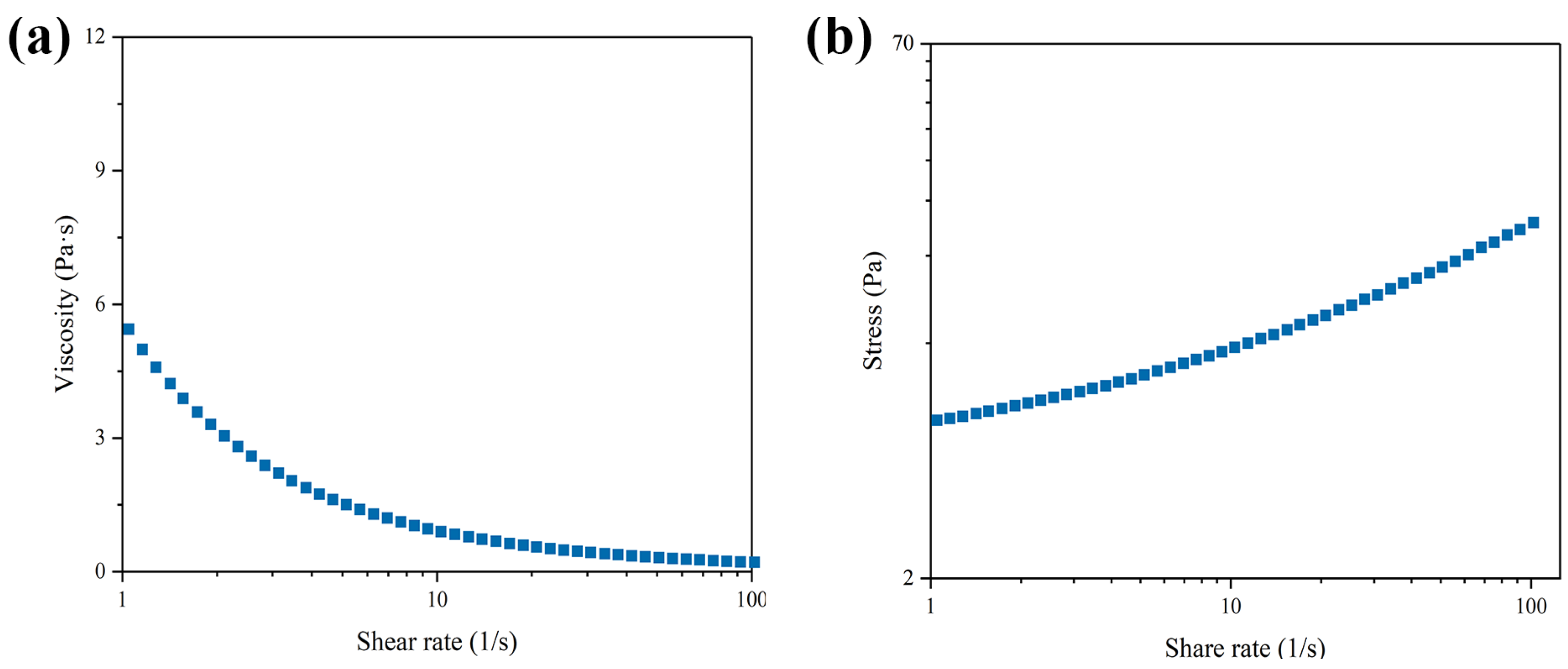
3.2. The Rheological Behavior of THRC Gels

The rheological behaviors of THRC gels were investigated using an Anton Paar rheometer. Within the range of 1 to 100 s−1, the viscosity of the THRC gels gradually decreased with an increasing shear rate, indicating the presence of shear-thinning behavior (Figure 1a). A double logarithmic plot of the shear rate versus viscosity in the range of 1 s−1 to 100 s−1 approximated a straight line with the equation y = 0.2864x + 0.7033 (Figure 1b). According to Equation (1), we obtain n = 0.2846, which is less than 1. These results demonstrated that the THRC gel exhibited non-Newtonian pseudoplastic behavior.

3.3. Skin Irritation Evaluation of THRC Gels on Rabbits

The skin irritation by a single administration assessment was performed by an in vivo rabbit test to evaluate the safety of THRC gels applied to the skin. The shaved rabbit skin was first divided into four parts. The scores of skin edema and erythema for the normal/broken area where the gel was applied were both “0” in each rabbit at any time point. PDII was calculated as 0 according to the scores, indicating that the skin irritation caused by the THRC gels was negligible (Table 3). The results indicated that the THRC gels exhibited no skin irritation for either normal or damaged skin.

3.4. DermaLab Combo Evaluation of the Performance of THRC Gels on the Repair of Sunburned Skin

A UV-mediated acute sunburned skin model was established according to the method described in Section 2.6. The THRC gels and commercialized collagen dressings were applied to the severe sunburned sites, respectively. Meanwhile, the untreated group was the model group.
A Combo dermatoscope was utilized to evaluate the representative macroscopic appearance of sunburn at different points (Figure 2). At the first day after model preparation, all groups except the blank group exhibited typical sunburn reactions, including redness, swelling, scaling, and epidermal peeling, with no significant differences between these experimental groups. On day 3, the THRC gels group showed complete scabs on the sunburned sites, which began to fall off. The commercially available products group had scabs with slight epidermal damage. The redness and swelling of the treated group basically subsided. The model group skin cracked due to inflammation and dryness. On day 5, the scabs of the THRC gels group were completely removed, and skin essentially returned to normal but there was slight dryness. The skin of the commercially available products group crusted and quickly fell off. The model group skin was crusted completely with slight redness. On day 7, the skin with THRC gels returned to normal. The scabs of the commercially available product group were removed completely but slight dryness remained, while the scabs of the model group were not completely removed. The results demonstrated that the sunburn model was successfully constructed. Compared to the untreated group, the treated groups indicated much accelerated repair of the skin wounds. Notably, THRC gels showed superior efficacy in repairing sun-damaged skin compared to commercialized collagen dressings.
Using the DermaLab Combo skin analysis system, the dermis density, EI, hydration, and TEWL of the skin were measured. One day after UV radiation, no significant difference was observed among the different groups expect the blank group, no matter the dermis density, EI, hydration, or TEWL (Figure 3b–e). The representative ultrasound scanning images as well as dermis density were obtained by skin ultrasonic inspection at different time points (Figure 3a,b). In the treated group with THRC gels, the dermis density increased along with the extending of experiment time, increasing to 32.70 ± 3.39, 44.53 ± 4.19, and 57.81 ± 6.11, respectively, at 3, 5, and 7 days, while the dermis density in the commercially available product group increased to 30.63 ± 3.92, 38.91 ± 5.70, and 48.33 ± 3.29. The differences between these two groups were significant (p < 0.01) at 7 days. In the model group, the dermis density decreased to 22.36 ± 4.53, 23.99 ± 2.46, and 24.45 ± 3.49, respectively, at 3, 5, and 7 days. The result was consistent with the photodamaging descriptions reported in the literature, which indicated the successful establishment of the sunburned animal model [11,36]. Along with the extending of experiment time, the EI in the THRC gels decreased to 7.91 ± 0.97, 4.46 ± 0.60, and 3.34 ± 0.46, respectively, at 3, 5, and 7 days. Meanwhile, in the commercially available product group, the reduction in EI was 9.62 ± 0.56, 7.16 ± 0.33, and 3.95 ± 0.82 at 3, 5, and 7 days (Figure 3c). The differences between these two groups were significant at 3 days. The hydration had a similar trend to that of dermis density (Figure 3b). At 3, 5, and 7 days, the increment of hydration in the THRC gel group was 136.61 ± 14.34 μS, 198.81 ± 18.42 μS, and 290.07 ± 13.36 μS, respectively. The hydration of the commercially available product group decreased to 91.95 ± 16.43 μS because of cracking skin at 3 days, but increased to 156.37 ± 20.67 μS and 221.33 ± 23.95 μS at 5 and 7 days. The hydration of skin showed significance along with the experiment time between these two groups. The TEWL paralleled EI (Figure 3e). At 5 days postoperatively, the TEWL of the THRC gels group showed no significant difference from the blank group. The data attained by the DermaLab Combo skin analysis system indicated that UV radiation impacts skin parameters including the dermis density, EI, hydration, and TEWL. Additionally, the THRC gels enhanced the skin conditions, while the efficiency was better than the commercialized collagen dressings.

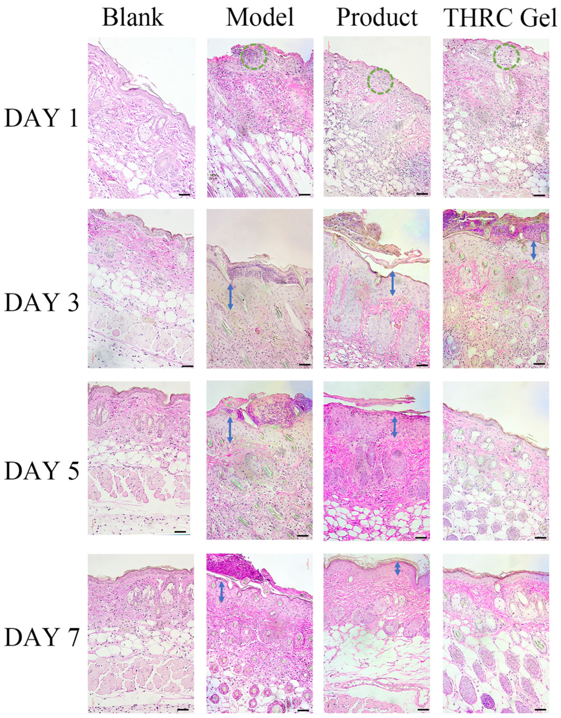
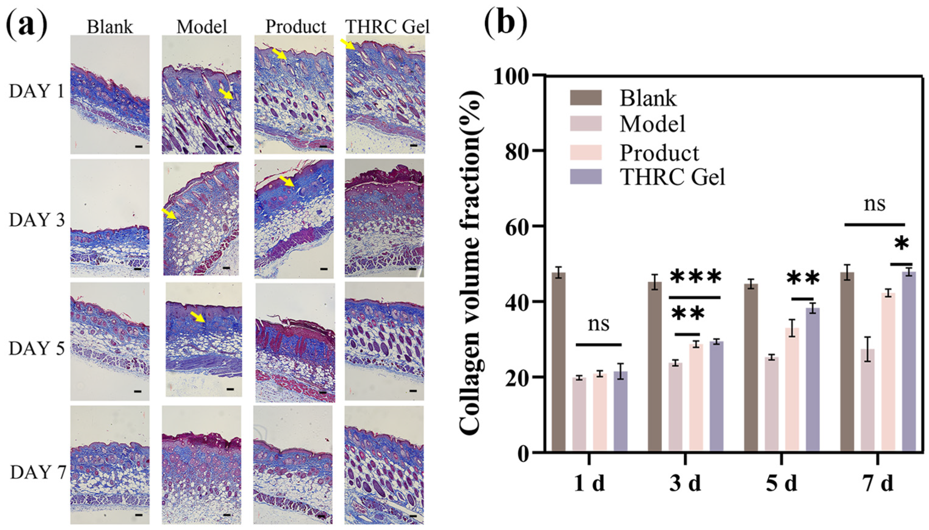
3.5. Histological Analysis to Investigate the Repair Ability of THRC Gels

H&E staining indicated the biocompatibility of the THRC gels, revealing no obvious inflammation or granuloma observed (Figure 4). Throughout the experiment, the blank group’s skin remained healthy, showing no inflammatory reactions. The epidermal layer remained continuous and intact, with cells arranged in an orderly fashion, and there were no spines at the epidermal–dermal junction. On the first day, the histopathological analysis revealed sunburn symptoms in all groups except the blank group. Keratinocytes were vacuolated, numerous sun-damaged cells were present in the stratum spinosum, spines were evident at the epidermal–dermal junction, and dermal edema along with inflammatory cell infiltration led to a blurred epidermal–dermal boundary. By day 3, notable epidermal changes occurred in the UV radiation groups. Increased melanocytes and inflammatory cells were evident in the epidermis, with the highest counts observed in the model group, followed by the commercially available product group, and then the THRC gels group. Keratinization was incomplete in the model group, whereas both the commercially available product and the THRC gels groups exhibited crusted keratinization. By day 5, minimal change was observed in the model group compared to day 3, except for the presence of scabs. The scabs of the commercially available product group began to fall off, accompanied by epidermis thinning, reduction in inflammatory cells, and subsiding dermal edema. The THRC gels group basically returned to a normal state. By day 7, the epidermis of the commercially available product group was slightly thickened compared to the blank group. The THRC gels group returned to normal. These results demonstrated that THRC gels displayed enhanced epithelization capability, accelerated wound healing, and improved healing efficacy, effectively reducing the risk of infection in sunburned skin.
Masson staining images of the untreated group showed abundant collagen debris and short aggregates, which could result from collagen polypeptide chain breakage and the degradation of collagen fibers due to severe UV irradiation (Figure 5a). In contrast, the treated groups both showed a significant increase in the amount of well-ordered dense collagen fibers with interspersing fibroblasts, which well resembled the normal dermis. Collagen volume fraction results further illustrated remarkable collagen deposition in the treated groups (Figure 5b). The results indicated that THRC gel promoted collagen deposition to accelerate sunburn wound healing.

3.6. Effect of THRC Gels on Hyp, MDA, SOD, and GSH Levels

Hyp, a characteristic amino acid in the collagen sequence, was measured to quantify collagen content. The content of Hyp in the blank group served as the standard. Compared to the blank group, the untreated group exhibited a significant reduction in Hyp content due to UV damage (Figure 6a). Following treatment with THRC gels, the Hyp content was increased, indicating collagen enrichment within 7 days. Similarly, treatment with the commercially available products also increased Hyp content. These results suggested that THRC gels possessed the power to accelerate the healing of severe UV-mediated skin injury by promoting the regeneration of dermal collagen. Additionally, the result was superior to those obtained with commercialized collagen dressings.
Exposure to UV radiation would generate large amounts of reactive oxygen species (ROS) and causes DNA damage in skin tissue, leading to oxidative stress [37,38]. MDA, a major product of lipid peroxidation, significantly increases due to UV radiation, resulting in the oxidation of macromolecules [39]. Thus, the MDA content was evaluated to discuss the degree of sunburn in skin tissue. Compared to the blank group, the model group showed a significant increase in MDA content (1.72 ± 0.14 nmol/g), suggesting that UV radiation induces lipid peroxidation in the skin (Figure 6b). Meanwhile, both of the two treated groups showed a significant decrease in MDA content, with the values of 1.22 ± 0.11 nmol/g (THRC gel) and 1.40 ± 0.05 nmol/g (product group). Notably, the MDA content in the THRC gel group was not significantly different from the blank group.
The activities of SOD and GSH were further investigated to study the effect of THRC gels on the repair of sunburned skin. Excessive UV radiation has also been reported to induce a decrease in SOD and GSH activity, disrupting the physiological equilibrium of the skin [11,39]. The activities of SOD and GSH decreased to 151.35 U/g and 74.67 ± 2.37 mg/g in the model group, respectively, displaying significant decreases compared to the blank group (Figure 6c,d). The SOD activity in the treated groups showed a significant difference compared to the model group. Specifically, the THRC gel group exhibited a SOD activity of 209.77 ± 5.07 U/g, while the product group showed an activity of 183.38 ± 2.39 U/g. The GSH activity significantly increased to 113.86 ± 3.02 (THRC gel) and 105.67 ± 4.67 (product). These findings suggest that THRC gels are more effective in revitalizing sunburned skin than the commercial product, likely by significantly increasing SOD and GSH levels while inhibiting MDA overexpression.

4. Discussion

Sunburn is arguably the most prevalent skin disease induced by UV irradiation particularly in high-latitude regions, and it has been considered as a crucial risk factor for skin cancer [10,40]. As the main component of the dermis, collagen degradation has been identified as a critical feature of acute sunburn. Consequently, THRC gels with high biocompatibility and bioactivity exhibit notable protective effects against UV-induced sunburn. Compared to traditional liquid dressings, gels treating sunburn wounds may offer enhanced efficiency. Initially, the gel forms a protective film on the wound surface, isolating it from bacterial contamination, preventing fluid loss, and fostering an optimal environment for cell proliferation, thereby accelerating tissue healing. Additionally, it adheres to the wound longer than a liquid dressing, facilitating healing. It maintains a moist environment on the wound surface and can be repeatedly administered in situ, avoiding new injuries due to dressing adhesion to the wound surface during dressing changes, and effectively alleviating patient discomfort [23]. Our results revealed that THRC gels represented a promising and more effective strategy for sunburn treatment.
We have reported the development of biocompatible and nonirritating gels composed of triple-helical recombinant collagen, HA, and glycerol for the accelerated healing of photodamaged acute skin wounds and use in cosmetics. The physicochemical evaluations of THRC gels over different time periods revealed their remarkable stability, attributed to the triple-helix conformation [19]. The skin irritation test of New Zealand rabbits further demonstrated that the THRC gels are highly safe and non-irritating.
The sunburn healing efficacy of THRC gels was examined using a UV-induced acute skin injury mice model, characterized by exfoliation, erythema, dryness, and epidermis damage. DermaLab Combo results demonstrated that the THRC gels may improve wound healing by accelerating inflammation [41]. Skin barrier damage is characterized by the diminishment of hydration and increment of TEWL, while the moisture content in the skin typically determines the health of skin [39]. THRC, HA, and glycerol are efficient moisturizers since the THRC gels can enhance the hydration of the stratum corneum, and film-forming properties contribute to mitigate TEWL. The histological analysis achieved a consistent result. H&E and Masson staining images indicated that the untreated group showed epidermal damage, cellular infiltration, and dermal inflammation. In contrast, 5 days of treatment with THRC gels resulted in the robust revitalization of sunburned mice skin, evidenced by the prominent acceleration of epithelization and collagen deposition, whereas the reported fish scale extract natural hydrogel showed persistent redness on day 5 [42]. Furthermore, THRC gels remarkably promoted the arrangement and deposition of collagen fibers, which could be accomplished by supplementing contact collagen, reducing denatured collagen, and stimulating collagen regeneration according to our previous research [31].
UV radiation causes the overexpression of ROS, which is a reduction product of oxygen in the body and could induce the activation of signaling molecules and pathways such as the nuclear factor-κB (NF-κB) and mitogen-activated protein kinase (MAPK) pathway [3]. As one of the main causes that could clear ROS generated by UV radiation, SOD is important in cellular overall defense mechanisms. GSH, a master antioxidant inside the cells, is essential for preventing oxidative cell damage, and could be detected and considered as an antioxidant activity index [39]. Additionally, long-term exposure to UV accelerates lipid peroxidation, resulting in increased MDA content. The THRC gels showed higher levels of SOD and GSH as well as lower MDA content, which meant the most potent antioxidant effect. Therefore, it could be deduced that THRC gels possessed a remarkable antioxidant performance, which was beneficial to prevent the photodamaging caused by UV radiation.

5. Conclusions

In conclusion, we have demonstrated a novel type of THRC gel for the enhanced healing of sunburned skin. The THRC gels displayed great stability and rheological behavior, showing no aggregation over a wide range of temperatures and time periods, with a pH that met the required standards. Non-invasive combo evaluations revealed that the THRC gels contributed to recovery of dermis density, EI, hydration, and TEWL values of sunburned skin to normal levels. The histological analysis further indicated that THRC gels significantly enhanced the repair of sunburned skin by preventing inflammatory reactions and promoting collagen regeneration. In addition, THRC gel demonstrated excellent resistance to oxidative stress, including the inhibition of lipid peroxidation and increased SOD and GSH activity. Notably, the THRC gels demonstrated a faster recovery rate compared to commercial collagen dressings, highlighting their superior efficacy in treating sunburn wounds. These highly biocompatible and bioactive THRC gels offer an effective treatment strategy for sunburn and daily skincare, with broad applications in cosmetics and dermatology.

Author Contributions

Y.Y.: Methodology, Software, Investigation, Data curation; B.T.: Methodology, Investigation, Visualization; Y.X.: Resources; P.L.: Supervision; L.Y.: Writing—review and editing, Supervision, Funding acquisition; J.X.: Writing—review and editing, Supervision, Project administration, Funding acquisition. All authors have read and agreed to the published version of the manuscript.

Funding

This research was funded by the National Natural Science Foundation of China (22074057 and 22205089), and the Natural Science Foundation of Gansu Province (20YF3FA025 and 23JRRA1096).

Institutional Review Board Statement

All animal experiments were performed with protocols approved by the ethics committee of the College of Chemistry and Chemical Engineering at Lanzhou University (No. G09, 20220711).

Informed Consent Statement

Not applicable.

Data Availability Statement

Data are available on request from the corresponding author.

Conflicts of Interest

The authors declare no conflicts of interest. The funders had no role in the design of the study; in the collection, analyses, or interpretation of data; in the writing of the manuscript; or in the decision to publish the results.

References

  1. Choi, S.-I.; Jung, T.-D.; Cho, B.-Y.; Choi, S.-H.; Sim, W.-S.; Han, X.; Lee, S.J.; Kim, Y.-C.; Lee, O.-H. Anti-photoaging effect of fermented agricultural by-products on ultraviolet B-irradiated hairless mouse skin. Int. J. Mol. Med. 2019, 44, 559–568. [Google Scholar] [CrossRef] [PubMed]
  2. Indirapriyadarshini, R.; Kanimozhi, G.; Natarajan, D.; Jeevakaruniyam, S.J. Andrographolide protects acute ultraviolet-B radiation-induced photodamages in the mouse skin. Arch. Dermatol. Res. 2023, 315, 1197–1205. [Google Scholar] [CrossRef] [PubMed]
  3. Wei, M.; He, X.; Liu, N.; Deng, H. Role of reactive oxygen species in ultraviolet-induced photodamage of the skin. Cell Div. 2024, 19, 1. [Google Scholar] [CrossRef] [PubMed]
  4. Zhang, X.; Feng, C.; Wang, S.; Wang, Y.; Fu, Z.; Zhang, Y.; Sun, H.; Xie, C.; Fu, Y.; Tao, J.; et al. A novel amphibian-derived peptide alleviated ultraviolet B-induced photodamage in mice. Biomed. Pharmacother. 2021, 136, 111258. [Google Scholar] [CrossRef] [PubMed]
  5. Wu, S.; Hu, Y.; Bai, W.; Zhao, J.; Huang, C.; Wen, C.; Deng, L.; Lu, D. Cyanidin-3-o-glucoside inhibits UVA-induced human dermal fibroblast injury by upregulating autophagy. Photodermatol. Photoimmunol. Photomed. 2019, 35, 360–368. [Google Scholar] [CrossRef] [PubMed]
  6. Daré, R.G.; Nakamura, C.V.; Ximenes, V.F.; Lautenschlager, S.O.S. Tannic acid, a promising anti-photoaging agent: Evidences of its antioxidant and anti-wrinkle potentials, and its ability to prevent photodamage and MMP-1 expression in L929 fibroblasts exposed to UVB. Free Radic. Biol. Med. 2020, 160, 342–355. [Google Scholar] [CrossRef] [PubMed]
  7. Zhang, L.; Liang, S.; Zhang, Z.; Wang, K.; Cao, J.; Yao, M.; Qin, L.; Qu, C.; Miao, J. Protective Effects of ζ-Carotene-like Compounds against Acute UVB-Induced Skin Damage. Int. J. Mol. Sci. 2023, 24, 13970. [Google Scholar] [CrossRef] [PubMed]
  8. Gilchrest, B.A. Photoaging. J. Investig. Dermatol. 2013, 133, E2–E6. [Google Scholar] [CrossRef] [PubMed]
  9. Fillon, M. Melanoma threats from ultraviolet exposure vary by state. CA Cancer J. Clin. 2020, 70, 233–235. [Google Scholar] [CrossRef] [PubMed]
  10. Young, A.R.; Claveau, J.; Rossi, A.B. Ultraviolet radiation and the skin: Photobiology and sunscreen photoprotection. J. Am. Acad. Dermatol. 2017, 76 (Suppl. 1), S100–S109. [Google Scholar] [CrossRef] [PubMed]
  11. Wang, J.; Qiu, H.; Xu, Y.; Gao, Y.; Tan, P.; Zhao, R.; Liu, Z.; Tang, Y.; Zhu, X.; Bao, C.; et al. The biological effect of recombinant humanized collagen on damaged skin induced by UV-photoaging: An in vivo study. Bioact. Mater. 2022, 11, 154–165. [Google Scholar] [CrossRef] [PubMed]
  12. Bukhari, S.N.A.; Roswandi, N.L.; Waqas, M.; Habib, H.; Hussain, F.; Khan, S.; Sohail, M.; Ramli, N.A.; Thu, H.E.; Hussain, Z. Hyaluronic acid, a promising skin rejuvenating biomedicine: A review of recent updates and pre-clinical and clinical investigations on cosmetic and nutricosmetic effects. Int. J. Biol. Macromol. 2018, 120 Pt B, 1682–1695. [Google Scholar]
  13. Wang, J.; Duan, X.; Zhong, D.; Zhang, M.; Li, J.; Hu, Z.; Han, F. Pharmaceutical applications of chitosan in skin regeneration: A review. Int. J. Biol. Macromol. 2024, 261 Pt 1, 129064. [Google Scholar] [CrossRef] [PubMed]
  14. Amorim, S.; Reis, C.A.; Reis, R.L.; Pires, R.A. Extracellular Matrix Mimics Using Hyaluronan-Based Biomaterials. Trends Biotechnol. 2021, 39, 90–104. [Google Scholar] [CrossRef] [PubMed]
  15. Assunção, M.; Yiu, C.H.K.; Wan, H.-Y.; Wang, D.; Ker, D.F.E.; Tuan, R.S.; Blocki, A. Hyaluronic acid drives mesenchymal stromal cell-derived extracellular matrix assembly by promoting fibronectin fibrillogenesis. J. Mater. Chem. B 2021, 9, 7205–7215. [Google Scholar] [CrossRef] [PubMed]
  16. Ku, H.J.; Park, J.H.; Kim, S.H.; Park, J.-W. Isocitrate dehydrogenase 2 deficiency exacerbates dermis damage by ultraviolet-B via ΔNp63 downregulation. Biochim. Biophys. Acta Mol. Basis Dis. 2018, 1864, 1138–1147. [Google Scholar] [PubMed]
  17. Fu, C.; Shi, S.; Tian, J.; Gu, H.; Yao, L.; Xiao, J. Non-denatured yak type I collagen accelerates sunburned skin healing by stimulating and replenishing dermal collagen. Biotechnol. Rep. 2023, 37, e00778. [Google Scholar] [CrossRef] [PubMed]
  18. Fu, C.; Shi, S.; Wei, N.; Fan, Y.; Gu, H.; Liu, P.; Xiao, J. Biocompatible Triple-Helical Recombinant Collagen Dressings for Accelerated Wound Healing in Microneedle-Injured and Photodamaged Skin. Cosmetics 2023, 10, 31. [Google Scholar] [CrossRef]
  19. Ilamaran, M.; Janeena, A.; Valappil, S.; Ramudu, K.N.; Shanmugam, G.; Niraikulam, A. A self-assembly and higher order structure forming triple helical protein as a novel biomaterial for cell proliferation. Biomater. Sci. 2019, 7, 2191–2199. [Google Scholar] [CrossRef] [PubMed]
  20. Aljghami, M.E.; Saboor, S.; Amini-Nik, S. Emerging Innovative Wound Dressings. Ann. Biomed. Eng. 2019, 47, 659–675. [Google Scholar] [CrossRef] [PubMed]
  21. Ghomi, E.R.; Khalili, S.; Khorasani, S.N.; Neisiany, R.E.; Ramakrishna, S. Wound dressings: Current advances and future directions. J. Appl. Polym. Sci. 2019, 136, 47738. [Google Scholar] [CrossRef]
  22. Kumar, M.; Kumar, D.; Garg, Y.; Mahmood, S.; Chopra, S.; Bhatia, A. Marine-derived polysaccharides and their therapeutic potential in wound healing application—A review. Int. J. Biol. Macromol. 2023, 253 Pt 6, 127331. [Google Scholar] [CrossRef] [PubMed]
  23. Shi, C.; Wang, C.; Liu, H.; Li, Q.; Li, R.; Zhang, Y.; Liu, Y.; Shao, Y.; Wang, J. Selection of Appropriate Wound Dressing for Various Wounds. Front. Bioeng. Biotechnol. 2020, 8, 182. [Google Scholar] [CrossRef] [PubMed]
  24. Andrade, T.D.A.; Heimfarth, L.; Santos, D.M.D.; Santos, M.R.V.D.; Albuquerque-Júnior, R.L.C.D.; Santos-Neto, A.G.D.; Araujo, G.R.S.D.; Lira, A.A.M.; Matos, S.S.; Frank, L.A.; et al. Hesperetin-Based Hydrogels Protect the Skin against UV Radiation-Induced Damage. AAPS PharmSciTech 2022, 23, 170. [Google Scholar] [CrossRef] [PubMed]
  25. Huixia, H.; Fan, Y.; Shanshan, Z.; Zhao, L.; Zaiman, L.; Linghui, Y.; Jianxi, X. Bone morphogenetic protein-2 loaded triple helix recombinant collagen-based hydrogels for enhancing bone defect healing. Biomed. Mater. 2024, 19, 035029. [Google Scholar]
  26. Panyachariwat, N.; Jimtaisong, A.; Saewan, N. Antioxidative Potentials of Eleutherine bulbosa Bulb and Its Utilization in Topical Cosmetic Emulsion. Cosmetics 2024, 11, 111. [Google Scholar] [CrossRef]
  27. QB/T 2872-2017; Light Industry Standard of the People’s Republic of China. Mask. Ministry of Industry and Information Technology of PRC: Beijing, China, 2017. Available online: https://www.chinesestandard.net/PDF.aspx/QBT2872-2017 (accessed on 5 August 2024).
  28. GB/T 13531.1-2008; National Standard of the People’s Republic of China. General Methods on Determination of Cosmetics—Determination of pH. General Administration of Quality Supervision, Inspection and Quarantine: Beijing, China, 2008. Available online: https://www.chinesestandard.net/PDF.aspx/GBT13531.1-2008 (accessed on 5 August 2024).
  29. Costa, G.M.D.A.; Campos, P.M.B.G.M. Development of Cosmetic Formulations Containing Olive Extract and Spirulina sp.: Stability and Clinical Efficacy Studies. Cosmetics 2024, 11, 68. [Google Scholar] [CrossRef]
  30. GB/T 16886.10-2017; National Standard of the People’s Republic of China. Biological Evaluation of Medical Devices—Part 10: Test for Irritation and Skin Sensitization. General Administration of Quality Supervision, Inspection and Quarantine of the People’s Republic of China: Beijing, China, 2017. Available online: https://www.chinesestandard.net/PDF.aspx/GBT16886.10-2017 (accessed on 5 August 2024).
  31. Li, J.; Huang, W.; He, H.; Shi, S.; Sun, X.; Xiao, J. Biocompatible and bioactive hydrogels of recombinant fusion elastin with low transition temperature for improved healing of UV-irradiated skin. J. Mater. Chem. B 2023, 11, 6975–6982. [Google Scholar] [CrossRef] [PubMed]
  32. Ariffin, N.H.M.; Hasham, R. Assessment of non-invasive techniques and herbal-based products on dermatological physiology and intercellular lipid properties. Heliyon 2020, 6, e03955. [Google Scholar] [CrossRef] [PubMed]
  33. Abdlaty, R.; Fang, Q. Skin erythema assessment techniques. Clin. Dermatol. 2021, 39, 591–604. [Google Scholar] [CrossRef] [PubMed]
  34. Shi, S.; Zhang, J.; Quan, S.; Yang, Y.; Yao, L.; Xiao, J. A highly biocompatible and bioactive transdermal nano collagen for enhanced healing of UV-damaged skin. Int. J. Biol. Macromol. 2024, 272 Pt. 1, 132857. [Google Scholar] [CrossRef]
  35. Wang, Q.; Yan, H.; Yao, L.; Xie, Y.; Liu, P.; Xiao, J. A highly bioactive THPC-crosslinked recombinant collagen hydrogel implant for aging skin rejuvenation. Int. J. Biol. Macromol. 2024, 266 Pt. 2, 131276. [Google Scholar] [CrossRef]
  36. Wang, Y.-J.; Chang, C.-C.; Lu, M.-E.; Wu, Y.-H.; Shen, J.-W.; Chiang, H.-M.; Lin, B.-S. Photoaging and Sequential Function Reversal with Cellular-Resolution Optical Coherence Tomography in a Nude Mice Model. Int. J. Mol. Sci. 2022, 23, 7009. [Google Scholar] [CrossRef] [PubMed]
  37. Biernacki, M.; Brzóska, M.M.; Markowska, A.; Gałażyn-Sidorczuk, M.; Cylwik, B.; Gęgotek, A.; Skrzydlewska, E. Oxidative Stress and Its Consequences in the Blood of Rats Irradiated with UV: Protective Effect of Cannabidiol. Antioxidants 2021, 10, 821. [Google Scholar] [CrossRef] [PubMed]
  38. Wölfle, U.; Esser, P.R.; Simon-Haarhaus, B.; Martin, S.F.; Lademann, J.; Schempp, C.M. UVB-induced DNA damage, generation of reactive oxygen species, and inflammation are effectively attenuated by the flavonoid luteolin in vitro and in vivo. Free Radic. Biol. Med. 2011, 50, 1081–1093. [Google Scholar] [CrossRef] [PubMed]
  39. Lu, W.; Kong, C.; Cheng, S.; Xu, X.; Zhang, J. Succinoglycan riclin relieves UVB-induced skin injury with anti-oxidant and anti-inflammatory properties. Int. J. Biol. Macromol. 2023, 235, 123717. [Google Scholar] [CrossRef] [PubMed]
  40. Shimizu, T. The Role of Macrophage Migration Inhibitory Factor (MIF) in Ultraviolet Radiation-Induced Carcinogenesis. Cancers 2010, 2, 1555–1564. [Google Scholar] [CrossRef] [PubMed]
  41. Xu, Q.; Torres, J.E.; Hakim, M.; Babiak, P.M.; Pal, P.; Battistoni, C.M.; Nguyen, M.; Panitch, A.; Solorio, L.; Liu, J.C. Collagen- and hyaluronic acid-based hydrogels and their biomedical applications. Mater. Sci. Eng. R Rep. 2021, 146, 100641. [Google Scholar] [CrossRef] [PubMed]
  42. Liu, Y.; Qin, D.; Wang, H.; Zhu, Y.; Bi, S.; Liu, Y.; Cheng, X.; Chen, X. Effect and mechanism of fish scale extract natural hydrogel on skin protection and cell damage repair after UV irradiation. Colloids Surf. B Biointerfaces 2023, 225, 113281. [Google Scholar] [CrossRef] [PubMed]
Figure 1. The rheological behavior of the THRC gel. (a) The viscosity measurement; (b) a double logarithmic plot of the shear rate versus viscosity.
Cosmetics 11 00138 g001
Figure 2. Combo dermatoscope images of the skin in blank, model, product, and THRC gel groups on days 1, 3, 5, and 7 at the magnification of 50×.
Cosmetics 11 00138 g002
Figure 3. The Combo non-invasive evaluation of the performance of THRC gels on the repair of sunburned skin. (a) Ultrasonic image; (b) dermis density; (c) EI; (d) hydration; and (e) TEWL of mice skin in different groups including blank, model (untreated), and two treatment groups (product and THRC gels) on days 1, 3, 5, and 7. * p < 0.05, ** p < 0.01, *** p < 0.001, ns > 0.05, n = 6.
Cosmetics 11 00138 g003
Figure 4. H&E staining to evaluate the repair ability of THRC gels on days 1, 3, 5, and 7. Green circles (apoptotic keratinocytes or sunburned cells); blue double arrows (the thickness of epidermis). Scale bar: 70 µm.
Cosmetics 11 00138 g004
Figure 5. Masson staining evaluation of the ability of THRC gel to promote collagen regeneration. Masson staining images (a) and collagen fraction volume (b) of the blank, model, product, and THRC gel groups on days 1, 3, 5, and 7. Yellow arrows (collagen fiber breakage). Scale bar: 40 µm. * p < 0.05, ** p < 0.01, *** p < 0.001, ns > 0.05, n = 3.
Cosmetics 11 00138 g005
Figure 6. The quantitative analysis of the effect of THRC gels on the repair of sunburned skin. The content of Hyp (a), MDA (b), SOD (c), and GSH (d) of the blank, model, product, and THRC gel groups on day 7. * p < 0.05, ** p < 0.01, *** p < 0.001, ns > 0.05, n = 3.
Cosmetics 11 00138 g006
Table 1. The main information of experimental groups.
GroupUV RadiationTreatment
blank--
model+-
product+0.1 g/day
THRC gel+0.1 g/day
Table 2. The stability of THRC gels at different times (n = 3).
TimepH (25 °C)Centrifugation TestHeat TestFreeze Test
0 w6.43 ± 0.03no significant
difference
no significant
difference
no significant
difference
4 w6.57 ± 0.05no significant
difference
no significant
difference
no significant
difference
8 w6.44 ± 0.08no significant
difference
no significant
difference
no significant
difference
12 w6.50 ± 0.04no significant
difference
no significant
difference
no significant
difference
Table 3. Results of the skin irritation test to the THRC gels (n = 3).
GroupScorePDIIThe Intensity of
Irritation
control00no irritating
normal00no irritating
damage00no irritating
positive control1.660.55mildly irritating
Disclaimer/Publisher’s Note: The statements, opinions and data contained in all publications are solely those of the individual author(s) and contributor(s) and not of MDPI and/or the editor(s). MDPI and/or the editor(s) disclaim responsibility for any injury to people or property resulting from any ideas, methods, instructions or products referred to in the content.

