Article

Trifluorometyl Phenethyl Mesalazine (TFM) Acts as an Antioxidant and Improves Facial Skin Wrinkles and Whitening

1
GNT Pharma Co., Ltd., Yongin 17096, Republic of Korea
2
Department of Systems Biology, Yonsei University College of Life Science and Biotechnology, Seoul 03722, Republic of Korea
3
Division of Life Sciences, College of Life Sciences, Korea University, Seoul 02841, Republic of Korea
*
Author to whom correspondence should be addressed.
Cosmetics 2024, 11(5), 158; https://doi.org/10.3390/cosmetics11050158
Submission received: 4 June 2024 / Revised: 30 August 2024 / Accepted: 10 September 2024 / Published: 13 September 2024

Abstract

:
Oxidative stress is a primary contributor to human skin aging, while antioxidants are known to mitigate skin damage related to aging processes. In this study, we investigated the possible action of trifluoromethyl phenethyl mesalazine (TFM), an antioxidizing agent, on skin aging processes, including skin wrinkles and pigmentation. Our data revealed that TFM exerted a strong free radical scavenging capability and notably inhibited melanin production. Moreover, TFM downregulated the UV-induced production of matrix metalloproteinase-1 and interleukin-6 in cultured human skin fibroblasts. Furthermore, in a clinical study with 24 women, TFM significantly reduced skin wrinkles and improved skin brightness compared to a placebo. These findings highlight the previously unrecognized effects of TFM on skin health by mitigating skin aging processes associated with oxidative damage.

1. Introduction

Human skin, like all other organs, undergoes intrinsic as well as extrinsic processes of aging. The clinical manifestations of intrinsic aging include dehydrated skin, fine wrinkles, and decreased suppleness, whereas those of extrinsic aging include thick and deep wrinkles and, in severe cases, loss of elasticity, pigmentary problems, and skin cancers [1,2]. Intrinsic and extrinsic aging processes of the skin overlap and are strongly related to the accumulation of free radicals, causing oxidative damage in the skin [3]. Although the molecular mechanisms of skin aging are still poorly understood, uncontrolled oxidative stress plays a pivotal role in its pathogenesis. Due to frequent exposure to oxidative stress, the skin has developed mechanisms to cope with the unwanted high concentrations of oxygen and oxygen metabolites [4]. Antioxidant defense, in which antioxidants and enzymes directly react with reactive oxygen species (ROS) to prevent them from reaching their biological target, is one of the most powerful skin protection systems [5]. Since free radicals are suggested to be the source of aging factors that induce wrinkle formation, antioxidants are widely used as topical supplements to prevent ROS-induced skin damage and enhance the antioxidant capacity of skin. Furthermore, given that oxidative stress promotes melanin production and thereby accelerates skin pigmentation [6], antioxidants can inhibit the reaction for melanin production and exert skin whitening properties [7,8]. Notably, a variety of cosmetic formulations have been designed to include antioxidizing agents to improve skin health by blocking the aging processes.
The predominant antioxidants in topical cosmetic formulations include non-enzymatic antioxidants such as coenzyme Q10 [9], phenolic compounds [10], vitamin E [11], and vitamin C [12] and its derivatives. There is substantial evidence that these antioxidants can modify and prevent the processes of chronological aging and photoaging [6,13]. However, many of these antioxidants have restricted applications due to various issues, including toxicity, low activity, and usage limits. TFM has been shown to act as a direct spin-trapping molecule, blocking oxidative stress and inflammation [14]. It also functions as an antagonist of microsomal prostaglandin E synthase-1 (mPGES-1) [14]. Given that TFM is a ROS scavenging agent that inhibits inflammatory responses [14,15], it is intriguing to investigate the potential actions of TFM on skin damage related to aging processes.
In this study, we demonstrated that TFM exhibits strong scavenging activity against UV-induced free radical production and has a protective effect on the UV-induced production of MMP-1 in human skin cells. Furthermore, TFM inhibited the melanin production induced by α-melanocyte-stimulating hormone (α-MSH) in melanoma cells. In addition, TFM treatment significantly reduced skin wrinkles as well as improved skin whitening in human clinical studies. These findings suggest that TFM, with its anti-aging and skin-whitening properties, has a great potential to be used as a key material in cosmeceuticals for human skin health.

2. Materials and Methods

2.1. Preparation of TFM

TFM was prepared by the high-pressure homogenization. Briefly, hydrogenated lecithin, water (32.95%), polyglyceryl-10 oleate (10%), glycerin (50%), 1,2-hexadiol (2%), and trifluoromethyl phenethyl mesalazine (0.05%) were agitated at 70 °C with a homomixer (Tokushu Kika Kogyo, Co., Ltd., Tokyo, Japan). This mixture was homogenized with a microfluidizer (Microfluidics, Westwood, MA, USA) at 70 °C.

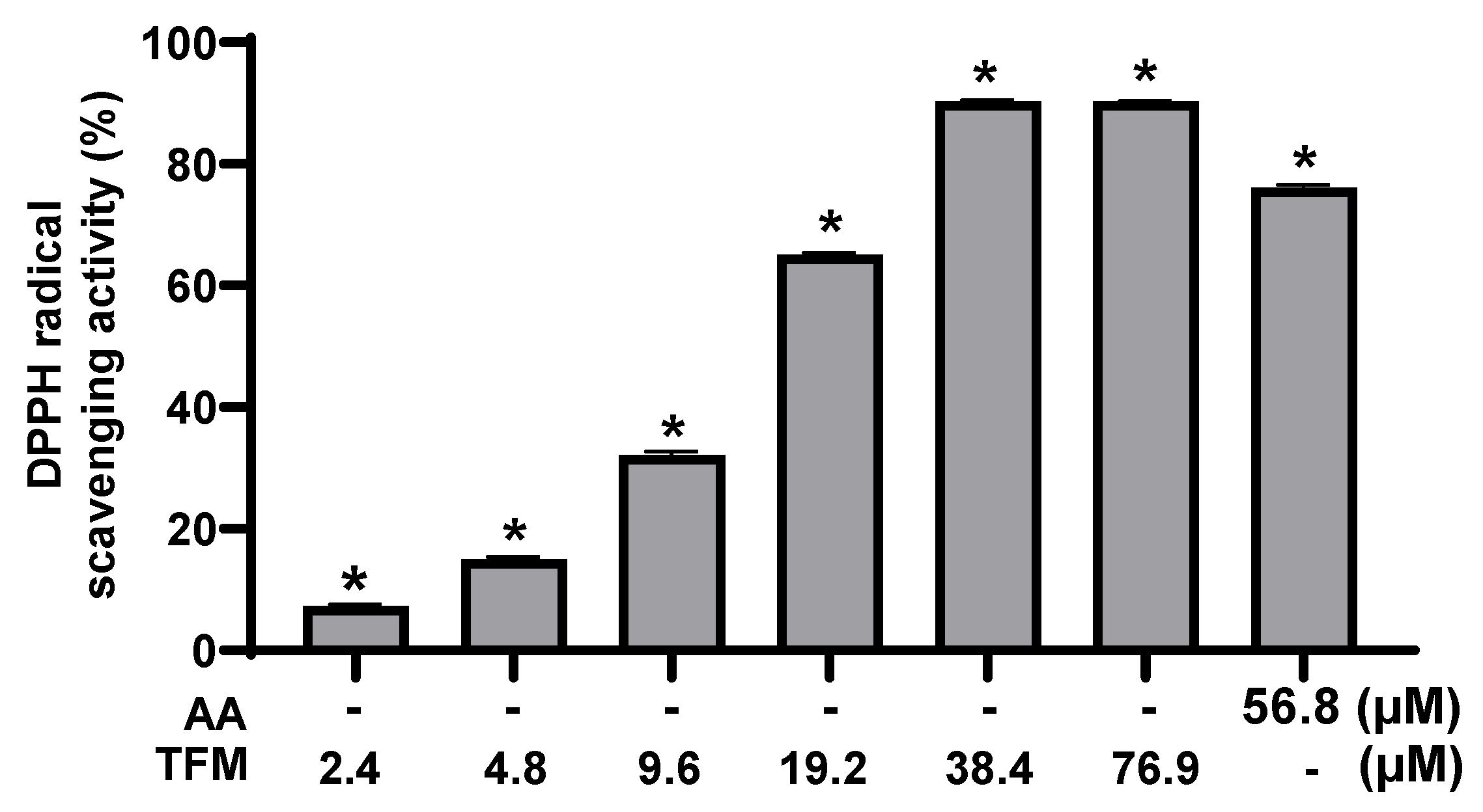
2.2. Radical Scavenging Assay Using 1,1-Diphenyl-2-Picrylhydrazyl (DPPH)

DPPH (1.2 mg/mL in CH3OH) (Sigma, St. Louis, MO, USA) was mixed with L-ascorbic acid (56.8 mM) or various concentrations of TFM, allowed to stay for 30 min under dark conditions, and then quantified for free radical generation with the use of a microplate reader (Molecular Devices, LCC, San Jose, CA, USA) as described previously [16].

2.3. Cell Culture

Normal human dermal fibroblasts (NHDFs) were purchased from Lonza (Allendale, NJ, USA). The cells were maintained in fibroblast basal medium (FBM, Lonza, NJ, USA) with 2% fetal bovine serum (FBS, Lonza, NJ, USA), 0.1% insulin (Lonza, NJ, USA), 0.1% recombinant human FGF-B (Lonza, NJ, USA), and 0.1% amphotericin-B (GA-1000) in a humidified 5% CO2 incubator at 37 °C. B16F1 melanoma cells were purchased from the ATCC (Manassas, VA, USA) and cultivated in DMEM containing 100 unit/mL penicillin-streptomycin and 10% FBS in a humidified 5% CO2 incubator at 37 °C.

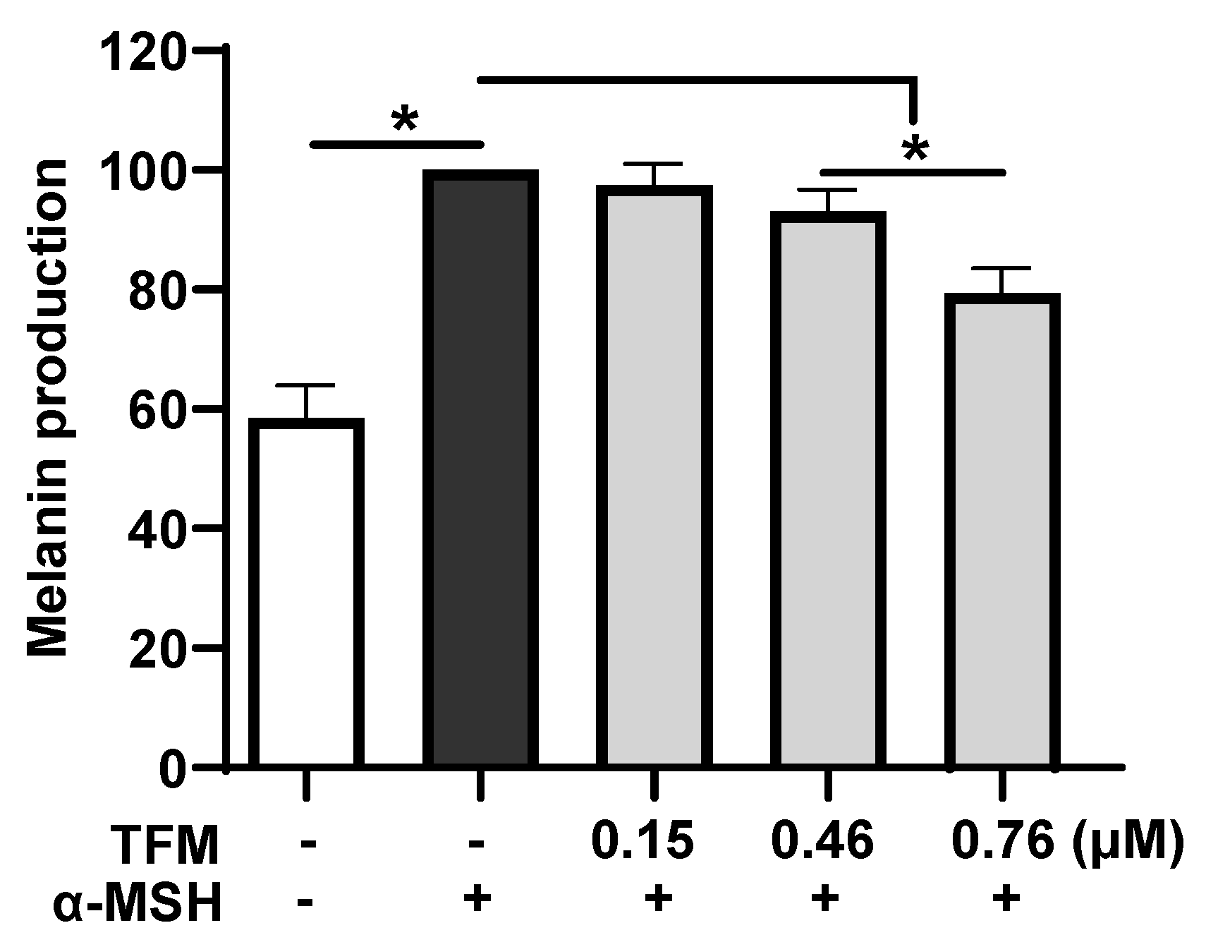
2.4. Quantitation of Cellular Melanin

B16F1 melanoma cells were treated for 72 h with 100 nM α-MSH in the absence or presence of various concentrations of TFM. Following the treatment, culture medium was collected and quantified for extracellular melanin using a microplate reader (Molecular Devices, LCC, CA, USA). For intracellular melanin, the cells were harvested and then treated with 1 N NaOH plus 10% DMSO at 65 °C for 1 h. The melanin content was quantified using a microplate reader (Molecular Devices, LCC, CA, USA). The melanin content was normalized to the quantity of cellular total protein.

2.5. Cell Viability Assay

NHDF cells (2 × 10⁴ cells/well in 24-well plates) were cultured in serum-free fibroblast basal medium containing indicated concentrations of TFM for 24 h. The effect of TFM on viability of NHDF cells was assessed with the use of an EZCYTOX kit (Dail-Lab Service, Seoul, Korea) in accordance with the manufacturer’s instructions.

2.6. Assay of Procollagen Production

NHDF cells (2 × 10⁴ cells/well in 24-well plates) in serum-free fibroblast basal medium were left untreated or treated with indicated concentrations of TFM for 24 h. Then, the supernatants were taken from each well and assayed for abundance of procollagen using a procollagen type I C-peptide enzyme immunoassay assay kit (Takara Bio Inc., Tokyo, Japan).

2.7. UV-Induced MMP-1 Production

Normal human dermal fibroblasts (NHDFs) cultivated in 24-well plates (3.0 × 10⁴ cells/well) were irradiated to ultraviolet B (UVB; 40 mJ). After 48 h of UVB irradiation, culture media were collected and quantified for abundance of MMP-1 by using an ELISA kit (Abcam, Cambridge, MA, USA).

2.8. Quantification of IL-6 Production

NHDF cells (1 × 10⁵ cells/well in a 12-well plate) were pre-treated with the indicated concentrations of TFM for 2 h, then exposed to UVB (30 mJ/cm2). The cells were then incubated in fresh culture media with the indicated concentrations of TFM. After 24 h of the treatment, culture supernatants were collected and assayed for IL-6 protein levels with the use of a human IL-6 ELISA kit (Invitrogen, Carlsbad, CA, USA), according to the manufacturer’s instructions.

2.9. In Vivo Skin Irritation Test

A clinical skin irritation test was performed at the P&K Skin Research Center (Seoul, Korea). This open-label study included thirty-three healthy participants, aged 20 to 50, who had no physical illnesses, including infectious skin diseases. TFM was attached to the IQ Ultimate chamber (Chemotechnique Diagnostics, Vellinge, Sweden) and was applied to the test subjects for 24 h. At 1 or 24 h following patch removal, skin reactivity and irritation were assessed using the Frosch and Kligman, Cosmetic Toiletry and Fragrance Association (CTFA) guideline, and Draize technique, respectively.

2.10. Clinical Study Designs for Skin Wrinkle and Whitening Assessment

The anti-wrinkle and anti-whitening efficacies of TFM were compared in 24 women (average age 48.5; two women who discontinued the study protocol were excluded). We carried out this study in accordance with the Guideline for Efficacy Evaluation of Functional Cosmetics published by the Korean Ministry of Food and Drug Safety (KMFDS), which requires a comparative assessment of vehicles in the absence or presence of active ingredients in a minimum of twenty participants for statistical analysis. This study was approved by the institutional review board of the Ethics Committee of the P&K Skin Research Centre, Seoul, Korea (approval no: P2001-844). Twenty-four women were treated twice daily for 8 weeks with either a topical TFM ampoule (32 ppm TFM-containing serum) or a placebo (serum only) on the right or left side of the face, respectively. Twenty-two participants completed the study, while the other two participants withdrew from the study due to personal reasons.

2.11. Assessment of Skin Wrinkles

Assessment of skin wrinkles was conducted through visual examination by dermatologists and instrumental examination of skin-replica images. For the visual assessment, skin examinations of the crow’s feet area were conducted with a double-blinded method based on the Guideline for Efficacy Evaluation of Functional Cosmetics (KMFDS). For the instrumental examination of skin-replica images, replicas were made using a silicone-based solution, according to the manufacturer’s instructions (Courage and Khazaka, Köln, Germany). These replicas were taken from the crow’s feet area and analyzed with a Skin Visiometer SV700 (Courage and Khazaka). This device analyzed the topography of the skin surface based on skin wrinkle parameters: skin roughness (R1), maximum roughness (R2), average roughness (R3), smoothness depth (R4), and arithmetic average roughness (R5). Wrinkles in the crow’s feet region were assessed using the PRIMOS CR (Canfield Imaging Systems, NJ, USA) by photographing the areas of interest. The images are subsequently analyzed for the Ra value (average roughness; mm). These measurements were performed at 0, 4, and 8 weeks after topical application of TFM or placebo.

2.12. Assessment of Skin Brightness

Analysis of skin brightness [brightness factor or luminance (L*) based on the definition by the International Commission on Illumination] was performed three times using a tristimulus colorimeter (Spectrophotometer CM700-d, Konica Minolta, Japan) at the same spot of the cheek area. The measurements were conducted at 0, 4, and 8 weeks after topical application of TFM or placebo. Melanin index (M.I.) were measured at 0, 4, and 8 weeks after TFM or placebo treatment with use of Mexameter® MX18 (Courage and Khazaka Electronic GmbH, Cologne, Germany).

2.13. Statistical Analysis

The significance of the measured values was determined using SPSS software (version 19.0) (SPSS Inc., Chicago, IL, USA). Following repeated measurements, the data were analyzed by analysis of variance (ANOVA) with a Bonferroni correction. The Friedman test and Mann–Whitney U test was used to examine within-group data and between-group data, respectively. A p-value less than 0.05 was considered statistically significant.

3. Results

3.1. Antioxidant Capacity of TFM

To investigate a possible action of TFM on oxidative damage, we assessed its potential to scavenge free radicals using the DPPH· scavenging assay, a widely recognized method for evaluating antioxidant capacity in diverse samples [17]. Our data revealed that TFM significantly reduced the generation of DPPH· free radicals with an IC50 of 14.7 mM (Figure 1). These results suggest that TFM is an effective scavenger of free radicals, thereby functioning as an antioxidizing agent.

3.2. Effect of TFM on α-MSH-Induced Melanin Production

Next, given the strong free-radical scavenging activity of TFM, we examined its effect on melanin production. It has been shown that free radical generation can lead to abnormal melanin production [18,19]. Our data indicated that α-MSH treatment increased melanin production in B16 cells, and this increase was reduced by TFM (Figure 2). These results suggest that TFM downregulates the α-MSH-induced production of melanin in the cells.

3.3. Effect of TFM on the UV-Induced Production of MMP-1

Multiple lines of evidence indicate that UV irradiation increases the production and release of MMPs in skin cells through ROS generation [20,21] and that MMPs play a pivotal role in the breakdown of extracellular matrix proteins in human skin [22]. Therefore, we examined whether TFM could modulate UV-induced MMP-1 production in NHDF cells. UV irradiation of NHDF cells led to an increased abundance of MMP-1 protein in the media, and this increase was significantly reduced by treating the cells with TFM (Figure 3). These results suggest that TFM negatively modulates the UV-induced production of MMP-1 in normal human skin cells. Interestingly, TFM did not affect the production of procollagen, a major substrate of MMP-1, in the cells under basal conditions (Figure S1).
Next, given that UV-induced oxidative stress is a well-known inducer of skin inflammation [23], we tested the potential action of TFM on the UV-induced generation of interleukin-6 (IL-6), a proinflammatory cytokine, in NHDF cells. UV exposure increased the production of IL-6 in the human skin cells, and this increase was significantly reduced after TFM treatment (Figure 4). These results indicate that TFM negatively regulates the inflammatory response following UV exposure in skin cells.

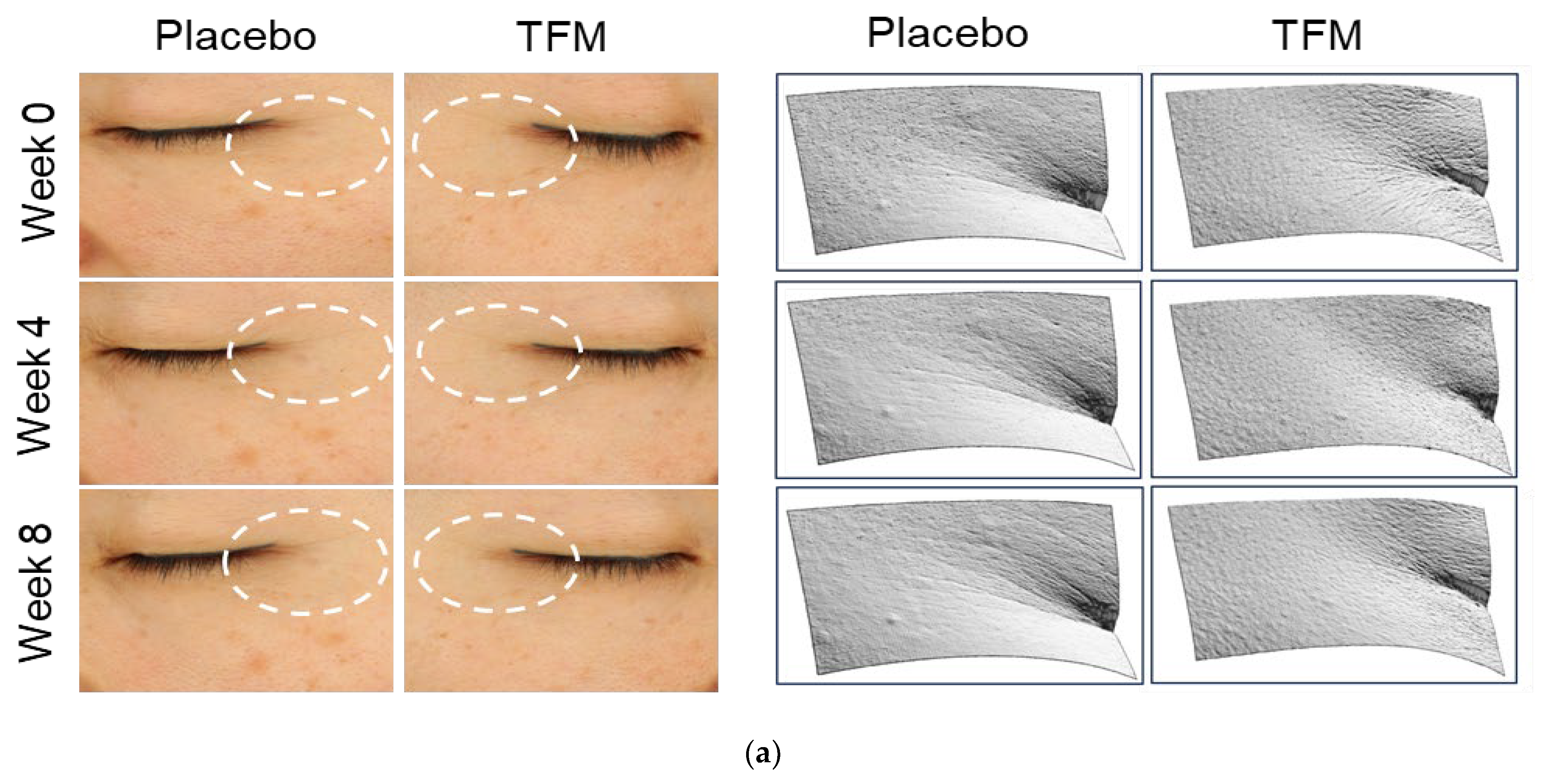
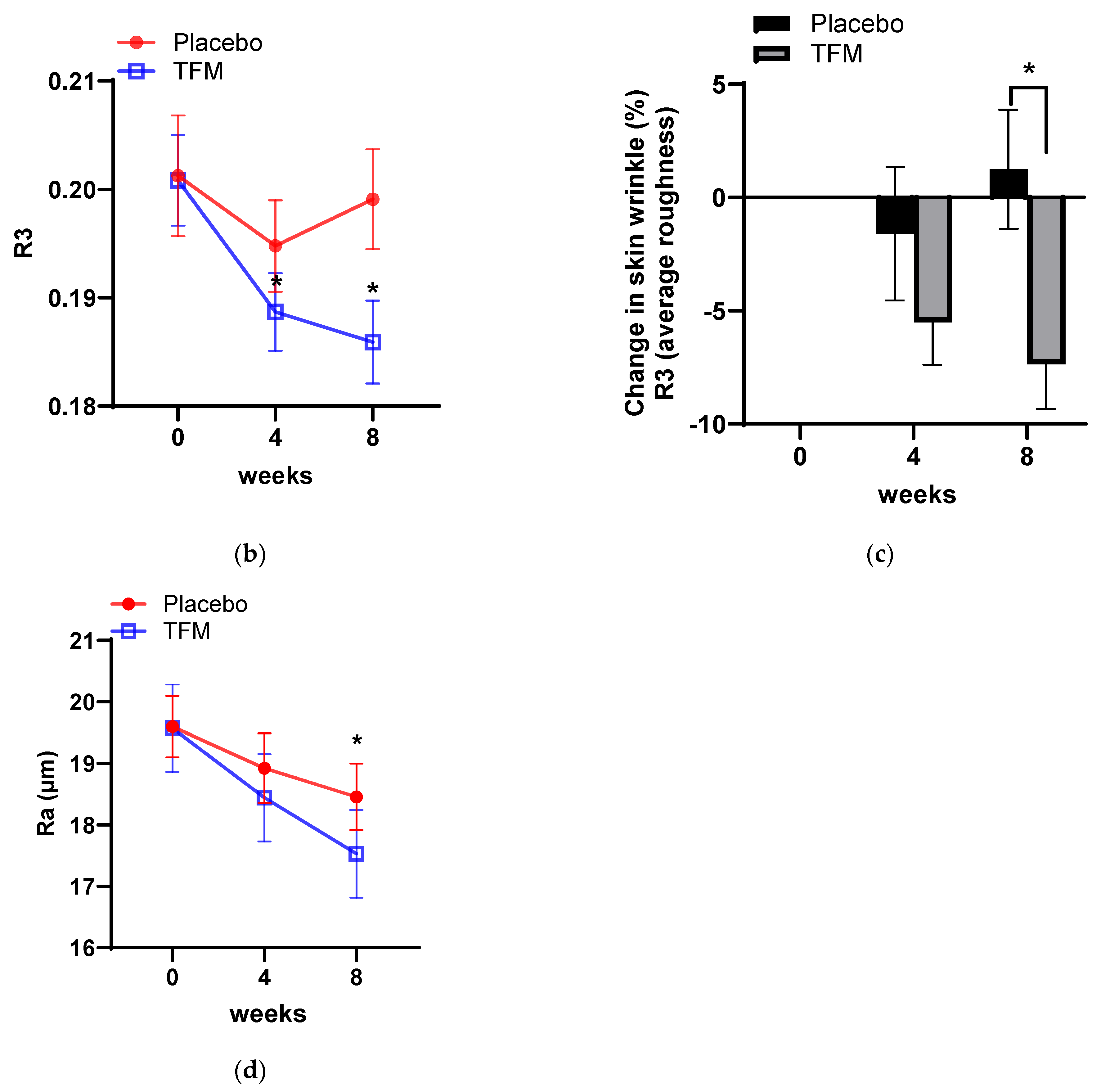
3.4. Clinical Effects of TFM on Skin Wrinkle Changes

Next, we decided to examine the possible effects of TFM on wrinkles around the eyes of human subjects by performing image analysis using replica and visiometer techniques. We found that TFM treatment resulted in a significant improvement in skin wrinkle changes compared to placebo administration (Figure 5A). The visiometer data of eye wrinkles demonstrated that the average roughness R3 was significantly improved in TFM-treated groups at 4 and 8 weeks compared with baseline, whereas no significant improvement was shown in placebo groups (Figure 5B). After 8 weeks of the treatment, there was a significant improvement in the R3 parameter (average roughness) in TFM-treated groups compared to placebo treatment (Figure 5C). Other parameters of skin wrinkles (R1, R2, R4, R5) were also significantly improved after 8 weeks of TFM treatment, compared to before treatment (Table 1). Furthermore, the images of eye wrinkle analysis revealed that the average roughness in the TFM treatment showed significantly higher values compared to the placebo treatment (Figure 5D). Collectively, these results suggested that TFM treatment reduces skin wrinkles in human subjects.

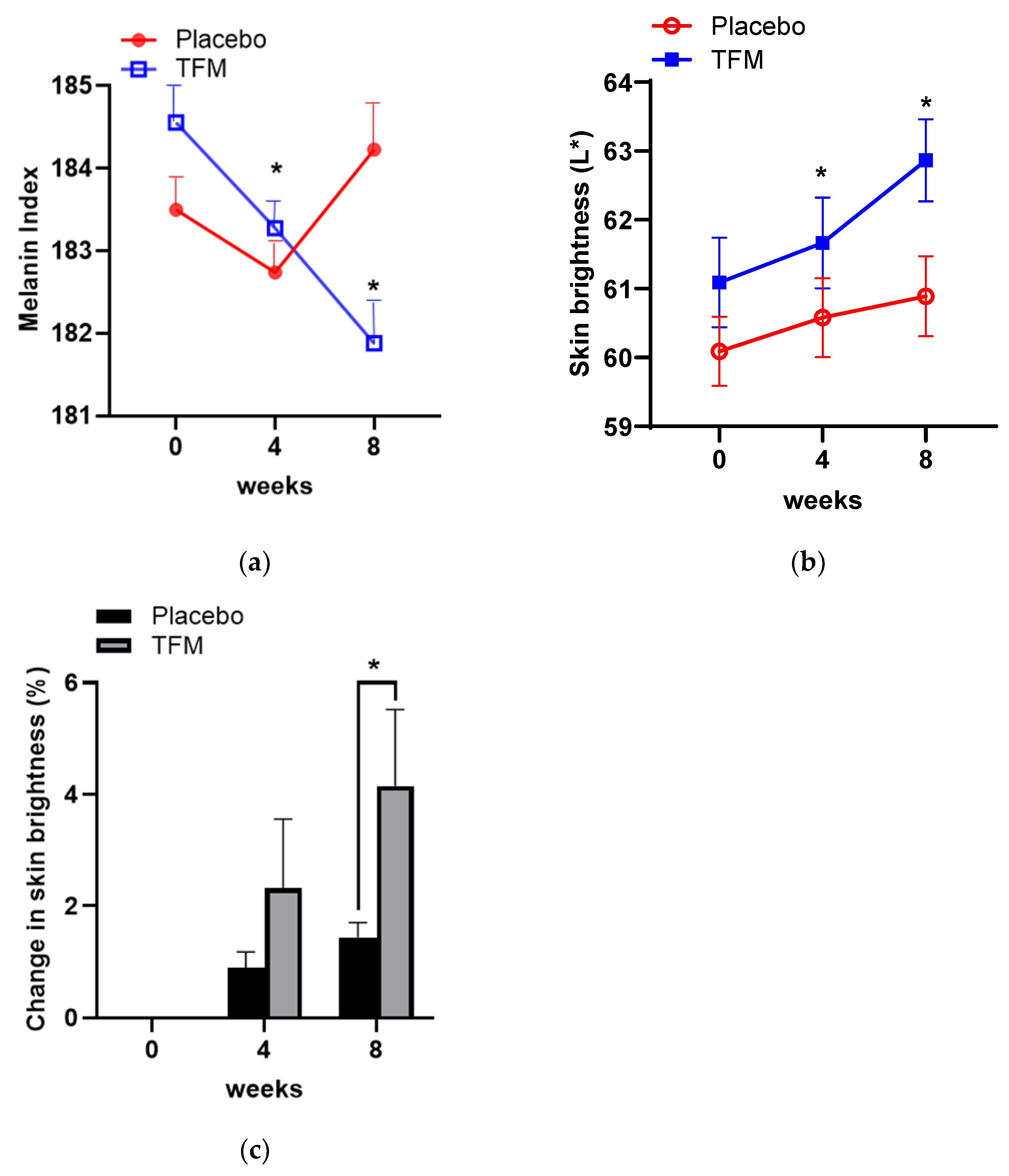
3.5. Effects of TFM on Skin Whitening

Next, to better understand the action of TFM on skin whitening, we investigated whether TFM could affect the abundance of melanin in the skin of human subjects. We assessed skin melanin changes at 4 and 8 weeks after TFM treatment with a Mexameter. Our results indicated that TFM treatment markedly decreased the melanin index (MI) values compared to placebo (Figure 6A). In separate experiments, we measured the L* value, which is known as the most sensitive parameter of the trichromatic values to skin color change [24]. We observed significant enhancement in skin brightness (L* scores) in human subjects treated with TFM for both 4 and 8 weeks (Figure 6B).

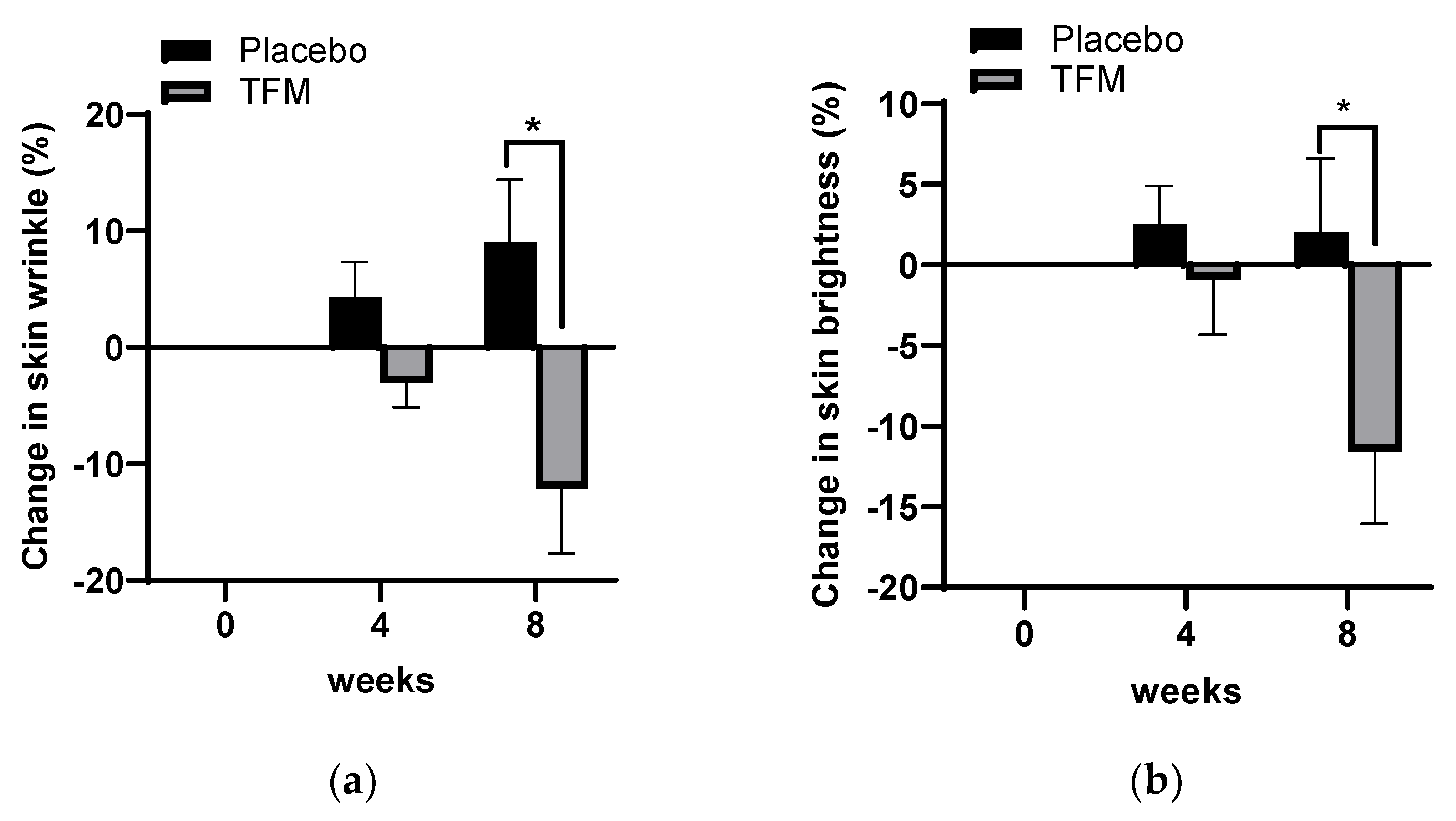
3.6. Investigator’s Assessments on Skin Wrinkles and Brightness

Next, we carried out a visual assessment analysis of skin wrinkles and brightness after 4 or 8 weeks of treatment with TFM. TFM treatment for 8 weeks significantly improved both skin wrinkles (Figure 7A) and skin whitening (Figure 7B).

3.7. In Vivo Evaluation of Skin Compatibility

We, next, investigated the skin compatibility of TFM in humans to evaluate potential irritative events resulting from topical application of TFM. In the in vivo skin irritation test, TFM did not cause any irritation to the test subjects. Additionally, none of the test participants showed erythema, edema, or papules on their skin following TFM treatment, suggesting that it is safe for topical application. Additionally, TFM did not affect cell viability in the in vitro cytotoxicity test using NHDF (Figure S2).

4. Discussion

In this study, our findings highlight the antioxidative and protective effects of TFM on human skin, demonstrating significant improvements in visible signs of skin aging, including wrinkles and pigmentation. The skin, serving as the largest organ and the forefront barrier against environmental challenges, is incessantly exposed to factors that contribute to oxidative stress and, consequently, aging [4]. This stress, compounded by UV radiation, leads to ROS production, which in turn accelerates the breakdown of the structural integrity of the skin, manifesting as wrinkles, pigmentation anomalies, and diminished elasticity [25,26,27]. In this study, we present the potential of TFM as a novel intervention to counteract these unwanted effects.
The capability of TFM to scavenge free radicals underscores its efficacy as a potent antioxidative agent. This action of TFM is particularly significant given the pivotal role of ROS in initiating and propagating the molecular events that facilitate skin aging [4]. By neutralizing free radicals, TFM offers a direct mechanism to prevent the cascade of oxidative damage that compromises the functionality of skin cells and the integrity of the extracellular matrix. Furthermore, the ability of TFM to inhibit melanin production induced by α-MSH is noteworthy. α-MSH, by activating the melanocortin 1 receptor, triggers melanogenesis in melanocytes [28]. Overproduction of melanin in melanocytes and the resulting hyperpigmentation of skin cause a variety of unwanted skin conditions, including melasma, freckles, age spots, solar lentigines, and a variety of hyperpigmentation syndromes [29,30]. Thus, hyperpigmentation is a common sign of skin aging and can be exacerbated by UV exposure [29]. Indeed, through ROS-related signaling, α-MSH and UV radiation have been shown to upregulate the translation of tyrosinase, thereby enhancing melanin overproduction [31]. Antioxidants and ROS scavengers are deemed critical for reducing hyperpigmentation or preventing UV-induced melanogenesis [32]. Our findings, indicating a reduction in α-MSH-dependent melanin production, suggest that TFM employs a two-pronged strategy in skincare by combating oxidative stress and inhibiting the enzymatic pathways leading to pigmentation. This is especially relevant for aging skin plagued by age spots and uneven skin tone [33].
The clinical effectiveness of TFM in diminishing skin wrinkles and enhancing skin luminosity further cements its role as a potent anti-aging agent. These effects, likely mediated by its antioxidative and anti-inflammatory properties, align with the theoretical perspective that links oxidative stress and inflammation to the aging phenotype. The observed improvements in skin texture and pigmentation support the argument that tackling oxidative stress can significantly influence the reversal or slowing of the signs of aging. These findings hold considerable implications for the cosmeceutical industry, which is in constant pursuit of effective and safe ingredients that provide anti-aging benefits without adverse effects [34]. Regarding the safety of TFM, it is noteworthy that the topical application of TFM does not cause skin irritation or other apparent side effects. Additionally, it was previously reported that TFM does not cause gastric damage [14], which is one of the most reported side effects of mesalazine [35].
The limitations of current antioxidants, including their stability and potential irritation effects, necessitate the exploration of new compounds [36,37]. TFM, with its antioxidative, anti-melanogenic, and anti-inflammatory properties, emerges as an appealing novel addition. Its efficacy in a clinical setting over an 8-week period in this study highlights its suitability for prolonged use in skincare formulations. Additionally, this research into TFM contributes to the ongoing exploration of the mechanisms behind skin aging. By detailing the role of TFM in modulating UV-induced MMP-1 production, the study enriches our understanding of how antioxidants, including TFM, can prevent oxidative stress from disrupting skin homeostasis, leading to collagen breakdown and, consequently, skin laxity. Nevertheless, while the results presented are promising, they underscore the need for further investigation. The limitations of this study, including its duration and sample size, point to the necessity for larger-scale, long-term studies to fully understand TFM’s safety, efficacy, and mechanism of action. Comparative analyses with existing antioxidants would also be beneficial in situating TFM within the wider landscape of skincare solutions.
In conclusion, TFM emerges as a novel and efficacious agent for anti-aging cosmeceutical applications, with significant antioxidative, anti-melanogenic, and anti-wrinkle properties. Its capacity to improve skin health by addressing oxidative damage and melanin production suggests a multifaceted role in skincare regimens aimed at combating aging. As the quest for effective anti-aging solutions persists, the role of TFM in skin health and its potential integration into skincare practices merit further investigation. Ongoing and future studies will provide deeper insight into the full potential of TFM, providing a scientific basis for its application in addressing skin aging and improving skin aesthetics.

5. Conclusions

This study has demonstrated that TFM exhibits its antioxidative, anti-melanogenic, and anti-wrinkle properties, making it a good candidate for cosmeceutical applications for preventing skin aging. TFM effectively scavenges free radicals, inhibits melanin production, and reduces UV-induced skin damage, along with visible improvements in wrinkle reduction as well as enhanced skin brightness. These findings suggest that TFM plays a crucial role in skincare regimens designed to prevent the adverse effects of oxidative stress and aging.

Supplementary Materials

The following supporting information can be downloaded at: https://www.mdpi.com/article/10.3390/cosmetics11050158/s1, Figure S1: Effects of TFM on pro-collagen production in NHDF cells.; Figure S2: Effects of TFM on cell viability.

Author Contributions

Investigation, J.M.L.; writing—original draft preparation, S.W.; writing—review and editing, E.-J.C.; software, J.H.L.; methodology, J.-H.S.; review, Y.J.O.; resources, B.J.G. All authors have read and agreed to the published version of the manuscript.

Funding

This research received no external funding.

Institutional Review Board Statement

This study was approved by the Institutional Review Board of P&K Research Center (P2001-844).

Informed Consent Statement

Informed consent was obtained from all subjects involved in the study.

Data Availability Statement

The original contributions presented in the study are included in the article/Supplementary Materials, further inquiries can be directed to the corresponding author/s.

Acknowledgments

This work was supported partially by a Korea University grant (E.-J.C.).

Conflicts of Interest

The authors are employees of GNT Pharma Co., Ltd. The authors declare that the research was conducted in the absence of any commercial or financial relationships that could be construed as a potential conflict of interest. The funders had no role in the design of the study; in the collection, analyses or interpretation of data; in the writing of the manuscript; or in the decision to publish the results.

References

  1. Krutmann, J.; Schikowski, T.; Morita, A.; Berneburg, M. Environmentally-Induced (Extrinsic) Skin Aging: Exposomal Factors and Underlying Mechanisms. J. Investig. Dermatol. 2021, 141, 1096–1103. [Google Scholar] [CrossRef] [PubMed]
  2. Zhang, S.; Duan, E. Fighting against Skin Aging: The Way from Bench to Bedside. Cell Transplant. 2018, 27, 729–738. [Google Scholar] [CrossRef] [PubMed]
  3. Hanson, K.M.; Clegg, R.M. Observation and quantification of ultraviolet-induced reactive oxygen species in ex vivo human skin. Photochem. Photobiol. 2002, 76, 57–63. [Google Scholar] [CrossRef] [PubMed]
  4. Rinnerthaler, M.; Bischof, J.; Streubel, M.K.; Trost, A.; Richter, K. Oxidative stress in aging human skin. Biomolecules 2015, 5, 545–589. [Google Scholar] [CrossRef] [PubMed]
  5. Pugliese, P.T. The skin’s antioxidant systems. Dermatol. Nurs. 1998, 10, 401–416; quiz 417–418. [Google Scholar]
  6. Masaki, H. Role of antioxidants in the skin: Anti-aging effects. J. Dermatol. Sci. 2010, 58, 85–90. [Google Scholar] [CrossRef]
  7. Kang, S.H.; Jeon, Y.D.; Cha, J.Y.; Hwang, S.W.; Lee, H.Y.; Park, M.; Lee, B.R.; Shin, M.K.; Kim, S.J.; Shin, S.M.; et al. Antioxidant and skin-whitening effects of aerial part of Euphorbia supina Raf. Extract. BMC Complement. Altern. Med. 2018, 18, 256. [Google Scholar] [CrossRef]
  8. Kim, Y.J. Antimelanogenic and antioxidant properties of gallic acid. Biol. Pharm. Bull. 2007, 30, 1052–1055. [Google Scholar] [CrossRef]
  9. Knott, A.; Achterberg, V.; Smuda, C.; Mielke, H.; Sperling, G.; Dunckelmann, K.; Vogelsang, A.; Kruger, A.; Schwengler, H.; Behtash, M.; et al. Topical treatment with coenzyme Q10-containing formulas improves skin’s Q10 level and provides antioxidative effects. Biofactors 2015, 41, 383–390. [Google Scholar] [CrossRef]
  10. Kim, H.H.; Jeong, S.H.; Park, M.Y.; Bhosale, P.B.; Abusaliya, A.; Kim, H.W.; Seong, J.K.; Ahn, M.; Park, K.I.; Kim, G.S. Antioxidant effects of phenolic compounds in through the distillation of Lonicera japonica & Chenpi extract and anti-inflammation on skin keratinocyte. Sci. Rep. 2023, 13, 20883. [Google Scholar] [CrossRef]
  11. Barbosa, E.; Faintuch, J.; Machado Moreira, E.A.; Goncalves da Silva, V.R.; Lopes Pereima, M.J.; Martins Fagundes, R.L.; Filho, D.W. Supplementation of vitamin E, vitamin C, and zinc attenuates oxidative stress in burned children: A randomized, double-blind, placebo-controlled pilot study. J. Burn Care Res. 2009, 30, 859–866. [Google Scholar] [CrossRef] [PubMed]
  12. Enescu, C.D.; Bedford, L.M.; Potts, G.; Fahs, F. A review of topical vitamin C derivatives and their efficacy. J. Cosmet. Dermatol. 2022, 21, 2349–2359. [Google Scholar] [CrossRef] [PubMed]
  13. Davinelli, S.; Bertoglio, J.C.; Polimeni, A.; Scapagnini, G. Cytoprotective Polyphenols Against Chronological Skin Aging and Cutaneous Photodamage. Curr. Pharm. Des. 2018, 24, 99–105. [Google Scholar] [CrossRef]
  14. Shin, J.H.; Lee, Y.A.; Lee, J.K.; Lee, Y.B.; Cho, W.; Im, D.S.; Lee, J.H.; Yun, B.S.; Springer, J.E.; Gwag, B.J. Concurrent blockade of free radical and microsomal prostaglandin E synthase-1-mediated PGE2 production improves safety and efficacy in a mouse model of amyotrophic lateral sclerosis. J. Neurochem. 2012, 122, 952–961. [Google Scholar] [CrossRef]
  15. Chun, H.; Im, H.; Kang, Y.J.; Kim, Y.; Shin, J.H.; Won, W.; Lim, J.; Ju, Y.; Park, Y.M.; Kim, S.; et al. Severe reactive astrocytes precipitate pathological hallmarks of Alzheimer’s disease via H(2)O(2)(-) production. Nat. Neurosci. 2020, 23, 1555–1566. [Google Scholar] [CrossRef] [PubMed]
  16. Wang, F.; Tan, J.; Jiang, R.; Li, F.; Zheng, R.; Yu, L.; Luo, L.; Zheng, Y. DPPH Radical Scavenging Activity of New Phenolics from the Fermentation Broth of Mushroom Morehella importuna. Molecules 2023, 28, 4760. [Google Scholar] [CrossRef] [PubMed]
  17. Baliyan, S.; Mukherjee, R.; Priyadarshini, A.; Vibhuti, A.; Gupta, A.; Pandey, R.P.; Chang, C.M. Determination of Antioxidants by DPPH Radical Scavenging Activity and Quantitative Phytochemical Analysis of Ficus religiosa. Molecules 2022, 27, 1326. [Google Scholar] [CrossRef]
  18. Baumann, L. How to Use Oral and Topical Cosmeceuticals to Prevent and Treat Skin Aging. Facial Plast. Surg. Clin. North Am. 2018, 26, 407–413. [Google Scholar] [CrossRef]
  19. Maddodi, N.; Jayanthy, A.; Setaluri, V. Shining light on skin pigmentation: The darker and the brighter side of effects of UV radiation. Photochem. Photobiol. 2012, 88, 1075–1082. [Google Scholar] [CrossRef]
  20. Pittayapruek, P.; Meephansan, J.; Prapapan, O.; Komine, M.; Ohtsuki, M. Role of Matrix Metalloproteinases in Photoaging and Photocarcinogenesis. Int. J. Mol. Sci. 2016, 17, 868. [Google Scholar] [CrossRef]
  21. Wlaschek, M.; Heinen, G.; Poswig, A.; Schwarz, A.; Krieg, T.; Scharffetter-Kochanek, K. UVA-induced autocrine stimulation of fibroblast-derived collagenase/MMP-1 by interrelated loops of interleukin-1 and interleukin-6. Photochem. Photobiol. 1994, 59, 550–556. [Google Scholar] [CrossRef] [PubMed]
  22. Kahari, V.M.; Saarialho-Kere, U. Matrix metalloproteinases in skin. Exp. Dermatol. 1997, 6, 199–213. [Google Scholar] [CrossRef] [PubMed]
  23. Brand, R.M.; Wipf, P.; Durham, A.; Epperly, M.W.; Greenberger, J.S.; Falo, L.D., Jr. Targeting Mitochondrial Oxidative Stress to Mitigate UV-Induced Skin Damage. Front. Pharmacol. 2018, 9, 920. [Google Scholar] [CrossRef] [PubMed]
  24. Huang, W.S.; Wang, Y.W.; Hung, K.C.; Hsieh, P.S.; Fu, K.Y.; Dai, L.G.; Liou, N.H.; Ma, K.H.; Liu, J.C.; Dai, N.T. High correlation between skin color based on CIELAB color space, epidermal melanocyte ratio, and melanocyte melanin content. PeerJ 2018, 6, e4815. [Google Scholar] [CrossRef] [PubMed]
  25. Kammeyer, A.; Luiten, R.M. Oxidation events and skin aging. Ageing Res. Rev. 2015, 21, 16–29. [Google Scholar] [CrossRef]
  26. Pinnell, S.R. Cutaneous photodamage, oxidative stress, and topical antioxidant protection. J. Am. Acad. Dermatol. 2003, 48, 1–19. [Google Scholar] [CrossRef]
  27. Scharffetter-Kochanek, K.; Brenneisen, P.; Wenk, J.; Herrmann, G.; Ma, W.; Kuhr, L.; Meewes, C.; Wlaschek, M. Photoaging of the skin from phenotype to mechanisms. Exp. Gerontol. 2000, 35, 307–316. [Google Scholar] [CrossRef]
  28. Dall’Olmo, L.; Papa, N.; Surdo, N.C.; Marigo, I.; Mocellin, S. Alpha-melanocyte stimulating hormone (alpha-MSH): Biology, clinical relevance and implication in melanoma. J. Transl. Med. 2023, 21, 562. [Google Scholar] [CrossRef]
  29. Kang, H.Y.; Lee, J.W.; Papaccio, F.; Bellei, B.; Picardo, M. Alterations of the pigmentation system in the aging process. Pigment Cell Melanoma Res. 2021, 34, 800–813. [Google Scholar] [CrossRef]
  30. Pillaiyar, T.; Namasivayam, V.; Manickam, M.; Jung, S.H. Inhibitors of Melanogenesis: An Updated Review. J. Med. Chem. 2018, 61, 7395–7418. [Google Scholar] [CrossRef]
  31. Peng, H.Y.; Lin, C.C.; Wang, H.Y.; Shih, Y.; Chou, S.T. The melanogenesis alteration effects of Achillea millefolium L. essential oil and linalyl acetate: Involvement of oxidative stress and the JNK and ERK signaling pathways in melanoma cells. PLoS ONE 2014, 9, e95186. [Google Scholar] [CrossRef] [PubMed]
  32. Chou, S.T.; Chang, W.L.; Chang, C.T.; Hsu, S.L.; Lin, Y.C.; Shih, Y. Cinnamomum cassia essential oil inhibits alpha-MSH-induced melanin production and oxidative stress in murine B16 melanoma cells. Int. J. Mol. Sci. 2013, 14, 19186–19201. [Google Scholar] [CrossRef] [PubMed]
  33. Chin, T.; Lee, X.E.; Ng, P.Y.; Lee, Y.; Dreesen, O. The role of cellular senescence in skin aging and age-related skin pathologies. Front. Physiol. 2023, 14, 1297637. [Google Scholar] [CrossRef]
  34. Gao, X.H.; Zhang, L.; Wei, H.; Chen, H.D. Efficacy and safety of innovative cosmeceuticals. Clin. Dermatol. 2008, 26, 367–374. [Google Scholar] [CrossRef] [PubMed]
  35. Scholar, E. Mesalamine. xPharm: The Comprehensive Pharmacology Reference; Elsevier: Amsterdam, The Netherlands, 2009; pp. 1–6. [Google Scholar]
  36. Kang, S.; Duell, E.A.; Fisher, G.J.; Datta, S.C.; Wang, Z.Q.; Reddy, A.P.; Tavakkol, A.; Yi, J.Y.; Griffiths, C.E.; Elder, J.T.; et al. Application of retinol to human skin in vivo induces epidermal hyperplasia and cellular retinoid binding proteins characteristic of retinoic acid but without measurable retinoic acid levels or irritation. J. Investig. Dermatol. 1995, 105, 549–556. [Google Scholar] [CrossRef]
  37. Montagnani Marelli, M.; Marzagalli, M.; Moretti, R.M.; Beretta, G.; Casati, L.; Comitato, R.; Gravina, G.L.; Festuccia, C.; Limonta, P. Vitamin E delta-tocotrienol triggers endoplasmic reticulum stress-mediated apoptosis in human melanoma cells. Sci. Rep. 2016, 6, 30502. [Google Scholar] [CrossRef]
Figure 1. Antioxidant capacity of TFM. The DPPH∙ free radical scavenging activity of TFM at various concentrations was examined using ascorbic acid (AA) as a positive control. Data are the mean ± SD (n = 3). *, p < 0.05.
Cosmetics 11 00158 g001
Figure 2. TFM inhibits α-MSH-induced melanin production in B16 melanoma cells. B16 cells were left untreated or treated with 100 nM α-MSH in the absence or presence of indicated concentrations of TFM for 72 h, and then lysed. The lysates and supernatants of culture media were assayed for melanin content, as described in Materials and Methods. Data are the mean ± SD (n = 3). *, p < 0.05.
Cosmetics 11 00158 g002
Figure 3. Effect of TFM on UV-induced MMP-1 production in human dermal fibroblasts. NHDF cells in 24-well plates were irradiated to ultraviolet B (UVB; 40 mJ) in the presence or absence of TFM, as indicated. After 48 h of UVB irradiation, culture supernatants were examined for the abundance of MMP-1 by using a human MMP-1 ELISA kit. Data are the mean ± SD (n = 3). *, p < 0.05.
Cosmetics 11 00158 g003
Figure 4. Action of TFM on UV-induced IL-6 production in human dermal fibroblasts. NHDF cells were exposed to UVB (40 mJ) without or with TFM, as indicated. After 48 h of UVB irradiation, culture supernatants were assayed for the abundance of IL-6 with an ELISA kit. Data are expressed as the mean ± SD of relative IL-6 production in UVB-irradiated cells compared to non-irradiated control (a). Percentages of inhibition by TFM were also shown (b). Data were analyzed with one-way ANOVA followed by Tukey post hoc test. *, p < 0.05, ***, p < 0.001.
Cosmetics 11 00158 g004
Figure 5. Clinical effects of TFM on skin wrinkle changes. (a) Representative images of the subjects at 0, 4, and 8 weeks after TFM treatment. (b) R3 values in skin replica analysis data were obtained using the Skin-Visiometer 600 at 4 and 8 weeks after treatment with placebo and TFM. *, p-value < 0.05 (repeated measure ANOVA, post hoc Bonferroni correction). (c) Change in skin wrinkle R3 values in skin replica analysis data using the Skin-Visiometer 600 at 4 and 8 weeks after treatment with placebo and TFM. Percentage of change = (after − before)/before × 100. *, p-value < 0.05; independent t-test. (d) Change in Crow’s feet scores of eye wrinkles (crow’s feet) using PRISMO CR before and after TFM treatment. *, p-value < 0.05; Mann–Whitney U test.
Cosmetics 11 00158 g005aCosmetics 11 00158 g005b
Figure 6. Effects of TFM on skin brightness and melanin. (a) Changes in the melanin index (M.I.) were examined at 0, 4, and 8 weeks after TFM treatment using a Mexameter. *, p < 0.05 by repeated measures ANOVA, post hoc Bonferroni correction. (b) Changes in skin color lightness at 0, 4, and 8 weeks after TFM treatment. *, p < 0.05 by repeated measures ANOVA, post hoc Bonferroni correction. (c) The rate of change in the skin brightness. Percentage of change = (after − before)/before × 100. *, p < 0.025 by Friedman test, post hoc Wilcoxon signed-rank test with Bonferroni correction.
Cosmetics 11 00158 g006
Figure 7. Investigator’s assessments on skin wrinkles and brightness after TFM treatment. (a) The rate of change in the visual evaluation of eye wrinkles. Percentage of change = (after − before)/before × 100. *, p-value < 0.05 (Mann–Whitney U). (b) The rate of change in the visual evaluation of whitening. Percentage of change = (after − before)/before × 100. *, p-value < 0.05 (Mann–Whitney U).
Cosmetics 11 00158 g007
Table 1. Effect of TFM on skin wrinkle changes.
Evaluation
Parameter
Time-Point
(week)
TFM-Treated GroupPlacebo Group
MeanMean
R100.385 ± 0.0600.361 ± 0.046
40.353 ± 0.054 *0.370 ± 0.052
80.328 ± 0.051 *#0.388 ± 0.049 *
R200.257 ± 0.0270.250 ± 0.035
40.243 ± 0.023 *0.251 ± 0.024
80.237 ± 0.030 *#0.256 ± 0.030
R400.200 ± 0.0410.193 ± 0.034
40.195 ± 0.0440.200 ± 0.036
80.177 ± 0.043 #0.251 ± 0.035
R500.058 ± 0.0190.053 ± 0.012
40.054 ± 0.0170.055 ± 0.013
80.047 ± 0.012 *#0.062 ± 0.015
*, p-value < 0.05 compared with the 0-week point data to each corresponding group; Mann–Whitney U test. #, p-value < 0.05 compared with the placebo data to each corresponding group; Mann–Whitney U test.
Disclaimer/Publisher’s Note: The statements, opinions and data contained in all publications are solely those of the individual author(s) and contributor(s) and not of MDPI and/or the editor(s). MDPI and/or the editor(s) disclaim responsibility for any injury to people or property resulting from any ideas, methods, instructions or products referred to in the content.

