Communication

Inhibitory Effect of Probiotic Metabolites on Seborrheic Dermatitis and Acne-Related Pathogenic Bacteria

School of Chemistry and Molecular Engineering, East China University of Science and Technology, 130 Meilong Road, Shanghai 200237, China
*
Authors to whom correspondence should be addressed.
Cosmetics 2025, 12(1), 3; https://doi.org/10.3390/cosmetics12010003
Submission received: 12 November 2024 / Revised: 12 December 2024 / Accepted: 25 December 2024 / Published: 31 December 2024
(This article belongs to the Section Cosmetic Dermatology)

Abstract

:
The topical application of probiotic metabolites has shown positive effects in the treatment of skin diseases; however, the effectiveness is strain dependent. Comparing the pathogen inhibitory effects of probiotic strains with different genetic backgrounds and analyzing their key metabolites can provide insights about the potential of applying probiotics for skincare. In this study, we investigated the fermentation growth inhibition of 18 commercial probiotic strains on the skin pathogens Malassezia furfur (M. furfur) and Cutibacterium acnes (C. acnes) in vitro. We found that most Bifidobacterium animalis subsp. lactis (B. lactis) and Lacticaseibacillus rhamnosus (L. rhamnosus) strains exhibited strong inhibition of M. furfur and C. acnes, which lasted up to 100 h. The main antibacterial metabolites observed were molecules below 10,000 Da in molecular weight, including peptides and organic acids (lactic acid, acetic acid, propionic acid, and butyric acid). The synergistic effect of organic acid combinations lowered the minimum inhibitory concentration (MIC). The composition of these antimicrobial metabolites varied among strains, which demonstrated the strain-dependent pathogenic inhibitory effects. This study provides insights into the application potential of using probiotic metabolites against seborrheic dermatitis and acne-related pathogenic bacteria.

1. Introduction

The skin acts as a crucial barrier against external pathogens while supporting a complex microbial ecosystem, known as the skin microbiome. The skin microbiome comprises over 1000 different microorganisms, including bacteria, fungi, and viruses, with approximately 1 billion microorganisms per square meter of skin [1]. These microorganisms form a symbiotic relationship with the skin, and they can be classified as either resident or transient bacteria, depending on the duration of residence. Most microorganisms inhabit the skin surface and appendages, forming a stable microbial flora [2] to prevent harmful bacteria or pathogens from colonizing [3].
Recent studies suggest that the composition and distribution of different microorganisms maintain a dynamic balance. These microorganisms interact with the host skin and immune system through proliferation, secretion, and other processes [4,5,6,7]. Once this balance is disrupted, it may lead to dysbiosis and result in skin diseases such as acne, atopic dermatitis, and seborrheic dermatitis (SD) [2,8,9,10,11,12,13,14,15,16,17,18,19,20,21,22].
Unlike treatment with traditional antibiotics and immunological drugs, which can increase the risk of pathogenic bacteria drug resistance, probiotic therapy has emerged as a promising approach for the prevention and treatment of skin diseases and wound protection without such risk [23]. Applications in human medicine and animal health have demonstrated that the topical or oral use of probiotics restores skin microbial homeostasis, improves the skin barrier, and promotes the synthesis of antimicrobial short peptides to treat skin inflammation [24]. Unfortunately, the topical use of probiotics on the resident microbial communities of the skin, which often plays important roles in opportunistic infections [9,10,11], has not been well studied [25,26,27]. The efficacy of different probiotics strains, dose, inhibition duration, and key functional metabolites remain unclear.
In this study, we conducted a comparative analysis of the pathogens’ inhibitory effects exhibited by various probiotics. In order to explore the inhibitory potential probiotic strains against the opportunistic pathogenic microorganisms M. furfur and C. acnes, 18 selected probiotics were tested to identify the most effective probiotic strains and their key metabolites that could alleviate skin diseases such as seborrheic dermatitis and acne (As shown in Figure 1). We isolated four bacterial strains among them for further investigation of their antimicrobial activities, with a specific focus on two types of metabolites: peptides and organic acids. The results revealed that the combined effect of organic acid mixtures can effectively reduce the minimum inhibitory concentration (MIC).

2. Materials and Methods

2.1. Preparation of Pathogenic Bacteria

M. furfur (BNCC 337308) and C. acnes (BNCC 186335) were purchased from BeNa Culture Collection. M. furfur was cultivated on modified Dixon glycine medium (0.6% peptone, 3.6% malt extract, 2% ox-bile, 1% Tween-40, 0.2% glycerol, 0.2% oleic acid, 7 mmol glycine, and 2% agar) at 30 °C for 48 h. C. acnes was cultivated on brain heart infusion 1053 medium at 37 °C for 48 h and in anaerobic jars (anaerobically).

2.2. Preparation of Probiotic Strains

Eighteen probiotic strains were obtained from Danisco (China) Holding Co., Ltd (Shanghai, China). Strains were cultivated in commercial MRS (DeMan, Rogosa and Sharpe) broth (Oxoid, UK) at 37 °C for 24 h. After fermentation, the culture broth was centrifuged at 5500 rpm for 5 min, and the supernatant was then filtered using a 0.22 μm filter (PALL) membrane to remove bacterial cells completely. The supernatant was finally stored at 4 °C until use.

2.3. Growth Inhibition Test

The probiotic supernatant was added to the medium of M. furfur or C. acnes at different concentrations, followed by adding 1% of the fresh pathogen cultures, while the same amount of sterile water instead of probiotic supernatant was employed as the control. The growth of M. furfur and C. acnes was measured every 24 h at OD600 (nm) using a Anthos2010 microplate reader (Anthos Labtec Instruments GmbH, Wals-siezenheim, Austria).

2.4. Identification of Active Metabolites

Probiotic strains that showed growth inhibition in the previous study (2.3 Growth Inhibition Test) were then isolated to further investigate their antimicrobial activities, with a specific focus on two types of metabolites: peptides and organic acids.
To obtain the antimicrobial peptides, the supernatants of the probiotic strains were sterilized via microfiltration, followed by precipitation using an ammonium sulfate solution (70% to 80% saturation) at 4 °C. The precipitate and supernatant were then separated via centrifugation. After that, the precipitate was dissolved in sodium phosphate buffer at pH 6.0 and dialyzed (dialysis bag, 10,000/200 Da molecular weight cut-off (MWCO)) to obtain crude protein extract containing proteins larger than 10,000 Da. The supernatant was dialyzed the same way. The crude extracts obtained from the precipitate and supernatant after dialysis were used for the growth inhibition assay.
To obtain the antimicrobial organic acids, the supernatants of the four bacterial strains were neutralized to pH 6.5 with 0.1 M NaOH and then used for the growth inhibition assay. The contents of lactic acid, acetic acid, propionic acid, and butyric acid in the supernatants of the four bacterial strains were determined using HPLC analysis. Based on the organic acid content of each sample, a solution containing only organic acids and deionized water was prepared, sterilized, and used as a control for the growth inhibition assay.

3. Results

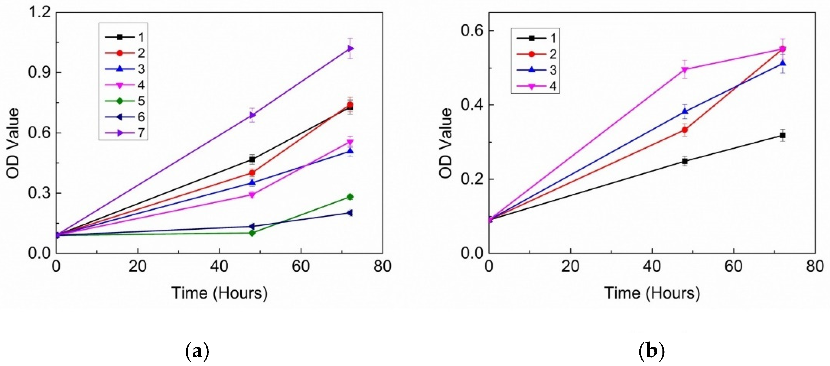
3.1. Inhibitory Effects of 18 Probiotic Strains on Skin Pathogens

The growth inhibitory results of the supernatant from the fermentation of 18 probiotic strains presented in Table 1 demonstrate that three strains of B. lactis and three strains of L. rhamnosus exhibited significant growth inhibition of M. furfur and C. acnes, with an inhibition rate exceeding 90% within 48 h. B. lactis maintained a high inhibition rate over 72 h, while the inhibitory effect of L. rhamnosus decreased to approximately 60%. L. plantarum and B. animalis performed well in the initial 48 h, but their antibacterial effects decreased to 60% or less after 72 h. Most strains exhibited prolonged inhibitory effects on C. acnes growth, especially B. lactis and L. rhamnosus. L. salivarius and L. plantarum also demonstrated relatively high inhibitory effects. The minimum inhibitory concentrations (MICs) of the probiotic supernatants required for inhibiting the growth of M. furfur and C. acnes were 20–25% and 8–12%, respectively.
Out of the eighteen bacterial strains evaluated for their antimicrobial performances based on their inhibition effectiveness and duration, HN019 exhibited the highest efficacy, with an inhibition rate of over 90% against both pathogenic bacteria after 72 h. Bi07 and B420 showed the highest long-term inhibition rates against M. furfur, with a slightly lower long-term inhibition rate against C. acnes compared to HN019. Conversely, Lp115 exhibited the opposite pattern, with a slightly lower long-term inhibition rate against M. furfur and the highest long-term inhibition rate against C. acnes. Therefore, these four strains were selected for further investigation regarding their antimicrobial properties.

3.2. Antibacterial Proteins and Peptides

The dialyzed samples (10,000 MWCO) of B420, Bi07, HN019, and Lp115 showed no inhibition on M. furfur and C. acnes. The crude extract of B420 protein even promoted the growth of C. acnes. However, the dialyzed samples (200 Da MWCO) exhibited antibacterial effects, and the results are shown in Figure 2.
For the growth inhibition test of M. furfur, the crude supernatant extracts of B420, Bi07, HN019, and Lp115 demonstrated a significant inhibitory effect. The growth inhibition rate of the crude supernatant extracts of HN019 and Lp115 reached 90% in 48 h and above, while those of B420 and Bi-07 were approximately 60%. In the case of the crude protein extract, Bi07 and HN019 also exhibited an inhibitory effect at approximately 40% to 50%. However, the inhibition rate of all samples declined at 72 h, ranging from 1% to 20%.
In the growth inhibition test of C. acnes, the crude protein extract of B420 showed an inhibition rate of 61% at 48 h, while no inhibition was observed in the crude protein extracts of other strains. The 48 h inhibition rate of the supernatant from the Bi07 crude protein extracts was 40%, and that of HN019 was 28%. By 72 h, the inhibition rate of these samples decreased to 10% to 40%.

3.3. Inhibitory Effects of Organic Acids

Further investigation of the organic acids produced by strains B420, Bi07, HN019, and Lp115 revealed that the supernatants of the four bacterial strains, neutralized to pH 6.5 with 0.1 M NaOH, exhibited partial inhibition. The inhibition rates ranged from 40% to 80% at 48 h and 0% to 50% at 72 h.
HPLC analysis of the fermentation broth of the four probiotics presented (Table 2) showed that lactic acid and acetic acid were the most abundant organic acids, accounting for 88% to 94% of the total content. Propionic acid accounted for 5% to 10%, while butyric acid comprised only 0.2% to 1%. The contents of organic acids varied significantly among the strains. For example, the lactic acid content in Lp115 was four times higher than that in B420, and the acetic acid content in B420 was over two times higher than that in Lp115.
The results of the growth inhibition test of M. furfur and C. acnes using the prepared organic acid mixture are presented in Figure 3.
The organic acid mixtures derived from the four selected strains demonstrated significant inhibition effects, with inhibition rates ranging from 53% to 90% at 72 h. Sample 2 exhibited the highest inhibition rate at 90%, while Samples 1 and 3 exhibited inhibition rates around 70%.
The organic acid mixture of the four samples exhibited notable inhibition effects, with inhibition rates ranging from 90% to 95% at 72 h. Sample 1 exhibited the highest inhibition rate at 96%.

4. Discussion

The personal care industry is increasingly utilizing probiotics and their metabolites as bioactive ingredients to enhance skin health via topical application, Lactobacillus is the most commonly used genus of probiotics [28,29,30]. In this study, B. lactis strains demonstrated greater effectiveness in inhibiting the growth of M. furfur and C. acnes compared to L. rhamnosus, indicating the potential of being used as probiotics for skin care.
Previous studies have confirmed that the antibacterial substance compounds of the found probiotics in probiotic metabolites include organic acids, bacteriocins, ethyl acetate, and phenyllactic acid [28,29,30,31,32]. This study further suggests that these antibacterial compounds are likely to be complex mixtures with varied antibacterial functions, differing in types and proportions across strains (Table 3). These compounds may primarily consist of peptides and organic acids. Additionally, it was observed that the metabolites with inhibitory effects on M. furfur and C. acnes were predominantly concentrated in substances with a molecular weight below 10,000 Da. It is speculated that these substances primarily disrupt the cell membrane structure and intracellular environment homeostasis, ultimately leading to cell death [33].
The primary discovery of this study was the diverse effects resulting from different combinations of organic acids in the antimicrobial tests. The combined impact of organic acid mixtures notably reduced the minimum inhibitory concentration (MIC). In the antimicrobial test against M. furfur, the antibacterial effect of the sample (B420) with the lowest organic acid mixture is better than that of the sample (Lp115) with the highest content. Moreover, the antimicrobial effects of the two samples (Bi07 and Lp115) with the highest total organic acid contents differed by 40%, while the sample with the lowest total organic acid content exhibited intermediate antimicrobial effect. This test also revealed that a single component is not the sole determinant of antimicrobial efficacy. For instance, although sample B420 had the lowest lactic acid content, its antimicrobial effect reached 70%. Conversely, sample Lp115, despite having a slightly higher lactic acid content than Bi07, yielded a 40% lower antimicrobial effect, the lowest among the four samples at 54%. A similar trend was observed for acetic acid. despite its acetic acid content being only 65% of that in B420, it exhibited the most potent antimicrobial effect, indirectly indicating the synergistic antimicrobial effects of several organic acids. Joana Salomskiene’s research revealed that L. Lactis 140/2 showcased the highest antimicrobial activity by producing a complex of organic acids [34], including lactic acid, citric acid, benzoic acid, and tartaric acid. This suggests that organic acids produced by strains such as Bi07 may also have complex antimicrobial effects, necessitating further research.
To date, most studies measured the antibacterial effect over a 48 h period. The extended measurement period in our study allowed for a better understanding of the dosage and treatment time. Notably, the sample of B. lactis HN019 had a prolonged inhibitory effect on both M. furfur and C. acnes, achieving a 100% inhibition rate for 72 h. Therefore, B. lactis HN019 could be the potential candidate for further probiotic application studies in skin microbiome modulation.

5. Conclusions

In this study, 10 strains among 18 tested probiotics showed inhibitory effects on M. furfur and C. acnes. Our findings indicated that the fermented supernatant, containing a mixture of organic acids, peptides, and other substances, exhibited inhibitory effects on M. furfur and C. acnes. The main active substances responsible for these effects were found to have a molecular weight smaller than 10,000 Da. Different probiotics produced antimicrobial substances in varying quantities. The inhibition activity of the fermentates lasted between 0 to 100 h, and it decreased gradually throughout the treatment. To obtain the active substances from probiotic metabolites, a membrane filtration method with a 10,000 Da MWCO (molecular weight cut-off) membrane can be used during production. This process not only yields a product with bacteriostatic properties but also results in reduced color and taste compared to conventional methods, making it highly versatile and applicable in various settings.

Author Contributions

Conceptualization, Q.M. and Y.S.; methodology, Q.M. and C.X.; software, Q.M.; validation, Q.M. and C.X.; formal analysis, Q.M.; investigation, Q.M.; resources, Y.S. and Z.W.; data curation, Q.M.; writing—original draft preparation, Q.M.; writing—review and editing, Q.M. and Y.S.; visualization, Q.M.; supervision, Y.S.; project administration, Z.W.; funding acquisition, Y.S. All authors have read and agreed to the published version of the manuscript.

Funding

This work was supported by the National Natural Science Foundation of China (No. 22308094), the Independent Research Fund of the Yunnan Characteristic Plant Extraction Laboratory (2022YKZY004), the Fundamental Research Funds for the Central Universities (2022ZFJH004 and 222201717003), and the National Natural Science Foundation of China for Innovative Research Groups (No. 51621002).

Institutional Review Board Statement

Not applicable.

Informed Consent Statement

Not applicable.

Data Availability Statement

The original contributions presented in this study are included in the article material. Further inquiries can be directed to the corresponding authors.

Conflicts of Interest

The authors declare no conflict of interest.

References

  1. Sender, R.; Fuchs, S.; Milo, R. Revised Estimates for the Number of Human and Bacteria Cells in the Body. PLoS Biol. 2016, 14, e1002533. [Google Scholar] [CrossRef] [PubMed]
  2. Grice, E.A.; Segre, J.A. The skin microbiome. Nat. Rev. Microbiol. 2011, 9, 244. [Google Scholar] [CrossRef] [PubMed]
  3. Byrd, A.L.; Belkaid, Y.; Segre, J.A. The human skin microbiome. Nat. Rev. Microbiol. 2018, 16, 143–155. [Google Scholar] [CrossRef] [PubMed]
  4. Elias, P.M.; Choi, E.H. Interactions among stratum corneum defensive functions. Exp. Dermatol. 2005, 14, 719–726. [Google Scholar] [CrossRef]
  5. Chehoud, C.; Rafail, S.; Tyldsley, A.S.; Seykora, J.T.; Lambris, J.D.; Grice, E.A. Complement modulates the cutaneous microbiome and inflammatory milieu. Proc. Natl. Acad. Sci. USA 2013, 110, 15061–15066. [Google Scholar] [CrossRef]
  6. Sugimoto, S.; Iwamoto, T.; Takada, K.; Okuda, K.-I.; Tajima, A.; Iwase, T.; Mizunoe, Y. Staphylococcus epidermidis Esp Degrades Specific Proteins Associated with Staphylococcus aureus Biofilm Formation and Host-Pathogen Interaction. J. Bacteriol. 2013, 195, 1645–1655. [Google Scholar] [CrossRef]
  7. Bomar, L.; Brugger, S.D.; Yost, B.H.; Davies, S.S.; Lemon, K.P. Corynebacterium accolens Releases Antipneumococcal Free Fatty Acids from Human Nostril and Skin Surface Triacylglycerols. mBio 2016, 7, e01725-15. [Google Scholar] [CrossRef]
  8. Iebba, V.; Totino, V.; Gagliardi, A.; Santangelo, F.; Schippa, S. Eubiosis and Dysbiosis: The Two Sides of the Microbiota. New Microbiol. Off. J. Ital. Soc. Med. Virol. (SIVIM) 2016, 39, 1–12. [Google Scholar]
  9. Coenye, T.; Peeters, E.; Nelis, H.J. Biofilm formation by Propionibacterium acnes is associated with increased resistance to antimicrobial agents and increased production of putative virulence factors. Res. Microbiol. 2007, 158, 386–392. [Google Scholar] [CrossRef]
  10. Cogen, A.L.; Nizet, V.; Gallo, R.L. Skin microbiota: A source of disease or defence? Br. J. Dermatol. 2008, 158, 442–455. [Google Scholar] [CrossRef]
  11. Wang, Y.; Hata, T.R.; Tong, Y.L.; Kao, M.-S.; Zouboulis, C.C.; Gallo, R.L.; Huang, C.-M. The Anti-Inflammatory Activities of Propionibacterium acnes CAMP Factor-Targeted Acne Vaccines. J. Investig. Dermatol. 2018, 138, 2355–2364. [Google Scholar] [CrossRef] [PubMed]
  12. Cho, I.; Blaser, M.J. The human microbiome: At the interface of health and disease. Nat. Rev. Genet. 2012, 13, 260–270. [Google Scholar] [CrossRef] [PubMed]
  13. Byrd, A.L.; Deming, C.; Cassidy, S.K.B.; Harrison, O.J.; Ng, W.-I.; Conlan, S.; Program, N.C.S.; Belkaid, Y.; Segre, J.A.; Kong, H.H. Staphylococcus aureusand Staphylococcus epidermidis strain diversity underlying pediatric atopic dermatitis. Sci. Transl. Med. 2017, 9, eaal4651. [Google Scholar] [CrossRef] [PubMed]
  14. Lin, Q.; Panchamukhi, A.; Li, P.; Shan, W.; Zhou, H.; Hou, L.; Chen, W. Malassezia and Staphylococcus dominate scalp microbiome for seborrheic dermatitis. Bioprocess Biosyst. Eng. 2021, 44, 965–975. [Google Scholar] [CrossRef]
  15. DeAngelis, Y.M.; Saunders, C.W.; Johnstone, K.R.; Reeder, N.L.; Coleman, C.G.; Kaczvinsky, J.R.; Gale, C.; Walter, R.; Mekel, M.; Lacey, M.P.; et al. Isolation and Expression of a Malassezia globosa Lipase Gene, LIP1. J. Investig. Dermatol. 2007, 127, 2138–2146. [Google Scholar] [CrossRef]
  16. Hay, R.J. Malassezia, dandruff and seborrhoeic dermatitis: An overview. Br. J. Dermatol. 2011, 165 (Suppl. S2), 2–8. [Google Scholar] [CrossRef]
  17. Borda, L.J.; Wikramanayake, T.C. Seborrheic Dermatitis and Dandruff: A Comprehensive Review. J. Clin. Investig. Dermatol. 2015, 3, 10. [Google Scholar]
  18. Turner, G.A.; Hoptroff, M.; Harding, C.R. Stratum corneum dysfunction in dandruff. Int. J. Cosmet. Sci. 2012, 34, 298–306. [Google Scholar] [CrossRef]
  19. Kim, S.Y.; Kim, S.H.; Kim, S.N.; Kim, A.-R.; Kim, Y.R.; Kim, M.J.; Park, W.-S.; Lee, J.H.; Jung, W.H.; Lee, Y.W.; et al. Isolation and identification of Malassezia species from Chinese and Korean patients with seborrheic dermatitis and in vitro studies on their bioactivity on sebaceous lipids and IL-8 production. Mycoses 2016, 59, 274–280. [Google Scholar] [CrossRef]
  20. Sommer, B.; Overy, D.P.; Kerr, R.G. Identification and characterization of lipases from Malassezia restricta, a causative agent of dandruff. FEMS Yeast Res. 2015, 15, fov078. [Google Scholar] [CrossRef]
  21. Dawson, T.L. Malassezia globosa and restricta: Breakthrough Understanding of the Etiology and Treatment of Dandruff and Seborrheic Dermatitis through Whole-Genome Analysis. J. Investig. Dermatol. Symp. Proc. 2007, 12, 15–19. [Google Scholar] [CrossRef] [PubMed]
  22. Tanaka, A.; Cho, O.; Saito, C.; Saito, M.; Tsuboi, R.; Sugita, T. Comprehensive pyrosequencing analysis of the bacterial microbiota of the skin of patients with seborrheic dermatitis. Microbiol. Immunol. 2016, 60, 521–526. [Google Scholar] [CrossRef] [PubMed]
  23. Fuchs-Tarlovsky, V.; Marquez-Barba, M.F.; Sriram, K. Probiotics in dermatologic practice. Nutrition 2016, 32, 289–295. [Google Scholar] [CrossRef] [PubMed]
  24. Katz, L.; Baltz, R.H. Natural product discovery: Past, present, and future. J. Ind. Microbiol. Biotechnol. 2016, 43, 155–176. [Google Scholar] [CrossRef]
  25. Al-Ghazzewi, F.H.; Tester, R.F. Effect of konjac glucomannan hydrolysates and probiotics on the growth of the skin bacterium Propionibacterium acnes in vitro. Int. J. Cosmet. Sci. 2010, 32, 139–142. [Google Scholar] [CrossRef]
  26. Guéniche, A.; Cathelineau, A.C.; Bastien, P.; Esdaile, J.; Martin, R.; Queille Roussel, C.; Breton, L. Vitreoscilla filiformis biomass improves seborrheic dermatitis. J. Eur. Acad. Dermatol. Venereol. 2008, 22, 1014–1015. [Google Scholar] [CrossRef]
  27. Guéniche, A.; Hennino, A.; Goujon, C.; Dahel, K.; Bastien, P.; Martin, R.; Jourdain, R.; Breton, L.; Gueniche, A.; Hennino, A.; et al. Improvement of atopic dermatitis skin symptoms by Vitreoscilla filiformis bacterial extract. Eur. J. Dermatol. 2006, 16, 380–384. [Google Scholar]
  28. Lay, C.L.; Coton, E.; Blay, G.L.; Chobert, J.M.; Haertlé, T.; Choiset, Y.; Long, N.V.; Meslet-Cladière, L.; Mounier, J. Identification and quantification of antifungal compounds produced by lactic acid bacteria and propionibacteria. Int. J. Food Microbiol. 2016, 239, 79–85. [Google Scholar] [CrossRef]
  29. Qiao, N.; Yu, L.; Zhang, C.; Wei, C.; Zhao, J.; Zhang, H.; Tian, F.; Zhai, Q.; Chen, W. A comparison of the inhibitory activities of Lactobacillus and Bifidobacterium against Penicillium expansum and an analysis of potential antifungal metabolites. FEMS Microbiol. Lett. 2020, 367, fnaa130. [Google Scholar] [CrossRef]
  30. Kang, K.H.; Shin, H.J.; Park, Y.H.; Lee, T.S. Studies on the antibacterial substances produced by lactic acid bacteria: Purification and some properties of antibacterial substance ’Bifilong’ produced by B. longum. Korean J. Dairy Sci. 1989, 1, 204–216. [Google Scholar]
  31. Angelescu, I.R.; Grosu-Tudor, S.S.; Cojoc, L.R.; Maria, G.M.; Chirițoiu, G.N.; Munteanu, C.V.A.; Zamfir, M. Isolation, characterization, and mode of action of a class III bacteriocin produced by Lactobacillus helveticus 34.9. World J. Microbiol. Biotechnol. 2022, 38, 220. [Google Scholar] [CrossRef] [PubMed]
  32. Ahmad, C.; Natascha, C.; Haiqin, C.; Jianxin, Z.; Jian, T.; Hao, Z.; Wei, C. Bifidin I—A new bacteriocin produced by Bifidobacterium infantis BCRC 14602: Purification and partial amino acid sequence—ScienceDirect. Food Control 2010, 21, 746–753. [Google Scholar]
  33. Nissen-Meyer, J.; Rogne, P.; Oppegard, C.; Haugen, H.; Kristiansen, P. Structure-Function Relationships of the Non-Lanthionine-Containing Peptide (class II) Bacteriocins Produced by Gram-Positive Bacteria. Curr. Pharm. Biotechnol. 2009, 10, 19–37. [Google Scholar] [CrossRef]
  34. Salomskiene, J.; Jonkuviene, D.; Macioniene, I.; Abraitiene, A.; Zeime, J.; Repeckiene, J.; Vaiciulyte-Funk, L. Differences in the occurence and efficiency of antimicrobial compounds produced by lactic acid bacteria. Eur. Food Res. Technol. 2019, 245, 569–579. [Google Scholar] [CrossRef]
Figure 1. Schematic illustration of the study on the inhibitory effect of probiotic metabolites on seborrheic dermatitis and acne-associated pathogenic bacteria.
Cosmetics 12 00003 g001
Figure 2. (a) The 0–72 h growth inhibition of the six samples on M. furfur. Sample 1: crude protein extract of Bi-07; Sample 2: crude protein extract of HN019; Sample 3: crude supernatant extract of B420; Sample 4: crude supernatant extract of Bi-07; Sample 5: crude supernatant extract of HN019; Sample 6: crude supernatant extract of Lp-115; Sample 7: positive control. (b) The 0–72 h growth inhibition of six samples on C. acnes. Sample 1: crude protein extract of B420; Sample 2: crude supernatant extract of Bi-07; Sample 3: crude supernatant extract of HN019; Sample 4: positive control.
Cosmetics 12 00003 g002
Figure 3. (a) The 0–72 h growth inhibition of M. furfur. (b) The 0–72 h growth inhibition of C. acnes. Samples 1 to 4 contained only the organic acids found in the supernatants of the four strains 1-B420, 2-Bi07, 3-HN019, and 4-Lp115, respectively, as determined via HPLC analysis. Sample 5 was the assay control.
Cosmetics 12 00003 g003
Table 1. The growth inhibition results of 18 probiotic strains on M. furfur and C. acnes. ++ indicates that the inhibition rate exceeds 90%. + indicates that the inhibition rate is between 40% and 90%. − indicates that the inhibition rate is less than 40%.
NameStrainGrowth Inhibition of M. furfurGrowth Inhibition of C. acnes
48 h72 h96 h48 h72 h96 h
Bifidobacterium lactisHN019++++++++++++
Bifidobacterium lactisBl-04+++
Bifidobacterium lactisB420++++++++++
Bifidobacterium lactisBi-07++++++++++
Bifidobacterium animalisBb-12++++++
Lactobacillus brevisLbr-35+++
Lactobacillus acidophilusNCFM++++
Lactobacillus plantarumLp-115+++++++++
Lactobacillus paracaseiLpc-37+++
Lactobacillus rhamnosusHN001+++++++
Lactobacillus acidophilusLa-14++
Lactobacillus caseiLc-11++++
Lactobacillus rhamnosusGG++++++
Lactobacillus salivariusLs-33+++++
Lactobacillus rhamnosusLr-32++++++
Lactobacillus reuteri1E1++
Lactobacillus fermentumSBS-1+++
Lactobacillus bulgaricusLb-87+++
Table 2. The concentrations of lactic acid, acetic acid, propionic acid, and butyric acid in the supernatants of the four selected strains via HPLC analysis.
No.StrainLactic Acid (mg/L)Acetic Acid (mg/L)Propionic Acid (mg/L)Butyric Acid (mg/L)
1B4205.649.261.820.2
2Bi-0717.396.251.650.17
3HN01910.828.851.060.05
4Lp-11519.884.041.800.06
Table 3. The growth inhibition of M. furfur of the active metabolites of the four strains.
No.StrainGrowth Inhibition of M. furfur
Crude Protein ExtractCrude Supernatant ExtractsOrganic Acid Mixture
1B420-55%72%
2Bi-0731%50%92%
3HN01930%79%71%
4Lp-115-88%54%
Disclaimer/Publisher’s Note: The statements, opinions and data contained in all publications are solely those of the individual author(s) and contributor(s) and not of MDPI and/or the editor(s). MDPI and/or the editor(s) disclaim responsibility for any injury to people or property resulting from any ideas, methods, instructions or products referred to in the content.

