Article

Hair Growth-Promoting Effects of Astragalus sinicus Extracts in Human Follicle Dermal Papilla Cells

1
Research Institute for Biomedical and Health Science, Konkuk University, Chungju 27478, Republic of Korea
2
Korea Research Institute BioScience Co., Ltd., 362-20, Seongju-ro, Gamgok-myeon, Eumseong-gun, Chungcheongbuk-do 27600, Republic of Korea
*
Author to whom correspondence should be addressed.
Cosmetics 2025, 12(1), 6; https://doi.org/10.3390/cosmetics12010006
Submission received: 24 November 2024 / Revised: 15 December 2024 / Accepted: 30 December 2024 / Published: 4 January 2025

Abstract

:
Hair loss is influenced by various stresses and disruptions in the hair growth cycle within hair follicles. The activation of these follicles is primarily driven by dermal papilla cells, which play a central role in the progression of the hair growth cycle and formation. Many studies are being carried out on various natural plants to improve hair loss. In this study, we assessed the potential of Astragalus sinicus, known as a pharmacological herb, in mitigating damage to human follicle dermal papilla cells (HFDPCs) caused by dihydrotestosterone (DHT). We found that Astragalus sinicus extracts (ASEs) facilitated wound healing and elevated alkaline phosphatase expression in DHT-damaged HFDPCs. ASEs also significantly reduced reactive oxygen species (ROS) generated by DHT and increased ATP levels by restoring mitochondrial membrane potential in damaged cells. Furthermore, we demonstrated that ASEs substantially enhanced the phosphorylation levels of the AKT/ERK pathway and activated the Wnt signaling pathway in DHT-damaged HFDPCs. Taken together, we suggest that ASEs may be a potential ingredient for enhancing hair loss treatment.

1. Introduction

Hair is found on various parts of the human body and performs several critical functions, including body protection, heat insulation, camouflage, sebaceous secretion, sensory perception, and social interactions [1]. That is why hair loss is an important concern for many people today. Hair loss is characterized by a reduction in hair density, hair thinning, or both, and can result from both hormonal and non-hormonal causes [2]. Androgenetic alopecia (AGA), commonly known as male pattern baldness, is a multifactorial condition characterized by a specific pattern of baldness [3].
AGA is characterized by the gradual shrinking of hair follicles, which occurs due to alterations in the hair growth cycle dynamics. This leads to the transformation of the terminal hair follicle into villus-like hairs, with a gradual shortening in the anagen phase and a lengthening in the telogen phase [4]. The condition is mainly driven by 5-alpha-reductase and its byproduct, DHT, which induces miniaturization of hair follicles and shortens successive anagen cycles [5]. Increased oxidative stress affects the pathogenesis of androgenic alopecia and the secretion of known follicle-inhibitory factors [6,7]. However, the underlying molecular mechanisms of androgen-related hair loss remain largely unknown [8].
Hair follicles are living mini-organs composed of dermal and epidermal cells. Dermal papilla cells, derived from the mesodermal layer, play a crucial role in the formation of human hair follicles [9]. HFDPCs are the major regulatory cells within the hair follicle, inducing hair formation and growth through reciprocal interactions with epithelial cells [10]. HFDPCs are located at the bottom of a hair follicle and control the hair growth cycle. There are three phases in the cycle of hair growth: growth (anagen), regression (catagen), and rest (telogen) [11]. Accelerating the telogen-to-anagen transition or delaying the anagen-to-catagen shift can mitigate hair miniaturization and prevent hair loss [12]. Pathways involved in regulating follicle cycle development primarily include Wnt/β-catenin, sonic hedgehog (SHH), and transformation growth factor -β (TGF-β). Especially, the Wnt/β-catenin pathway is pivotal for follicle morphogenesis and primary follicle formation [13,14]. The activated Wnt/β-catenin pathway plays key roles in morphogenesis, growth, and regeneration of follicles [15]. Additionally, alkaline phosphatase (ALP), which is crucial for growth phase induction and wound-induced hair neogenesis, is increased by Wnt/β-catenin signaling [16].
Although there are various drugs and treatment strategies to improve such hair loss, it has been reported that chemicals contained in natural products have properties that stimulate hair growth [17]. Polyphenols, abundantly present in plants, are gaining recognition for their significant contributions to health, and more than 1000 species of polyphenols have been identified by technological advances in recent years [18]. Polyphenols and other antioxidants have been documented for their roles in preventing hair loss and promoting hair growth [19].
Astragalus sinicus is a perennial herbaceous legume native to East Asia, including Japan, southern China, and central Korea [20]. It is primarily cultivated in rice paddies, where it enhances soil fertility through nitrogen fixation, benefiting rice cultivation [21]. Research on Astragalus sinicus reveals that its tender young leaves and stems are edible and have been traditionally utilized in medicine to address ailments such as excessive sweating, hypertension, and diabetes [22]. Astragalus sinicus chemical components contain chemical components and structures such as triterpenoid saponins, flavonoids, and glycosides [23]. Previous reports have shown positive results regarding its anti-inflammatory and antioxidant properties [24,25]. It has been hypothesized that chemical components like flavonoids may have a beneficial effect on improving hair loss [26].
In this study, we investigated whether Astragalus sinicus extracts (ASEs) can be used as a potential cosmetic and therapeutic agent to improve HFDPC damaged by DHT and promote hair growth.

2. Materials and Methods

2.1. Preparation of ASEs

Astragalus sinicus was collected in early May from the farmland Hampyeong-gun, Jeollanam-do. It was naturally dried under sunlight for 2 days. Two kg of dried aerial parts of Astragalus sinicus were then extracted with 40 L of 70% ethanol/water mixture (v/v) at 80 °C for 6 h. The extract was filtered with a filter cloth, and the filtrate was concentrated using a vacuum evaporator under the following conditions: 10~20 kPa, 40 rpm, and 50 °C. Subsequently, it was freeze-dried for 48 h, yielding 0.5 kg of ASE powder. The freeze-dried powder was vacuum-sealed and stored frozen for experimental use.

2.2. Cell Culture

HFDPCs acquired from PromoCell (Heidelberg, Germany) were cultured using a dermal papilla cell growth medium, supplemented with a mix of growth factors and 1% streptomycin/penicillin in a 5% CO2 incubator at 37 °C. Additionally, ready-to-use HFDPC Medium and a Detach kit from PromoCell (Heidelberg, Germany) were used for cell cultivation. The Detach kit, composed of HEPES BSS solution, Trypsin/EDTA solution, and trypsin neutralization solution (Heidelberg, Germany), was utilized to enhance the subculturing environment of HFDPCs. Cells were detached using the Detach kit and transferred to 75 mm tissue culture flasks once they reached 80–90% confluence, typically every 3 days.

2.3. Cell Viability Assay

An EZ-cytox cell-based assay kit (DoGenBio, Seoul, Republic of Korea) was utilized to assess cell viability. HFDPCs (2 × 10⁴ cells per well) were cultured in a microplate and incubated for 24 h. HFDPCs were treated with ASEs at concentrations of 5, 10, and 50 ppm for 24 h. Ten μL EZ-cytox reagent was added to each well, and 100 μL dermal papilla cell growth medium was introduced into each well for 1 h at 37 °C incubation. The absorbance at 450 nm was recorded using a Synergy HTX microplate reader (BioTek, Winooski, VT, USA).

2.4. Cell Proliferation Assay

HFDPCs were seeded in a confocal dish and incubated for 24 h. Cells were exposed to 5 µM DHT, 1 µM minoxidil, and 100 ppm ASEs with 10 uM working solution of EdU for 24 h. Following the treatment, 1 mL of 3.7% formaldehyde was added and left to incubate for 15 min. Each sample was incubated for 15 min at room temperature. Each sample was washed twice with 3% BSA in PBS, and 1 mL of PBS with 0.5% Triton X-100 was added, and samples were incubated for 20 min. Each sample was then washed twice with 3% BSA in PBS, adding 0.5 mL of Click-iT® reaction cocktail each time, and incubated for 30 min. Each sample was washed once with 3% BSA in PBS, 1 mL of 1X Hoechst 33,342 solution was added, and samples were incubated for 30 min. Finally, each sample was washed twice with PBS, 1 mL PBS was added, and the samples were measured with fluorescence imaging. FITC and DAPI measurements were performed using an Eclipse Ti2 fluorescence live-cell imaging microscope (Nikon, Tokyo, Japan). Fluorescence signal intensities were quantified using Fiji Image J software 1.8.0 (win 64 bit) for analysis.

2.5. Wound Healing Assay

HFDPCs were seeded in a 6-well plate. The cells were incubated at 37 °C until they reached approximately 80% confluence after 24 h. A 200 µL pipette tip was then used to create a horizontal scratch through the center of the confluent cell layer. The cells were washed once with DPBS, and a fresh medium was added. HFDPCs were subsequently treated with 5 µM DHT, 1 µM minoxidil, and ASEs at concentrations of 10 ppm, 50 ppm, and 100 ppm, respectively. Wound healing was monitored at 0 and 24 h marks post-wound creation. Images of both treated and untreated control cells were captured using phase-contrast microscopy (Microscope ECLIPSE Ts2, Nikon, Tokyo, Japan).

2.6. Alkaline Phosphatase Staining (ALP) Assay

HFDPCs were seeded in a 24-well plate, and 500 μL of culture medium was added to each well. Consequently, HFDPCs were treated with 5 µM DHT, 1 µM minoxidil, and ASEs at concentrations of 100 ppm. Then, 1× PBST was prepared using 0.05% Tween 20. Fixing solution (0.4 mL) was applied to each well of a plate and incubated for 2 min. After incubation, the fixing solution was removed and the fixed cells were washed twice with 1 mL of 1× PBST. Following the final wash, the remaining liquid was aspirated and 0.4 mL of AP Staining Solution was added to each well. The cells were incubated for 24 h, ensuring they were protected from light during this period. Subsequently, the cells were rinsed with DPBS. The colonies exhibiting purple staining were subsequently counted and compared to unstained colonies under a Nikon light microscope (ECLIPSE Ts2, Nikon, Tokyo, Japan). The degree of staining was quantified using Fiji Image J software (win 64 bit) for analysis.

2.7. Measurement of Intracellular ROS

HFDPCs were seeded at 2 × 10⁴ cells/mL in a confocal dish and incubated for 24 h. The cells were exposed to 5 µM DHT, 1 µM minoxidil, and 100 ppm ASEs individually for 24 h. Cells were rinsed twice with DPBS, after which the 20 mM 2,7-Dichlorofluorescin diacetate (DCF-DA) solution provided in the H2DCFDA-Cellular ROS Assay Kit (Abcam, Cambridge, UK) was diluted to a final concentration of 10 µM and incubated for 20 min. After staining, the cells were again washed with DPBS, and 500 µL of DPBS was added for fluorescence measurement. FITC and DIC measurements were performed using an Eclipse Ti2 fluorescence live-cell imaging microscope (Nikon, Tokyo, Japan). Fluorescence signal intensities were quantified using Fiji Image J software (win 64 bit) for analysis.

2.8. Measurement of Membrane Potential in Mitochondria

HFDPCs were plated in a confocal dish and incubated for 24 h. Cells were individually treated with 5 µM DHT, 1 µM minoxidil, and 100 ppm ASEs for 24 h. Cells were washed with DPBS, followed by diluting the 1 mM JC-1 solution from the JC-1—Mitochondrial Membrane Potential Assay Kit (Abcam, Cambridge, UK) to a final concentration of 10 µM and incubating for 15 min. The cells were washed out again with DPBS, and 500 µL of DPBS was added for fluorescence analysis. RITC, FITC, and DIC measurements were taken using an Eclipse Ti2 fluorescence live-cell imaging microscope (Nikon, Tokyo, Japan), followed by merging the images. Fluorescence signal intensities were quantified using Fiji Image J software (win 64 bit) for analysis.

2.9. Live Cell ATP Assay

HFDPCs were seeded at 2.5 × 10⁴ cells/mL in confocal dishes and incubated for 24 h. Cells were treated with 5 µM DHT, 1 µM minoxidil, and 100 ppm ASEs, individually, for 24 h. Once the cells were ready, they were washed twice with DPBS. Subsequently, to prepare the ATP Red™ working solution, 1 mL of cell culture medium was added to every 5 µL of ATP Red™ Stock Solution from the Cell Meter™ Live Cell ATP Assay Kit (AAT Bioquest, Pleasanton, CA, USA). A volume of 500 µL of the working solution was applied, and the plate was incubated for 30 min. After removing the ATP Red™ working solution, the cells were washed twice with DPBS. Subsequently, MitoLite™ Green FM (AAT Bioquest, Pleasanton, CA, USA) was diluted from 1 mM to 100 nM to prepare the MitoLite™ Green FM staining solution. A volume of 500 µL was then added and incubated for 30 min. Cells were imaged using an Eclipse Ti2 fluorescence live-cell imaging microscope (Nikon, Tokyo, Japan) with RITC and FITC filters. Fluorescence signal intensities were quantified using Fiji Image J software (win 64 bit) for analysis.

2.10. Western Blot Analysis

HFDPCs were plated in dishes and incubated for 24 h. Cells were treated with DHT 5 µM, 1 µM minoxidil, and ASEs (10, 50, and 100 ppm) for 24 h. Cells were washed with DPBS, and protein extraction was performed using RIPA buffer. The proteins were sonicated for 5 min and centrifuged at 12,000 rpm, 10 min, and 4 °C. The supernatants were harvested, and the total protein concentration was assessed by the Pierce™ BCA Protein Assay Kit (Thermo Fisher Scientific, Waltham, MA, USA). The proteins were equalized with 4× LDS sample buffer diluted in RIPA buffer and heated on a heat block at 70 °C for 10 min. The membranes were incubated by 5% non-fat dry milk in TBS-T with 0.1% Tween 20. After blocking, primary antibodies including phospho-ERK/ERK, phospho-AKT/AKT, phospho-GSK-3β/GSK-3β, and β-catenin (Cell Signaling Technology, Danvers, MA, USA) were incubated overnight on the shaker. After that, the secondary antibody was incubated for 2 h and washed with TBS-T for 10 min. The immunoreactive bands were finally detected using the ECL reagent (1:1), the images were captured using the Invitrogen iBright 1500 (Waltham, MA, USA), and the data were analyzed with Fiji ImageJ software (Windows 64-bit).

2.11. Statistical Analysis

The data are shown as the mean ± standard deviation (SD) of three independent experiments. All statistical analyses, including t-tests and nonparametric tests, were conducted using GraphPad Prism 8.0.1 software (San Diego, CA, USA) through a t-test to compare two groups and analysis of variance (ANOVA) to analyze differences among multiple groups.

3. Results

3.1. The Effects of ASEs on Cell Viability and Proliferation in HFDPCs

To study the effects of ASEs on cell viability in HFDPCs, we treated the cells with ASEs at concentrations of 10 ppm, 50 ppm, and 100 ppm for 24 h. Cell viability was assessed using the EZ-cytox assay. The results demonstrated that ASE treatment markedly enhanced cell viability in HFDPCs compared to the control group (Figure 1A). A significant increase in the EdU-positive cells was observed in the ASE-treated group compared to the negative control (Figure 1B). Based on these findings, we also demonstrated that the cell proliferation rate was enhanced by ASEs at 24 and 48 h compared to the DHT-damaged HFDPCs (Figure 1C).

3.2. ASEs Improved Cellular Migration of HFDPCs Damaged by DHT

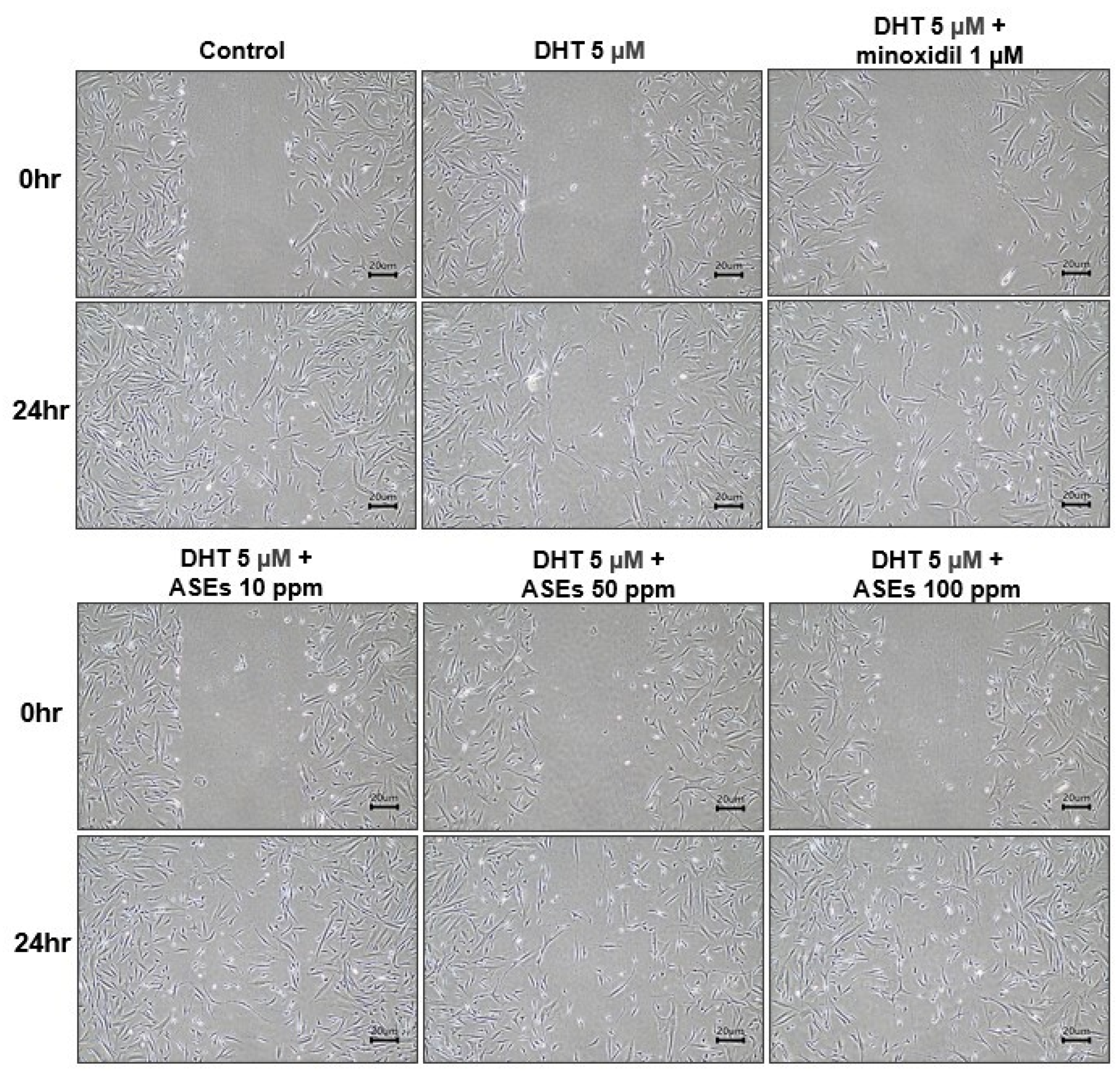
Assessing the mobility of HFDPCs is crucial for hair growth and regeneration, particularly in their contribution to follicle structure formation [10]. Thus, we conducted a wound healing assay to assess the effects of ASEs on cell migration in HFDPCs damaged by DHT. After 24 h, the DHT-treated group exhibited suppressed cell growth, whereas the group treated with both DHT and minoxidil showed a marked increase in cell migration. Furthermore, ASEs exhibited a significant dose-dependent enhancement in wound healing (Figure 2).

3.3. ASEs Improved the ALP Activity of HFDPCs Damaged by DHT

Alkaline phosphatase (ALP), the prominent marker of HFDPCs, was identified as a critical marker for hair growth promotion [27,28]. It has been suggested that the hair inductivity of HFDPCs is closely related to ALP activity [29]. HFDPCs are essential for hair growth and regeneration in regressed hair follicles. As expected, we observed a significant increase in ALP expression in the group treated with 1 μM minoxidil, the positive control, compared to the group treated with DHT-damaged HFDPCs. Similarly, a substantial increase in ALP expression was observed in the group treated with 100 ppm ASEs (Figure 3.)

3.4. ASEs Reduced DHT-Induced ROS Levels in HFDPCs

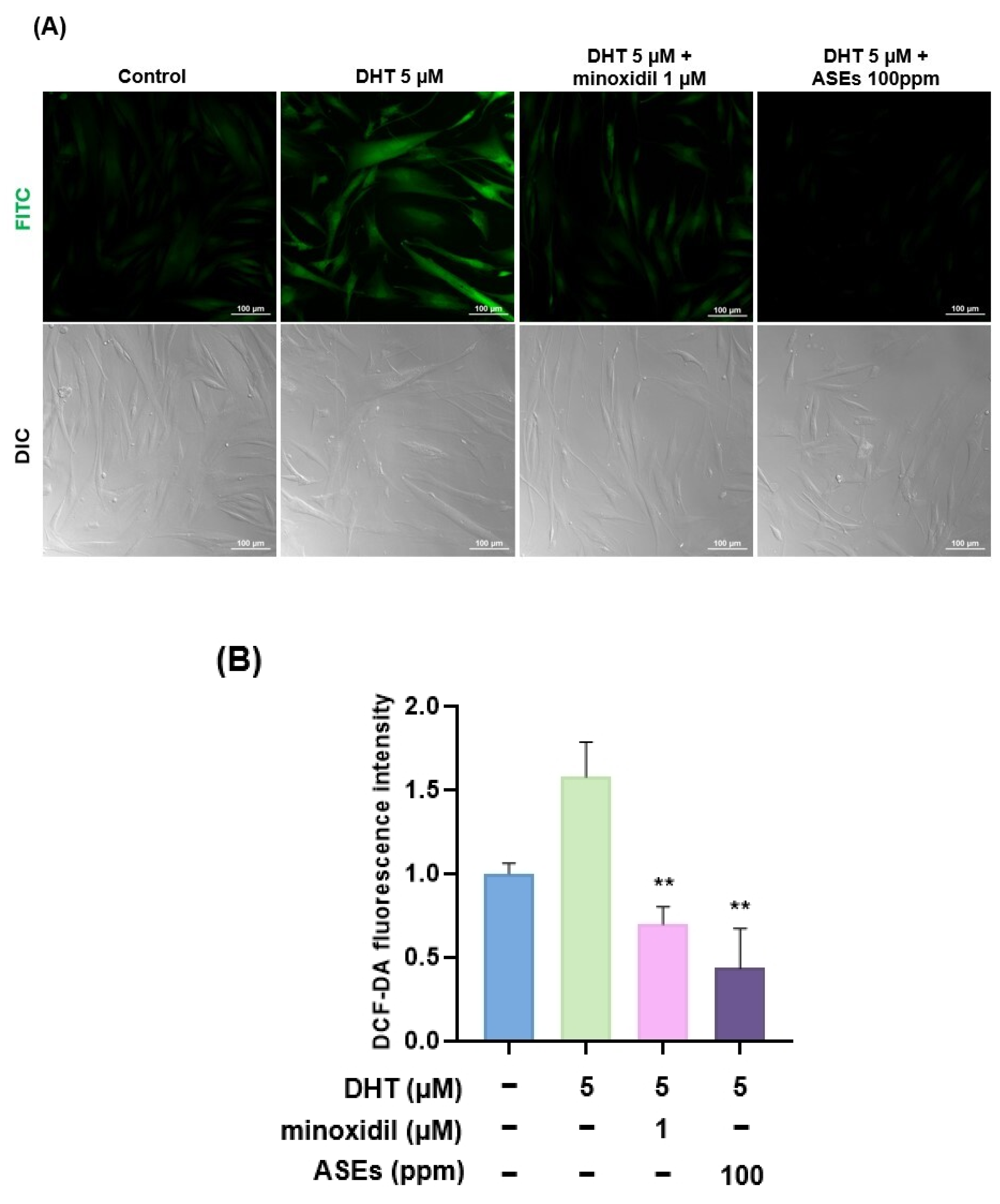
ROS production has endogenous and exogenous sources [30]. Stress and external oxidants, including air pollutants, cigarette smoke, and ionizing radiation, influence the redox (reduction-oxidation) status of cells [31,32]. Stress-induced alopecia is driven by factors such as androgen excess, genetic factors, and exposure to oxidative stress, which leads to the formation of ROS [33]. DHT elevates intracellular ROS levels, leading to cell death and growth arrest [34]. We hypothesized that ASEs would reduce ROS in HFDPCs damaged by DHT. Thus, we compared the ROS levels between the DHT-treated and the ASE-treated groups damaged by DHT with DCF-DA staining. Interestingly, we observed that the ASE-treated group had lower cellular ROS levels compared to the DHT-treated group (Figure 4).

3.5. ASEs Restored Mitochondrial Potential and ATP Levels in DHT-Damaged HFDPCs

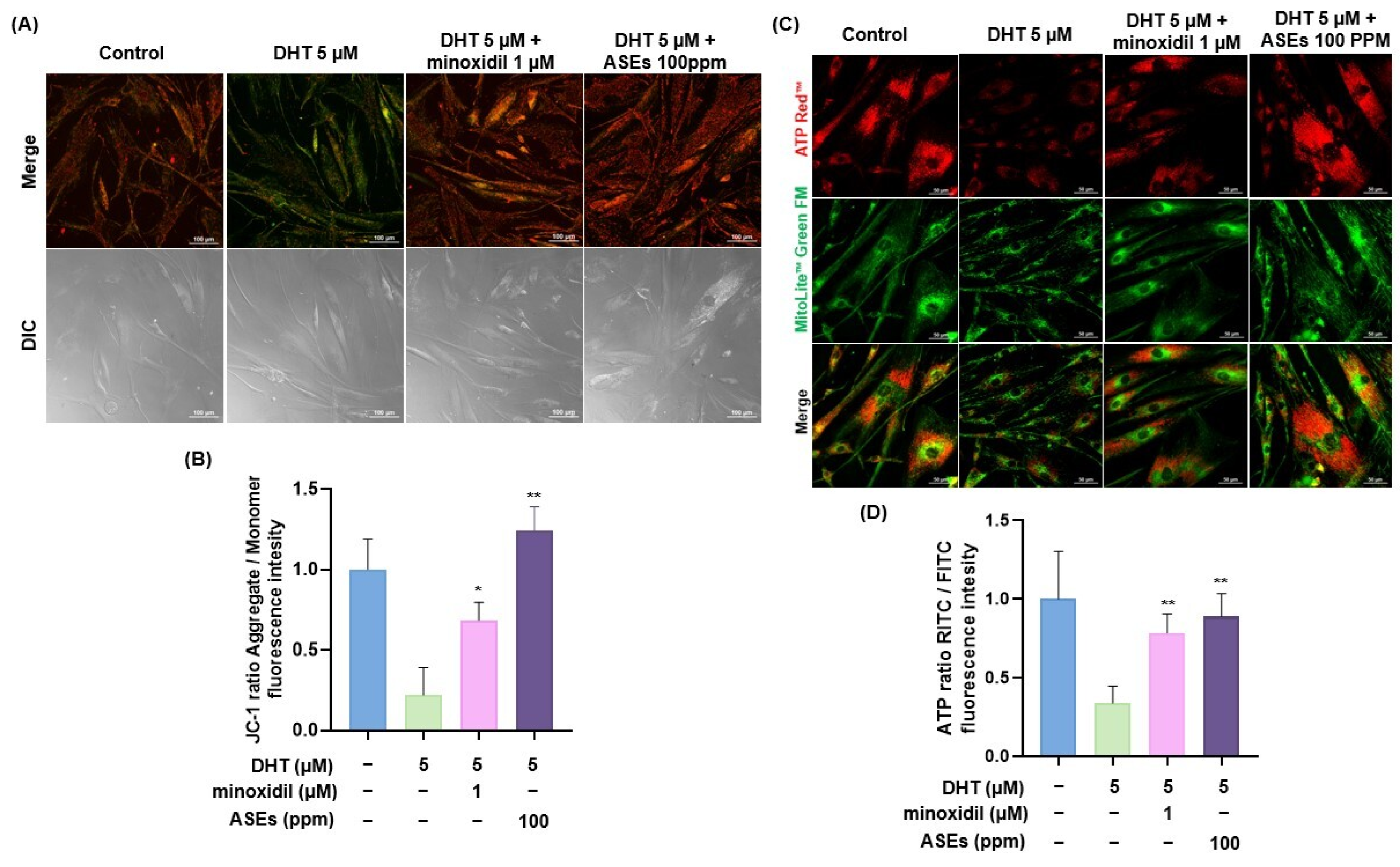
The anagen phase of hair growth involves cell division in the dermal papilla, which increases energy demands and reliance on mitochondrial respiration [35,36]. Consequently, mitochondrial function plays a critical role in hair growth regulation [37,38]. We assessed changes in mitochondrial membrane potential in DHT-damaged HDFPCs treated with ASEs using the JC-1 assay.
Depending on the membrane potential, JC-1 dye accumulates in the mitochondria, with normal mitochondrial membrane potential indicated by red fluorescence from JC-1 aggregates and damaged membrane potential indicated by green fluorescence from JC-1 monomers. We observed strong red fluorescence in ASEs damaged by 5 uM DHT. In contrast, the group treated with 5 uM DHT alone exhibited green fluorescence compared to the control group (Figure 5A,B). These results suggest that ASEs restored mitochondrial potential in HFPDCs damaged by DHT.
Based on this observation, we hypothesized that ASEs would increase ATP activity through enhanced mitochondrial function. The key function of mitochondria is the production of ATP via oxidative phosphorylation [39]. Therefore, disruption of mitochondrial oxidative phosphorylation may inhibit the differentiation and proliferation of hair stem cells and delay hair regeneration [40,41]. To test this hypothesis, we measured ATP activity in the mitochondria of DHT-damaged HFDPCs treated with ASEs using Live Cell ATP assays, indicating that living cells are indicated by red fluorescence. We observed that ATP activity was higher in ASE-treated cells damaged by 5 uM DHT compared to the DHT-treated group (Figure 5C,D).
These results support our hypothesis that ASEs increased ATP activity through enhanced mitochondrial function.

3.6. ASEs Were Involved in the ERK/AKT Signaling Pathway and Wnt/β-Catenin Pathway in DHT-Damaged HFDPCs

The ERK and AKT signaling pathways serve as critical modulators of cellular proliferation [42,43]. The ERK signaling pathway is essential for promoting cell proliferation in various cell types [44,45], including HFDPCs, and enhances hair induction by activating the AKT pathway [46,47]. The Wnt/β-catenin pathway is critical for the development and growth of hair follicles [48,49]. β-catenin expression in both hair follicle mesenchymal cells and epithelial cells plays vital roles in sustaining hair follicle development, as well as in the proliferation and differentiation of hair follicle stem cells [50,51]. In addition, GSK-3β signaling is necessary for regulating diverse biological processes, including cell proliferation, hair growth, and hair regeneration [52,53].
To investigate the signaling mechanism of ASEs, we conducted a Western blot analysis to assess their impact on the phosphorylation of ERK, AKT, β-catenin, and GSK-3β. In the DHT-treated group, the phosphorylation levels of p-AKT, p-AKT, p-GSK-3β, and the expression levels of β-catenin were decreased (Figure 6). In contrast, in DHT-treated HFDPCs exposed to ASEs, the phosphorylation levels of p-AKT, p-ERK, and p-GSK-3β and the expression levels of β-catenin were elevated dose-dependently (Figure 6).
These results indicate that ASEs activated the ERK/AKT signaling pathway and the GSK-3β/β-catenin signaling pathway in HFDPCs damaged by DHT.

4. Discussion

Alopecia is associated with various psychosocial issues, including depression, low self-esteem, and reduced social engagement [54,55]. As a result, there is an ongoing demand for novel and effective treatments for hair loss [56,57]. AGA is a prevalent hair loss condition influenced by genetic and hormonal factors, characterized by androgen-driven progressive thinning of scalp hair in a specific pattern [58,59]. The current treatments for AGA are oral finasteride and topical minoxidil. Oral minoxidil is increasingly used and has proven efficacy [60,61]. There are current limitations of DHT-targeted therapy. Finasteride and minoxidil are the most used DHT-targeted therapies that focus on reducing the enzymatic activity of 5α-reductase (finasteride) or increasing hair follicle blood flow (minoxidil) [62]. However, finasteride fails to address the inflammatory and oxidative stress responses that affect the miniaturization of hair follicles [63]. Minoxidil improves blood supply but does not directly regulate the cellular signaling pathway or inhibit inflammatory cytokine activity [64]. Therefore, natural extracts are gaining attention as potential safe alternatives for improving hair loss.
Astragalus sinicus is a traditional legume crop in the genus Astragalus [65]. Astragalus sinicus has an important role in maintaining soil fertility [66]. However, since no study has investigated the potential of ASEs to improve hair loss in HFDPCs damaged by DHT, we attempted this study for the first time.
In our study, we investigated whether ASEs could increase hair growth in DHT-damaged HFDPCs. Treatment with ASEs increased the proliferation rate (Figure 1C) and activated the migration of cells in HFDPCs damaged by DHT (Figure 2). In addition, alkaline phosphatase activity, a marker of the hair follicle cell growth cycle, was enhanced (Figure 3). ROS is a key factor in hair loss, and ASEs were shown to effectively suppress ROS levels compared to the DHT-damaged group (Figure 4). Mitochondria are membrane-bound organelles that play crucial roles in various cellular processes, including ATP production, metabolism, calcium signaling, redox signaling, and apoptosis [67,68]. Interestingly, ASEs restored mitochondrial function in DHT-damaged HFDPCs (Figure 5A). In addition, ASEs increased ATP activity by utilizing the membrane potential recovery capacity of mitochondria (Figure 5C). Activation of the Wnt/β-catenin pathway promotes hair cycle and regeneration [69,70]. In the absence of the Wnt pathway activation, β-catenin accumulates in the cytoplasm, where it is ubiquitinated and degraded by GSK-3β, which forms a complex with APC/Axin/GSK-3β [71,72]. The elevated levels of β-catenin in the nucleus activate target genes, including cyclin D1, which facilitates cell proliferation [72,73]. Based on this, we confirmed that treatment with ASEs enhanced the expression level of β-catenin and increased the phosphorylation level of GSK-3β (Figure 6). AKT is a critical protein that functions downstream of the P13K pathway to regulate the cellular response to external stimuli [74,75]. Phosphorylated AKT is found in mitochondria and nuclei, where it interacts with different molecules [74,76]. We demonstrated that ASEs inhibit β-catenin degradation, activate its nuclear migration, and induce cell growth through the ERK/AKT pathway, suggesting that ASEs may effectively prevent hair loss (Figure 7).
According to a recent study, natural remedies have gained popularity due to their reduced effects and side effects on the scalp, thanks to their complex vegetable-based ingredients [77]. ASEs provide a unique opportunity to integrate into existing and new hair regeneration strategies. ASEs can complement natural compounds such as caffeine or biotin, creating a comprehensive, multi-target approach to scalp health and hair follicle activation [78]. Although ASEs show potential for hair improvement effects, further research such as ex-vivo hair organ culture is needed. Additionally, more studies are necessary to ensure effective penetration and bioavailability with ASEs. Clinical trials are also needed to evaluate the safety and efficacy of hair loss.

5. Conclusions

ASEs may potentially address the current gap in hair enhancement methods by contributing to oxidative stress relief and promoting HFDPCs through the activation of the Wnt/β-Catenin pathway. Therefore, ASEs could serve as promising ingredients for hair-promoting effects.

Author Contributions

Conceptualization, C.Y.J. and D.W.S.; formal analysis, C.Y.J. and M.Y.G.; resources, I.-K.K., M.-R.P., H.W.L. and Y.-K.K.; funding acquisition, D.W.S.; investigation, C.Y.J. and M.Y.G.; methodology, C.Y.J.; writing—original draft, D.W.S.; writing—review and editing, D.W.S. All authors have read and agreed to the published version of the manuscript.

Funding

This research was supported by “Regional Innovation Strategy (RIS)” through the National Research Foundation of Korea (NRF) funded by the Ministry of Education (MOE) (2021RIS-001).

Institutional Review Board Statement

Not applicable.

Informed Consent Statement

Not applicable.

Data Availability Statement

The data from this study are available upon demand from the corresponding author.

Acknowledgments

The scientific illustration was created using BioRender (Toronto, ON, Canada).

Conflicts of Interest

I.K., M.P., H.W.L., and Y.K are employees of Korea Research Institute BioScience Co., Ltd. The remaining authors declare no conflicts of interest. The authors declare that the research was conducted in the absence of any commercial or financial relationships that could be construed as a potential conflict of interest. The funders had no role in the design of the study; in the collection, analyses or interpretation of data; in the writing of the manuscript; or in the decision to publish the results.

References

  1. Shimizu, Y.; Ntege, E.H.; Sunami, H.; Inoue, Y. Regenerative medicine strategies for hair growth and regeneration: A narrative review of literature. Regen. Ther. 2022, 21, 527–539. [Google Scholar] [CrossRef]
  2. Choi, B.Y. Targeting Wnt/β-Catenin Pathway for Developing Therapies for Hair Loss. Int. J. Mol. Sci. 2020, 21, 4915. [Google Scholar] [CrossRef]
  3. Fu, D.; Huang, J.; Li, K.; Chen, Y.; He, Y.; Sun, Y.; Guo, Y.; Du, L.; Qu, Q.; Miao, Y.; et al. Dihydrotestosterone-induced hair regrowth inhibition by activating androgen receptor in C57BL6 mice simulates androgenetic alopecia. Biomed. Pharmacother. 2021, 137, 111247. [Google Scholar] [CrossRef]
  4. Kaliyadan, F.; Nambiar, A.; Vijayaraghavan, S. Androgenetic alopecia: An update. Indian J. Dermatol. Venereol. Leprol. 2013, 79, 613–625. [Google Scholar] [CrossRef] [PubMed]
  5. Nestor, M.S.; Ablon, G.; Gade, A.; Han, H.; Fischer, D.L. Treatment options for androgenetic alopecia: Efficacy, side effects, compliance, financial considerations, and ethics. J. Cosmet. Dermatol. 2021, 20, 3759–3781. [Google Scholar] [CrossRef]
  6. Upton, J.H.; Hannen, R.F.; Bahta, A.W.; Farjo, N.; Farjo, B.; Philpott, M.P. Oxidative stress-associated senescence in dermal papilla cells of men with androgenetic alopecia. J. Investig. Dermatol. 2015, 135, 1244–1252. [Google Scholar] [CrossRef]
  7. Trueb, R.M. Oxidative stress and its impact on skin, scalp and hair. Int. J. Cosmet. Sci. 2021, 43 (Suppl. S1), S9–S13. [Google Scholar] [CrossRef] [PubMed]
  8. Martinez-Jacobo, L.; Villarreal-Villarreal, C.D.; Ortiz-Lopez, R.; Ocampo-Candiani, J.; Rojas-Martinez, A. Genetic and molecular aspects of androgenetic alopecia. Indian J. Dermatol. Venereol. Leprol. 2018, 84, 263–268. [Google Scholar] [CrossRef] [PubMed]
  9. Taghiabadi, E.; Nilforoushzadeh, M.A.; Aghdami, N. Maintaining Hair Inductivity in Human Dermal Papilla Cells: A Review of Effective Methods. Ski. Pharmacol. Physiol. 2020, 33, 280–292. [Google Scholar] [CrossRef]
  10. Abreu, C.M.; Cerqueira, M.T.; Pirraco, R.P.; Gasperini, L.; Reis, R.L.; Marques, A.P. Rescuing key native traits in cultured dermal papilla cells for human hair regeneration. J. Adv. Res. 2020, 30, 103–112. [Google Scholar] [CrossRef] [PubMed]
  11. Gao, R.; Yu, Z.; Lv, C.; Geng, X.; Ren, Y.; Ren, J.; Wang, H.; Ai, F.; Zhang, B.; Yue, B.; et al. Medicinal and edible plant Allium macrostemon Bunge for the treatment of testosterone-induced androgenetic alopecia in mice. J. Ethnopharmacol. 2023, 315, 116657. [Google Scholar] [CrossRef]
  12. Kim, S.M.; Kang, J.; Yoon, H.; Choi, Y.K.; Go, J.S.; Oh, S.K.; Ahn, M.; Kim, J.; Koh, Y.S.; Hyun, J.W.; et al. HNG, A Humanin Analogue, Promotes Hair Growth by Inhibiting Anagen-to-Catagen Transition. Int. J. Mol. Sci. 2020, 21, 4553. [Google Scholar] [CrossRef] [PubMed]
  13. Tang, X.; Cao, C.; Liang, Y.; Han, L.; Tu, B.; Yu, M.; Wan, M. Adipose-Derived Stem Cell Exosomes Antagonize the Inhibitory Effect of Dihydrotestosterone on Hair Follicle Growth by Activating Wnt/β-Catenin Pathway. Stem Cells Int. 2023, 2023, 5548112. [Google Scholar] [CrossRef]
  14. Li, Y.; Zhang, K.; Ye, J.; Lian, X.; Yang, T. Wnt10b promotes growth of hair follicles via a canonical Wnt signalling pathway. Clin. Exp. Dermatol. 2011, 36, 534–540. [Google Scholar] [CrossRef]
  15. Enshell-Seijffers, D.; Lindon, C.; Kashiwagi, M.; Morgan, B.A. Β-Catenin Activity in the Dermal Papilla Regulates Morphogenesis and Regeneration of Hair. Dev. Cell. 2010, 18, 633–642. [Google Scholar] [CrossRef]
  16. Ryu, Y.C.; Kim, Y.; Park, J.; Choi, S.; Kim, G.; Kim, E.; Hwang, Y.; Kim, H.; Bak, S.S.; Lee, J.E.; et al. Wnt/β-catenin signaling activator restores hair regeneration suppressed by diabetes mellitus. BMB Rep. 2022, 55, 559–564. [Google Scholar] [CrossRef]
  17. Kesika, P.; Sivamaruthi, B.S.; Thangaleela, S.; Bharathi, M.; Chaiyasut, C. Role and Mechanisms of Phytochemicals in Hair Growth and Health. Pharmaceuticals 2023, 16, 206. [Google Scholar] [CrossRef]
  18. Sun, M.; Deng, Y.; Cao, X.; Xiao, L.; Ding, Q.; Luo, F.; Huang, P.; Gao, Y.; Liu, M.; Zhao, H. Effects of Natural Polyphenols on Skin and Hair Health: A Review. Molecules 2022, 27, 7832. [Google Scholar] [CrossRef] [PubMed]
  19. Trakoolthong, P.; Ditthawuttikul, N.; Sivamaruthi, B.S.; Sirilun, S.; Rungseevijitprapa, W.; Peerajan, S.; Chaiyasut, C. Antioxidant and 5α-Reductase Inhibitory Activity of Momordica charantia Extract, and Development and Characterization of Microemulsion. Appl. Sci. 2022, 12, 4410. [Google Scholar] [CrossRef]
  20. Zhang, X.; Xu, J.; Si, L.; Cao, K.; Wang, Y.; Li, H.; Wang, J. Cloning, Identification, and Functional Analysis of the Chalcone Isomerase Gene from Astragalus sinicus. Genes 2023, 14, 1400. [Google Scholar] [CrossRef] [PubMed]
  21. Lv, M.; Wang, Y.; Chen, X.; Qin, W.; Shi, W.; Song, W.; Chen, J.; Xu, C. The moderate substitution of Astragalus sinicus returning for chemical fertilizer improves the N cycle function of key ecological bacterial clusters in soil. Front. Microbiol. 2023, 13, 1067939. [Google Scholar] [CrossRef] [PubMed]
  22. Lim, D.; Choi, D.; Choi, O.; Cho, K.; Kim, R.; Choi, H.; Cho, H. Effect of Astragalus sinicus L. seed extract on antioxidant activity. J. Ind. Eng. Chem. 2011, 17, 510–516. [Google Scholar] [CrossRef]
  23. D’Avino, D.; Cerqua, I.; Ullah, H.; Spinelli, M.; Di Matteo, R.; Granato, E.; Capasso, R.; Maruccio, L.; Ialenti, A.; Daglia, M.; et al. Beneficial Effects of Astragalus membranaceus (Fisch.) Bunge Extract in Controlling Inflammatory Response and Preventing Asthma Features. Int. J. Mol. Sci. 2023, 24, 10954. [Google Scholar] [CrossRef] [PubMed]
  24. Kim, B.; Oh, I.; Kim, J.; Jeon, J.; Jeon, B.; Shin, J.; Kim, T. Anti-inflammatory activity of compounds isolated from Astragalus sinicus L. in cytokine-induced keratinocytes and skin. Exp. Mol. Med. 2014, 46, e87. [Google Scholar] [CrossRef] [PubMed]
  25. Liu, S.; Wang, W.; Chen, J.; Ma, Z.; Xiao, Y.; Chen, Z.; Zhang, Y.; Du, X.; Mu, Y. Weed suppression and antioxidant activity of Astragalus sinicus L. decomposition leachates. Front. Plant. Sci. 2022, 13, 1013443. [Google Scholar] [CrossRef] [PubMed]
  26. Bassino, E.; Gasparri, F.; Munaron, L. Protective Role of Nutritional Plants Containing Flavonoids in Hair Follicle Disruption: A Review. Int. J. Mol. Sci. 2020, 21, 523. [Google Scholar] [CrossRef] [PubMed]
  27. Lee, S.; Yoon, J.; Shin, S.H.; Zahoor, M.; Kim, H.J.; Park, P.J.; Park, W.; Min, D.S.; Kim, H.; Choi, K. Valproic acid induces hair regeneration in murine model and activates alkaline phosphatase activity in human dermal papilla cells. PLoS ONE 2012, 7, e34152. [Google Scholar] [CrossRef]
  28. Ohyama, M.; Zheng, Y.; Paus, R.; Stenn, K.S. The mesenchymal component of hair follicle neogenesis: Background, methods and molecular characterization. Exp. Dermatol. 2010, 19, 89–99. [Google Scholar] [CrossRef] [PubMed]
  29. Li, F.; Liu, H.; Wu, X.; Song, Z.; Tang, H.; Gong, M.; Liu, L.; Li, F. Tetrathiomolybdate Decreases the Expression of Alkaline Phosphatase in Dermal Papilla Cells by Increasing Mitochondrial ROS Production. Int. J. Mol. Sci. 2023, 24, 3123. [Google Scholar] [CrossRef]
  30. Liu, K.; Hua, S.; Song, L. PM2.5 Exposure and Asthma Development: The Key Role of Oxidative Stress. Oxidative Med. Cell. Longev. 2022, 2022, 3618806. [Google Scholar] [CrossRef]
  31. Sahiner, U.M.; Birben, E.; Erzurum, S.; Sackesen, C.; Kalayci, O. Oxidative stress in asthma: Part of the puzzle. Pediatr. Allergy Immunol. 2018, 29, 789–800. [Google Scholar] [CrossRef]
  32. Nakamoto, K.; Watanabe, M.; Saito, M.; Kasuga, K.; Miyaoka, C.; Yoshida, Y.; Kobayashi, F.; Nunokawa, H.; Aso, J.; Nakamoto, Y.; et al. Serum Derivatives-Reactive Oxygen Metabolite Levels as a Marker of Clinical Conditions in Patients with Bronchial Asthma, COPD, or Asthma-COPD Overlap: A Prospective Study. J. Clin. Med. 2024, 13, 6022. [Google Scholar] [CrossRef] [PubMed]
  33. Tantawy, M.; Khabir, A.A.; Mahsoub, N.; Zohdy, M. Serum Paroxonase 1 level may be an Indicator and Predictor of the Severity of Androgenetic Alopecia. Int. J. Trichology 2021, 13, 26–31. [Google Scholar] [CrossRef] [PubMed]
  34. Shin, S.; Kim, K.; Lee, M.J.; Lee, J.; Choi, S.; Kim, K.; Ko, J.; Han, H.; Kim, S.Y.; Youn, H.J.; et al. Epigallocatechin Gallate-Mediated Alteration of the MicroRNA Expression Profile in 5alpha-Dihydrotestosterone-Treated Human Dermal Papilla Cells. Ann. Dermatol. 2016, 28, 327–334. [Google Scholar] [CrossRef] [PubMed]
  35. Natarelli, N.; Gahoonia, N.; Aflatooni, S.; Bhatia, S.; Sivamani, R.K. Dermatologic Manifestations of Mitochondrial Dysfunction: A Review of the Literature. Int. J. Mol. Sci. 2024, 25, 3303. [Google Scholar] [CrossRef]
  36. Stout, R.; Birch-Machin, M. Mitochondria’s Role in Skin Ageing. Biology 2019, 8, 29. [Google Scholar] [CrossRef]
  37. Wu, H.; Fan, X.; Hu, C.; Chao, Y.; Liu, C.; Chang, J.; Sen, Y. Comparison of mitochondrial transplantation by using a stamp-type multineedle injector and platelet-rich plasma therapy for hair aging in naturally aging mice. Biomed. Pharmacother. 2020, 130, 110520. [Google Scholar] [CrossRef] [PubMed]
  38. Kloepper, J.E.; Baris, O.R.; Reuter, K.; Kobayashi, K.; Weiland, D.; Vidali, S.; Tobin, D.J.; Niemann, C.; Wiesner, R.J.; Paus, R. Mitochondrial function in murine skin epithelium is crucial for hair follicle morphogenesis and epithelial-mesenchymal interactions. J. Investig. Dermatol. 2015, 135, 679–689. [Google Scholar] [CrossRef]
  39. Richani, D.; Dunning, K.R.; Thompson, J.G.; Gilchrist, R.B. Metabolic co-dependence of the oocyte and cumulus cells: Essential role in determining oocyte developmental competence. Hum. Reprod. Update 2021, 27, 27–47. [Google Scholar] [CrossRef] [PubMed]
  40. Tang, Y.; Luo, B.; Deng, Z.; Wang, B.; Liu, F.; Li, J.; Shi, W.; Xie, H.; Hu, X.; Li, J. Mitochondrial aerobic respiration is activated during hair follicle stem cell differentiation, and its dysfunction retards hair regeneration. PeerJ 2016, 4, e1821. [Google Scholar] [CrossRef]
  41. Lan, S.; Liu, F.; Zhao, G.; Zhou, T.; Wu, C.; Kou, J.; Fan, R.; Qi, X.; Li, Y.; Jiang, Y.; et al. Cyclosporine A increases hair follicle growth by suppressing apoptosis-inducing factor nuclear translocation: A new mechanism. Fundam. Clin. Pharmacol. 2015, 29, 191–203. [Google Scholar] [CrossRef]
  42. Huang, H.; Lin, H.; Huang, M. Lactoferrin promotes hair growth in mice and increases dermal papilla cell proliferation through Erk/Akt and Wnt signaling pathways. Arch. Dermatol. Res. 2019, 311, 411–420. [Google Scholar] [CrossRef] [PubMed]
  43. Rastegar, H.; Ahmadi Ashtiani, H.; Aghaei, M.; Ehsani, A.; Barikbin, B. Combination of herbal extracts and platelet-rich plasma induced dermal papilla cell proliferation: Involvement of ERK and Akt pathways. J. Cosmet. Dermatol. 2013, 12, 116–122. [Google Scholar] [CrossRef] [PubMed]
  44. Li, W.; Man, X.; Li, C.; Chen, J.; Zhou, J.; Cai, S.; Lu, Z.; Zheng, M. VEGF induces proliferation of human hair follicle dermal papilla cells through VEGFR-2-mediated activation of ERK. Exp. Cell Res. 2012, 318, 1633–1640. [Google Scholar] [CrossRef] [PubMed]
  45. Li, W.; Lu, Z.; Man, X.; Li, C.; Zhou, J.; Chen, J.; Yang, X.; Wu, X.; Cai, S.; Zheng, M. VEGF upregulates VEGF receptor-2 on human outer root sheath cells and stimulates proliferation through ERK pathway. Mol. Biol. Rep. 2012, 39, 8687–8694. [Google Scholar] [CrossRef]
  46. Lee, Y.R.; Bae, S.; Kim, J.Y.; Lee, J.; Cho, D.; Kim, H.; An, I.; An, S. Monoterpenoid Loliolide Regulates Hair Follicle Inductivity of Human Dermal Papilla Cells by Activating the Akt/β-Catenin Signaling Pathway. J. Microbiol. Biotechnol. 2019, 29, 1830–1840. [Google Scholar] [CrossRef]
  47. Feutz, A.; Barrandon, Y.; Monard, D. Control of thrombin signaling through PI3K is a mechanism underlying plasticity between hair follicle dermal sheath and papilla cells. J. Cell Sci. 2008, 121, 1435–1443. [Google Scholar] [CrossRef]
  48. Lee, S.B.; Lee, J.W.; Lee, H.; Lee, K.; Hwang, H.W.; Shin, H.; Byun, J.W.; Shin, J.; Choi, G.S. Dickkopf-related Protein 2 Promotes Hair Growth by Upregulating the Wnt/β-catenin Signaling Pathway in Human Dermal Papilla Cells. Ann. Dermatol. 2024, 36, 292–299. [Google Scholar] [CrossRef] [PubMed]
  49. Andl, T.; Reddy, S.T.; Gaddapara, T.; Millar, S.E. WNT signals are required for the initiation of hair follicle development. Dev. Cell 2002, 2, 643–653. [Google Scholar] [CrossRef]
  50. Yang, Y.; Li, Y.; Wang, Y.; Wu, J.; Yang, G.; Yang, T.; Gao, Y.; Lu, Y. Versican gene: Regulation by the β-catenin signaling pathway plays a significant role in dermal papilla cell aggregative growth. J. Dermatol. Sci. 2012, 68, 157–163. [Google Scholar] [CrossRef]
  51. Xiong, Y.; Liu, Y.; Song, Z.; Hao, F.; Yang, X. Identification of Wnt/β-catenin signaling pathway in dermal papilla cells of human scalp hair follicles: TCF4 regulates the proliferation and secretory activity of dermal papilla cell. J. Dermatol. 2014, 41, 84–91. [Google Scholar] [CrossRef]
  52. Kim, Y.; Lee, J.O.; Lee, J.M.; Lee, M.; Kim, H.; Chung, H.; Kim, D.; Lee, J.; Kim, B.J. Low Molecular Weight Collagen Peptide (LMWCP) Promotes Hair Growth by Activating the Wnt/GSK-3β/β-Catenin Signaling Pathway. J. Microbiol. Biotechnol. 2024, 34, 17–28. [Google Scholar] [CrossRef] [PubMed]
  53. Lee, J.O.; Kim, Y.; Lee, J.M.; Suk, J.M.; Jung, I.; Choi, S.Y.; Yoo, K.H.; Seok, J.; Kim, B.J. AP collagen peptides (APCPs) promote hair growth by activating the GSK-3β/β-catenin pathway and improve hair condition. Exp. Dermatol. 2024, 33, e15137. [Google Scholar] [CrossRef] [PubMed]
  54. Han, S.; Byun, J.; Lee, W.; Kang, H.; Kye, Y.; Kim, K.; Kim, D.; Kim, M.; Kim, S.; Kim, H.; et al. Quality of life assessment in male patients with androgenetic alopecia: Result of a prospective, multicenter study. Ann. Dermatol. 2012, 24, 311–318. [Google Scholar] [CrossRef] [PubMed]
  55. Gupta, S.; Goyal, I.; Mahendra, A. Quality of Life Assessment in Patients with Androgenetic Alopecia. Int. J. Trichology 2019, 11, 147–152. [Google Scholar] [CrossRef]
  56. English, R.S.J.; Ruiz, S.; DoAmaral, P. Microneedling and Its Use in Hair Loss Disorders: A Systematic Review. Dermatol. Ther. 2022, 12, 41–60. [Google Scholar] [CrossRef]
  57. Ocampo-Garza, S.S.; Fabbrocini, G.; Ocampo-Candiani, J.; Cinelli, E.; Villani, A. Micro needling: A novel therapeutic approach for androgenetic alopecia, A Review of Literature. Dermatol. Ther. 2020, 33, e14267. [Google Scholar] [CrossRef] [PubMed]
  58. Stevens, J.; Khetarpal, S. Platelet-rich plasma for androgenetic alopecia: A review of the literature and proposed treatment protocol. Int. J. Women’s Dermatol. 2018, 5, 46–51. [Google Scholar] [CrossRef]
  59. Gentile, P.; Garcovich, S. Systematic Review of Platelet-Rich Plasma Use in Androgenetic Alopecia Compared with Minoxidil®, Finasteride®, and Adult Stem Cell-Based Therapy. Int. J. Mol. Sci. 2020, 21, 2702. [Google Scholar] [CrossRef] [PubMed]
  60. Devjani, S.; Ezemma, O.; Kelley, K.J.; Stratton, E.; Senna, M. Androgenetic Alopecia: Therapy Update. Drugs 2023, 83, 701–715. [Google Scholar] [CrossRef]
  61. Adil, A.; Godwin, M. The effectiveness of treatments for androgenetic alopecia: A systematic review and meta-analysis. J. Am. Acad. Dermatol. 2017, 77, 136–141.e5. [Google Scholar] [CrossRef]
  62. Gasmi, A.; Mujawdiya, P.K.; Beley, N.; Shanaida, M.; Lysiuk, R.; Lenchyk, L.; Noor, S.; Muhammad, A.; Strus, O.; Piscopo, S.; et al. Natural Compounds Used for Treating Hair Loss. Curr. Pharm. Des. 2023, 29, 1231–1244. [Google Scholar] [CrossRef] [PubMed]
  63. Gupta, A.K.; Talukder, M.; Williams, G. Comparison of oral minoxidil, finasteride, and dutasteride for treating androgenetic alopecia. J. Dermatol. Treat. 2022, 33, 2946–2962. [Google Scholar] [CrossRef] [PubMed]
  64. Suchonwanit, P.; Thammarucha, S.; Leerunyakul, K. Minoxidil and its use in hair disorders: A review. Drug Des. Dev. Ther. 2019, 13, 2777–2786. [Google Scholar] [CrossRef] [PubMed]
  65. Zheng, C.; Liu, C.; Ren, W.; Li, B.; Lu, Y.; Pan, Z.; Cao, W. Flower and pod development, grain-setting characteristics and grain yield in Chinese milk vetch (Astragalus sinicus L.) in response to pre-anthesis foliar application of paclobutrazol. PLoS ONE 2021, 16, e0245554. [Google Scholar] [CrossRef]
  66. Mukhtar, I.; Chen, R.; Cheng, Y.; Chen, J. First report of powdery mildew on Astragalus sinicus (Chinese milk vetch) caused by Erysiphe trifoliorum in China. Plant Dis. 2021, 106, 2535. [Google Scholar] [CrossRef]
  67. Tan, W.J.T.; Song, L. Role of mitochondrial dysfunction and oxidative stress in sensorineural hearing loss. Hear. Res. 2023, 434, 108783. [Google Scholar] [CrossRef] [PubMed]
  68. Baek, J.; Kim, Y.; Lee, K.; Kim, U. Mitochondrial redox system: A key target of antioxidant therapy to prevent acquired sensorineural hearing loss. Front. Pharmacol. 2023, 14, 1176881. [Google Scholar] [CrossRef]
  69. Huang, P.; Yan, R.; Zhang, X.; Wang, L.; Ke, X.; Qu, Y. Activating Wnt/β-catenin signaling pathway for disease therapy: Challenges and opportunities. Pharmacol. Ther. 2019, 196, 79–90. [Google Scholar] [CrossRef]
  70. Han, S.H.; Jo, K.W.; Kim, Y.; Kim, K. Piperonylic Acid Promotes Hair Growth by Activation of EGFR and Wnt/β-Catenin Pathway. Int. J. Mol. Sci. 2024, 25, 10774. [Google Scholar] [CrossRef] [PubMed]
  71. Wang, F.; He, G.; Liu, M.; Sun, Y.; Ma, S.; Sun, Z.; Wang, Y. Pilose antler extracts promotes hair growth in androgenetic alopecia mice by activating hair follicle stem cells via the AKT and Wnt pathways. Front. Pharmacol. 2024, 15, 1410810. [Google Scholar] [CrossRef]
  72. Kang, J.; Kim, M.; Lee, J.; Jeon, Y.; Hwang, E.; Koh, Y.; Hyun, J.; Kwon, S.; Yoo, E.; Kang, H. Undariopsis peterseniana Promotes Hair Growth by the Activation of Wnt/β-Catenin and ERK Pathways. Mar. Drugs 2017, 15, 130. [Google Scholar] [CrossRef]
  73. Kang, J.; Choi, Y.K.; Han, S.; Nam, H.; Lee, G.; Kang, J.; Koh, Y.S.; Hyun, J.W.; Yoo, E.; Kang, H. 5-Bromo-3,4-dihydroxybenzaldehyde Promotes Hair Growth through Activation of Wnt/β-Catenin and Autophagy Pathways and Inhibition of TGF-β Pathways in Dermal Papilla Cells. Molecules 2022, 27, 2176. [Google Scholar] [CrossRef]
  74. Nam, G.; Jo, K.; Park, Y.; Kawk, H.W.; Yoo, J.; Jang, J.D.; Kang, S.M.; Kim, S.; Kim, Y. Bacillus/Trapa japonica Fruit Extract Ferment Filtrate enhances human hair follicle dermal papilla cell proliferation via the Akt/ERK/GSK-3β signaling pathway. BMC Complement. Altern. Med. 2019, 19, 104–108. [Google Scholar] [CrossRef]
  75. Fu, H.; Li, W.; Weng, Z.; Huang, Z.; Liu, J.; Mao, Q.; Ding, B. Water extract of cacumen platycladi promotes hair growth through the Akt/GSK3β/β-catenin signaling pathway. Front. Pharmacol. 2023, 14, 1038039. [Google Scholar] [CrossRef]
  76. Nam, G.H.; Jo, K.; Park, Y.; Kawk, H.W.; Yoo, J.; Jang, J.D.; Kang, S.M.; Kim, S.; Kim, Y. The peptide AC 2 isolated from Bacillus-treated Trapa japonica fruit extract rescues DHT (dihydrotestosterone)-treated human dermal papilla cells and mediates mTORC1 signaling for autophagy and apoptosis suppression. Sci. Rep. 2019, 9, 16903. [Google Scholar] [CrossRef]
  77. Sharma, A.; Mohapatra, H.; Arora, K.; Babbar, R.; Arora, R.; Arora, P.; Kumar, P.; Algin Yapar, E.; Rani, K.; Meenu, M.; et al. Bioactive Compound-Loaded Nanocarriers for Hair Growth Promotion: Current Status and Future Perspectives. Plants 2023, 12, 3739. [Google Scholar] [CrossRef] [PubMed]
  78. Popescu, M.N.; Berteanu, M.; Beiu, C.; Popa, L.G.; Mihai, M.M.; Iliescu, M.G.; Stanescu, A.M.A.; Ionescu, A.M. Complementary Strategies to Promote Hair Regrowth in Post-COVID-19 Telogen Effluvium. Clin. Cosmet. Investig. Dermatol. 2022, 15, 735–743. [Google Scholar] [CrossRef] [PubMed]
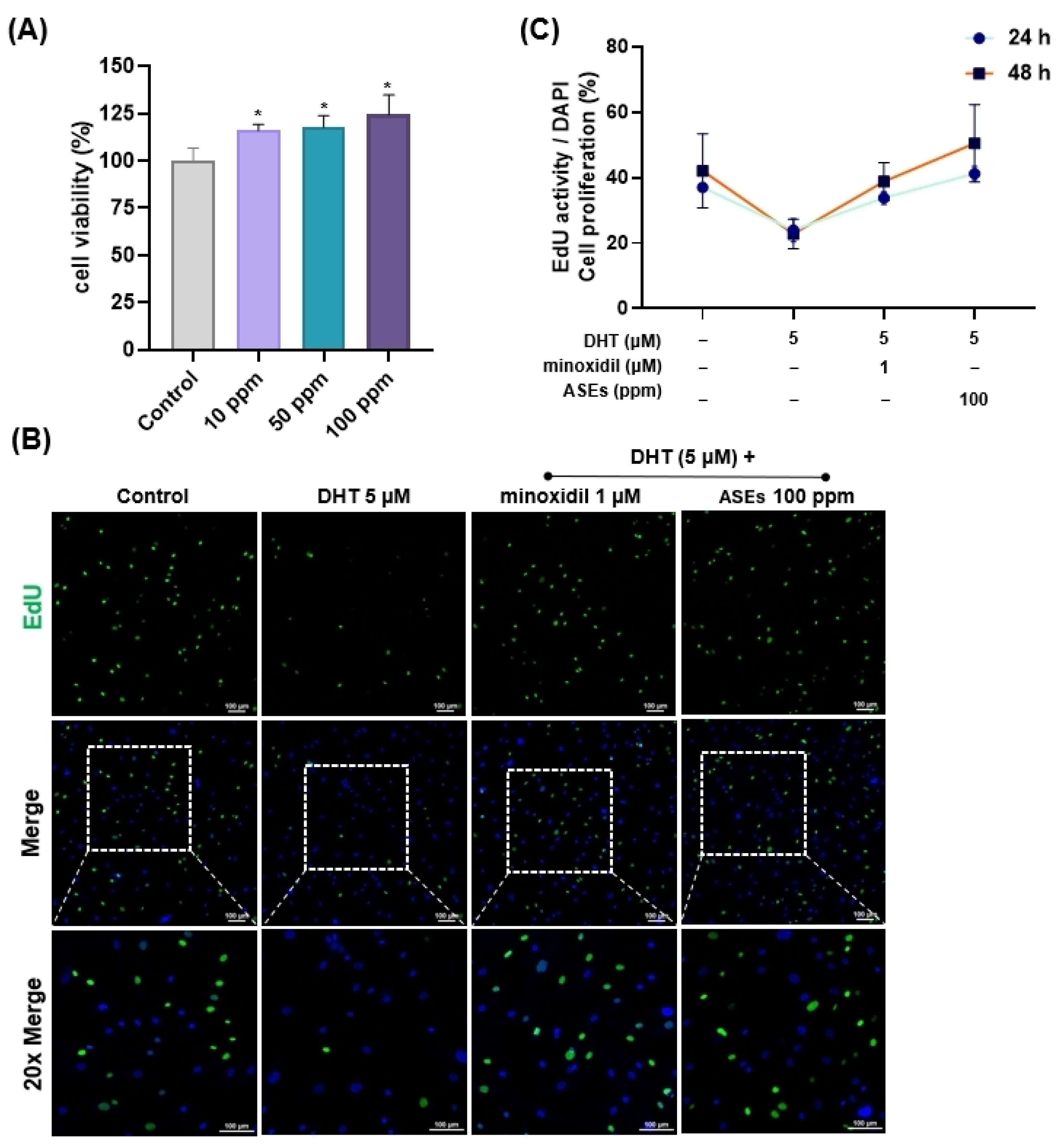
Figure 1. Cell viability and cell proliferation of ASEs in HFDPCs. (A) Cell viability of ASEs at different concentrations, was assessed by EZ-cytox assay. (B) Cells were treated with 5 μM DHT, 1 μM minoxidil, or 100 ppm ASEs for 48 h. Cell proliferation was determined by EdU staining assay (scale bars, 100 µm). (C) Cell proliferation was measured by EdU staining assay after 24 and 48 h. Cell viability and proliferation were determined as the percentage (%) of viable cells compared to the untreated cells. All data are presented as mean ± SD (n = 3). These images were acquired from three independent experiments. * p < 0.05 compared to the control group.
Cosmetics 12 00006 g001
Figure 2. The wound healing effect of ASEs in DHT-damaged HFDPCs. The wound healing assay was conducted for 24 h with HFDPCs damaged by 5 μM DHT. Cells were treated with ASEs at concentrations of 10, 50, and 100 ppm, as well as with 1 μM minoxidil, for 24 h (scale bars, 20 µm). Each image was captured using a phase-contrast microscope. These images were acquired from three independent experiments (n = 3).
Cosmetics 12 00006 g002
Figure 3. Effects of ASEs on the expression levels of ALP in DHT-damaged HFDPCs. (A) The ALP assay was performed on HFDPCs damaged by 5 μM DHT. HFDPCs were pre-treated with 100 ppm ASEs and 1 μM minoxidil for 24 h, followed by exposure to 5 μM DHT to induce damage (scale bars, 20 µm). (B) ALP activity was increased in HFDPCs treated with ASEs. These images were acquired from three independent experiments (n = 3). ** p < 0.01, *** p < 0.001 compared to the only DHT-treated group.
Cosmetics 12 00006 g003
Figure 4. Effects of ASEs on ROS levels in HFDPCs damaged by DHT. The measurement of ROS levels was performed for HFDPCs damaged by 5 μM DHT. (A) One hundred ppm ASEs and 1 μM minoxidil were pre-treated for 24 h followed by 5 μM DHT damage. (B) Reduced ROS levels in damaged HFDPCs treated with DHT from ASEs. DCF-DA images were captured using an Eclipse Ti2 fluorescence live-cell imaging microscope (scale bars, 100 µm), and the intensity of the green fluorescence (FITC) correlates with the concentration of ROS. These images were acquired from three independent experiments (n = 3). ** p < 0.01 compared to the only DHT-treated group.
Cosmetics 12 00006 g004
Figure 5. Effects of ASEs on mitochondrial potential and ATP activity in DHT-damaged HFDPCs. (A) HFDPCs were pre-treated with 100 ppm ASEs and 1 μM minoxidil for 24 h. JC-1 images were captured using Eclipse Ti2 fluorescence live-cell imaging (scale bars, 100 µm). Red puncta (RITC) indicates hyperpolarized mitochondria while green puncta (FITC) indicates depolarized mitochondria. (B) Mitochondrial membrane potential was increased in HFDPCs treated with ASEs. (C) ATP assay was performed under the same conditions. Red puncta (RITC) represents ATP activity while green puncta (FITC) represents mitochondria dye (scale bars, 50 µm). (D) ATP activity was higher in HFDPCs treated with ASEs. These images were acquired from three independent experiments (n = 3). * p < 0.05, ** p < 0.01 compared to the only DHT-treated group.
Cosmetics 12 00006 g005
Figure 6. Effects of ASEs on the phosphorylation level of AKT/ERK, β-Catenin, and GSK-3β in HFDPCs damaged by DHT. HFDPCs treated with ASEs (10, 50, and 100 ppm) for 24 h were harvested and analyzed for p-ERK, ERK, p-AKT, AKT, p-GSK, GSK, β-catenin, and β-actin using Western blot assay. (A) Relative expression band of each protein in the ASE-treated group. (B) Bar graph showing relative expression levels of AKT, ERK, β-Catenin, and GSK-3β in ASE-treated groups. All data were shown as the mean ± SD (n = 3). * p < 0.05, ** p < 0.01, compared to the only DHT-treated group.
Cosmetics 12 00006 g006
Figure 7. A schematic diagram shows how ASEs activate the ERK/AKT and β-catenin pathway. ASEs suppressed ROS in HFDPC damaged by DHT and activated the ERK/AKT pathway and β-catenin. This intervention led to the improvement and prevention of hair loss.
Cosmetics 12 00006 g007
Disclaimer/Publisher’s Note: The statements, opinions and data contained in all publications are solely those of the individual author(s) and contributor(s) and not of MDPI and/or the editor(s). MDPI and/or the editor(s) disclaim responsibility for any injury to people or property resulting from any ideas, methods, instructions or products referred to in the content.