Article

Solid Lipid Nanoparticles for Skin Delivery of Trans-Resveratrol: Impact of Preparation Methods on Formulation Stability

by
Andressa Tardim Cruz
1,
Leonardo Delello Di Filippo
1,
Jonatas Lobato Duarte
1,
Antonio José Guillot
2,
Alberto Pérez-García
3,
Ana Melero
2 and
Marlus Chorilli
1,*
1
Department of Drugs and Medicines, School of Pharmaceutical Sciences, Sao Paulo State University “Julio de Mesquita Filho”, Araraquara 14800903, SP, Brazil
2
Department of Pharmacy and Pharmaceutical Technology and Parasitology, University of Valencia, 46010 Valencia, Spain
3
Servicio de Cirugía Plástica y Quemados, Hospital Universitario y Politécnico La Fe, 46026 Valencia, Spain
*
Author to whom correspondence should be addressed.
Cosmetics 2025, 12(1), 7; https://doi.org/10.3390/cosmetics12010007
Submission received: 1 October 2024 / Revised: 10 December 2024 / Accepted: 17 December 2024 / Published: 7 January 2025
(This article belongs to the Special Issue Nanotechnology Advances in Cosmetics)

Abstract

:
Trans-resveratrol (RES) is a natural polyphenol known for its antioxidant, anti-inflammatory, and anti-aging properties, making it highly valuable in cosmetic applications. Solid lipid nanoparticles (SLNs) offer a promising solution to enhance RES’s stability and cutaneous availability. This study aimed to develop and characterize SLNs encapsulating RES for enhanced skin delivery. Multiple methodologies were evaluated to determine the impact of preparation methods on formulation stability. SLNs were formulated using stearic acid, soy phosphatidylcholine, polysorbate 80, cetyltrimethylammonium bromide, and poloxamer 407, with variations in heating temperatures and homogenization techniques. Stability assessments were conducted over 90 days, examining organoleptic properties of the hydrodynamic diameter, polydispersity index, and zeta potential. Encapsulation efficiency and skin permeation studies were performed to investigate the efficacy of SLNs in delivering RES. Results demonstrated that formulations prepared with Ultra Turrax at 24,000 rpm and heating at higher temperatures exhibited enhanced stability and smaller particle sizes. The selected formulations, F1 (prepared at 80 °C) and F2 (prepared at 70 °C) presented encapsulation efficiencies of 70% and 72%, respectively. Skin permeation studies confirmed the ability of SLNs to facilitate RES delivery through the skin. The study concludes that SLNs are suitable carriers for RES skin delivery, offering improved stability and sustained release, thus representing a promising approach for topical applications to leverage RES’s cutaneous therapeutic benefits.

1. Introduction

Trans-resveratrol (3, 5, 4’-trihydroxy-trans-stilbene) is a naturally occurring polyphenolic compound in various plants, including grapes, berries, and peanuts. Its wide range of biological activities, including antioxidant, anti-inflammatory, anti-cancer, and cardioprotective effects, has sparked significant interest. In dermatology, where it can combat skin aging, inflammation, and oxidative stress, trans-resveratrol (RES) holds immense potential [1,2].
Despite its wide variety of properties, the biological effectiveness of RES is limited by its physical–chemical properties, such as poor water solubility, chemical instability, and rapid metabolism, which result in low bioavailability and low skin permeation [3]. This is where solid lipid nanoparticles (SLNs) step in, offering an innovative and efficient approach for RES skin delivery. SLNs are nano-sized particles used in drug delivery, composed of biodegradable and biocompatible solid lipids and surfactants that can encapsulate lipophilic drugs, protecting them from degradation and enhancing their bioavailability [4].
SLNs could offer several advantages for the topical delivery of RES. They can provide a controlled release mechanism, improve drug stability, and enhance skin penetration. Owing to their composition and size, SLNs can better interact with the stratum corneum, facilitating deeper penetration of the encapsulated drug. Additionally, SLNs can form a protective hydrophobic film on the skin, reducing water loss and enhancing hydration, essential for maintaining skin health and increasing drug permeation. These properties of SLNs hold great promise for the future of RES skin delivery from topical cosmetic products [5].
Several studies have explored lipid-based nanocarriers for the topical delivery of resveratrol, focusing on their physical properties and biological activities. Liu et al., (2021) demonstrated that resveratrol-loaded nanostructured lipid carrier (NLC) hydrogels effectively enhanced anti-UV and antioxidant efficacy, emphasizing the stability and epidermal accumulation of resveratrol to protect against UV-induced oxidative damage [6]. Similarly, Soldati et al., (2018) explored the controlled release of resveratrol from solid lipid nanoparticles (SLNs), reporting increased antioxidant activity and skin permeation compared to unencapsulated resveratrol [7]. Gokce et al., (2012) compared SLNs and NLCs, finding that NLCs penetrated deeper into the epidermis and had a superior antioxidant profile [8]. More recently, Samprasit et al., (2024) highlighted the comparative properties of lipid-based carriers such as NLCs and nanoemulsions, showcasing their respective advantages in topical delivery and antioxidant performance [9].
The current study distinguishes itself from previous investigations by exploring the impact of preparation methods on the formulation stability and skin delivery performance of resveratrol-loaded solid lipid nanoparticles (SLNs), emphasizing how varying preparation parameters, such as homogenization speed and temperature, influence key physicochemical properties like particle size, zeta potential, and encapsulation efficiency.

2. Materials and Methods

2.1. Development of SLNs

SLNs were prepared using adapted methods described by Pardeike, Hommoss, and Müller (2009) [10], Lim et al., (2004) [11], and Mehnert and Mäder (2001) [4]. To develop the SLN formulations (Table 1), three methodologies were tested to evaluate the impact of preparation methods on the final stability. Initially, a pre-emulsion was prepared by melting the lipid phase at 5, 10, or 15 °C above the lipid’s melting point and dispersing it in an aqueous surfactant solution heated to the same temperatures. This process was consistent across all tested formulations and methodologies. Then, the pre-emulsions were homogenized using a magnetic stirrer at 1000 rpm or an Ultra Turrax at 11,000 or 24,000 rpm for 2 min. After homogenization, the pre-emulsions were sonicated for 20 min at 25% amplitude. To eliminate titanium particles from the sonication process, the formulations were centrifuged at 3500 rpm for 10 min [12].

2.2. Time Stability Testing of Formulations

The formulations were stored in a refrigerator (4 ± 2 °C) and evaluated for the hydrodynamic diameter, polydispersity index, and zeta potential. Analyses were conducted at 24 h, 7, 15, 30, 60, and 90 days after preparation.

2.3. Hydrodynamic Diameter Polydispersity Index and Zeta Potential Analysis

The nanoparticles’ hydrodynamic diameter and polydispersity index were determined by dynamic light scattering (DLS) using a Zetasizer Nano NS (Malvern Instruments, Worcesershire, UK). Nanoparticle dispersions were diluted in Milli-Q water (10 μL/mL) and maintained in dust-free cuvettes. Measurements were conducted at a scattering angle of 90°, a temperature of 25 °C, a laser wavelength of 633 nm, and a refractive index of 1.311. Three measurements were taken to calculate the mean and standard deviation of the diameter and polydispersity index.
SLNs were diluted in Milli-Q water (10 μL/mL) for zeta potential analysis. Electrophoretic mobility determined Zeta potential using the Helmholtz–Smoluchowski equation with a Zetasizer Nano NS (Malvern Instruments, Malvern, UK). Samples were placed in an electrophoretic cell, and three determinations of surface potential were conducted for each sample to calculate the mean and standard deviation.

2.4. Encapsulation Efficiency

An analytical curve of RES was determined in triplicate using a UV spectrophotometer (Carry 60, Agilent Technologies, Santa Clara, USA) at a wavelength of 306 nm, within a working range of 1 to 20 μg/mL. The encapsulation efficiency (EE%) was determined using an adapted method described by Gokce et al., (2012) [8]. A cellulose acetate membrane (0.45 µm pore size, Chromafil Xta) was used to separate the free RSV. The membrane was placed in a centrifuge tube, forming a donor compartment. The tube was filled with 13 mL of a 1:1 ethanol–water solution. Each formulation (300 µL, n = 3) was placed in the donor compartment and centrifuged at 5000 rpm for 1 h. After centrifugation, a 1 mL aliquot from the receptor compartment was diluted with 1 mL ethanol–water solution (1:1). The RES content was analyzed by UV spectroscopy. The EE% was calculated using Equation (1).
E E % = ( W i W f W i ) × 100

2.5. Skin Permeation

Permeation experiments were conducted using human abdominal skin samples obtained from surgical corrections. Patients gave consent, and the protocol was approved by the Research Ethics Committee of the University of Valencia, Spain (protocol H1462978691586). Excess fat and connective tissues were removed, and samples were stored at −26 °C for less than three months. Epidermal membranes were prepared by the heat-separation technique by immersing the skin in water at 60 °C for 2 min and then carefully separating the epidermis from the dermis.
In vitro permeation studies were conducted using glass Franz diffusion cells with an available diffusion area of 1.76 cm2 and a 12 mL receptor compartment placed in a heating/stirring module. Skin samples were mounted on the diffusion cells with the stratum corneum side in contact with the donor compartment and the dermal side in the receptor compartment. They were equilibrated for 1 h, and air bubbles were removed from the diffusion cell. The receptor solution was an aqueous polysorbate of 80 solutions (2.0% w/v) to ensure sink conditions throughout the test [13]. The cells were covered with parafilm to prevent evaporation. Samples were taken at specific time intervals from the receptor compartment, followed by replacement with fresh receptor solution. Permeation samples were analyzed “as is”. At the end of the experiments, a 1% red phenol solution was added to test the integrity of the skin samples. RES concentration in the receptor solution was determined by a HPLC-validated method (SM). The column used was a C18 Luna (250 mm × 4.6 mm I.D., 5 µm; Phenomenex, USA), with a mobile phase composed of water and acetonitrile (60:40).

3. Results and Discussion

The preparation methodologies significantly influenced the hydrodynamic diameter, zeta potential, and overall stability of the SLN formulations. Three distinct methods were tested, namely magnetic stirring, Ultra Turrax homogenization at low speed (11,000 rpm), and Ultra Turrax homogenization at high speed (24,000 rpm). These methods were applied to F1 and F2 formulations at varying heating temperatures (70 °C, 75 °C, and 80 °C).
F1 and F2 formulations differ in their compositions by the presence of cetyltrimethylammonium bromide (CTAB) in F2 formulations, a cationic surfactant. This difference in composition impacted several analyzed parameters, including hydrodynamic diameter, ZP, and stability.
Particle size is crucial for effective skin delivery. Smaller particles enhance skin penetration by ensuring increased contact with the stratum corneum due to an amphiphilic surface and higher contact area [14,15]. For F1, the particle size varied significantly with all temperatures and the type of mechanical stirring (Table 2). The magnetic stirrer at 75 °C consistently produced larger particles, reaching a maximum of 1262 nm at 90 days. In contrast, at 70 °C, smaller particle sizes were observed, peaking at 358.5 nm by 90 days. The use of a Turrax at 11,000 rpm demonstrated better control over particle size, yielding smaller particles, with sizes generally below 400 nm at 70, 75, and 80 °C compared to the use of the magnetic stirrer. The most no2 reduction occurred at Turrax 24,000 rpm, where particles at 70 °C measured 248.5 nm at 1 day, further decreasing to 228.6 nm after 7 days. The Turrax at 24,000 rpm provides higher energy input than lower rpm settings. This higher energy input leads to better particle size reduction and a more uniform distribution. The increased shear force at 24,000 rpm effectively breaks down lipid droplets into smaller particles, resulting in particle sizes of 248.5 nm at 1 day and 228.6 nm after 7 days. In contrast, lower energy input, such as that produced by a magnetic stirrer or lower rpm settings, tends to generate larger particles and higher polydispersity indices, affecting stability and uniformity. These findings align with previous studies indicating that lower energy input during nanoparticle preparation results in larger particle sizes and higher polydispersity indices, affecting stability and uniformity [4].
In contrast, Ultra Turrax homogenization at 11,000 rpm produced smaller particle sizes (300–500 nm) but showed instability over 90 days due to insufficient energy input, leading to aggregation. The Ultra Turrax homogenization at 24,000 rpm achieved the smallest and most stable particles. F1 formulations had particle sizes between 300 and 500 nm. The literature shows that a finer dispersion can be obtained by increasing energy by raising the production temperature, enhancing the stirring speed, prolonging emulsification time, or utilizing more intense ultrasound power. These techniques help reduce particle size and improve dispersion uniformity by supplying sufficient energy to overcome interfacial tension, generating more stable particles [16].
The particle size of F2 varied significantly depending on the energy input method and temperature (Table 3). When using the magnetic stirrer, at 70 °C, the initial particle size was large, starting at 905.0 nm on day 1 and increasing further to 1307.4 nm by day 90. The presence of CTAB contributed to positive zeta potentials, which likely provided some stabilization for these larger particles, but the energy supplied by the magnetic stirrer was insufficient to prevent the formation of larger aggregates. At higher temperatures, such as 75 °C and 80 °C, smaller initial particles were observed, measuring 273.9 nm and 442.0 nm, respectively, though both conditions exhibited significant particle growth over time.
In contrast, the Turrax at 11,000 rpm produced considerably smaller particles across all temperatures. At 70 °C, particles began at 242.5 nm, grew to 332.1 nm by day 15, and stabilized around 504.5 nm by day 50. Similar patterns were observed at 75 °C and 80 °C, with slightly larger particle sizes but still smaller than those formed using the magnetic stirrer. The Turrax at 24,000 rpm produced the smallest and most stable particles for F2, with initial sizes at 70 °C as low as 169.3 nm on day 1 and only increasing to 301.7 nm by day 90. The high shear forces, combined with the stabilizing influence of CTAB, effectively minimized particle growth and maintained a more stable particle size over time (Table 3). Similar observations have been made in studies where higher shear forces during homogenization led to smaller and more stable nanoparticles [10].
The preparation method significantly impacts the size of SLNs. The smaller particle size and narrow size distribution achieved with high-speed Ultra Turrax homogenization suggest enhanced potential for effective skin penetration and consistent delivery of resveratrol. This is crucial for therapeutic applications, where consistent and prolonged drug delivery is desired. The smaller particle sizes achieved through high-energy methods can facilitate better skin absorption and enhanced therapeutic efficacy [17].
Zeta potential, indicating surface charge, is a critical indicator of particle stability. Higher absolute ZP values suggest stronger repulsive forces, reducing aggregation. This electrostatic stabilization is essential for maintaining the integrity and efficacy of nanoparticle formulations over time [18].
Magnetic stirring produced formulations with lower absolute ZP values, implying weak electrostatic stabilization. Ultra Turrax at 11,000 rpm improved these values but stability remained suboptimal. At 24,000 rpm, Ultra Turrax yielded the highest absolute ZP values, with −20 to −30 mV for F1 and positive values for F2 due to CTAB, demonstrating superior electrostatic stabilization and possible reduced aggregation (Table 2). This finding is consistent with the literature, which shows that higher ZP values are associated with better stability due to increased electrostatic repulsion between particles [19,20].
Magnetic stirring resulted in high PdI values, showing broad particle size distribution. Ultra Turrax at 11,000 rpm improved PdI but with some heterogeneity. The 24.000 rpm method achieved the lowest PdI values (~0.25 for F1 and below 0.2 for F2), indicating a narrow particle size distribution and high uniformity, maintaining stability over 90 days (Table 2 and Table 3). A low PdI value indicates a homogeneous nanoparticle population, which is crucial for reproducibility and efficacy in drug delivery systems [17,21].
The uniformity in particle size distribution is essential for ensuring consistent drug release and absorption. A narrow size distribution minimizes the variability in drug delivery, leading to more predictable therapeutic outcomes. This consistency is vital for clinical applications, where dose precision and reliability are paramount [22].
The EE% is crucial for evaluating the effectiveness of SLNs. The Ultra Turrax method at 24,000 rpm achieved the highest EE% values, with 70% for F1 and 72% for F2. This aligns with previous studies [8], showing that high-energy methods enhance active compound dispersion within the lipid matrix. CTAB in F2 likely contributed to a slightly higher EE% due to improved interaction between the lipid matrix and the active compound. High encapsulation efficiencies are essential for maximizing the therapeutic potential of encapsulated drugs, particularly for compounds like resveratrol that suffer from poor water solubility and stability [23]; in this way, we chose the F2 formulation to perform the permeation study.
Permeation studies have highlighted significant differences between free RES in Tween 80 solution and encapsulated RES (F2). Free RES skin permeation was significantly higher than encapsulated RES, thus confirming the ability of the particles to retain the encapsulated RES (Figure 1). The high standard deviation observed in both profiles is due to the non-uniformity of the skin tissue and the low permeated amounts, which were close to the quantification limit. In addition, free RES presents low solubility and high instability, affecting reproducibility. As the concentration in both formulations is the same, the permeability coefficient calculated by dividing the flow by the dose is also higher for the RES solution. However, the solution is close to saturation, which indicates a high thermodynamic activity, promoting its potential for enhanced skin permeation. This is not the case in the encapsulated form, where two fractions can be observed, namely the encapsulated remaining RES and the released RES. In this case, the main objective was to achieve a controlled release and prolonged permeability of RES over time. These findings align with the literature, where nanoparticle encapsulation significantly reduced skin permeation by a controlled release [24,25].
The improved permeation rates suggest that encapsulated RES can effectively reach the systemic compartment, potentially enhancing its therapeutic effects for skin conditions such as aging and inflammation. This improved permeation is likely due to the lipophilic nature of the SLNs, which facilitates better interactions with the drug. It is also expected that these SLNs will also interact with the stratum corneum due to their lipid nature, thus improving its hydration [26].
The enhanced delivery seen with encapsulated RES emphasizes the potential of SLNs to modify the bioavailability of topically applied drugs, leading to more efficient treatments for various dermatological conditions and harnessing the full therapeutic potential of resveratrol [27,28].
The permeation flux and permeation coefficient for the free resveratrol solution and the encapsulated resveratrol formulation reveal key differences in their skin delivery performance. The free resveratrol solution showed a higher permeation flux of 145 ± 060 µg/cm2/h compared to the encapsulated resveratrol formulation, which had a flux of 098 ± 028 µg/cm2/h. This indicates that free resveratrol is more immediately available for diffusion across the skin due to the absence of a delivery matrix. Conversely, the encapsulated resveratrol formulation demonstrated a higher permeation coefficient of 16,157 × 10−6 ± 67,201 × 10−7 cm/s compared to 10,843 × 10−6 ± 64,995 × 10−7 cm/s for the free resveratrol solution. This suggests that the solid lipid nanoparticles enhance resveratrol’s ability to partition through the skin layers despite a lower overall flux. The lipid nanoparticles facilitate better interaction with the stratum corneum, allowing for deeper penetration and more efficient delivery over time. The lower flux but higher permeation coefficient of the encapsulated formulation supports the notion of a controlled release mechanism, where the resveratrol is gradually delivered. This controlled release helps maintain stability and prolong therapeutic or cosmetic effects, making the encapsulated formulation advantageous for topical applications where sustained efficacy is desired.

4. Conclusions

Variations in SLN preparation methods and heating temperatures significantly influenced the formulations’ hydrodynamic diameter, zeta potential, and polydispersity index. Using the Ultra-Turrax at 24,000 rpm resulted in smaller and more uniform particles than magnetic stirring. Specifically, Formulation F1 (prepared at 80 °C) and Formulation F2 (prepared at 70 °C) exhibited the best stability and reproducibility, with 70% and 72% encapsulation efficiencies, respectively.
Furthermore, the encapsulated resveratrol (RES) formulations demonstrated controlled and sustained skin permeation compared to free RES in solution. This behavior suggests that solid lipid nanoparticles (SLNs) can effectively enhance RES’s stability, delivery, and potential efficacy for topical applications. These findings highlight the importance of optimizing preparation parameters to achieve stable and efficient SLN formulations for the enhanced skin delivery of resveratrol.

Author Contributions

Conceptualization, M.C.; methodology, M.C., A.M., A.J.G. and A.P.-G.; formal analysis, A.T.C., J.L.D. and L.D.D.F.; investigation, A.T.C.; resources, M.C. and A.M.; writing—original draft preparation, J.L.D. and L.D.D.F.; writing—review and editing, M.C. and A.M.; supervision, M.C. and A.M.; project administration, M.C.; funding acquisition, M.C. All authors have read and agreed to the published version of the manuscript.

Funding

This research was funded by Sao Paulo Research Foundation (FAPESP), grant number 17/05566-3, 17/24365-9 and 12/19568-4.

Data Availability Statement

The raw data supporting the conclusions of this article will be made available by the authors on request.

Acknowledgments

We would like to thank Roberta Balansin Rigon for the technical support.

Conflicts of Interest

The authors declare no conflict of interest.

References

  1. Diaz-Gerevini, G.T.; Repossi, G.; Dain, A.; Tarres, M.C.; Das, U.N.; Eynard, A.R. Beneficial Action of Resveratrol: How and Why? Nutrition 2016, 32, 174–178. [Google Scholar] [CrossRef] [PubMed]
  2. Frémont, L. Biological Effects of Resveratrol. Life Sci. 2000, 66, 663–673. [Google Scholar] [CrossRef] [PubMed]
  3. Baur, J.A.; Sinclair, D.A. Therapeutic Potential of Resveratrol: The in vivo Evidence. Nat. Rev. Drug Discov. 2006, 5, 493–506. [Google Scholar] [CrossRef] [PubMed]
  4. Mehnert, W.; Mäder, K. Solid Lipid Nanoparticles: Production, Characterization and Applications. Adv. Drug Deliv. Rev. 2012, 64, 83–101. [Google Scholar] [CrossRef]
  5. Müller, R.H.; Mäder, K.; Gohla, S. Solid Lipid Nanoparticles (SLN) for Controlled Drug Delivery—A Review of the State of the Art. Eur. J. Pharm. Biopharm. 2000, 50, 161–177. [Google Scholar] [CrossRef]
  6. Miao, L.; Daozhou, L.; Ying, C.; Qibing, M.; Siyuan, Z. A Resveratrol-Loaded Nanostructured Lipid Carrier Hydrogel to Enhance the Anti-UV Irradiation and Anti-Oxidant Efficacy. Colloids Surf. B Biointerfaces 2021, 204, 111786. [Google Scholar] [CrossRef]
  7. Soldati, P.P.; Polonini, H.C.; Paes, C.Q.; Restrepob, J.A.S.; Creczynksi-Pasa, T.B.; Chaves, M.G.A.M.; Brandão, M.A.F.; Pittella, F.; Raposo, N.R.B. Controlled Release of Resveratrol from Lipid Nanoparticles Improves Antioxidant Effect. In Proceedings of the IFAC-PapersOnLine; Elsevier B.V.: Amsterdam, The Netherlands, 2018; Volume 51, pp. 16–21. [Google Scholar]
  8. Gokce, E.H.; Korkmaz, E.; Dellera, E.; Sandri, G.; Cristina Bonferoni, M.; Ozer, O. Resveratrol-Loaded Solid Lipid Nanoparticles versus Nanostructured Lipid Carriers: Evaluation of Antioxidant Potential for Dermal Applications. Int. J. Nanomed. 2012, 7, 1841. [Google Scholar] [CrossRef]
  9. Samprasit, W.; Suriyaamporn, P.; Sriamornsak, P.; Opanasopit, P.; Chamsai, B. Resveratrol-Loaded Lipid-Based Nanocarriers for Topical Delivery: Comparative Physical Properties and Antioxidant Activity. OpenNano 2024, 19. [Google Scholar] [CrossRef]
  10. Pardeike, J.; Hommoss, A.; Müller, R.H. Lipid Nanoparticles (SLN, NLC) in Cosmetic and Pharmaceutical Dermal Products. Int. J. Pharm. 2009, 366, 170–184. [Google Scholar] [CrossRef]
  11. Lim, S.J.; Lee, M.K.; Kim, C.K. Altered Chemical and Biological Activities of All-Trans Retinoic Acid Incorporated in Solid Lipid Nanoparticle Powders. J. Control Release 2004, 100, 53–61. [Google Scholar] [CrossRef]
  12. Agayan, R.R.; Horvath, T.; McNaughton, B.H.; Anker, J.N.; Kopelman, R. Optical Manipulation of Metal-Silica Hybrid Nanoparticles. Opt. Trapp. Opt. Micromanipulation 2004, 5514, 502–513. [Google Scholar] [CrossRef]
  13. Detoni, C.B.; Souto, G.D.; da Silva, A.L.M.; Pohlmann, A.R.; Guterres, S.S. Photostability and Skin Penetration of Different E -Resveratrol-Loaded Supramolecular Structures. Photochem. Photobiol. 2012, 88, 913–921. [Google Scholar] [CrossRef]
  14. Souto, E.B.; Fangueiro, J.F.; Fernandes, A.R.; Cano, A.; Sanchez-Lopez, E.; Garcia, M.L.; Severino, P.; Paganelli, M.O.; Chaud, M.V.; Silva, A.M. Physicochemical and Biopharmaceutical Aspects Influencing Skin Permeation and Role of SLN and NLC for Skin Drug Delivery. Heliyon 2022, 8, e08938. [Google Scholar] [CrossRef] [PubMed]
  15. Labouta, H.I.; El-Khordagui, L.K.; Kraus, T.; Schneider, M. Mechanism and Determinants of Nanoparticle Penetration through Human Skin. Nanoscale 2011, 3, 4989–4999. [Google Scholar] [CrossRef]
  16. Hou, D.; Xie, C.; Huang, K.; Zhu, C. The Production and Characteristics of Solid Lipid Nanoparticles (SLNs). Biomaterials 2003, 24, 1781–1785. [Google Scholar] [CrossRef]
  17. Jores, K.; Mehnert, W.; Drechsler, M.; Bunjes, H.; Johann, C.; Mäder, K. Investigations on the Structure of Solid Lipid Nanoparticles (SLN) and Oil-Loaded Solid Lipid Nanoparticles by Photon Correlation Spectroscopy, Field-Flow Fractionation and Transmission Electron Microscopy. J. Control Release 2004, 95, 217–227. [Google Scholar] [CrossRef]
  18. Honary, S.; Zahir, F. Effect of Zeta Potential on the Properties of Nano-Drug Delivery Systems—A Review (Part 1). Trop. J. Pharm. Res. 2013, 12, 255–264. [Google Scholar] [CrossRef]
  19. Müller, R.H.; Radtke, M.; Wissing, S.A. Nanostructured Lipid Matrices for Improved Microencapsulation of Drugs. Int. J. Pharm. 2002, 242, 121–128. [Google Scholar] [CrossRef]
  20. Rawat, M.; Jain, A.; Mishra, A.; Muthu, M.; Singh, S. Development of Repaglinide Loaded Solid Lipid Nanocarrier: Selection of Fabrication Method. Curr. Drug Deliv. 2010, 7, 44–50. [Google Scholar] [CrossRef]
  21. Danaei, M.; Dehghankhold, M.; Ataei, S.; Hasanzadeh Davarani, F.; Javanmard, R.; Dokhani, A.; Khorasani, S.; Mozafari, M.R. Impact of Particle Size and Polydispersity Index on the Clinical Applications of Lipidic Nanocarrier Systems. Pharmaceutics 2018, 10, 57. [Google Scholar] [CrossRef]
  22. Kim, J.-K.; Howard, M.D.; Dziubla, T.D.; Rinehart, J.J.; Jay, M.; Lu, X. Uniformity of Drug Payload and Its Effect on Stability of Solid Lipid Nanoparticles Containing an Ester Prodrug. ACS Nano 2011, 5, 209–216. [Google Scholar] [CrossRef] [PubMed]
  23. Souto, E.B.; Müller, R.H. Lipid Nanoparticles: Effect on Bioavailability and Pharmacokinetic Changes. Handb. Exp. Pharmacol. 2010, 197, 115–141. [Google Scholar] [CrossRef]
  24. Ourique, A.F.; Melero, A.; de Bona da Silva, C.; Schaefer, U.F.; Pohlmann, A.R.; Guterres, S.S.; Lehr, C.-M.; Kostka, K.-H.; Beck, R.C.R. Improved Photostability and Reduced Skin Permeation of Tretinoin: Development of a Semisolid Nanomedicine. Eur. J. Pharm. Biopharm. 2011, 79, 95–101. [Google Scholar] [CrossRef] [PubMed]
  25. Melero, A.; Ferreira Ourique, A.; Stanisçuaski Guterres, S.; Raffin Pohlmann, A.; Lehr, C.-M.; Ruver Beck, R.C.; Schaefer, U. Nanoencapsulation in Lipid-Core Nanocapsules Controls Mometasone Furoate Skin Permeability Rate and Its Penetration to the Deeper Skin Layers. Ski. Pharmacol. Physiol. 2014, 27, 217. [Google Scholar] [CrossRef]
  26. Wissing, S. Cosmetic Applications for Solid Lipid Nanoparticles (SLN). Int. J. Pharm. 2003, 254, 65–68. [Google Scholar] [CrossRef]
  27. Fang, J.-Y.; Fang, C.-L.; Liu, C.-H.; Su, Y.-H. Lipid Nanoparticles as Vehicles for Topical Psoralen Delivery: Solid Lipid Nanoparticles (SLN) versus Nanostructured Lipid Carriers (NLC). Eur. J. Pharm. Biopharm. 2008, 70, 633–640. [Google Scholar] [CrossRef]
  28. Garcês, A.; Amaral, M.H.; Sousa Lobo, J.M.; Silva, A.C. Formulations Based on Solid Lipid Nanoparticles (SLN) and Nanostructured Lipid Carriers (NLC) for Cutaneous Use: A Review. Eur. J. Pharm. Sci. 2018, 112, 159–167. [Google Scholar] [CrossRef]
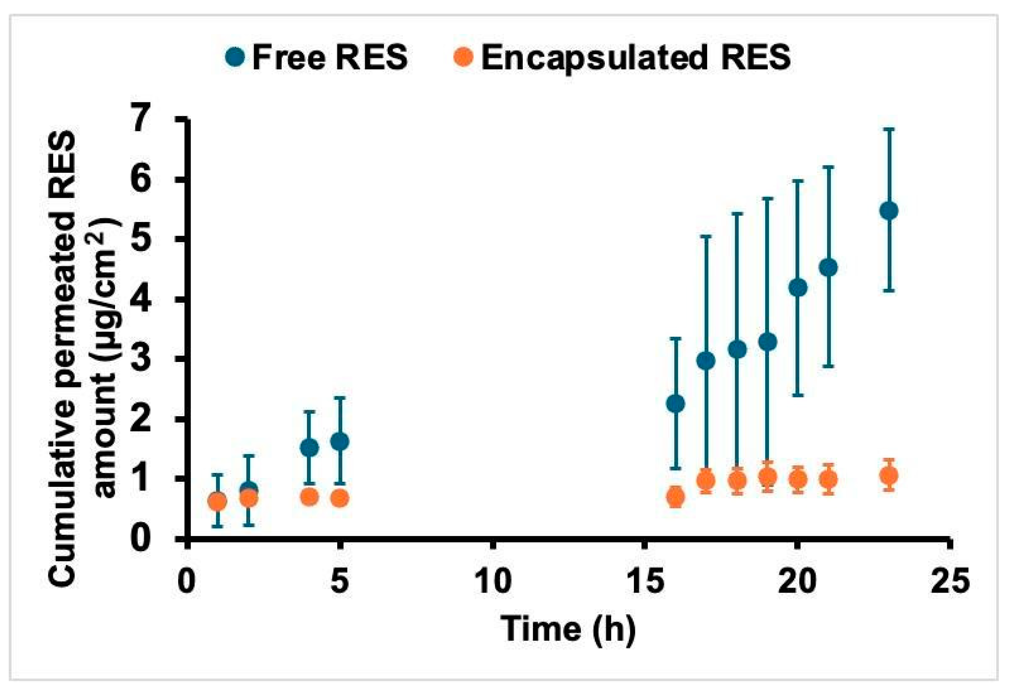
Figure 1. Cumulative amounts of resveratrol from encapsulated RSV (F2) and free RES in Tween 80 solution.
Cosmetics 12 00007 g001
Table 1. Composition of the developed formulations *.
Components (%)
F1F1-RESF2F2-RES
Stearic acid (SA)5.05.05.05.0
Soy phosphatidylcholine (SPC)1.21.21.21.2
Polysorbate 80 (T80)3.53.53.53.5
Cetyltrimethylammonium bromide (CTAB)--0.450.45
Poloxamer 407 ® (P407)0.10.10.10.1
Glycerin (GLI)0.150.150.150.15
Trans-resveratrol (RES)-0.25-0.25
Phenonip ®0.750.750.750.75
Ethanol P.A.1.251.251.251.25
Ultra-pure waterq.s.p.q.s.p.q.s.p.q.s.p.
* q.s.p.—quantity sufficient to make 10 mL, ® Registered trademark.
Table 2. Particle size, polydispersity index, and zeta potential of F1.
Magnetic Stirrer
DaysSize (nm)PdIZP (mV)
70 °C75 °C80 °C70 °C75 °C80 °C70 °C75 °C80 °C
1295.41068.1316.70.40.40.2−28.3−16.7−18.3
7287.4412.5319.20.20.40.3−23.8−18.2−22.0
15312.1846.2341.90.20.30.3−19.5−19.0−21.2
30317.01167.6329.00.20.30.3−18.9−16.7−20.0
50311.41197.7366.10.20.40.3−19.9−22.1−20.8
90358.51262.0378.60.30.30.3−20.7−22.6−22.2
Turrax 11,000 rpm
DaysSize (nm)PdIZP (mV)
70 °C75 °C80 °C70 °C75 °C80 °C70 °C75 °C80 °C
1313.1313.6284.60.30.20.2−20.4−19.1−18.9
7317.6323.3266.80.40.20.2−18.6−15.9−19.3
15318.9323.3285.80.40.20.3−19.8−15.9−19.5
30319.6330.3313.60.20.30.3−21.2−17.6−19.7
50769.9336.8745.30.30.20.3−19.7−20.9−24.2
90788.4347.01100.80.40.20.3−20.9−20.8−20.3
Turrax 24,000 rpm
DaysSize (nm)PdIZP (mV)
70 °C75 °C80 °C70 °C75 °C80 °C70 °C75 °C80 °C
1248.5367.6272.40.20.30.2−22.2−21.3−22.5
7228.6381.0285.80.20.30.2−19.8−20.0−20.9
15365.7384.5298.50.30.20.2−21.2−20.7−21.2
30246.9387.8314.40.20.20.2−19.4−20.3−18.6
50291.1388.5317.90.20.30.2−20.1−20.3−25.1
90316.6393.1330.90.20.30.2−16.3−20.6−22.9
Table 3. Particle size, polydispersity index, and zeta potential of F2.
Magnetic Stirrer
DaysSize (nm)PdIZP (mV)
70 °C75 °C80 °C70 °C75 °C80 °C70 °C75 °C80 °C
1905.0273.9442.00.50.30.327.626.325.2
7835.4346.8610.00.40.30.426.527.924.5
15928.8401.9665.00.40.40.425.329.227.4
301171.5455.3694.60.40.40.421.730.728.7
501225.0518.0712.10.50.50.532.728.928.7
901307.4535.5782.70.50.50.527.228.228.3
Turrax 11,000 rpm
DaysSize (nm)PdIZP (mV)
70 °C75 °C80 °C70 °C75 °C80 °C70 °C75 °C80 °C
1242.5219.4298.70.20.30.222.526.527.9
7302.6501.5358.30.30.30.314.729.532.6
15332.1584.2420.50.30.40.318.728.323.6
30459.4662.7455.20.30.30.424.828.729.5
50504.5683.1452.70.40.40.523.231.531.9
90556.1765.6611.90.40.40.524.230.923.1
Turrax 24,000 rpm
DaysSize (nm)PdIZP (mV)
70 °C75 °C80 °C70 °C75 °C80 °C70 °C75 °C80 °C
1169.3183.0251.70.20.20.226.125.128.4
7198.2311.7273.80.20.30.232.428.830.5
15219.5335.0318.50.20.30.322.025.326.7
30257.3392.1354.20.20.30.331.724.732.7
50274.1396.8430.50.20.30.426.127.930.8
90301.7402.3478.80.20.30.427.329.331.7
Disclaimer/Publisher’s Note: The statements, opinions and data contained in all publications are solely those of the individual author(s) and contributor(s) and not of MDPI and/or the editor(s). MDPI and/or the editor(s) disclaim responsibility for any injury to people or property resulting from any ideas, methods, instructions or products referred to in the content.