Article

Recombinant Type XVII Collagen Inhibits EGFR/MAPK/AP-1 and Activates TGF-β/Smad Signaling to Enhance Collagen Secretion and Reduce Photoaging

1
Xi’an Giant Biogene Technology Co., Ltd., Xi’an 710069, China
2
Engineering Research Center of Western Resource Innovation Medicine Green Manufacturing, Ministry of Education, School of Chemical Engineering, Northwest University, Xi’an 710127, China
3
Shaanxi Key Laboratory of Biomaterials and Synthetic Biology, Shaanxi R & D Center of Biomaterials and Fermentation Engineering, School of Chemical Engineering, Northwest University, Xi’an 710127, China
4
Biotech & Biomed Research Institute, Northwest University, Xi’an 710127, China
5
Shaanxi Green Bio-Manufacturing Future Industry Research Institute, Northwest University, Xi’an 710127, China
*
Authors to whom correspondence should be addressed.
Cosmetics 2025, 12(2), 59; https://doi.org/10.3390/cosmetics12020059
Submission received: 12 February 2025 / Revised: 21 March 2025 / Accepted: 24 March 2025 / Published: 27 March 2025
(This article belongs to the Section Cosmetic Dermatology)

Abstract

:
Studies have consistently shown that long-wave ultraviolet A (UVA) radiation triggers skin photoaging, which is evident as reduced elasticity, a loss of firmness, and signs of aging. There is an urgent need to investigate photoaging mechanisms to devise protective strategies against UVA. The present study aimed to explore the effects of recombinant type XVII collagen on UVA-induced skin aging and uncover its molecular mechanisms, thereby laying a solid theoretical foundation for precise treatments and prevention. We therefore modeled photoaging damage in HaCaT cells and evaluated collagen-related protein and gene expression levels via western blot analysis and real-time quantitative polymerase chain reaction analysis. Immunofluorescent staining was also used to assess collagen secretion and basement membrane protein expression. Recombinant type XVII collagen significantly boosted type IV and type XVII collagen, laminin alpha 5, and integrin β1 production, thus counteracting UVA-induced collagen decline. The polymerase chain reaction analysis revealed matrix metalloproteinase (MMP) downregulation and tissue inhibitor of metalloproteinase (TIMP) upregulation. Modulating the transforming growth factor (TGF)-β/Smad and epidermal growth factor receptor (EGFR)/mitogen-activated protein kinase (MAPK)/activator protein-1 (AP-1) pathways suppressed photoaging. Together, our findings suggest that recombinant type XVII collagen ameliorates UVA-induced damage by reversing MMP and TIMP gene expression, thereby preventing collagen degradation and enhancing basement membrane secretion. These results offer a theoretical basis for potent anti-photoaging products, thus paving the way for innovative solutions against UVA-induced skin aging.

1. Introduction

Up to 80% of skin aging can reportedly be attributed to photoaging, which is primarily caused by the sun’s ultraviolet (UV) rays: UVA and UVB. Of these, UVA rays penetrate deeply into the skin, damaging DNA and causing mutations that lead to long-term damage and an elevated risk of skin cancer. Photoaging not only increases cancer risk but also results in visible signs of aging such as wrinkles, pigmentation changes, and a loss of elasticity [1]. For example, prolonged UV exposure causes collagen breakdown. Collagen is crucial for skin firmness and elasticity, whereas elastin enables the skin to stretch and revert to its original shape. Damaged collagen and elastin fibers lead to loose and wrinkled skin [2]. Furthermore, UVA exposure inhibits dermal cells from synthesizing collagen and promotes the production of matrix metalloproteinases (MMP), thus leading to collagen degradation and the destruction of the elasticity and support structure of the skin [3]. By understanding the causes and effects of photoaging, proactive measures may be adopted to protect the skin and delay the aging process.
A previous comprehensive investigation revealed that the mitogen-activated protein kinase (MAPK) signaling cascade is of fundamental importance in the cellular response to UV irradiation [4]. The MAPK superfamily encompasses extracellular signal-regulated kinase (ERK), p38 MAPK, and c-Jun N-terminal kinase (JNK), each of which plays distinct yet interconnected roles. Of note, ERK and p38 MAPK function upstream in the activation of activator protein-1 (AP-1) via transactivation mechanisms [5]. Upon activation, the AP-1 signaling axis stimulates the expression and production of a spectrum of MMPs. Consequently, the regulation of MMP expression is predominantly governed by the intricate MAPK signal transduction pathway [6]. The initiation of MAPK signaling is preceded by the phosphorylation of tyrosine residues located within the epidermal growth factor receptor (EGFR), a transmembrane protein consisting of an extracellular domain, a transmembrane segment, and an intracellular region [7]. UV radiation catalyzes the phosphorylation of tyrosine residues in the intracellular segment of EGFR, thereby enabling the recruitment of the adaptor protein growth factor receptor-bound protein 2. This interaction facilitates the translocation of guanine nucleotide-releasing factor (SOS) from the cytoplasm to the plasma membrane. SOS subsequently catalyzes the activation of GTP, which binds to the RAS small GTPase, thus leading to the sequential activation of RAF and MAPK kinase (MEK). This series of events ultimately triggers the MAPK kinase kinase/MEK/MAPK signaling cascade, encompassing the activation of ERK, JNK, and p38 MAPK [8,9]. ERK signaling is instrumental in the activation and upregulation of c-Fos expression. Concurrently, JNK and p38 MAPK pathways exhibit a synergistic effect, phosphorylating c-Jun to enable its nuclear translocation and the formation of the active AP-1 heterodimer with c-Fos [10]. This resultant activation of AP-1 promotes MMP gene transcription, ultimately enhancing MMP synthesis and facilitating increased collagen degradation [11]. Together, these findings contribute to a deeper understanding of the intricate signaling networks that govern cellular responses to UV radiation.
Exposure to UVA radiation also exerts inhibitory effects on the transforming growth factor-β (TGF-β)/Smad signaling pathway, leading to reduced collagen synthesis and decreased collagen levels in the body, which in turn precipitate the onset of photoaging in skin tissue. TGF-β is a pivotal regulator that maintains collagen stability by stimulating type I and type III pro-collagen synthesis and inhibiting MMP expression [12]. In physiological contexts, TGF-β binds to the TGF-β type I and type II receptors, which possess serine/threonine kinase activity on the cellular surface. This binding results in Smad protein phosphorylation. Subsequently, activated Smad2 and Smad3 form a complex with Smad4 and translocate to the nucleus, thereby regulating the synthesis and secretion of collagen and the extracellular matrix. Concurrently, UVA radiation-induced activation of AP-1 downregulates TGF-β type II receptors and induces the expression of Smad7, an inhibitor of the TGF-β/Smad signaling pathway [13]. Overexpression of Smad7 inhibits TGF-β expression, which then inhibits the downstream phosphorylation of Smad2/3, effectively disrupting the TGF-β/Smad signaling cascade [14].
The main group of enzymes responsible for the degradation of collagen and other proteins in extracellular matrices comprises matrix metalloproteinases (MMPs) and tissue inhibitors of metalloproteinases (TIMPs) [15]. UV-induced AP-1 activation enhances the expression of MMP-1, MMP-3, and MMP-9 [16]. In the skin, epidermal keratinocytes and dermal fibroblasts mainly secrete MMP-1 (also known as interstitial collagenase or collagenase 1), which is a collagenase that degrades type I and type III fibrillar collagen into specific fragments at a single site within the central triple helix. In addition, MMP-1 is stimulated by the excessive production of reactive oxygen species and plays a key role in photoaging [17]. Both MMP-2 (also known as gelatinase A or 72-kDa type IV collagenase) and MMP-9 (also known as gelatinase B or 92-kDa type IV collagenase) are members of the MMP gelatinase subgroup, and their expression is largely dependent on AP-1 activation. They cleave type IV collagen and degrade other substrates, such as type V, type VII, and type X collagen as well as fibronectin and elastin [18,19]. MMP-3 (also known as stromelysin-1) degrades many extracellular matrix proteins, such as type IV, type V, type IX, and type X collagen as well as gelatin, fibrillin-1, fibronectin, laminin, and proteoglycans [20]. Naturally occurring inhibitors, known as TlMPs, are important factors that control MMP activity. Notably, tissue destruction in disease processes is often correlated with an imbalance of MMPs over TIMPs. The major inhibitors are TIMP-I and TIMP-2 [21], which bind to MMP complexes in a 1:1 ratio; each complex has a specific inhibitory effect on active MMPs [21].
Type XVII collagen (COL17) is a transmembrane protein that constitutes the hemidesmosome structure within the basement membrane, serving as a crucial junction between the dermis and epidermis of the skin [22]. This protein facilitates the transmission of information among stem cells, adjacent cells, and the extracellular matrix while also regulating cellular homeostasis, aging processes, and tissue repair mechanisms [23]. Unlike other fibrillar collagens, COL17 primarily mediates adhesion between keratinocytes and the basement membrane, thereby playing a fundamental role in maintaining the structural integrity of the epidermis [24]. Extensive research has demonstrated that COL17 can augment epidermal thickness, bolster barrier function, and promote epidermal regeneration [25]. Furthermore, it plays a pivotal role in skin anti-aging processes such as by ameliorating wrinkles and enhancing the expression of crucial type IV collagen (COL4) and basement membrane-associated proteins within the basement membrane zone, ultimately stabilizing the structural integrity of this zone [26,27]. Xiang et al. [28] found a positive correlation between reduced COL17 expression and epidermal aging in human skin through in vivo experiments. In vivo clonal analysis and in vitro 3D modeling in mice by N Liu et al. [24] found that the forced maintenance of COL17A1 rescues skin organ ageing. Y Liu et al. [23] demonstrated that COL17 is the only hemidesmosomal component to undergo a marked reduction during the aging process. Their findings suggest that this change in the level of COL17 is mainly driven by intrinsic aging and photoaging. Sun et al. [29] found that mRNA and protein expression levels of COL17α1 in mice decreased with age and hypothesized that this may be responsible for epidermal thinning and slow wound healing in elderly skin. This evidence further underscores the critical relationship between photoaging-induced cellular senescence and the regulation of COL17.
Nevertheless, despite the well-documented physiological roles of COL17 in skin tissue, its associations with natural skin aging, photoaging, and the effects of UV irradiation have received limited attention. Additionally, although many recent studies have been conducted on the role of COL17 in bullous pemphigoid [30,31,32] and wound repair [33], there remains a paucity of research regarding the secretory mechanisms of COL17 and the inhibition of its degradation.
The present study, therefore, investigated the effects of recombinant COL17 on UVA-induced photoaging mechanisms, with a focus on classical signaling pathways and basement membrane proteins, in a HaCaT cell model. Our aim was to provide a theoretical basis for anti-aging cosmetics.

2. Materials and Methods

2.1. Experimental Raw Materials

The recombinant COL17 was derived from the self-developed product of Xi’an Juzi Biotechnology Co., Ltd., Xi’an, China which was obtained by fermentation and purification. The collagenous domain of human type XVII collagen (GPPGQKGE MGTPGPKGDR GPAGPPGHPG PPGPRGHKGE KGDKGDQ) was tandemly repeated six times, codon-optimized for yeast expression, and synthesized (BeijingTsingke Biotech Co., Ltd., Beijing, China). The gene was cloned into the pPICZαA vector, linearized with PmeI, and transformed into P. pastoris X-33 via electroporation. Transformants were selected on YPDS/zeocin (100 μg/mL) plates and verified by colony PCR. For protein production, recombinant strains were cultured in YPD medium (OD600 = 25 ± 3) and scaled up in a 5 L bioreactor (30 °C, pH 5.5). Methanol induction (28 °C, pH 5.0) at >200 g/L cell density yielded 10.32 g/L recombinant protein after 48 h (BCA assay). Purification protocol: (1) Initial filtration was done through a hollow fiber column to remove impurities. (2) Concentration was done by ultrafiltration membrane (30 kDa). (3) Purification was done via cation-exchange chromatography (pH 6.5) with a 0.5 M NaCl elution buffer, utilizing an MMC filler (Bestchrom Biosciences Ltd., Shanghai, China) and a Hambone protein purification system. The column was loaded and eluted with a 0 to 100% gradient of 1 M NaCl, collecting the protein at the elution peak. (4) Protein content was analyzed by 10% SDS-PAGE (Figure 1), and the purified proteins were pooled and desalted via ultrafiltration. (5) The desalted samples were freeze-dried using a vacuum freeze dryer to obtain the final recombinant COL17 protein samples.

2.2. Cell Lines and Materials

HaCaT cells (Cat. no. iCell-ho66) were purchased from Shanghai Gaining Biotechnology Co. (Shanghai, China). Dulbecco’s modified essential cell culture medium (DMEM; Sigma-Aldrich, St Louis, MO, USA), 10% fetal bovine serum (Lonza, Basel, Switzerland), and 1% penicillin-streptomycin solution (Biological Industries, Beit HaEmek, Israel) were used for the cell culture studies. Cells were cultured in a conventional culture environment (37 °C, 5% CO2) using complete DMEM medium and were passaged at 80–85% confluency. The HaCaT cells were routinely tested to be negative for mycoplasma contamination using a MycoAlert PLUS Mycoplasma Detection Kit (Cat. no. LT07-705, Lonza, Basel, Switzerland).

2.3. Selection of an Optimal UVA Dose for Photoaging

HaCaT cells were seeded onto 96-well (1 × 10⁴ cells/well) and 6-well (4 × 10⁵ cells/well) plates and incubated overnight at 37 °C and 5% CO2. When HaCaT cells reached 60% confluence, the cells were washed with phosphate-buffered saline (PBS) and placed under UVA (20 J/cm2) light (365 nm, PL-DY600, Beijing Precise Technology Co., Ltd., Beijing, China) for 0, 5, 10, 25, 30, and 45 min. After 24 h, the supernatant was discarded, and 0.5 mg/mL MTT solution was added to the cells in 96-well plates and incubated for 4 h at 37 °C in darkness. Subsequently, 150 µL dimethyl sulfoxide was added to each well to dissolve the formaldehyde product. The absorbance in each well was then measured at 490 nm using an enzyme-labeled instrument (Thermo Fisher Scientific, USA). Proteins were extracted from cells cultured in 6-well plates using radioimmunoprecipitation assay lysis buffer, and the expression of COL17 was detected. Samples without UVA irradiation were used as controls. Each group was subjected to three independent analyses.

2.4. Establishment of a UVA Irradiation-Induced Model of Cell Damage

The model was constructed by subjecting cells to UVA (20 J/cm2, 25 min) irradiation, as described in Section 2.3. After UVA irradiation, the cells were changed to a serum-free medium containing different concentrations of recombinant COL17. The unirradiated control group did not receive any treatments, and the UVA-treated group was only treated with UVA irradiation. Following a 24 h incubation period for all experimental groups, the experimental protocol was terminated, and cellular samples were systematically collected for subsequent analysis. All cell experiments were performed three times.

2.5. RT-qPCR

At the end of cell modeling, RNA was extracted using the TRIzol reagent (Invitrogen, Carlsbad, CA, USA). In total, 0.1 μg of the total RNA was reverse-transcribed using the SuperScript III First-Strand Synthesis System (Invitrogen) for RT-qPCR. Molecular analyses were performed using a StepOnePlus Real-Time PCR System (Applied Biosystems, Waltham, MA, USA). Beta-actin (ACTB) was used as the housekeeping gene. Changes in gene expression are expressed as the fold change relative to the unirradiated control group. The mRNA data were normalized using ACTB mRNA, and statistical analysis was performed using the 2−ΔΔCt formula. The primer sequences were as follows: MMP1, forward, 5′-CCCAAGGACATCTACAGC-3′ and reverse, 5′-CTCTGGGATCAACGTCAG-3′; MMP2, forward, 5′-TGACGGTAAGGACGGACTC-3′ and reverse, 5′-ATACTTCACACGGACCACTTG-3′; MMP3, forward, 5′-CCCAAGGACATCTACAGC-3′ and reverse, 5′-CTCTGGGATCAACGTCAG-3′; MMP9, forward, 5′-CTGGGCAGATTCCAAACCT-3′ and reverse, 5′-TACACGCGAGTGAAGGTGAG-3′; TIMP1, forward, 5′-AATTCCGACCTCGTCATCAG-3′ and reverse, 5′-GTTGTGGGACCTGTGGAAGT-3′; TIMP2, forward, 5′-GTAGTGATCAGGGCCAAAGC-3′ and reverse, 5′-GGGGGCCGTGTAGATAAACT-3′; ACTB, forward, 5′-CCTTCCTGGGCATGGAGTC-3′ and reverse, 5′-TGATCTTCATTGTGCTGGGTG-3′. Thermal cycling conditions were as follows: pre-denaturation at 95 °C for 5 min, denaturation at 95 °C for 10 s, annealing at 58 °C for 15 s, and extension at 72 °C for 20 s.

2.6. Western Blot Analysis

Total protein was extracted using radioimmunoprecipitation assay lysis buffer. Sodium dodecyl sulfate polyacrylamide gel electrophoresis (10%) was then used to separate the proteins from each sample; these were then transferred to a polyvinylidene fluoride membrane. The membrane was blocked in 5% bovine serum albumin before being treated overnight with primary antibodies against Anti-Collagen XVII (Abcam, ab184996), EGFR (Abcam, Cambridge, UK, ab52894), RAS (Abcam, ab206969), A-RAF (Abcam, ab314539); p-EGFR (Abcam, ab316155), HER2/ERBB2 (Cell Signaling Technology [CST], Danvers, MA, USA, #2165), p-HER2/ERBB2 (CST, #2243), MEK1/2 (CST, #9122), p-MEK1/2 (CST, #9154), p38 MAPK (CST, #8690), p-p38 MAPK (CST, #4511), SAPK/JNK (CST, #9252), p-SAPK/JNK (CST, #4668), p-c-Jun (CST, #3270), p-c-Fos (CST, #5348), p44/42 MAPK (ERK1/2; CST, #4695), c-Fos (CST, #2250), TGF-β (CST, #3711), Smad2/3 (CST, #5678), p-Smad2/3 (CST, #8828), Smad4 (CST, #46535), β-actin (CST, #4967), and Smad7 (Z8-B; Santa Cruz Biotechnology, Dallas, TX, USA, sc-101152). All antibodies were diluted at 1:1000. The next morning, the membrane was washed and treated with secondary antibodies. Enhanced chemiluminescence was used to detect the bound antibodies, and an ImageQuant LAS 4000 mini (GE Healthcare, Chicago, IL, USA) was used to capture the images. The internal control was β-actin.

2.7. Immunofluorescent Staining

The levels of COL4 and COL17 in UVA-irradiated HaCaT cells were detected using immunofluorescent staining. HaCaT cells were cultured on glass coverslips and exposed to UVA (20 J/cm2, 25 min). Following UVA irradiation, the cells were treated with recombinant COL17. After 24 h of incubation, the HaCaT cells were collected and immobilized in 4% paraformaldehyde for 10 min at room temperature. The paraformaldehyde was then discarded, and the cells were washed twice with PBS. Next, the cells were fixed on glass coverslips and stained at 4 °C overnight with anti-COL17 antibody (ab231939; Abcam), anti-COL4 antibody (ab6586; Abcam), recombinant anti-LAMA5 antibody (ab318962; Abcam), and anti-integrin β1 antibody (ab24693; Abcam) conjugated with Alexa Fluor 488 (ab150077; Abcam) and ProLong Gold Antifade Reagent with 4′,6-diamidino-2-phenylindole (cat. no. 28718-90-3; Merck Millipore, Burlington, MA, USA) to visualize the nuclei. The cell fixation and staining procedures were performed using an Immunofluorescence Application Solutions Kit (cat. no. #12727; CST) according to the manufacturer’s instructions.

2.8. Statistical Analysis

Statistical analysis was performed using IBM SPSS Statistics for Windows, version 24.0 (IBM Corp., Armonk, NY, USA). The independent samples t-test was used to compare data between two groups; findings were presented using GraphPad Prism 8 for MacOS (GraphPad Software, Boston, MA, USA) and Photoshop (Adobe Systems, San Jose, CA, USA). The p-values were considered significant at <0.05. All experiments were conducted independently and were repeated at least three times.

3. Results

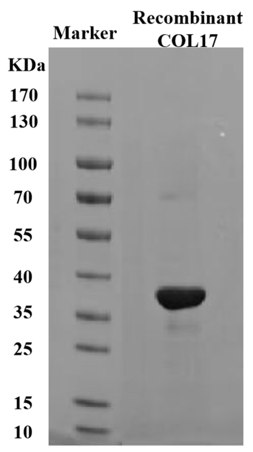
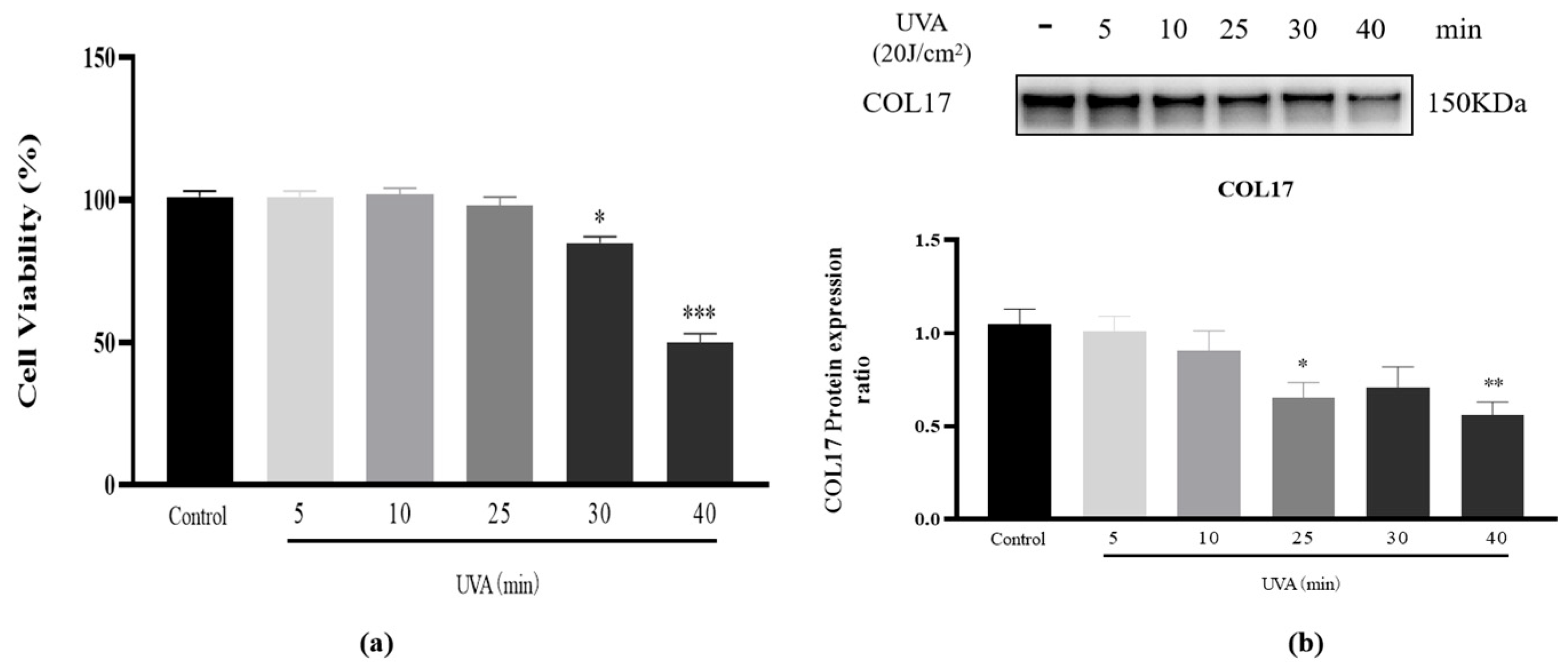
3.1. Cell Viability and COL17 Expression After UVA (20 J/cm2) Irradiation

According to reported UVA irradiation conditions [34], different irradiation times were explored to find conditions for establishing a photoaging model of HaCaT cells. The MTT assay was used to evaluate the cytotoxicity of UVA on HaCaT cells and the optimal treatment time. Cell viability results showed that after 5, 10, 25, and 30 min, UVA irradiation and continued culturing for 24 h did not lead to significant cytotoxicity, with cell viability greater than 80% (Figure 2a). The key role of COL17 in regulating skin aging was also mentioned in a previous article [23]. Western blot evaluation showed that COL17 levels decreased significantly after 25 min UVA irradiation (Figure 2b), indicating that the photoaging cell model induced by UVA irradiation was successfully constructed. However, 40 min UVA irradiation caused a decrease in cell viability (<80%). Therefore, UVA irradiation for 25 min was selected as the optimal experimental condition.

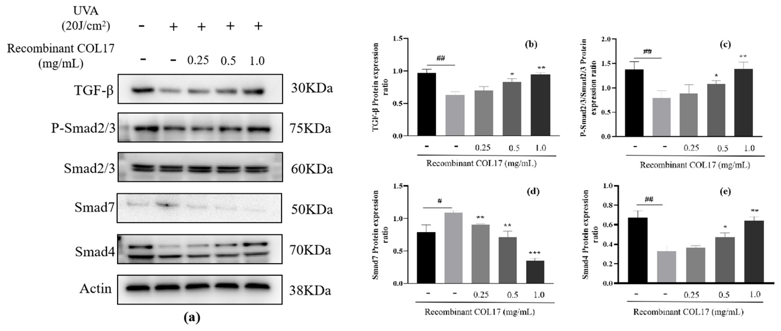
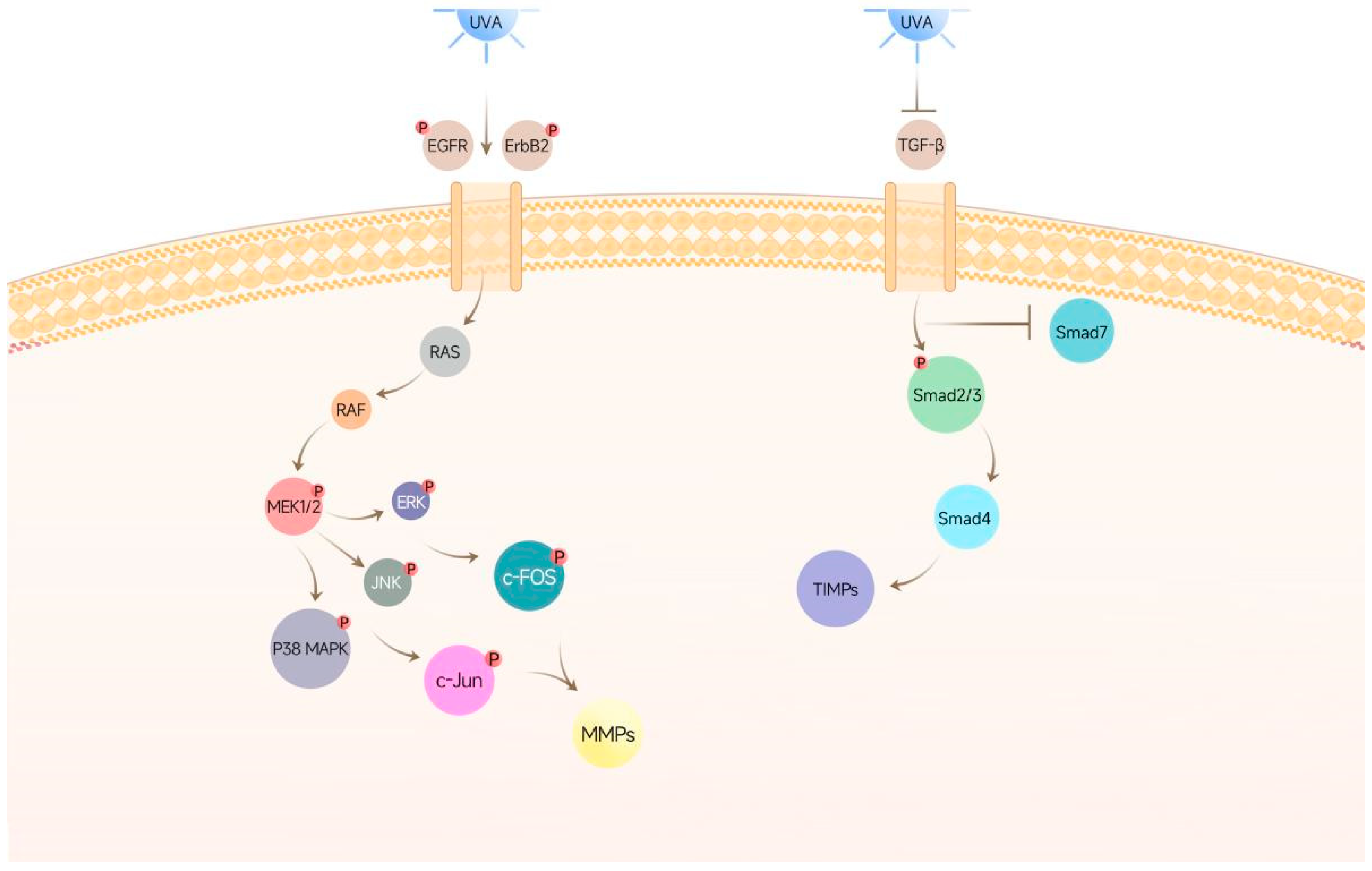
3.2. Effects of Recombinant COL17 on the TGF-β/Smad Pathway

The TGF-β/Smad pathway is a major signal transduction pathway that regulates the expression of collagen synthesis genes. After UVA irradiation, the expression of TGF-β and phosphorylated (p)-Smad2/3 and Smad4 is downregulated, and the expression of Smad7 is upregulated, thereby destroying collagen synthesis in skin tissue [35]. To elucidate the stimulatory effects of recombinant COL17 on TGF-β and its downstream signaling proteins, Smad proteins were analyzed. The expression of TGF-β and Smad4 and the phosphorylation rates of Smad2/3 were reduced by 35.26%, 42.62%, and 50.66%, respectively, after UVA irradiation. Conversely, Smad7, an inhibitor of Smad2/3 activation and nuclear translocation, increased by 26.84% (Figure 3 and Figure S1). Treatment with recombinant COL17 (0.25, 0.5, or 1.0 mg/mL) dose-dependently increased TGF-β, Smad4, and p-Smad2/3 levels compared to UVA-exposed cells without treatment. Additionally, Smad7 levels decreased by 67.36% at the highest COL17 concentration (1.0 mg/mL). These results demonstrate that recombinant COL17 promotes collagen synthesis by regulating the TGF-β/Smad pathway, effectively mitigating UVA-induced skin photoaging.

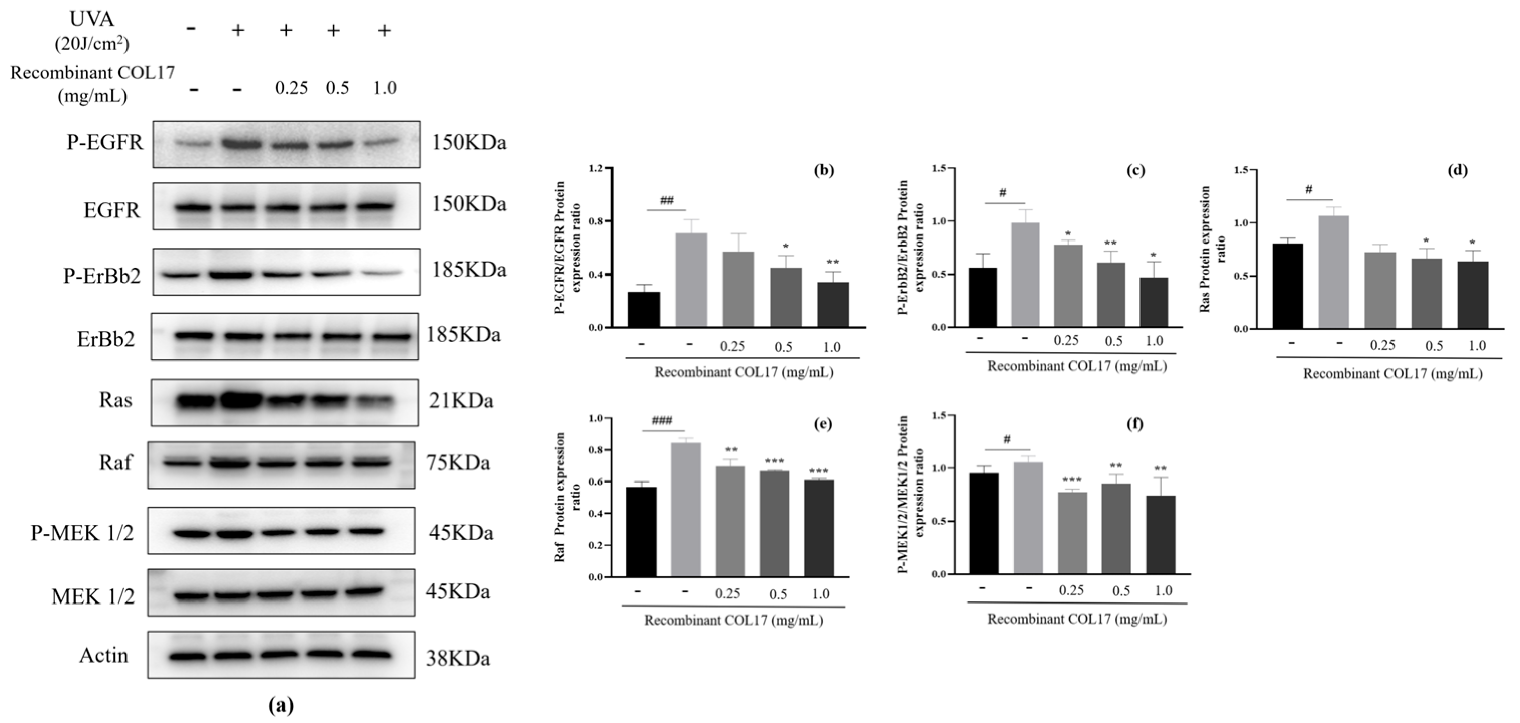
3.3. Effects of Recombinant COL17 on the EGFR/MAPK/AP-1 Pathway

Activation of the RAS/ERK1/2 signaling pathway through EGFR has been studied extensively at both molecular and biochemical levels [3]. Previous studies indicate that the EGFR signal intensity decreases with skin aging and that EGFR inhibits COL17 proteolysis by activating tissue inhibitor of metalloproteinase 1 (TIMP-1). TIMP-1 promotes human keratinocyte stem cell motility and enhances COL17 production [1]. In this study, we examined the expression of RAS and other downstream proteins in the EGFR/ERBB2 pathway, as well as related proteins in the MAPK signaling cascade. UVA exposure significantly suppressed the phosphorylation of EGFR and ERBB2, leading to the activation of RAS/RAF/ERK1/2 signaling pathway proteins (Figure 4, Figure 5, Figures S2 and S3). Furthermore, UVA exposure activated the classical downstream MAPK signaling pathway, increasing the phosphorylation of JNK, p38, ERK, and AP-1 (a heterodimer of c-Fos and c-Jun). Recombinant COL17 treatment concentration-dependently reduced EGFR and ERBB2 phosphorylation (Figure 4a–c). At 1.0 mg/mL, it decreased RAS, RAF, and p-ERK1/2 levels by 40.28%, 27.85%, and 29.70%, respectively (Figure 4d–f). High-dose treatment also reduced JNK, p38, and ERK phosphorylation by 57.19%, 27.39%, and 68.71%, with JNK returning to control levels (Figure 5a–d). Similarly, it reduced p-c-Fos and p-c-Jun levels by 53.16% and 52.83%, nearly restoring control levels (Figure 5e,f). Given the established role of AP-1 signaling in MMP production [30], we further examined MMP and TIMP expression to validate these findings.

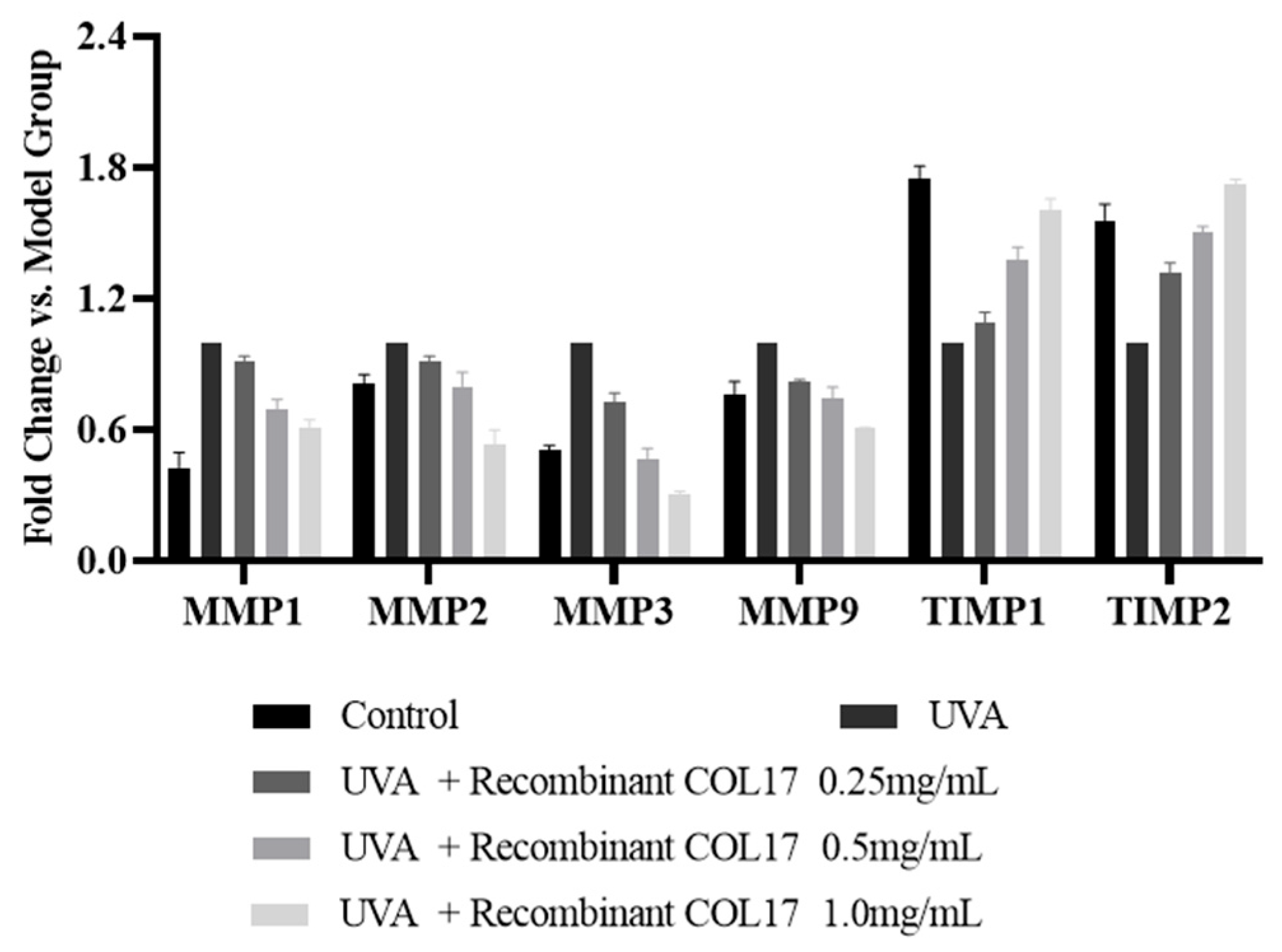
3.4. Effects of Recombinant COL17 on MMP and TIMP Expression in HaCaT Cells

The MMP and TIMP mRNA expression levels were assessed (Figure 6, Table S3). The observed alterations in MMP and TIMP expression in the recombinant COL17 treatment groups were compared with those in the UVA-treated group and are expressed as fold changes relative to the UVA-treated group. Compared with the non-irradiated control cells, UVA exposure increased MMP mRNA levels by 0.5- to 1.5-fold, which was accompanied by a decrease in TIMP mRNA levels. Notably, recombinant COL17 reversed this effect on MMP mRNA expression in a dose-dependent manner. Specifically, in the high-concentration (1.0 mg/mL of recombinant COL17) group, the expression levels of MMP2, MMP3, MMP9, TIMP1, and TIMP2 were restored to those of the unirradiated control, effectively reversing the changes induced by UVA exposure. Collectively, these findings suggest that recombinant COL17 exerts an inhibitory effect on collagen proteolysis through the activation of EGFR-mediated secreted TIMPs.

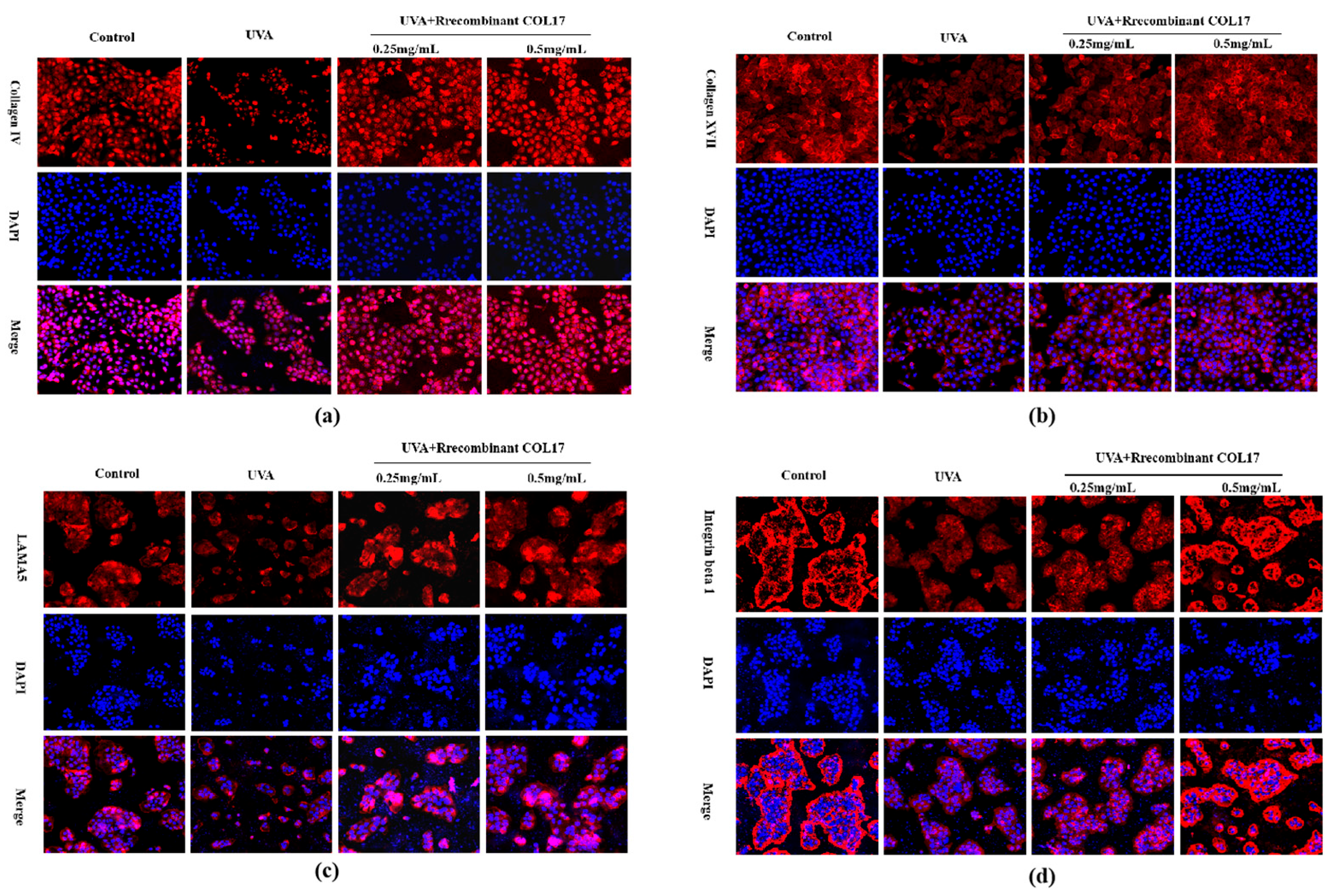
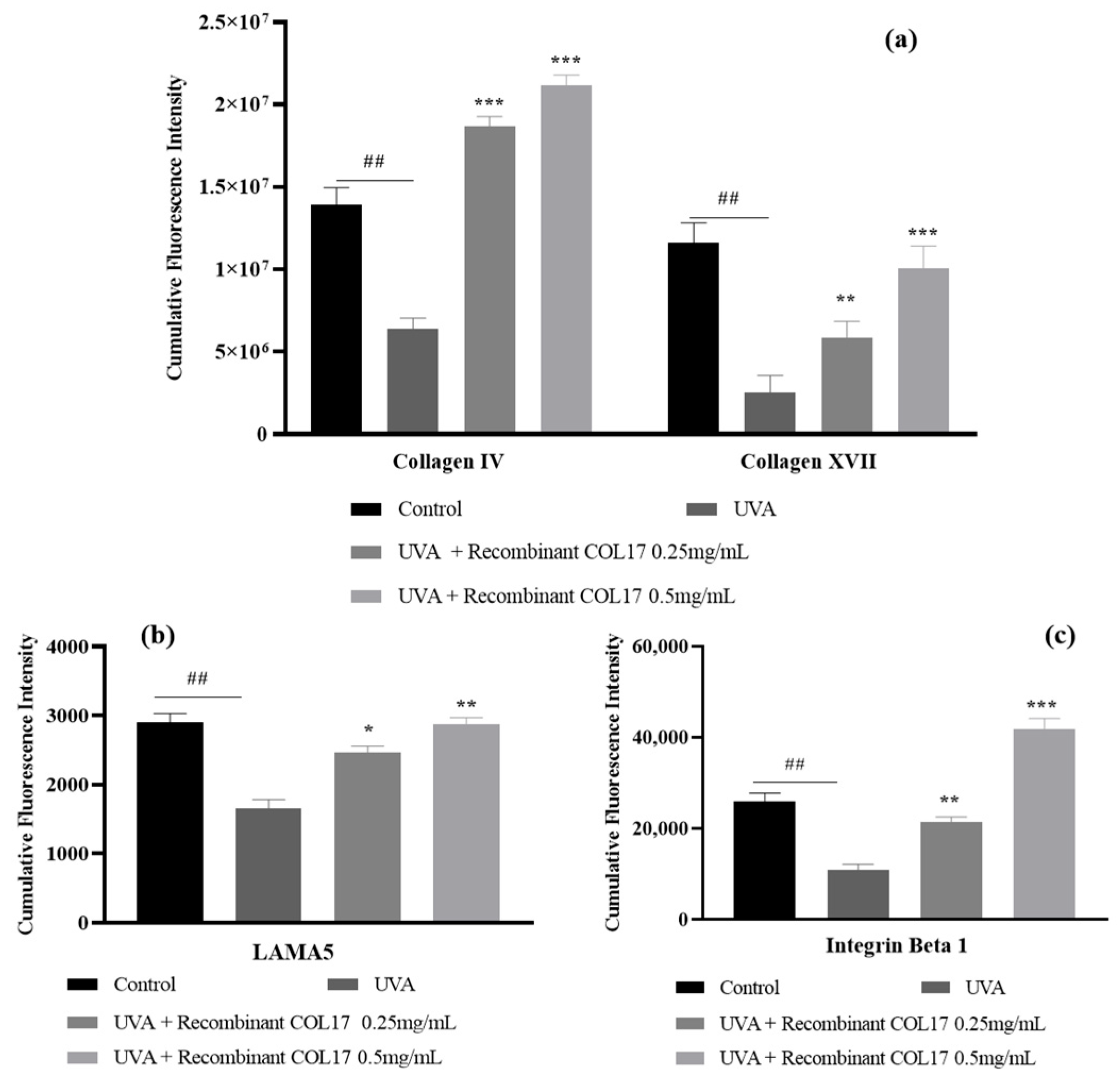
3.5. Effects of Recombinant COL17 on the UVA-Mediated Expression of Basement Membrane-Associated Proteins

Overexpression of MMPs and reduced expression of TIMPs, coupled with reduced collagen levels, contribute to skin photoaging. To assess the impact of recombinant COL17 on collagen production, we examined UVA-irradiated HaCaT cells. The results show that UVA exposure significantly decreased collagen levels in HaCaT cells (Figure 7a), but recombinant COL17 treatment mitigated this reduction effectively. COL4, a major component of the basement membrane, is synthesized intracellularly and contributes to the extracellular collagen matrix [27]. Recombinant COL17 also alleviated the UVA-induced decline in COL4 levels (Figure 7b). Laminin, a key basement membrane glycoprotein, supports epidermal cell adhesion to the basement membrane [29]. Similarly, integrin β1, a cell membrane receptor, is involved in extracellular matrix adhesion, cell proliferation, migration, and differentiation. Recombinant COL17 counteracted the UVA-induced reduction of laminin alpha 5 (LAMA5) and integrin β1 (Figure 7c,d).
After staining, the cumulative immunofluorescent intensities of the cells were calculated under the microscope by selecting different fields of view (n = 3). The fluorescent intensities of COL4, COL17, LAMA5, and integrin β1 were significantly decreased in HaCaT cells after UVA irradiation, and treatment with 0.25 mg/mL and 0.5 mg/mL recombinant COL17 increased the fluorescent intensity of each protein (Figure 8, Table S4).

4. Discussion

Skin undergoes inevitable aging processes with accumulating years, and approximately 80% of this aging is attributed to chronic exposure to UV radiation. Specifically, UVA rays can penetrate deep into the dermis and trigger the excessive production of metalloproteinases. These enzymes subsequently break down collagen fibers, generate reactive oxygen species, and induce oxidative stress. Such perturbations lead to damage of the cellular membrane and DNA, which manifests as skin laxity, roughness, hyperpigmentation, wrinkles, and other signs of aging [1,3]. Previous studies have documented the efficacy of various bioactive compounds, including doxercalciferol [36], santamarine [37], and resveratrol [38], in mitigating photoaging-induced skin damage through mechanisms such as anti-inflammatory actions, antioxidant properties, and the inhibition of metalloproteinase expression. However, despite its significant market potential, research on the anti-photodamage mechanisms of genetically engineered collagen remains limited. In this study, the anti-photoaging properties of recombinant COL17 were investigated in UVA-irradiated HaCaT cells, and the underlying molecular mechanisms were elucidated.
In recent years, there have been many reports on the underlying mechanisms of skin photoaging, including a variety of signal transduction channels and molecules. The signal transduction channels are mainly divided into the MAPK, nuclear factor κB, nuclear factor erythroid 2-related factor 2/antioxidant response element, and TGF-β/Smad signaling pathways. The molecular mechanisms mainly involve DNA damage, oxidative stress, the inflammatory response, structural changes in collagen, or the degradation and reduction of related protein production [39]. It has been reported that UV radiation can activate EGFR and other cell surface-related receptors, affect MAPK and phosphatidylinositol 3-kinase/protein kinase B signaling pathways, upregulate AP-1 expression, and induce MMP synthesis, thereby leading to increased collagen degradation. This simultaneously prevents the binding of TGF-β to its receptor, resulting in the blockage of collagen synthesis and the subsequent appearance of skin aging phenomena such as sagging and wrinkles [40]. In the present study, UVA irradiation promoted MMP1 expression by activating both the EGFR/MAPK/AP-1 and TGF-β/Smad signaling pathways in HaCaT cells to degrade collagen (Figure 9). However, the mechanisms by which EGFR influences the MAPK signaling pathway, ultimately leading to collagen degradation, remain relatively unknown. Similarly, there is a scarcity of reports on the active substances that may be capable of reversing this phenomenon.
Jagodzik et al. and Bahar et al. [8,9] speculated that UV radiation may catalyze the phosphorylation of tyrosine residues in the intracellular segment of EGFR, which then promotes the activation of GTP catalyzed by SOS. Combined with RAS-related enzymes, this may then lead to the continuous activation of RAF and MEK. RAS is the main component of the MAPK/ERK1/2 signaling pathway, which controls cell proliferation, differentiation, survival, and migration [41]. It also transmits extracellular stimulation signals to cells through the phosphorylation cascade of RAF, MEK, and ERK protein kinases and finally regulates AP-1 signaling [5,42]. In the present study, recombinant COL17 inhibited the UVA-induced phosphorylation of EGFR and ERBB2, resulting in the inhibition of MAPK and AP-1 activation in HaCaT cells, as indicated by the decreased phosphorylation of EGFR and RAS/RAF/MEK1/2 signaling (Figure 4). The inhibition of MEK activation also indirectly affected the activity of three MAPK signaling pathways, including ERK, JNK, and p38, and ultimately reduced the nuclear translocation of p-c-Fos and p-c-Jun (Figure 5), resulting in downregulated MMP expression. We therefore speculate that recombinant COL17 ameliorates UVA-induced collagen degradation by inhibiting the RAS/RAF/MEK1/2 signaling pathway from EGFR to MMPs. However, the specific regulation mechanism of EGFR on its downstream signaling, even the direct effect on MMPs and TIMPs, may need further verification, using methods such as EGFR activation and inhibition experiments and gene knockout/knockdown experiments.
It has recently been reported that MAPK activation leads to increased production of the transcription factor AP-1 [43]. Moreover, a key role of AP-1 in the transcriptional upregulation of MMP-1, MMP-2, MMP-3, and MMP-9 has also been reported [6], and MMP overexpression leads to structural changes and degradation of collagen fibers in the dermis and accelerates skin aging [4,32]. Nonetheless, it has also been reported that EGFR regulates COL17A1 proteolysis by affecting TIMP expression [44]. In the present comprehensive study, the expression levels of MMP and TIMP mRNA were meticulously quantified using RT-qPCR techniques (Figure 6). Recombinant COL17 emerged as a potent agent that was able to mitigate the UVA radiation-induced abnormal activation of EGFR on the cellular membrane. This, in turn, suppressed the activation of the RAS/RAF/ERK signaling cascade, which subsequently influenced a cascade of nuclear events. These complex pathways also played a pivotal role in regulating MMP upregulation, thereby effectively hindering collagen degradation and delaying skin aging processes. Although the signaling pathways regulating MMPs and TIMPs have been extensively studied, developing specific inhibitors or activators targeting these pathways remains challenging. In addition, photoaging involves the interaction of multiple signaling pathways. In the future, it will be necessary to comprehensively analyze the regulatory network of MMPs and TIMPs by analyzing the synergistic or antagonistic effects of different signaling pathways.
On the basis of numerous reports linking UVA irradiation with the TGF-β/Smad pathway, we also evaluated the effects of recombinant COL17 on TGF-β and its downstream activator, the Smad2/3 complex [45]. Upon the activation of TGF-β signaling, the p-Smad2/3 complex forms another complex with Smad4 and is transported to the nucleus to initiate collagen expression [46]. The results of the present study confirmed that UVA irradiation of HaCaT cells significantly reduced TGF-β levels and inhibited Smad2/3 complex phosphorylation and Smad4 trafficking. Conversely, UVA irradiation increased the levels of Smad7, a natural negative regulator of TGF-β/Smad signaling. The addition of recombinant COL17 to the culture medium after UVA irradiation reversed the inhibitory effect of UVA on the TGF-β signaling pathway. Although the mechanisms of the TGF-β/Smad signaling pathway on photoaging have been reported, the interaction mechanism of the Tgf-β/Smad signaling pathway with MAPK, EGFR, NF-κB, and other pathways has not been fully elucidated, especially the synergistic or antagonistic effects on photodamage. Moreover, most current studies are based on cell experiments and lack the support of animal models or clinical data, thus making it difficult to fully reveal the role of the TGF-β/Smad pathway in photodamage.
Although the above experiments demonstrate the effects of recombinant COL17 on two signaling pathways under a UVA photoaging model and preliminarily explore the mechanism of collagen degradation by UVA irradiation, the specific effects of recombinant COL17 on collagen or basement membrane proteins still require further elucidation. A recent study has shown that recombinant COL17 enhances keratinocyte migration and adhesion and increases the expression of ECM components, thereby protecting the integrity of the basement membrane [47]. We, therefore, evaluated the potential pro-collagenolytic effects of recombinant COL17 by analyzing the fluorescent staining of relevant proteins in the cells. Xiang et al. [28] suggested that reduced COL17A1 expression is positively correlated with impaired keratinocyte regeneration, reduced dermal–epidermal junctions, and thin epidermis in aged human skin (i.e., epidermal aging). Vazquez [48], Wang J et al. [12] investigated the relationship between skin COL4—a basement membrane component—and changes in basement membranes during aging and noted that COL4 decreases with age. In addition, Amano [49] proposed that repeated damage to the basement membrane at the dermal–epidermal junction of skin exposed to sunlight may destroy the stability of the skin. Laminin and integrin β1 are crucial substances for the connections between the epidermis and dermis and the maintenance of skin structure and are thus of great importance for repairing the basement membrane and delaying skin aging. In our study of UVA-irradiated HaCaT cells, the secretion of COL4, COL17, integrin β1, and LAMA5 was affected by UVA, and the fluorescence intensity decreased by 40–80%, especially for COL17. Following treatment with recombinant COL17 at concentrations of 0.25 and 0.5 mg/mL, a significant upregulation in protein secretion was observed, surpassing baseline levels. Notably, at the lowest dose of 0.25 mg/mL, the secretion of LAMA5 and integrin β1 increased by 48% and 97%, respectively. Particularly striking were the increases in COL4 and COL17, which exceeded 100%, demonstrating statistically significant enhancements (Figure 7 and Figure 8). These observations are similar to the results of an earlier study by Jeong et al. [26]. Although recombinant COL17 showed superior pro-collagen and pro-basement membrane protein secretion in our preliminary study, how it compares with classical anti-aging and pro-collagen synthesis components such as retinol, vitamin C derivatives, and acetyl-hexapeptide-8 is unknown and should be examined in future work.
In summary, recombinant COL17 exhibited significant potential as a viable candidate for incorporation into future anti-aging skincare formulations. Nevertheless, the mechanistic interplay between EGFR/MAPK/AP-1 and TGF-β/Smad signaling pathways and their regulatory effects on skin basement membrane proteins remains insufficiently elucidated, underscoring the need for more comprehensive research. Subsequent investigations should utilize UVA-induced skin damage models in hairless mice, with established anti-aging compounds serving as comparative controls, to delineate the mechanisms by which recombinant COL17 mitigates photoaging (photodamage) and modulates the secretion of basement membrane proteins. Furthermore, clinical trials are warranted to evaluate the therapeutic efficacy of recombinant COL17 in addressing skin damage induced by concurrent UVA and UVB exposure. The synergistic combination of recombinant COL17 with other bioactive agents may further augment its anti-aging properties, presenting an innovative approach for formulating advanced skincare solutions.

5. Conclusions

Recombinant COL17 can inhibit skin photoaging and delay skin aging by inhibiting the EGFR/MAPK/AP-1 signaling pathway, inhibiting MMP secretion, activating TGF-β/Smad signaling, upregulating TIMP levels, increasing basement membrane protein expression, and promoting COL4 and COL17 secretion. The present findings revealed a variety of signaling pathways related to collagen synthesis and degradation and lay a foundation for future photoaging skincare and clinical applications.

Supplementary Materials

The following supporting information can be downloaded at https://www.mdpi.com/article/10.3390/cosmetics12020059/s1: Figures S1–S3: Original images of Western Blot; Tables S1 and S2: Gray value data of Western Blot; Table S3: Date on mRNA expression levels; Table S4: Immunofluorescence intensity data.

Author Contributions

Y.H. contributed to the experimental basis, paper conception, data, and writing of the manuscript. S.Y. provided support for the logic and theory of the paper. R.X. provided support for the visualization, conceptualization, and research. Y.Z. provided support for changes in the formatting of pictures, charts, and references of the article. Y.D. provided support for the immunofluorescent staining experimental technology. Z.D. and D.F. commented, edited, and supervised the writing. All authors have read and agreed to the published version of the manuscript.

Funding

This work was funded by the National Key R&D Program of China (No. 2022YFC2104800).

Institutional Review Board Statement

This study did not require ethical approval.

Informed Consent Statement

No human studies were involved in this study.

Data Availability Statement

Data will be made available on request.

Acknowledgments

We thank the research and development team of Xi’an Juzi Biotechnology Co., Ltd., for the construction, fermentation, and purification of the recombinant collagen type XVII.

Conflicts of Interest

Authors Ying He, Shiyu Yin, Ru Xu, Yan Zhao and Yuhang Du were employed by the company Xi’an Giant Biogene Technology Co., Ltd. The remaining authors declare that the research was conducted in the absence of any commercial or financial relationships that could be construed as a potential conflict of interest.

References

  1. Gromkowska-Kępka, K.J.; Puścion-Jakubik, A.; Markiewicz-Żukowska, R.; Socha, K. The impact of ultraviolet radiation on skin photoaging—Review of in vitro studies. J. Cosmet. Dermatol. 2021, 20, 3427–3431. [Google Scholar] [CrossRef] [PubMed]
  2. Fitsiou, E.; Pulido, T.; Campisi, J.; Alimirah, F.; Demaria, M. Cellular Senescence and the Senescence-Associated Secretory Phenotype as Drivers of Skin Photoaging. J. Investig. Dermatol. 2021, 141, 1119–1126. [Google Scholar] [CrossRef] [PubMed]
  3. Battie, C.; Jitsukawa, S.; Bernerd, F.; Del Bino, S.; Marionnet, C.; Verschoore, M. New insights in photoaging, UVA induced damage and skin types. Exp. Dermatol. 2014, 23, 7–12. [Google Scholar] [CrossRef] [PubMed]
  4. Silvers, A.L.; Bachelor, M.A.; Bowden, G.T. The Role of JNK and p38 MAPK Activities in UVA-Induced Signaling Pathways Leading to AP-1 Activation and c-Fos Expression. Neoplasia 2003, 5, 319–329. [Google Scholar] [CrossRef]
  5. Zhang, M.; Zhang, T.; Tang, Y.; Ren, G.; Zhang, Y.; Ren, X. Concentrated growth factor inhibits UVA-induced photoaging in human dermal fibroblasts via the MAPK/AP-1 pathway. Biosci. Rep. 2020, 40, BSR20193566. [Google Scholar] [CrossRef]
  6. Oh, J.H.; Joo, Y.H.; Karadeniz, F.; Ko, J.; Kong, C.-S. Syringaresinol Inhibits UVA-Induced MMP-1 Expression by Suppression of MAPK/AP-1 Signaling in HaCaT Keratinocytes and Human Dermal Fibroblasts. Int. J. Mol. Sci. 2020, 21, 3981. [Google Scholar] [CrossRef]
  7. He, Y.-Y.; Council, S.E.; Feng, L.; Chignell, C.F. UVA-Induced Cell Cycle Progression Is Mediated by a Disintegrin and Metalloprotease/Epidermal Growth Factor Receptor/AKT/Cyclin D1 Pathways in Keratinocytes. Cancer Res. 2008, 68, 3752–3758. [Google Scholar] [CrossRef]
  8. Jagodzik, P.; Tajdel-Zielinska, M.; Ciesla, A.; Marczak, M.; Ludwikow, A. Mitogen-Activated Protein Kinase Cascades in Plant Hormone Signaling. Front. Plant Sci. 2018, 9, 1387. [Google Scholar] [CrossRef]
  9. Bahar, M.E.; Kim, H.J.; Kim, D.R. Targeting the RAS/RAF/MAPK pathway for cancer therapy: From mechanism to clinical studies. Signal. Transduct. Target. Ther. 2023, 8, 455. [Google Scholar] [CrossRef]
  10. Hong, J.-A.; Bae, D.; Oh, K.-N.; Oh, D.-R.; Kim, Y.; Kim, Y.; Jeong Im, S.; Choi, E.-J.; Lee, S.-G.; Kim, M.; et al. Protective effects of Quercus acuta Thunb. fruit extract against UVB-induced photoaging through ERK/AP-1 signaling modulation in human keratinocytes. BMC Complement. Med. Ther. 2022, 22, 6. [Google Scholar] [CrossRef]
  11. Qin, Z.; Balimunkwe, R.M.; Quan, T. Age-related reduction of dermal fibroblast size upregulates multiple matrix metalloproteinases as observed in aged human skin in vivo. Br. J. Dermatol. 2017, 177, 1337–1348. [Google Scholar] [CrossRef] [PubMed]
  12. Wang, J.; Huang, H.; Tao, K.; Guo, L.; Hu, X.; Chang, H. Novel Thermus thermophilus and Bacillus subtilis mixed-culture ferment extract provides potent skin benefits in vitro and protects skin from aging. J. Cosmet. Dermatol. 2024, 23, 4334–4342. [Google Scholar] [CrossRef] [PubMed]
  13. Quan, T.; He, T.; Kang, S.; Voorhees, J.J.; Fisher, G.J. Solar Ultraviolet Irradiation Reduces Collagen in Photoaged Human Skin by Blocking Transforming Growth Factor-&#x3b2; Type II Receptor/Smad Signaling. Am. J. Pathol. 2004, 165, 741–751. [Google Scholar] [CrossRef]
  14. Chen, B.; Li, R.; Yan, N.; Chen, G.; Qian, W.; Jiang, H.L.; Ji, C.; Bi, Z.G. Astragaloside IV controls collagen reduction in photoaging skin by improving transforming growth factor-β/Smad signaling suppression and inhibiting matrix metalloproteinase-1. Mol. Med. Rep. 2015, 11, 3344–3348. [Google Scholar] [CrossRef]
  15. Jabłońska-Trypuć, A.; Matejczyk, M.; Rosochacki, S. Matrix metalloproteinases (MMPs), the main extracellular matrix (ECM) enzymes in collagen degradation, as a target for anticancer drugs. J. Enzym. Inhib. Med. Chem. 2016, 31, 177–183. [Google Scholar] [CrossRef]
  16. Pittayapruek, P.; Meephansan, J.; Prapapan, O.; Komine, M.; Ohtsuki, M. Role of Matrix Metalloproteinases in Photoaging and Photocarcinogenesis. Int. J. Mol. Sci. 2016, 17, 868. [Google Scholar] [CrossRef]
  17. Wang, Y.; Chen, H.; Wang, W.; Wang, R.; Liu, Z.-L.; Zhu, W.; Lian, S. N-terminal 5-mer peptide analog P165 of amyloid precursor protein inhibits UVA-induced MMP-1 expression by suppressing the MAPK pathway in human dermal fibroblasts. Eur. J. Pharmacol. 2014, 734, 1–8. [Google Scholar] [CrossRef]
  18. Yu, S.-C.; Chiu, W.-C.; Loe, P.Y.; Chien, Y.-W. Effects of Hydroxysafflor Yellow A (HSYA) on UVA-Induced Damage in HaCaT Keratinocytes. Int. J. Mol. Sci. 2024, 25, 7573. [Google Scholar] [CrossRef]
  19. Oh, J.H.; Karadeniz, F.; Seo, Y.; Kong, C.-S. Isopimpinellin inhibits UVA-induced overproduction of MMPs via suppression of MAPK/AP-1 signaling in human dermal fibroblasts. Food Sci. Biotechnol. 2024, 33, 3579–3589. [Google Scholar] [CrossRef]
  20. Yao, C.; Lee, D.H.; Oh, J.-H.; Kim, M.-K.; Kim, K.H.; Park, C.-H.; Chung, J.H. Poly(I:C) induces expressions of MMP-1, -2, and -3 through various signaling pathways including IRF3 in human skin fibroblasts. J. Dermatol. Sci. 2015, 80, 54–60. [Google Scholar] [CrossRef]
  21. Chen, Y.-L.; Zhang, M.-H.; Su, L.-L.; Sun, L.-C.; Qiu, X.-J.; Lin, D.; Zhang, L.-J.; Jin, T.; Cao, M.-J. Relationships of Matrix Metalloproteinase 1 and a Tissue Inhibitor of Metalloproteinase to Collagen Metabolism in Haliotis discus hannai. J. Agric. Food Chem. 2022, 70, 14886–14897. [Google Scholar] [CrossRef]
  22. Laronha, H.; Caldeira, J. Structure and Function of Human Matrix Metalloproteinases. Cells 2020, 9, 1076. [Google Scholar] [CrossRef] [PubMed]
  23. Liu, Y.; Ho, C.; Wen, D.; Sun, J.; Huang, L.; Gao, Y.; Li, Q.; Zhang, Y. Targeting the stem cell niche: Role of collagen XVII in skin aging and wound repair. Theranostics 2022, 12, 6446–6454. [Google Scholar] [CrossRef] [PubMed]
  24. Liu, N.; Matsumura, H.; Kato, T.; Ichinose, S.; Takada, A.; Namiki, T.; Asakawa, K.; Morinaga, H.; Mohri, Y.; De Arcangelis, A.; et al. Stem cell competition orchestrates skin homeostasis and ageing. Nature 2019, 568, 344–350. [Google Scholar] [CrossRef] [PubMed]
  25. Kroeger, J.; Hoppe, E.; Galiger, C.; Has, C.; Franzke, C.-W. Amino acid substitution in the C-terminal domain of collagen XVII reduces laminin-332 interaction causing mild skin fragility with atrophic scarring. Matrix Biol. 2019, 80, 72–84. [Google Scholar] [CrossRef]
  26. Jeong, S.; Yoon, S.; Kim, S.; Jung, J.; Kor, M.; Shin, K.; Lim, C.; Han, H.S.; Lee, H.; Park, K.-Y.; et al. Anti-Wrinkle Benefits of Peptides Complex Stimulating Skin Basement Membrane Proteins Expression. Int. J. Mol. Sci. 2020, 21, 73. [Google Scholar]
  27. Jacków, J.; Löffek, S.; Nyström, A.; Bruckner-Tuderman, L.; Franzke, C.-W. Collagen XVII Shedding Suppresses Re-Epithelialization by Directing Keratinocyte Migration and Dampening mTOR Signaling. J. Investig. Dermatol. 2016, 136, 1031–1041. [Google Scholar] [CrossRef]
  28. Xiang, Y.; Liu, Y.; Yang, Y.; Yan, Y.; Kim, A.J.; Guo, C.; Fisher, G.J.; Quan, T. Reduced expression of Collagen 17A1 in naturally aged, photoaged, and UV-irradiated human skin in vivo: Potential links to epidermal aging. J. Cell Commun. Signal. 2022, 16, 421–432. [Google Scholar] [CrossRef]
  29. Has, C.; Kern, J.S. Collagen XVII. Dermatol. Clin. 2010, 28, 61–66. [Google Scholar] [CrossRef]
  30. Wang, Y.; Kitahata, H.; Kosumi, H.; Watanabe, M.; Fujimura, Y.; Takashima, S.; Osada, S.-I.; Hirose, T.; Nishie, W.; Nagayama, M.; et al. Collagen XVII deficiency alters epidermal patterning. Lab. Investig. 2022, 102, 581–588. [Google Scholar] [CrossRef]
  31. Wang, Y.; Hess, M.E.; Tan, Y.; Esser, P.R.; Nyström, A.; Boerries, M.; Sayar, S.B.; Has, C. Alterations in the microenvironment of junctional epidermolysis bullosa keratinocytes: A gene expression study. Matrix Biol. 2025, 135, 12–23. [Google Scholar] [CrossRef] [PubMed]
  32. Pigors, M.; Patzelt, S.; Reichhelm, N.; Dworschak, J.; Khil’chenko, S.; Emtenani, S.; Bieber, K.; Hofrichter, M.; Kamaguchi, M.; Goletz, S.; et al. Bullous pemphigoid induced by IgG targeting type XVII collagen non-NC16A/NC15A extracellular domains is driven by Fc gamma receptor- and complement-mediated effector mechanisms and is ameliorated by neonatal Fc receptor blockade. J. Pathol. 2024, 262, 161–174. [Google Scholar] [CrossRef] [PubMed]
  33. Liu, Y.; Ho, C.; Wen, D.; Sun, J.; Liu, Y.; Li, Q.; Zhang, Y.; Gao, Y. PKM2-mediated collagen XVII expression is critical for wound repair. JCI Insight 2025, 10, e184457. [Google Scholar] [CrossRef] [PubMed]
  34. Hwang, Y.P.; Oh, K.N.; Yun, H.J.; Jeong, H.G. The flavonoids apigenin and luteolin suppress ultraviolet A-induced matrix metalloproteinase-1 expression via MAPKs and AP-1-dependent signaling in HaCaT cells. J. Dermatol. Sci. 2011, 61, 23–31. [Google Scholar] [CrossRef]
  35. Iriyama, S.; Yasuda, M.; Nishikawa, S.; Takai, E.; Hosoi, J.; Amano, S. Decrease of laminin-511 in the basement membrane due to photoaging reduces epidermal stem/progenitor cells. Sci. Rep. 2020, 10, 12592. [Google Scholar] [CrossRef]
  36. Ge, Y.; Li, M.; Bai, S.; Chen, C.; Zhang, S.; Cheng, J.; Wang, X. Doxercalciferol alleviates UVB-induced HaCaT cell senescence and skin photoaging. Int. Immunopharmacol. 2024, 127, 111357. [Google Scholar] [CrossRef]
  37. Oh, J.H.; Kim, J.; Karadeniz, F.; Kim, H.R.; Park, S.Y.; Seo, Y.; Kong, C.-S. Santamarine Shows Anti-Photoaging Properties via Inhibition of MAPK/AP-1 and Stimulation of TGF-β/Smad Signaling in UVA-Irradiated HDFs. Molecules 2021, 26, 3585. [Google Scholar] [CrossRef]
  38. Cui, B.; Wang, Y.; Jin, J.; Yang, Z.; Guo, R.; Li, X.; Yang, L.; Li, Z. Resveratrol Treats UVB-Induced Photoaging by Anti-MMP Expression, through Anti-Inflammatory, Antioxidant, and Antiapoptotic Properties, and Treats Photoaging by Upregulating VEGF-B Expression. Oxid. Med. Cell Longev. 2022, 2022, 6037303. [Google Scholar] [CrossRef]
  39. Berneburg, M.; Plettenberg, H.; Krutmann, J. Photoaging of human skin. Photodermatol. Photoimmunol. Photomed. 2000, 16, 239–244. [Google Scholar] [CrossRef]
  40. Martínez-Carpio, P.A.; Trelles, M.A. Cutaneous epidermal growth factor receptor system following ultraviolet irradiation: Exploring the role of molecular mechanisms. Photodermatol. Photoimmunol. Photomed. 2010, 26, 250–256. [Google Scholar] [CrossRef]
  41. Jeon, H.; Tkacik, E.; Eck, M.J. Signaling from RAS to RAF: The Molecules and Their Mechanisms. Annu. Rev. Biochem. 2024, 93, 289–316. [Google Scholar] [CrossRef] [PubMed]
  42. Surve, S.; Watkins, S.C.; Sorkin, A. EGFR-RAS-MAPK signaling is confined to the plasma membrane and associated endorecycling protrusions. J. Cell Biol. 2021, 220, e202107103. [Google Scholar] [CrossRef] [PubMed]
  43. Han, S.H.; Ballinger, E.; Choung, S.-Y.; Kwon, J.Y. Anti-Photoaging Effect of Hydrolysates from Pacific Whiting Skin via MAPK/AP-1, NF-κB, TGF-β/Smad, and Nrf-2/HO-1 Signaling Pathway in UVB-Induced Human Dermal Fibroblasts. Mar. Drugs 2022, 20, 308. [Google Scholar] [CrossRef] [PubMed]
  44. Nanba, D.; Toki, F.; Asakawa, K.; Matsumura, H.; Shiraishi, K.; Sayama, K.; Matsuzaki, K.; Toki, H.; Nishimura, E.K. EGFR-mediated epidermal stem cell motility drives skin regeneration through COL17A1 proteolysis. J. Cell Biol. 2021, 220, e202012073. [Google Scholar] [CrossRef]
  45. Honda, A.; Abe, R.; Makino, T.; Norisugi, O.; Fujita, Y.; Watanabe, H.; Nishihira, J.; Iwakura, Y.; Yamagishi, S.-i.; Shimizu, H.; et al. Interleukin-1β and macrophage migration inhibitory factor (MIF) in dermal fibroblasts mediate UVA-induced matrix metalloproteinase-1 expression. J. Dermatol. Sci. 2008, 49, 63–72. [Google Scholar] [CrossRef]
  46. Liu, Z.; Li, Y.; Song, H.; He, J.; Li, G.; Zheng, Y.; Li, B. Collagen peptides promote photoaging skin cell repair by activating the TGF-β/Smad pathway and depressing collagen degradation. Food Funct. 2019, 10, 6121–6134. [Google Scholar] [CrossRef]
  47. Wang, J.; Lin, S.; Wei, Y.; Ye, Z. Recombinant human collagen XVII protects skin basement membrane integrity by inhibiting the MAPK and Wnt signaling pathways. Mol. Med. Rep. 2025, 31, 100. [Google Scholar] [CrossRef]
  48. Vázquez, F.; Palacios, S.; Alemañ, N.; Guerrero, F. Changes of the basement membrane and type IV collagen in human skin during aging. Maturitas 1996, 25, 209–215. [Google Scholar] [CrossRef]
  49. Amano, S. Possible Involvement of Basement Membrane Damage in Skin Photoaging. J. Investig. Dermatol. Symp. Proc. 2009, 14, 2–7. [Google Scholar] [CrossRef]
Figure 1. 10% SDS-PAGE analysis of recombinant COL17, rainbow protein marker (10-170 kda, #26616, Thermo Fisher Scientific, Waltham, MA, USA).
Cosmetics 12 00059 g001
Figure 2. Cytotoxicity of UVA (20 J/cm2) on HaCaT cells (a). After UVA irradiation of HaCaT cells for 5, 10, 25, 30, and 40 min, cell viability was assessed by the MTT assay. (b) The effect of UVA irradiation on COL17 expression in HaCaT cells. These cells were irradiated with UVA (20 J/cm2) for 5, 10, 25, 30, and 40 min, and proteins were extracted and evaluated by western blot analysis for COL17 expression. All data are presented as the mean ± standard deviation of at least three independent experiments,* p < 0.05, ** p < 0.01, *** p < 0.001 versus control cells (no irradiation).
Cosmetics 12 00059 g002
Figure 3. Recombinant COL17 stimulates the TGF-β/Smad signaling pathway in UVA irradiation HaCaT cells. HaCaT cells were irradiated with UVA (20 J/cm2) followed by treatment with 0.25, 0.5, or 1.0 mg/mL of recombinant COL17. Western blot analysis (a) was used to evaluate the expression of TGF-β (b), p-Smad2/3/Smad2/3 (c), Smad7 (d), and Smad4 (e). All data are indicated as the mean ± standard deviation of at least three independent experiments. # p < 0.05, ## p < 0.01 versus control cells (no irradiation); * p < 0.05, ** p < 0.01, *** p < 0.001 versus UVA-irradiated cells.
Cosmetics 12 00059 g003
Figure 4. Recombinant COL17 stimulates the EGFR/RAS/MAPK/AP-1 signaling pathway in UVA irradiation HaCaT cells. HaCaT cells were irradiated with UVA (20 J/cm2) followed by treatment with 0.25, 0.5, or 1.0 mg/mL of recombinant COL17. Western blot analysis (a) was used to evaluate the expression levels of p-EGFR/EGFR (b), p-ERBB2/ERBB2 (c), RAS (d), RAF (e), and p-ERK1/2/ERK1/2 (f). All data are presented as the mean ± standard deviation of at least three independent experiments. # p < 0.05, ## p < 0.01, ### p < 0.001 versus control cells (no irradiation); * p < 0.05, ** p < 0.01, *** p < 0.001 versus UVA-irradiated cells.
Cosmetics 12 00059 g004
Figure 5. Recombinant COL17 stimulates the EGFR/RAS/MAPK/AP-1 signaling pathway in UVA irradiation HaCaT cells. HaCaT cells were subjected to UVA irradiation (20 J/cm2) and subsequently treated with 0.25, 0.5, or 1.0 mg/mL of recombinant COL17. Western blot analysis (a) was used to evaluate the expression levels of p-JNK/JNK (b), p-p38/p38 (c), p-ERK1/2/ERK1/2 (d), p-c-Jun/c-Jun (e), and p-Fos/Fos (f). All data are presented as the mean ± standard deviation of at least three independent experiments. # p < 0.05, ## p < 0.01, ### p < 0.001 versus control cells (no irradiation); * p < 0.05, ** p < 0.01, *** p < 0.001 versus UVA-irradiated cells.
Cosmetics 12 00059 g005
Figure 6. HaCaT cells were treated with UVA and/or recombinant COL17 for 24 h before MMP and TIMP expression levels were determined using real-time quantitative polymerase chain reaction (RT-qPCR) analysis.
Cosmetics 12 00059 g006
Figure 7. Effects of recombinant COL17 on collagen production in UVA irradiation (20 J/cm2) HaCaT cells. Images of HaCaT cells were obtained after staining with fluorescein isothiocyanate-conjugated antibody (red) and 4′,6-diamidino-2-phenylindole (DAPI; blue) to highlight the nuclei. The merged images show magenta, indicating partial colocalization of the protein with the nucleus. (a) COL4. (b) COL17. (c) Recombinant anti-LAMA5. (d) Recombinant anti-integrin β1.
Cosmetics 12 00059 g007
Figure 8. Cumulative fluorescent intensity of collagen in different fields of view after immunofluorescent staining, calculated using Image-Pro software 6.0. Values are presented as the mean ± standard deviation (n = 3). ## p < 0.01 versus control cells (no irradiation); * p < 0.05, ** p < 0.01, *** p < 0.001 versus UVA-treated cells. (a) COL4 and COL17. (b) Recombinant anti-LAMA5. (c) Recombinant anti-integrin β1.
Cosmetics 12 00059 g008
Figure 9. Schematic diagram of the molecular mechanisms underlying UVA-induced skin photoaging.
Cosmetics 12 00059 g009
Disclaimer/Publisher’s Note: The statements, opinions and data contained in all publications are solely those of the individual author(s) and contributor(s) and not of MDPI and/or the editor(s). MDPI and/or the editor(s) disclaim responsibility for any injury to people or property resulting from any ideas, methods, instructions or products referred to in the content.

