Article

Investigation of the Anti-Aging Effects of Composite Nanocarriers Based on Autophagy Regulation and Oxidative Stress Inhibition

by
Min Liu
1,
Lei Ye
1,
Lingling Jiang
1,*,
Xi Wang
2,3,
Cui Sun
1,
Jiuyan Zheng
1 and
Wei Liu
2,3
1
Suzhou Misifu Cosmetic Co., Ltd., Suzhou 215299, China
2
National Engineering Research Center for Nanomedicine, Huazhong University of Science and Technology, Wuhan 430075, China
3
College of Life Science and Technology, Huazhong University of Science and Technology, Wuhan 430074, China
*
Author to whom correspondence should be addressed.
Cosmetics 2025, 12(2), 83; https://doi.org/10.3390/cosmetics12020083
Submission received: 10 January 2025 / Revised: 3 April 2025 / Accepted: 11 April 2025 / Published: 17 April 2025
(This article belongs to the Special Issue Skin Anti-Aging Strategies)

Abstract

:
Focusing on the anti-aging mechanism and efficient utilization of anti-aging active ingredients in the skin is an excellent strategy to mitigate aging. In this study, ribose/collagen/decarboxylated carnitine hydrochloride/palmitoyl tripeptide-1 composite nanocarriers (RCDP NCs) were synthesized using transdermal drug delivery nanotechnology. The drug delivery of composite nanocarriers and the anti-aging mechanism of RCDP NCs were studied through transdermal behavior, cell uptake, cell proliferation, antioxidant enzyme activity, lipid oxidation product expression, β—galactosidase content, autophagy vesicle number, autophagy-related protein expression, and other indicators. The results showed that the composite nanocarriers on the skin could reach a dermal depth of 460.0 μm (4 h). The uptake of RCDP NCs by keratinocytes and fibroblasts increased by 47.37% and 89.11% (4 h), respectively. RCDP NCs promoted cell proliferation, enhanced the activities of the main antioxidant enzymes, and reduced the production of the lipid oxidation product malondialdehyde (MDA). Sequestosome-1 protein (p62) decreased, whereas both the ratio of microtubule-associated protein light chain 3 II/microtubule-associated protein light chain 3 I (LC3II/LC3I) and the number of autophagosomes increased, indicating that RCDP NCs promoted autophagy. The drug delivery nanotechnology in this study achieved better transdermal application of active ingredients, which could mitigate skin aging faster and more effectively.

1. Introduction

Skin aging is a physiological phenomenon characterized by degenerative alterations in cutaneous morphology and functionality resulting from various factors. As human longevity increases and an aging society emerges, public awareness regarding aesthetics has progressively heightened. Consequently, the investigation of the fundamental mechanisms underlying skin aging and methods to mitigate this process has become a prominent focus within cosmetic research [1]. The complexity of skin aging involves multiple concurrent mechanisms; therefore, a singular efficacy approach cannot adequately address all manifestations of aging [2]. Thus, a comprehensive examination of the mechanisms involved in skin aging, combined with the development of skincare materials that exhibit multiple efficacy pathways, can facilitate the creation of more potent anti-aging cosmetic ingredients.
Free radical accumulation induces oxidative stress, which can potentially damage lipids, proteins, nucleic acids, and cellular organelles. The intracellular buildup of non-functional or deteriorated organelles and proteins adversely impacts cellular integrity, thereby promoting cellular senescence [3]. When the accumulation of damage from free radicals exceeds the body’s reparative capacity, alterations in cellular differentiation occur or may be completely abolished. This results in cellular rupture, apoptosis, and subsequent skin aging [4]. Autophagy plays a crucial role in maintaining intracellular homeostasis under various stress conditions; it functions as a survival mechanism against both endogenous and exogenous stressors [5]. However, cellular senescence is associated with reduced autophagic activity. This decline allows for the continuous accumulation of damaged organelles and proteins, further accelerating the process of senescence. Therefore, moderate activation and regulation of autophagy are beneficial for delaying senescence [6].
Ribose, a pentose sugar, plays a crucial role in stimulating the synthesis of adenosine triphosphate, rejuvenating the cellular energy supply, enhancing cellular respiration, and improving cutaneous elasticity as well as the appearance of wrinkles [7,8,9]. Recombinant human collagen III (hereafter referred to as collagen, sequence: GERGAPGFRGPAGPNGIPGEKGPAGERGAP) represents a functional domain fragment derived from human collagen III. This recombinant form exhibits high activity and affinity, with notable transdermal performance. The curved triple helical structure of recombinant human collagen III facilitates superior adhesion to melanocytes, fibroblasts, macrophages, and other cell types, resulting in enhanced cellular attachment. Furthermore, this structural configuration promotes self-assembly into collagen fibers. The abundant integrin binding sites within collagen interact with cellular surface receptors, thereby augmenting integrin binding and subsequently regulating processes such as cellular recognition, adhesion, and migration [10,11]. Decarboxy carnosine HCL demonstrates multiple functionalities, including antioxidant effects, anti-glycation properties, pollution protection capabilities, and anti-aging benefits. This compound protects cutaneous cells, diminishes wrinkles, and exerts protective effects on enzymatic activity and collagen integrity [12]. Palmitoyl tripeptide-1 promotes extracellular matrix synthesis, including collagen and glycosaminoglycan production, thereby reducing wrinkle depth, volume, and density [13].
Nanocarriers possess the capability to encapsulate both hydrophilic and lipophilic active substances. Their nanoscale dimensions and favorable deformability confer an enhanced capacity to penetrate physiological tissue barriers [14]. When incorporated into cosmetic or transdermal drug delivery formulations, nanocarriers significantly improve the cutaneous penetration of active ingredients. The simultaneous encapsulation of multiple mechanistically distinct active components within a single nanocarrier achieves sustained release, prolonged efficacy, and synergistic effects through multi-target mechanisms, substantially enhancing overall effectiveness [15,16]. Consequently, this research focused on the development of ribose/collagen/decarboxy carnosine HCL/palmitoyl tripeptide-1 composite nanocarriers (RCDP NCs), which simultaneously incorporate ribose, collagen, decarboxy carnosine, and palmitoyl tripeptide-1. Free RCDP was employed as a control to evaluate the transdermal penetration, cellular uptake, anti-oxidative capacity, anti-aging effects, and autophagic regulation of RCDP NCs.
The present investigation is predicated on the efficient utilization of established active ingredients that demonstrate proven anti-aging efficacy. A comprehensive examination of the anti-aging mechanisms associated with these ingredient combinations has been conducted. The selection criteria for active ingredient combinations encompassed: small molecular collagen active fragments capable of effective cellular recognition, adhesion promotion, and migration enhancement; ribose for cellular autophagy stimulation; decarboxy carnosine HCL, which exhibits potent antioxidant and anti-glycation properties; and palmitoyl tripeptide-1, which enhances extracellular matrix component synthesis, including collagen and glycosaminoglycan. These bioactive constituents are encapsulated within nanocarriers that possess a bilayer vesicular structure for optimized delivery.
The efficiency optimization approach incorporates bioavailability enhancement through penetration-promoting technology, achieves multi-mechanism coordination and synergistic anti-aging effects via a scientific combination of ingredients, augments the stability of anti-aging active ingredients through nanocarrier encapsulation, prevents efficacy reduction through multidimensional optimization, and ultimately maximizes the cutaneous anti-aging effects of these active ingredients.

2. Materials and Methods

2.1. Main Materials, Reagents, and Instruments

Bama pig skin was procured from the Zhifu District Yourong Biological Studio, Yantai, Shandong province, China. Human immortalized keratinocytes (HaCaT) and human skin fibroblasts (HSF) were obtained from the Kunming Cell Bank at the Chinese Academy of Sciences, Kunming, Yunnan province, China.
Ribose was acquired from Shanghai Zhirou Chemical Co., Ltd., Shanghai, China. Decarboxy carnosine HCL and palmitoyl tripeptide-1 were purchased from Nanjing Leon Biotechnology Co., Ltd., Nanjing, Jiangsu province, China. Recombinant human collagen Ⅲ (trade name: micoreCol.Ⅲ) was sourced from Shanxi Jinbo Bio-Pharmaceutical Co., Ltd., Taiyuan, Shanxi province, China. Dulbecco’s modified eagle medium (DMEM), trypsin, double antibody, fetal bovine serum, and phosphate-buffered saline (PBS) solution were purchased from Thermo Fisher Scientific, Waltham, MA, USA. Cell Counting Kit-8 (CCK-8), reactive oxygen species detection kit, β-galactosidase staining kit, CAT, GSH-Px, SOD, and MDA detection kits were acquired from Beyotime Biotechnology Co., Ltd., Shanghai, China. The bicinchoninic acid protein concentration assay kit, glyceraldehyde-3-phosphate dehydrogenase (GAPDH) primary antibody (rabbit source), microtubule-associated protein 1 light chain 3 (LC3) primary antibody (rabbit source), Sequestosome-1 (also known as P62) primary antibody (rabbit source), and horseradish peroxidase-conjugated affinipure goat anti-rabbit IgG (H&L) were purchased from Servicebio Biotechnology Co., Ltd., Wuhan, Hubei province, China. All other reagents were supplied by Sinopharm Group, Shanghai, China.
The SHJ-6A digital magnetic stirring water bath was acquired from Changzhou Yineng Experimental Instrument Factory, Changzhou, China. The BB150 carbon dioxide cell incubator was purchased from Thermo Fisher Scientific, Waltham, MA, USA. The SW-CJ-2FD ultra-clean worktable was obtained from Suzhou Purification Equipment Co., Ltd., Suzhou, Jiangsu province, China. The Zetasizer/Nano-ZS90 Particle Size Analyzer was procured from Malvern Panalytical, Marvin, UK. The multi-label microplate reader was supplied by Perkin Elmer, Waltham, MA, USA. The FV3000 laser confocal microscope and flow cytometer were purchased from Olympus, Tokyo, Japan, and Beckman, Brea, CA, USA, respectively. The MF52-N inverted microscope was obtained from Guangzhou Mingmei Optoelectronic Technology Co., Ltd., Guangzhou, Guangdong province, China. The CT15RE desktop high-speed refrigerated centrifuge and HT7700 transmission electron microscope were supplied by Hitachi, Tokyo, Japan. The WB electrophoresis apparatus and chemiluminescence imaging system were acquired from Servicebio Biotechnology Co., Ltd., Wuhan, Hubei province, China.

2.2. Experimental Methods

2.2.1. Preparation Method of RCDP NCs

For the preparation of 1000.00 g of RCDP NCs, 20.00 g of ribose, 0.30 g of palmitoyl tripeptide-1, 100.00 g of glycerol, 150.00 g of pentylene glycol, 100.00 g of ethoxydiglycol, 100.00 g of polysorbate 80, 100.00 g of PEG-40 hydrogenated castor oil, and 30.00 g of lecithin were accurately weighed. These components were subsequently stirred at 55 °C until a homogeneous mixed solution A was formed. In a separate container, 10.00 g of decarboxy carnosine HCl, 1.00 g of collagen, 5.00 g of sodium bisulfite, and 383.70 g of water were accurately weighed and stirred at 55 °C to create a homogeneous mixed solution B. Mixed solution A was then gradually incorporated into mixed solution B and thoroughly stirred at 55 °C until a uniform mixed solution C was achieved. This final mixture was subjected to homogenization twice in a high-pressure homogenizer at 850 bar, resulting in the formation of RCDP NCs containing numerous vesicles with bilayer structures. The resultant RCDP NCs were diluted to 0.1 g/mL with pure water without filtration. The particle size, polydispersity index (PDI), and Zeta potential of these nanocarriers were subsequently measured using a Malvern particle size analyzer. Measurements were conducted at a test temperature of 25 °C with an equilibration time of 120 s.

2.2.2. Percutaneous Penetration

Rhodamine B (RhoB) was utilized as a fluorescent marker in place of the functional component to prepare rhodamine B composite nanocarriers (RhoB NCs), while an equivalent concentration (0.20 g/L) of rhodamine B solution (Free RhoB) was formulated. For the preparation of 1000.00 g of RhoB NCs, 100.00 g of glycerol, 150.00 g of pentylene glycol, 100.00 g of ethoxydiglycol, 100.00 g of polysorbate 80, 100.00 g of PEG-40 hydrogenated castor oil, and 30.00 g of lecithin were accurately weighed. These components were subsequently stirred at 55 °C until a homogeneous mixed solution A was formed. In a separate container, 0.20 g of rhodamine B, 5.00 g of sodium bisulfite, and 414.80 g of water were accurately weighed and stirred at 55 °C to create a homogeneous mixed solution B. Mixed solution A was then gradually incorporated into mixed solution B and thoroughly stirred at 55 °C until a uniform mixed solution C was achieved. The final mixture was homogenized twice in an 850-bar high-pressure homogenizer to obtain RhoB NCs.
Panama pig-isolated skin was secured between the receiving and supply chambers of the diffusion cell. Subsequently, 0.5 g of RhoB NCs essence (5% RhoB NCs) and Free RhoB essence (containing an equivalent RhoB concentration as in RhoB NCs) were separately applied to the supply chamber, with PBS serving as the receiving solution. After 2 and 4 h of penetration and diffusion, residual samples were gently removed from the skin surface, and the target area of the skin was excised and thoroughly rinsed, and excess moisture was eliminated. The specimens were cryosectioned, and the resultant slices were examined via laser confocal microscopy, with representative areas selected for imaging. Fluorescence signals were quantitatively analyzed using ImageJ software (Version 1.54) to determine penetration depth and fluorescence intensity.

2.2.3. Quantification of Cellular Uptake

Cellular uptake behavior was evaluated using laser confocal microscopy. HaCaT cells were cultured in 35 mm confocal dishes at a density of 2.0 × 10⁵ cells per dish, while HSF cells were seeded at 1.6 × 10⁵ cells per dish. After 24 h of cultivation, DMEM medium containing Free RhoB or RhoB NCs was introduced for subsequent 2 h and 4 h incubation periods. Following incubation, the medium was aspirated, and cells were washed three times with PBS solution, fixed with paraformaldehyde, and stained with 4′,6-diamidino-2-phenylindole solution for 15 min. Observations were conducted under a 60-fold objective using laser confocal microscopy. Flow cytometry was employed for the quantitative assessment of cellular uptake. HaCaT cells were seeded in 6-well plates at 2.0 × 10⁵ cells per well, whereas HSF cells were plated at 1.6 × 10⁵ cells per well. The treatment protocol remained consistent with previous methods. After 2 h and 4 h cultivation periods, cell pellets were harvested, resuspended in 0.5 mL of cold PBS solution, and mean fluorescence intensity within cells was determined via flow cytometry.

2.2.4. Cell Proliferation Activity

HaCaT and HSF cells were seeded in 96-well plates at densities of 8 × 10³ and 4 × 10³ cells per well, respectively, followed by 24 h of cultivation. Each well received 100 μL of DMEM complete medium containing either RCDP NCs (at concentrations of 200, 400, and 800 mg/L) or Free RCDP (with functional component concentrations equivalent to those in RCDP NCs). Control wells were supplemented with 100 μL of DMEM complete medium only. Three replicates were maintained for each experimental group. After 48 h of cultivation, cell proliferation rates were assessed using the CCK-8 method.

2.2.5. β-Galactosidase Staining Observation

HSF cells at passage 15 were seeded in 6-well plates at 1.6 × 10⁵ cells per well. Following 24 h of cultivation, the control group received only DMEM complete medium. Treatment groups were administered DMEM complete medium containing either RCDP NCs (400 mg/L) or Free RCDP (with functional component concentrations equivalent to those in RCDP NCs). After 48 h of cultivation, cells from each group were washed three times with PBS according to kit instructions. Subsequently, 1.0 mL of β-galactosidase staining fixative was added to each well, followed by room temperature fixation for 15 min and three PBS washes. Thereafter, 1.0 mL of β-galactosidase staining working solution was applied, and samples were incubated overnight at 37 °C. Staining outcomes were visualized microscopically.

2.2.6. Inhibition of Oxidative Stress

HSF cells were seeded in 24-well plates at 8 × 10⁴ cells per well in 500 μL of medium. After 24 h of cultivation, the supernatant was removed. The model group received DMEM medium containing 0.6 mmol/L H2O2. Treatment groups were administered DMEM complete medium containing both 0.6 mmol/L H2O2 and RCDP NCs (at concentrations of 200, 400, and 800 mg/L) or various concentrations of Free RCDP (with functional component concentrations equivalent to those in RCDP NCs). The control group received H2O2-free DMEM medium. Each experimental group comprised three replicate wells and was maintained for 24 h. Cellular CAT activity, SOD activity, GSH-Px activity, and MDA content were determined according to the respective kit protocols.

2.2.7. Autophagy-Related Protein Expression in Cells

HSF cells were seeded into 10 mm cell culture dishes at a density of 8 × 10⁵ cells per dish, with 10 mL of medium in each dish. Following 24 h of incubation, the supernatant was aspirated. Subsequently, DMEM complete medium containing either 400 mg/L or 800 mg/L of RCDP NCs or Free RCDP (at concentrations equivalent to the functional components in RCDP NCs) was introduced, and the cells were cultured for an additional 48 h. Cellular protein was extracted from each experimental group using protein lysate, and protein concentration was quantified using a protein determination kit. For electrophoretic analysis, 60 μg of protein per lane was loaded. Following sodium dodecyl sulfate-polyacrylamide gel electrophoresis, proteins were transferred to polyvinylidene fluoride membranes using a semi-dry transfer methodology. The membranes were incubated with primary antibodies against P62, LC3I, LC3II, and GAPDH at appropriate dilutions overnight at 4 °C. After three 10 min washes with tris-buffered saline containing tween 20, horseradish peroxidase-conjugated goat anti-rabbit secondary antibody was applied. A 1:1 chemiluminescence solution was prepared and applied to the membrane positioned on the exposure plate. Images were captured and analyzed using an automated fluorescence and visible light gel imaging analysis system.

2.2.8. Autophagosomes

HSF cells were seeded into 10 mm cell culture dishes at a density of 8 × 10⁵ cells per dish, with 10.0 mL of medium per dish. After 24 h of cultivation, the supernatant was removed. DMEM complete medium containing 400 mg/L of either RCDP NCs or Free RCDP was then introduced, and the cells were maintained in culture for 48 h. The cells were subsequently harvested using trypsin digestion and centrifuged until aggregation occurred. The cellular pellet obtained through centrifugation was dissociated and fixed with 2.5% glutaraldehyde in phosphoric acid buffer, then stored at 4 °C overnight. Following multiple rinses with PBS, the specimens were post-fixed with 1% osmic acid for 1–2 h. The cellular samples underwent gradient dehydration through a series of increasing ethanol and acetone concentrations. The samples were then embedded, sectioned, and examined for autophagic structures via transmission electron microscopy.

2.2.9. Data Processing

Statistical analysis was performed using the Statistical Package for the Social Sciences version 20.0 software. All quantitative data were presented as mean ± standard deviation (SD). Comparisons between experimental groups were conducted using one-way analysis of variance. Statistical significance was established at p < 0.05.

3. Results

3.1. Particle Size, PDI, and Zeta Potential

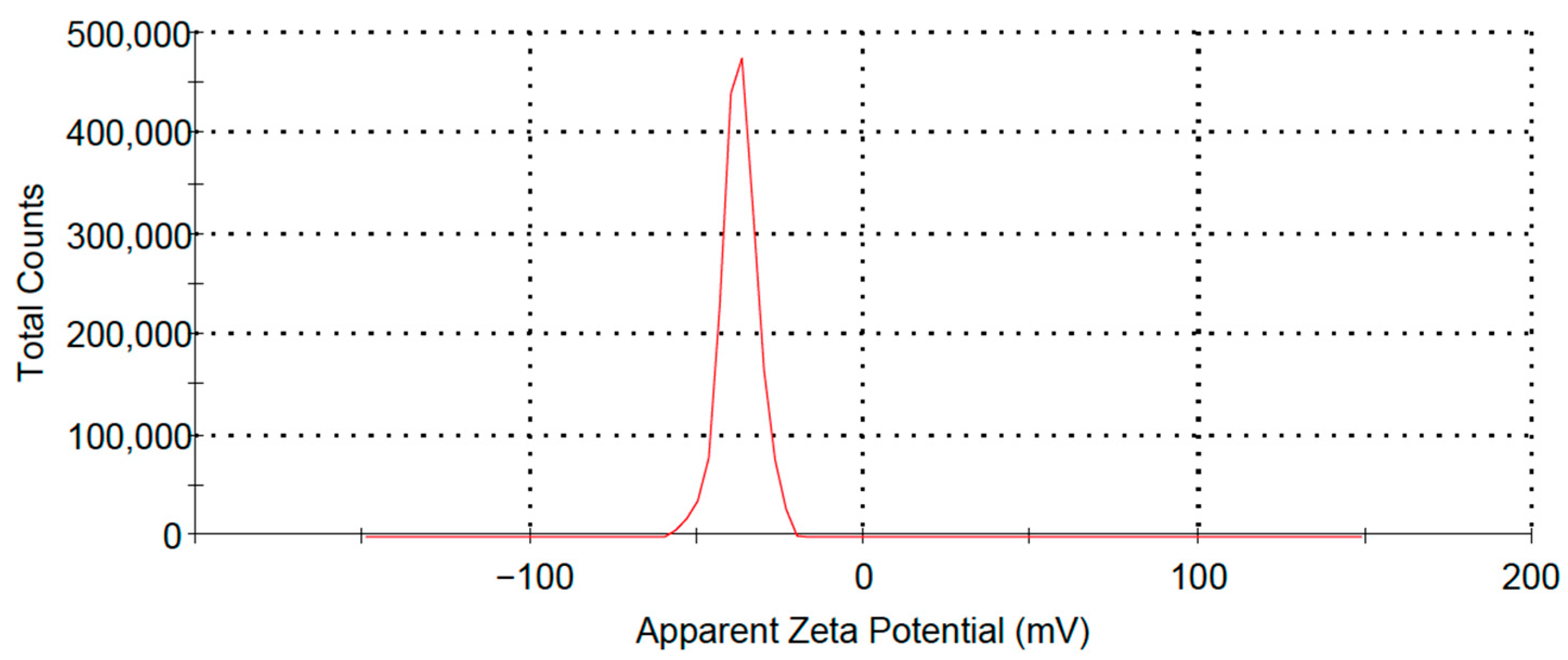
The RCDP NCs synthesized in this study were observed as a light yellow transparent liquid, characterized by an average particle size of 33.1 nm, a PDI of 0.292, and a Zeta potential of (−34.2 ± 0.7) mV. As illustrated in Figure 1, the size distribution by number displays a narrow range, indicating uniform vesicle dimensions, with nearly all particles measuring below 50 nm in diameter. Figure 2 demonstrates a similarly narrow range for Zeta potential distribution, with vesicle surfaces predominantly exhibiting negative charges, thereby confirming the stability of the RCDP NCs.

3.2. In Vitro Skin Penetration

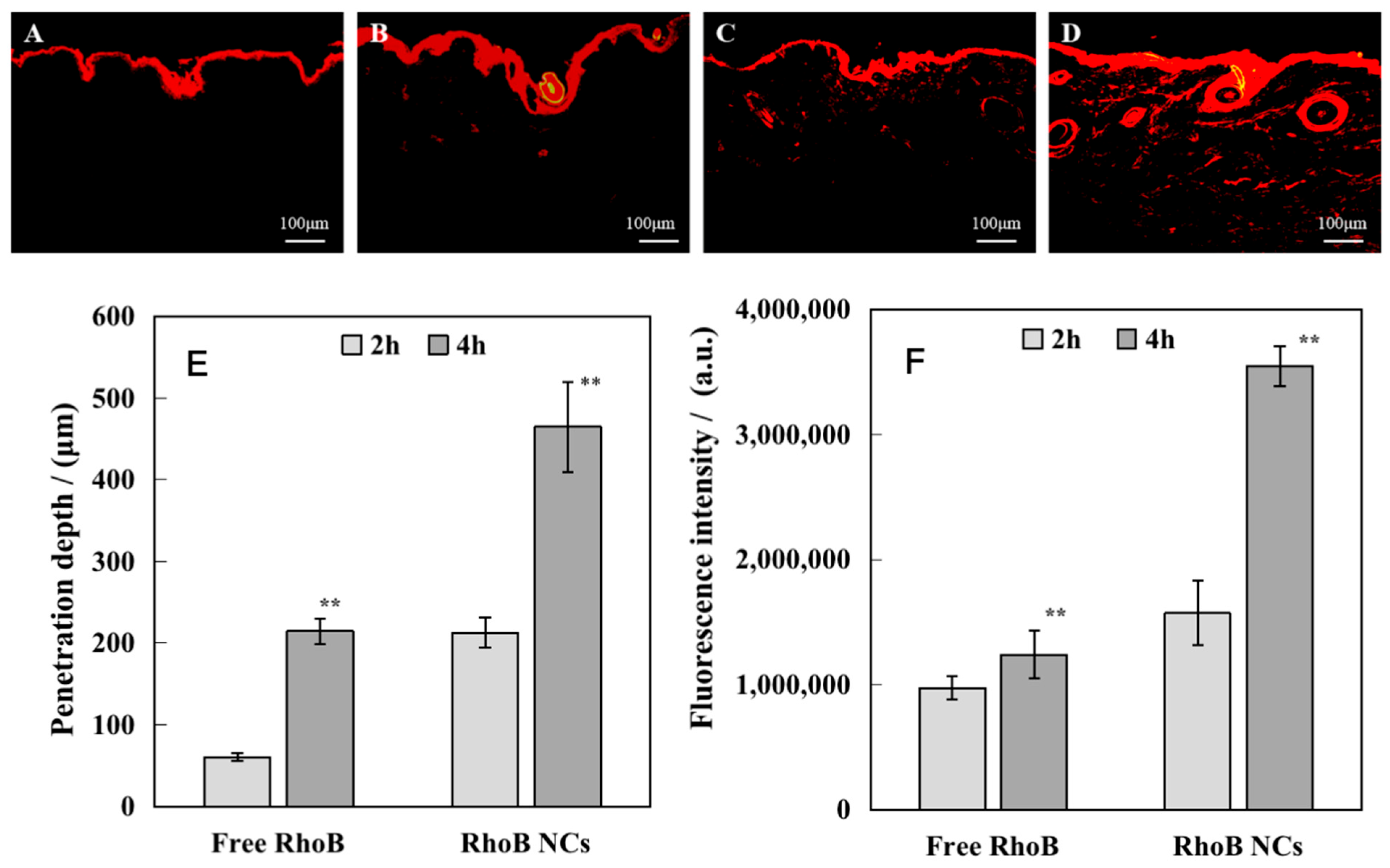
The stratum corneum constitutes a formidable epidermal barrier against environmental elements, resulting in limited transdermal penetration of therapeutic agents and impeding their delivery to the primary anti-aging target site—the dermis. To assess the dermal penetration capability of RCDP NCs, RhoB was encapsulated within the nanocarriers, and both penetration depth and fluorescence intensity were assessed at various time intervals utilizing the vertical Franz diffusion cell methodology. The experimental results are presented in Figure 3.
As observed in Figure 3, Free RhoB within 2 h was predominantly localized in the stratum corneum and was unable to penetrate this barrier. In contrast, RhoB NCs successfully penetrated the stratum corneum barrier within the same timeframe. With extended temporal exposure, the fluorescence penetration depth of RhoB NCs in the skin significantly increased within 4 h, ultimately reaching a depth of 460.0 μm. The fluorescence intensity and penetration depth of RhoB NCs in the skin were significantly greater than those observed for Free RhoB. These findings illustrate that nanocarriers can facilitate the rapid and efficient delivery of encapsulated components to deep dermal tissues.

3.3. Cell Proliferation

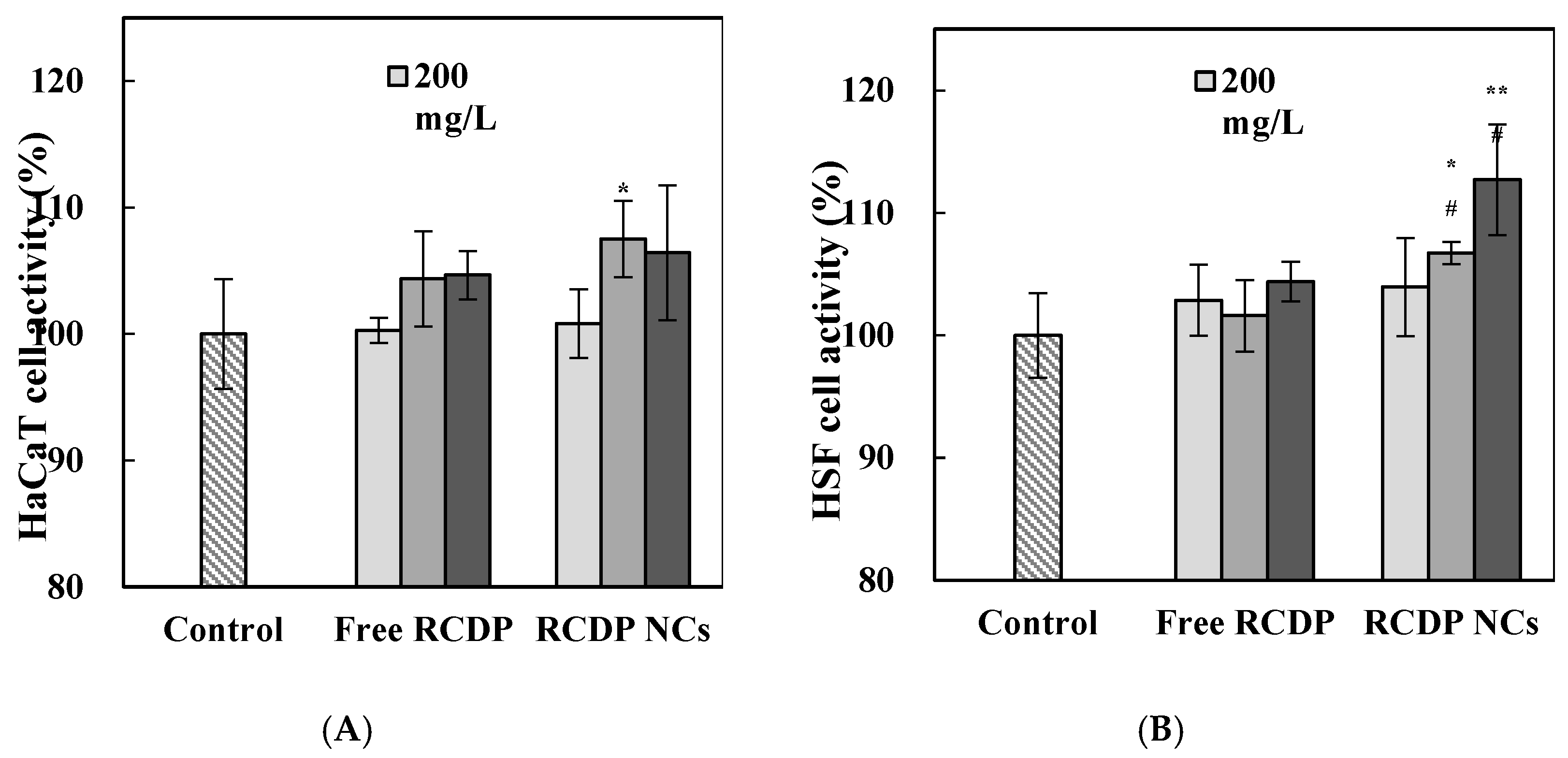
The rapid remodeling of the skin epidermis and dermis significantly impacts the metabolic state of skin renewal. Consequently, we systematically investigated the proliferative effects of Free RCDP and RCDP NCs on HaCaT and HSF cells. The resulting data are presented in Figure 4.
As illustrated in Figure 4A, RCDP NCs at a concentration of 400 mg/L significantly enhanced the proliferation of HaCaT cells compared to the control group (p < 0.05). Figure 4B indicates that RCDP NCs at concentrations of both 400 mg/L and 800 mg/L markedly stimulated HSF cell proliferation relative to the control group (p < 0.01 or p < 0.05). Furthermore, when compared with Free RCDP, HSF cell proliferation was significantly increased at RCDP NC concentrations of 400 mg/L and 800 mg/L (p < 0.05).

3.4. The Uptake of RCDP NCs by Cells

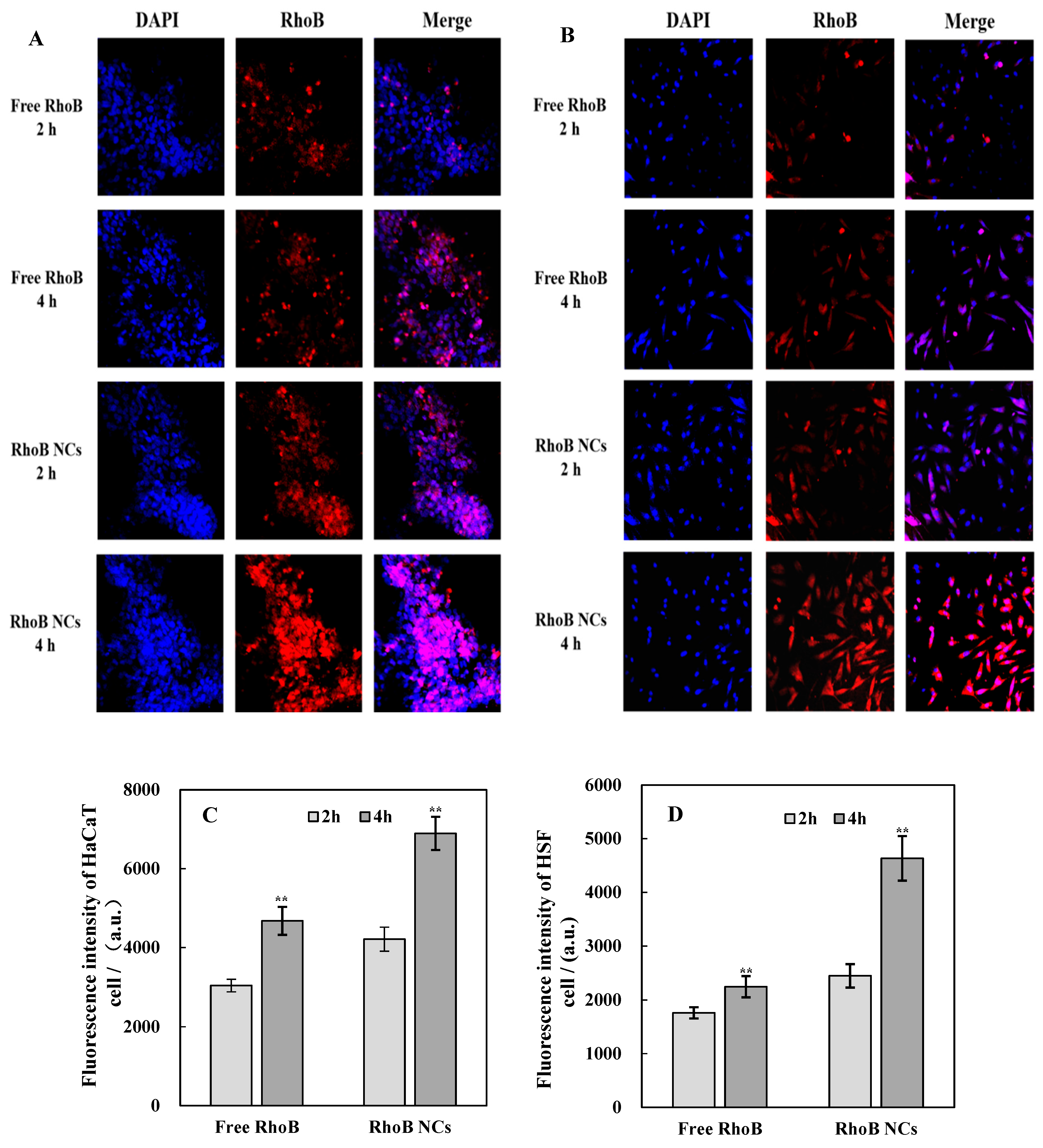
To assess the efficacy of RCDP NCs in delivering functional components into HaCaT and HSF cells, it was deemed essential to investigate the cellular entry behavior of these nanocarrier-encapsulated components [17]. The cellular uptake patterns of Free RhoB and RhoB NCs by HaCaT and HSF cells were analyzed using laser confocal microscopy and flow cytometry. RhoB NCs were utilized for both qualitative and quantitative assessments. The experimental results are presented in Figure 5.
With extended incubation periods, the fluorescence intensity within both HaCaT and HSF cells was significantly enhanced. As illustrated in Figure 5A,B, minimal fluorescence intensity was observed in the Free RhoB solution group after 2 h of incubation. In contrast, the RhoB NCs group exhibited significantly stronger fluorescence intensity compared to the Free RhoB group at the same time point. After 4 h of incubation, the fluorescence intensity in the RhoB NCs group was further increased. According to Figure 5C, following incubation periods of 2 h and 4 h, the mean fluorescence intensity of HaCaT cells treated with RhoB NCs measured 4213 and 6894, respectively. These values represented increases of 53.73% and 47.37%, respectively, compared with the Free RhoB group. Figure 5D demonstrates that after incubation periods of 2 h and 4 h, the mean fluorescence intensity of HSF cells treated with RhoB NCs was quantified at 2451 and 4635, respectively. These measurements indicated increases of 27.60% and 89.11%, respectively, compared with the Free RhoB group. These data confirm that RCDP NCs can effectively deliver encapsulated bioactive substances into targeted skin cells.

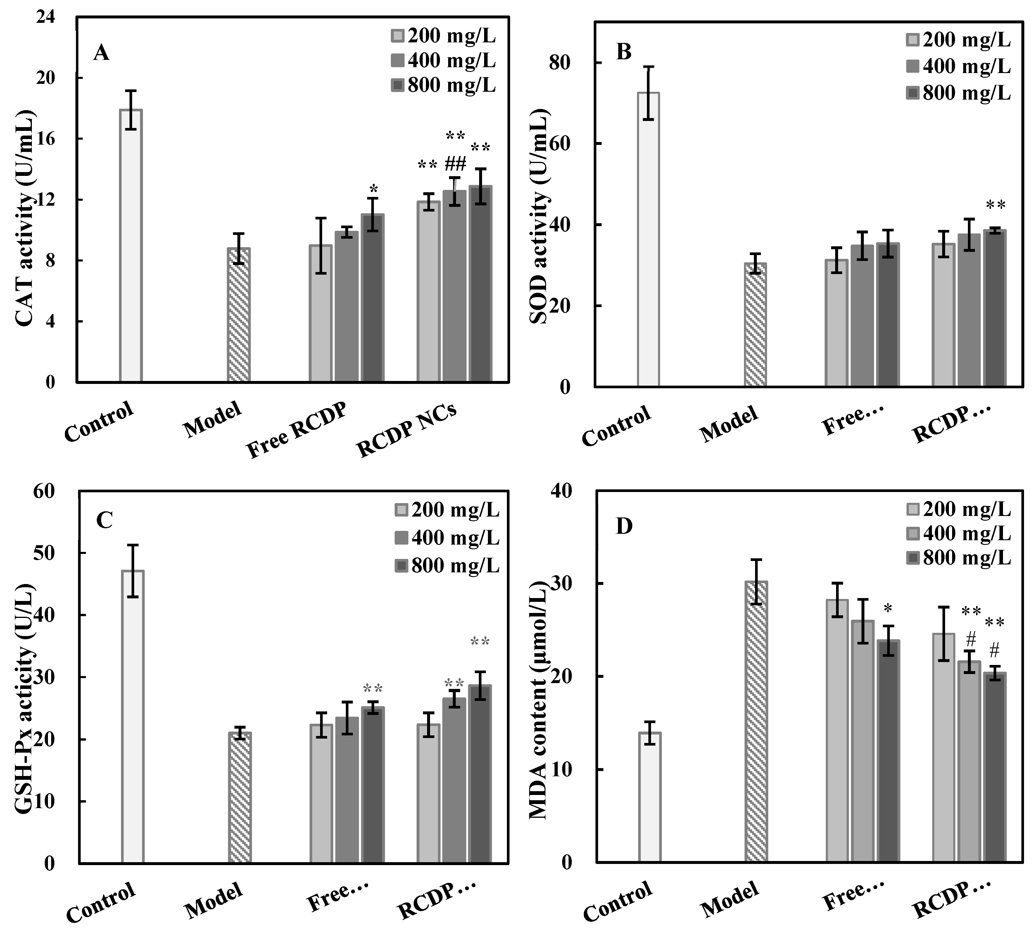
3.5. Effect of RCDP NCs on Cellular Oxidative Stress

Oxidative stress is widely recognized as a significant contributor to skin aging. This phenomenon can lead to the degradation and alteration of various intracellular molecules, including lipids, nucleic acids, and proteins, ultimately resulting in apoptosis and the progression of skin aging [18]. The effects of Free RCDP and RCDP NCs on oxidative parameters within the oxidized cell model are illustrated in Figure 6.
As illustrated in Figure 6, HSF cells in the model group exhibited a significant increase in the activities of CAT, SOD, and GSH-Px, accompanied by a decrease in MDA content following exposure to 0.6 mmol/L H2O2. The CAT activity analysis revealed that 800 mg/L Free RCDP and all three concentrations of RCDP NCs (200 mg/L, 400 mg/L, 800 mg/L) significantly enhanced CAT activity compared to the model group (p < 0.05 or p < 0.01). Furthermore, at a concentration of 400 mg/L, RCDP NCs demonstrated significantly higher CAT activity in oxidatively damaged cells compared to Free RCDP (p < 0.01). In terms of SOD activity, RCDP NCs at 800 mg/L significantly increased SOD activity relative to the model group (p < 0.01). The assessment of GSH-Px activity demonstrated that Free RCDP at 800 mg/L and RCDP NCs at concentrations of 400 mg/L and 800 mg/L significantly increased GSH-Px activity compared to the model group (p < 0.01). The measurement of MDA content indicated that Free RCDP at 800 mg/L and RCDP NCs at concentrations of 400 mg/L and 800 mg/L significantly reduced MDA levels compared to the model group (p < 0.05 or p < 0.01). Additionally, RCDP NCs at 400 mg/L and 800 mg/L demonstrated significantly lower MDA content compared to Free RCDP (p < 0.05). These findings suggest that RCDP NCs possess superior antioxidant properties compared to Free RCDP at equivalent concentrations.

3.6. β-Galactosidase

β-galactosidase serves as a classical biomarker for cellular senescence. This enzyme interacts with the 5-bromo-4-chloro-3-indolyl-β-D-galactopyranoside substrate, generating dark blue products that are commonly utilized to identify senescent cells [19]. The results of β-galactosidase staining in HSF cells treated with Free RCDP and RCDP NCs are presented in Figure 7.
As illustrated in Figure 7, the cells of the 15th generation displayed positive β-galactosidase staining with a prominent dark blue coloration, indicating substantial cellular senescence. Following treatment with Free RCDP and RCDP NCs, the blue coloration of the cells was noticeably attenuated. The RCDP NC treatment resulted in markedly lighter coloration compared to Free RCDP treatment, suggesting a more pronounced reduction in the senescent state of HSF cells after RCDP NCs administration.

3.7. Expression of Autophagy-Related Proteins in Cells

LC3, a homologous protein associated with autophagy-related genes, typically exists in two forms: LC3I and LC3II. During autophagy activation, LC3I is converted into LC3II [20]. P62 serves as a specific substrate that can be degraded through its binding to ubiquitinated proteins, subsequently forming a complex with the LC3II protein and undergoing degradation via the autophagic lysosomal pathway. Elevated levels of P62 expression are correlated with reduced autophagy activity [21]. The expression profiles of the autophagy-related proteins LC3I and LC3II, along with the autophagy degradation-related protein P62, as determined by western blot analysis, are presented in Figure 8.
As illustrated in Figure 8, the autophagy-related protein P62 content was significantly reduced in both the Free RCDP and RCDP NC groups compared to the control group (p < 0.01). Concurrently, there was a substantial increase in the ratio of autophagy-related proteins LC3II/LC3I (p < 0.01). When compared with equivalent concentrations of Free RCDP, the RCDP NCs group exhibited a more pronounced decrease in P62 protein expression (p < 0.01) and a greater increase in the LC3II/LC3I ratio (p < 0.01). These findings indicate that RCDP NCs effectively enhance the autophagic capacity of HSF cells with superior efficacy relative to their free components.

3.8. Observation of Autophagosomes Using Transmission Electron Microscopy

Cellular autophagy involves the encapsulation of damaged or misfolded proteins and organelles within membranous structures, forming double-membrane vesicles that subsequently fuse with lysosomes for content degradation [22]. Autophagosomes within cellular structures were visualized using transmission electron microscopy, and the results are presented in Figure 9.
Transmission electron microscopy observations, as illustrated in Figure 9, revealed a minimal presence of autophagosomes in the cytoplasm of cells from the control group (A,B). In contrast, the cytoplasm of cells treated with Free RCDP exhibited an increased number of autophagosomes compared to the control group (C,D). Notably, the highest density of autophagosomes was observed in the cytoplasm of cells treated with RCDP NCs (E,F). These microscopic findings further substantiate that RCDP NCs enhance the autophagic capacity of HSF cells more effectively than their free components.

4. Discussion

Human aging is regulated by genetically predetermined programs and is simultaneously influenced by cumulative internal and external “wear and tear”. Both mechanisms manifest distinctly at the cellular and molecular levels. The skin, which functions as the outermost organ of the human body, undergoes aging processes that follow comparable patterns. Moreover, skin aging characteristics—such as wrinkles, sagging, pigmentation, and textural roughness—are more visible and measurable [23,24]. As quality of life standards elevate and aesthetic consciousness intensifies, the societal desire to delay cutaneous aging and preserve youthfulness has strengthened considerably. Through advanced research methodologies and technological innovations, scientists have conducted comprehensive investigations into skin aging mechanisms and developed targeted anti-aging interventions [25,26,27]. Although numerous active ingredients with demonstrated anti-aging properties are incorporated into cosmetic formulations and exhibit efficacy through single or multiple pathways, the outcomes frequently fail to meet consumer expectations. The fundamental challenge in addressing skin aging resides in elucidating the anti-aging mechanisms and optimizing the utilization efficiency of active ingredients [28,29,30,31]. In this research context, efficient utilization encompasses maximizing the effectiveness of anti-aging compounds on the skin through enhanced bioavailability via penetration technologies, achieving synergistic anti-aging effects through scientifically validated ingredient combinations, and augmenting the stability of active compounds to prevent efficacy degradation.
The present study was designed based on the optimization of the utilization efficiency of established anti-aging active ingredients. The formulation incorporates small molecular collagen active fragments selected for their superior cellular recognition and adhesion properties, promoting cellular migration [10,11]; ribose for enhancing cellular autophagy [7,8,9]; decarboxylcarnosine HCl for its exceptional antioxidant and anti-glycation capabilities [12]; and palmitoyl tripeptide-1 for facilitating the synthesis of extracellular matrix components, including collagen and glycosaminoglycan [13]. These active compounds were encapsulated within nanocarriers possessing bilayer vesicular structures for targeted delivery. Transdermal penetration assessments and cellular uptake investigations of RCDP NCs demonstrated that these carriers significantly enhanced penetration efficiency, depth, and cellular utilization of the anti-aging compounds.
The experimental results revealed that RCDP NCs increased the activities of SOD, CAT, and GSH-Px enzymes, thereby reducing free radical production in cells under oxidative stress. The observed decrease in MDA levels indicated that RCDP NCs inhibited oxidative stress reactions and minimized cellular damage. Furthermore, RCDP NCs decreased the autophagy degradation-related protein p62 content, elevated the ratio of autophagy-related protein LC3II/LC3I, and increased autophagosome formation. During autophagy, p62 serves as a key ligating protein. p62 is selectively wrapped into the autophagosome and subsequently degraded by protein hydrolases in the autophagic lysosome, and its expression level negatively correlates with autophagic activity [32]. LC3I animalizes or binds to autophagosome membranes and is converted into LC3II, so that an increase in the LC3II/LC3I ratio indicates an increase in autophagy [33,34]. Through autophagy activation, RCDP NCs facilitated the clearance of damaged proteins and senescent organelles, consequently delaying cellular aging processes. Additionally, RCDP NCs stimulated proliferation in keratinocytes and fibroblasts, thereby enhancing metabolic functions in aging skin. After treating 15 generations of HSF aging cells with RCDP NCs, a significant reduction in β-galactosidase, a cell aging biomarker, was observed, indicating substantial improvement in cellular senescence status.
In this study, RCDP NCs primarily delayed cellular senescence through potent antioxidant activity and autophagy promotion. The antioxidant properties enhanced inherent cellular defense mechanisms, mitigating damage to proteins, lipids, and organelles caused by reactive oxygen species and other free radicals. Autophagy functioned as a critical survival mechanism for aging cells responding to internal and external stressors [35]. By eliminating damaged proteins and organelles, reducing oxidative stress-induced damage from various factors, and removing damaged proteins and senescent mitochondria among other organelles, RCDP NCs comprehensively delayed cellular senescence, promoted cellular proliferation, and retarded skin aging progression. Compared with previous studies on anti-aging active ingredients [36,37], this research not only explored the anti-aging mechanisms but also emphasized the efficient utilization of these compounds. This integrated approach presents greater potential for achieving desired anti-aging effects in subsequent skin aging treatments, thereby yielding enhanced economic benefits and market value.

5. Conclusions

In this study, RCDP NCs were successfully synthesized using transdermal drug delivery nanotechnology. The evaluation of transdermal behavior revealed that RCDP NCs exhibited superior penetration efficiency, reaching a dermal depth of 460.0 μm within 4 h, significantly outperforming free components. Enhanced cellular uptake was observed in both HaCaT and HSF, with fluorescence intensity increasing by 47.37% and 89.11%, respectively, after 4 h of incubation, confirming the nanocarriers’ ability to effectively deliver bioactive compounds into target skin cells.
RCDP NCs demonstrated potent anti-aging effects by promoting cell proliferation in HaCaT and HSF cells, elevating antioxidant enzyme activities (CAT, SOD, GSH-Px), and reducing lipid oxidation product (MDA) levels. Notably, β-galactosidase staining indicated a marked reduction in senescent HSF cells following RCDP NC treatment. Furthermore, autophagy regulation studies revealed that RCDP NCs decreased the expression of the autophagy degradation-related protein P62, increased the LC3II/LC3I ratio, and enhanced autophagosome formation, indicating robust activation of autophagy pathways. These mechanisms collectively contributed to delaying cellular senescence and mitigating oxidative stress-induced damage.
Compared to Free RCDP, RCDP NCs exhibited enhanced stability, bioavailability, and synergistic anti-aging efficacy through multi-target mechanisms. The integration of nanotechnology with scientifically formulated active ingredients optimized transdermal delivery, cellular uptake, and functional performance. These findings underscore the potential of RCDP NCs as promising candidates for anti-aging cosmetic formulations, offering significant advancements in addressing skin aging through improved ingredient utilization and mechanistic coordination. Further research and clinical validation are warranted to translate these innovations into practical skincare applications.

Author Contributions

Conceptualization, L.Y. and C.S.; data curation, M.L., L.Y., X.W. and W.L.; formal analysis, M.L.; investigation, M.L. and L.J.; methodology, M.L.; project administration, M.L. and J.Z.; resources, L.Y., J.Z. and W.L.; supervision, L.Y. and C.S.; validation, L.J.; writing—original draft, L.J. and X.W.; writing—review and editing, M.L. and L.J. All authors have read and agreed to the published version of the manuscript.

Funding

This research received no external funding.

Institutional Review Board Statement

Not applicable.

Informed Consent Statement

Not applicable.

Data Availability Statement

The datasets supporting this article’s conclusions are available from the corresponding author upon reasonable request.

Acknowledgments

The authors thank Jiuyan Zheng for testing funding and Wei Liu for polishing the English in the manuscript.

Conflicts of Interest

Min Liu, Lei Ye, Lingling Jiang, Cui Sun and Jiuyan Zheng were all employed by Suzhou Misifu Cosmetic Co., Ltd. The remaining authors declare that the research was conducted in the absence of any commercial or financial relationships that could be construed as a potential conflict of interest.

References

  1. Warsito, M.F.; Kusumawati, I. The impact of herbal products in the prevention, regeneration and delay of skin aging. Adv. Exp. Med. Biol. 2019, 1178, 155–174. [Google Scholar] [CrossRef] [PubMed]
  2. Zhao, X.J.; Hong, Y.H.; Liu, W. Nanocarrier technology-from transdermal drug delivery to functional cosmetics. Deterg. Cosmet. 2016, 44, 12–16. [Google Scholar] [CrossRef]
  3. Martinez-Lopez, N.; Athonvarangku, D.; Singh, R. Autophagy and aging. Adv. Exp. Med. Biol. 2015, 847, 73–87. [Google Scholar] [CrossRef] [PubMed]
  4. Palmer, D.M.; Kitchin, J.S. Oxidative damage, skin aging, antioxidants and a novel antioxidant rating system. J. Drugs Dermatol. 2010, 9, 11–15. [Google Scholar] [PubMed]
  5. Gu, Y.; Han, J.; Jiang, C.; Zhang, Y. Biomarkers, oxidative stress and autophagy in skin aging. Ageing Res. Rev. 2020, 59, 101036. [Google Scholar] [CrossRef]
  6. Mariño, G.; Niso-Santano, M.; Baehrecke, E.H.; Kroemer, G. Self-consumption: The interplay of autophagy and apoptosis. Nat. Rev. Mol. Cell Biol. 2014, 15, 81–94. [Google Scholar] [CrossRef] [PubMed]
  7. Roupe, G. Skin of the aging human being. Lakartidningen 2001, 98, 1091–1095. [Google Scholar] [PubMed]
  8. Muggleton-Harris, A.L.; Defuria, R. Age-dependent metabolic changes in cultured human fibroblasts. In Vitro Cell Dev. Biol. 1985, 21, 271–276. [Google Scholar] [CrossRef]
  9. Shecterle, L.M.; St Cyr, J.A. Dermal benefits of topical D-ribose. Clin. Cosmet. Investig. Dermatol. 2009, 2, 151–152. [Google Scholar] [CrossRef]
  10. Wang, J.; Hu, H.; Wang, J.; Qiu, H.; Gao, Y.; Xu, Y.; Liu, Z.; Tang, Y.; Song, L.; Ramshaw, J.; et al. Characterization of recombinant humanized collagen type Ⅲ and its influence on cell behavior and phenotype. Collagen Leather 2023, 5, 84–96. [Google Scholar] [CrossRef]
  11. Hua, C.; Zhu, Y.; Xu, W.; Ye, S.; Zhang, R.; Lu, L.; Jiang, S. Characterization by high-resolution crystal structure analysis of a triple-helix region of human collagen type III with potent cell adhesion activity. Biochem. Biophys. Res. Commun. 2019, 508, 1018–1023. [Google Scholar] [CrossRef] [PubMed]
  12. Baek, J.H.; Kong, U.S.; Lee, G.M. Cosmetic Composition for Preventing Skin Wrinkles, Containing Carcinine(Decarboxy Carnosine 2HCl) Having Collagen Synthetic and Antioxidant Effects. GB Patent Application No. KR20030010259, 11 September 2024. [Google Scholar]
  13. Yang, F.; Zhang, X.; Wang, H.; Guo, M.; Zhang, J.; Feng, X.; Yu, J.; Yang, J.; Zhu, J.; Wang, Y. Comprehensive evaluation of the efficacy and safety of a new multi-component anti-aging topical eye cream. Skin Res. Technol. 2024, 30, e13790. [Google Scholar] [CrossRef]
  14. Zhang, S.; Zhou, H.; Chen, X.; Zhu, S.; Chen, D.; Luo, D.; Chen, S.; Liu, W. Microneedle Delivery Platform Integrated with Codelivery Nanoliposomes for Effective and Safe Androgenetic Alopecia Treatment. ACS Appl. Mater. Interfaces 2024, 16, 15701–15717. [Google Scholar] [CrossRef]
  15. Zhou, H.; Luo, D.; Chen, D.; Tan, X.; Bai, X.; Liu, Z.; Yang, X.; Liu, W. Current Advances of Nanocarrier Technology-Based Active Cosmetic Ingredients for Beauty Applications. Clin. Cosmet. Investig. Dermatol. 2021, 14, 867–887. [Google Scholar] [CrossRef] [PubMed]
  16. Wang, Z.P.; Shen, H.H.; Luo, D.; Chen, D.; Sheng, J.; Liu, W. Preparation and efficacy evaluation of nanoliposomes for co-delivery of anti-alopecia agents. Chin. Surf. Deter. Cos. 2020, 50, 396–401. [Google Scholar] [CrossRef]
  17. Kim, B.S.; Na, Y.G.; Choi, J.H.; Kim, I.; Lee, E.; Kim, S.-Y.; Lee, J.-Y.; Cho, C.-W. The Improvement of Skin Whitening of Phenylethyl Resorcinol by Nanostructured Lipid Carriers. Nanomaterials 2017, 7, 241. [Google Scholar] [CrossRef]
  18. Wang, M.; Charareh, P.; Lei, X.; Zhong, J.L. Autophagy: Multiple Mechanisms to Protect Skin from Ultraviolet Radiation-Driven Photoaging. Oxidative Med. Cell. Longev. 2019, 2019, 8135985. [Google Scholar] [CrossRef]
  19. Han, S.; Li, H.; Luo, F.; Chen, X.; Cen, Y.; Liu, P.; Chen, Z.; Lan, T.; Lin, J. Inhibitory Effect of Seawater Pearl Hydrolysate on UVA-Induced Photoaging of Human Skin Fibroblasts. Evid. Based. Complement. Alternat. Med. 2022, 2022, 1558288. [Google Scholar] [CrossRef]
  20. Misovic, M.; Milenkovic, D.; Martinovic, T.; Ciric, D.; Bumbasirevic, V.; Kravic-Stevovic, T. Short-term exposure to UV-A, UV-B, and UV-C irradiation induces alteration in cytoskeleton and autophagy in human keratinocytes. Ultrastruct. Pathol. 2013, 37, 241–248. [Google Scholar] [CrossRef]
  21. Jung, C.H.; Ro, S.H.; Cao, J.; Otto, N.M.; Kim, D.-H. mTOR regulation of autophagy. FEBS Lett. 2010, 584, 1287–1295. [Google Scholar] [CrossRef]
  22. Klionsky, D.J.; Emr, S.D. Autophagy as a regulated pathway of cellular degradation. Science 2000, 290, 1717–1721. [Google Scholar] [CrossRef]
  23. Gilchrest, B.A. A review of skin aging and its medical therapy. Br. J. Dermatol. 1996, 135, 867–875. [Google Scholar] [CrossRef] [PubMed]
  24. Gilchrest, B.A.; Garmys, M.; Yaar, M.A. Aging and photoaging effectgene expression in cultured human Keratinocyte. Arch. Dermatol. 1994, 130, 82–86. [Google Scholar] [CrossRef]
  25. Boismal, F.; Serror, K.; Dobos, G.; Zuelgaray, E.; Bensussan, A.; Michel, L. Skin aging: Pathophysiology and innovative therapies. Med. Sci. 2020, 36, 1163–1172. [Google Scholar] [CrossRef]
  26. Csekes, E.; Račková, L. Skin Aging, Cellular senescence and natural polyphenols. Int. J. Mol. Sci. 2021, 22, 12641. [Google Scholar] [CrossRef] [PubMed]
  27. Fisher, G.J.; Kang, S.; Varani, J.; Bata-Csorgo, Z.; Wan, Y.; Datta, S.; Voorhees, J.J. Mechanisms of photoaging and chronological skin aging. Arch. Dermatol. 2002, 138, 1462–1470. [Google Scholar] [CrossRef] [PubMed]
  28. Pozos-Nonato, S.; Domínguez-Delgado, C.L.; Campos-Santander, K.A.; Benavides, A.A.; Pacheco-Ortin, S.M.; Higuera-Piedrahita, R.I.; Resendiz-González, G.; Molina-Trinidad, E.M. Novel nanotechnological strategies for skin anti-aging. Curr. Pharm. Biotechnol. 2023, 24, 1397–1419. [Google Scholar] [CrossRef]
  29. He, X.; Gao, X.; Guo, Y.; Xie, W. Research progress on bioactive factors against skin aging. Int. J. Mol. Sci. 2024, 25, 3797. [Google Scholar] [CrossRef]
  30. He, X.; Wan, F.; Su, W.; Xie, W. Research progress on skin aging and active Ingredients. Molecules 2023, 28, 5556. [Google Scholar] [CrossRef]
  31. Pintea, A.; Manea, A.; Pintea, C.; Vlad, R.-A.; Bîrsan, M.; Antonoaea, P.; Rédai, E.M.; Ciurba, A. Peptides: Emerging candidates for the prevention and treatment of skin senescence: A review. Biomolecules 2025, 15, 88. [Google Scholar] [CrossRef]
  32. Alotaibi, M.R.; As, S.H.; Alaqil, F.A.; Almutairi, M.; Alhazzani, K.; Sulaiman, A.A.; Isab, A.A.; Alotaibi, N.H. A newly synthesizedplatinum-based compound (PBC-Ⅱ) increases chemosensitivity of HeLa ovarian cancer cells via inhibition of autophagy. Saudi Pharm. J. 2019, 27, 1203–1209. [Google Scholar] [CrossRef] [PubMed]
  33. Chen, S.M.; Gong, Y.C.; Sui, L.; Chu, M.; Dong, Y. Role of tangeretin on autophagy in human gastric cancer AGS cells and its mechanism. Chin. Pharmacol. Bull. 2019, 35, 1671–1676. [Google Scholar] [CrossRef]
  34. Qi, M.; Tan, B.E. Molecular mechanism of autophagy regulating oxidative stress in animals. Chin. J. Anim. Nut. 2020, 32, 10. [Google Scholar]
  35. Sun, D.; Wu, R.; Zheng, J.; Li, P.; Yu, L. Polyubiquitin chain-induced p62 phase separation drives autophagic cargo segregation. Cell Res. 2018, 28, 405–415. [Google Scholar] [CrossRef]
  36. Ferreira, M.S.; Magalhães, M.C.; Oliveira, R.; Sousa-Lobo, J.M.; Almeida, I.F. Trends in the Use of Botanicals in Anti-Aging Cosmetics. Molecules 2021, 26, 3584. [Google Scholar] [CrossRef]
  37. Cronin, H.; Draelos, Z.D. Top 10 botanical ingredients in 2010 anti-aging creams. J. Cosmet. Dermatol. 2010, 9, 218–225. [Google Scholar] [CrossRef]
Figure 1. Size distribution by number of RCDP NCs.
Cosmetics 12 00083 g001
Figure 2. Zeta potential distribution of RCDP NCs.
Cosmetics 12 00083 g002
Figure 3. Percutaneous penetration of Free RhoB and RhoB NCs, (A). Free RhoB infiltration for 2 h (10×); (B). Free RhoB permeates for 4 h (10×); (C). RhoB NCs permeate for 2 h (10×); (D). RhoB NCs permeate for 4 h (10×); (E). Penetration depth of Free RhoB and RhoB NCs at 2 h and 4 h of penetration; (F). Fluorescence intensity of Free RhoB and RhoB NCs at 2 h and 4 h of penetration. (Note: compared with Free RhoB at the same time, ** p < 0.01).
Cosmetics 12 00083 g003
Figure 4. Effects of Free RCDP and RCDP NCs on the proliferation of HaCaT (A); Effects of Free RCDP and RCDP NCs on HSF proliferation (B). (Note: compared with the control, ** p < 0.01, * p < 0.05; compared with the same concentration of Free RCDP, # p < 0.05.).
Cosmetics 12 00083 g004
Figure 5. Cell uptake of Free RhoB and RhoB NCs. (A) The uptake of Free RhoB and RhoB NCs by HaCaT was observed using laser confocal microscopy (60×). (B) The uptake of Free RhoB and RhoB NCs by HSF was observed using laser confocal microscopy (60×). (C) Quantitative analysis of HaCaT cell uptake using flow cytometry; (D) Quantitative analysis of HSF cell uptake using flow cytometry. (Note: compared with Free RhoB at the same time, ** p < 0.01).
Cosmetics 12 00083 g005
Figure 6. Effects of Free RCDP and RCDP NCs on oxidation indices of HSF cells, (A) CAT activity; (B) SOD activity; (C) GSH-Px activity; (D) MDA content. (Note: compared with the control, ** p < 0.01, * p < 0.05; compared with the same concentration of Free RCDP, ## p < 0.01, # p < 0.05).
Cosmetics 12 00083 g006
Figure 7. β-galactosidase staining of HSF cells, (A) The 15th generation HSF cells in the control group (10×); (B) Free RCDP group of the 15th generation HSF cells (10×); (C) RCDP NC group of the 15th generation HSF cells (10×).
Cosmetics 12 00083 g007
Figure 8. Expression of autophagy-related proteins in HSF cells treated with Free RCDP and RCDP NCs. (A) Expression of LC3I, LC3II, and P62 proteins; (B) P62/GAPDH protein expression ratio; (C) LC3II/LC3I protein expression ratio. (Note: compared with the control, ** p < 0.01; compared with the same concentration of Free RCDP, ## p < 0.01.).
Cosmetics 12 00083 g008
Figure 9. Autophagic lysosomes of HSF cells observed via transmission electron microscopy: (A) represents the control group (2500×), (B) represents the control group (7000×), (C) represents the Free RCDP group (2500×), (D) represents the Free RCDP group (7000×), (E) represents the RCDP NCs group (2500×), and (F) represents the RCDP NCs group (7000×). The black arrows indicate autophagic lysosomes.
Cosmetics 12 00083 g009
Disclaimer/Publisher’s Note: The statements, opinions and data contained in all publications are solely those of the individual author(s) and contributor(s) and not of MDPI and/or the editor(s). MDPI and/or the editor(s) disclaim responsibility for any injury to people or property resulting from any ideas, methods, instructions or products referred to in the content.