Abstract
This study analyzed the antioxidant activity, cell viability, and human skin primary irritation test using the hot-water extracts of the Syzygium samarangense. As a result of the recent warmer climate, tropical plants have flourished on Jeju Island, and S. samarangense is one of these plants known to have biological activities. In this study, the hot-water extract of S. samarangense leaf and branch was analyzed. Antioxidant activity was measured by DPPH (2,2-diphenyl-1-picrylhydrazyl) and ABTS (2,2′-azino-bis(3-ethyl-benzthiazoline-6-sulfonic acid)) assays, and the DMPD (dimethyl-4-phenylenediamine) radical scavenging activity, nitrite scavenging activity, ferrous-ion chelating activity, cupric reducing antioxidant capacity, reducing power assay, ferric reducing antioxidant power, total phenol content, and total flavonoid content were also measured. In addition, cell viability was measured by MTT assay in human keratinocyte cells (HaCaT), and the safety of the extract for use on the skin was evaluated in the human skin primary irritation test. The antioxidant activities, except DMPD radical scavenging activity and ferrous-ion chelating activity, were stronger in the branch extract than in leaf extract, and the total phenol and flavonoid contents were also higher in the branch extract. Slight irritation was observed in the human skin primary irritation test. However, it was possible to observe sufficient antioxidant capacity at a concentration lower than the concentration used in the irritation test; therefore, if the concentration of the extract is appropriately adjusted, this suggests that it is a possible natural material suitable for use in cosmetics.
1. Introduction
The predicted changes in climate change suggest that by the end of the 21st century, the average temperature will have risen by approximately 4 °C, and the central area of Korea will have a subtropical climate [1]. Consumption of tropical and subtropical vegetables is expected to increase from 20,625 tons to 37,879 tons in 2020, and the cultivation area is also expected to increase from 548 ha to 1073 ha [2]. Indeed, the local governments in the southern region of Korea, where the climate is warm, are intensively promoting subtropical crops as alternatives for future agriculture. In Jeju Island in the southern region, which has the warmest climatic conditions in Korea, there is a favorable environment for the cultivation of subtropical crops, and there is an Agricultural Research Center for Climate Change that oversees the cultivation and distribution of subtropical crops [3]. Tropical and subtropical fruits, such as papaya, artichoke, and avocado, are now cultivated in Jeju island.
S. samarangense is native to Malaysia, but it is grown widely in Southeast Asia, including the Philippines. Recently, it has also been grown in Korea’s Jeju Island, owing to the recent warmer climate [4]. It is known that the effects of S. samarangense are hepatoprotective against alcohol [5], antioxidant activity [6,7], whitening, and wrinkle improvement [8]. In addition, although research on ethanol and methanol extracts of leaf and branch of S. samarangense has been published, the effects on hot-water extracts have rarely been reported.
Human aging is governed by various factors; free radicals have been found to be one of the causes of aging, alongside various diseases [7]. Therefore, owing to their capacity to remove free radicals, the use of antioxidants that protect against oxidative substances generated by oxidative stress in vivo is increasing [9], and functional cosmetics is an active area of research [10].
As the skin ages, the structure and physiological functions of the skin deteriorate, resulting in aging. Skin aging is caused by various factors, such as a decrease in the number of biological bonds in the skin cells, changes in the structure of the cutaneous stratum corneum, decreased differentiation of epidermal cells, and the inhibition of protein synthesis and intercellular substances by fibroblasts in the dermis, and the most important of them is free radicals. Therefore, to suppress skin aging, it is important to establish a barrier against free radicals in these skins.
Therefore, in this study, various assays, such as the DPPH (2,2-diphenyl-1-picrylhydrazyl) radical scavenging assay and the ABTS (2,2′-azino-bis(3-ethyl-benzthiazoline-6-sulfonic acid)) radical cation scavenging assay, were performed to evaluate the antioxidant activity of the extracts of leaf and branch of S. samarangense, and the safety of the extracts was evaluated through the MTT assay and human skin primary irritation test. In addition, HPLC fingerprint analysis was conducted to study the active ingredients of the S. samarangense. Based on these studies, we aimed to investigate the applicability of hot-water extracts of the leaf and branch of S. samarangense as a cosmetic ingredient.
2. Materials and Methods
2.1. Preparation of Extracts
S. samarangense used in this study was collected on Jeju Island in 2016. The collected leaf and branch were extracted at 65 °C for 8 h in distilled water. Each extract was filtered and then concentrated using a vacuum concentrator. The concentrated extract was freeze-dried at −20 ℃, dissolved in distilled water for use in the experiment.
2.2. DPPH Radical Scavenging Activity
The DPPH radical scavenging experiment is a simple, convenient, and widely used antioxidant screening method. The assay was performed, as previously described [11], with some modifications. The assay was conducted in a 96-well microtiter plate, and a plate reader was used to measure absorbance at 515 nm. Each well contained 20 µL of various samples and 180 µL of the solution of DPPH (0.2 mM, Sigma Aldrich, St. Louis, MO, USA). After incubation of the plate at room temperature for 15 min, the absorbance was measured. The radical scavenging activity was calculated from the following equation and expressed as a percentage. Each reaction was measured in triplicate.
Scavenging activity (%) = (Acontrol − Asample)/Acontrol × 100
2.3. ABTS Radical Scavenging Activity
The assay was performed, as previously described [12], with some modifications. The assay was conducted in a 96-well microtiter plate, and a plate reader was used to measure the absorbance at 700 nm. ABTS (14 mM, Sigma Aldrich, St. Louis, MO, USA) and potassium persulfate (4.9 mM, Sigma Aldrich, St. Louis, MO, USA) were dissolved in distilled water and reacted in the dark for 16 h at room temperature to form ABTS cation radicals. Prior to use in the assay, the ABTS radical cations were diluted with 95% ethanol for an initial absorbance of approximately 0.7 ± 0.02 at 700 nm. Each well contained 20 µL of various samples and 180 µL of the solution of ABTS cation radical. After incubation of the plate at room temperature for 15 min, the absorbance was measured. The radical scavenging activity was calculated from the following equation and expressed as a percentage. Each reaction was measured in triplicate.
Scavenging activity (%) = (Acontrol − Asample)/Acontrol × 100
2.4. DMPD (Dimethyl-4-Phenylenediamine) Radical Scavenging Activity
The assay was performed, as previously described [13], with some modifications. The assay was conducted in a 96-well microtiter plate, and a plate reader was used to measure the absorbance at 515 nm. DMPD (200 mM, Sigma Aldrich, St. Louis, MO, USA) was prepared by dissolving in distilled water, and sodium acetate buffer (0.1 M, Biosesang) was added to this solution, and iron (III) chloride (50 mM, Sigma Aldrich, St. Louis, MO, USA) was added to obtain a colored radical cation. Each well contained 20 µL of various samples and 180 µL of DMPD cation radical solution, and the absorbance was measured. The radical scavenging activity was calculated from the following equation and expressed as a percentage. Each reaction was measured in triplicate.
Scavenging activity (%) = (Acontrol − Asample)/Acontrol × 100
2.5. Nitrite Scavenging Activity
The assay was performed, as previously described [14], with some modifications. The assay was conducted in a 96-well microtiter plate, and a plate reader was used to measure the absorbance at 540 nm. Each well contained 20 μL of various samples and 90 μL of sodium nitroprusside (62.5 mM, Sigma Aldrich, St. Louis, MO, USA) and incubated for 30 min at room temperature. Then, 90 µL of Griess reagent (Sigma Aldrich, St. Louis, MO, USA) was added and incubated at room temperature for 15 min, and the absorbance was measured. The nitrite scavenging activity was calculated from the following equation and expressed as a percentage. Each reaction was measured in triplicate.
Scavenging activity (%) = (Acontrol − Asample)/Acontrol × 100
2.6. Ferrous-Ion Chelating Activity
The assay was performed, as previously described [15], with some modifications. The assay was conducted in a 96-well microtiter plate, and a plate reader was used to measure the absorbance at 562 nm. Each well contained 100 μL of various samples, 10 μL of FeCl2·4H2O (1 mM, Sigma Aldrich, St. Louis, MO, USA), and 90 μL of ferrozine (2.5 mM, Sigma Aldrich, St. Louis, MO, USA) and incubated for 1 h at room temperature. Then, the absorbance was measured. The chelating activity was calculated from the following equation and expressed as a percentage. Each reaction was measured in triplicate.
Chelating activity (%) = (Acontrol − Asample)/Acontrol × 100
2.7. Cupric Reducing Antioxidant Capacity (CUPRAC)
The CUPRAC assays measure the copper ion reducing power, and the assay was performed, as previously described [16], with some modifications. The assay was conducted in a 96-well microtiter plate, and a plate reader was used to measure the absorbance at 450 nm. Each well contained 20 μL of various samples, 60 μL of CuCl2·4H2O (5 mM, Sigma), 60 μL of necouproine (1 M, Sigma Aldrich, St. Louis, MO, USA), and ammonium acetate buffer (1 M, pH 7, Biosesang). The plate was incubated for 30 min at room temperature, and the absorbance was measured.
2.8. Reducing Power Assay
The assay was performed, as previously described [17], with some modifications. The assay was conducted in a 96-well microtiter plate, and a plate reader was used to measure the absorbance at 700 nm. Each tube contained 100 μL of various samples, 300 μL of 1% (w/v) potassium ferricyanide (Sigma Aldrich, St. Louis, MO, USA), and 300 μL of phosphate buffer (0.2 M, Sigma Aldrich, St. Louis, MO, USA) and incubated for 30 min at 50 °C; 10% (w/v) trichloroacetic acid was added to the mixture and centrifuged at 3000 rpm for 3 min. Then, 0.1% (w/v) FeCl3 was added to the supernatant, the solutions were transferred to a 96-well microplate, and the absorbance was measured.
2.9. Ferric Reducing Antioxidant Power (FRAP)
The assay was performed, as previously described [18], with some modifications. The assay was conducted in a 96-well microtiter plate, and a plate reader was used to measure the absorbance at 590 nm. Acetate buffer (300 mM, Biosesang), 2,4,6-Tris(2-pyridyl)-s-triazine (TPTZ, 10 mM, Sigma), and FeCl2·6H2O (20 mM, Sigma Aldrich, St. Louis, MO, USA) were prepared, mixed in a 10:1:1 ratio immediately before the experiment and heated at 37 °C for 10 min. Each well contained 20 μL of various samples and 180 μL of FRAP solution and incubated for 30 min at room temperature in the dark, and the absorbance was measured. FeSO4·7H2O was used as a standard, and the content of Fe2+ was calculated using the calibration curve obtained here.
2.10. Total Phenol Contents
The assay was performed, as previously described [19], with some modifications. The assay was conducted in a 96-well microtiter plate, and a plate reader was used to measure the absorbance at 700 nm. Each tube contained 100 μL of various samples, 100 μL of Folin–Ciocalteu’s phenol reagent (Sigma Aldrich, St. Louis, MO, USA), and 900 μL of distilled water and incubated for 30 min at room temperature. Thereafter, 200 µL of Na2CO3 (2 M, Sigma Aldrich, St. Louis, MO, USA) was added and incubated for 1 h at room temperature. After transferring to a 96 well plate, the absorbance was measured. Gallic acid was used as a standard, and the content of phenol was calculated using the calibration curve obtained here.
2.11. Total Flavonoid Contents
The assay was performed, as previously described [20], with some modifications. The assay was conducted in a 96-well microtiter plate, and a plate reader was used to measure the absorbance at 420 nm. Each well contained 20 μL of various samples, 20 μL of 5% (w/v) NaNO2, 140 μL of 10% (w/v) AlCl3·6H2O, and NaOH (1 M, Sigma Aldrich, St. Louis, MO, USA) and incubated for 20 min at 37 °C. Then, the absorbance was measured. Quercetin was used as a standard, and the content of flavonoid was calculated using the calibration curve obtained here.
2.12. Cell Cultures
Human keratinocytes (HaCaT) were obtained from the Korea Cell Line Bank. Cells were cultured in Dulbecco’s modified Eagle’s medium (DMEM) supplemented with 10% fetal bovine serum, 100 units/mL penicillin, and 100 μg/mL streptomycin. Gibco (Grand Island, NY, USA) was used, and 4 × 10⁵ cells/dish were cultured and incubated in an incubator at 37 °C and 5% CO2.
2.13. Cell Viability Assay
After incubation, the cells (1 × 10⁵ cells/well) were seeded in 24-well plates and incubated for 24 h. The culture solution was removed, the medium containing the sample was replaced, and the cells were incubated for a further 24 h. The culture solution was removed, and 400 μg/mL of MTT reagent (0.4 mg/mL) was added, incubated for 4 h at 37 °C, 5% CO2 incubator. The 800 μg/mL of DMSO was added, and the absorbance was measured at 570 nm. Cell viability was calculated as a percentage compared to the control.
2.14. Skin Primary Irritation Test
This test was performed on 34 men or women between 20 and 60 years of age who met the criteria for subject selection and did not meet the exclusion criteria as determined through examination of the background and medical history of the subjects. The test site was wiped with 70% ethanol on the cotton and dried. Then, 20 µL of the test substance was applied on a van der bend to an area on the subject’s back. After 24 h, the patch was removed, and the first evaluation was performed 20 min after patch removal, and the second evaluation was performed after 24 h. The primary skin irritation response was evaluated in accordance with the Personal Care Products Council (PCPC) guidelines (Table 1). The skin reaction results for each test substance were calculated from the formula shown below. The average reactivity of each calculated test substance was determined, as shown in Table 2.

Table 1.
The grading system for skin primary irritation test.

Table 2.
Determination criteria for skin primary irritation [21].
2.15. HPLC Fingerprint
In the case of myricitrin, HPLC analysis was performed by gradient elution with 3% acetic acid aqueous solution and MeOH, and the HPLC conditions are shown in Table 3. In the case of ρ-coumaric acid, water:MeOH:acetic acid = 65:34:1, and it was separated by an isocratic elution method. HPLC conditions are shown in Table 3.

Table 3.
HPLC conditions for the separation of hot-water extracts of leaf and branch of S. samarangense.
2.16. Statistical Analyses
Data were presented as mean values ± standard deviation. The half maximal inhibitory concentration (IC50) values were estimated by a nonlinear regression algorithm (SigmaPlot version 12.0).
3. Results
3.1. DPPH Radical Scavenging Activity
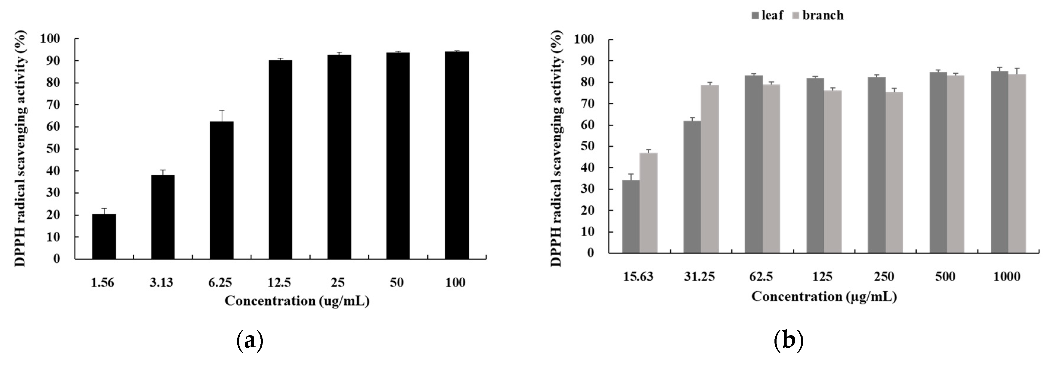
DPPH is a water-soluble free radical that forms a stable, purple color, and when it reacts with an antioxidant, the color dissipates. It is used to search for antioxidants using this principle [22]. In this study, the results of measuring the scavenging activity of DPPH radicals by dissolving hot-water extracts of leaf and branch of S. samarangense in distilled water at a concentration of 15.63 µg/mL to 1000 µg/mL are shown in Figure 1. The half-maximal inhibitory concentration (IC50) values of L-ascorbic acid (the control), leaf, and branch were 4.55 ± 0.034 µg/mL, 24.158 ± 0.685 µg/mL, and 21.352 ± 4.683 µg/mL, respectively. Although the extracts were less effective than L-ascorbic acid, their activity was increased depending on the concentration; in addition, the branch extract had stronger antioxidant activity than the leaf extract.
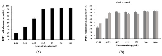
Figure 1.
DPPH (2,2-diphenyl-1-picrylhydrazyl) radical scavenging activities of (a) L-ascorbic acid and (b) hot-water extracts of the S. samarangense. An appropriate amount of ascorbic acid was used as a positive control. The results were expressed as the mean ± SD of data obtained from three independent experiments.
3.2. ABTS Radical Scavenging Activity
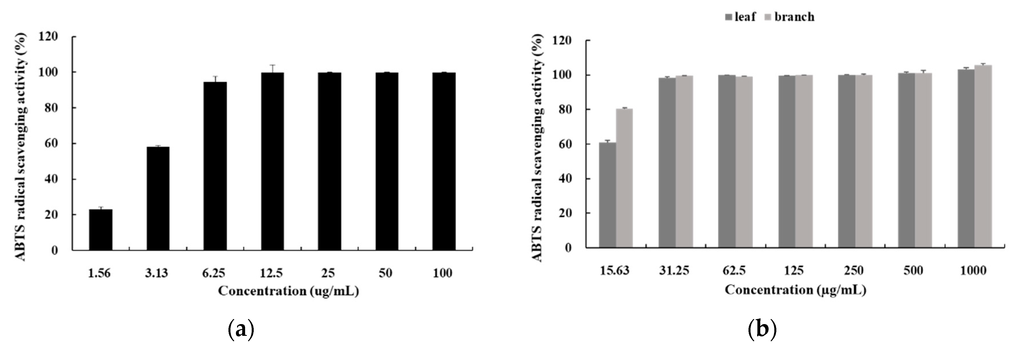
The ABTS radical scavenging activity is the same as DPPH radical, but DPPH is free radical, whereas ABTS is cation [23]. In this study, the results of measuring the scavenging activity of ABTS radicals by dissolving the hot-water extracts of leaf and branch of S. samarangense in distilled water at a concentration of 15.63 µg/mL to 1000 µg/mL are shown in Figure 2. The IC50 value of L-ascorbic acid (the control) was 2.732 ± 0.099 µg/mL, and the values of the leaf and branch were below 10 µg/mL. The extracts showed stronger antioxidant power than in the DPPH assay, and it was expected that the extract could be a useful functional material as it showed similar activity to L-ascorbic acid.
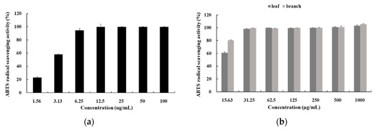
Figure 2.
ABTS (2,2′-azino-bis(3-ethyl-benzthiazoline-6-sulfonic acid)) radical scavenging activities of (a) L-ascorbic acid and (b) hot-water extracts from S. samarangense. An appropriate amount of ascorbic acid was used as a positive control. The results were expressed as the mean ± SD of data obtained from three independent experiments.
3.3. DMPD Radical Scavenging Activity
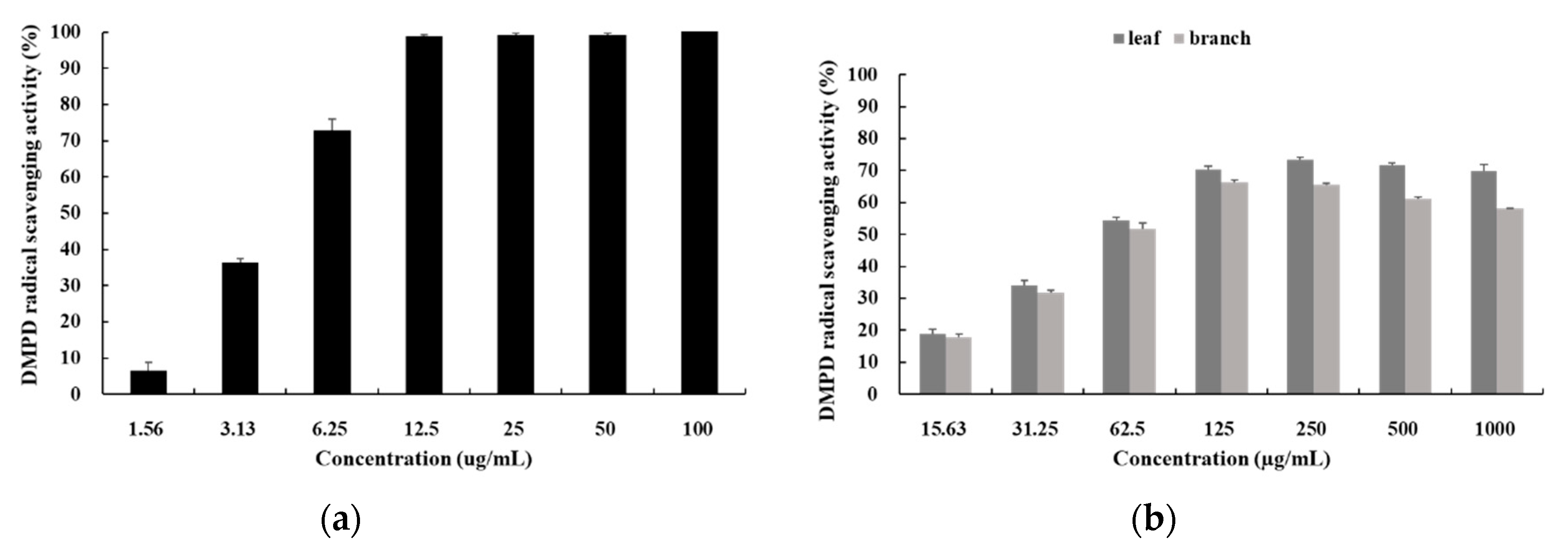
The reaction of the compound DMPD in the presence of a suitable oxidant solution leads to the formation of a colored solution of the DMPD radical cation. Antioxidant compounds, which are able to transfer a hydrogen atom to the DMPD radical cation, cause decoloration of the solution [24]. In this study, the results of measuring the scavenging activity of DMPD radicals by dissolving hot-water extracts of leaf and branch of S. samarangense in distilled water at a concentration of 15.63 µg/mL to 1000 µg/mL are shown in Figure 3. The IC50 values of L-ascorbic acid (the control), leaf, and branch were 4.116 ± 0.092 µg/mL, 53.269 ± 0.386 µg/mL, and 57.278 ± 1.186 µg/mL, respectively. Although the extracts were less effective than L-ascorbic acid, their activity was increased depending on the concentration; in addition, the leaf extract had stronger antioxidant activity than the branch activity.
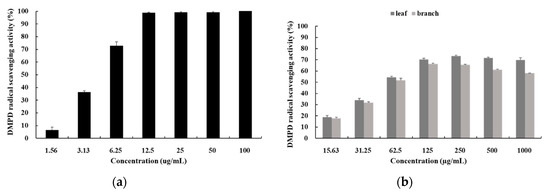
Figure 3.
DMPD (dimethyl-4-phenylenediamine) radical scavenging activities of (a) L-ascorbic acid and (b) hot-water extracts from S. samarangense. An appropriate amount of ascorbic acid was used as a positive control. The results were expressed as the mean ± SD of data obtained from three independent experiments.
3.4. Nitrite Scavenging Activity
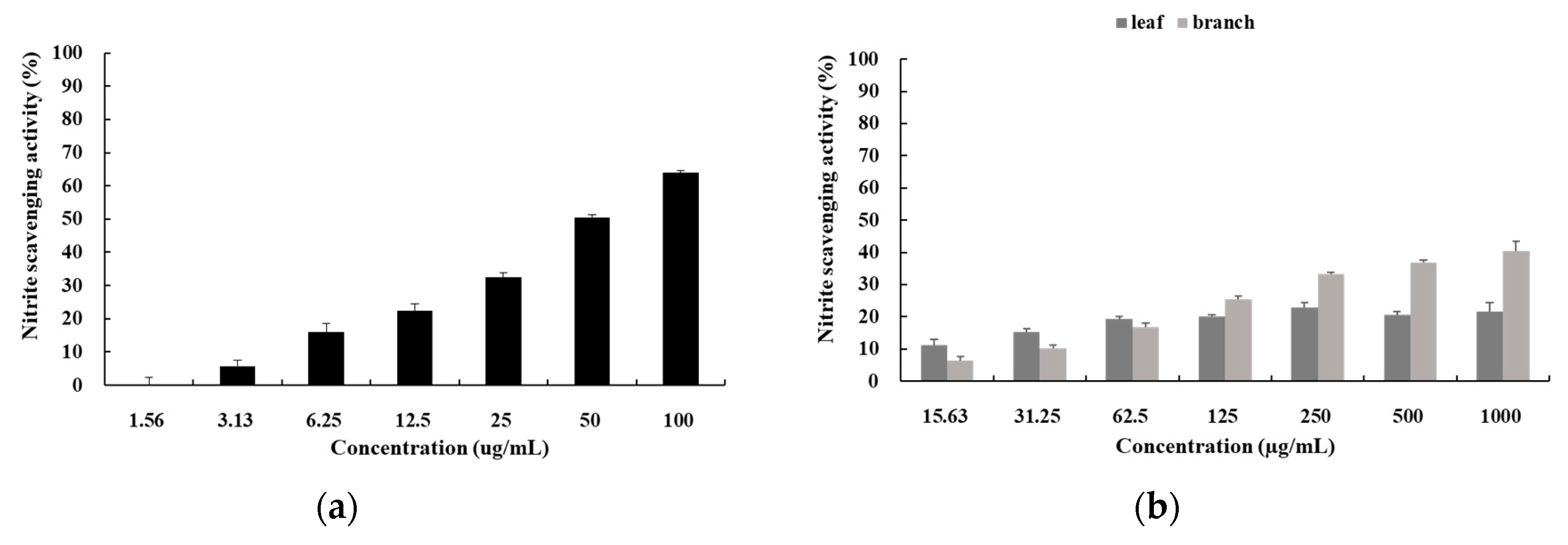
Nitrite is added to prevent toxin production and color development and to prevent rancidity during the processing and storage of meat products and seafood [25]. However, nitrite oxidizes the iron in hemoglobin to produce methemoglobin or combine an amine to produce nitrosamine, a carcinogen [26]. In this study, the results of measuring the scavenging activity of nitrite by dissolving hot-water extracts of leaf and branch of S. samarangense in distilled water at a concentration of 15.63 µg/mL to 1000 µg/mL are shown in Figure 4. The IC50 value of L-ascorbic acid as control was 52.588 ± 1.027 µg/mL, and the values of the leaf and branch were more than 1000 µg/mL. Although the extract was less effective than L-ascorbic acid, its activity was increased depending on the concentration; in addition, the branch extract showed stronger antioxidant activity than the leaf extract.
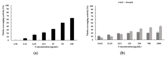
Figure 4.
Nitrite scavenging activities of (a) L-ascorbic acid and (b) hot-water extracts from S. samarangense. An appropriate amount of ascorbic acid was used as a positive control. The results were expressed as the mean ± SD of data obtained from three independent experiments.
3.5. Ferrous-Ion Chelating Activity
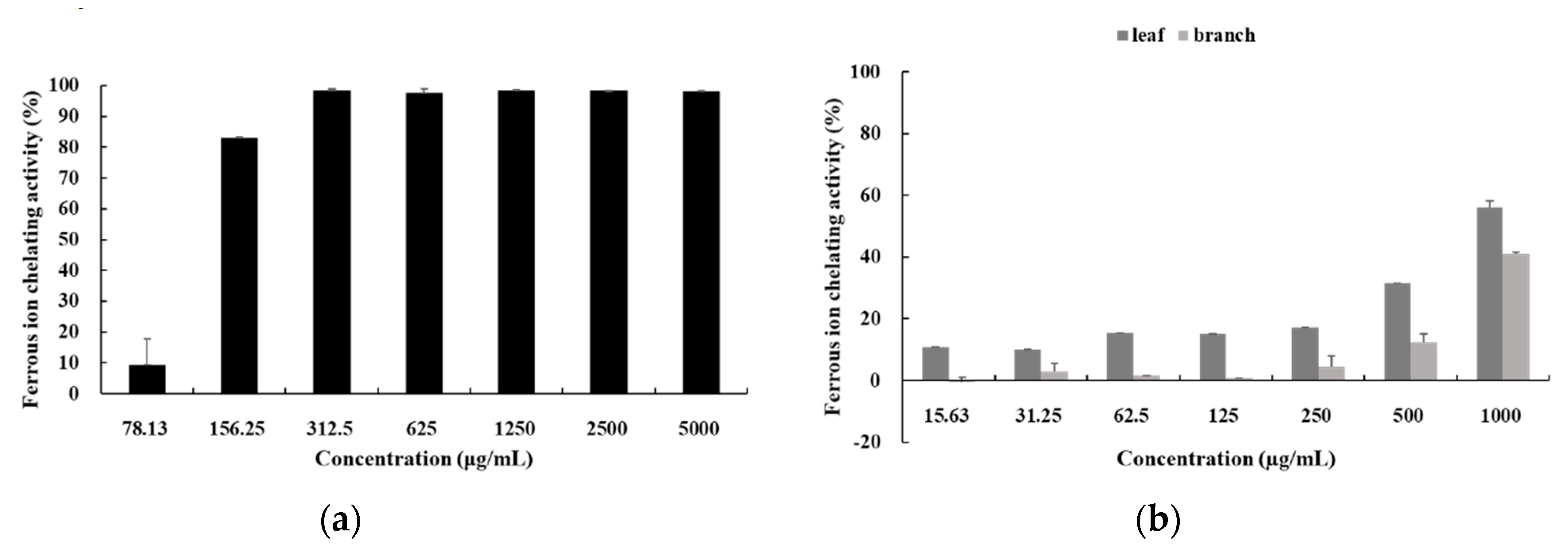
Ferrozine forms a complex with Fe2+, which produces a reddish solution. At this time, if a substance with a chelating effect is present in the sample, the formation of the Fe2+-ferrozine complex is hindered, and color development is inhibited [27]. In this study, the results of measuring the chelating activity by dissolving hot-water extracts of leaf and branch of S. samarangense in distilled water at a concentration of 15.63 µg/mL to 1000 µg/mL are shown in Figure 5. The IC50 value of EDTA—the control—was 121.283 ± 2.857 µg/mL, and the values of the leaf and branch extracts were more than 1000 µg/mL. Although the extract was less effective than L-ascorbic acid, its activity was increased depending on the concentration; in addition, the leaf extract had stronger antioxidant activity than the branch extract.
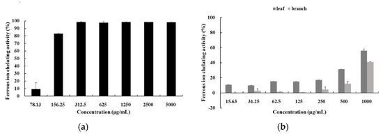
Figure 5.
Fe2+ ion chelating activities of (a) EDTA and (b) hot-water extracts from S. samarangense. An appropriate amount of EDTA was used as a positive control. The results were expressed as the mean ± SD of data obtained from three independent experiments.
3.6. Cupric Reducing Antioxidant Capacity (CUPRAC)
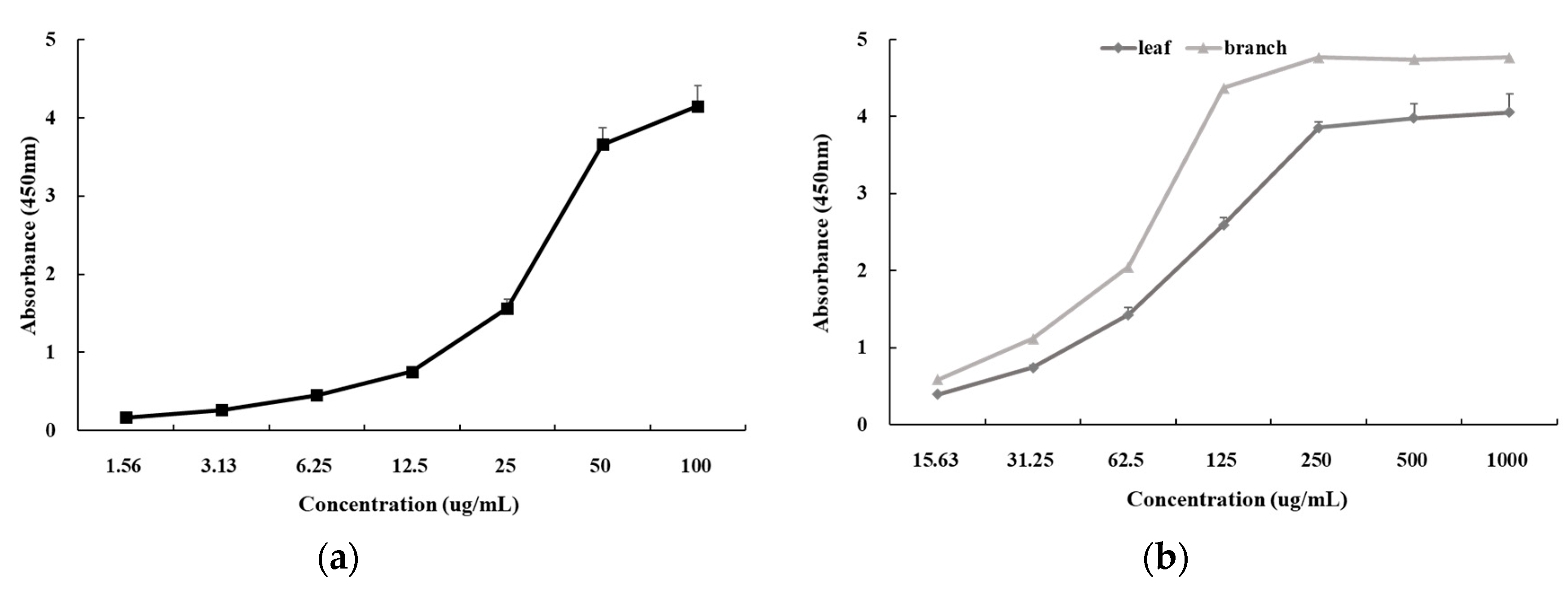
This experiment measured the activity of copper ion reducing power through changes in absorbance [28]. In this study, the results of measuring the reducing antioxidant capacity by dissolving hot-water extracts of leaf and branch of S. samarangense in distilled water at a concentration of 15.63 µg/mL to 1000 µg/mL are shown in Figure 6. The absorbance values of L-ascorbic acid (the control), leaf, and branch were 4.2 at 100 µg/mL, 4.1 at 1000 µg/mL, and 4.8 at 1000 µg/mL, respectively. Although the extract was less effective than L-ascorbic acid, its activity was increased depending on the concentration; in addition, the branch extract had stronger antioxidant activity than the leaf extract.
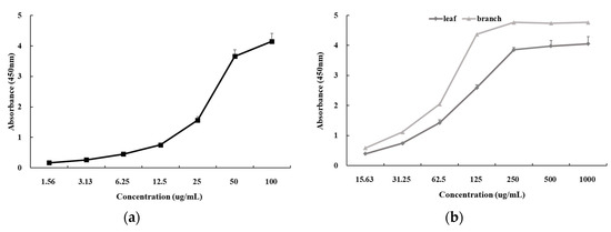
Figure 6.
Cupric reducing antioxidant capacity of (a) L-ascorbic acid and (b) hot-water extracts from S. samarangense. An appropriate amount of ascorbic acid was used as a positive control. The results were expressed as the mean ± SD of data obtained from three independent experiments.
3.7. Reducing Power Assay
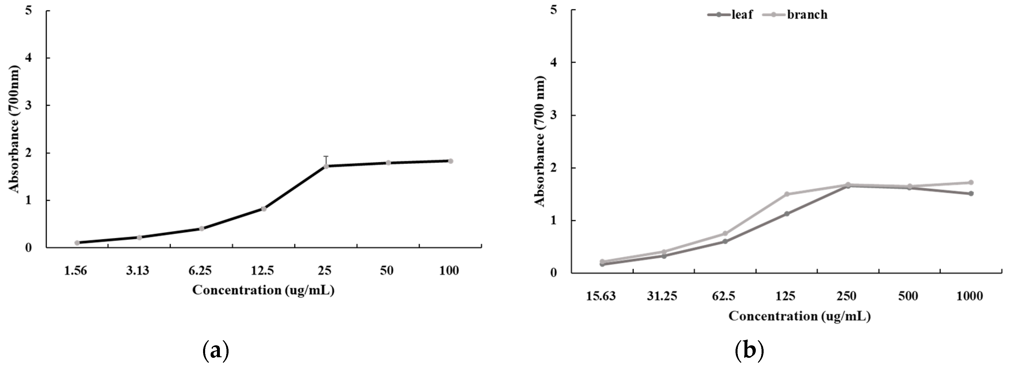
The reducing power assay is a method to measure the reducing power reduced from Fe3+ to Fe2+ [26]. In this study, the results of measuring the reducing power by dissolving hot-water extracts of leaf and branch of S. samarangense in distilled water at a concentration of 15.63 µg/mL to 1000 µg/mL are shown in Figure 7. The absorbance values of L-ascorbic acid (the control), leaf, and branch were 1.8 at 100 µg/mL, 1.5 at 1000 µg/mL, and 1.7 at 1000 µg/mL, respectively. Although the extract was less effective than L-ascorbic acid, its activity was increased depending on the concentration; in addition, the branch extract had stronger antioxidant activity than the leaf extract.
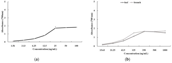
Figure 7.
Reducing power of (a) L-ascorbic acid and (b) hot-water extracts from S. samarangense. An appropriate amount of ascorbic acid was used as a positive control. The results were expressed as the mean ± SD of data obtained from three independent experiments.
3.8. Ferric Reducing Antioxidant Power (FRAP)
The FRAP assay is a recently developed means to measure antioxidant capacity, which uses the principle of the reduction of ferric tripyridyltriazine (Fe3+-TPTZ) complex to a blue ferrous tripyridyltriazine (Fe2+-TPTZ) by a reducing agent at a low pH [29]. The results of the FRAP were using FeSO4 as a standard substance, as shown in Table 4. Although the extract was less effective than L-ascorbic acid, it had FRAP activity, and the branch extract had a stronger reducing power than leaf extract.

Table 4.
Ferric reducing antioxidant power (FRAP) values of ascorbic acid and hot-water extracts from S. samarangense.
3.9. Total Phenol Content and Total Flavonoid Content
The total phenol content was determined using the principle that Folin-reagent is reduced to blue molybdenum, owing to the polyphenol component contained in the extract; the results are shown in Table 5 [30]. Phenolic compounds have various physiological activities, such as antioxidant and anti-cancer properties. In this study, the hot-water extracts of leaf and branch of S. samarangense were prepared by dissolving in distilled water, and the phenol content of the extract was converted according to the standard curve using gallic acid as a standard substance. The total phenolic content was 66.778 ± 1.64 mg GAE/g in the leaf extract and 76.3820 ± 1.085 mg GAE/g in the branch extract; the branch extract contained a higher phenol content than the leaf extract.

Table 5.
Total phenol content and flavonoid content of hot-water extracts from S. samarangense.
The total flavonoid content uses the principle that the flavonoid contained in the extract is turned yellow by a strong oxidizing agent; the results are shown in Table 5 [30]. Flavonoids are pigments present in various plants and are known to show the effects, such as pathogen inhibition, UV protection, anti-mutation, antiviral, and anti-inflammatory effects. In this study, the hot-water extracts of leaf and branch of S. samarangense were prepared by dissolving in distilled water, and the flavonoid content of the extract was converted according to the standard curve using quercetin as a standard substance. The total flavonoid content was 40.8076 ± 2.226 mg QE/g in the leaf extract, and 78.057 ± 3.576 mg QE/g in the branch extract; the branch extract contained a higher total flavonoid content than the leaf extract.
3.10. Cell Viability Assay
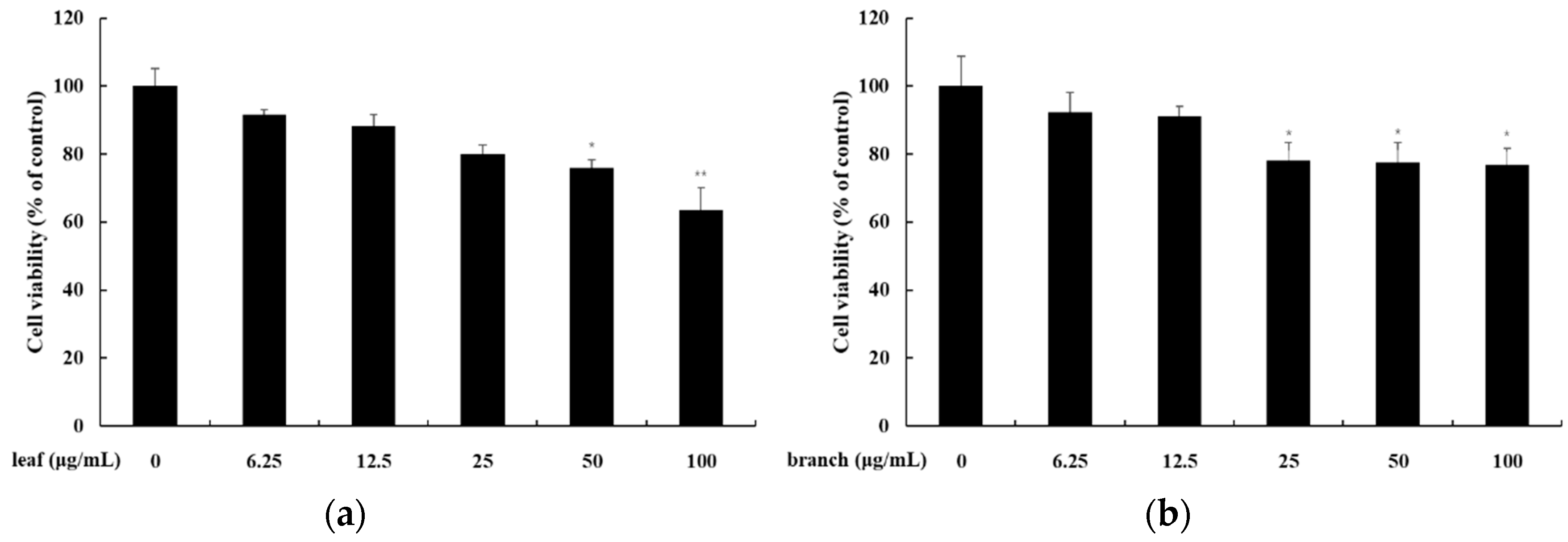
To investigate the cytotoxicity of extracts to human keratinocyte HaCaT cells, cell viability was measured by using the MTT assay. MTT is transported into living cells and is reduced to formazan by the metabolism of cells and becomes purple [31]. In this study, the hot-water extracts of leaf and branch of S. samarangense were prepared by dissolving in distilled water, and the results of treating the sample are shown in Figure 8. The leaf extract showed 80% cell viability at 25 µg/mL, and the branch extract showed 78% cell viability at 25 µg/mL.
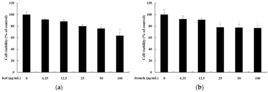
Figure 8.
The effect of hot-water extracts from S. samarangense on the viability of HaCaT (human keratinocyte cells). Cells were each treated with (a) the leaf of S. samarangense (6.25, 12.5, 25, 50, and 100 µg/mL) and (b) the branch of S. samarangense (6.25, 12.5, 25, 50, and 100 µg/mL) for 24 h. The cell viability was determined by MTT assay. The data were presented as the mean ± standard deviation (SD) of at least four independent experiments (n = 3). * p < 0.05, ** p < 0.01, *** p < 0.001 versus control.
3.11. Skin Primary Irritation Test
The hot-water extracts of leaf and branch of S. samarangense at a concentration of 100 µg/mL were applied to a patch and tested for skin contact for 24 h. Then, the patch was removed after 48 h, and the extracts were observed (Table 6). Six subjects showed a stimulus-response of +1 grade in response to the leaf extract. Therefore, in terms of skin primary irritation test, the test substance was considered to be a substance of the medium stimulation category. Two subjects showed a stimulus-response of +1 grade in response to the branch extract. Therefore, in terms of skin primary irritation test, the test substance was considered to be a substance of the low stimulation category.

Table 6.
Results of human skin primary irritation test (n = 34).
3.12. HPLC Analysis
In order to analyze the content of myricitrin and ρ-coumaric acid using HPLC, the retention time was compared with a standard substance and quantified. As a result, myricitrin contained 15.6 mg/g in leaf extract and 0.2 mg/g in the branch extract (Figure 9). ρ-coumaric acid was 0.36 mg/g in leaf extract and was not found in the branch extract (Figure 10). The leaf and branch extract contained myricitrin and ρ-coumaric acid, so it could be expected to have anti-oxidant effects, consistent with the actual experimental results.

Figure 9.
HPLC chromatogram of (a) myricitrin and hot-water extracts of the leaf (b) and branch (c) of S. samarangense.

Figure 10.
HPLC chromatogram of (a) ρ-coumaric acid and hot-water extracts of the leaf (b) and branch (c) of S. samarangense.
4. Discussion
Ingredients for natural cosmetics tend to emphasize the familiar impression with new and surprising functionality. As a representative trend, the fact that inner beauty, represented by eating cosmetics, has established itself as a global trend centering on developed countries. Due to the familiarity of consumers, tropical or subtropical fruit is an attractive resource for cosmetics’ developers as a target for developing cosmetic ingredients [8]. Therefore, based on these ideas, we tried to confirm whether Jeju wax apple could be a cosmetic ingredient through various experiments.
Whether or not Jeju wax apple possesses antioxidant properties is very important from the perspective of developing cosmetic ingredients. This is because many antioxidant ingredients, including vitamin C, have a close relationship with the efficacy of suppressing wrinkles and melanin production and inflammatory skin diseases on the face and skin and can be applied to various cosmetics [32,33].
In this study, the DPPH radical scavenging, ABTS radical scavenging, DMPD radical scavenging, nitrite scavenging, and ferrous-ion chelating activity of the hot-water extracts of leaf and branch of S. samarangense were analyzed. The leaf and branch of S. samarangense were extracted with distilled water at 65 ℃. In addition, cupric reducing antioxidant capacity, reducing power assay, ferric reducing antioxidant power (FRAP), total phenol contents, and total flavonoid content were also measured.
First, the IC50 value of DPPH radical scavenging activity was four times larger for the leaf and branch extracts than ascorbic acid. In addition, the IC50 value of ABTS radical scavenging activity was similar for the leaf and branch extracts and ascorbic acid. DPPH uses free radicals in DPPH radical scavenging activity, whereas ABTS evaluates the scavenging ability of cation radicals; thus, the difference is believed to be due to the different phenolic substances that bind each of the radicals [34]. In these two antioxidant experiments, S. samarangense worked well as an antioxidant because it is superior to other subtropical plants that grow on Jeju Island—Mangifera indica, Momordica charantia, and Curcuma longa [35,36,37]. The IC50 value of DMPD radical scavenging activity was approximately 13 times higher for the leaf and branch extracts than for ascorbic acid, and the IC50 value of ferrous-ion chelating activity was approximately 10 times higher than that of EDTA. The IC50 value of the nitrite scavenging activity was approximately 20 times higher for the leaf and branch extracts than for ascorbic acid. In these experiments, it was seen that although the extracts had weaker activity than ascorbic acid, the activity increased depending on the concentration.
The IC50 values of cupric reducing antioxidant capacity, reducing power assay, and ferric reducing antioxidant power (FRAP) were approximately 10 times larger than that of leaf and branch extracts compared to ascorbic acid. However, in all four experiments, the activity increased depending on the concentration, and the activity of the branch extract was stronger than that of the leaf extract.
Second, the total phenolic content was approximately 1.1 times greater in the branch extract than the leaf extract, and the total flavonoid content was approximately 1.9 times greater. This result supported the finding that the antioxidant activity of the branch was superior to that of the leaf in most antioxidant experiments. There is a strong correlation between antioxidant activity with total phenolic and total flavonoid content. Phenolic compounds are important plant constituents with redox properties responsible for antioxidant activity. This is because hydroxyl groups having a natural structure contained in plant extracts generally promote free radical scavenging [38].
Next, in order to test the application of the cosmetic ingredients of the hot water extract derived from wax apple’s leaf and branch, we conducted an MTT experiment on human keratinocytes and a primary skin irritation test on human skin. As shown in Figure 8, the cell viability of leaf and branch extracts, using human keratinocyte HaCaT, was slightly lowered from 92% to 64% when the leaf was treated at a concentration 6.25–100 µg/mL. In the branch also, the cell viability was lowered from 92% to 77%, indicating lower toxicity than the leaf. Besides, in the skin primary irritation test, the leaf extract was classified as a medium stimulation category, and the branch extract was classified as a low stimulation category. In the case of the leaf extract, skin irritation was observed, but sufficient antioxidant activity was observed at a concentration lower than the applied concentration of 100 µg/mL. For the branch extract, skin irritation did not appear after 48 h in the primary skin irritation test, and the antioxidant activity was generally better than for the leaf extract. Therefore, it is suggested if an appropriate concentration of branch extract is applied, it may be a suitable natural material for use in cosmetics.
Finally, we performed HPLC fingerprint analysis to confirm the standard components for the antioxidant efficacy of Jeju wax apple extract. Since myricitrin and ρ-coumaric acid have been reported as the ingredients in the Syzygium plant that are effective antioxidant agents, they were used as standard substances [39]. Using the conditions described in the experimental section, myricitrin and ρ-coumaric acid were well resolved from the wax apple extract with excellent peak shapes.
Considering these results, we suggested that Jeju wax apple extracts be considered possible antioxidant candidates for topical application.
Author Contributions
Investigation: S.B.H., S.B.; writing—original draft preparation: S.B.H.; writing—review and editing: S.B.H., C.-G.H.; supervision: C.-G.H. All authors have read and agreed to the published version of the manuscript.
Funding
This research received no external funding.
Acknowledgments
This work was supported by the Technology Development Program (S2707107) funded by the Ministry of SMEs and Startups (MSS, Korea).
Conflicts of Interest
The authors declare no conflict of interest.
References
- Kwak, T.S.; Ki, J.H.; Kim, Y.E.; Jeon, H.M.; Kim, S.J. A Study of GIS prediction model of domestic fruit cultivation location changes by the global warming -six tropical and sub-tropical fruits-. J. Korea Spatial Inform. Syst. Soc. 2008, 10, 93–106. [Google Scholar]
- Seong, K.C.; Kim, C.H.; Son, D.; Wi, S.H.; Lim, C.G.; Cheon, S.J.; Kim, S.Y.; Choi, B.Y.; Seo, J.S.; Song, M.G.; et al. The present state and future prospect of tropical/subtropical vegetables in Korea. Korean Soc. Hortic. Sci. 2014, 5, 56. [Google Scholar]
- Kim, C.Y.; Kim, Y.H.; Han, S.H.; Ko, H.C. Current situations and prospects on the cultivation program of tropical and subtropical crops in Korea. Plant Resour. Soc. Korea 2019, 32, 45–52. [Google Scholar]
- Kim, M.J.; Lee, J.Y.; Kim, S.S.; Seong, K.C.; Lim, C.K.; Park, K.J.; An, H.J.; Choi, Y.H.; Kim, S.Y.; Lee, N.H.; et al. Anti-inflammatory activity of wax apple (Syzygium samarangense) extract from Jeju Island. Korean Soc. Biotechnol. Bioeng. 2017, 32, 245–250. [Google Scholar]
- Zhang, Y.J.; Zhou, T.; Wang, F.; Zhou, Y.; Li, Y.; Zhang, J.J.; Zheng, J.; Xu, D.P.; Li, H.B. The effects of Syzygium samarangense, Passiflora edulis and Solanum muricatum on alcohol-induced liver injury. Int. J. Mol. Sci. 2016, 17, 1616. [Google Scholar] [CrossRef] [PubMed]
- Soubir, T. Antioxidant activities of some local bangladeshi fruits (Artocarpus heterophyllus, Annona squamosa, Terminalia bellirica, Syzygium samarangense, Averrhoa carambola and Olea europa). Chin. J. Biotechnol. 2007, 23, 257–261. [Google Scholar]
- Simirgiotis, M.J.; Adachi, S.; To, S.; Yang, H.; Reynertson, K.A.; Basile, M.J.; Gil, R.R.; Weinstein, I.B.; Kennelly, E.J. Cytotoxic chalcones and antioxidants from the fruits of Syzygium samarangense (Wax Jambu). Food Chem. 2008, 107, 813–819. [Google Scholar] [CrossRef]
- Kim, M.J.; Hyun, J.M.; Kim, S.S.; Seong, K.C.; Lim, C.K.; Kang, J.S. In vitro screening of subtropical plants cultivated in Jeju Island for cosmetic ingredients. Orient. J. Chem. 2006, 32, 807–815. [Google Scholar] [CrossRef][Green Version]
- Park, S.H.; Cho, C.H.; Ahn, B.Y. A study on the application of Gastrodiae rhizoma for food stuffs-effects of Gastrodiae rhizoma on the regional cerebral blood flow and blood pressure. J. East Asian Soc. Diet Life 2007, 17, 554–562. [Google Scholar]
- Rhim, T.J. In vitro antioxidant activity of Sanguisorbae Radix ethanol extracts. Korean J. Plant Res. 2013, 26, 149–158. [Google Scholar] [CrossRef][Green Version]
- Blois, M.S. Antioxidant determinations by the use of a stable free radical. Nature 1958, 181, 1199–1200. [Google Scholar] [CrossRef]
- Re, R.; Pellegrini, N.; Proteggente, A.; Pannala, A.; Yang, M.; Rice-Evans, C. Antioxidant activity applying an improved ABTS radical cation decolorization assay. Free Radic. Biol. Med. 1999, 26, 1231–1237. [Google Scholar] [CrossRef]
- Fogliano, V.; Verde, V.; Randazzo, G.; Ritieni, A. Method for measuring antioxidant activity and its application to monitoring the antioxidant capacity of wines. J. Agric. Food Chem. 1999, 47, 1035–1040. [Google Scholar] [CrossRef] [PubMed]
- Kato, H.; Lee, I.E.; Van Chuyen, N.; Kim, S.B.; Hayase, F. Inhibition of nitrosamine formation by nondialyzable melanoidins. Agr. Biol. Chem. 1987, 51, 1333–1338. [Google Scholar]
- Dinis, T.C.; Madeira, V.M.; Almeida, L.M. Action of phenolic derivatives (acetaminophen, salicylate, and 5-aminosalicylate) as inhibitors of membrane lipid peroxidation and as peroxyl radical scavengers. Arch. Biochem. Biophys. 1994, 315, 161–169. [Google Scholar] [CrossRef] [PubMed]
- Apak, R.; Güçlü, K.; Özyürek, M.; Karademir, S.E. Novel total antioxidant capacity index for dietary polyphenols and vitamins C and E, using their cupric ion reducing capability in the presence of neocuproine: CUPRAC method. J. Agric. Food Chem. 2004, 52, 7970–7981. [Google Scholar] [CrossRef]
- Yildirim, A.; Mavi, A.; Kara, A.A. Determination of antioxidant and antimicrobial activities of Rumex crispus L. extracts. J. Agric. Food Chem. 2001, 49, 4083–4089. [Google Scholar] [CrossRef]
- Jeong, J.W.; Lee, Y.C.; Jung, S.W.; Lee, K.M. Flavor components of citron juice as affected by the extraction method. Korean J. Food Sci. Technol. 1994, 26, 709–712. [Google Scholar]
- Tawaha, K.; Alali, F.Q.; Gharaibeh, M.; Mohammad, M.; El-Elimat, T. Antioxidant activity and total phenolic content of selected Jordanian plant species. Food Chem. 2007, 104, 1372–1378. [Google Scholar] [CrossRef]
- Kim, I.S.; Yang, M.R.; Lee, O.H.; Kang, S.N. Antioxidant activities of hot water extracts from various spices. Int. J. Mol. Sci. 2011, 12, 4120–4131. [Google Scholar] [CrossRef]
- An, S.M.; Ham, H.; Choi, E.J.; Shin, M.K.; An, S.S.; Kim, H.O.; Koh, J.S. Primary irritation index and safety zone of cosmetics: Retrospective analysis of skin patch tests in 7440 Korean women during 12 years. Int. J. Cosmet. Sci. 2014, 36, 62–67. [Google Scholar] [CrossRef] [PubMed]
- Ancerewicz, J.; Migliavacca, E.; Carrupt, P.A.; Testa, B.; Brée, F.; Zini, R.; Tillement, J.P.; Labidalle, S.; Guyot, C.; Chauvet, A.M.; et al. Structure–property relationships of trimetazidine derivatives and model compounds as potential antioxidants. Free Radic. Biol. Med. 1998, 25, 113–120. [Google Scholar] [CrossRef]
- Song, W.Y.; Byeon, S.J.; Choi, J.H. Anti-oxidative and anti-inflammatory activities of Sasa borealis extracts. J. Agric. Life Sci. 2015, 49, 145–154. [Google Scholar] [CrossRef]
- Asghar, M.N.; Khan, I.U.; Arshad, M.N.; Sherin, L. Evaluation of antioxidant activity using an improved DMPD radical cation decolorization assay. Acta. Chim. Slov. 2007, 54, 295–300. [Google Scholar]
- Jin, T.Y.; Park, J.R.; Kim, J.H. Electron donating abilities, nitrite scavenging effects and antimicrobial activities of Smilax china leaf. J. Korean Soc. Food Sci. Nutr. 2004, 33, 621–625. [Google Scholar]
- Kim, D.B.; Shin, G.H.; Lee, J.S.; Lee, O.H.; Park, I.J.; Cho, J.H. Antioxidant and nitrite scavenging activities of Acanthopanax senticosus extract fermented with different mushroom mycelia. Korean J. Food Sci. Technol. 2014, 46, 205–212. [Google Scholar] [CrossRef]
- Kim, E.M.; Won, S.I. Functional composition and antioxidative activity from different organs of native Cirsium and Carduus genera. Korean J. Food Cook Sci. 2009, 25, 406–414. [Google Scholar]
- Rhim, T.J.; Choi, M.Y.; Park, H.J. Antioxidative activity of Rumex crispus L. extract. Korean J. Plant Res. 2012, 25, 568–577. [Google Scholar] [CrossRef][Green Version]
- Kim, K.H.; Kim, H.J.; Byun, M.W.; Yook, H.S. Antioxidant and antimicrobial activities of ethanol extract from six vegetables containing different sulfur compounds. J. Korean Soc. Food Sci. Nutr. 2012, 41, 577–583. [Google Scholar] [CrossRef]
- Hwang, J.S.; Lee, B.H.; An, W.; Jeong, H.R.; Kim, Y.E.; Lee, I.; Lee, H.; Kim, D.O. Total phenolics, total flavonoids, and antioxidant capacity in the leaves, bulbs, and roots of Allium hookeri. Korean J. Food Sci. Technol. 2015, 47, 261–266. [Google Scholar] [CrossRef]
- Jang, S.C.; Park, T.J.; Kim, M.S.; Hong, H.H.; Kim, M.J.; Kim, S.Y. The Anti-inflammatory Activity of Corydalis platycarpa (Maxim.) Makino Crude Extract from Jeju Island. Korean Soc. Biotechnol. Bioeng. J. 2018, 33, 253–260. [Google Scholar]
- Cao, C.; Xiao, Z.; Wu, Y.; Ge, C. Diet and skin aging-from the perspective of food nutrition. Nutrients 2020, 12, 870. [Google Scholar] [CrossRef] [PubMed]
- Khan, B.A.; Akhtar, N.; Menaa, B.; Menaa, A.; Braga, V.A.; Menaa, F. Relative free radicals scavenging and enzymatic activities of Hippophae rhamnoides and Cassia fistula extracts: Importance for cosmetic, food and medicinal applications. Cosmetics 2017, 4, 3. [Google Scholar] [CrossRef]
- Ilyasov, I.R.; Beloborodov, V.L.; Selivanova, I.A.; Terekhov, R.P. ABTS/PP Decolorization assay of antioxidant capacity reaction pathways. Int. J. Mol. Sci. 2020, 21, 1131. [Google Scholar] [CrossRef] [PubMed]
- Boo, H.J.; Kim, J.A.; Chun, J.Y. Quality characteristics and antioxidative activity of different parts of bitter melon (Momordica charantia L.). J. Korean Soc. Food Sci. Nutr. 2019, 48, 418–423. [Google Scholar] [CrossRef]
- Kim, H.J.; Lee, J.W.; Kim, Y.D. Antimicrobial activity and antioxidant effect of Curcuma longa, Curcuma aromatica and Curcuma zedoaria. Korean J. Food Preserv. 2011, 18, 219–225. [Google Scholar] [CrossRef]
- Ku, T.K.; Yoo, I.S.; Park, A.N. Antioxidant effects of bioactive mango leaves. J. Kor. Soc. Cosm. 2014, 20, 847–8512. [Google Scholar]
- Aryal, S.; Baniya, M.K.; Danekhu, K.; Kunwar, P.; Gurung, R.; Koirala, N. Total phenolic content, flavonoid content and antioxidant potential of wild vegetables from Western Nepal. Plants 2019, 8, 96. [Google Scholar] [CrossRef]
- Sobeh, M.; Petruk, G.; Osman, S.; El Raey, M.A.; Imbimbo, P.; Monti, D.M.; Wink, M. Isolation of myricitrin and 3,5-di-O-methyl gossypetin from Syzygium samarangense and evaluation of their involvement in protecting keratinocytes against oxidative stress via activation of the Nrf-2 pathway. Molecules 2019, 24, 1839. [Google Scholar] [CrossRef]
© 2020 by the authors. Licensee MDPI, Basel, Switzerland. This article is an open access article distributed under the terms and conditions of the Creative Commons Attribution (CC BY) license (http://creativecommons.org/licenses/by/4.0/).

