Article

Heart Rate Variability during Virtual Reality Activity in Individuals after Hospitalization for COVID-19: A Cross-Sectional Control Study

by
Cinthia Mucci Ribeiro
1,2,†,
Renata de Andrade Gomes
1,†,
Carlos Bandeira de Mello Monteiro
3,4,
Rodrigo Martins Dias
1,
Amanda Orasmo Simcsik
3,
Luciano Vieira de Araújo
4,
Laura Cristina Pereira Maia
5,
Adriana Paulino de Oliveira
5,
Bruna Leal de Freitas
5,
Helen Dawes
6,7,
Celso Ferreira
1,
Íbis Ariana Peña de Moraes
2,3,6,8,* and
Talita Dias da Silva
1,3,8
1
Medicine (Cardiology) at Escola Paulista de Medicina, Federal University of São Paulo (EPM/UNIFESP), São Paulo 03828-000, Brazil
2
Fisioclin, Physiotherapy Hospital Company and Care, São Paulo 04004-030, Brazil
3
Rehabilitation Sciences, Faculty of Medicine, University of São Paulo (FMUSP), São Paulo 01246-903, Brazil
4
Physical Activity Sciences, School of Arts, Science and Humanities of University of São Paulo (EACH-USP), São Paulo 03828-000, Brazil
5
Department of Physiotherapy, Municipal University of São Caetano do Sul (USCS), São Caetano do Sul 09521-160, Brazil
6
Exeter Biomedical Research Centre, College of Medicine and Health, St Lukes Campus, University of Exeter, Exeter EX1 2LU, UK
7
Department of Paediatrics, University of Oxford, Oxford OX3 9DU, UK
8
Faculty of Medicine, City of São Paulo University (UNICID), São Paulo 05424-140, Brazil
*
Author to whom correspondence should be addressed.
These authors contributed equally to this work.
Electronics 2023, 12(8), 1925; https://doi.org/10.3390/electronics12081925
Submission received: 26 January 2023 / Revised: 10 March 2023 / Accepted: 23 March 2023 / Published: 19 April 2023

Abstract

:
(1) Background: COVID-19 can lead to many complications, including cardiorespiratory complications and dysautonomia. This can be assessed by heart rate variability (HRV), which reflects the autonomic nervous system. There are different possibilities for physical rehabilitation after COVID, one of which that has been growing fast is the use of Virtual reality (VR) for rehabilitation. VR may represent an innovative and effective tool to minimize deficits that could lead to permanent disabilities in patients of outpatient rehabilitation services. The aim of this protocol is to establish whether practicing a task using a VR game with body movements influences physiological variables, such as heart rate, HRV, oxygen saturation, blood pressure, and perceptual variables during exercise in individuals post-hospitalization for COVID. (2) Methods: This cross-sectional study evaluated individuals divided into two groups, a post-hospitalization for COVID-19 group and a healthy control group. Subjects underwent one session of a VR task, and physiological variables, including HRV, were measured during rest, VR activity, and recovery. In addition, considering the influence of age in HRV and the impact of COVID-19, we divided participants by age. (3) Results: In all HRV indices and in both groups, an increase in sympathetic and a decrease in parasympathetic activity were found during VR. Additionally, the older post-COVID-19 group performed worse in non-linear indices, peripheral oxygen saturation, and rating of perceived exertion (RPE). (4) Conclusions: The VR game positively affects physiological variables and can therefore be utilized as a secure physical activity in both healthy individuals and individuals after hospitalization for COVID-19. COVID-19 affects the autonomic nervous system of older patients’ post-hospitalization, which may be partly due to a higher BMI and the reduced exercise capacity in this population, affecting their ability to perform exercise activities. Other important observations were the higher RPE in COVID-19 patients during and after exercise, which may reflect altered physiological and autonomic responses. Taken together with the high reporting of fatigue after COVID-19, this is an important finding, and considering that RPE is usually lower during VR exercise compared to non-VR strengthens the potential for the use of VR in COVID-19 patients.

1. Introduction

Since the new human coronavirus (severe acute respiratory syndrome coronavirus 2-SARS-CoV-2) appeared in 2019, with a rapidly spreading characteristic, it has been causing health complications worldwide, such as acute respiratory distress syndrome, sepsis, and multiple organ failure, leading to admission to the intensive care unit [1]. Recent studies have documented signs and symptoms among patients with COVID that are common in both the acute and long-term COVID phases, including tachycardia, blood pressure lability, muscle fatigue, and dyspnea, which may be related to abnormalities in the autonomic nervous (ANS) and cardiovascular systems, and can potentially offer a unifying pathophysiology for a number of the acute, subacute, and long-term sequelae of SARS-CoV-2 infection, representing an interventional target [2]. Another point to consider is that, during infection with the coronavirus, an inflammatory pathway is induced by the virus, whereupon the sympathetic nervous system is activated by the vagus nerve inflammatory reflex. Heart rate variability (HRV) is a reflection of the ANS: sympathetic and parasympathetic (vagus nerve) activity and circulating hormones. The fast beat-to-beat changes can be attributed to changes in vagal activity. The heart has integrating nervous centers of its own in the cardiac ganglionic plexuses, where the integration of autonomic influences and instantaneous cardiac demands takes place. The activity of the vagus nerve can be indexed by the measurement of HRV [3,4]. HRV is a simple, reliable, low-cost, and non-invasive measure capable of capturing cardiac autonomic impulses. It is a measure that can be used to assess the modulation of the ANS under physiological conditions and during physical activity [5,6]; changes in HRV provide a sensitive and early indicator of health compromises [5,6,7].
Further, different studies have reported that problems resulting from periods of hospitalization could be responsible for changes in various systems of the human body, leading to some patients requiring inclusion in a rehabilitation program at home after hospitalization [8]. Consequently, it is of utmost importance that after any period of hospital stay, patients continue to receive follow-ups and management of symptoms they may have developed [9], as well as rehabilitation to manage the symptoms of the disease. The global rehabilitation of these patients should be considered during the post-hospitalization follow-up [10]. According to Lew, Oh-Park, and Cifu [11], after hospitalization, patients can present low physical fitness, shortness of breath after exertion, and muscle atrophy (including respiratory, and trunk and limb muscles) and a program of physical rehabilitation could provide important improvement [8].
Although there are different possibilities for physical rehabilitation after COVID, one possibility that has been increasing in popularity fast is the use of Virtual reality (VR) for rehabilitation. VR may represent an innovative and effective tool to minimize deficits that could lead to permanent disabilities in patients of outpatient rehabilitation services [8,12,13,14]. The use of VR in post-COVID individuals is better described in our protocol [15], with positive results. We can emphasize the benefits reported in recent studies where VR has emerged as a promising treatment tool in the field of rehabilitation. Some studies demonstrate the efficacy of VR in rehabilitation post-COVID-19, such as the study of Ostrowska et al. [16] that presented an improvement in quality of life and a faster return to independence. Groenveld et al. [17] published a study verifying the feasibility of VR exercises at home for the post-COVID-19 condition and reported good feasibility and appreciation from participants who perceived positive health and quality of life improvements. A systematic review from Marzaleh et al. [18] about VR applications for the rehabilitation of COVID-19 patients reported that VR may enhance functional and cognitive consequences from COVID-19, providing high contentment levels among patients with the task proposed, and the ability for this population to take charge of their own healthcare. Moreover, providing home-based rehabilitation is more practicable, cost-effective, and even safer than in-hospital rehabilitation and this, probably, will guide the future for rehabilitation (see more in Vibhuti et al. [19]). Thus, embedding VR in virtual care platforms could assist in overcoming some barriers and stimulating the spread of VR therapy, both for post-COVID-19 patients in the present and, possibly, for other patients with similar rehabilitation needs in the future [20,21]. Considering the above, we developed a protocol to investigate the possibility of using and the potential for an effect of a non-immersive VR intervention in individuals’ post-hospitalization for COVID. Thus, we analyzed one group of individuals post-hospitalization for COVID and one group of individuals without any confirmed COVID diagnosis or symptoms. All participants performed a movement task using non-immersive VR software called MoveHero that has already been used to increase physical activity with different populations such as Cerebral Palsy [22], Down Syndrome [23], and spinal cord injury [24].
The aim of this protocol was to establish whether practicing a task using a VR game with body movements influences physiological variables such as heart rate, HRV, oxygen saturation, blood pressure, and perceptual variables during exercise, and perceived exertion in individuals post-hospitalization for COVID. In addition, considering the influence of age in HRV [25] and the impact of COVID-19 [26], we divided participants by age and compared data from the post-COVID participants with the control group to investigate the possible influence of COVID-19 on physiological variables [27]. We hypothesized that practicing a task using a VR game would positively influence physiological variables for both groups (post-hospitalization for COVID and control group) and the post-COVID group would present worse performance in the cardiorespiratory variables when compared with the control group.

2. Materials and Methods

2.1. Study Design and Location

This was a cross-sectional study, with data that make up part of a previously published trial protocol [15]. From September 2020 to September 2021, 107 subjects were assessed, but 7 refused to participate and 6 dropped out after inclusion, totaling 94 participants who were evaluated, 49 patients with a previous diagnosis and hospitalization for COVID-19 and 45 subjects who were selected for the control group (who had not been previously diagnosed with COVID-19), matched by sex and age with the experimental group. The study groups were evaluated up to 3 months after hospitalization and none had been previously vaccinated. No patient was discontinued during the evaluations. This study was approved by the Federal University of São Paulo Committee (CAAE: 50099521.7.0000.5505) and all participants agreed to the research and signed the consent form. The study was registered on ClinicalTrials.gov NCT04537858. The study is written up according to the STROBE guidelines [28].

2.2. Inclusion Criteria

The participants were invited to participate in the study through calls on social networks. Telephone contact was made to schedule a day and time for the researcher to go to the patient’s house. The researcher requested the use of a room in the house that is ventilated.
Post-COVID group: Participants included in this group were admitted and stayed for at least 7 days in the hospital, for confirmed COVID-19, or for suspected COVID-19 that was later confirmed by reverse transcription-polymerase chain reaction, immunoglobulin M and immunoglobulin G, and hospitalization resulting from the diagnosis. These participants were required to have been discharged from hospital for a maximum of 3 months.
Control group: Participants included in this group had no known previous diagnosis of COVID-19 and were matched by age and sex with the post-COVID group.
The participants of both groups signed the free and informed consent form; were aged from 25 to 80 years; did not have cardiac arrhythmias, atrioventricular block, or use a cardiac pacemaker; had no congenital anomalies, such as congenital heart disease or pulmonary malformations; were not using drugs that could interfere with the ANS; did not present previous neurological and cardiorespiratory conditions.
Moreover, the participants were required to present the capacity to remain in a standing position, stand and sit without any support or help, and present the cognitive function to understand the game task (i.e., understanding the task was assessed through the ability of the participant to perform the task correctly after three supported attempts with explanations and demonstrations from the evaluator). Moreover, all participants needed to present the MRC (Medical Research Council) with a score of at least 36 (has enough ability to overcome gravity).
To determine the sample size, a sample calculation was performed, assuming the following parameters: alpha of 5%, beta of 20% (power = 80%), and difference between groups of 10% regarding the values of the HRV indices. With these data, we calculated at least 45 individuals in each group for the study.

2.3. Exclusion Criteria

Only participants who dropped out after signing the consent form were excluded.

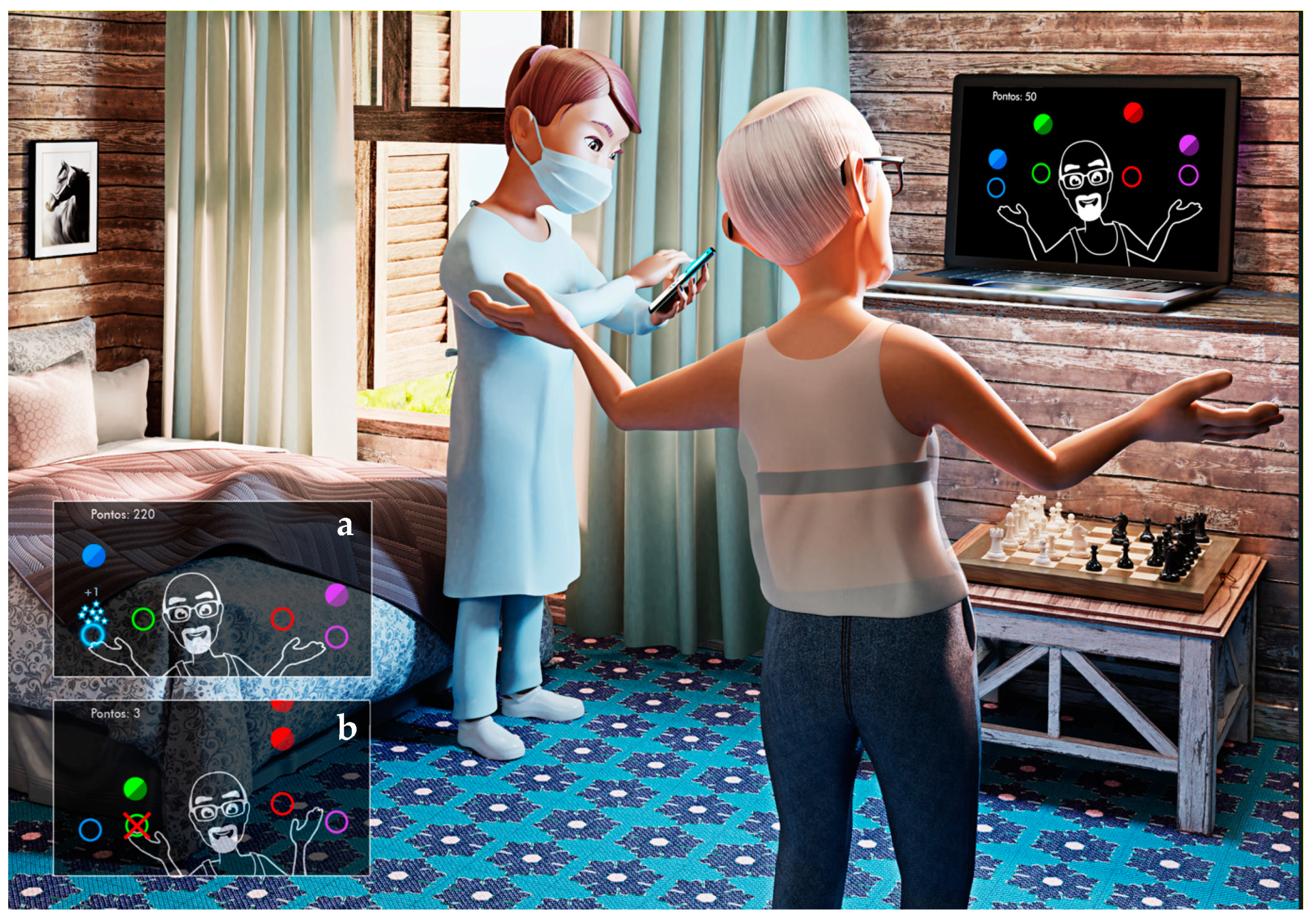
2.4. MoveHero—VR Activity

MoveHero is a virtual game developed by the School of Arts, Sciences, and Humanity at the University of São Paulo [15,29,30,31]. The game presents balls that fall, in 4 imaginary columns on the computer screen, in the rhythm of a predefined piece of music. The task is to not let the balls fall, but the balls can only be touched when they reach 1 of the 4 parallel circles, called targets. The participant is required to “touch” the target as the sphere passes through it. Errors occur when the participant touches the target before or after the ball passes through it. The game captures the participant’s movements through a webcam, so the participant moves his/her arms at a distance of 1.5 m from the computer. The participant receives feedback on hits and scores while playing. The game is a physical activity that involves agility, balance, and reasoning (Figure 1).

2.5. Characterization

To characterize the sample, an anamnesis was performed using a questionnaire to collect information such as age, sex, comorbidities, medications, and habits such as smoking and physical activity prior to the hospitalization; additionally, hospitalization information (i.e., in this study, we used only information provided by the hospitals responsible for the treatment) such as time in days, use of sedatives, use of chloroquine, use of oxygen, and invasive and non-invasive mechanical ventilation; finally, the use of oxygen and need of rehabilitation after hospitalization. Scales were used such as the Medical Research Council—MRC, for muscle strength [32,33]; the Barthel Index, to verify mobility, balance, and functional independence [34,35,36].

2.6. Physiological Variables

After resting, VR activity, and recovery, the following measures were collected: Systolic blood pressure (SBP), Diastolic blood pressure (DBP), Peripheral oxygen saturation (SpO2), and Rating of perceived exertion (RPE), using a Littmann stethoscope, Fingertip pulse finger oximeter, and Premium manual pressure device.

2.7. Procedures

Before carrying out the HRV collection, the participants received some instructions as follows: they should not take any of the following substances the day before the test and on the day of the test: alcohol, caffeine, nicotine, chocolate, soda, energy drinks, and, whenever possible, medications; they should also not have done any strenuous physical exercise and should have had a good night’s sleep [37].
After characterization of the sample, for HRV collection, a Polar brand strap was placed on the participant’s chest (Polar V800) [38], and the data were recorded through the Elite HRV app [39], where it remained until the end of the protocol. Initially, participants were placed in a sitting position at rest for 15 min to assess HRV. Subsequently, the physiological variables were measured, and then the participants performed the VR activity using the MoveHero game, in the orthostatic position for 10 min [40]. If the patient felt fatigue or tiredness, they were allowed to sit in a chair, and the physiological variables were measured again. Lastly, the patient remained seated in a chair for 10 min to capture recovery HRV, and a final measure of physiological variables was performed. The circulation of people was not permitted in the room during the data collection, to reduce the anxiety of the subjects and to avoid recording errors. All tests took approximately 1 h to complete. To minimize the influence of age, participants were divided into 3 subgroups according to age: 25–40 years, 41–60 years, and 61–80 years (Figure 2).

2.8. Age Groups

We divided the group considering age due to the following: 1) HRV and age: according to Sammito and Böckelmann [25], HRV is age-related, and as it decreases with age, when evaluating HRV, it is important to differentiate people by each group of 10 years. Thus, in order to minimize the bias related to age and keep an appropriate number of participants, we decided to divide them into 3 groups, aged between 25 and 40, between 41 and 60, and between 61 and 80. 2) COVID-19 impact with age: older age is an important determinant of disease severity and progression and is considered the major predictor of mortality in COVID-19. Chen et al. [41] hypothesized that an age-related decline and dysregulation of immune function (i.e., immunosenescence and inflammaging) play a major role in contributing to heightened vulnerability to severe COVID-19 outcomes in older adults.

2.9. Heart Rate Variability (HRV)

The data obtained from the HRV monitor were transferred to a computer and 256 consecutive RR intervals were analyzed. Digital filtering was performed to eliminate ectopic, artifact, and premature beats. Only series with more than 95% sinus heartbeats were included in the study. The HRV analysis was performed by linear methods, through time and frequency domains, and non-linear methods, using symbolic dynamic analysis [37,40].
The linear indices were performed to measure RR intervals during a given time: in the time domain, the Mean HR—average heart rate; SDNN—standard deviation of all normal RR intervals recorded in a time interval, expressed in ms, which expresses the global variability; RMSSD—the root-mean-square of the differences between adjacent normal RR intervals in a time interval, expressed in ms, representing parasympathetic activity. In the frequency domain, the following variables were measured: the LF index—low-frequency component reflecting the global action (vagal and sympathetic components) on the heart, but with a predominance of the sympathetic, including variations between 0.04 and 0.15 Hz; HF—high-frequency component indicating the action of the vagus nerve on the heart, including variations from 0.15 to 0.4 Hz; finally, the LF/HF ratio—the relationship between these two indices, reflecting the sympathovagal balance [40].
The non-linear analysis was performed using the Symbolic Dynamics Analysis method, with four indices [42], measured by grouping the patterns with 3 symbols into four types of clusters; the rate of occurrence for each pattern was defined as %0 V (no variation) reflecting only sympathetic modulation, %1 V (one variation) reflecting global variability by sympathetic and parasympathetic modulation, %2 LV (two like variations), and %2 UV (two unlike variations), both of which exclusively reflect parasympathetic modulation (vagal) [43,44,45].

2.10. Data Analysis

For the independent variables, the chi-square test was used for categorical variables and ANOVA for continuous variables. As dependent variables, we considered the physiological variables (Heart rate, Systolic blood pressure, Diastolic blood pressure, Peripheral oxygen saturation), perceptual variables (Rating of perceived exertion), and all HRV indices (time domain and frequency domain indices). Data were submitted to MANOVA with 2 (Groups: control and post-COVID) by 3 (Ages: 25–40 years, 41–60 years, 61–80 years) by 3 (Moments: Rest, VR Activity, and Recovery) with repeated measures in the last factor. The LSD post hoc test was used (Least Significant Difference).
The graph data were presented as the mean and standard error. The partial Eta squared (ŋp2) was reported to measure the effect size and interpreted as small (effect size > 0.01), medium (effect size > 0.06), or large (effect size > 0.14) [46]. The statistical package used was SPSS, version 26.0. p-values < 0.05 were considered significant.

3. Results

In total, 94 participants were evaluated. The demographic data are shown in Table 1.

3.1. Physiological Variables

MANOVA revealed a significant effect for Group (F5,84 = 8.31; p < 0.001, ηp2 = 0.33; Wilks’ λ = 0.669), Age (F10,168 = 2.86; p = 0.003, ηp2 = 0.14; Wilks’ λ = 0.730), and Moment (F10,79 = 30.42; p < 0.001, ηp2 = 0.79; Wilks’ λ = 0.206), with no interaction between factors. Separate follow-up repeated measures (RM-ANOVAs) for Systolic blood pressure (SBP), Diastolic blood pressure (DBP), Peripheral oxygen saturation (SpO2), and Rating of perceived exertion (RPE) are reported in the paragraphs below (Figure 3).
Main effects were found for the following factors: Moment (SBP F2,166 = 22.12; p < 0.001, ηp2 = 0.20; DBP F2,166 = 22.22; p < 0.001, ηp2 = 0.20; RPE F2,166 = 109.83; p < 0.001, ηp2 = 0.55), Group (SpO2 F1,88 = 14.98; p < 0.001, ηp2 = 0.14; RPE F1,88 = 20.41; p < 0.001, ηp2 = 0.18), and Age (SpO2 F2,88 = 7.18; p = 0.001, ηp2 = 0.14; SBP F2,88 = 5.71; p = 0.005, ηp2 = 0.11; DBP F2,88 = 3.80; p = 0.026, ηp2 = 0.08), in addition to a Group and Moment interaction (RPE F2,88 = 12.75; p < 0.001, ηp2 = 0.12). Post hoc comparisons showed no differences between groups for SBP and DBP, but there was an increase in both from Rest to VR Activity (p < 0.001), and a decrease from VR Activity to Recovery (p < 0.001), without differences from Rest to Recovery. The 25–40 yo age group presented a lower SBP and DBP than the 61–80 yo group did (p = 0.001 and p = 0.018), and a lower DBP than the 41–60 yo group did (p = 0.016).
For SpO2, there was a difference between Groups: the control group had a higher SpO2 than the post-COVID group did in two Age groups, 25–40 yo (p = 0.044) and 41–60 yo (p = 0.001), and at two specific Moments in the latter Age: Rest (p = 0.045) and VR Activity (p < 0.001). In addition, the control group did not present a difference in SpO2 between Moments, but the post-COVID group in the 41–60 yo age group presented a significant decrease from Rest to VR Activity (p = 0.011), followed by an increase from VR Activity to Recovery (p = 0.009). Finally, for RPE, in general, the control group had a lower RPE than the post-COVID group did. Considering the Moments, there was no difference for the control group, but the post-COVID group presented a significant increase in the RPE from Rest to VR Activity (p = 0.034), followed by a decrease from VR Activity to Recovery (p = 0.037), without a difference between Rest and Recovery. Although the 41–60 yo group presented a statistical difference in SpO2 compared to the other groups during the activity, it was not below the normal limits recommended in the literature [47].

3.2. Heart Rate Variability

3.2.1. Linear Indices—Time and Frequency Domain

MANOVA revealed main effects for Age (F26,152 = 2.51; p < 0.001, ηp2 = 0.30; Wilks’ λ = 0.489) and Moment (F39,50 = 10.95; p < 0.001, ηp2 = 0.89; Wilks’ λ = 0.105), as well as an interaction between Moment and Age (F78,100 = 1.49; p = 0.030, ηp2 = 0.53; Wilks’ λ = 0.214). MANOVA did not find main effects for the Group factor. Separate follow-up repeated measures (RM-ANOVAs) for the time domain and frequency domain are reported in the paragraphs below (Figure 4).
Considering time domain indices, main effects were found for the following factors: Moment (Mean HR F3,264 = 70.67; p < 0.001, ηp2 = 0.44; RMSSD F3,264 = 25.12; p < 0.001, ηp2 = 0.22; SDNN F3,264 = 40.00; p < 0.001, ηp2 = 0.31) and Age (RMSSD F2,88 = 5.99; p = 0.004, ηp2 = 0.12; SDNN F2,88 = 11.12; p < 0.001, ηp2 = 0.20). ANOVA did not find main effects for Group, and no interactions were found between the factors. Post hoc comparisons showed that in all Ages, there was a decrease in SDNN and RMSSD from Rest to VR Activity, and an increase from VR Activity to Recovery, with no difference between the Rest and Recovery Moments. The mean HR index showed an inversely proportional pattern. In all Groups (control and post-COVID) and at all Moments (Rest, VR Activity, and Recovery), the 25–40 yo age group had a higher SDNN and RMSSD than the 41–60 yo (p < 0.001) and 61–40 yo age groups did (p < 0.001).
For frequency domain indices, main effects were found for the following factors: Moment (LF ms2 F3,264 = 29.50; p < 0.001, ηp2 = 0.25; HF ms2 F3,264 = 14.38; p < 0.001, ηp2 = 0.14; LF/HF F3,264 = 3.39; p = 0.019, ηp2 = 0.03) and Age (LF ms2 F2,88 = 16.61; p < 0.001, ηp2 = 0.27; HF ms2 F2,88 = 3.88; p = 0.024, ηp2 = 0.08; LF/HF F2,88 = 4.06; p = 0.020, ηp2 = 0.08), and an interaction between Moment and Age (LF ms2 F6,264 = 4.33; p = 0.001, ηp2 = 0.09; LF/HF F6,264 = 2.70; p = 0.015, ηp2 = 0.05). ANOVA did not find main effects for Group. Post hoc comparisons showed that in the 25–40 yo age group, there was a decrease in the LF ms2 and HF ms2 from Rest to VR Activity, and an increase from VR Activity to Recovery, while from Rest to Recovery, all participants maintained an increase in both indices. In the 41–60 yo age group, there was an increase in LF ms2 and LF ms2 from VR Activity to Recovery; in HF ms2, there was also a decrease from Rest to VR Activity; in the 61–80 yo age group, there was no difference between the Moments for LF ms2, but there was a decrease from Rest to VR Activity in HF ms2. In all Groups (control and post-COVID) and at all Moments (Rest, VR Activity, and Recovery), the 25–40 yo age group had a higher LF ms2 and HF ms2 than the 41–60 yo (p < 0.001) and 61–80 yo groups did (p < 0.001), and for LF/HF, both 25–40 yo and 41–60 yo groups had a higher value than that of the 61–80 yo age group (p = 0.027 and p = 0.010).

3.2.2. Non-Linear Indices—Symbolic Dynamic Analysis

For the symbolic dynamic indices (Figure 5), main effects were found for the following factors: Moment (%0 V: F2,176 = 60.27; p < 0.001, ηp2 = 0.40; %1 V: F2,176 = 48.34; p < 0.001, ηp2 = 0.35; %2 LV: F2,176 = 55.45; p < 0.001, ηp2 = 0.38; %2 UV: F2,176 = 10.42; p < 0.001, ηp2 = 0.10), Age (%0 V: F2,88 = 7.48; p = 0.001, ηp2= 0.14; %1 V: F2,88 = 7.96; p = 0.001, ηp2 = 0.15; %2 LV: F1,88 = 10.49; p < 0.001, ηp2 = 0.19), and Group (%0 V: F1,88 = 4.08; p = 0.046, ηp2 = 0.04; %1 V: F1,88 = 14.76; p < 0.001, ηp2 = 0.14; %2 LV: F1,88 = 7.64; p = 0.007, ηp2 = 0.08). In addition, there were interactions between the factors Moment and Group (%0 V: F2,176 = 3.91; p = 0.023, ηp2 = 0.04; %2 LV: F2,176 = 4.92; p = 0.010, ηp2 = 0.05), and Age and Group (%1 V: F2,88 = 5.25; p = 0.007, ηp2 = 0.10). Considering the %0 V index, the post-COVID group presented higher values (M = 61) compared to the control group (M = 55), indicating greater sympathetic activity in this group. The 25–40 yo age group showed lower values when compared to the 41–60 yo (p < 0.001) and 61–80 yo groups (p < 0.001). In all Ages and both Groups, there was an increase in %0 V from Rest to VR Activity, and a decrease from VR Activity to Recovery. Post hoc comparisons showed that the 61–80 yo age group had a higher %0 V at Rest and Recovery than the control group did (p = 0.001; p = 0.005).
In turn, for the %1 V and %2 LV indices, the post-COVID group presented lower values (M = 28.1 and M = 1.5) compared to the control group (M = 34.9, p < 0.001 and M = 2.3, p = 0.007), indicating worse global variability and parasympathetic activity in this group. The 25–40 yo age group showed higher values when compared to the 41–60 yo and 61–80 yo groups in both indices. Considering moments, an inverse pattern from %0 V was found to %1 V, %2 LV, and %2 UV. Post hoc comparisons showed that the Age 61–80 yo post-COVID group had lower values of %1 V than the control group at all moments (Rest p = 0.001; VR Activity p = 0.006, Recovery p < 0.001), indicating worse global variability. For %2 LV, the post-COVID group showed lower values in the Recovery for Age 25–40 yo and Rest and Recovery for Age 61–80 yo than the control group, indicating worse parasympathetic activity.

4. Discussion

In the current study, we investigated the influence of using a VR game in physiological variables for individuals’ post-hospitalization for COVID compared with a control group. Confirming our hypothesis, practicing a task using a VR game positively influenced physiological variables for both groups (post-COVID group and control group). However, we not only found differences between groups (control group and post-COVID group), but also greater sympathetic activation, less global variation and parasympathetic activity, and differences in some physiological variables in the older post-COVID group (61–80 yo). These results are discussed below.

4.1. Physiological Variables and HRV before, during, and after the Virtual Reality Game

Initially, our results compared three moments: rest, physical activity, and recovery, and showed that during practicing a VR game, all participants (post-COVID hospitalization and control group) presented increased Heart rate (HR), Systolic blood pressure (SBP), Diastolic blood pressure (DBP), Peripheral oxygen saturation (SpO2), and Rating of perceived exertion (RPE). Considering HRV, we found a positive effect of the practice of physical activity, showing increased sympathetic and decreased parasympathetic activity with the movement performed during the task in VR. This increase in physiological variables was expected and is an important result to support the use of a VR game to provide enough physical activity that can benefit recovery. Within a cardiovascular rehabilitation program, there is evidence of an improvement in cardiorespiratory functional capacity from aerobic exercise, which is one of the factors responsible for reducing total mortality [48,49]. Other studies support that an intervention program, with aerobic exercises, improves physical conditioning and physical performance (heart hate, blood pressure, and oxygen saturation) [50].
The use of VR in rehabilitation is a modern treatment concept that is based on the use of games and tasks in virtual environments to stimulate physical and cognitive functions in individuals with different types of disabilities [51], and to help individuals to obtain improvements in physical performance. Recent studies report that the impact of exercise causes improvements in autonomic modulation [5,24,31,52]. In our study, we demonstrated that patients with COVID-19, as in the control group, when subjected to VR games, demonstrate HRV behavior similar to individuals undergoing conventional physical activity.
Overall, the use of VR in rehabilitation for patients with cardiac conditions has the potential to exert a positive impact on an individual’s physiological, psychological, and rehabilitative outcomes compared with traditional exercise [53]. Eichhorn et al. [54], using a virtual task very similar to the one used in our study, verified an increase in heart rate through movements of the upper limbs in the virtual environment. Furthermore, studies show that interventions using VR can improve ergometry test results, metabolic equivalents, resistance to fatigue, and quality of life in patients with ischemic heart disease [55,56], in addition to decreasing symptoms of anxiety and depression [57,58], increasing adherence, reducing stress levels, and increasing patient engagement in their rehabilitation program [59]. VR technology can also allow for remote rehabilitation, providing patients with more convenient and accessible healthcare.

4.2. Comparison of HRV between Groups and Different Ages

HRV represents the activity and balance of the ANS and its ability to react to internal and external stimuli. As a measure of general body homeostasis, HRV is linked to lifestyle factors and is associated with morbidity and mortality. A high variability in heart rate is a sign of good adaptation, characterizing a healthy individual, with efficient autonomic mechanisms, while low variability is often an indicator of abnormal and insufficient adaptation of the ANS [40].
Although some authors found a significantly lower parasympathetic activity of HRV (RMSSD) in patients’ post-hospitalization for COVID-19 submitted to standing up from the supine position [60], during the analysis of linear HRV indices, we did not observe any significant difference between the control group and the post-COVID group. This may have occurred because the records representing sympathetic and parasympathetic activities do not allow us to distinguish when changes in HRV are due to increased sympathetic tone or loss of vagal tone [40].
Although HRV is commonly analyzed using linear models, interest in nonlinear methods (symbolic analysis) has increased in recent years [43]. This methodology differs from standard approaches because it considers the qualitative properties of a single heart rate series [61]. The mechanisms involved in cardiovascular regulation are interconnected in a nonlinear theory and, therefore, nonlinear analysis can provide additional information [62,63] about the complexity, or randomness, of a time series, and describe elements that manifest behaviors that are extremely sensitive to the initial conditions, or difficult to repeat, but still deterministic [64,65]. The study of Solinski et al. [66] explained that differences after COVID-19 may occur and be caused by changes in activity of the parasympathetic ANS, as well as by the coupling of respiratory rhythm with heart rate due to an increase in pulmonary arterial vascular resistance in several post-COVID patients. However, our results found differences considering the non-linear analysis and rating of perceived exertion, and only the age group of 61–80 years presented greater sympathetic activity, worse global variability, and parasympathetic activity considering the indices (indices %1 V, %2 LV, %0 V), and a greater rating of perceived exertion, during the VR activity. These results can be supported by Erdal et al. [67] who studied post-COVID autonomic dysfunction, and reported that symptoms of autonomic dysfunction are very common in the post-infectious period of COVID-19 and were associated with age. The most common symptom in the acute phase was fatigue and its presence increased the risk of autonomic dysfunction by 2.2 times in the post-COVID period, but was only evident in older patients.
We can report that the group of 61–80 years had three significant differences compared to the other age groups: 1—needed more non-invasive mechanical ventilation during hospitalization, 2—presented the lowest MRC score, and 3—presented the highest BMI. Thus, according to Medrinal et al. [68], the period of mechanical ventilation is influenced by muscle weakness and is directly linked to age. The decrease in strength becomes more evident from the sixth decade of life [69] and, with the hyper-inflammatory response caused by SARS-CoV-2 and exacerbation of the immune senescence process, enhances the endothelial damage and mitochondrial dysfunction and autophagy, and induces myofibril breakdown and muscle degradation, mainly in older individuals. The aftermath of the acute and complex immunological SARS-CoV-2-related phenomena, augmented by anosmia, ageusia, and altered microbiota, may lead to decreased food intake and exacerbated catabolism. According to Piotrowicz et al. [70], older individuals already present fragility, and the imposed physical inactivity due to lock-down, quarantine, and acute hospitalization with bedrest would intensify the acute sarcopenia process, and this could be responsible for the lower MRC and increased mechanical ventilation time. Another factor that may have contributed to the slower post-exertion recovery and influenced the non-linear analysis in the older group is the fact that they have a higher BMI. It is known that obesity is a strong and independent risk factor for a greater severity of COVID-19 [71]. Recent investigations have indicated that obesity is associated with a poor prognosis for SARS-CoV-2 infection, possible hospitalization in intensive care, and disease progression, associated with decreased physical fitness, increased fatigue, and tiredness on exertion [72].
Considering the above explanation, our results can highlight the importance of the management of patients post-COVID-19 infection, emphasizing special support for older patients. Although we found interesting results, we can point out some limitations of the present study: (1) this study evaluated the individuals only once after hospital discharge. Thus, the participants were evaluated at a single time point (cross-sectional study) and a follow-up protocol could provide deeper information on the impact of COVID-19 on the physiological variables over time; (2) another limitation was evaluating patients who were admitted from different hospitals at a time of protocol changes due to the emergence of COVID-19, and more standardized information could allow effective research results; (3) an anamnesis with more details about the previous life characterization should be performed and could contribute to a better understanding of the influence of the physiological variables analyzed; (4) we used a laboratory-specific motor task, and other VR tasks should be assessed in the future to provide different task possibilities for rehabilitation. Hence, we believe that future studies should analyze the effects of different VR games as a type of physical activity, using HRV and physiological variable assessments in patients’ post-hospitalization for COVID-19, especially in the older population.

5. Conclusions

The most important result of this study is that the VR game positively affected physiological variables, so it can be utilized as a secure physical activity, in both healthy individuals and individuals after hospitalization for COVID-19. The effect of VR reflected changes similar to physical activity, showing increased sympathetic and decreased parasympathetic activity during the task in VR. COVID-19 affects the ANS system of older patients’ post-hospitalization, which may be partly due to their higher BMI and reduced exercise capacity affecting their ability to perform exercise activities. Other important observations were the higher RPE in COVID-19 patients during and after exercise, which may reflect altered physiological and autonomic responses. Taken together with the high reporting of fatigue after COVID-19, this is an important finding, and considering that RPE is usually lower during VR exercise compared to non-VR strengthens the potential for the use of VR in COVID-19 patients.

Author Contributions

Conceptualization, C.M.R., R.d.A.G., C.B.d.M.M., Í.A.P.d.M. and C.F.; methodology, C.M.R., R.d.A.G., Í.A.P.d.M. and H.D.; software, L.V.d.A.; validation, L.V.d.A.; formal analysis, Í.A.P.d.M. and T.D.d.S.; investigation, C.M.R. and R.d.A.G.; resources, Í.A.P.d.M., H.D. and C.B.d.M.M.; data curation, Í.A.P.d.M. and T.D.d.S.; writing—original draft preparation, C.M.R., R.d.A.G., R.M.D., A.O.S., L.C.P.M., A.P.d.O., B.L.d.F. and C.F.; writing—review and editing, C.M.R., R.d.A.G., R.M.D., A.O.S., L.C.P.M., A.P.d.O., B.L.d.F., H.D. and T.D.d.S.; supervision, Í.A.P.d.M., H.D., C.F. and T.D.d.S.; project administration, Í.A.P.d.M. and C.B.d.M.M.; funding acquisition, Í.A.P.d.M., H.D. and C.F. All authors have read and agreed to the published version of the manuscript.

Funding

This study was financed in part by the Coordenação de Aperfeiçoamento de Pessoal de Nível Superior—Brasil: Finance Code 001 and by Conselho Nacional de Desenvolvimento Científico e Tecnológico, Brazil (CNPq) process number 442456/2016-6.

Institutional Review Board Statement

The study was conducted in accordance with the Declaration of Helsinki and approved by the Research Ethics Committee of the Federal University of São Paulo (CAAE 50099521.7.0000.5505). It was registered on ClinicalTrials.gov (NCT04537858).

Informed Consent Statement

Informed consent was obtained from all subjects involved in the study.

Data Availability Statement

The data presented in this study are available on request from the corresponding author. The data are not publicly available, due to other studies in progress.

Acknowledgments

We would like to thank the Coordenação de Aperfeiçoamento de Pessoal de Nível Superior, Brazil (CAPES) and the Conselho Nacional de Desenvolvimento Científico e Tecnológico, Brazil (CNPq).

Conflicts of Interest

The authors declare no conflict of interest.

References

  1. Wang, T.J.; Chau, B.; Lui, M.D.; Lam, G.-T.; Lin, N.; Humbert, S. Physical Medicine and Rehabilitation and Pulmonary Rehabilitation for COVID-19. Am. J. Phys. Med. Rehabil. 2020, 99, 769–774. [Google Scholar] [CrossRef] [PubMed]
  2. Goldstein, E.; Lipsitch, M.; Cevik, M. On the Effect of Age on the Transmission of SARS-CoV-2 in Households, Schools, and the Community. J. Infect. Dis. 2020, 223, 362–369. [Google Scholar] [CrossRef] [PubMed]
  3. How the vagus nerve produces beat-to-beat heart rate variability; experiments in rabbits to mimic in vivo vagal patterns. J. Clin. Transl. Res. 2015, 1, 190–204. [CrossRef]
  4. Mol, M.B.A.; Strous, M.T.A.; van Osch, F.H.M.; Vogelaar, F.J.; Barten, D.G.; Farchi, M.; Foudraine, N.A.; Gidron, Y. Heart-rate-variability (HRV), predicts outcomes in COVID-19. PLoS ONE 2021, 16, e0258841. [Google Scholar] [CrossRef] [PubMed]
  5. Silveira, A.C.; Moraes, Í.A.P.; Vidigal, G.P.; Simcsik, A.O.; Rosa, R.M.; Favero, F.M.; Fernandes, S.M.S.; Garner, D.M.; Araújo, L.V.; Massa, M.; et al. Cardiac Autonomic Modulation in Subjects with Amyotrophic Lateral Sclerosis (ALS) during an Upper Limb Virtual Reality Task: A Prospective Control Trial. BioMed Res. Int. 2022, 2022, 4439681. [Google Scholar] [CrossRef]
  6. Dias, R.M.; Moraes, Í.A.P.; Dantas, M.T.A.P.; Fernani, D.C.G.L.; Fontes, A.M.G.G.; Silveira, A.C.; Barnabé, V.; Fernandes, M.; Martinelli, P.M.; Monteiro, C.B.M.; et al. Influence of Chronic Exposure to Exercise on Heart Rate Variability in Children and Adolescents Affected by Obesity: A Systematic Review and Meta-Analysis. Int. J. Environ. Res. Public Health 2021, 18, 11065. [Google Scholar] [CrossRef]
  7. Moraes, Í.A.P.; Silva, T.D.; Massetti, T.; Menezes, L.D.C.; Ribeiro, V.F.; Tropiano, L.M.C.C.; Barnabé, V.; Hoshi, R.A.; Monteiro, C.B.M.; Fernandes, M. Fractal correlations and linear analyses of heart rate variability in healthy young people with different levels of physical activity. Cardiol. Young 2019, 29, 1236–1242. [Google Scholar] [CrossRef]
  8. Imamura, M.; Mirisola, A.R.; de Quadros Ribeiro, F.; De Pretto, L.R.; Alfieri, F.M.; Delgado, V.R.; Battistella, L.R. Rehabilitation of patients after COVID-19 recovery: An experience at the Physical and Rehabilitation Medicine Institute and Lucy Montoro Rehabilitation Institute. Clinics 2021, 76, e2804. [Google Scholar] [CrossRef]
  9. Grigoletto, I.; Cavalheri, V.; de Lima, F.F.; Ramos, E.M.C. Recovery after COVID-19: The potential role of pulmonary rehabilitation. Braz. J. Phys. Ther. 2020, 24, 463–464. [Google Scholar] [CrossRef]
  10. Smondack, P.; Gravier, F.; Prieur, G.; Repel, A.; Muir, J.-F.; Cuvelier, A.; Combret, Y.; Medrinal, C.; Bonnevie, T. Physiotherapy and COVID-19. From Intensive Care Unit to Home Care–An Overview of International Guidelines. Revue Des Maladies Respiratoires 2020, 37, 811–822. [Google Scholar] [CrossRef]
  11. Lew, H.L.; Oh-Park, M.; Cifu, D.X. The War on COVID-19 Pandemic: Role of Rehabilitation Professionals and Hospitals. Am. J. Phys. Med. Rehabil. 2020, 99, 571–572. [Google Scholar] [CrossRef] [PubMed]
  12. Bond, S.; Laddu, D.R.; Ozemek, C.; Lavie, C.J.; Arena, R. Exergaming and Virtual Reality for Health: Implications for Cardiac Rehabilitation. Curr. Probl. Cardiol. 2021, 46, 100472. [Google Scholar] [CrossRef]
  13. García-Bravo, S.; Cuesta-Gómez, A.; Campuzano-Ruiz, R.; López-Navas, M.J.; Domínguez-Paniagua, J.; Araújo-Narváez, A.; Barreñada-Copete, E.; García-Bravo, C.; Flórez-García, M.T.; Botas-Rodríguez, J.; et al. Virtual reality and video games in cardiac rehabilitation programs. A systematic review. Disabil. Rehabil. 2021, 43, 448–457. [Google Scholar] [CrossRef] [PubMed]
  14. Cuesta-Gómez, A.; Sánchez-Herrera-Baeza, P.; Oña-Simbaña, E.D.; Martínez-Medina, A.; Ortiz-Comino, C.; Balaguer-Bernaldo-De-Quirós, C.; Jardón-Huete, A.; Cano-De-La-Cuerda, R. Effects of virtual reality associated with serious games for upper limb rehabilitation in patients with multiple sclerosis: Randomized controlled trial. J. Neuroeng. Rehabil. 2020, 17, 90. [Google Scholar] [CrossRef] [PubMed]
  15. da Silva, T.D.; de Oliveira, P.M.; Dionizio, J.B.; de Santana, A.P.; Bahadori, S.; Dias, E.D.; Ribeiro, C.M.; de Andrade Gomes, R.; Ferreira, M.; Ferreira, C.; et al. Comparison Between Conventional Intervention and Non-immersive Virtual Reality in the Rehabilitation of Individuals in an Inpatient Unit for the Treatment of COVID-19: A Study Protocol for a Randomized Controlled Crossover Trial. Front. Psychol. 2021, 12, 622618. [Google Scholar] [CrossRef] [PubMed]
  16. Ostrowska, P.M.; Śliwiński, M.; Studnicki, R.; Hansdorfer-Korzon, R. Telerehabilitation of Post-Stroke Patients as a Therapeutic Solution in the Era of the COVID-19 Pandemic. Healthcare 2021, 9, 654. [Google Scholar] [CrossRef]
  17. Groenveld, T.; Achttien, R.; Smits, M.; de Vries, M.; van Heerde, R.; Staal, B.; van Goor, H. COVID Rehab Group Feasibility of Virtual Reality Exercises at Home for Post–COVID-19 Condition: Cohort Study. JMIR Rehabil. Assist. Technol. 2022, 9, e36836. [Google Scholar] [CrossRef]
  18. Marzaleh, M.A.; Peyravi, M.; Azhdari, N.; Bahaadinbeigy, K.; Sharifian, R.; Samad-Soltani, T.; Sarpourian, F. Virtual reality applications for rehabilitation of COVID-19 patients: A systematic review. Health Sci. Rep. 2022, 5, e853. [Google Scholar] [CrossRef]
  19. Vibhuti; Kumar, N.; Kataria, C. Efficacy assessment of virtual reality therapy for neuromotor rehabilitation in home environment: A systematic review. Disabil. Rehabil. Assist. Technol. 2021, 1–21. [Google Scholar] [CrossRef]
  20. Dębska, M.; Polechoński, J.; Mynarski, A.; Polechoński, P. Enjoyment and Intensity of Physical Activity in Immersive Virtual Reality Performed on Innovative Training Devices in Compliance with Recommendations for Health. Int. J. Environ. Res. Public Health 2019, 16, 3673. [Google Scholar] [CrossRef]
  21. Dimbwadyo-Terrer, I.; Trincado-Alonso, F.; de los Reyes-Guzmán, A.; Aznar, M.A.; Alcubilla, C.; Pérez-Nombela, S.; del Ama-Espinosa, A.; Polonio-López, B.; Gil-Agudo, Á. Upper limb rehabilitation after spinal cord injury: A treatment based on a data glove and an immersive virtual reality environment. Disabil. Rehabil. Assist. Technol. 2016, 11, 462–467. [Google Scholar] [CrossRef] [PubMed]
  22. de Mello Monteiro, C.B.; Massetti, T.; da Silva, T.D.; van der Kamp, J.; de Abreu, L.C.; Leone, C.; Savelsbergh, G.J. Transfer of motor learning from virtual to natural environments in individuals with cerebral palsy. Res. Dev. Disabil. 2014, 35, 2430–2437. [Google Scholar] [CrossRef] [PubMed]
  23. de Mello Monteiro, C.B.; da Silva, T.D.; de Abreu, L.C.; Fregni, F.; de Araujo, L.V.; Ferreira, F.H.I.B.; Leone, C. Short-term motor learning through non-immersive virtual reality task in individuals with down syndrome. BMC Neurol. 2017, 17, 71. [Google Scholar] [CrossRef]
  24. Dias, E.D.; de Menezes, L.D.C.; da Silva, T.D.; da Silva, N.M.; Vidal, P.R.; Brondane, B.R.; Padula, N.; Gaspar, R.C.; Santos, S.; Auricchio, J.R.; et al. Comparison of cardiac autonomic modulation of athletes and non-athletes individuals with spinal cord injury at rest and during a non-immersive virtual reality task. Spinal Cord 2021, 59, 1294–1300. [Google Scholar] [CrossRef] [PubMed]
  25. Sammito, S.; Böckelmann, I. Reference values for time- and frequency-domain heart rate variability measures. Heart Rhythm. 2016, 13, 1309–1316. [Google Scholar] [CrossRef] [PubMed]
  26. Marin, B.G.; Aghagoli, G.; Lavine, K.; Yang, L.; Siff, E.J.; Chiang, S.S.; Salazar-Mather, T.P.; Dumenco, L.; Savaria, M.C.; Aung, S.N.; et al. Predictors of COVID -19 severity: A literature review. Rev. Med. Virol. 2021, 31, 1–10. [Google Scholar] [CrossRef]
  27. Kang, S.-J.; Jung, S.I. Age-Related Morbidity and Mortality among Patients with COVID-19. Infect. Chemother. 2020, 52, 154–164. [Google Scholar] [CrossRef]
  28. von Elm, E.; Altman, D.G.; Egger, M.; Pocock, S.J.; Gøtzsche, P.C.; Vandenbroucke, J.P. The Strengthening the Reporting of Observational Studies in Epidemiology (STROBE) Statement: Guidelines for Reporting Observational Studies. PLoS Med. 2007, 4, e296. [Google Scholar] [CrossRef]
  29. Da Silva, T.D.; Fontes, A.M.G.G.; De Oliveira-Furlan, B.S.; Roque, T.T.; Lima, A.I.I.; De Souza, B.M.M.; Alberissi, C.A.D.O.; Silveira, A.C.; De Moraes, Í.A.P.; Collett, J.; et al. Effect of Combined Therapy of Virtual Reality and Transcranial Direct Current Stimulation in Children and Adolescents with Cerebral Palsy: A Study Protocol for a Triple-Blinded Randomized Controlled Crossover Trial. Front. Neurol. 2020, 11, 953. [Google Scholar] [CrossRef]
  30. Martins, F.P.A.; Massetti, T.; Crocetta, T.B.; Lopes, P.B.; da Silva, A.A.; Figueiredo, E.F.; de Abreu, L.C.; da Silva, T.D.; de Mello Monteiro, C.B. Analysis of motor performance in individuals with cerebral palsy using a non-immersive virtual reality taskA pilot study. Neuropsychiatr. Dis. Treat. 2019, 15, 417–428. [Google Scholar] [CrossRef]
  31. Moraes, Í.A.P.; Lima, J.A.; Silva, N.M.; Simcsik, A.O.; Silveira, A.C.; Menezes, L.D.C.; Araújo, L.V.; Crocetta, T.B.; Voos, M.C.; Tonks, J.; et al. Effect of Longitudinal Practice in Real and Virtual Environments on Motor Performance, Physical Activity and Enjoyment in People with Autism Spectrum Disorder: A Prospective Randomized Crossover Controlled Trial. Int. J. Environ. Res. Public Health 2022, 19, 14668. [Google Scholar] [CrossRef] [PubMed]
  32. Schefold, J.C.; Wollersheim, T.; Grunow, J.J.; Luedi, M.M.; Z’Graggen, W.J.; Weber-Carstens, S. Muscular weakness and muscle wasting in the critically ill. J. Cachex-Sarcopenia Muscle 2020, 11, 1399–1412. [Google Scholar] [CrossRef] [PubMed]
  33. Craig, P.; Dieppe, P.; Macintyre, S.; Michie, S.; Nazareth, I.; Petticrew, M. Developing and evaluating complex interventions: The new Medical Research Council guidance. BMJ 2008, 337, a1655. [Google Scholar] [CrossRef] [PubMed]
  34. Mahoney, F.I.; Barthel, D.W. Functional evaluation: The Barthel Index. Md. State Med. J. 1965, 14, 61–65. [Google Scholar] [PubMed]
  35. Novak, S.; Johnson, J.; Greenwood, R. Barthel revisited: Making guidelines work. Clin. Rehabil. 1996, 10, 128–134. [Google Scholar] [CrossRef]
  36. Houlden, H.; Edwards, M.; McNeil, J.; Greenwood, R. Use of the Barthel Index and the Functional Independence Measure during early inpatient rehabilitation after single incident brain injury. Clin. Rehabil. 2006, 20, 153–159. [Google Scholar] [CrossRef]
  37. Catai, A.M.; Pastre, C.M.; de Godoy, M.F.; da Silva, E.; Takahashi, A.C.D.M.; Vanderlei, L.C.M. Heart rate variability: Are you using it properly? Standardisation checklist of procedures. Braz. J. Phys. Ther. 2019, 24, 91–102. [Google Scholar] [CrossRef]
  38. Giles, D.; Draper, N.; Neil, W. Validity of the Polar V800 heart rate monitor to measure RR intervals at rest. Eur. J. Appl. Physiol. 2015, 116, 563–571. [Google Scholar] [CrossRef]
  39. Perrotta, A.S.; Jeklin, A.T.; Hives, B.A.; Meanwell, L.E.; Warburton, D.E. Validity of the Elite HRV Smartphone Application for Examining Heart Rate Variability in a Field-Based Setting. J. Strength Cond. Res. 2017, 31, 2296–2302. [Google Scholar] [CrossRef]
  40. Vanderlei, L.C.M.; Pastre, C.M.; Hoshi, R.A.; de Carvalho, T.D.; de Godoy, M.F. Basic Notions of Heart Rate Variability and Its Clinical Applicability. Braz. J. Cardiovasc. Surg. 2009, 24, 205–217. [Google Scholar] [CrossRef]
  41. Chen, Y.; Klein, S.L.; Garibaldi, B.T.; Li, H.; Wu, C.; Osevala, N.M.; Li, T.; Margolick, J.B.; Pawelec, G.; Leng, S.X. Aging in COVID-19: Vulnerability, immunity and intervention. Ageing Res. Rev. 2020, 65, 101205. [Google Scholar] [CrossRef] [PubMed]
  42. De Moura-Tonello, S.C.G.; Carvalho, V.; De Godoy, M.F.; Porta, A.; Leal, Â.M.d.O.; Bocchi, E.A.; Catai, A.M. Evaluation of Cardiac Autonomic Modulation Using Symbolic Dynamics after Cardiac Transplantation. Braz. J. Cardiovasc. Surg. 2019, 34, 572–580. [Google Scholar] [CrossRef] [PubMed]
  43. Dias, R.M.; Hoshi, R.A.; Vanderlei, L.C.M.; Monteiro, C.B.d.M.; Alvarez, M.P.B.; Crocetta, T.B.; Grossklauss, L.F.; Fernani, D.C.G.L.; Dantas, M.T.A.P.; Martins, F.P.A.; et al. Influence of Different Types of Corticosteroids on Heart Rate Variability of Individuals with Duchenne Muscular Dystrophy—A Pilot Cross Sectional Study. Life 2021, 11, 752. [Google Scholar] [CrossRef] [PubMed]
  44. Porta, A.; Tobaldini, E.; Guzzetti, S.; Furlan, R.; Montano, N.; Gnecchi-Ruscone, T. Assessment of cardiac autonomic modulation during graded head-up tilt by symbolic analysis of heart rate variability. Am. J. Physiol. Circ. Physiol. 2007, 293, H702–H708. [Google Scholar] [CrossRef] [PubMed]
  45. Molina, G.E.; Fontana, K.E.; Porto, L.G.G.; Junqueira, L.F. Post-exercise heart-rate recovery correlates to resting heart-rate variability in healthy men. Clin. Auton. Res. 2016, 26, 415–421. [Google Scholar] [CrossRef]
  46. Lakens, D. Calculating and reporting effect sizes to facilitate cumulative science: A practical primer for t-tests and ANOVAs. Front. Psychol. 2013, 4, 863. [Google Scholar] [CrossRef]
  47. Gottlieb, J.; Capetian, P.; Hamsen, U.; Janssens, U.; Karagiannidis, C.; Kluge, S.; Nothacker, M.; Roiter, S.; Volk, T.; Worth, H.; et al. German S3 Guideline: Oxygen Therapy in the Acute Care of Adult Patients. Respiration 2021, 101, 214–252. [Google Scholar] [CrossRef]
  48. De Carvalho, T.; Milani, M.; Ferraz, A.S.; Da Silveira, A.D.; Herdy, A.H.; Hossri, C.A.C.; e Silva, C.G.S.; de Araújo, C.G.S.; Rocco, E.A.; Teixeira, J.A.C.; et al. Diretriz Brasileira de Reabilitação Cardiovascular2020. Arq. Bras. De Cardiol. 2020, 114, 943–987. [Google Scholar] [CrossRef]
  49. Haykowsky, M.J.; Timmons, M.P.; Kruger, C.; McNeely, M.; Taylor, D.A.; Clark, A.M. Meta-Analysis of Aerobic Interval Training on Exercise Capacity and Systolic Function in Patients with Heart Failure and Reduced Ejection Fractions. Am. J. Cardiol. 2013, 111, 1466–1469. [Google Scholar] [CrossRef] [PubMed]
  50. Mediano, M.F.F.; Mendes, F.d.S.N.S.; Pinto, V.L.M.; da Silva, G.M.S.; da Silva, P.S.; Carneiro, F.M.; Sangenis, L.H.C.; Saraiva, R.M.; Xavier, S.S.; do Brasil, P.E.A.A.; et al. Cardiac rehabilitation program in patients with Chagas heart failure: A single-arm pilot study. Rev. Da Soc. Bras. De Med. Trop. 2016, 49, 319–328. [Google Scholar] [CrossRef]
  51. Monteiro, C.B.D.M.; Dawes, H.; Deutsch, J.E. Editorial: Virtual Reality for Sensorimotor Rehabilitation of Neurological Health Conditions Across the Lifespan. Front. Neurol. 2021, 12, 1985. [Google Scholar] [CrossRef] [PubMed]
  52. Alvarez, M.P.B.; da Silva, T.D.; Favero, F.M.; Valenti, V.E.; Raimundo, R.D.; Vanderlei, L.C.M.; Garner, D.M.; de Mello Monteiro, C.B. Autonomic Modulation in Duchenne Muscular Dystrophy during a Computer Task: A Prospective Control Trial. PLoS ONE 2017, 12, e0169633. [Google Scholar] [CrossRef] [PubMed]
  53. Qian, J.; McDonough, D.J.; Gao, Z. The Effectiveness of Virtual Reality Exercise on Individual’s Physiological, Psychological and Rehabilitative Outcomes: A Systematic Review. Int. J. Environ. Res. Public Health 2020, 17, 4133. [Google Scholar] [CrossRef]
  54. Eichhorn, S.; Koller, V.; Schreiber, U.; Mendoza, A.; Krane, M.; Lange, R. Development of an Exergame for individual rehabilitation of patients with cardiovascular diseases. Australas. Phys. Eng. Sci. Med. 2013, 36, 441–447. [Google Scholar] [CrossRef] [PubMed]
  55. Vieira, Á.; Melo, C.; Machado, J.; Gabriel, J. Virtual reality exercise on a home-based phase III cardiac rehabilitation program, effect on executive function, quality of life and depression, anxiety and stress: A randomized controlled trial. Disabil. Rehabil. Assist. Technol. 2017, 13, 112–123. [Google Scholar] [CrossRef] [PubMed]
  56. García-Bravo, S.; Cano-de-la-Cuerda, R.; Domínguez-Paniagua, J.; Campuzano-Ruiz, R.; Barreñada-Copete, E.; López-Navas, M.J.; Araujo-Narváez, A.; García-Bravo, C.; Florez-Garcia, M.; Botas-Rodríguez, J.; et al. Effects of Virtual Reality on Cardiac Rehabilitation Programs for Ischemic Heart Disease: A Randomized Pilot Clinical Trial. Int. J. Environ. Res. Public Health 2020, 17, 8472. [Google Scholar] [CrossRef] [PubMed]
  57. Szczepańska-Gieracha, J.; Jóźwik, S.; Cieślik, B.; Mazurek, J.; Gajda, R. Immersive Virtual Reality Therapy as a Support for Cardiac Rehabilitation: A Pilot Randomized-Controlled Trial. Cyberpsychol. Behav. Soc. Netw. 2021, 24, 543–549. [Google Scholar] [CrossRef]
  58. Bashir, Z.; Misquith, C.; Shahab, A.; Has, P.; Bukhari, S. The impact of Virtual Reality on Anxiety and Functional Capacity in Cardiac Rehabilitation: A Systematic Review and Meta-analysis. Curr. Probl. Cardiol. 2023, 48, 101628. [Google Scholar] [CrossRef]
  59. van der Kruk, S.R.; Zielinski, R.; MacDougall, H.; Hughes-Barton, D.; Gunn, K.M. Virtual reality as a patient education tool in healthcare: A scoping review. Patient Educ. Couns. 2022, 105, 1928–1942. [Google Scholar] [CrossRef]
  60. Shah, B.; Kunal, S.; Bansal, A.; Jain, J.; Poundrik, S.; Shetty, M.K.; Batra, V.; Chaturvedi, V.; Yusuf, J.; Mukhopadhyay, S.; et al. Heart rate variability as a marker of cardiovascular dysautonomia in post-COVID-19 syndrome using artificial intelligence. Indian Pacing Electrophysiol. J. 2022, 22, 70–76. [Google Scholar] [CrossRef]
  61. Perseguini, N.M.; Takahashi, A.C.M.; Rebelatto, J.R.; Silva, E.; Borghi-Silva, A.; Porta, A.; Montano, N.; Catai, A.M. Spectral and symbolic analysis of the effect of gender and postural change on cardiac autonomic modulation in healthy elderly subjects. Braz. J. Med. Biol. Res. 2011, 44, 29–37. [Google Scholar] [CrossRef] [PubMed]
  62. Jokinen, V.; Tapanainen, J.M.; Seppänen, T.; Huikuri, H.V. Temporal changes and prognostic significance of measures of heart rate dynamics after acute myocardial infarction in the beta-blocking era. Am. J. Cardiol. 2003, 92, 907–912. [Google Scholar] [CrossRef] [PubMed]
  63. Huikuri, H.V.; Mäkikallio, T.H.; Perkiömäki, J. Measurement of heart rate variability by methods based on nonlinear dynamics. J. Electrocardiol. 2003, 36, 95–99. [Google Scholar] [CrossRef] [PubMed]
  64. Godoy, M.F.; Takakura, I.T.; Correa, P.R. The Relevance of Nonlinear Dynamic Analysis (Chaos Theory) to Predict Morbidity and Mortality in Patients Undergoing Surgical Myocardial Revascularization. Arqu Ciênc Saúde 2005, 12, 161–171. [Google Scholar]
  65. Scheff, J.D.; Griffel, B.; Corbett, S.A.; Calvano, S.E.; Androulakis, I.P. On heart rate variability and autonomic activity in homeostasis and in systemic inflammation. Math. Biosci. 2014, 252, 36–44. [Google Scholar] [CrossRef] [PubMed]
  66. Soliński, M.; Pawlak, A.; Petelczyc, M.; Buchner, T.; Aftyka, J.; Gil, R.; Król, Z.J.; Żebrowski, J.J. Heart rate variability comparison between young males after 4–6 weeks from the end of SARS-CoV-2 infection and controls. Sci. Rep. 2022, 12, 8832. [Google Scholar] [CrossRef]
  67. Erdal, Y.; Atalar, A.C.; Gunes, T.; Okluoglu, T.; Yavuz, N.; Emre, U. Autonomic dysfunction in patients with COVID-19. Acta Neurol. Belg. 2022, 122, 885–891. [Google Scholar] [CrossRef]
  68. Medrinal, C.; Prieur, G.; Bonnevie, T.; Gravier, F.-E.; Mayard, D.; Desmalles, E.; Smondack, P.; Lamia, B.; Combret, Y.; Fossat, G. Muscle weakness, functional capacities and recovery for COVID-19 ICU survivors. BMC Anesthesiol. 2021, 21, 64. [Google Scholar] [CrossRef]
  69. Pícoli, T.D.S.; De Figueiredo, L.L.; Patrizzi, L.J. Sarcopenia e envelhecimento. Fisioter. Em Mov. 2011, 24, 455–462. [Google Scholar] [CrossRef]
  70. Piotrowicz, K.; Gąsowski, J.; Michel, J.-P.; Veronese, N. Post-COVID-19 acute sarcopenia: Physiopathology and management. Aging Clin. Exp. Res. 2021, 33, 2887–2898. [Google Scholar] [CrossRef]
  71. Bezerra, K.M.G.; Berto, C.M.; Da Silva, F.A.A.; Silva, L.M.d.S.; Marques, A.P.D.O. Impacto da obesidade na mortalidade de idosos com COVID-19. Rev. Bras. De Ciências Do Envelhec. Hum. 2020, 17. [Google Scholar] [CrossRef]
  72. Freire, A.P.C.F.; Lira, F.S.; Morano, A.E.V.A.; Pereira, T.; Coelho-E-Silva, M.-J.; Caseiro, A.; Christofaro, D.G.D.; Júnior, O.M.; Dorneles, G.P.; Minuzzi, L.G.; et al. Role of Body Mass and Physical Activity in Autonomic Function Modulation on Post-COVID-19 Condition: An Observational Subanalysis of Fit-COVID Study. Int. J. Environ. Res. Public Health 2022, 19, 2457. [Google Scholar] [CrossRef] [PubMed]
Figure 1. Representative design of the accomplishment of the MoveHero activity in the use of the webcam interface. (a) Demonstration of hit performed by the participant. (b) Miss performed by the participant.
Electronics 12 01925 g001
Figure 2. Study design. MRC: Medical Research Council; yo: years-old; HRV: Heart rate variability; VR: Virtual Reality; SBP: Systolic blood pressure; DBP: Diastolic blood pressure; SpO2: Peripheral oxygen saturation; RPE: Rating of perceived exertion.
Electronics 12 01925 g002
Figure 3. Representation of the mean and standard error of Physiological variables between groups (Control and post-COVID), ages (25–49 yo, 41–60 yo, and 61–80 yo), and moments (Rest, VR activity, and Recovery). yo: years-old; VR: Virtual Reality; SBP: Systolic blood pressure; DBP: Diastolic blood pressure; SpO2: Peripheral oxygen saturation; RPE: Rating of perceived exertion; * p < 0.05 between groups.
Electronics 12 01925 g003
Figure 4. Representation of the mean and standard error of time and frequency domain indices of HRV between groups (Control and post-COVID), ages (25–49 yo, 41–60 yo, and 61–80 yo), and moments (Rest, VR activity, and Recovery). Yo: years-old; VR: Virtual Reality; HR: heart rate; RMSSD: Root-mean-square of the squared differences between successive RR intervals; SDNN: Standard deviation of the mean of all RR intervals over a period; HF: high frequency; LF: low frequency; LF/HF: low frequency and high frequency ratio; ms2: millisecond squared.
Electronics 12 01925 g004
Figure 5. Representation of the mean and standard error of symbolic dynamic indices of HRV between groups (Control and post-COVID), ages (25–49 yo, 41–60 yo, and 61–80 yo), and moments (Rest, VR activity, and Recovery). Yo: years-old; VR: Virtual Reality; %0 V: no variation—reflects sympathetic modulation, %1 V: one variation—reflects global variability, %2 LV: two like variations, and %2 UL: two unlike variations—reflect parasympathetic modulation; * p < 0.05 between groups.
Electronics 12 01925 g005
Table 1. Demographic data.
Variables25–40 yo41–60 yo61–80 yo
ControlPost-COVIDControlPost-COVIDControlPost-COVID
n = 14n = 12n = 18n = 18n = 13n = 19
Mean ± SDMean ± SDMean ± SDMean ± SDMean ± SDMean ± SD
Age34.2 ± 5.233.9 ± 4.652.3 ± 5.651.5 ± 6.069.3 ± 6.768.8 ± 5.6
BMI25.4 ± 4.728.7 ± 3.225.6 ± 3.530.2 ± 4.73 *27.4 ± 4.933.5 ± 5.7 *
MRC60.0 ± 0.060.0 ± 0.059.9 ± 0.556.4 ± 14.159.8 ± 0.548.2 ± 22.1 *
Barthel index100.0 ± 0.0100.0 ± 0.0100.0 ± 0.099.2 ± 3.5100.0 ± 0.095.5 ± 14.0
n (%)n (%)n (%)n (%)n (%)n (%)
Sex
Male6 (42.9)5 (41.7)6 (33.3)8 (44.4)8 (61.5)10 (52.6)
Female8 (57.1)7 (58.3)12 (66.7)10 (55.6)5 (38.5)9 (47.8)
Comorbidities
Systemic arterial hypertension0 (0.0)2 (16.7)0 (0.0)2 (11.1)8 (61.5)15 (78.9)
Diabetes mellitus0 (0.0)0 (0.0)4 (22.2)1 (5.6)1 (7.7)8 (42.1) *
Congestive heart failure0 (0.0)0 (0.0)0 (0.0)2 (11.1)2 (15.4)1 (5.3)
Habits
Smoker0 (0.0)0 (0.0)1 (5.6)2 (11.1)1 (7.7)0 (0.0)
PA pre-pandemic 3 (21.4)4 (33.3)3 (16.7)4 (22.2)4 (30.8)5 (26.3)
PA during pandemic3 (21.4)1 (8.3)1 (5.6)3 (16.7)1 (7.7)0 (0.0)
Medications
Antihypertensive0 (0.0)1 (8.3)5 (27.8)3 (16.7)9 (69.2)10 (52.6)
Hospitalization for COVIDMean ± SD or n (%)Mean ± SD or n (%)Mean ± SD or n (%)
Time (days)-10.7 ± 10.2-10.4 ± 12.3-20.2 ± 19.4
Received sedative-4 (33.3)-3 (16.7)-8 (42.1)
Received chloroquine-7 (58.3)-7 (38.9)-6 (31.6)
Oxygen catheter-10 (83.3)-14 (77.8)-15 (78.9)
Non-rebreathing oxygen mask-0 (0.0)-4 (22.2)-2 (10.5)
High-flow nasal cannula oxygen -0 (0.0)-1 (5.6)-4 (21.1)
Non-invasive ventilation-1 (8.3)-2 (11.1)-11 (57.9) §
Invasive mechanical ventilation-1 (8.3)-1 (5.6)-6 (31.6)
Oxygen after hospitalization-0 (0.0)-0 (0.0)-6 (31.6) §
Post-hospitalization rehabilitation-6 (50.0) §-10 (55.6)-17 (89.5)
SD: Standard deviation; yo: years-old; BMI: Body mass index; MRC: Medical research council; PA: Physical activity; * p < 0.05 between Control and post-COVID; § p < 0.05 compared to other ages.
Disclaimer/Publisher’s Note: The statements, opinions and data contained in all publications are solely those of the individual author(s) and contributor(s) and not of MDPI and/or the editor(s). MDPI and/or the editor(s) disclaim responsibility for any injury to people or property resulting from any ideas, methods, instructions or products referred to in the content.

