
Design, Synthesis and Antimicrobial Potential of Conjugated Metallopeptides Targeting DNA

 , ,
, ,  , , ,
, , ,  and
and
Abstract
:1. Introduction
2. Materials and Methods
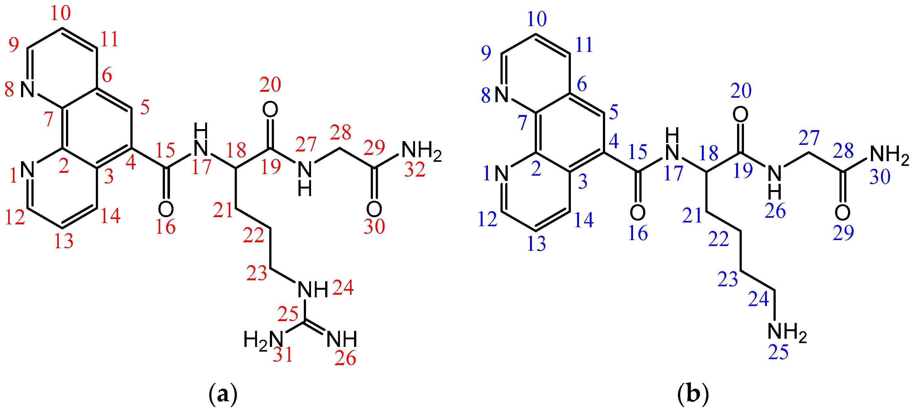
2.1. Synthesis and Characterization
2.2. Assessment of Biological Activity and Interactions with Biomolecules
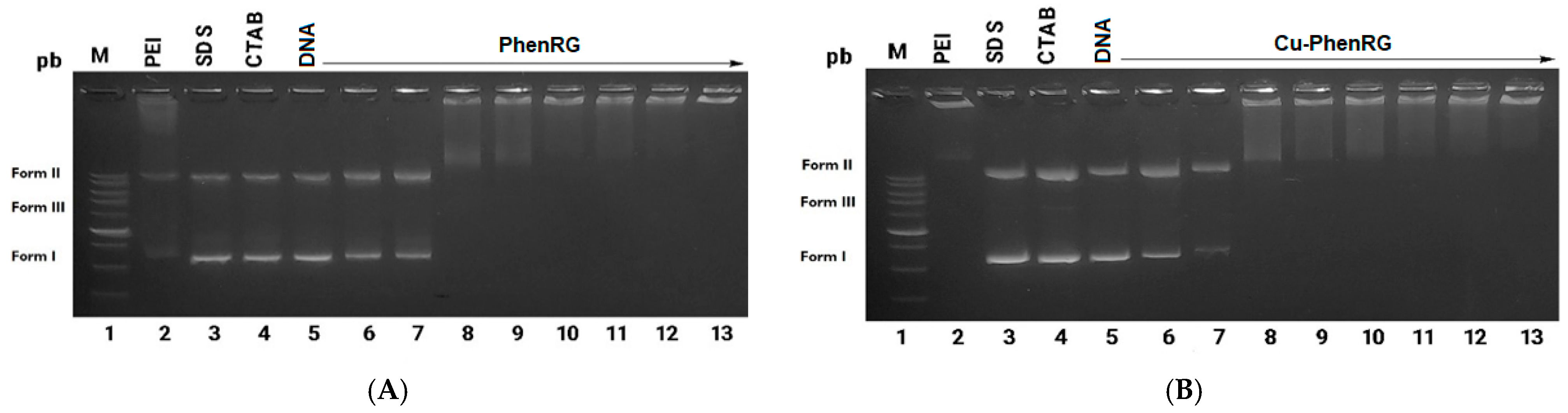
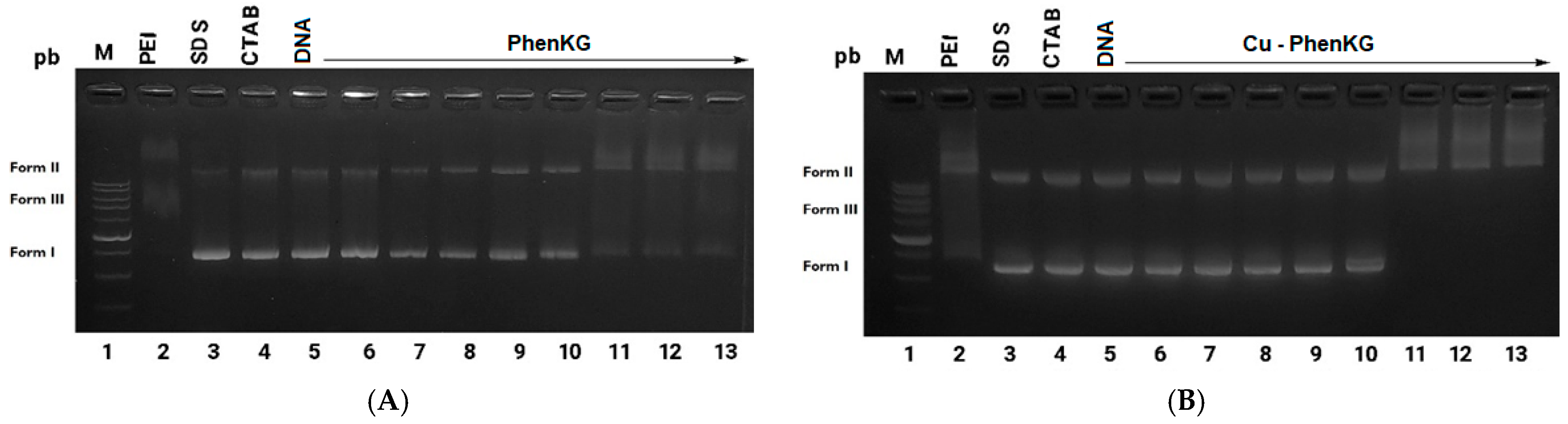
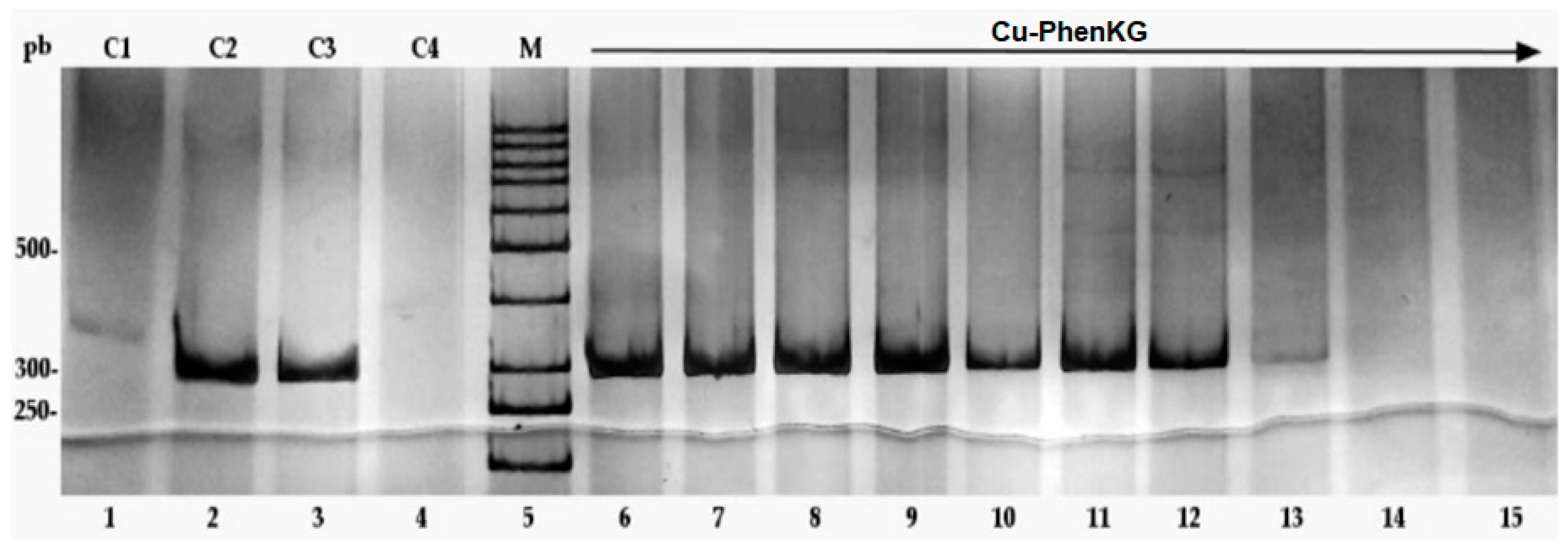
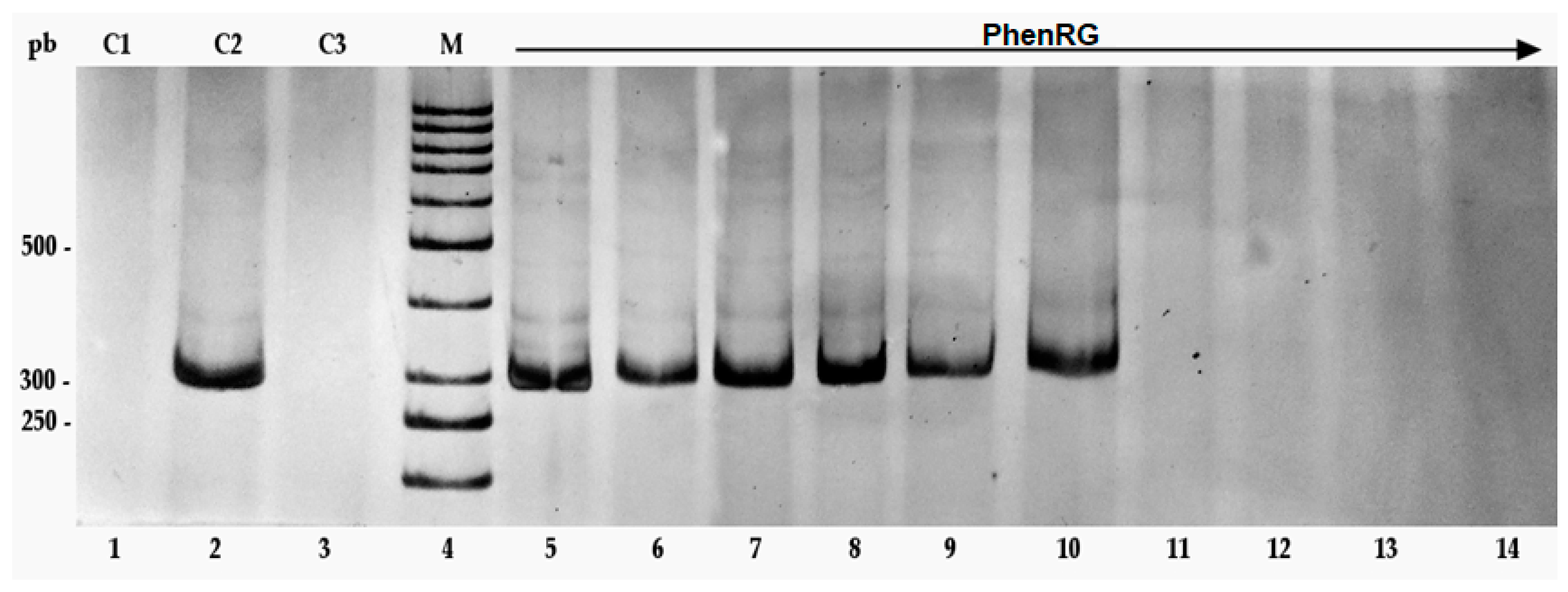
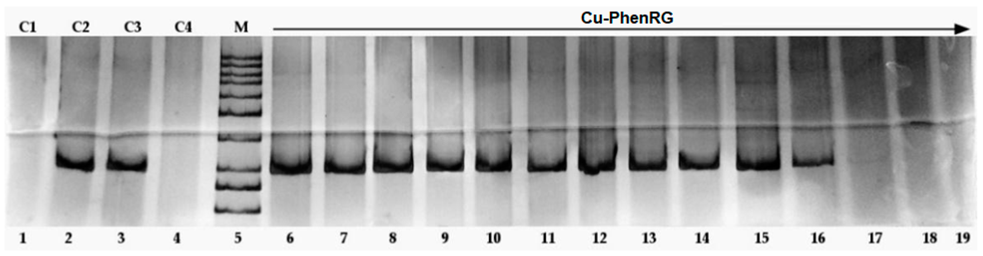
Interactions of Peptides and Metalloconjugates with Bacterial DNA
2.3. Calculation of Molecular and Pharmacokinetic Parameters
3. Results and Discussion
3.1. Synthesis and Characterization
3.2. Studies of Biological Activity and Interactions with Biomolecules
3.3. Calculation of Molecular and Pharmacokinetic Parameters
4. Conclusions
Supplementary Materials
Author Contributions
Funding
Institutional Review Board Statement
Informed Consent Statement
Data Availability Statement
Acknowledgments
Conflicts of Interest
References
- Rogers Van Katwyk, S.; Grimshaw, J.M.; Nkangu, M.; Nagi, R.; Mendelson, M.; Taljaard, M.; Hoffman, S.J. Government Policy Interventions to Reduce Human Antimicrobial Use: A Systematic Review and Evidence Map. PLoS Med. 2019, 16, e1002819. [Google Scholar] [CrossRef] [PubMed]
- Juliano, S.A.; Serafim, L.F.; Duay, S.S.; Heredia Chavez, M.; Sharma, G.; Rooney, M.; Comert, F.; Pierce, S.; Radulescu, A.; Cotten, M.L.; et al. A Potent Host Defense Peptide Triggers DNA Damage and Is Active against Multidrug-Resistant Gram-Negative Pathogens. ACS Infect. Dis. 2020, 6, 1250–1263. [Google Scholar] [CrossRef] [PubMed]
- Bergkessel, M.; Forte, B.; Gilbert, I.H. Small-Molecule Antibiotic Drug Development: Need and Challenges. ACS Infect. Dis. 2023, 9, 2062–2071. [Google Scholar] [CrossRef] [PubMed]
- Valenti, G.E.; Alfei, S.; Caviglia, D.; Domenicotti, C.; Marengo, B. Antimicrobial Peptides and Cationic Nanoparticles: A Broad-Spectrum Weapon to Fight Multi-Drug Resistance Not Only in Bacteria. Int. J. Mol. Sci. 2022, 23, 6108. [Google Scholar] [CrossRef] [PubMed]
- Patrzykat, A.; Friedrich, C.L.; Zhang, L.; Mendoza, V.; Hancock, R.E.W. Sublethal Concentrations of Pleurocidin-Derived Antimicrobial Peptides Inhibit Macromolecular Synthesis in Escherichia coli. Antimicrob. Agents Chemother. 2002, 46, 605–614. [Google Scholar] [CrossRef] [PubMed]
- Rivera-Sánchez, S.P.; Agudelo-Góngora, H.A.; Oñate-Garzón, J.; Flórez-Elvira, L.J.; Correa, A.; Londoño, P.A.; Londoño-Mosquera, J.D.; Aragón-Muriel, A.; Polo-Cerón, D.; Ocampo-Ibáñez, I.D. Antibacterial Activity of a Cationic Antimicrobial Peptide against Multidrug-Resistant Gram-Negative Clinical Isolates and Their Potential Molecular Targets. Molecules 2020, 25, 5035. [Google Scholar] [CrossRef] [PubMed]
- Futaki, S.; Suzuki, T.; Ohashi, W.; Yagami, T.; Tanaka, S.; Ueda, K.; Sugiura, Y. Arginine-Rich Peptides. J. Biol. Chem. 2001, 276, 5836–5840. [Google Scholar] [CrossRef] [PubMed]
- Lengacher, R.; Marlin, A.; Śmiłowicz, D.; Boros, E. Medicinal Inorganic Chemistry—Challenges, Opportunities and Guidelines to Develop the next Generation of Radioactive, Photoactivated and Active Site Inhibiting Metal-Based Medicines. Chem. Soc. Rev. 2022, 51, 7715–7731. [Google Scholar] [CrossRef] [PubMed]
- Abd-El-Aziz, A.S.; Agatemor, C.; Etkin, N. Antimicrobial Resistance Challenged with Metal-Based Antimicrobial Macromolecules. Biomaterials 2017, 118, 27–50. [Google Scholar] [CrossRef]
- Łodyga-Chruścińska, E. Tetrazole Peptides as Copper(II) Ion Chelators. Coord. Chem. Rev. 2011, 255, 1824–1833. [Google Scholar] [CrossRef]
- D’Accolti, M.; Bellotti, D.; Dzień, E.; Leonetti, C.; Leveraro, S.; Albanese, V.; Marzola, E.; Guerrini, R.; Caselli, E.; Rowińska-Żyrek, M.; et al. Impact of C- and N-terminal protection on the stability, metal chelation and antimicrobial properties of calcitermin. Sci. Rep. 2023, 13, 18228. [Google Scholar] [CrossRef] [PubMed]
- Berdis, A.J. Inhibiting DNA Polymerases as a Therapeutic Intervention against Cancer. Front. Mol. Biosci. 2017, 4, 78. [Google Scholar] [CrossRef]
- Long, E.C.; Fang, Y.-Y.; Lewis, M.A. DNA Minor Groove Recognition by Ni(II)· and Cu(II)·Gly-Gly-His Derived Metallopeptides. In ACS Symposium Series; Oxford University Press: Oxford, UK, 2009; pp. 219–241. [Google Scholar] [CrossRef]
- O’Shaughnessy, M.; Hurley, J.; Dillon, S.C.; Herra, C.; McCarron, P.; McCann, M.; Devereux, M.; Howe, O. Antibacterial Activity of Metal–Phenanthroline Complexes against Multidrug-Resistant Irish Clinical Isolates: A Whole Genome Sequencing Approach. J. Biol. Inorg. Chem. 2022, 28, 153–171. [Google Scholar] [CrossRef] [PubMed]
- Oñate-Garzón, J.; Manrique-Moreno, M.; Trier, S.; Leidy, C.; Torres, R.; Patiño, E. Antimicrobial activity and interactions of cationic peptides derived from Galleria mellonella cecropin D-like peptide with model membranes. J. Antibiot. 2017, 70, 238–245. [Google Scholar] [CrossRef]
- Bolin, D.R.; Sytwu, I.; Humiec, F.; Meienhfer, J. Preparation of Oligomer-free Nα-Fmoc and Nα-urethane Amino Acids. Int. J. Pept. Protein Res. 1989, 33, 353–359. [Google Scholar] [CrossRef]
- Novabiochem® Letters 2/06; Merck Millipore: Burlington, MA, USA, 2006; pp. 1–4.
- Torres-García, C.; Pulido, D.; Carceller, M.; Ramos, I.; Royo, M.; Nicolás, E. Solid-Phase Synthesis of Phenylalanine Containing Peptides Using a Traceless Triazene Linker. J. Org. Chem. 2012, 77, 9852–9858. [Google Scholar] [CrossRef] [PubMed]
- Caballero, A.B.; Terol-Ordaz, L.; Espargaró, A.; Vázquez, G.; Nicolás, E.; Sabaté, R.; Gamez, P. Histidine-Rich Oligopeptides To Lessen Copper-Mediated Amyloid-β Toxicity. Chem. A Eur. J. 2016, 22, 7268–7280. [Google Scholar] [CrossRef] [PubMed]
- Kaiser, E.; Colescott, R.L.; Bossinger, C.D.; Cook, P.I. Color Test for Detection of Free Terminal Amino Groups in the Solid-Phase Synthesis of Peptides. Anal. Biochem. 1970, 34, 595–598. [Google Scholar] [CrossRef]
- He, Y.; Zheng, Q.; Huang, H.; Ji, Y.; Lin, Z. Synergistic Synthesis of Hydrophilic Hollow Zirconium Organic Frameworks for Simultaneous Recognition and Capture of Phosphorylated and Glycosylated Peptides. Anal. Chim. Acta 2022, 1198, 339552. [Google Scholar] [CrossRef]
- Clinical and Laboratory Standards Institute. Methods for Dilution Antimicrobial Susceptibility Test for Bacteria That Grow Aerobically, Approved Standard, 9th ed.; CLSI: Wayne, PA, USA, 2012. [Google Scholar]
- Gajic, I.; Kabic, J.; Kekic, D.; Jovicevic, M.; Milenkovic, M.; Mitic Culafic, D.; Trudic, A.; Ranin, L.; Opavski, N. Antimicrobial Susceptibility Testing: A Comprehensive Review of Currently Used Methods. Antibiotics 2022, 11, 427. [Google Scholar] [CrossRef]
- Polo-Cerón, D. Cu(II) and Ni(II) Complexes with New Tridentate NNS Thiosemicarbazones: Synthesis, Characterisation, DNA Interaction, and Antibacterial Activity. Bioinorg. Chem. Appl. 2019, 2019, 3520837. [Google Scholar] [CrossRef] [PubMed]
- Rivera Franco, N.; Braga, Y.; Espitia Fajardo, M.; Barreto, G. Identifying New Lineages in the Y Chromosome of Colombian Amazon Indigenous Populations. Am. J. Phys. Anthropol. 2020, 172, 165–175. [Google Scholar] [CrossRef] [PubMed]
- Daina, A.; Michielin, O.; Zoete, V. SwissADME: A Free Web Tool to Evaluate Pharmacokinetics, Drug-Likeness and Medicinal Chemistry Friendliness of Small Molecules. Sci. Rep. 2017, 7, 42717. [Google Scholar] [CrossRef] [PubMed]
- Rappe, A.K.; Casewit, C.J.; Colwell, K.S.; Goddard, W.A.; Skiff, W.M. UFF, a Full Periodic Table Force Field for Molecular Mechanics and Molecular Dynamics Simulations. J. Am. Chem. Soc. 1992, 114, 10024–10035. [Google Scholar] [CrossRef]
- Trott, O.; Olson, A.J. AutoDock Vina: Improving the Speed and Accuracy of Docking with a New Scoring Function, Efficient Optimization, and Multithreading. J. Comput. Chem. 2009, 31, 455–461. [Google Scholar] [CrossRef]
- Gasteiger, J.; Marsili, M. A New Model for Calculating Atomic Charges in Molecules. Tetrahedron Lett. 1978, 19, 3181–3184. [Google Scholar] [CrossRef]
- Jo, S.; Kim, T.; Iyer, V.G.; Im, W. CHARMM-GUI: A Web-based Graphical User Interface for CHARMM. J. Comput. Chem. 2008, 29, 1859–1865. [Google Scholar] [CrossRef] [PubMed]
- Huang, J.; MacKerell, A.D. CHARMM36 All-Atom Additive Protein Force Field: Validation Based on Comparison to NMR Data. J. Comput. Chem. 2013, 34, 2135–2145. [Google Scholar] [CrossRef] [PubMed]
- Yao, G.; Huang, Q. Theoretical and Experimental Study of the Infrared and Raman Spectra of L-Lysine Acetylation. Spectrochim. Acta Part A Mol. Biomol. Spectrosc. 2022, 278, 121371. [Google Scholar] [CrossRef]
- Seshadri, S.; Khurana, R.; Fink, A.L. [36] Fourier Transform Infrared Spectroscopy in Analysis of Protein Deposits. In Methods in Enzymology; Academic Press: Cambridge, MA, USA, 1999; pp. 559–576. [Google Scholar] [CrossRef]
- Kose, A.; Erkan, S.; Tümer, M. A Series of Phenanthroline-Imine Compounds: Computational, OLED Properties and Fluorimetric Sensing of Nitroaromatic Compounds. Spectrochim. Acta A Mol. Biomol. Spectrosc. 2023, 286, 122006. [Google Scholar] [CrossRef]
- Fulmer, G.R.; Miller, A.J.M.; Sherden, N.H.; Gottlieb, H.E.; Nudelman, A.; Stoltz, B.M.; Bercaw, J.E.; Goldberg, K.I. NMR Chemical Shifts of Trace Impurities: Common Laboratory Solvents, Organics, and Gases in Deuterated Solvents Relevant to the Organometallic Chemist. Organometallics 2010, 29, 2176–2179. [Google Scholar] [CrossRef]
- Fan, K.; Hsu, C.; Chen, Y. Mass Spectrometry in the Discovery of Peptides Involved in Intercellular Communication: From Targeted to Untargeted Peptidomics Approaches. Mass Spectrom. Rev. 2022, 42, 2404–2425. [Google Scholar] [CrossRef] [PubMed]
- Shi, J.; Chen, C.; Wang, D.; Wang, Z.; Liu, Y. The Antimicrobial Peptide LI14 Combats Multidrug-Resistant Bacterial Infections. Commun. Biol. 2022, 5, 926. [Google Scholar] [CrossRef] [PubMed]
- Rodríguez, C.A.; Vesga, O. Staphylococcus Aureus Resistente a Vancomicina. Biomédica 2005, 25, 575. [Google Scholar] [CrossRef]
- Turnbull, P.C.B.; Sirianni, N.M.; LeBron, C.I.; Samaan, M.N.; Sutton, F.N.; Reyes, A.E.; Peruski, L.F. MICs of Selected Antibiotics for Bacillus anthracis, Bacillus cereus, Bacillus thuringiensis, and Bacillus mycoidesfrom a Range of Clinical and Environmental Sources as Determined by the Etest. J. Clin. Microbiol. 2004, 42, 3626–3634. [Google Scholar] [CrossRef] [PubMed]
- Bencini, A.; Lippolis, V. 1,10-Phenanthroline: A Versatile Building Block for the Construction of Ligands for Various Purposes. Coord. Chem. Rev. 2010, 254, 2096–2180. [Google Scholar] [CrossRef]
- Norouzi, P.; Mirmohammadi, M.; Houshdar Tehrani, M.H. Anticancer Peptides Mechanisms, Simple and Complex. Chem. Biol. Interact. 2022, 368, 110194. [Google Scholar] [CrossRef] [PubMed]
- Yasir, M.; Dutta, D.; Willcox, M.D.P. Enhancement of Antibiofilm Activity of Ciprofloxacin against Staphylococcus Aureus by Administration of Antimicrobial Peptides. Antibiotics 2021, 10, 1159. [Google Scholar] [CrossRef] [PubMed]
- Choosakoonkriang, S.; Lobo, B.A.; Koe, G.S.; Koe, J.G.; Middaugh, C. Russell. Biophysical Characterization of PEI/DNA Complexes. J. Pharm. Sci. 2003, 92, 1710–1722. [Google Scholar] [CrossRef]
- Yang, S.; Zhou, X.; Li, R.; Fu, X.; Sun, P. Optimized PEI-based Transfection Method for Transient Transfection and Lentiviral Production. Curr. Protoc. Chem. Biol. 2017, 9, 147–157. [Google Scholar] [CrossRef]
- Hellman, L.M.; Fried, M.G. Electrophoretic Mobility Shift Assay (EMSA) for Detecting Protein–Nucleic Acid Interactions. Nat. Protoc. 2007, 2, 1849–1861. [Google Scholar] [CrossRef] [PubMed]
- Davies, A.M. A Ca2+-Induced Mitochondrial Permeability Transition Causes Complete Release of Rat Liver Endonuclease G Activity from Its Exclusive Location within the Mitochondrial Intermembrane Space. Identification of a Novel Endo-Exonuclease Activity Residing within the Mitochondrial Matrix. Nucleic Acids Res. 2003, 31, 1364–1373. [Google Scholar] [CrossRef] [PubMed]
- McKie, S.J.; Desai, P.R.; Seol, Y.; Allen, A.M.; Maxwell, A.; Neuman, K.C. Topoisomerase VI Is a Chirally-Selective, Preferential DNA Decatenase. eLife 2022, 11, e67021. [Google Scholar] [CrossRef] [PubMed]
- Meloni, B.P.; Mastaglia, F.L.; Knuckey, N.W. Cationic Arginine-Rich Peptides (CARPs): A Novel Class of Neuroprotective Agents With a Multimodal Mechanism of Action. Front. Neurol. 2020, 11, 108. [Google Scholar] [CrossRef] [PubMed]
- Anastassopoulou, J. Metal–DNA Interactions. J. Mol. Struct. 2003, 651–653, 19–26. [Google Scholar] [CrossRef]
- Lane, D.; Prentki, P.; Chandler, M. Use of Gel Retardation to Analyze Protein-Nucleic Acid Interactions. Microbiol. Rev. 1992, 56, 509–528. [Google Scholar] [CrossRef] [PubMed]
- Artika, I.M.; Dewi, Y.P.; Nainggolan, I.M.; Siregar, J.E.; Antonjaya, U. Real-Time Polymerase Chain Reaction: Current Techniques, Applications, and Role in COVID-19 Diagnosis. Genes 2022, 13, 2387. [Google Scholar] [CrossRef] [PubMed]
- Sidstedt, M.; Rådström, P.; Hedman, J. PCR inhibition in qPCR, dPCR and MPS—mechanisms and solutions. Anal. Bioanal. Chem. 2020, 412, 2009–2023. [Google Scholar] [CrossRef]
- Agarwal, P.; Huckle, J.; Newman, J.; Reid, D.L. Trends in Small Molecule Drug Properties: A Developability Molecule Assessment Perspective. Drug Discov. Today 2022, 27, 103366. [Google Scholar] [CrossRef]


| Compound | MIC (µg/mL) | |||||
|---|---|---|---|---|---|---|
| S. aureus | B. cereus | L. monocytogenes | P. aeruginosa | S. typhimurium | E. coli | |
| ATCC 25923 | ATCC 10876 | ATCC 19115 | ATCC 27853 | ATCC 14028 | ATCC 25922 | |
| PhenKG | >2 | >2 | >2 | >2 | >2 | >2 |
| Cu-PhenKG | 0.5 | >2 | 2 | >2 | 2 | 2 |
| PhenRG | >1.25 | >1.25 | >1.25 | >1.25 | >1.25 | >1.25 |
| Cu-PhenRG | 1.25 | >1.25 | 1.25 | >1.25 | 0.63 | 1.25 |
| Cip | ≤0.5 | ≤0.5 | ≤0.5 | ≤0.5 | ≤0.5 | ≤0.5 |
| Vancomycin | ≤4 * | 3 * | ≤2 * | - | - | - |
| Compound | Binding Energy (kJ/mol) |
|---|---|
| Mitoxantrone | −6.8 |
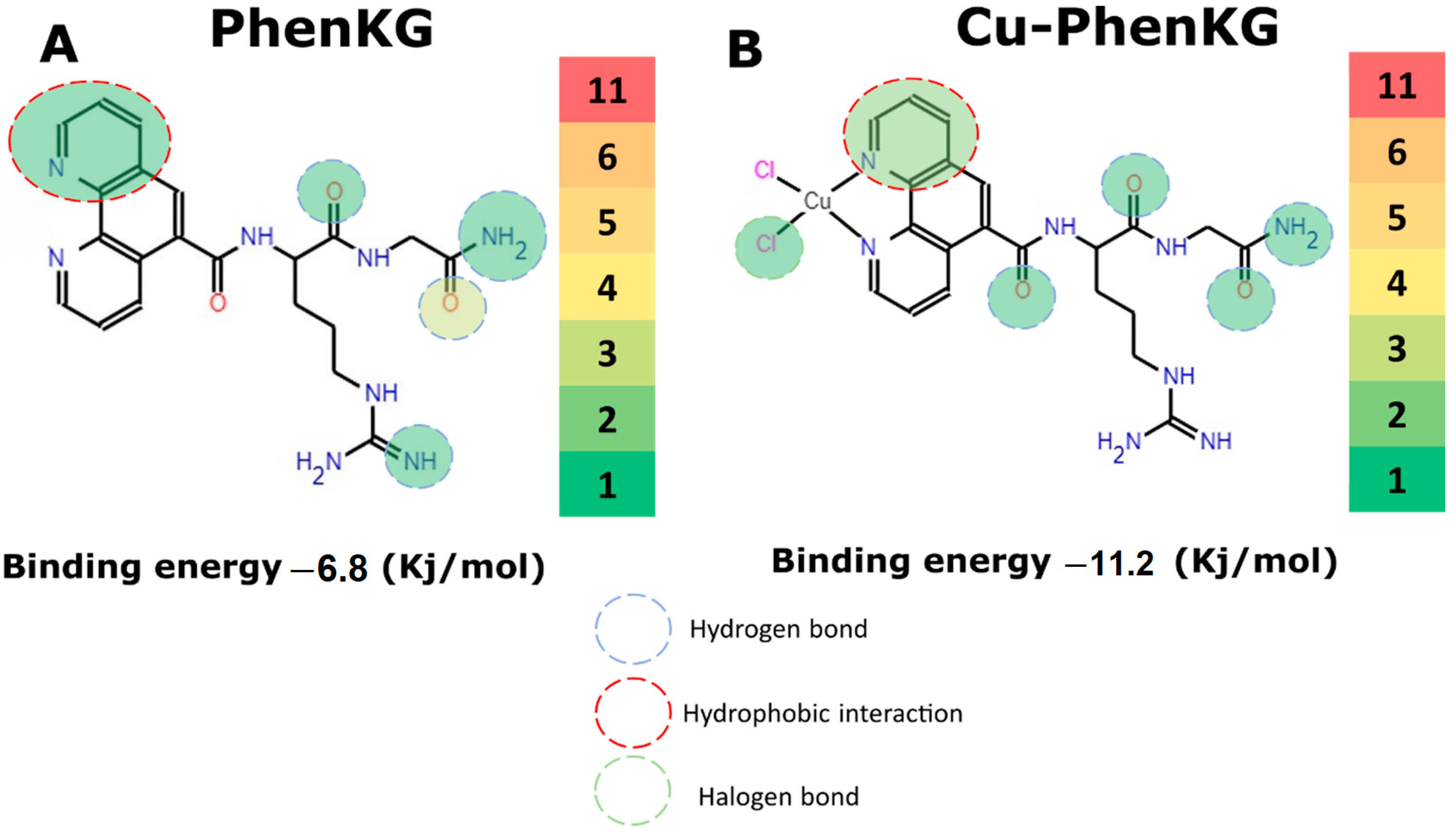
| PhenKG | −6.8 |
| Cu-PhenKG | −11.2 |
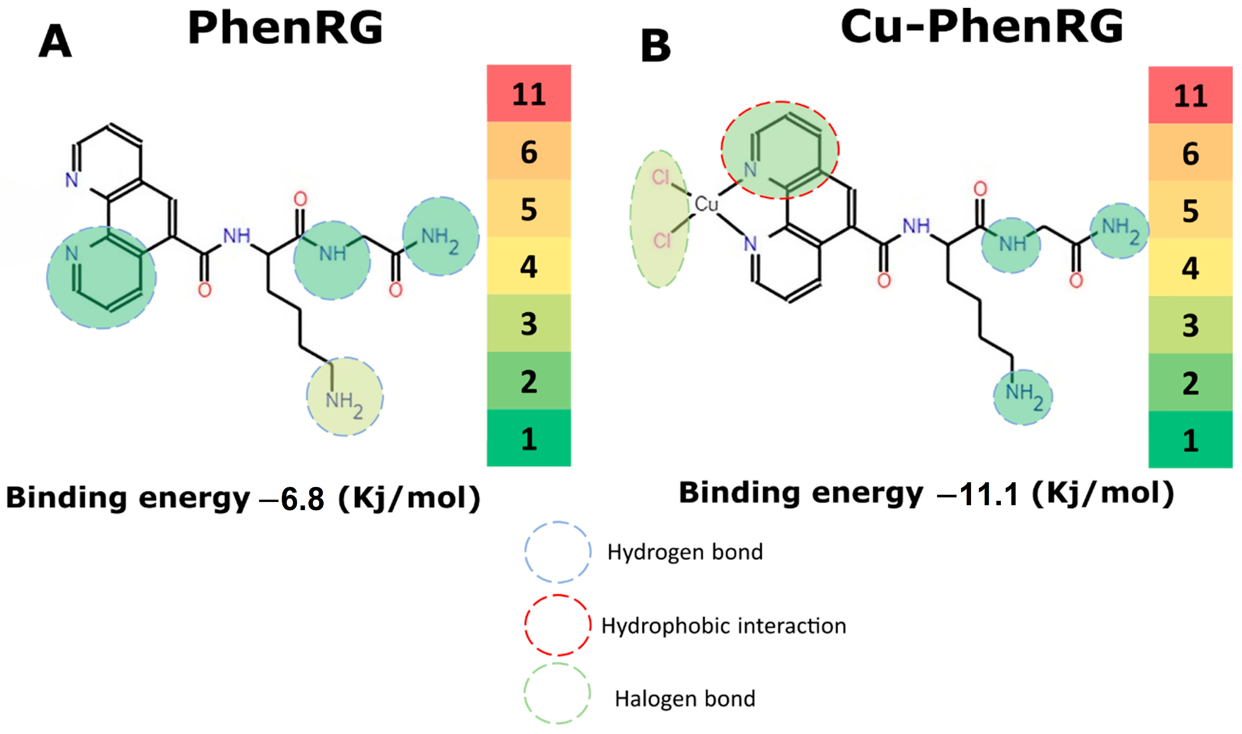
| PhenRG | −6.8 |
| Cu-PhenRG | −11.1 |
| Compound | Molecular Weight (g/mol) | Rotary Bonds | H-Aceptor Bonds | H-Donor Bonds | LogS | TPSA | Log Po/w (MLOGP) | Lipinski Filter |
|---|---|---|---|---|---|---|---|---|
| (ESOL) | (Å2) | |||||||
| PhenKG | 408.46 | 11 | 6 | 4 | Soluble | 153.09 | −0.41 | Yes; 0 violation |
| PhenRG | 436.47 | 12 | 6 | 6 | Very Soluble | 188.97 | −0.79 | No; 2 violations: N or O > 10, NH or OH > 5 |
| Compound | Absortion GI | Substrate P-gp | Inhibitor | ||||
|---|---|---|---|---|---|---|---|
| CYP1A2 | CYP2C19 | CYP2C9 | CYP2D6 | CYP3A4 | |||
| PhenKG | Low | Yes | No | No | No | No | No |
| PhenRG | Low | No | No | No | No | No | No |
Disclaimer/Publisher’s Note: The statements, opinions and data contained in all publications are solely those of the individual author(s) and contributor(s) and not of MDPI and/or the editor(s). MDPI and/or the editor(s) disclaim responsibility for any injury to people or property resulting from any ideas, methods, instructions or products referred to in the content. |
© 2024 by the authors. Licensee MDPI, Basel, Switzerland. This article is an open access article distributed under the terms and conditions of the Creative Commons Attribution (CC BY) license (https://creativecommons.org/licenses/by/4.0/).
Share and Cite
Moreno-Ramirez, M.C.; Arias-Bravo, A.S.; Aragón-Muriel, A.; Godoy, C.A.; Liscano, Y.; Garzón, J.O.; Polo-Cerón, D. Design, Synthesis and Antimicrobial Potential of Conjugated Metallopeptides Targeting DNA. Sci. Pharm. 2024, 92, 21. https://doi.org/10.3390/scipharm92020021
Moreno-Ramirez MC, Arias-Bravo AS, Aragón-Muriel A, Godoy CA, Liscano Y, Garzón JO, Polo-Cerón D. Design, Synthesis and Antimicrobial Potential of Conjugated Metallopeptides Targeting DNA. Scientia Pharmaceutica. 2024; 92(2):21. https://doi.org/10.3390/scipharm92020021
Chicago/Turabian StyleMoreno-Ramirez, Maria Camila, Adriana Stefania Arias-Bravo, Alberto Aragón-Muriel, César Alonso Godoy, Yamil Liscano, Jose Oñate Garzón, and Dorian Polo-Cerón. 2024. "Design, Synthesis and Antimicrobial Potential of Conjugated Metallopeptides Targeting DNA" Scientia Pharmaceutica 92, no. 2: 21. https://doi.org/10.3390/scipharm92020021