Article

Antioxidant Activity of the Medicinal Plant Urtica dioica L.: Extraction Optimization Using Response Surface Methodology and Protective Role in Red Blood Cells

Department of Biomedical Science, School of Health and Sports Sciences, New York College, 138 Egnatia, 54622 Thessaloniki, Greece
*
Author to whom correspondence should be addressed.
Sci. Pharm. 2024, 92(3), 45; https://doi.org/10.3390/scipharm92030045
Submission received: 14 July 2024 / Revised: 3 August 2024 / Accepted: 9 August 2024 / Published: 14 August 2024

Abstract

:
Free radicals are highly reactive compounds that lead to the onset of a variety of serious health conditions, known as “oxidative stress”. Antioxidants, on the other hand, act as defense mechanisms to fight the accumulation of free radicals and maintain cell homeostasis. Urtica dioica L. is a medicinal plant with unique antioxidant properties, mainly attributable to the presence of polar phenolic compounds. This study aimed to optimize the combination of determinant factors toward the maximum extraction of antioxidants from U. dioica L. Furthermore, it investigated the protective role of the extract on red blood cells that were exposed to oxidative stress. The extraction optimization was performed using Response Surface Methodology and the in vitro antioxidant activity of the extract was determined with Folin–Ciocalteu, FRAP, and DPPH assays. Based on the results, the highest value of antioxidant activity and polar phenolic compounds was recorded using 34% (v/v) ethanol as a solvent in an ultrasonic process carried out at 42 °C for 87 min. In addition, UV–Vis spectrum of the extract revealed the presence of chlorophylls, carotenoids, and flavonoid glycosides. This study also provided insight into the in vitro protective effect of the plant extract on red blood cells morphology under oxidative stress conditions. The findings highlighted the good predictability of the extraction model and the potential role of the extract as an antioxidant product.

Graphical Abstract

1. Introduction

The health of the human body is strongly associated with the normal function of the cells by maintaining balance between the chemical and biological processes that occur within the cellular structure. Among the various essential components, free radicals can be found as supporting mechanisms of cell signaling, immune response, and energy production. However, any increase in the concentration of free radicals above normal levels is detrimental to cell homeostasis, leading to a state of “oxidative stress” [1]. There are three principal reactions that can lead to the formation of radicals. The first two categories involve the transfer of electrons, while the third one is associated with electrons separation. Each case is presented below [2].
  • Radical cation formation:
  • A: → A●+ + e          (Reaction 1)
  • Radical anion formation:
  • B: +e → :B●−         (Reaction 2)
  • Radical fragments formation:
  • A:B → A + B          (Reaction 3)
The main substances responsible for cell lysis are reactive oxygen species (ROS) and reactive nitrogen species (RNS), both generated via the action of free radicals. The natural antioxidant defense system of the human body can be overwhelmed when the generation rate of ROS and RNS surpasses its ability to neutralize and eliminate them, leading to the cell death [3,4]. The exogenous factors of the current daily life, like environmental pollutants, tobacco smoke, and UV radiation, aid the release of free radical precursors. The latter are introduced into the human cells and, through cellular metabolic activity, free radicals are generated as by-products [4]. An increase in oxidative stress can have detrimental effects on biological systems, causing the degradation of lipids, nucleic acids, proteins, and DNA [2]. The permanent modifications of genetic material, as a consequence of the oxidative damage, lead to the first stages of mutagenesis, carcinogenesis, and aging [1].
Antioxidant compounds can be effective in the inhibition and treatment of chronic disorders, by blocking or slowing down the destruction of biomolecules with the transfer of electrons to the free radicals [5]. Natural origin antioxidants, especially from plants, are generally considered a much preferable source as compared to the ones that are synthetically produced [6,7]. The phytochemicals in plants have high antioxidant activity due to their redox potential and chemical structure, resulting in the neutralization of free radicals and the chelation of transitional metals [8].
The use of natural products in medicine has been evident since the 19th century, long before synthetic drugs were developed, and continues to grow today, as many plant-derived products play a key role in contemporary drug discovery and development [9]. The most widespread medicinal plant from the 19th century is Urtica dioica L., which belongs to the Urticaceae family of Angiosperms (flowering plants) and the genus Urtica [10]. U. dioica finds a wide application in a variety of industrial fields. Besides its well-known role as a food ingredient with high nutritional value, it is also used as a fertilizer to enhance the growth of different plants [11]. Furthermore, U. dioica as a medicinal plant holds a rich source of pharmacologically relevant bioactive compounds. The healing properties of the plant are primarily attributed to the presence of polar phenolic compounds, and more especially chlorogenic acid. Other bioactive constituents can also be found, including terpenoids, sphingolipids, steroids, lignans, flavonoids, and alkaloids. These compounds are recognized for their antioxidant, anti-inflammatory, and antiviral capacity, among other properties [12]. This unique composition has led U. dioica to be recognized in ethnopharmacology for its therapeutic benefits in both preventing and treating various illnesses [10]. Although various efforts have been made to recover the antioxidants from the plant, discrepancies in the optimum extraction conditions have been evident among studies in the literature, mainly due to the lack of consideration for certain extraction parameters and their combined effect on the process [13,14,15]. As a consequence, until now, there has been no standard extraction procedure in the literature for this plant.
The aim of the current study was to explore the combined effect of determinant factors on the optimum extraction conditions of antioxidants from the medicinal plant U. dioica L. The ultrasound-assisted extraction process was optimized by employing Response Surface Methodology (RSM) and the in vitro antioxidant activity of the extract was determined by Folin–Ciocalteu, FRAP, and DPPH assays. Furthermore, in vitro blood tests were conducted under oxidative stress conditions to assess the direct effect of the antioxidant extract on red blood cells.

2. Materials and Methods

2.1. Reagents and Biological Materials

Sodium carbonate anhydrous (≥99.5%), iron (III) chloride anhydrous (>98%), and crystallized phenol (>99.5%) were obtained from Labbox Labware S.L. (Barcelona, Spain). Folin–Ciocalteu reagent was provided by Sordalab (Étampes, France). Bradford assay solution, 1,1-Diphenyl-2-picrylhydrazyl (DPPH) (>97%), and 2,4,6-Tri(2-pyridyl)-1,3,5-triazine (TPTZ) (>98%) were purchased from TCI Europe N.V. (Zwijndrecht, Belgium). Sodium acetate anhydrous was supplied by Lach-Ner (98.7%) (Neratovice, Czech Republic). The iron (II) sulfate heptahydrate (99.5%), gallic acid anhydrous (≥99%), and bovine serum albumin (≥96%) standards were obtained from Glentham Life Sciences (Corsham, UK), while the saccharose (≥99.8%) standard was from Penta Chemicals (Praha, Czech Republic). Ethanol absolute (≥99.8%), hydrochloric acid (37%), and sulfuric acid (≥95%) were purchased from Fisher Scientific (Loughborough, UK). Methanol (≥99.8%) was obtained from Chem-Lab (Zedelgem, Belgium). Commercial sheep blood defibrinated was provided by TCS Biosciences (Buckingham, UK). Commercial Urtica dioica L. leaves were supplied in dry form by Green Family (Thessaloniki, Greece).

2.2. Apparatus

The instruments used to carry out the experimental procedures were a VWR UV-1600PC Spectrophotometer (Radnor, PA, USA), a Skymen 010S Ultrasonic Cleaner (Shenzhen, China), a Milwaukee MW151 MAX pH/ORP/Temp Logging Bench Meter (Szeged, Hungary), a Bresser Science TFM 201/301 + Infinity Microscope (Rhede, Germany), an Axis ACN220G Analytical Balance (Gdańsk, Poland), a Biosan MSH-300 Magnetic Stirrer with hot plate (Riga, Latvia), a Biosan Vortex V-1 plus (Riga, Latvia), and a Rohnson R-5795 Hand Blender (Prague, Czech Republic).

2.3. Ultrasound-Assisted Extraction of Antioxidants

Dried leaves of U. dioica were ground with a blender at a frequency of 50 Hz. Then, 1 g of the processed material was added to a falcon tube of 50 mL capacity and mixed with 20 mL of the solvent, the composition of which was based on the experimental design (ethanol and/or water). The falcon tube was transferred to an ultrasonic processor (frequency 50 Hz, power 60 W), where specific extraction time and extraction temperature level were tested according to the experimental design. After the extraction, the Buchner funnel filtration process was performed by passing the extract through a polyurethane filter (pore size 8–12 μm) and the filtrate was collected for further analysis.

2.4. Experimental Design of the Extraction Process

The extraction conditions were optimized as part of the experimental design process in order to obtain the highest antioxidant activity. To achieve this goal, an unblocked full factorial face-centered central composite design of the RSM was developed using the Minitab software (free trial version 22.1, State College, PA, USA). The model tested the effect of three extraction process factors (Xi), namely, ethanol content (X1, % v/v), extraction time (X2, min), and extraction temperature (X3, °C). As illustrated in Table 1, the three levels of each factor were coded as −1, 0, and +1, corresponding to the low-, mid-, and high-level of Xi, respectively. The mid-level was calculated from the equation shown as a footnote in Table 1. The entire design consisted of 20 experimental runs, which were carried out in a random sequence to maximize the effect of unexplained variability in the observed responses. Six experiments were conducted at the center of the design as replicates for the estimation of error.
The antioxidant activity of each extract was determined by the Folin–Ciocalteu (Section 2.6), FRAP (Section 2.7) and DPPH assays (Section 2.8), and then the second-order polynomial equation was fitted to the respective responses YFolin (mg/L), YFRAP (g/L), and YDPPH (%) as shown below.
Υ = β0 + β1X1 + β2X2 + β3X3 + β11X12 + β22X22 + β33X32 + β12X1X2 + β13X1X3 + β23X2X3
where Y is the dependent variable; X1, X2, and X3 are the independent variables as mentioned above; and β0, β1β23 represent the estimated coefficients with β0 having a role of a scaling constant.

2.5. Scale-Up of the Optimum Extraction Conditions

A 5-fold scale-up of the extraction process was performed at the optimum conditions proposed by the model in order to achieve the maximum antioxidant activity. Firstly, 5 g of ground dried leaves and 100 mL of 34% (v/v) ethanol were added to a 250 mL Erlenmeyer flask. Afterwards, the flask was transferred to an ultrasonic processor and the extraction was performed at 42 °C for 87 min. Following that, filtration was carried out using the Buchner funnel technique and the filtrate was used for further analysis (Section 2.6, Section 2.7, Section 2.8, Section 2.9, Section 2.10, Section 2.11, Section 2.12).

2.6. Folin–Ciocalteu Assay

The total polar phenol content in the plant extracts was determined using the Folin–Ciocalteu method [16]. In brief, 5 mL of distilled water, 200 μL of the extract, and 500 μL of Folin–Ciocalteu reagent were carefully transferred to a 10 mL volumetric flask. After allowing the mixture to stand for 3 min in the dark, 1 mL of a 37% (w/v) Na2CO3 aqueous solution was added, and the flask was filled up to the mark with water. The resulting mixture was left to stand for 1 h in the dark and then the absorbance was recorded at 725 nm against a blank containing 200 μL of solvent instead of the extract. Quantification was carried out using linear regression analysis with standard gallic acid solutions (25–400 mg/L), and the results were expressed as gallic acid equivalents (mg/L).

2.7. FRAP Assay

The reducing power of the plant extracts was determined using the Ferric Reducing Antioxidant Power (FRAP) method [17]. To prepare the FRAP reagent solution, a mixture of 0.012 M FeCl3, 10 mM TPTZ, and 0.3 M acetate buffer (pH 3.6) was combined at a 1:1:10 ratio. Subsequently, 100 μL of the extract were combined with 3 mL of the FRAP reagent solution in small test tubes. The mixture was then incubated in a water bath at 37 °C for 30 min followed by the absorbance recording at 593 nm against a blank containing 100 μL of solvent instead of the extract. Quantification took place with the aid of linear regression analysis, based on standard ferrous sulfate solutions (0.2–3 g/L), and the results were expressed as ferrous sulfate equivalents (g/L).

2.8. DPPH Radical Scavenging Activity Assay

The radical scavenging activity of the plant extracts was evaluated using the DPPH method [18]. Particularly, 0.1 mL of the extract was added to 3 mL of a 0.1 mM DPPH methanolic solution. Next, the sample mixture was left for 30 min in the dark, followed by the measurement of the absorbance at 517 nm. A control solution was prepared using 0.1 mL of solvent instead of the extract and subjected to the same assay conditions. A blank containing 3 mL of methanol instead of the DPPH solution was also made for each extract in order to autozero the spectrophotometer. The antioxidant activity of the extract was determined using the following equation and expressed as the percentage of radical scavenging activity (%).
R a d i c a l   s c a v e n g i n g   a c t i v i t y % = ( A C o n t r o l A S a m p l e A C o n t r o l ) × 100 %
where ASample is the absorbance of the sample and AControl is the absorbance of the control.

2.9. Dubois Assay

The total soluble sugar content in the plant extracts was analyzed by the Dubois method [19]. Briefly, 2 mL of the extract were mixed with 1 mL of phenol aqueous solution (1%, w/v) and 5 mL of concentrated H2SO4 in a test tube. Then, the mixture was left for 15 min, followed by vortexing and further incubation for another 15 min at 25–30 °C. In the final step, the absorbance was measured at 490 nm against a blank containing 2 mL of solvent instead of the extract. Quantification was performed using linear regression analysis with standard sucrose solutions (50–500 mg/L) and the results were expressed as sucrose equivalents (g/L).

2.10. Bradford Assay

The protein content in the plant extracts was determined with the Bradford protein method [20]. In particular, 0.5 mL of the extract were mixed with 1.5 mL of Bradford reagent (4-fold diluted with distilled water) inside a test tube. The mixture was left to stand for 5 min and then the absorbance was measured at 595 nm against a blank containing 0.5 mL solvent instead of the extract. Quantification was carried out using linear regression analysis with standard BSA solutions (0.5–4 g/L), and the results were expressed as BSA equivalents (g/L).

2.11. UV-Vis Spectrum

The UV-Vis absorbance spectrum of the plant extract was recorded in the region 200–800 nm after appropriate dilution using the M. Wave professional software (version 1.0.23, M.R.C. Laboratory Equipment, Harlow, UK).

2.12. Red Blood Cells Test

The antioxidant effects of the plant extract were evaluated on red blood cells experiencing oxidative stress. To achieve this goal, various factors have been taken into account. Firstly, based on the literature, a 30 μM concentration of the pro-oxidant H2O2 was selected as an effective way to induce oxidative stress in the red blood cells [21]. Also, to assess the influence of a plant extract on this oxidative stress condition, it was estimated that approximately 150 mL of the extract could be consumed by an adult with a total blood volume of 5 L [22]. The latter would result in a 3% (v/v) concentration of extract in the blood. Considering all the above, three different sample preparations were tested in the experiment, corresponding to (a) sheep blood, (b) sheep blood supplemented with H2O2 (30 μM), as well as (c) sheep blood supplemented with H2O2 (30 μM) and plant extract (3%, v/v). Each sample preparation was placed into test tubes and incubation was followed in a water bath at 37 °C for 30 min [21]. Afterward, the morphology of the red blood cells in the processed samples was carefully examined under a microscope at 100× magnification. Additionally, total red blood cells in each sample were counted using a Neubauer-improved hemocytometer (Marienfeld Superior, Lauda-Königshofen, Germany).

2.13. Statistical Analysis

Minitab software (free trial version 22.1, State College, PA, USA) was used to assess the model quality of fitting by analyzing the adjusted and predicted coefficients of determination (R2-adj and R2-pred, respectively), calculating the significance of each parameter via F-test (p-values), and observing the lack-of-fit of the model. Coefficient p-values lower than 0.05 were considered statistically significant. Reduced models were obtained by retaining only the significant terms (p < 0.05) and those that support the hierarchical principle. The beta coefficient was established by minimizing the sum of squared differences between observed and predicted values. To verify the proposed model, the experiment was repeated at the optimum combination of the three factors for the maximization of the responses. The findings of the latter were compared with the values predicted by the model.
The degree of the linear relationship between the total polar phenol content and the antioxidant activity was assessed by Pearson’s correlation coefficient (R) using the Microsoft Excel spreadsheet (Microsoft 365, Redmond, WA, USA). The p-value was used to determine the significance of Pearson’s correlation (p < 0.01) based on a t-distribution with n − 2 degrees of freedom.

3. Results and Discussion

3.1. Planning the Extraction Process

The traditional methods used to perform extraction of bioactive ingredients from plants, including maceration, distillation, decoction, heat reflux, and Soxhlet extraction, seem to have various disadvantages such as high energy consumption requirement, loss of volatile components, and difficulty in the control of the extraction efficiency. A very promising alternative method that was found to be very effective for this purpose is ultrasound-assisted extraction. Notably, during the ultrasonic process various mechanical phenomena take place (like fragmentation, erosion, detexturation, and sonoporation) that lead to the disruption of the plant’s cell wall and, consequently, the release of bioactive compounds. Due to these properties, ultrasound-assisted extraction offers several exceptional advantages against other methods, such as shorter extraction time, lower solvent consumption, improved extraction rates, and enhanced extraction quality. The additional benefits of this method are its economical and environmentally friendly nature [23]. Considering all the above, an ultrasound-assisted extraction technique was chosen in the current research for the extraction of antioxidants from the plant U. dioica.
When it comes to the selection of a proper extraction solvent, it has to be taken into account that over the past decade industries have been increasingly employing green solvents in their processes. Among them, water has emerged as the most suitable green solvent in terms of safety and cost. Regarding the extraction of polar bioactive compounds (e.g., polyphenols), the exploitation of water mixtures with polar organic solvents seems to be a more effective approach rather than water’s sole use [24]. In order to assess which green organic solvent has the highest potential for the extraction of antioxidants from U. dioica, a preliminary study was carried out comparing the efficacy of methanol, ethanol, acetone, and ethyl acetate. Among them, ethanol was found to be the most suitable solvent to obtain the maximum antioxidant activity in the extract (data not shown). Thus, the first factor in the extraction process modeling was decided to be the concentration of ethanol in mixtures with water.
The time and temperature were also two other important factors for the process design. A minimum and a maximum time period of 15 min and 90 min, respectively, were chosen in order to assess the effect of time on the migration of the plant antioxidants to the solvent (i.e., extraction yield). The temperature has also an influence on the extraction as the plant tissues become softened through heating, leading to the disruption of the cell membranes [25]. To assess this effect, a range of 30 °C to 75 °C was tested, considering that the maximum temperature in the design should not exceed the boiling point of ethanol (78 °C).

3.2. Predictive Modeling of the Extraction Process

3.2.1. Response Variables and Accuracy of Predictive Equations

RSM design was successfully applied to select the best combination of ethanol content (X1, % v/v), extraction time (X2, min), and extraction temperature (X3, °C) to maximize the responses YFolin (mg/L), YFRAP (g/L), and YDPPH (%). The experimental data obtained for each response variable are presented in Table 2.
Multiple regression analysis was employed to fit the second-order polynomial equation (Equation (2)) for each response (Yi) using uncoded values. The resulting models were simplified by removing non-significant terms (p ≥ 0.05) to form Equations (3)–(5) displayed in Table 3.
In the case of YFolin, the non-significant terms X22, X32, X1X2, X1X3, and X2X3 were omitted from Equation (3). In a similar way, for YFRAP, the non-significant terms X3, X22, X32, X1X2, X1X3, and X2X3 terms were excluded from Equation (4). In addition, Equation (5) related to YDPPH was simplified by eliminating the non-significant terms X22, X1X2, and X2X3.
Notably, the lack-of-fit value for the responses YFolin, YFRAP, and YDPPH was statistically non-significant (p = 0.106, 0.102, and 0.106, respectively), meaning that there are no differences between the observed response values and the values predicted by the model. This indicates a good fit of the model for the data and that the results of the regression analysis are reliable. Additionally, both the adjusted and predicted coefficients of determination for all the responses are high and close to each other (R2-adj 92.97–97.52 and R2-pred 82.58–90.42). This highlights that the models can accurately explain the effect of the three factors on YFolin, YFRAP, and YDPPH and can exert strong predictability for any new observations.

3.2.2. Assessment of Factors Significance Using Pareto Charts

Pareto charts were created based on the t-values to examine the effects of each factor as well as of their interactions on the YFolin, YFRAP, and YDPPH responses (Figure 1).
The linear term of % ethanol content (X1) exhibited the strongest negative influence on all three response variables. In YFolin, the quadratic effect of X1 (X12) was found to be significantly positive (Figure 1A). This signifies that the negative impact of X1 on the response decelerates beyond its middle level (50%, v/v). On the other hand, a negative effect of X12 was observed for YFRAP and YDPPH (Figure 1B,C), indicating that as the concentration of ethanol increases, its influence becomes even more pronounced. Another significant factor was the extraction time (X2), which exhibited a positive linear trend for YFolin, YFRAP, and YDPPH. In contrast, the quadratic term of time (X22) had a non-significant effect on the three responses, indicating that a plateau was reached when the factors approached their set maximum value (90 min). Additionally, the negative influence of the extraction temperature (X3) and its quadratic term (X32) highlighted that heating had a negative impact on all three responses and this effect decelerates at values above its middle level (52.5 °C).
Significantly positive interactions were found between X1 and X3 for YDPPH (Figure 1C), indicating that the positive effect of ethanol’s presence becomes more evident when heating is used. Considering the negative effect of X3 as a sole factor on YDPPH, the previous outcome suggests that the optimum temperature for the response is relatively low. Oppositely, non-significant interactions were assigned to X1X3 on YFolin and YFRAP, as well as to X1X2 and X2X3 on the three responses.

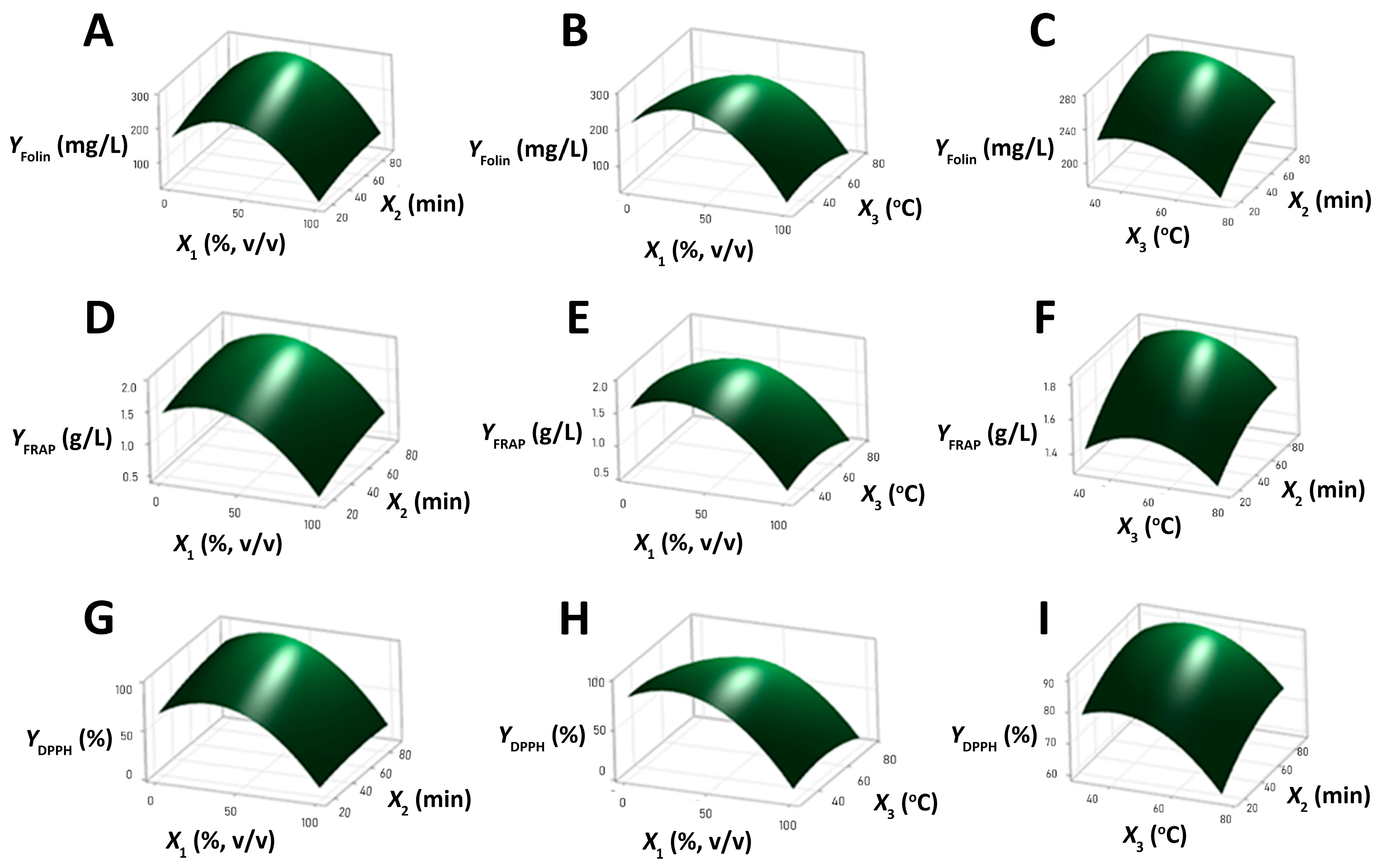
3.2.3. Profiling of Factors Influence Using 3D Response Surface Plots and Pearson Correlation Analysis

Three-dimensional response surface plots were constructed to enhance understanding of the connections between the response variables and the independent factors as well as to highlight the trends toward the optimum extraction conditions. As shown in the surface plots for YFolin (Figure 2A,B), YFRAP (Figure 2D,E), and YDPPH (Figure 2G,H), ethanol content (X1, % v/v) appears to be the most critical parameter for the extraction of antioxidants. Specifically, it seems that low levels of ethanol in the model promote the extraction of antioxidants from the plant. However, extraction efficiency decreases dramatically with higher ethanol content, particularly when it exceeds the middle level (i.e., >50%, v/v). To explain the above phenomena, the polarity of the phenolic components in the plant must be taken into account. U. dioica is known to contain highly polar compounds, like chlorogenic acid and 2-O-caffeoylmalic acid [26]. These polar compounds are easily extracted in low ethanol content, along with less polar phenolic compounds, such as terpenoids and flavonoids. However, when the ethanol content increases, the solubility of the highly polar compounds decreases, resulting in reduced responses [27].
The extraction time (X2, min) appears to have a positive impact on YFolin (Figure 2A,C), YFRAP (Figure 2D,F), and YDPPH (Figure 2G,I). In particular, there is a notable increase in the extraction of antioxidants over time, with the rate of extraction appearing to be almost exponential. This trend continues until it reaches a plateau close to the high-level area (up to 90 min) in the model. Indeed, ultrasound-assisted extraction is known to aid the release of antioxidants from plant materials through the formation of air bubbles, the generation of heat, and the distribution of the components by mechanical agitation. Nevertheless, research has demonstrated that prolonged exposure to ultrasonic waves can lead to the degradation of phenolic compounds [28,29].
The extraction temperature (X3, °C) also has a significant influence on YFolin (Figure 2B,C), YFRAP (Figure 2E,F), and YDPPH (Figure 2H,I), displaying a similar trend with the X1 but with less pronounced effects. Indeed, low heating seems to significantly favor the extraction process. However, when the temperature reaches the middle level (52.5 °C) and beyond (75 °C), a detrimental effect on the recovery of antioxidants becomes evident. The positive effect of low-level X3 on the responses is associated with the fact that under the conventional extraction temperatures of 20 °C to 50 °C, heating favors the mass transfer and solubility of antioxidants. On the other hand, higher temperature values lead to the degradation of phenolic compounds and, consequently, to the reduction in antioxidant activity. The latter effect of high heating is intensified by the combined increase in the extraction time as under these conditions, phenolic compounds are readily oxidized [30].
All the above greatly strengthen the rationale and precision of the proposed model as the antioxidant activity is uniformly influenced by each factor in all the assay methods. It is also remarkable to note that the impact trend of each factor was the same across all three responses (YFolin, YFRAP, and YDPPH), denoting a linear relationship between the changes in the phenol content and the antioxidant activity of the extract. The latter was proved by Pearson’s correlation analysis (Figure 3), which showed a very strong positive relationship between YFolin and YFRAP values (R = 0.98, p = 0.000) as well as between YFolin and YDPPH values (R = 0.98, p = 0.000). This finding validates that the antioxidant activity of the extract is mainly attributable to the presence of phenolic compounds.

3.2.4. Prediction of the Optimum Extraction Conditions

Based on the results of the experimental design, the optimum conditions for the ultrasound-assisted extraction of antioxidants from U. dioica correspond to the use of 34% (v/v) ethanolic solution as a solvent at the extraction temperature of 42 °C for 87 min. These conditions are expected to provide the combined maximization of all three responses, YFolin (285.3 mg/L), YFRAP (1.88 g/L), and YDPPH (97.5%). The further verification of the model took place by performing the extraction process under the optimum conditions. The experimental findings for YFolin (285.8 ± 1.4 mg/L), YFRAP (1.87 ± 0.04 g/L), and YDPPH (94.9 ± 0.4%) fit well with the predicted ones, showcasing the strong validity of the model.
The optimum solvent appears green in nature, as it is primarily composed of water (66%) and a small amount of ethanol (34%). This can be further strengthened by the fact that ethanol, a widely available reagent in chemical, biological, and medical research, can be economically produced via the fermentation of a variety of renewable feedstocks (e.g., sugarcane leaves, cheese whey) [31]. Also, the mild temperature condition of 42 °C, which was found to be optimum, supports the use of low thermal extraction power and, consequently, favors the economic aspects of the process. The latter finding is promising, considering that a recent study using an aqueous mixture of citric acid and lactic acid proposed a high temperature of 70 °C for 60 min as optimum for the extraction of bioactive constituents from the plant [13]. This highlights the positive effect of having an organic solvent present on the reduction in heating energy consumption.
Notably, an optimization study of the antioxidant activity of U. dioica extracts showcased water as the optimum solvent against methanol and ethanol. However, the study did not test the combination of water with the organic solvents or the effect of temperature on the process, resulting in a quite prolonged extraction time of 195 min [14]. In contrast, the current research modeled with RSM all the possible combinations of water and ethanol along with their sole use, while at the same time revealed that mild heating is critical for high extraction efficiency within a shorter amount of time (87 min vs. 195 min). Another investigation that did not take into account the effect of temperature on the process found that methanol, a solvent with a similar relative polarity index (0.762) to ethanol (0.654), is most effective at a concentration of 54% [15]. This indicates that temperature as an additional extraction factor potentially results in a decrease in the organic solvent percentage.

3.3. Scale-Up of the Optimum Extraction Process and Detection of Biomolecules in the Extract

A five-fold scale-up of the extraction process was carried out to validate the results obtained on the small scale. The optimum extraction conditions proposed by the model were employed in order to achieve the maximum antioxidant activity in the U. dioica extract. The optimum extract obtained after the process scale-up was further assessed for its total soluble sugar content, total protein content, and UV–Vis spectrum profile.
Notably, scaling-up of the process did not influence the YFolin (287.2 ± 2.0 mg/L), YFRAP (1.84 ± 0.04 g/L), and YDPPH (94.7 ± 0.3%), supporting the effectiveness of the model at a higher scale. In addition, experimental findings using the Dubois assay highlight that the total soluble sugar content in the U. dioica extract was 3.6 ± 0.0 g/L. According to yjr literature, this level is primarily characterized by the presence of monosaccharides, such as glucose and rhamnose, the disaccharide sucrose, and the sugar alcohol inositol [32]. As for total protein content, a concentration of 1.5 ± 0.0 g/L was determined in the U. dioica extract by the Bradford assay. Previous studies have shown that this plant contains a variety of amino acids, including valine, leucine, isoleucine, glutamic acid, proline, and phenylalanine [32].
The UV–Vis spectrum of the U. dioica extract is illustrated in Figure 4. The peaks at 370 nm and 595 nm are indicative of the presence of chlorophyll, the common dye found in plants. These peaks are attributed to the specific wavelength ranges of 370–450 nm and around 600 nm, which are characteristic of chlorophyll’s absorption spectrum [33]. In addition, the band detected at 430 nm in the plant sample indicates the presence of carotenoids, specifically lutein and β-carotene along with their isomers, which are known as the major categories of carotenoids found in U. dioica [34]. Furthermore, the plant extract showed high absorption bands in the UV region (from 231 to 330 nm). The absorption maximum identified at 330 nm can be attributed to the structure of flavonoid glycosides [35], while the high UV absorption at 231 nm and 290 nm verifies the existence of phenolic acids [36]. The presence of phenolic acids and flavonoids in the extract is in agreement with the literature data focused on the comprehensive characterization of the plant [26,37], confirming the effectiveness of the extraction procedure. Focusing on the main phenolic compounds in the plant, i.e., chlorogenic acid and 2-O-caffeoylmalic acid, they hold strong antioxidant potential against free radicals. Notably, the antioxidant mechanism of chlorogenic acid lies in a single electron transfer followed by proton transfer at 3′OH and 4′OH positions of its structure [38]. On the other hand, the double hydrogen atom transfer mechanism was previously proposed for the antioxidant activity of the caffeic acid ester, 2-O-caffeoylmalic acid [39]. Future research dedicated to the determination of the compositional changes in the optimum extract in the presence of free radicals is expected to provide further insight into the mechanistic aspects of U. dioica antioxidant activity.

3.4. Effect of the Optimum Extract in Red Blood Cells

In physiological conditions, red blood cells are able to self-support a variety of defense mechanisms catalyzed by antioxidant enzymes, like SOD, catalase, glutathione peroxidase, glutathione peroxidase, and glutathione reductase. The action of these enzymes protects the constituents of erythrocytes from free radicals and, in particular, from ROS and RNS. In order to maintain redox homeostasis, this antioxidant defense system prevents oxidative cell damage by balancing the activity of erythrocyte enzymes. ROS (like H2O2) are recognized as toxic intermediates in a wide range of pathologies. In particular, H2O2 has been identified as a key metabolite due to its high stability, diffusion, and role in the signaling pathways of the cell. Therefore, red blood cells have been the primary focus of metabolic activity research, including the impact of H2O2 [21].
The in vitro blood test was conducted to determine the erythrocyte abnormalities in the presence of H2O2 and plant extract. Specifically, in Figure 5A, a microscopic image of sheep blood is illustrated. The red blood cells exhibited normal morphology, appearing spherical and lacking a nucleus. These cells can carry oxygen and nutrients around the body with the aid of hemoglobin and transport carbon dioxide back to the lungs [40]. The total cell count of erythrocytes in the sheep blood was found to be 6.1 × 10⁶ cells/μL.
Figure 5B highlights the changes in the morphology of the sheep blood due to the presence of H2O2 (30 μM). H2O2 is one of the most important ROS, generated as a result of the incomplete reduction in oxygen during cellular metabolism. An unstable level of H2O2 can cause cell membrane damage and eventually lead to the destruction of nucleic acids [41]. The altered morphology of erythrocytes was associated with the formation of echinocytes, which are characterized by the distortion and shrinkage of the cells. As a result, the red blood cells that maintained a normal structure corresponded to 1.9 × 10⁶ cells/μL, i.e., three-fold lower than the initial value. In addition to the detected changes, there might be a release of some key elements (like hemoglobin) with the destruction of the cell membrane. All of the above are responsible for the cause of oxidative stress and, consequently, the development of various diseases [21].
Figure 5C illustrates the structure of the red blood cells after the incubation of sheep blood with H2O2 (30 μM) and the U. dioica extract (3%, v/v). The microscopic image shows similarities with Figure 5A as most of the erythrocytes have remained unaffected and the rest have a slight deformity that is characteristic of elliptocytes morphology. This reflected a total count of normal red blood cells of 4.5 × 10⁶ cells/μL, a level close to the initial one. In comparison with Figure 5B, it seems that the plant extract stabilizes and prevents the destruction of the cell membrane, leading to overall protection from oxidative stress. Up to now, research on the antioxidant activity of U. dioica has been mainly focused on in vivo animal studies, where multiple factors could influence the detected changes [42]. The current study, on the other hand, provides valuable insight into the direct positive effect of the U. dioica extract in red blood cells without the interference of exogenous factors. The future use of the product in in vivo studies would provide information on the contribution of additional biomarkers to the findings.

4. Conclusions

The RSM was successfully implemented for the optimization of the extraction conditions to achieve the maximum antioxidant activity in the extract of the medicinal plant U. dioica. The reliability and good predictability of the model were highlighted by both the R2-adj and R2-pred coefficients for all the responses. The results showed that the most critical factor was the presence of ethanol at low levels in order to achieve the maximum recovery of antioxidants. The principal components that contributed to the antioxidant potential of the extract were the polar phenolic compounds. Other antioxidants present were flavonoid glycosides, carotenoids, and chlorophylls. Furthermore, in vitro blood testing using H2O2 (30 μM) as an oxidative stress factor showed cellular changes in the absence of the plant extract. On the other hand, in the presence of the plant extract (3%, v/v), non-significant changes were detected under oxidative stress conditions due to the protective role of the antioxidant compounds on the erythrocytes. This research provided a new insight into the determinant factors for the optimum extraction of antioxidants from the medicinal U. dioica plant as well as the potential role of the extract itself as an antioxidant product. Further investigation regarding the pharmacokinetics of the plant’s bioactive compounds and the purity/safety of the extract is critical to provide scientific evidence for its potential use as a natural medicine.

Author Contributions

Conceptualization, E.P.; methodology, A.S. and E.P.; software, A.S. and E.P.; formal analysis, A.S.; investigation, A.S.; data curation, A.S. and E.P.; writing—original draft preparation, A.S.; writing—review and editing, E.P.; visualization, A.S.; supervision, E.P. All authors have read and agreed to the published version of the manuscript.

Funding

This research received no external funding.

Institutional Review Board Statement

Not applicable.

Informed Consent Statement

Not applicable.

Data Availability Statement

The original contributions presented in the study are included in the article; further inquiries can be directed to the corresponding author.

Conflicts of Interest

The authors declare no conflicts of interest.

References

  1. Sies, H. Oxidative Stress: Concept and Some Practical Aspects. Antioxidants 2020, 9, 852. [Google Scholar] [CrossRef] [PubMed]
  2. Halliwell, B.; Gutteridge, J.M.C. Free Radicals in Biology and Medicine, 5th ed.; Oxford University Press: Oxford, UK, 2015. [Google Scholar]
  3. Weidinger, A.; Kozlov, A.V. Biological Activities of Reactive Oxygen and Nitrogen Species: Oxidative Stress versus Signal Transduction. Biomolecules 2015, 5, 472–484. [Google Scholar] [CrossRef] [PubMed]
  4. Pizzino, G.; Irrera, N.; Cucinotta, M.; Pallio, G.; Mannino, F.; Arcoraci, V.; Squadrito, F.; Altavilla, D.; Bitto, A. Oxidative Stress: Harms and Benefits for Human Health. Oxidative Med. Cell. Longev. 2017, 2017, 8416763. [Google Scholar] [CrossRef]
  5. Flieger, J.; Flieger, W.; Baj, J.; Maciejewski, R. Antioxidants: Classification, Natural Sources, Activity/Capacity Measurements, and Usefulness for the Synthesis of Nanoparticles. Materials 2021, 14, 4135. [Google Scholar] [CrossRef] [PubMed]
  6. Lévuok-Mena, K.P.; Patiño-Ladino, O.J.; Prieto-Rodríguez, J.A. In Vitro Inhibitory Activities against α-Glucosidase, α-Amylase, and Pancreatic Lipase of Medicinal Plants Commonly Used in Chocó (Colombia) for Type 2 Diabetes and Obesity Treatment. Sci. Pharm. 2023, 91, 49. [Google Scholar] [CrossRef]
  7. Phromnoi, K.; Sinchaiyakij, P.; Khanaree, C.; Nuntaboon, P.; Chanwikrai, Y.; Chaiwangsri, T.; Suttajit, M. Anti-Inflammatory and Antioxidant Activities of Medicinal Plants Used by Traditional Healers for Antiulcer Treatment. Sci. Pharm. 2019, 87, 22. [Google Scholar] [CrossRef]
  8. Kumar, A.; Pradeep, N.; Kumar, M.; Jose, A.; Tomer, V.; Oz, E.; Proestos, C.; Zeng, M.; Elobeid, T.; K, S.; et al. Major Phytochemicals: Recent Advances in Health Benefits and Extraction Method. Molecules 2023, 28, 887. [Google Scholar] [CrossRef]
  9. Said, M. Capacity Development of Human Resource in Local Government to Improve Public Service Quality. J. Ilm. Adm. Publik (JIAP) 2015, 1, 8–13. [Google Scholar] [CrossRef]
  10. Semwal, P.; Rauf, A.; Olatunde, A.; Singh, P.; Zaky, M.Y.; Islam, M.M.; Khalil, A.A.; Aljohani, A.S.M.; Al Abdulmonem, W.; Ribaudo, G. The Medicinal Chemistry of Urtica Dioica L.: From Preliminary Evidence to Clinical Studies Supporting Its Neuroprotective Activity. Nat. Prod. Bioprospect. 2023, 13, 16. [Google Scholar] [CrossRef]
  11. Taheri, Y.; Quispe, C.; Herrera-Bravo, J.; Sharifi-Rad, J.; Ezzat, S.M.; Merghany, R.M.; Shaheen, S.; Azmi, L.; Prakash Mishra, A.; Sener, B.; et al. Urtica Dioica-Derived Phytochemicals for Pharmacological and Therapeutic Applications. Evid.-Based Complement. Altern. Med. 2022, 2022, 4024331. [Google Scholar] [CrossRef]
  12. Bhusal, K.K.; Magar, S.K.; Thapa, R.; Lamsal, A.; Bhandari, S.; Maharjan, R.; Shrestha, S.; Shrestha, J. Nutritional and Pharmacological Importance of Stinging Nettle (Urtica Dioica L.): A Review. Heliyon 2022, 8, E09717. [Google Scholar] [CrossRef]
  13. Koraqi, H.; Qazimi, B.; Khalid, W.; Stanoeva, J.P.; Sehrish, A.; Siddique, F.; Çesko, C.; Ali Khan, K.; Rahim, M.A.; Hussain, I.; et al. Optimized Conditions for Extraction, Quantification and Detection of Bioactive Compound from Nettle (Urtica Dioica L.) Using the Deep Eutectic Solvents, Ultra-Sonication and Liquid Chromatography-Mass Spectrometry (LC-DAD-ESI-MS/MS). Int. J. Food Prop. 2023, 26, 2171–2185. [Google Scholar] [CrossRef]
  14. Flórez, M.; Cazón, P.; Vázquez, M. Antioxidant Extracts of Nettle (Urtica Dioica) Leaves: Evaluation of Extraction Techniques and Solvents. Molecules 2022, 27, 6015. [Google Scholar] [CrossRef] [PubMed]
  15. Vajić, U.J.; Grujić-Milanović, J.; Živković, J.; Šavikin, K.; Godevac, D.; Miloradović, Z.; Bugarski, B.; Mihailović-Stanojević, N. Optimization of Extraction of Stinging Nettle Leaf Phenolic Compounds Using Response Surface Methodology. Ind. Crops Prod. 2015, 74, 912–917. [Google Scholar] [CrossRef]
  16. Singleton, V.L.; Orthofer, R.; Lamuela-Raventós, R.M. Analysis of Total Phenols and Other Oxidation Substrates and Antioxidants by Means of Folin-Ciocalteu Reagent. Methods Enzymol. 1999, 299, 152–178. [Google Scholar]
  17. Chaves, N.; Santiago, A.; Alías, J.C. Quantification of the Antioxidant Activity of Plant Extracts: Analysis of Sensitivity and Hierarchization Based on the Method Used. Antioxidants 2020, 9, 76. [Google Scholar] [CrossRef] [PubMed]
  18. Nguyen, N.H.; Nguyen, M.T.; Nguyen, H.D.; Pham, P.D.; Thach, U.D.; Trinh, B.T.D.; Nguyen, L.T.T.; Dang, S.V.; Do, A.T.; Do, B.H. Antioxidant and Antimicrobial Activities of the Extracts from Different Garcinia Species. Evid.-Based Complement. Altern. Med. 2021, 2021, 5542938. [Google Scholar] [CrossRef]
  19. Dubois, M.; Gilles, K.A.; Hamilton, J.K.; Rebers, P.A.; Smith, F. Colorimetric Method for Determination of Sugars and Related Substances. Anal. Chem. 1956, 28, 350–356. [Google Scholar] [CrossRef]
  20. Papadaki, E.; Roussis, I.G. Assessment of Antioxidant and Scavenging Activities of Various Yogurts Using Different Sample Preparation Procedures. Appl. Sci. 2022, 12, 9283. [Google Scholar] [CrossRef]
  21. Revin, V.V.; Gromova, N.V.; Revina, E.S.; Samonova, A.Y.; Tychkov, A.Y.; Bochkareva, S.S.; Moskovkin, A.A.; Kuzmenko, T.P. The Influence of Oxidative Stress and Natural Antioxidants on Morphometric Parameters of Red Blood Cells, the Hemoglobin Oxygen Binding Capacity, and the Activity of Antioxidant Enzymes. Biomed. Res. Int. 2019, 2019, 2109269. [Google Scholar] [CrossRef]
  22. Gawron-Gzella, A.; Chanaj-Kaczmarek, J.; Cielecka-Piontek, J. Yerba Mate—A Long but Current History. Nutrients 2021, 13, 3706. [Google Scholar] [CrossRef] [PubMed]
  23. Yusoff, I.M.; Mat Taher, Z.; Rahmat, Z.; Chua, L.S. A Review of Ultrasound-Assisted Extraction for Plant Bioactive Compounds: Phenolics, Flavonoids, Thymols, Saponins and Proteins. Food Res. Int. 2022, 157, 111268. [Google Scholar] [CrossRef] [PubMed]
  24. Plaskova, A.; Mlcek, J. New Insights of the Application of Water or Ethanol-Water Plant Extract Rich in Active Compounds in Food. Front. Nutr. 2023, 10, 1118761. [Google Scholar] [CrossRef]
  25. Che Sulaiman, I.S.; Basri, M.; Fard Masoumi, H.R.; Chee, W.J.; Ashari, S.E.; Ismail, M. Effects of Temperature, Time, and Solvent Ratio on the Extraction of Phenolic Compounds and the Anti-Radical Activity of Clinacanthus Nutans Lindau Leaves by Response Surface Methodology. Chem. Cent. J. 2017, 11, 54. [Google Scholar] [CrossRef]
  26. Pinelli, P.; Ieri, F.; Vignolini, P.; Bacci, L.; Baronti, S.; Romani, A. Extraction and HPLC Analysis of Phenolic Compounds in Leaves, Stalks, and Textile Fibers of Urtica Dioica L. J. Agric. Food Chem. 2008, 56, 9127–9132. [Google Scholar] [CrossRef] [PubMed]
  27. Huaman-Castilla, N.L.; Martínez-Cifuentes, M.; Camilo, C.; Pedreschi, F.; Mariotti-Celis, M.; Pérez-Correa, J.R. The Impact of Temperature and Ethanol Concentration on the Global Recovery of Specific Polyphenols in an Integrated HPLE/RP Process on Carménère Pomace Extracts. Molecules 2019, 24, 3145. [Google Scholar] [CrossRef] [PubMed]
  28. Liu, Y.; She, X.R.; Huang, J.B.; Liu, M.C.; Zhan, M.E. Ultrasonic-Extraction of Phenolic Compounds from Phyllanthus Urinaria: Optimization Model and Antioxidant Activity. Food Sci. Technol. 2018, 38, 286–293. [Google Scholar] [CrossRef]
  29. Rashad, S.; El-Chaghaby, G.; Lima, E.C.; Simoes, G.; Reis, D. Optimizing the Ultrasonic-Assisted Extraction of Antioxidants from Ulva Lactuca Algal Biomass Using Factorial Design. Biomass. Convers. Biorefin. 2021, 13, 5681–5690. [Google Scholar] [CrossRef]
  30. Dai, J.; Mumper, R.J. Plant Phenolics: Extraction, Analysis and Their Antioxidant and Anticancer Properties. Molecules 2010, 15, 7313–7352. [Google Scholar] [CrossRef]
  31. Sharma, S.; Kundu, A.; Basu, S.; Shetti, N.P.; Aminabhavi, T.M. Sustainable Environmental Management and Related Biofuel Technologies. J. Environ. Manag. 2020, 273, 111096. [Google Scholar] [CrossRef]
  32. Dhouibi, R.; Affes, H.; Ben Salem, M.; Hammami, S.; Sahnoun, Z.; Zeghal, K.M.; Ksouda, K. Screening of Pharmacological Uses of Urtica Dioica and Others Benefits. Prog. Biophys. Mol. Biol. 2020, 150, 67–77. [Google Scholar] [CrossRef]
  33. Kume, A. Importance of the Green Color, Absorption Gradient, and Spectral Absorption of Chloroplasts for the Radiative Energy Balance of Leaves. J. Plant Res. 2017, 130, 501–514. [Google Scholar] [CrossRef]
  34. Guil-Guerrero, J.L.; Rebolloso-Fuentes, M.M.; Torija Isasa, M.E. Fatty Acids and Carotenoids from Stinging Nettle (Urtica Dioica L.). J. Food Compos. Anal. 2003, 16, 111–119. [Google Scholar] [CrossRef]
  35. Akbay, P.; Basaran, A.A.; Undeger, U.; Basaran, N. In Vitro Immunomodulatory Activity of Flavonoid Glycosides from Urtica Dioica L. Phytother. Res. 2003, 17, 34–37. [Google Scholar] [CrossRef]
  36. Repajić, M.; Cegledi, E.; Kruk, V.; Pedisić, S.; Çınar, F.; Bursać Kovačević, D.; Žutić, I.; Dragović-Uzelac, V. Accelerated Solvent Extraction as a Green Tool for the Recovery of Polyphenols and Pigments from Wild Nettle Leaves. Processes 2020, 8, 803. [Google Scholar] [CrossRef]
  37. Tarasevičienė, Ž.; Vitkauskaitė, M.; Paulauskienė, A.; Černiauskienė, J. Wild Stinging Nettle (Urtica Dioica L.) Leaves and Roots Chemical Composition and Phenols Extraction. Plants 2023, 12, 309. [Google Scholar] [CrossRef]
  38. Izu, G.O.; Njoya, E.M.; Tabakam, G.T.; Nambooze, J.; Otukile, K.P.; Tsoeu, S.E.; Fasiku, V.O.; Adegoke, A.M.; Erukainure, O.L.; Mashele, S.S.; et al. Unravelling the Influence of Chlorogenic Acid on the Antioxidant Phytochemistry of Avocado (Persea Americana Mill.) Fruit Peel. Antioxidants 2024, 13, 456. [Google Scholar] [CrossRef] [PubMed]
  39. Lu, L.; Luo, K.; Luan, Y.; Zhao, M.; Wang, R.; Zhao, X.; Wu, S. Effect of Caffeic Acid Esters on Antioxidant Activity and Oxidative Stability of Sunflower Oil: Molecular Simulation and Experiments. Food Res. Int. 2022, 160, 111760. [Google Scholar] [CrossRef] [PubMed]
  40. Gell, D.A. Structure and Function of Haemoglobins. Blood Cells Mol Dis 2018, 70, 13–42. [Google Scholar] [CrossRef]
  41. Gaikwad, R.; Thangaraj, P.R.; Sen, A.K. Direct and Rapid Measurement of Hydrogen Peroxide in Human Blood Using a Microfluidic Device. Sci. Rep. 2021, 11, 2960. [Google Scholar] [CrossRef]
  42. Jaiswal, V.; Lee, H.J. Antioxidant Activity of Urtica Dioica: An Important Property Contributing to Multiple Biological Activities. Antioxidants 2022, 11, 2494. [Google Scholar] [CrossRef] [PubMed]
Figure 1. Pareto charts of the standardized main and interaction effects of ethanol content (X1, % v/v), extraction time (X2, min), and extraction temperature (X3, °C) on (A) YFolin (mg/L), (B) YFRAP (g/L), and (C) YDPPH (%). Dark and light blue horizontal bars correspond to the positive and negative effects of responses, respectively. The symbols placed on the right side of the reference line (vertical red bar) correspond to statistically significant effects (p < 0.05).
Scipharm 92 00045 g001
Figure 2. Three-dimensional surface plots for YFolin (mg/L) (AC), (B) YFRAP (g/L) (DF), and (C) YDPPH (%) (GI) as a function of ethanol content (X1, % v/v), extraction time (X2, min), and extraction temperature (X3, °C). In all cases, the third factor was kept constant at its middle level.
Scipharm 92 00045 g002
Figure 3. Pearson’s correlation between (A) total polar phenol content and antioxidant activity using the FRAP assay and (B) total polar phenol content and antioxidant activity using the DPPH assay.
Scipharm 92 00045 g003
Figure 4. UV–Vis spectrum of the 10-fold diluted extract of Urtica dioica L. (ultrasound-assisted extraction with 34% v/v ethanol at 42 °C for 87 min) recorded in the region 200–800 nm.
Scipharm 92 00045 g004
Figure 5. Microscopic image of (A) sheep blood, (B) sheep blood supplemented with H2O2 (30 μM), as well as (C) sheep blood supplemented with H2O2 (30 μM) and plant extract (3%, v/v) at 100× magnification.
Scipharm 92 00045 g005
Table 1. Coded and uncoded values of factors used in the Response Surface Methodology design for the extraction of plant antioxidants.
FactorVariablesLevels
Coded Values a
−101
Actual Values
Ethanol content (%, v/v)X1050100
Extraction time (min)X21552.590
Extraction temperature (°C)X33052.575
a  C o d e d v a l u e = a c t u a l l e v e l [ ( h i g h l e v e l + l o w l e v e l ) / 2 ] ( h i g h l e v e l + l o w l e v e l ) / 2 .
Table 2. Complex central composite design to investigate the optimal combination of the independent variables (X1, X2, X3) and mean value of the responses (YFolin, YFRAP, YDPPH).
RunFactorResponse (Experimental/Predicted)
X1X2X3YFolinYFRAPYDPPH
15052.552.5253.2/259.91.68/1.7390.0/87.8
25052.552.5294.6/259.91.86/1.7394.5/87.8
3052.552.5192.8/205.61.49/1.6175.4/77.6
409075169.0/163.31.37/1.3563.2/61.0
55052.552.5288.2/259.91.86/1.7383.3/87.8
65052.552.5256.4/259.91.74/1.7390.7/87.8
7509052.5262.4/270.61.81/1.8191.1/89.5
809030222.0/225.81.61/1.6080.9/84.5
9501552.5199.2/225.81.40/1.5270.3/78.6
105052.552.5271.4/259.91.73/1.7390.6/87.8
115052.530265.0/256.61.71/1.6585.0/86.2
1201530180.8/183.41.37/1.3975.3/71.7
13100903055.6/60.50.70/0.7810.2/8.5
14100157519.0/6.50.33/0.314.1/−1.1
155052.552.5265.2/259.91.73/1.7390.7/87.8
165052.570173.0/216.21.32/1.5165.4/70.9
17100303028.0/24.90.36/0.345.5/6.0
1810052.552.549.6/71.60.75/0.7413.7/18.2
1901575123.0/109.31.33/1.2241.8/41.8
20100907565.2/53.80.72/0.676.0/7.9
Table 3. Second-order polynomial equations, in terms of uncoded units, fitted for YFolin, YFRAP, and YDPPH.
Polynomial Equations R2adjR2pred
YFolin = 110.4 + 2.908X1 + 1.33X2 + 3.17X3 − 0.04851X12(3)92.9782.58
YFRAP = 0.708 + 0.01026X1 + 0.00835X2 − 0.000221X12(4)94.8283.05
YDPPH = 45.7 + 0.805X1 + 0.389X2 + 1.209X3 − 0.01594X12 − 0.01813X32 +0.00507X1X3(5)97.5290.42
Disclaimer/Publisher’s Note: The statements, opinions and data contained in all publications are solely those of the individual author(s) and contributor(s) and not of MDPI and/or the editor(s). MDPI and/or the editor(s) disclaim responsibility for any injury to people or property resulting from any ideas, methods, instructions or products referred to in the content.

