Article

Mycobacterium avium subsp. paratuberculosis Proteome Changes Profoundly in Milk

by
Kristina J. H. Kleinwort
1,
Bernhard F. Hobmaier
1,
Ricarda Mayer
2,
Christina Hölzel
2,3,
Roxane L. Degroote
1,
Erwin Märtlbauer
2,
Stefanie M. Hauck
4 and
Cornelia A. Deeg
1,*
1
Chair of Physiology, Department of Veterinary Sciences, LMU Munich, D-82152 Martinsried, Germany
2
Chair of Hygiene and Technology of Milk, Department of Veterinary Sciences, LMU Munich, D-85764 Oberschleißheim, Germany
3
Institute of Animal Breeding and Husbandry, Faculty of Agricultural and Nutritional Sciences, CAU Kiel, D-24098 Kiel, Germany
4
Research Unit Protein Science, Helmholtz Center Munich, German Research Center for Environmental Health, D-80939 Munich, Germany
*
Author to whom correspondence should be addressed.
Metabolites 2021, 11(8), 549; https://doi.org/10.3390/metabo11080549
Submission received: 21 July 2021 / Revised: 10 August 2021 / Accepted: 18 August 2021 / Published: 20 August 2021
(This article belongs to the Special Issue Multi-Omics Methods in Dairy Research)

Abstract

:
Mycobacterium avium subspecies paratuberculosis (MAP) are detectable viable in milk and other dairy products. The molecular mechanisms allowing the adaptation of MAP in these products are still poorly understood. To obtain information about respective adaptation of MAP in milk, we differentially analyzed the proteomes of MAP cultivated for 48 h in either milk at 37 °C or 4 °C or Middlebrook 7H9 broth as a control. From a total of 2197 MAP proteins identified, 242 proteins were at least fivefold higher in abundance in milk. MAP responded to the nutritional shortage in milk with upregulation of 32% of proteins with function in metabolism and 17% in fatty acid metabolism/synthesis. Additionally, MAP upregulated clusters of 19% proteins with roles in stress responses and immune evasion, 19% in transcription/translation, and 13% in bacterial cell wall synthesis. Dut, MmpL4_1, and RecA were only detected in MAP incubated in milk, pointing to very important roles of these proteins for MAP coping with a stressful environment. Dut is essential and plays an exclusive role for growth, MmpL4_1 for virulence through secretion of specific lipids, and RecA for SOS response of mycobacteria. Further, 35 candidates with stable expression in all conditions were detected, which could serve as targets for detection. Data are available via ProteomeXchange with identifier PXD027444.

1. Introduction

Mycobacterium avium subsp. paratuberculosis (MAP) is the causative agent of paratuberculosis or Johne’s disease, a chronic granulomatous enteritis of cattle and small ruminants [1,2]. Due to its remarkable similarity to the pathology of Crohn’s disease in humans, it has been discussed for a long time as a possible cause of human morbidity, especially chronic inflammatory bowel diseases (IBD) such as Crohn’s disease and ulcerative colitis [3,4,5,6]. Although there is no definitive proof of MAP being involved in the pathogenesis of the human diseases, it is broadly accepted in the scientific community that human exposure to MAP, especially through contaminated milk and milk products, should be reduced [7]. MAP can either be shed directly into milk by infected cows or be introduced via fecal contamination [8,9,10,11]. Viable MAP were found in pasteurized milk, cheese, and even in dried milk products such as infant formula [12,13,14]. This might have been caused by post-processing contamination, but there is also evidence of MAP being able to survive the pasteurization process [15,16,17,18,19]. The mechanisms behind this tenacity, however, are not fully understood and have been vigorously discussed over the last years [8,20]. Theories include clumping, intracellular localization of MAP in milk, and formation of heat-resistant forms such as endospores [16,21,22].
It is common for bacteria to sense environmental changes and react to it with various stress response mechanisms [23]. Milk in particular is a rather hostile environment for bacteria, due to several mechanisms that inhibit bacterial growth such as the iron binding protein lactoferrin, antimicrobial peptides, and different nutrient sources compared to the bacteria’s physiological environment (i.e., lactose instead of glucose) [24,25]. Significant proteome changes after cultivation in milk compared to cultivation in a standard medium were shown for various bacteria, including Escherichia coli, Lactococcus lactis, and Streptococcus thermophilus [24,26,27]. Therefore, it is not unlikely for MAP to also adapt to such altered environmental conditions by changing several of its physiological properties. A good example for this environment-dependent variation are cell wall-deficient forms of MAP that were repeatedly isolated from human intestines [6,28,29,30]. In a previous study, we showed that after incubation of MAP with peripheral blood mononuclear cell (PBMC) of cows with different immunological phenotypes, the exoproteome of MAP changed according to the immunological phenotype of the leukocytes used for coincubation [31]. For milk, altered properties of MAP were shown by an in vitro experiment, in which incubation of MAP in milk led to faster translocation through a monolayer of Madin–Darby bovine kidney epithelial cells (MDBK) cells [32]. In addition to this, MAP cultured in milk had distinctly different gene expression profiles compared to those cultured in broth (different cell wall lipid profile and enhanced expression of cell invasion-associated genes), suggesting milk as a valid transmission route for MAP to calves and humans [33]. However, changes in gene expression do not necessarily always mean equivalent changes in the proteome. Therefore, it is important to validate those findings on a protein level. Understanding how the MAP proteome changes after being incubated in milk may elucidate the role of milk in MAP transmission, shed light to the question of why MAP can survive pasteurization, and may lead to new possible targets for the detection of MAP in milk and milk products. Therefore, in this study, we investigated possible differential proteome changes in MAP after incubation in milk at 37 °C (simulating the situation in the mammary gland) and 4 °C (for tank milk) in comparison to MAP from M7H9 broth at 37 °C.

2. Results

2.1. Identification of MAP Proteome with Label-Free Mass Spectrometry

To analyze if Mycobacterium avium ssp. paratuberculosis (MAP) changes its protein expression through incubation in milk, we performed a differential proteomics experiment using quantitative liquid chromatography–tandem mass spectrometry (LC–MS/MS) analysis of MAP incubated in Middlebrook 7H9 (M7H9) standard cell culture medium and milk at 37 °C and at 4 °C. Milk at 37 °C simulated the condition in cow’s udder, and milk at 4 °C the simulated condition in bulk tank. To eliminate the highly abundant milk proteins prior to measurement, we refiltrated the MAP samples after incubation with a novel method. A total of 2470 proteins were identified (Table S1), divided into 2197 MAP proteins and 273 bovine proteins. Of these 2197 MAP proteins, 785 (36%) were classified as “hypothetical proteins” in the gene bank entries (Figure 1). For further analyses, we searched and added the gene IDs for these candidates (Table S2).

2.2. Highly Differential MAP Proteomes after Incubation in Standard Medium and Milk

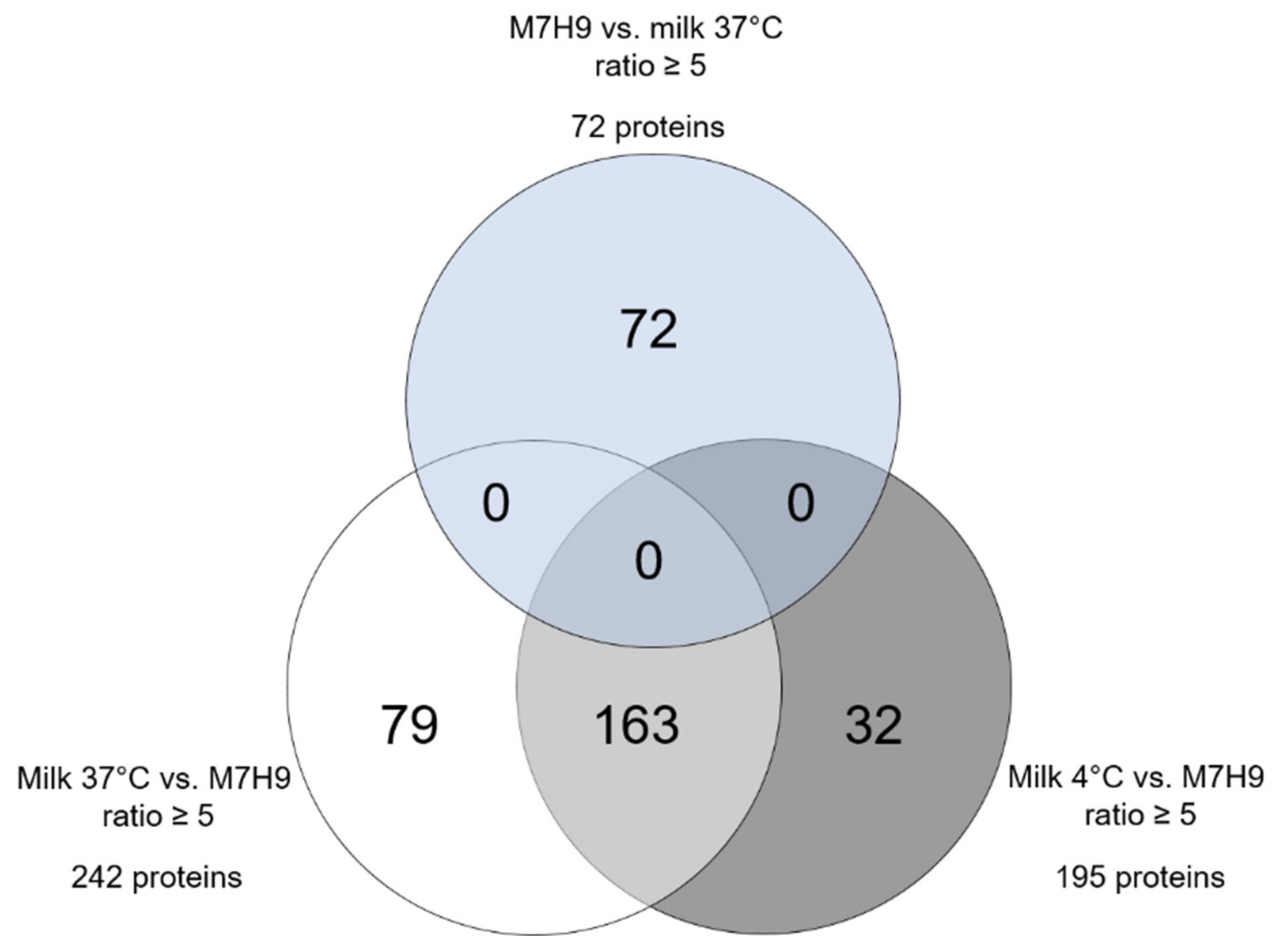
A large difference between proteomes of MAP incubated in milk and in M7H9 became evident, whereas the temperature of the milk exerted a less strong effect on differential protein expression (Figure 2). In total, 274 MAP proteins were fivefold higher abundant in MAP incubated in milk at both temperatures compared to MAP from M7H9 broth (Figure 2, white and gray circles). Therein, 195 proteins were higher abundant in MAP from milk at 4 °C compared to the MAP proteome in M7H9 medium (Figure 2, gray circle). A similar difference was detected between the proteome of MAP incubated in milk at 37 °C and MAP in medium, revealing 242 proteins that were at least five times higher in their abundance in milk at 37 °C (Figure 2, white circle). Out of those proteins, the majority (163 proteins; Figure 2, light gray) were higher in abundance in both milk samples, whereas 32 proteins were exclusively enriched in milk at 4 °C (Figure 2, dark gray) and 79 proteins only in milk at 37 °C (Figure 2, white). In contrast to the 274 higher abundant MAP proteins in milk, with 91 proteins, only one-third as many proteins showed fivefold increased abundance after incubation in M7H9 broth compared to milk at both temperatures. In more detail, 72 proteins were higher abundant in medium compared to milk at 37 °C (Figure 2, light blue) and 54 proteins compared to milk at 4 °C (data not shown).

2.3. Functional Affiliation of Differential MAP Proteins

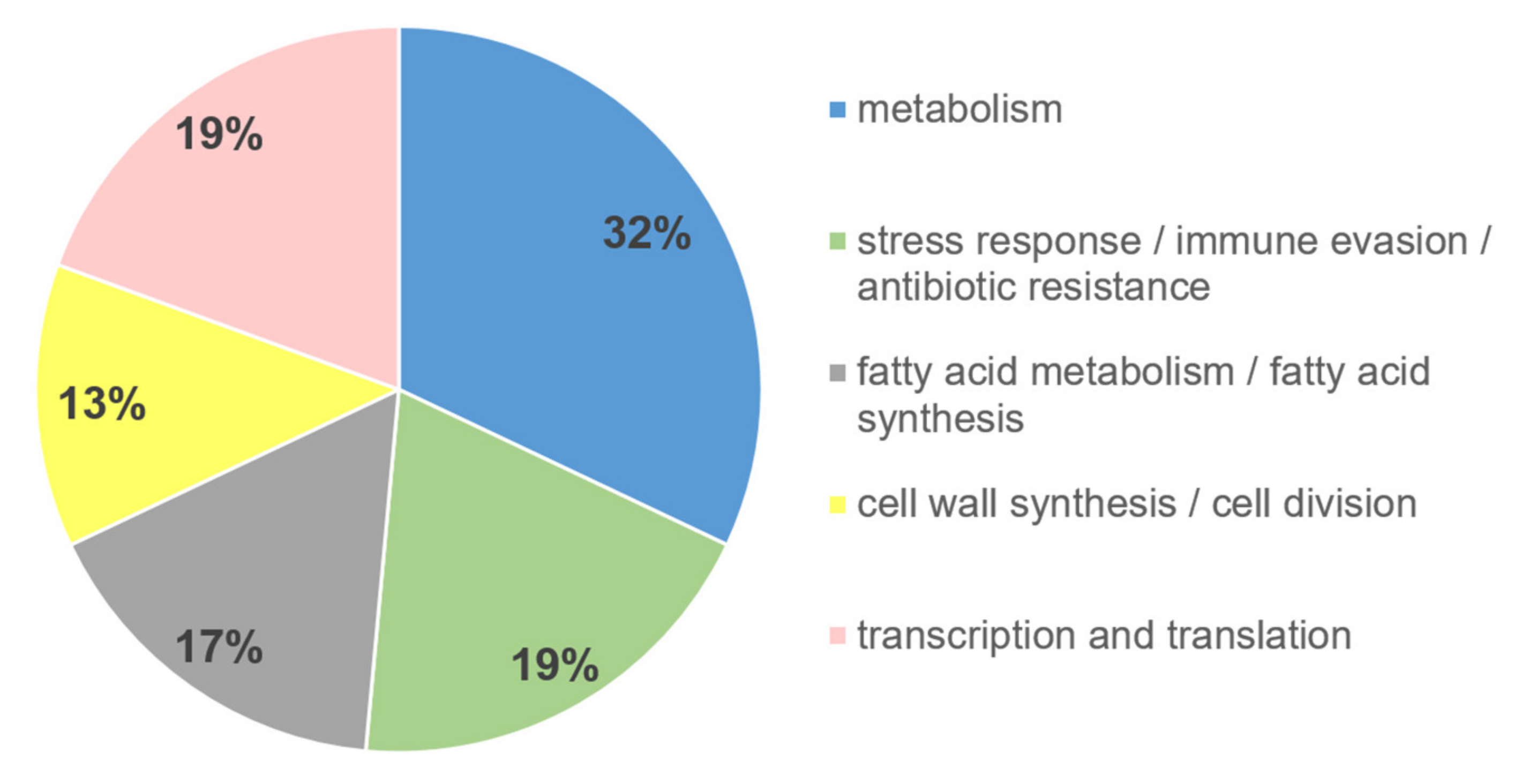
To further analyze the functional impact of the differentially (ratio ≥ 5) higher abundant MAP proteins in milk compared to M7H9 medium, we assigned the proteins to their function. The analysis revealed that 32% of the proteins were assigned to the function metabolism (Figure 3, blue), 19% to each stress response/immune evasion/antibiotic resistance (Figure 3, green) and to transcription/translation (Figure 3, pink), 17% to fatty acid metabolism/synthesis (Figure 3, gray), and 13% to cell wall synthesis/cell division (Figure 3, yellow).
Table 1 provides an overview of the top three proteins from each functional affiliation.

2.4. Stably Expressed Proteins as Possible Targets for Detection Methods

In order to find a possible target molecule for MAP detection in food, we screened the respective proteomes for molecules, which were stably expressed in all three environments tested. Thus, a group of 35 proteins that were highly abundant (>20 peptide counts) on one hand and quite equally expressed (ratio ≤ 2) in all three conditions on the other hand were identified (Table 2).

3. Discussion

MAP infections in ruminants reduce milk production and lead to premature removal as well as reduced proceeds from subsequent sales of respective MAP-infected animals. MAP belong to the slow-growing non-tuberculous mycobacteria (NTM) species and can cause opportunistic infections in animals and humans [34,35]. However, MAP is also described as a food-borne pathogen [6], thought to be mainly transmitted via milk and dairy products [7,14].
Since bacteria generally sense environmental changes and respond with various mechanisms [23], we were interested in changes in the MAP proteome after incubation in milk. With our study, we provide a comprehensive characterization of MAP proteome cultured in short-time high-temperature, defatted, and casein partially removed milk. The aim of this pretreatment procedure was to remove bacteria in order to avoid interference and to allow unambiguous assigning of proteins. Fat had to be removed because it impaired the filtration process. In milk of most species, approximately 97% of the fat is present in fat globules, which consist of triglycerides surrounded by a membrane [36]. Fat globules, but not other minor lipids such as cholesterol, phospholipids, diacylglycerol, and free fatty acids, were removed by the pretreatment, thus resembling conditions comparable to skimmed milk. The fact that caseins were detectable in the mass spectrometry data set (Table S1) indicates that a significant proportion of casein micelles were able to pass the filter. Thus, the experimental outcomes should also not have been influenced by lack of casein. According to the literature, heat-treated skimmed milk offers favorable conditions for metabolic activity of lactic acid bacteria [37]. Therefore, we did not expect major differences in the conditions for bacterial metabolism between our pretreated milk and milk in field conditions. Using label-free LC–MS/MS, we investigated changes in the MAP proteome after incubation in milk at 37 °C and 4 °C in comparison to MAP incubated in M7H9 broth at 37 °C in this study. Milk at 37 °C should simulate the condition in the bovine udder and milk at the 4 °C condition in the milk bulk tank, while M7H9 broth served as comparison as a standard cell culture medium for MAP. In this approach, a total of 2470 proteins were identified (Table S1). Therein, 273 proteins were from bovine origin and 2197 were MAP proteins, from which 785 were classified as “hypothetical proteins” in the gene bank entries (Figure 1). Hypothetical proteins are predicted to be expressed from an open reading frame, but there is no experimental evidence of translation into proteins and the associated function [38]. The expression of the 785 hypothetical proteins in MAP in vivo was thus confirmed by our study for the first time and underlines the analytical depth of the mass spectrometric analysis. Hypothetical proteins can be crucial for the pathogen survival and the severity of the associated disease in the host [39,40]. For many microorganisms, amongst others for Mycobacterium tuberculosis [41], hypothetical proteins were identified to have great importance, as they could serve as potential drug targets or vaccine candidates [42,43]. Thus, the high number of hypothetical proteins identified in our study also holds high potential as new candidates for detection methods of MAP and therefore adds important novel information about this genus on the protein level. Looking at the differentially expressed (ratio ≥ 5) MAP proteins between the three different approaches, we found a clear quantitative difference between the MAP proteome incubated in milk and in M7H9 medium. In total, 274 MAP proteins were at least fivefold higher in abundance in milk at both temperatures compared to M7H9 broth (Figure 2, white and gray circles), whereas after incubation in M7H9 broth, only one-third as many proteins (91 proteins) showed at least fivefold increase of abundance compared to milk at both temperatures. Interestingly, the temperature of the milk had less influence on the proteome changes (Figure 2). Thus, from the 274 differentially higher expressed MAP proteins in milk, with 163 proteins, the major part was higher abundant in both milk samples, irrespective of the temperature (Figure 2, light gray).
Since we were particularly interested in how MAP adapts to the milk environment, we looked more closely at the functions of the differentially higher abundant MAP proteins in milk. The major part of those proteins was assigned to the function cell metabolism (Figure 3, blue, 32%). Deoxyuridine 5′-triphosphate nucleotidohydrolase (Dut) was exclusively detected in MAP incubated in milk, but not in broth. The intact Dut protein is essential in NTM mycobacteria and plays an exclusive role for biosynthesis and growth [44].
In Saccharomyces cerevisiae, the inactivation of Dut was lethal because of genome instability [45]. We do not know why MAP upregulated Dut when cultivated in milk, but the concomitant higher abundance of key metabolic enzymes NAD-dependent aldehyde dehydrogenase AldA_1 and the uridylate kinase PyrH point to a stimulation of metabolic pathways.
In mammals, Alda_1 activates mitochondrial aldehyde dehydrogenase 2 (ALDH2), which is generally known for its protective functions [46]. Thus, ALDH2 attenuates oxidative damage of various cell types in humans, including cardiac, lung, and brain endothelial cells and hepatic cells, and it also reduces damage in various organs in animal models [46,47,48,49,50,51,52]. Inhibition of oxidative stress responses in hemorrhagic shock of alveolar damage and of immune cell infiltration were also observed through increased ALDH2 activity via AldA_1 in animal models [50,53]. Taken together, these data could point to stress for MAP in milk environment, resulting in upregulation of AldA_1 as a means to protect themselves against antimicrobial components of milk [24,25]. It is likely that the increased AldA_1 expression in MAP results in attenuating the immune responses. Interestingly, AldA_1 was found in high abundance in tylosin-resistant S. xylosus compared with tylosin-sensitive strains [54]. This relationship between increased AldA_1 expression in other bacteria and antibiotic resistance is also of special interest in the context of increased AldA_1 expression of MAP in milk. Another differentially regulated MAP protein allocated to the function metabolism was uridylate kinase (PyrH; Table 1; ratio milk 4 °C/M7H9: 21.2; ratio milk 37 °C/M7H9: 8.8), which catalyzes the phosphorylation of UMP to UDP, further serving as a precursor in the pyrimidine biosynthetic pathway [55]. Kim et al. found the PyrH gene expressed in vivo in Vibrio vulnificus during infection in humans [56]. They showed that PyrH inactivation reduced virulence of the bacteria [56]. Further research of this group confirmed an essential role for PyrH for the in vivo survival and growth of Vibrio vulnificus [57]. A study comparing the available bacterial genomes revealed that the PyrH gene is highly conserved between bacteria with no counterpart in eukaryotes [55]. In this context, the distinct higher expression of PyrH in MAP after incubation in milk also points to upregulation of immune evasion responses by MAP.
Another 19% of the differentially higher abundant MAP proteins in milk were assigned to the functional affiliations stress response/immune evasion/antibiotic resistance. The top three candidates were RecA (Table 1; ratio milk 4 °C/M7H9 and milk 37 °C/M7H9: infinite), RelA (Table 1; ratio milk 4 °C/M7H9: 313.2; ratio milk 37 °C/M7H9: 8371.5), and SecE (Table 1; ratio milk 4 °C/M7H9: 26.5; ratio milk 37 °C/M7H9: 25.4). RecA is important for DNA damage repair [58]. SecE translocates proteins across the plasma membrane [59]. RelA plays a central role in the ability of MAP to establish a persistent infection [60]. Deletion of RelA led to immune elimination of the mutant MAP [60].
Candidates enriched from transcription/translation pathway, DnAE, as well as XthA and SigG, belong to the stressome of MAP [61]. XthA acts on damaged DNA in bacterial base excision repair [62]. SigG is upregulated by DNA-damaging agents and by macrophage infection [63].
Top enriched candidates from fatty acid metabolism and synthesis were FadD5 (Table 1; ratio milk 4 °C/M7H9: 122.9; milk 37 °C/M7H9: 103.1), which may serve to recycle mycolic acids for the long-term survival of the tubercle bacilli [64], MmpL4_1 and LipN. MmpL4_1 (Table 1; ratio milk 4 °C/M7H9 and milk 37 °C/M7H9: infinite) is important for virulence of mycobacteria through secretion of specific lipids and participates in export of mycobacterium cell wall components to the bacterial surface [65,66]. LipN (Table 1; ratio milk 4 °C/M7H9: 6.0; milk 37 °C/M7H9: 5.9) is a fatty acid degradation lipase/esterase that was shown to be significantly upregulated in MAP shed in cow feces [61].
CrgA (Table 1; ratio milk 4 °C/M7H9: 14.1; milk 37 °C/M7H9: 18.5), MmpL11 (Table 1; ratio milk 4 °C/M7H9: 12.2; milk 37 °C/M7H9: 13.5), and YidC (Table 1; ratio milk 4 °C/M7H9: 6.7; milk 37 °C/M7H9: 7.4) were the top enriched candidates from cell wall synthesis/cell division. CrgA is related to peptidoglycan biosynthesis and cell shape maintenance [67]. MmpL11 is a conserved transporter of mycolic acid-containing lipids, which was shown to be important for biofilm formation in M. tuberculosis and M. smegmatis, as well as in non-replicating persistence in M. tuberculosis [68]. The membrane protein insertase YidC is required for the insertion and proper folding and complex formation of integral membrane proteins into the membrane of many bacteria [69].
Our analyses demonstrated significant changes in MAP proteome after cultivation in milk for 48 h. Higher abundant proteins have functions in protection and repair of MAP and point to a stressome reaction in milk. Milk was apparently sensed as a hostile environment and therefore induced respective counteractions by MAP.
Significant proteome changes after cultivation in milk compared to cultivation in a standard medium have already been shown for Escherichia coli, Lactococcus lactis, and Streptococcus thermophilus [24,26,27]. Our study confirms similar responses of MAP in milk and provides in-depth information about respective stress response of MAP in milk. Respective candidate proteins and pathways deserve further investigations to better understand interactions of MAP with milk. The next experiments should clarify if and how MAP survive in this environment over longer periods of time, since the survival of MAP in dairy products is unwanted. The better understanding of factors that prevent persistence of MAP could aid to inhibit resistance strategies of MAP. Perhaps subsequent analyses could also explain environment-dependent variations such as cell wall-deficient forms of MAP that were repeatedly isolated from human intestines [28,29,30].
Another focus of our study was on identifying possible candidate proteins for new detection methods for MAP, as there is still a lack of a rapid and sensitive detection method for MAP [70]. The gold standard for MAP detection is currently cultivation on agar [71]. This method has the advantage of detecting only live MAP, but takes weeks to produce a result, which is not suitable for detection in food [72]. Detection by PCR provides rapid results and is very sensitive, but it cannot distinguish between live and dead MAP [71,73]. In addition, false positive identifications may occur due to identification of non-MAP mycobacteria [74]. Thus, there is a need for rapid, sensitive, and highly specific detection of MAP, both in livestock and food. The solution could be an antibody-based assay. The candidate protein sought for this should ideally meet the following conditions: highly abundant expression in all conditions tested, MAP-specific epitope present, and being located at the bacterial surface. For this purpose, we first evaluated the most abundant proteins from the data set that were uniformly highly expressed between all conditions (ratio ≤ 2). This analysis yielded 35 proteins (Table 2), which will now be further investigated to identify MAP-specific conserved candidate proteins. In silico, all proteins with at least two unique peptides can be translated into DNA sequences and aligned with all contained total genomes and sequences of the Mycobacterium avium complex (MAC) in order to identify sequences that differ significantly from those of other MAC representatives unequal to MAP. In addition, the candidate protein should preferably be expressed at the MAP surface, as this would greatly facilitate the detection of this bacterium.

4. Materials and Methods

4.1. Bacteria

Mycobacterium avium subsp. paratuberculosis (MAP) strain DSM 44133 was obtained from the German Collection of Microorganisms and Cell Cultures (DSMZ, Braunschweig, Germany). MAP were cultivated for four weeks on Herrold’s egg yolk agar (HEYM) (BD Biosciences, Heidelberg, Germany).

4.2. Preparation of Milk

For defatting, fresh milk (1.5% fat, short time high temperature (72 °C, 20 s)) was centrifuged (20 min, 2000× g, 4 °C), and the resulting fat layer was removed. After filtration with a paper filter, milk was additionally filtered with 0.45 µm followed by 0.2 µm filters, partially removing casein. For contamination, control milk was incubated at 37 °C for 24 h and streaked out on blood agar, which was incubated at 37 °C for another 48 h. No microbial growth was detected.

4.3. Incubation of MAP in Liquids

MAP were harvested from HEYM agar by careful flushing with PBS. After washing three times with PBS (10 min, 16,000× g), 2 × 10⁹ cfu/mL MAP were inoculated into two aliquots of the prepared milk and in Middlebrook 7H9 (M7H9; Sigma-Aldrich, Taufkirchen, Germany) medium as liquid control medium. One preparation of the artificially contaminated milk and the control medium preparation were incubated at 37 °C, and the other aliquot of milk at 4 °C for 48 h. Milk at 37 °C should simulate the condition in the bovine udder and the milk at the 4 °C condition in the milk bulk tank, while M7H9 broth served as comparison as a standard cell culture medium for MAP.

4.4. Harvesting and Refiltration of MAP

All three assays were shortly warmed up to 38 °C in order to perform the following refiltration with as little pressure as possible. For refiltration of MAP, all preparations were first filtrated through 0.45 µm filters, followed by washing the filters two times with PBS. To obtain the MAP collected in the filters, we backflushed the corresponding filters (in opposite direction) with PBS. Extracted MAP-PBS suspension was centrifuged (30 min, 2500× g) and remaining supernatants were again centrifuged (10 min, 16,000× g) to make sure that all MAP were sedimented. Finally, related MAP pellets were pooled and washed three times with PBS (10 min, 16,000× g).

4.5. Lysis of MAP Samples

MAP pellets were resuspended in lysis buffer (1% Nonidet P-40, 10 mM NaCl, 10 mM Tris-HCl (pH 7.6)). For additional breakup of the robust MAP cell wall, samples were treated with silica beads (0.1 mm; 40 mg beads/100 µL lysate) in Ribolyser (Hybaid, Teddington, UK; level 6, 3 × 30 s). Heating (74 °C, 5 min) followed by ultrasonic treatment (6 × 30 s on ice) of samples gave maximum split-up.

4.6. Sample Preparation for LC–MS/MS Mass Spectrometry

A total of 10 µg total protein was digested with LysC and trypsin by a modified filter-aided sample preparation (FASP) [75] as follows. To every lysate, 1 µL 1 M dithiothreitol was added and incubated for 30 min at 60 °C. After cooling down, samples were diluted 1:3 in urea (UA)-buffer (8 M urea and 0.1 M Tris-HCl (pH 8.5) diluted in HPLC-grade water) and incubated with 10 µL of 300 mM iodoacetamide for 30 min at room temperature in the dark. After addition of 2 µL 1 M dithiothreitol, samples were transferred to 30 kDa cut-off centrifuge filters (Sartorius, Göttingen, Germany) and centrifuged (15 min, 14,000× g), followed by three washing steps with each 200 µL UA-buffer and 100 µL ammoniumbicarbonate (ABC) buffer (50 mM diluted in HPLC-grade water). Afterwards, the proteins were subjected to proteolysis with 1 µg Lys C (Lysyl Endopeptidase C) in 40 µL ABC buffer at 37 °C overnight. After centrifugation (15 min, 15,000× g) over new tubes and renewed washing with 20 µL ABC buffer, collected Lys-C fractions were acidified with 0.5% trifluoroacetic acid to pH 2 and frozen at −20 °C. Filter were subsequently incubated with 2 µg trypsin in 50 µL of ABC buffer at 37 °C overnight. Peptides of tryptic digestion were collected by centrifugation (15 min, 15,000× g) and additional washing with 20 µL ABC buffer containing 5% acetonitrile, and were also finally acidified with 0.5% trifluoroacetic acid to pH 2.

4.7. Mass Spectrometry

Acidified eluted peptides were analyzed in the data-dependent mode on a Q Exactive HF mass spectrometer (Thermo Fisher Scientific, Bremen, Germany) online coupled to a UItimate 3000 RSLC nano-HPLC (Dionex, Idstein, Germany). Samples were automatically injected and loaded onto the C18 trap column, and after 5 min eluted and separated on the C18 analytical column (75 µm ID × 15 cm, Acclaim PepMAP 100 C18. 100Å/size, LC Packings, Thermo Fisher Scientific, Bremen, Germany) by a 90 min non-linear acetonitrile gradient at a flow rate of 250 nL/min. MS spectra were recorded at a resolution of 60,000, and after each MS1 cycle, the 10 most abundant peptide ions were selected for fragmentation.

4.8. Protein Identification and Label-Free Quantification

Acquired MS spectra were imported into Progenesis software (version 2.5, Nonlinear Dynamics, Waters, Oslo, Norway) and analyzed as previously described [76,77]. After alignment, peak picking, exclusion of features with charge state of 1 and >7 and normalization, spectra were exported as Mascot Generic files and searched against a database containing all entries of Mycobacterium avium subspecies paratuberculosis from NCBI Protein database combined with the Ensembl bovine database (version 80) with Mascot (Matrix Science, Version 2.5.1). Search parameters used were 10 ppm peptide mass tolerance, 20 mmu fragment mass tolerance, one missed cleavage allowed, carbamidomethylation set as fixed modification, and methionine oxidation and deamidation of asparagine and glutamine as variable modifications. Mascot integrated decoy database search was set to a false discovery rate (FDR) of 1% when searching was performed on the concatenated mgf files with a percolator ion score cut-off of 13 and an appropriate significance threshold p. Peptide assignment was reimported to Progenesis Software. All unique peptides allocated to a protein were considered for quantification.

4.9. Data Analysis

Proteins with a ratio of at least fivefold in normalized abundance between samples were defined as differentially expressed. Venn diagram was calculated with Venny 2.1 open source tool (https://bioinfogp.cnb.csic.es/tools/venny/ (accessed on 14 May 2021)). To determine the function of the differentially expressed proteins (ratio ≥ 5) between milk at both temperatures and M7H9 broth, we first manually searched the respective proteins by accession number at Ensembl Bacteria (release 51). The functions of the domains indicated there were subsequently determined via InterPro (release 85.0; http://www.ebi.ac.uk/interpro/ (accessed on 20 May 2021)) and finally illustrated in a pie chart.

5. Conclusions

MAP proteomes from MAP incubated in milk show distinct modifications to this environment. The majority of changed proteins point to a stress response of MAP and a subsequent reaction to cope with nutrient changes and the hostile environment milk, leading to DNA damage and other impairments in MAP. However, there were also stably expressed proteins detected in this study that could be suited as targets for MAP detection.

Supplementary Materials

The following data are available online at https://www.mdpi.com/article/10.3390/metabo11080549/s1, Table S1: All identified proteins, Table S2: Hypothetical MAP proteins.

Author Contributions

Conceptualization, C.H., E.M. and C.A.D.; methodology, K.J.H.K., C.H., S.M.H. and C.A.D.; formal analysis, K.J.H.K., B.F.H., R.L.D. and S.M.H.; investigation, K.J.H.K. and R.M.; writing—original draft preparation, K.J.H.K. and B.F.H.; writing—review and editing, C.A.D.; visualization, K.J.H.K. and B.F.H.; supervision, C.H., E.M. and C.A.D.; project administration, C.H., E.M. and C.A.D.; funding acquisition, E.M. and C.A.D. All authors have read and agreed to the published version of the manuscript.

Funding

This research was funded by the German Ministry of Economics and Technology (via AiF) and the FEI, Project AiF 18388 N.

Institutional Review Board Statement

Not applicable.

Informed Consent Statement

Not applicable.

Data Availability Statement

The data presented in this study are available in Table S1: All identified proteins. The mass spectrometry proteomics data have been deposited to the ProteomeXchange Consortium via the PRIDE [78] partner repository with the dataset identifier PXD027444.

Acknowledgments

The authors would like to thank Andrea Stockmaier-Didier for critical discussions.

Conflicts of Interest

The authors declare no conflict of interest. The funders had no role in the design of the study; in the collection, analyses, or interpretation of data; in the writing of the manuscript; or in the decision to publish the results.

References

  1. Twort, F.W.; Ingram, G.L.Y. Further Experiments with the Mycobacterium Enteritidis Chronicæ Pseudo-Tuberculosæ Bovis, Johne, and with Vaccines Prepared from this Micro-Organism. Vet. J. 1900 1913, 69, 4–15. [Google Scholar] [CrossRef]
  2. Twort, F.W.; Ingram, G.L.Y. A method for isolating and cultivating the mycobacterium enteritidis chronicæ pseudotuberculosæ bovis, Jöhne, and some experiments on the preparation of a diagnostic vaccine for pseudo-tuberculous enteritis of bovines. Proc. R. Soc. B 1912, 84, 517–542. [Google Scholar] [CrossRef] [Green Version]
  3. Eslami, M.; Shafiei, M.; Ghasemian, A.; Valizadeh, S.; Al-Marzoqi, A.H.; Shokouhi Mostafavi, S.K.; Nojoomi, F.; Mirforughi, S.A. Mycobacterium avium paratuberculosis and Mycobacterium avium complex and related subspecies as causative agents of zoonotic and occupational diseases. J. Cell. Physiol. 2019, 234, 12415–12421. [Google Scholar] [CrossRef]
  4. Sechi, L.A.; Edow, C.T. Mycobacterium avium ss. paratuberculosis Zoonosis—The Hundred Year War—Beyond Crohn’s Disease. Front. Immunol. 2015, 6, 96. [Google Scholar] [CrossRef] [Green Version]
  5. Van der Sloot, K.W.J.; Voskuil, M.D.; Blokzijl, T.; Dinkla, A.; Ravesloot, L.; Visschedijk, M.C.; van Dullemen, H.M.; Festen, E.A.M.; Alizadeh, B.Z.; van Leer-Buter, C.; et al. Isotype specific antibody responses to Mycobacterium Avium subspecies Paratuberculosis antigens are associated with the use of biological therapy in Inflammatory Bowel Disease. J. Crohns Colitis 2020. [Google Scholar] [CrossRef]
  6. Aitken, J.M.; Phan, K.; Bodman, S.E.; Sharma, S.; Watt, A.; George, P.M.; Agrawal, G.; Tie, A.B.M. A Mycobacterium species for Crohn’s disease? Pathology 2021. [Google Scholar] [CrossRef]
  7. Kuenstner, J.T.; Naser, S.; Chamberlin, W.; Borody, T.; Graham, D.Y.; McNees, A.; Hermon-Taylor, J.; Hermon-Taylor, A.; Dow, C.T.; Thayer, W.; et al. The Consensus from the Mycobacterium avium ssp. paratuberculosis (MAP) Conference 2017. Front. Public Health 2017, 5, 208. [Google Scholar] [CrossRef] [PubMed]
  8. Mullan, W.M.A. Are we closer to understanding why viable cells of Mycobacterium avium subsp. paratuberculosis are still being reported in pasteurised milk? Int. J. Dairy Technol. 2019, 72, 332–344. [Google Scholar] [CrossRef]
  9. Rani, S.; Beaver, A.; Schukken, Y.H.; Pradhan, A.K. Modeling the effects of infection status and hygiene practices on Mycobacterium avium subspecies paratuberculosis contamination in bulk tank milk. Food Control 2019, 104, 367–376. [Google Scholar] [CrossRef]
  10. Sweeney, R.W.; Whitlock, R.H.; Rosenberger, A.E. Mycobacterium paratuberculosis cultured from milk and supramammary lymph nodes of infected asymptomatic cows. J. Clin. Microbiol. 1992, 30, 166–171. [Google Scholar] [CrossRef] [PubMed] [Green Version]
  11. Nigsch, A.; Robbe-Austerman, S.; Stuber, T.P.; Pavinski Bitar, P.D.; Gröhn, Y.T.; Schukken, Y.H. Who infects whom?—Reconstructing infection chains of Mycobacterium avium ssp. paratuberculosis in an endemically infected dairy herd by use of genomic data. PLoS ONE 2021, 16, e0246983. [Google Scholar] [CrossRef]
  12. Botsaris, G.; Swift, B.M.; Slana, I.; Liapi, M.; Christodoulou, M.; Hatzitofi, M.; Christodoulou, V.; Rees, C.E. Detection of viable Mycobacterium avium subspecies paratuberculosis in powdered infant formula by phage-PCR and confirmed by culture. Int. J. Food Microbiol. 2016, 216, 91–94. [Google Scholar] [CrossRef]
  13. Grant, I.R. Bacteriophage-Based Methods for Detection of Viable Mycobacterium avium subsp. paratuberculosis and Their Potential for Diagnosis of Johne’s Disease. Front. Vet. Sci. 2021, 8, 632498. [Google Scholar] [CrossRef] [PubMed]
  14. Hanifian, S. Behavior of Mycobacterium avium paratuberculosis in Lighvan cheese tracked by propidium monoazide qPCR and culture. LWT 2020, 133, 109886. [Google Scholar] [CrossRef]
  15. Chiodini, R.J.; Hermon-Taylor, J. The Thermal Resistance of Mycobacterium Paratuberculosis in Raw Milk under Conditions Simulating Pasteurization. J. Vet. Diagn. Investig. 1993, 5, 629–631. [Google Scholar] [CrossRef] [Green Version]
  16. Gerrard, Z.E.; Swift, B.M.C.; Botsaris, G.; Davidson, R.S.; Hutchings, M.R.; Huxley, J.N.; Rees, C.E.D. Survival of Mycobacterium avium subspecies paratuberculosis in retail pasteurised milk. Food Microbiol. 2018, 74, 57–63. [Google Scholar] [CrossRef]
  17. Grant, I.R.; Ball, H.J.; Rowe, M.T. Effect of higher pasteurization temperatures, and longer holding times at 72 °C, on the inactivation ofMycobacterium paratuberculosisin milk. Lett. Appl. Microbiol. 1999, 28, 461–465. [Google Scholar] [CrossRef] [PubMed]
  18. Paolicchi, F.; Cirone, K.; Morsella, C.; Gioffré, A. First isolation of Mycobacterium avium subsp Paratuberculosis from commercial pasteurized milk in Argentina. Braz. J. Microbiol. 2012, 43, 1034–1037. [Google Scholar] [CrossRef] [PubMed]
  19. Fechner, K.; Dreymann, N.; Schimkowiak, S.; Czerny, C.-P.; Teitzel, J. Efficacy of dairy on-farm high-temperature, short-time pasteurization of milk on the viability of Mycobacterium avium ssp. paratuberculosis. J. Dairy Sci. 2019, 102, 11280–11290. [Google Scholar] [CrossRef]
  20. Robertson, R.E.; Cerf, O.; Condron, R.J.; Donaghy, J.A.; Heggum, C.; Jordan, K. Review of the controversy over whether or not Mycobacterium avium subsp. paratuberculosis poses a food safety risk with pasteurised dairy products. Int. Dairy J. 2017, 73, 10–18. [Google Scholar] [CrossRef]
  21. Grant, I.R.; Williams, A.G.; Rowe, M.T.; Muir, D.D. Efficacy of Various Pasteurization Time-Temperature Conditions in Combination with Homogenization on Inactivation of Mycobacterium avium subsp. paratuberculosis in Milk. Appl. Environ. Microbiol. 2005, 71, 2853–2861. [Google Scholar] [CrossRef] [PubMed] [Green Version]
  22. Lamont, E.A.; Bannantine, J.P.; Armién, A.; Ariyakumar, D.S.; Sreevatsan, S. Identification and Characterization of a Spore-Like Morphotype in Chronically Starved Mycobacterium avium Subsp. Paratuberculosis Cultures. PLoS ONE 2012, 7, e30648. [Google Scholar] [CrossRef] [Green Version]
  23. Guo, M.S.; Gross, C.A. Stress-Induced Remodeling of the Bacterial Proteome. Curr. Biol. 2014, 24, R424–R434. [Google Scholar] [CrossRef] [Green Version]
  24. Lippolis, J.D.; Bayles, D.O.; Reinhardt, T.A. Proteomic Changes in Escherichia coli When Grown in Fresh Milk versus Laboratory Media. J. Proteome Res. 2009, 8, 149–158. [Google Scholar] [CrossRef]
  25. Um, J.; Manguy, J.; Anes, J.; Jacquier, J.-C.; Hurley, D.; Dillon, E.T.; Wynne, K.; Fanning, S.; O’Sullivan, M.; Shields, D.C. Enriching antimicrobial peptides from milk hydrolysates using pectin/alginate food-gels. Food Chem. 2021, 352, 129220. [Google Scholar] [CrossRef]
  26. Derzelle, S.; Bolotin, A.; Mistou, M.-Y.; Rul, F. Proteome Analysis of Streptococcus thermophilus Grown in Milk Reveals Pyruvate Formate-Lyase as the Major Upregulated Protein. Appl. Environ. Microbiol. 2005, 71, 8597–8605. [Google Scholar] [CrossRef] [PubMed] [Green Version]
  27. Gitton, C.; Meyrand, M.; Wang, J.; Caron, C.; Trubuil, A.; Guillot, A.; Mistou, M.-Y. Proteomic Signature of Lactococcus lactis NCDO763 Cultivated in Milk. Appl. Environ. Microbiol. 2005, 71, 7152–7163. [Google Scholar] [CrossRef] [Green Version]
  28. Chiodini, R.J.; Van Kruiningen, H.J.; Thayer, W.R.; Coutu, J.A. Spheroplastic phase of mycobacteria isolated from patients with Crohn’s disease. J. Clin. Microbiol. 1986, 24, 357–363. [Google Scholar] [CrossRef] [PubMed] [Green Version]
  29. Schwartz, D.; Shafran, I.; Romero, C.; Piromalli, C.; Biggerstaff, J.; Naser, N.; Chamberlin, W.; Naser, S.A. Use of short-term culture for identification of Mycobacterium avium subsp. paratuberculosis in tissue from Crohn’s disease patients. Clin. Microbiol. Infect. 2000, 6, 303–307. [Google Scholar] [CrossRef] [Green Version]
  30. Sechi, L.A.; Scanu, A.M.; Molicotti, P.; Cannas, S.; Mura, M.; Dettori, G.; Fadda, G.; Zanetti, S. Detection and isolation of Mycobacterium avium subspecies paratuberculosis from intestinal mucosal biopsies of patients with and without Crohn’s disease in Sardinia. Am. J. Gastroenterol. 2005, 100, 1529–1536. [Google Scholar] [CrossRef]
  31. Kleinwort, K.J.H.; Hauck, S.M.; Degroote, R.L.; Scholz, A.M.; Hölzel, C.; Maertlbauer, E.P.; Deeg, C. Peripheral blood bovine lymphocytes and MAP show distinctly different proteome changes and immune pathways in host-pathogen interaction. PeerJ 2019, 7, e8130. [Google Scholar] [CrossRef] [PubMed]
  32. Bermudez, L.E.; Petrofsky, M.; Sommer, S.; Barletta, R.G. Peyer’s Patch-Deficient Mice Demonstrate That Mycobacterium avium subsp. paratuberculosis Translocates across the Mucosal Barrier via both M Cells and Enterocytes but Has Inefficient Dissemination. Infect. Immun. 2010, 78, 3570–3577. [Google Scholar] [CrossRef] [Green Version]
  33. Alonso-Hearn, M.; Eckstein, T.M.; Sommer, S.; Bermudez, L.E. A Mycobacterium avium subsp. paratuberculosis LuxR regulates cell envelope and virulence. Innate Immun. 2010, 16, 235–247. [Google Scholar] [CrossRef] [PubMed] [Green Version]
  34. Johansen, M.D.; Herrmann, J.-L.; Kremer, L. Non-tuberculous mycobacteria and the rise of Mycobacterium abscessus. Nat. Rev. Microbiol. 2020, 18, 392–407. [Google Scholar] [CrossRef]
  35. Richter, E.; Wessling, J.; Lügering, N.; Domschke, W.; Rüsch-Gerdes, S. Mycobacterium aviumsubsp.paratuberculosisInfection in a Patient with HIV, Germany. Emerg. Infect. Dis. 2002, 8, 729–731. [Google Scholar] [CrossRef]
  36. Zhu, Z.; Guo, W. Recent developments on rapid detection of main constituents in milk: A review. Crit. Rev. Food Sci. Nutr. 2021, 61, 312–324. [Google Scholar] [CrossRef] [PubMed]
  37. Zhang, Y.-H.; Xu, D.; Liu, J.-Q.; Zhao, X.-H. Enhanced degradation of five organophosphorus pesticides in skimmed milk by lactic acid bacteria and its potential relationship with phosphatase production. Food Chem. 2014, 164, 173–178. [Google Scholar] [CrossRef]
  38. Ijaq, J.; Malik, G.; Kumar, A.; Das, P.S.; Meena, N.; Bethi, N.; Sundararajan, V.S.; Suravajhala, P. A model to predict the function of hypothetical proteins through a nine-point classification scoring schema. BMC Bioinform. 2019, 20, 14. [Google Scholar] [CrossRef]
  39. Kumar, K.; Prakash, A.; Tasleem, M.; Islam, A.; Ahmad, F.; Hassan, M.I. Functional annotation of putative hypothetical proteins from Candida dubliniensis. Gene 2014, 543, 93–100. [Google Scholar] [CrossRef]
  40. Desler, C.; Suravajhala, P.; Sanderhoff, M.; Rasmussen, M.; Rasmussen, L.J. In Silico screening for functional candidates amongst hypothetical proteins. BMC Bioinform. 2009, 10, 289. [Google Scholar] [CrossRef] [Green Version]
  41. Yang, Z.; Zeng, X.; Tsui, S.K.-W. Investigating function roles of hypothetical proteins encoded by the Mycobacterium tuberculosis H37Rv genome. BMC Genom. 2019, 20, 394. [Google Scholar] [CrossRef] [PubMed] [Green Version]
  42. Aguttu, C.; Okech, B.A.; Mukisa, A.; Lubega, G.W. Screening and characterization of hypothetical proteins of Plasmodium falciparum as novel vaccine candidates in the fight against malaria using reverse vaccinology. J. Genet. Eng. Biotechnol. 2021, 19, 103. [Google Scholar] [CrossRef]
  43. Sette, A.; Rappuoli, R. Reverse Vaccinology: Developing Vaccines in the Era of Genomics. Immunity 2010, 33, 530–541. [Google Scholar] [CrossRef] [PubMed] [Green Version]
  44. Griffin, J.E.; Gawronski, J.D.; De Jesus, M.A.; Ioerger, T.R.; Akerley, B.J.; Sassetti, C.M. High-Resolution Phenotypic Profiling Defines Genes Essential for Mycobacterial Growth and Cholesterol Catabolism. PLOS Pathog. 2011, 7, e1002251. [Google Scholar] [CrossRef] [Green Version]
  45. Guillet, M.; Van Der Kemp, P.A.; Boiteux, S. dUTPase activity is critical to maintain genetic stability in Saccharomyces cerevisiae. Nucleic Acids Res. 2006, 34, 2056–2066. [Google Scholar] [CrossRef] [PubMed]
  46. Zhang, T.; Zhao, Q.; Ye, F.; Huang, C.Y.; Chen, W.-M.; Huang, W.-Q. Alda-1, an ALDH2 activator, protects against hepatic ischemia/reperfusion injury in rats via inhibition of oxidative stress. Free Radic. Res. 2018, 52, 629–638. [Google Scholar] [CrossRef]
  47. Perez-Miller, S.; Younus, H.; Vanam, R.; Chen, C.-H.; Mochly-Rosen, D.; Hurley, T.D. Alda-1 is an agonist and chemical chaperone for the common human aldehyde dehydrogenase 2 variant. Nat. Struct. Mol. Biol. 2010, 17, 159–164. [Google Scholar] [CrossRef] [Green Version]
  48. Chen, C.-H.; Ferreira, J.C.; Gross, E.R.; Mochly-Rosen, D. Targeting Aldehyde Dehydrogenase 2: New Therapeutic Opportunities. Physiol. Rev. 2014, 94, 1–34. [Google Scholar] [CrossRef] [Green Version]
  49. Fu, S.-H.; Zhang, H.-F.; Yang, Z.B.; Li, T.-B.; Liu, B.; Lou, Z.; Ma, Q.-L.; Luo, X.-J.; Peng, J. Alda-1 reduces cerebral ischemia/reperfusion injury in rat through clearance of reactive aldehydes. Naunyn-Schmiedebergs Arch. Pharmacol. 2014, 387, 87–94. [Google Scholar] [CrossRef]
  50. Lu, Q.; Mundy, M.; Chambers, E.; Lange, T.; Newton, J.; Borgas, D.; Yao, H.; Choudhary, G.; Basak, R.; Oldham, M.; et al. Alda-1 Protects Against Acrolein-Induced Acute Lung Injury and Endothelial Barrier Dysfunction. Am. J. Respir. Cell Mol. Biol. 2017, 57, 662–673. [Google Scholar] [CrossRef] [PubMed]
  51. Zhu, Q.; He, G.; Wang, J.; Wang, Y.; Chen, W. Pretreatment with the ALDH2 agonist Alda-1 reduces intestinal injury induced by ischaemia and reperfusion in mice. Clin. Sci. 2017, 131, 1123–1136. [Google Scholar] [CrossRef] [Green Version]
  52. Patil, S.S.; Hernández-Cuervo, H.; Fukumoto, J.; Narala, V.R.; Saji, S.; Borra, M.; Alleyn, M.; Lin, M.; Soundararajan, R.; Lockey, R.; et al. Alda-1 attenuates hyperoxia-induced mitochondrial dysfunction in lung vascular endothelial cells. Aging 2019, 11, 3909–3918. [Google Scholar] [CrossRef] [PubMed]
  53. Hua, T.; Yang, M.; Zhou, Y.; Chen, L.; Wu, H.; Liu, R. Alda-1 Prevents Pulmonary Epithelial Barrier Dysfunction following Severe Hemorrhagic Shock through Clearance of Reactive Aldehydes. BioMed Res. Int. 2019, 2019, 2476252. [Google Scholar] [CrossRef] [Green Version]
  54. Liu, X.; Wang, J.; Chen, M.; Che, R.; Ding, W.; Yu, F.; Zhou, Y.; Cui, W.; Xiaoxu, X.; God’Spower, B.-O.; et al. Comparative proteomic analysis reveals drug resistance of Staphylococcus xylosus ATCC700404 under tylosin stress. BMC Vet. Res. 2019, 15, 224. [Google Scholar] [CrossRef]
  55. Briozzo, P.; Evrin, C.; Meyer, P.; Assairi, L.; Joly, N.; Bārzu, O.; Gilles, A.-M. Structure of Escherichia coli UMP Kinase Differs from That ofOther Nucleoside Monophosphate Kinases and Sheds New Light on EnzymeRegulation. J. Biol. Chem. 2005, 280, 25533–25540. [Google Scholar] [CrossRef] [Green Version]
  56. Kim, Y.R.; Lee, S.E.; Kim, C.M.; Kim, S.Y.; Shin, E.K.; Shin, D.H.; Chung, S.S.; Choy, H.E.; Progulske-Fox, A.; Hillman, J.D.; et al. Characterization and Pathogenic Significance of Vibrio vulnificus Antigens Preferentially Expressed in Septicemic Patients. Infect. Immun. 2003, 71, 5461–5471. [Google Scholar] [CrossRef] [Green Version]
  57. Lee, S.E.; Kim, S.Y.; Kim, C.M.; Kim, M.-K.; Kim, Y.R.; Jeong, K.; Ryu, H.-J.; Lee, Y.S.; Chung, S.S.; Choy, H.E.; et al. The pyrH Gene of Vibrio vulnificus Is an Essential In Vivo Survival Factor. Infect. Immun. 2007, 75, 2795–2801. [Google Scholar] [CrossRef] [PubMed] [Green Version]
  58. Brzostek, A.; Płociński, P.; Minias, A.; Ciszewska, A.; Gąsior, F.; Pawełczyk, J.; Dziadek, B.; Słomka, M.; Dziadek, J. Dissecting the RecA-(In)dependent Response to Mitomycin C in Mycobacterium tuberculosis Using Transcriptional Profiling and Proteomics Analyses. Cells 2021, 10, 1168. [Google Scholar] [CrossRef]
  59. Knyazev, D.G.; Kuttner, R.; Bondar, A.-N.; Zimmerman, M.; Siligan, C.; Pohl, P. Voltage Sensing in Bacterial Protein Translocation. Biomolecules 2020, 10, 78. [Google Scholar] [CrossRef] [PubMed] [Green Version]
  60. Abdellrazeq, G.S.; Elnaggar, M.M.; Bannantine, J.P.; Park, K.T.; Souza, C.D.; Backer, B.; Hulubei, V.; Fry, L.M.; Khaliel, S.A.; Torky, H.A.; et al. A Mycobacterium avium subsp. paratuberculosis relA deletion mutant and a 35 kDa major membrane protein elicit development of cytotoxic T lymphocytes with ability to kill intracellular bacteria. Vet. Res. 2018, 49, 53. [Google Scholar] [CrossRef] [PubMed] [Green Version]
  61. Wu, C.-W.; Schmoller, S.K.; Shin, S.J.; Talaat, A.M. Defining the Stressome of Mycobacterium avium subsp. paratuberculosis In Vitro and in Naturally Infected Cows. J. Bacteriol. 2007, 189, 7877–7886. [Google Scholar] [CrossRef] [Green Version]
  62. Khanam, T.; Rai, N.; Ramachandran, R. Mycobacterium tuberculosis class II apurinic/apyrimidinic-endonuclease/3′-5′ exonuclease III exhibits DNA regulated modes of interaction with the sliding DNA β-clamp. Mol. Microbiol. 2015, 98, 46–68. [Google Scholar] [CrossRef] [PubMed]
  63. Gaudion, A.; Dawson, L.; Davis, E.; Smollett, K. Characterisation of the Mycobacterium tuberculosis alternative sigma factor SigG: Its operon and regulon. Tuberculosis 2013, 93, 482–491. [Google Scholar] [CrossRef] [Green Version]
  64. Dunphy, K.Y.; Senaratne, R.H.; Masuzawa, M.; Kendall, L.V.; Riley, L.W. Attenuation ofMycobacterium tuberculosisFunctionally Disrupted in a Fatty Acyl–Coenzyme A Synthetase GenefadD5. J. Infect. Dis. 2010, 201, 1232–1239. [Google Scholar] [CrossRef] [PubMed] [Green Version]
  65. Domenech, P.; Reed, M.B.; Barry, C.E., 3rd. Contribution of the Mycobacterium tuberculosis MmpL Protein Family to Virulence and Drug Resistance. Infect. Immun. 2005, 73, 3492–3501. [Google Scholar] [CrossRef] [Green Version]
  66. Danelishvili, L.; Chinison, J.J.J.; Pham, T.; Gupta, R.; Bermudez, L.E. The Voltage-Dependent Anion Channels (VDAC) of Mycobacterium avium phagosome are associated with bacterial survival and lipid export in macrophages. Sci. Rep. 2017, 7, 7007. [Google Scholar] [CrossRef] [Green Version]
  67. Wang, R.; Ehrt, S. Rv0954 Is a Member of the Mycobacterial Cell Division Complex. Front. Microbiol. 2021, 12, 626461. [Google Scholar] [CrossRef]
  68. Melly, G.C.; Stokas, H.; Davidson, P.M.; Roma, J.S.; Rhodes, H.L.; Purdy, G.E. Identification of residues important for M. tuberculosis MmpL11 function reveals that function is modulated by phosphorylation in the C-terminal domain. Mol. Microbiol. 2021, 115, 208–221. [Google Scholar] [CrossRef]
  69. McDowell, M.A.; Heimes, M.; Sinning, I. Structural and molecular mechanisms for membrane protein biogenesis by the Oxa1 superfamily. Nat. Struct. Mol. Biol. 2021, 28, 234–239. [Google Scholar] [CrossRef] [PubMed]
  70. Hobmaier, B.F.; Lutterberg, K.; Kleinwort, K.J.H.; Mayer, R.; Hirmer, S.; Amann, B.; Hölzel, C.; Märtlbauer, E.P.; Deeg, C.A. Characterization of plant lectins for their ability to isolate Mycobacterium avium subsp. paratuberculosis from milk. Food Microbiol. 2019, 82, 231–239. [Google Scholar] [CrossRef]
  71. Slana, I.; Paolicchi, F.; Janštová, B.; Navrátilová, P.; Pavlík, I. Detection methods for Mycobacterium avium subsp paratuberculosis in milk and milk products: A review. Veterinární Med. 2008, 53, 283–306. [Google Scholar] [CrossRef] [Green Version]
  72. Sweeney, R.W.; Collins, M.T.; Koets, A.P.; McGuirk, S.M.; Roussel, A.J. Paratuberculosis (Johne’s Disease) in Cattle and Other Susceptible Species. J. Vet. Intern. Med. 2012, 26, 1239–1250. [Google Scholar] [CrossRef] [PubMed]
  73. Soumya, M.P.; Pillai, R.M.; Antony, P.X.; Mukhopadhyay, H.K.; Rao, V.N. Comparison of faecal culture and IS900 PCR assay for the detection of Mycobacterium avium subsp. paratuberculosis in bovine faecal samples. Vet. Res. Commun. 2009, 33, 781–791. [Google Scholar] [CrossRef] [PubMed]
  74. Park, H.-T.; Park, H.-E.; Jung, Y.-H.; Yoo, H.S. An ISMap 02—Like insertion sequence in Mycobacterium spp. interferes with specific detection of Mycobacterium avium subsp. paratuberculosis. Vet. Microbiol. 2018, 216, 1–6. [Google Scholar] [CrossRef]
  75. Grosche, A.; Hauser, A.; Lepper, M.F.; Mayo, R.; von Toerne, C.; Merl-Pham, J.; Hauck, S.M. The Proteome of Native Adult Müller Glial Cells from Murine Retina. Mol. Cell. Proteom. 2016, 15, 462–480. [Google Scholar] [CrossRef] [PubMed] [Green Version]
  76. Hauck, S.M.; Hofmaier, F.; Dietter, J.; Swadzba, M.E.; Blindert, M.; Amann, B.; Behler, J.; Kremmer, E.; Ueffing, M.; Deeg, C.A. Label-free LC-MSMS analysis of vitreous from autoimmune uveitis reveals a significant decrease in secreted Wnt signalling inhibitors DKK3 and SFRP2. J. Proteom. 2012, 75, 4545–4554. [Google Scholar] [CrossRef] [PubMed]
  77. Hauck, S.M.; Lepper, M.F.; Hertl, M.; Sekundo, W.; Deeg, C.A. Proteome Dynamics in Biobanked Horse Peripheral Blood Derived Lymphocytes (PBL) with Induced Autoimmune Uveitis. Proteomics 2017, 17, 1700013. [Google Scholar] [CrossRef]
  78. Perez-Riverol, Y.; Csordas, A.; Bai, J.; Bernal-Llinares, M.; Hewapathirana, S.; Kundu, D.J.; Inuganti, A.; Griss, J.; Mayer, G.; Eisenacher, M.; et al. The PRIDE database and related tools and resources in 2019: Improving support for quantification data. Nucleic Acids Res. 2019, 47, D442–D450. [Google Scholar] [CrossRef]
Figure 1. Overview of identified proteins. Of the total of 2470 proteins identified, 237 were classified as bovine and 2197 were linked to MAP. Furthermore, 785 of identified MAP proteins were previously not described on a proteomic level and therefore thus far classified as hypothetical proteins.
Metabolites 11 00549 g001
Figure 2. Differentially abundant (ratio ≥ 5) MAP proteins between milk and M7H9 medium. After incubation in milk at 37 °C (white circle) and 4 °C (gray circle), a total of 274 MAP proteins were at least fivefold higher in abundance compared to incubation in M7H9 broth. Most of the proteins (163, light gray) were enriched in milk at both temperatures, while 79 showed increased abundance only in milk at 37 °C (white) and 32 proteins only in milk at 4 °C (dark gray). In contrast, 72 proteins were at least fivefold higher abundant in M7H9 broth compared to milk at 37 °C (blue).
Metabolites 11 00549 g002
Figure 3. Functional affiliation of higher abundant MAP proteins in milk. The proteins fivefold higher in abundance in both milk at 37 °C and 4 °C compared to M7H9 broth can be functionally assigned to 32% metabolism (blue), 19% transcription and translation (pink), 19% stress response/immune evasion/antibiotic resistance (green), 17% each fatty acid metabolism/fatty acid synthesis (gray), and 13% cell wall synthesis/cell division (yellow).
Metabolites 11 00549 g003
Table 1. Selection of differentially higher abundant MAP proteins in milk assigned to functional affiliation.
Protein IDAccessionPeptide CountRatioFunctional
Affiliation
Milk 4 °C/M7H9Milk 37 °C/M7H9Milk 37 °C/Milk 4 °C
DutAAS051311infiniteinfinite0.5metabolism
AldA_1AAS02919175.923.40.3
PyrHAAS05263221.28.80.4
DnaE1AAS035742939.71365.01.5transcription and translation
SigGAAS06171123.742.91.8
XthAAAS06466117.116.61.0
CrgAAAS02330414.118.51.3cell wall synthesis/cell division
MmpL11AAS06187312.213.51.1
YidCAAS0689746.77.41.1
MmpL4_1AAS023931infiniteinfinite1.6fatty acid metabolism/fatty acid synthesis
FadD5AAS061512122.9103.10.8
LipNAAS0555426.05.91.0
RecAAAS051651infiniteinfinite0.7stress response/immune evasion/antibiotic resistance
RelAAAS033643313.28371.526.7
SecEAAS06660326.525.41.0
Table 2. Highly abundant proteins, which were similarly expressed in all different test conditions (milk 37 °C, milk 4 °C, M7H9 broth).
Protein IDAccessionPeptide CountRatio
Milk 4 °C/M7H9Milk 37 °C/M7H9Milk 4 °C/Milk 37 °C
1RpoCAAS06681551.01.40.7
2FasAAS04649411.40.72.0
3KgdAAS04853410.81.10.7
4RpoBAAS06680410.60.80.8
5MetEAAS04978390.70.90.8
6AceAbAAS03960340.91.30.7
7NAD-glutamate dehydrogenaseETB03859331.51.31.1
8Catalase/hydroperoxidase HPI(I)ETB03832321.20.61.8
9Aconitate hydrataseETB10875301.31.31.0
10GyrAAAS02323290.91.00.9
11Glutamate synthaseETB00293291.81.51.2
12RneAAS04584280.81.00.8
13MmpL3AAS06191280.80.80.9
143-Hydroxyacyl-CoA dehydrogenaseETB02135281.21.11.2
15IleSAAS03563270.90.91.1
16MAP_3698cAAS06248271.01.20.8
17Long-chain fatty acid-CoA ligaseETB02782270.50.60.9
18CtpIAAS06048261.41.11.3
19PckAAAS06196261.00.51.9
20ClpCAAS02778250.60.90.7
21TransketolaseETB00031251.00.81.2
22DNA topoisomerase IETB01242251.51.70.8
23ABC transporter ATP-binding proteinETB03858250.80.91.0
24PonA_1AAS02381240.60.90.6
25PolAAAS03639240.70.80.8
26ValSAAS04588240.70.80.8
27RhoAAS04781241.01.20.8
28SahHAAS05912241.82.00.9
29Pyruvate dehydrogenase E1ETB03102240.50.80.7
30ATP synthase subunit betaETB04389241.41.11.3
31PcaAAS02611231.61.31.2
32GltA2AAS03146230.90.71.2
33GlnEAAS04282221.61.70.9
34InfBAAS05224210.60.90.7
35MAP_3291cAAS05841210.71.10.7
Publisher’s Note: MDPI stays neutral with regard to jurisdictional claims in published maps and institutional affiliations.