Review

Vitamin B12 (Cobalamin): Its Fate from Ingestion to Metabolism with Particular Emphasis on Diagnostic Approaches of Acquired Neonatal/Infantile Deficiency Detected by Newborn Screening

Department of Pediatrics, University of Szeged, Korányi fasor 14-15, 6720 Szeged, Hungary
*
Author to whom correspondence should be addressed.
Metabolites 2022, 12(11), 1104; https://doi.org/10.3390/metabo12111104
Submission received: 21 October 2022 / Revised: 7 November 2022 / Accepted: 10 November 2022 / Published: 12 November 2022

Abstract

:
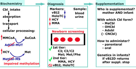
Acquired vitamin B12 (vB12) deficiency (vB12D) of newborns is relatively frequent as compared with the incidence of inherited diseases included in newborn screening (NBS) of different countries across the globe. Infants may present signs of vB12D before 6 months of age with anemia and/or neurologic symptoms when not diagnosed in asymptomatic state. The possibility of identifying vitamin deficient mothers after their pregnancy during the breastfeeding period could be an additional benefit of the newborn screening. Vitamin supplementation is widely available and easy to administer. However, in many laboratories, vB12D is not included in the national screening program. Optimized screening requires either second-tier testing or analysis of new urine and blood samples combined with multiple clinical and laboratory follow ups. Our scope was to review the physiologic fate of vB12 and the pathobiochemical consequences of vB12D in the human body. Particular emphasis was put on the latest approaches for diagnosis and treatment of vB12D in NBS.

Graphical Abstract

1. Introduction

VB12, also called cobalamin (Cbl), is present in several forms in nature. Cbl deficiency might be defined as absolute deficiency of vB12, or functional deficiency with the metabolite profile characteristic for inadequate cellular function of Cbl dependent enzymes (reviewed in several articles recently [1,2,3,4]). This review focuses on a nowadays still underdiagnosed condition, namely infantile acquired vB12D of maternal origin. Correct diagnosis of this condition is often challenging especially when considering the great inequity in the technical, financial background of different NBS programs and the differences in the socioeconomic status of pregnant women across the globe. By reviewing recent literature (with particular focus on the period 2018–2022), we aimed to discuss the biochemical background of the condition leading to the observed metabolic disturbances, the symptoms, prevalence, available diagnostic methods and guidelines for NBS, as well as options for treatment and follow-up.

2. From Uptake to Utilization

2.1. Chemistry and Dietary Source

From the chemical point of view, the corrin ring of the Cbl molecule is in covalent bond with the central cobalt ion (of various oxidation state). Above this plain, the beta-axial ligand bound to the cobalt ion determines specific reactions in which Cbl might be involved. Based on this latter ligand, molecules with distinct biochemical functions are distinguished like adenosyl-Cbl (AdoCbl), methyl-Cbl (MeCbl), hydroxy-Cbl (OHCbl), cyano-Cbl (CNCbl), and glutathionyl-Cbl (GSCbl). Most of these molecules are synthesized after intracellular processing of Cbl. AdoCbl and MeCbl are essential cofactors of methylmalonyl Coenzyme A (MMCoA) mutase (MUT) and methionine synthase (MS), respectively, enzymes in the intermediate metabolism of the human body.
Cbl rich diet includes animal products such as meat, fish, dairy products, liver and egg. Strict vegan or vegetarian diet without supplementation might lead to vB12D, already after 4 weeks of dietary change from omnivore to strict vegan diet [5,6]. In a study involving Dutch pregnant women (at 34–36 weeks of pregnancy) of mixed population including omnivore, lacto-ovo vegetarians and vegans, the main source of Cbl were dairy products, while eggs as Cbl source represented the minority of dietary Cbl source [7].

2.2. Fate of Cbl in the Gastrointestinal Trackt

The process from ingestion to ileal absorption is demonstrated on Figure 1 and Figure 2.
During absorption (Figure 2), the intrinsic factor (IF)-Cbl complex connects to its receptor cubam which is a heterodimer of the proteins amnionless and cubilin. The IF-Cbl complex is internalized by receptor mediated endocytosis. Later, cubam is recycled to the cell surface, while in parallel the pH of the vesicle turns acidic and becomes a lysosome. Low pH allows the dissociation of IF from Cbl, the released IF is degraded (reviewed by [9]). Intracellular trafficking from the lysosome to the cytoplasm involves the proteins LMBD1 and ABCD4. After its synthesis, ABCD4 protein is directed to the endoplasmic reticulum membrane, where it interacts with LMBD1 protein, this latter enabling the transition of the LMBD1-ABCD4 complex to the lysosomal membrane. ABCD4 is the key transporter of Cbl from the lysosomes to the cytosol where it is further processed in most cells [10]. Both proteins show high expression levels in several tissues, including small intestines [11].
Intracellular processing differences between gut epithelial cells or other peripheral, non-polarized cells are barely discussed in the literature. It is suspected that Cbl either undergoes processing to the biologically active cofactor forms (AdoCbl, MeCbl) that are finally used by enterocytes for specific reactions of metabolism, or passes the cell without major modifications (transcytosis) to enter the portal circulation (reviewed by [9]). The transport toward the blood stream occurs via ABCC1 (alternative name: MRP1 (multidrug resistance protein-1)), an ABC transporter located in the basolateral membrane of enterocytes. MRP1 binds and exports free Cbl. In the extracellular matrix free Cbl is bound to the apo-transcobalamin-2 molecule (TC2, Uniprot search term: P20062 or TCO2_HUMAN) which is excreted by cells to the extracellular matrix. The TC2-Cbl complex is later referred to as holo-transcobalamin (HoloTC) [8,9,12,13]. Experiments on bovine aortic endothelial cell cultures showed that endothelial cells excrete a significant concentration of TC2 and also express the HoloTC receptor (CD320) with which a complex Cbl uptake machinery is offered. The fate of HoloTC in endothelial cells might be transcytosis (HoloTC leaves the cell unmodified, ~5–10% of excreted Cbl), exocytosis asfree Cbl (via ABCC1, ~90–95% of excreted Cbl), metabolic modification into ready-to-use cofactors for enzymatic processes, or HoloTC is left intact within the cells. The possibility of choosing different intracellular fates is equally open to Cbl molecules with different moieties (AdoCbl, MeCbl, CNCbl, OHCbl) [14].
Some fraction of intestinal Cbl might be bound to luminal free TC2 and absorbed via CD320 receptor according to studies on murine small and large intestine, and human polarized colorectal cancer cell lines. Another, rather small fraction of Cbl is not absorbed at all, affecting microbiota metabolism and pathogenicity in large intestines. An even smaller amount is eliminated by the stool [15,16].

2.3. Intracellular Fate, Transport in Blood and Storage

Cytosolic processing of Cbl after release from the lysosome is a multistep procedure, in which apoenzymes bind and chaperonate different forms of Cbl (Figure 3). Many questions are yet to be answered, such as interaction between these proteins and with Cbl, their substrate specificity considering the beta-axial ligand of the corrin ring or oxidation state of the cobalt ion, or transport and recycling between intracellular organelles (from cytosol to mitochondria). Each of the proteins involved in intracellular processing is linked to a certain inherited Cbl disorder (Table 1). The aim of the process is to yield the proper amount of active coenzymes required for the reactions of the MUT and MS enzymes.
Transport of Cbl in blood occurs either in form of HolotTC (~20–25% of circulating Cbl) or TCN1-Cbl (~75–80% of circulating Cbl). HoloTC is considered the biologically available (“active”) Cbl in the circulation that can enter non-polarized cells via CD320 mediated endocytosis. On the other hand, TCN1-Cbl is considered to transport Cbl to hepatocytes, and allowing the storage or mobilization from hepatic Cbl buffer depending on Cbl storage levels of the body [4,12].
Kidneys might also serve as a Cbl reserve. The main receptor for reabsorbing Cbl in proximal tubular cells is megalin. Apart from this, cells also exhibit a remarkable intracellular TCN2, and apical CD320 expression in murine experiments. Thus excreted TCN2 can form HoloTC taken up by their receptors at the luminal surface of epithelial cells [16,17].

3. MUT and MS Catalyzed Reactions

The MUT enzyme (EC 5.1.99.1) is catalyzing the reaction in which MMCoA is converted to succinyl CoA (SuCoA); while the enzyme MS (EC 2.1.1.13) catalyzes the reaction where homocysteine (HCY) is converted to methionine (Met) (Figure 3). AdoCbl and MeCbl, respectively are the active Cbl cofactors of the latter enzymes. The above reactions yield substrates and regulatory molecules for a variety of reactions such as the anaplerotic molecule succinate for the tricarboxylic acid cycle or the methyl donor S-adenosyl methionine (SAM) for DNA methylation during epigenetic modifications. In case of insufficient availability of AdoCbl and MeCbl, clinical symptoms may be present as a consequence of the disturbed cellular metabolism. The impaired function of both enzymes might occur in Cbl deficiencies—regardless of the inherited or acquired origin of the condition—resulting in hematologic and/or neurologic symptoms of various degree (reviewed by [13]).

4. Pathobiochemical Consequences of vB12D

Considering the bone marrow, inadequate availability of MeCbl leads to accumulation of HCY and decreased Met, while the methyl donor methyl-THF is accumulating, thereby trapping the methyl group from other cellular methylation processes. This leads to abnormal nucleotide synthesis resulting in the histopathologic picture of macrocytic anemia (reviewed by [2]). Clinical descriptions from patients of various age show that hematologic effects are not necessarily present in vB12D [2,18,19,20]. According to a recent review on pernicious anemia (PA, a condition which evolves due to antibodies against IF, thereby preventing the intestinal absorption of Cbl), macrocytosis might be present years before development of anemia. On the other hand, ~30% of these patients never develop macrocytosis; concomitant iron deficiency anemia with coprevalence of ~20% might be a significant contributor to this. Interestingly, vB12D might also present in the form of conditions severely altering hemopoesis, such as pseudo-leukemia mimic syndrome or bone-marrow failure-like syndrome [21].
Neurocognitive symptoms might be linked to impaired methylation → disturbed myelination → increased inflammatory cytokine axis, leading in extreme cases to the atrophy of the spinal cord or cerebral tissue in different degree. The spectra of childhood neurologic symptoms associated with Cbl deficiency might include convulsion, muscular hypotonia, developmental delay, intellectual disability, dizziness, tingling sensation, syncope, or blurred vision [19]. Most of the functional neurologic disturbances can be reversed by parenteral vitamin B12 supplementation even in adults [18]. The effect of supplementation is also promising in pediatric children with autism spectrum disorder [22]. Coexistence of both hematologic and neuropsychiatric symptoms is not a prerequisite in vB12D.

5. Diagnostic Criteria of Vitamin B12 Deficiency

Standardized international definition or consensus guidelines for a vB12D are lacking. Laboratory evaluation of a vB12D allows determination of at least four distinct laboratory markers: total abundance of vB12, HoloTC, MMA and HCY [2,3,20]. There seems to be no single “perfect” biomarker; rather the combination of these markers in “four-variable”, “three-variable” (e.g., vB12 + HoloTC + MMA, HoloTC + MMA + HCY or vB12 + MMA + HCY) or “two-variable” (most often HoloTC + MMA) approaches, dependent on the laboratory infrastructure, financial sources and patient population [23,24]. Methylcitric acid (MCA) is an additional important biomarker in screening and diagnosis of propionic aciduria (PAuria) and MMAuria but is reportedly less useful in Cbl deficiencies and is rarely determined by general clinical laboratories [25].

5.1. VB12 Assays and Result Interpretation

Among analytical methods on vB12 determination, microbiological assays were the first to have found utility in the clinical laboratory, due to acceptable sensitivities and specificities [3]. This time-consuming technique does not provide a direct estimation of the vitamin levels but functional information that is dependent on utilization of the vitamin by the microbe used [26]. Of several strains, Lactobacillus leichmannii (ATCC 7830) was used most frequently due to the robustness of the assay [3,27]. Radioisotope-based dilution techniques were also published [28]. However, automated methods are most widely used currently for the assessment of vB12 status due to their high throughput and less laborious nature [3]. Competitive-binding luminescence-based assays (CBLA) for total (i.e., protein-bound plus free) vB12 are available since the early 1990’s and include electrochemiluminescent (Roche), chemiluminescent (Siemens, Abbott, Beckman Coulter, VITROS), enzyme-linked fluorescent (earlier Siemens and Abbott assays), or colorimetric techniques [3,29]. Serum vB12 assays are calibrated independently by manufacturers with traceability to an internally manufactured standard material rather than an internationally certified reference material [3]. This often results in poor agreement between commercially available assays for serum vB12, despite the ratification of The World Health Organization International Standard for serum B12 (03/178) in 2007 as a consensus of vB12 protein-binding assays [3,30]. Thus, assay-specific cutoffs are still generally used for the different vB12 assays, which hamper comparability of results from different settings, regions or countries [3].
Serum Cbl concentrations alone are weak indicators for functionally relevant intracellular Cbl deficiency [2]. A functional vB12 deficiency can be present in patients with apparently normal or even high serum vB12 levels, for example in the case of high levels of vB12-binding proteins [3]. The presence of macro-transcobalamin can reportedly increase vB12 results in various assays [31]. Erroneously high or normal total vB12 concentration may be caused by interfering anti-IF antibodies in pernicious anemia (where vB12 levels would be expected to decrease). This condition might also affect mothers of vB12 deficient infants detected by NBS [2,3,32,33].

5.2. HoloTC Assays and Result Interpretation

The second marker commonly used is HoloTC, the form of vB12 that is taken up by cells to meet metabolic demand, explaining the increasing use of assays on this biomarker [3,20]. HoloTC seems to be a helpful parameter to identify problems of absorption and to differentiate them from nutritional Cbl deficiency [2].
The first method for HoloTC was based on the enzyme-linked immunosorbent assay technique (ELISA) [34]. Currently, most clinical laboratories use ELISAs or (electro)chemiluminescent (ECLIA, CLIA) immunoassays for HoloTC [3]. In contrast with commercial automated total methods for vB12 that use assay-specific calibration with no international reference material, calibrators of commercially available assays for HoloTC are traceable back to common frozen primary reference calibrators which are held by Axis-Shield [3]. However, reference values are still strongly dependent on the assay method used [34]. In addition, some rare variants in the transcobalamin gene can reportedly interfere with the HoloTC determination, so that erroneously low HoloTC levels are measured, despite an absence of clinical deficiency and normal levels of all other laboratory markers [3]. In contrast with vB12, HoloTC results are reported to be relatively unaffected by assay interference from high-titer IF antibody concentrations [3]. However, HoloTC may still be erroneously normal in a small subset of patients with pernicious anaemia [35].

5.3. MMA Assays and Result Interpretation

The concentration of MMA in serum reflects the availability and utilization of AdoCbl in mitochondria. MMA in fresh or optimally stored urine or blood is considered the most sensitive marker of intracellular, functionally relevant Cbl depletion (with the limitation that urinary MMA is not reliable in individuals with impaired renal function) [2].
The first techniques for the measurement of MMA applied paper chromatography, thin layer chromatography, spectrophotometry and later ELISA, but were superseded by mass spectrometric methods due to markedly improved sensitivities and specificities. Full or semi-automation of liquid chromatography-tandem mass spectrometry (LC-MS/MS) methods for MMA is also possible [3]. Determination of MMA in NBS from dried blood spots (DBS) will be discussed in a separate section.
An elevated MMA is not specific for vB12 depletion or inborn errors of the Cbl metabolism. Among other disorders, a deficiency of the enzymes MUT (total or partial) or acyl-CoA synthetase family member 3 (ACSF3) can also result in MMA levels increased to various magnitudes [25]. Elevated MMA levels have also been reported in people with severe impairment of renal functionality due to impaired elimination [2,20].

5.4. HCY Assays and Result Interpretation

The total concentration of HCY in serum reflects the availability and utilization of MeCbl in the cytosol [1]. Historically, HCY was measured by means of paper chromatography, radio-enzymatic determination or ion-exchange amino acid analyzers. Today, automated enzyme immunoassays and high performance-LC methods (HPLC) with fluorescent or electrochemical detection are used, in addition to mass spectrometry coupled with gas chromatography (GC) or LC [3,36]. From the analytical perspective, the measurement of HCY is relatively challenging. The sulfhydryl group of HCY readily reacts with sulfhydryl groups of proteins, another HCY molecule or cysteine to form the corresponding disulfide compound. Therefore, analytical methods either determine free HCY without any reduction step (as in most immunoassays) or total HCY after reaction with a reducing agent (DTE (1,4-Dithioerythritol), DTT (DL-Dithiothreitol) or TCEP (Tris(2-carboxyethyl)phosphine hydrochloride)) to release HCY bound to proteins and other disulfides [18]. In order to avoid artificial alterations in serum HCY levels due to preanalytical issues, blood should be obtained in the fasting state, transported cooled (the freezing pack should not get in touch directly with the blood tube) and centrifuged as soon as possible, preferably within 30 min [37]. Measurement of HCY from DBS in the frame of NBS will be discussed in a separate section.
In addition to inborn errors of the Cbl metabolism, other congenital disorders can also manifest with elevations in serum HCY, including a cystathionine-β-synthase (CBS) or a methylenetetrahydrofolate reductase (MTHFR) deficiency [3]. Elevated HCY values can also occur as a consequence of folate or vitamin B6 deficiency, as well as impaired renal function, hypothyroidism, malignant tumors and certain medications [2,38]. In the aspect of age HCY seems more to be a marker of impaired Cbl metabolism in infants and toddlers, while it is associated primarily with abnormalities of folate metabolism in elderly. In this latter age group MMA is considered as primary marker of Cbl deficient cellular metabolism [1,39].
As a complement to the determination of HCY, a recent article reported an LC-MS/MS method for the determination of SAM and S-adenosylhomocysteine (SAH) in plasma and urine. The determination of SAM and SAH is complicated by the instability of SAM under neutral and alkaline conditions and the naturally low concentration (nanomolar range) of both SAM and SAH in plasma. The clinical applicability of the assay is yet to be determined [40].

5.5. General Considerations

Recommendations on the determination of vB12D include in optimal case measurement of serum vB12 or HoloTC combined with two functional markers, MMA and HCY. It is important to emphasize that HCY may also increase in folate deficiency; therefore, additional measurement of folic acid is needed for differential diagnosis [1]. This latter is also included in the mathematic formula for calculating the so called combined vB12 [23]. Cut-off values largely vary between studies. The low cut-off range of total vB12 in different centers was 118–221 pmol/L, for HoloTC 37–50 pmol/L. High cut-off of serum total HCY was in the interval 8–13.6 µmol/L (8 µmol/L when vB12D was determined based on single total HCY measurement without vB12 determination) and MMA in the range of 271–800 nmol/L [1]. Based on the results of the primary and functional markers low vB12, possible vB12D and probable vB12D might be distinguished. These result in subclinical vB12D, vB12D with possible or significant clinical manifestations, respectively [23]. According to the opinion of the authors, clinical laboratories should primarily define their cut-off values based on the diagnostic method they use, and literature data should only serve as orientation point.

6. Vitamin B12 Deficiency among Pregnant Women

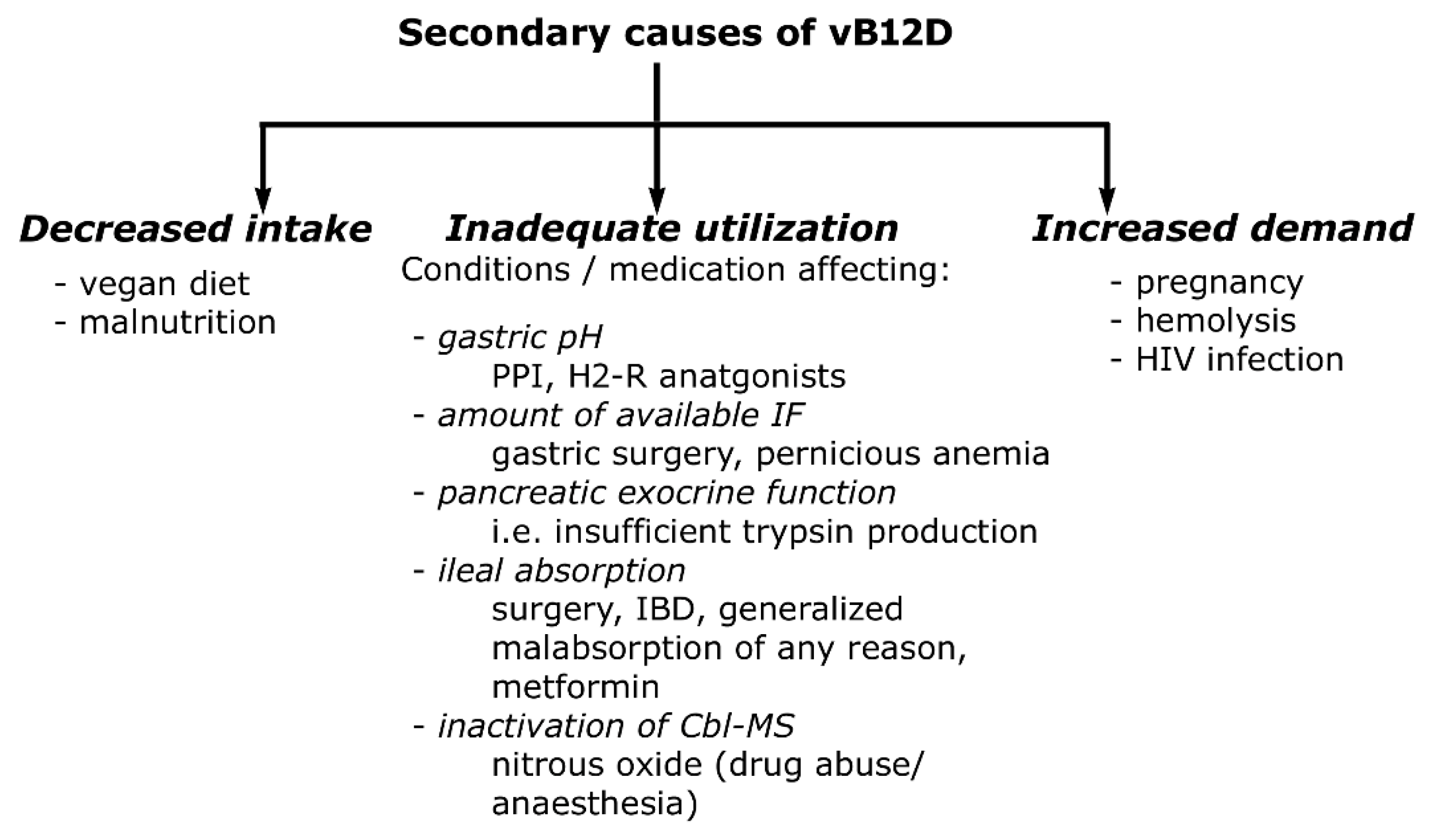
In most countries, screening of vB12D status is not compulsory during pregnancy [41,42]. Estimating and comparing the prevalence of this condition between geographic regions might be difficult because of the unstandardized definition of the laboratory assessment of vB12D [43]. However, the reported prevalence is relatively high, around 20% worldwide, especially in low-income areas where access to animal food is strongly limited [41,44]. In concordance with this, a positive correlation between the educational level of the mother and vB12 level was also observed [45]. The subclinical vB12D of the mothers is usually diagnosed only when their breastfed infant is identified during NBS with suspected vB12D. In these cases, metabolite elevations in DBS indicative of MMAuria/PAuria followed by further testing may shed light on the hidden vitamin deficiency of the mother [44,46]. Several gene products are involved in absorption, transport and intracellular trafficking of Cbl (Table 1). Nevertheless, the acquired vB12D of the mother results predominantly in vB12D of the breastfed infant [41,44,47]. Secondary causes of vB12D are summarized in Figure 4.
During pregnancy, concentrations of several biochemical markers are lower than those in non-pregnant women [48]. Hormonal changes resulting in physiologic hemodilution might be a factor contributing to this, in addition to fetal consumption and redistribution of the Cbl stores of the body [42]. Attempts to determine cutoff-values of vB12 in early, mid, and late pregnancy show a general tendency of a gradual decline of vB12 toward delivery, followed by a sharp rise during early lactation [48,49]. In parallel, TC1 declines more sharply, while HoloTC is maintained at a relatively stable level, albeit reports on change in HoloTC level during gestation remain controversial [50,51]. In population studies, these changes are accompanied by elevation in functional vB12D markers MMA and HCY [41,42,48,52]. In contrast, a Canadian study conducted on healthy pregnant women (not representing the entire population) noted the decrease of both MMA and HCY toward terminus [53]. According to these, trimester specific cutoffs were suggested for functional biomarkers [52]. Some studies hypothesized low Cbl status to be a side effect of the widespread prophylactic folate substitution in early pregnancy. There was, however, no proven evidence for a mechanistic link between the two. The corrected folate levels seemed to be more a co-occurrence with a non-corrected Cbl status [54].

7. Vitamin B12 Deficiency among Newborn

Cbl repletion of the newborn at the time of delivery is substantially determined by the Cbl status of the mother (involving both circulating and functional markers). There is positive correlation in each trimester between the Cbl status of the mother and newborn [55]. Moreover, this relationship (as early as the 18th gestational week), has a significant impact on the Cbl status of the breastfed infant 6 months post-partum [56]. However, even more enduring changes might occur according to a study in which the mid-pregnancy deficient vB12 levels of the mothers were accompanied with impaired cognitive function of their toddler as far as 24 months age [57]. Beside this, elevated HCY itself during the second and third trimester of pregnancy was inversely correlated to neurocognitive outcome of children at 30 months [58]. It seems that Cbl is accumulating in the fetus up till delivery, since serum vB12 concentration might be 2.5-fold higher in the newborn compared to its abundance in its mother [59]. It is yet to be evaluated whether nitrous oxide, an anesthetic gas commonly used during labor, could lead to transient elevations in the markers of a vB12D in NBS [60]. Even if nitrous oxide can irreversibly inactivate Cbl (when bound to MS—Figure 3.) by oxidizing the cobalt ion, only its regular use as drug was reported to be associated with neurologic symptoms in earlier papers [47,61]. However, recent results from the Norwegian NBS program suggested that nitrous oxide applied during labor might lead to vB12D [62]. After birth, infants on formula feeding receive an optimal supplementation of Cbl, while breastfed infants have to rely on the amount of Cbl the mammary glands excrete into the breastmilk, which show different dynamics in pre-term and full-term delivered infants [44,63]. Breastmilk Cbl peaks 4 weeks post-partum for infants born before the 34th gestational week. This is in contrast to full-term newborns where the peak is already at the time of birth, from which a gradual decline can be observed toward day 28 post-partum [63]. Recently, particular attention has been paid to the diagnosis of vB12D in NBS due to its relatively high prevalence, cheap and widely available treatment, and its capability to not only identify infants but also mothers with subclinical vitamin deficiency.

8. NBS Protocols and New Candidate Markers

NBS allows early recognition and treatment of numerous inborn errors of metabolism, thus facilitating the prevention of serious morbidity and mortality [64]. DBS is the standard sample type in NBS worldwide due to the small volume of blood needed, the ease of transportation and stability [65]. Expanded NBS took advantage of the implementation of MS/MS, allowing an increase in the number of screened disorders via simultaneous determination of several amino acids and acylcarnitines [64]. Among disorders of the propionate metabolism, PAuria and MMAuria are already included in NBS in numerous European countries and in all US states [66,67].
However, the characteristic alterations of an elevated propionylcarnitine (C3) and/or decreased Met may additionally be suggestive of a vB12D, that was previously considered an incidental finding and inappropriate for an inclusion in NBS until very recently [25,68,69]. The reason was that none of the first-tier parameters is specific for any of these conditions, which results in an elevation of the rate of false-positive results [25]. Even if false-positive samples cannot fully be avoided in NBS, their number has to be kept low to minimize parental anxiety, as well as costs and workload [64]. Moreover, even if C3 and Met are generally measured together with numerous other amino acids and acylcarnitines in a single assay, these parameters are often only evaluated if the country officially screens for these disorders [25].
Recently, an increasing number of publications suggested that an inclusion of a vB12D into NBS may not only be reasonable but feasible too. According to the authors, the proper selection of diagnostic analytes, cutoffs and strategies can facilitate detection of a vB12D. Thus, an inclusion of this condition into NBS is currently under evaluation in some countries including Germany [68,69]. The use of analyte ratios in the screening test such as the ratios Met/phenylalanine (Met/Phe) and C3/acetylcarnitine (C3/C2) can directly improve false-positive rates.
Diagnostic specificity can be further increased by confirmatory assays utilizing chromatographic separation of diagnostically important analytes (including MMA, MCA, HCY and rarely 3-hydroxy-propionic acid) from their isobaric substances, i.e., from those having the same molecular mass [25,68,69]. In addition to succinate, the only metabolite considered earlier as an interference for MMA, further five potential isobaric interferents have been identified recently [70]. The urinary organic acid assay by means of GC-MS performs well in separating isobaric interferences that could compromise diagnosis and an accurate quantification [65]. In contrast with the initial DBS sample being available for the NBS laboratory, urine has to be requested separately, which is associated with marked delays and significant increase of parental anxiety [25]. Thus, it is strongly recommended that confirmatory assays in NBS use the original DBS sample (second-tier tests) [25,64].
The very limited sample amount in DBS raises an analytical challenge for most diagnostic metabolites [65]. Thus, even if derivatization can be avoided for faster turnover times if sufficiently sensitive mass spectrometers are used [71], most assays for HCY, MMA and MCA in DBS use derivatization with n-butanolic HCl or silylated trifluoroacetamides [72,73,74]. HCY can be measured separately from MMA and MCA [71,75,76] or simultaneously in a single assay [28,29,30]. In contrast with MMA, MCA and HCY, vB12 is not determined from DBS but from serum, due to its very low concentration, high molecular mass and complex structure [3].
Considering the diagnostic value of the first- and second-tier markers, a vB12D can present with diverse alterations [25,69]. In a recent publication, the most sensitive first-tier parameters were a low Met/Phe (positive in 23 of 33 cases) and a low Met (20/33). Elevated C3/C2 (7/33), high Met/Phe (5/33) and high C3 (4/33) were less indicative for a vB12D. Of the second-tier biomarkers, HCY was found to show the best sensitivity, being elevated in 30 of 33 cases, while MMA and MCA were only elevated in 12 and 9 patients, respectively [69]. Using post-analytical methods, such as multivariate pattern recognition software developed by Collaborative Laboratory Integrated Reports (CLIR—Mayo Clinic), also revealed a clear demarcation of the C3/Gly ratio from the reference range in pathologic conditions affecting Cbl metabolism [77,78]. Thus, current knowledge suggests that instead of a single marker, a combination of appropriately selected first-tier cutoffs of metabolites and ratios, together with second-tier confirmatory assays for the determination of HCY, MMA and MCA are expected to facilitate the diagnosis of a vB12D in NBS.
Cut-off values defined for the above biomarkers varied according to different algorithms used by NBS laboratories. A single marker (HCY high cut-off: 8 µmol/L) was only used in Norway [39]. Most laboratories used the classical parameters C3 (high cut-off: 3.3–5.5 µmol/L), C3/C2 (high cut-off: 0.18–0.26), and C3/C16 (high cut-off: 1.5–2.0) [39,69,79,80,81]. When second tier MMA and HCY measurements were not available, additional serum vB12, folic acid, HCY and urinary MMA determinations were performed [79]. Cut-off values for additional first tier markers were: C3/C0: >0.23; C3/Met: >0.2; Met: <8–11 µmol/L, Met/Phe < 0.19–0.56. MMA and HCY second tier high cuf-offs varied between 0.4–4 µmol/L and 10–12 µmol/L, respectively [69,79,80,81]. None of these protocols was compared in a single study on the same population to test their efficiency; however, all identified vB12D in newborns with a prevalence that was previously underestimated at national level.

9. Treatment and Follow-Up

Maternal vB12 supplementation during gravidity and post-partum (until the 6th week) could serve as primary prevention strategy not only through better Cbl values of the newborn at birth but also due to an increase of Cbl in breastmilk [82]. Mothers receiving oral vB12 supplementation gave birth to infants whose language skills were significantly better at 30 months of age than those in the un-supplemented group [58]. VB12D in and after the post-partum period might lead to irreversible neurologic changes from as early as 6 months of age [41,77]. Therefore, it is advisable to establish the diagnosis and start supplementation as early as possible.
Infant formulas are fortified by vB12 and serve as adequate tools for vitamin administration [1,44]. In the case of breastfed infants, vitamin supplementation of both the mother and the infant is recommended, if investigations reveal a maternal vB12D [44,83]. Before prescription, one should consider the type of Cbl available and the way it can be administered. The palette consisting of the traditional CNCbl was later extended with nature biosimilar OHCbl, AdoCbl, and MeCbl. The bioavailability of all forms is similar, though biosimilar products should be favored due to the lack of cyanide load for the body [84].
Infantile parenteral administration of Cbl might occur in the form of intramuscular injection (most common), or via intravenous injection (usually as part of the water-soluble vitamin complex required during parenteral feeding—common in pre-term infants). A single intramuscular injection (400 µg OHCbl in infants < 8 months) may correct the impaired Cbl status and improve the already evolving clinical symptoms, such as motor dysfunction and regurgitations [85]. The less invasive, longer lasting oral supplementation scheme could also be an alternative; starting with higher doses for 3 days (500 µg Cbl/day), followed by reduction to the fifth of the original dose until the complete recovery of vB12 levels and functional markers. A maintenance dose of 5 µg/day can be ceased after introduction of animal protein-containing complementary food. Oral administration scheme was complemented with folate substitution during the first week, and laboratory checkup at 2 weeks (possible exclusion of genetic defects in Cbl absorption) after supplementation onset in a recent study [44]. According to the personal opinion of the authors, socio-economic status, parenteral compliance, geographic distances needed for checkups should all be considered before choosing the way of vB12 administration. A genetic defect affecting vB12 absorption or metabolism should be considered if cessation of the treatment induces a relapse in laboratory markers [44,85].

10. Conclusions

The vB12D is a condition that can severely affect quality of life if not diagnosed early or left untreated. In low-income regions with high prevalence of vB12D and less financial resources for NBS, primary prevention, such as mandatory food fortification, or recommendations in parallel with educations for vitamin supplementation during pregnancy could be the best choice [86]. Recent studies suggest that an extension of the NBS (when already using MS/MS methods) with vB12D may be feasible, and even financially affordable. According to the opinion of the authors, a first trimester check-up of the vB12 status combined with NBS for vB12D could be an optimal way in reduction of the condition’s prevalence in women and infants of developed countries. Pregnancy screening alone is not sufficient as vitamin supplementation of pregnant women may be suboptimal compared to that of newborns due to the different effectivity of the social net in the two groups in most countries. For optimal outcomes, pregnancy screening should be complemented with NBS that is performed along standardized algorithms, equal for the entire population of a region or a country. VB12 deficient newborns identified by the NBS benefit remarkably from the early supplementation in asymptomatic state, resulting in the prevention of long-term consequences for the child, as well as additional health care benefits for the mother and reduced expenses and load for the health care system.
Standardized protocols and consensus guidelines for vB12D are yet to be developed and, together with recent improvements in laboratory technology and clinical treatment, are expected to facilitate earlier diagnosis and a better quality of life of patients with vB12D.

Author Contributions

Conceptualization, visualization, M.K.; writing—original draft preparation, M.K. and P.M.; writing—review and editing, Z.G., I.L., Á.B., N.G., G.R. and C.B. All authors have read and agreed to the published version of the manuscript.

Funding

This research received no external funding.

Conflicts of Interest

The authors declare no conflict of interest.

References

  1. Hannibal, L.; Lysne, V.; Bjørke-Monsen, A.-L.; Behringer, S.; Grünert, S.C.; Spiekerkoetter, U.; Jacobsen, D.W.; Blom, H.J. Biomarkers and Algorithms for the Diagnosis of Vitamin B12 Deficiency. Front. Mol. Biosci. 2016, 3, 27. [Google Scholar] [CrossRef] [Green Version]
  2. Huemer, M.; Baumgartner, M.R. The Clinical Presentation of Cobalamin-related Disorders: From Acquired Deficiencies to Inborn Errors of Absorption and Intracellular Pathways. J. Inherit. Metab. Dis. 2019, 42, 686–705. [Google Scholar] [CrossRef] [PubMed]
  3. Harrington, D.J. Methods for Assessment of Vitamin B12. In Laboratory Assessment of Vitamin Status; Elsevier: Amsterdam, The Netherlands, 2019; pp. 265–299. [Google Scholar]
  4. Obeid, R.; Heil, S.G.; Verhoeven, M.M.; Van den Heuvel, E.G.; De Groot, L.C.; Eussen, S.J. Vitamin B12 Intake from Animal Foods, Biomarkers, and Health Aspects. Front. Nutr. 2019, 6, 93. [Google Scholar] [CrossRef] [PubMed] [Green Version]
  5. Lederer, A.-K.; Hannibal, L.; Hettich, M.; Behringer, S.; Spiekerkoetter, U.; Steinborn, C.; Gründemann, C.; Zimmermann-Klemd, A.M.; Müller, A.; Simmet, T. Vitamin B12 Status upon Short-Term Intervention with a Vegan Diet—A Randomized Controlled Trial in Healthy Participants. Nutrients 2019, 11, 2815. [Google Scholar] [CrossRef] [PubMed] [Green Version]
  6. Román, G.C.; Jackson, R.E.; Gadhia, R.; Román, A.N.; Reis, J. Mediterranean Diet: The Role of Long-Chain ω-3 Fatty Acids in Fish; Polyphenols in Fruits, Vegetables, Cereals, Coffee, Tea, Cacao and Wine; Probiotics and Vitamins in Prevention of Stroke, Age-Related Cognitive Decline, and Alzheimer Disease. Rev. Neurol. 2019, 175, 724–741. [Google Scholar] [CrossRef]
  7. Denissen, K.F.; Heil, S.G.; Eussen, S.J.; Heeskens, J.P.; Thijs, C.; Mommers, M.; Smits, L.J.; van Dongen, M.C.; Dagnelie, P.C. Intakes of Vitamin B-12 from Dairy Food, Meat, and Fish and Shellfish Are Independently and Positively Associated with Vitamin b-12 Biomarker Status in Pregnant Dutch Women. J. Nutr. 2019, 149, 131–138. [Google Scholar] [CrossRef] [Green Version]
  8. Uniprot. Available online: https://www.uniprot.org/ (accessed on 21 October 2022).
  9. Brito, A.; Habeych, E.; Silva-Zolezzi, I.; Galaffu, N.; Allen, L.H. Methods to Assess Vitamin B12 Bioavailability and Technologies to Enhance Its Absorption. Nutr. Rev. 2018, 76, 778–792. [Google Scholar] [CrossRef]
  10. Kitai, K.; Kawaguchi, K.; Tomohiro, T.; Morita, M.; So, T.; Imanaka, T. The Lysosomal Protein ABCD4 Can Transport Vitamin B12 across Liposomal Membranes in Vitro. J. Biol. Chem. 2021, 296, 100654. [Google Scholar] [CrossRef]
  11. Wang, D.; Eraslan, B.; Wieland, T.; Hallström, B.; Hopf, T.; Zolg, D.P.; Zecha, J.; Asplund, A.; Li, L.; Meng, C. A Deep Proteome and Transcriptome Abundance Atlas of 29 Healthy Human Tissues. Mol. Syst. Biol. 2019, 15, e8503. [Google Scholar] [CrossRef]
  12. Nielsen, M.J.; Rasmussen, M.R.; Andersen, C.B.; Nexø, E.; Moestrup, S.K. Vitamin B12 Transport from Food to the Body’s Cells—A Sophisticated, Multistep Pathway. Nat. Rev. Gastroenterol. Hepatol. 2012, 9, 345–354. [Google Scholar] [CrossRef]
  13. Froese, D.S.; Fowler, B.; Baumgartner, M.R. Vitamin B12, Folate, and the Methionine Remethylation Cycle—Biochemistry, Pathways, and Regulation. J. Inherit. Metab. Dis. 2019, 42, 673–685. [Google Scholar] [CrossRef] [PubMed] [Green Version]
  14. Hannibal, L.; Bolisetty, K.; Axhemi, A.; DiBello, P.M.; Quadros, E.V.; Fedosov, S.; Jacobsen, D.W. Transcellular Transport of Cobalamin in Aortic Endothelial Cells. FASEB J. 2018, 32, 5506–5519. [Google Scholar] [CrossRef] [PubMed]
  15. Rowley, C.A.; Kendall, M.M. To B12 or Not to B12: Five Questions on the Role of Cobalamin in Host-Microbial Interactions. PLoS Pathog. 2019, 15, e1007479. [Google Scholar] [CrossRef] [PubMed] [Green Version]
  16. Chen, Y.; Gu, X.; Zhang, Y.; Zhang, X.; Zhang, C.; Liu, M.; Sun, S.; Dong, N.; Wu, Q. CD320 Expression and Apical Membrane Targeting in Renal and Intestinal Epithelial Cells. Int. J. Biol. Macromol. 2022, 201, 85–92. [Google Scholar] [CrossRef] [PubMed]
  17. Birn, H.; Willnow, T.E.; Nielsen, R.; Norden, A.G.; Bönsch, C.; Moestrup, S.K.; Nexø, E.; Christensen, E.I. Megalin Is Essential for Renal Proximal Tubule Reabsorption and Accumulation of Transcobalamin-B12. Am. J. Physiol.-Ren. Physiol. 2002, 282, F408–F416. [Google Scholar] [CrossRef] [Green Version]
  18. Silva, B.; Velosa, A.; Barahona-Corrêa, J.B. Reversible Dementia, Psychotic Symptoms and Epilepsy in a Patient with Vitamin B12 Deficiency. BMJ Case Rep. CP 2019, 12, e229044. [Google Scholar] [CrossRef]
  19. Serin, H.M.; Arslan, E.A. Neurological Symptoms of Vitamin B12 Deficiency: Analysis of Pediatric Patients. Acta Clin. Croat. 2019, 58, 295. [Google Scholar] [CrossRef]
  20. Wolffenbuttel, B.H.; Wouters, H.J.; Heiner-Fokkema, M.R.; van der Klauw, M.M. The Many Faces of Cobalamin (Vitamin B12) Deficiency. Mayo Clin. Proc. Innov. Qual. Outcomes 2019, 3, 200–214. [Google Scholar] [CrossRef] [Green Version]
  21. Htut, T.W.; Thein, K.Z.; Oo, T.H. Pernicious Anemia: Pathophysiology and Diagnostic Difficulties. J. Evid.-Based Med. 2021, 14, 161–169. [Google Scholar] [CrossRef]
  22. Rossignol, D.A.; Frye, R.E. The Effectiveness of Cobalamin (B12) Treatment for Autism Spectrum Disorder: A Systematic Review and Meta-Analysis. J. Pers. Med. 2021, 11, 784. [Google Scholar] [CrossRef]
  23. Fedosov, S.N.; Brito, A.; Miller, J.W.; Green, R.; Allen, L.H. Combined Indicator of Vitamin B12 Status: Modification for Missing Biomarkers and Folate Status and Recommendations for Revised Cut-Points. Clin. Chem. Lab. Med. 2015, 53, 1215–1225. [Google Scholar] [CrossRef] [PubMed]
  24. Campos, A.J.; Risch, L.; Nydegger, U.; Wiesner, J.; Dyck, M.V.V.; Seger, C.; Stanga, Z.; Renz, H.; Risch, M. Diagnostic Characteristics of 3-Parameter and 2-Parameter Equations for the Calculation of a Combined Indicator of Vitamin B12 Status to Predict Cobalamin Deficiency in a Large Mixed Patient Population. Clin. Lab. 2020, 66, 10. [Google Scholar] [CrossRef] [PubMed]
  25. Forny, P.; Hörster, F.; Ballhausen, D.; Chakrapani, A.; Chapman, K.A.; Dionisi-Vici, C.; Dixon, M.; Grünert, S.C.; Grunewald, S.; Haliloglu, G. Guidelines for the Diagnosis and Management of Methylmalonic Acidaemia and Propionic Acidaemia: First Revision. J. Inherit. Metab. Dis. 2021, 44, 566–592. [Google Scholar] [CrossRef]
  26. Boxer, G.E.; Richards, J.C. Chemical Determination of Vitamin B12. 2. The Quantitative Isolation and Colorimetric Determination of Millimicrogram Quantities of Cyanide. Arch Biochem 1951, 30, 372–381. [Google Scholar] [PubMed]
  27. Peeler, H.T.; Yacowitz, H.; Norris, L.C. A Microbiological Assay for Vitamin B12 Using Lactobacillus Leichmannii. Proc. Soc. Exp. Biol. Med. 1949, 72, 515–521. [Google Scholar] [CrossRef] [PubMed]
  28. Barakat, R.; Ekins, R. Assay of Vitamin B12 in Blood: A Simple Method. Lancet 1961, 278, 25–26. [Google Scholar] [CrossRef]
  29. İspir, E.; Serdar, M.A.; Ozgurtas, T.; Gulbahar, O.; Akın, K.O.; Yesildal, F.; Kurt, İ. Comparison of Four Automated Serum Vitamin B12 Assays. Clin. Chem. Lab. Med. 2015, 53, 1205–1213. [Google Scholar] [CrossRef]
  30. Thorpe, S.J.; Heath, A.; Blackmore, S.; Lee, A.; Hamilton, M.; O’broin, S.; Nelson, B.C.; Pfeiffer, C. International Standard for Serum Vitamin B12 and Serum Folate: International Collaborative Study to Evaluate a Batch of Lyophilised Serum for B12 and Folate Content. Clin Chem Lab Med. 2007, 45, 380–386. [Google Scholar] [CrossRef]
  31. Duim, S.N.; Vlasveld, L.T.; Mezger, S.T.; Mingels, A.M.; Ramakers, C.R.; de Boer, D.; Heil, S.G.; Nexo, E.; van Rossum, A.P. Macro Transcobalamin Causing Raised Vitamin B12: Case-Based Laboratory Investigation. Ann. Clin. Biochem. 2022, 59, 302–307. [Google Scholar] [CrossRef]
  32. Scarpa, E.; Candiotto, L.; Sartori, R.; Radossi, P.; Maschio, N.; Tagariello, G. Undetected Vitamin B12 Deficiency Due to False Normal Assay Results. Blood Transfus. 2013, 11, 627. [Google Scholar]
  33. Wainwright, P.; Narayanan, S.; Cook, P. False-Normal Vitamin B12 Results in a Patient with Pernicious Anaemia. Clin. Biochem. 2015, 48, 1366–1367. [Google Scholar] [CrossRef] [PubMed]
  34. Nexo, E.; Christensen, A.-L.; Hvas, A.-M.; Petersen, T.E.; Fedosov, S.N. Quantification of Holo-Transcobalamin, a Marker of Vitamin B12 Deficiency. Clin. Chem. 2002, 48, 561–562. [Google Scholar] [CrossRef] [PubMed] [Green Version]
  35. Knoepfel, C.; Blanco, M.M.; Nydegger, U.; Risch, L.; Renz, H.; Risch, M. Failure of the Holotranscobalamin Assay in Vitamin B12-Deficient Patients. J. Lab. Med. 2018, 42, 141–147. [Google Scholar] [CrossRef]
  36. Alam, S.F.; Kumar, S.; Ganguly, P. Measurement of Homocysteine: A Historical Perspective. J. Clin. Biochem. Nutr. 2019, 65, 171–177. [Google Scholar] [CrossRef] [PubMed] [Green Version]
  37. Chernecky, C.C.; Berger, B.J. Laboratory Tests and Diagnostic Procedures; Elsevier Health Sciences: Amsterdam, The Netherlands, 2012; ISBN 1-4557-4502-2. [Google Scholar]
  38. Kim, J.; Kim, H.; Roh, H.; Kwon, Y. Causes of Hyperhomocysteinemia and Its Pathological Significance. Arch. Pharm. Res. 2018, 41, 372–383. [Google Scholar] [CrossRef] [PubMed]
  39. Ljungblad, U.W.; Paulsen, H.; Mørkrid, L.; Pettersen, R.D.; Hager, H.B.; Lindberg, M.; Astrup, H.; Eklund, E.A.; Bjørke-Monsen, A.-L.; Rootwelt, T. The Prevalence and Clinical Relevance of Hyperhomocysteinemia Suggesting Vitamin B12 Deficiency in Presumed Healthy Infants. Eur. J. Paediatr. Neurol. 2021, 35, 137–146. [Google Scholar] [CrossRef] [PubMed]
  40. Bravo, A.C.; Aguilera, M.N.L.; Marziali, N.R.; Moritz, L.; Wingert, V.; Klotz, K.; Schumann, A.; Grünert, S.C.; Spiekerkoetter, U.; Berger, U. Analysis of S-Adenosylmethionine and S-Adenosylhomocysteine: Method Optimisation and Profiling in Healthy Adults upon Short-Term Dietary Intervention. Metabolites 2022, 12, 373. [Google Scholar] [CrossRef]
  41. Behere, R.V.; Deshmukh, A.S.; Otiv, S.; Gupte, M.D.; Yajnik, C.S. Maternal Vitamin B12 Status during Pregnancy and Its Association with Outcomes of Pregnancy and Health of the Offspring: A Systematic Review and Implications for Policy in India. Front. Endocrinol. 2021, 12, 619176. [Google Scholar] [CrossRef]
  42. Rashid, S.; Meier, V.; Patrick, H. Review of Vitamin B12 Deficiency in Pregnancy: A Diagnosis Not to Miss as Veganism and Vegetarianism Become More Prevalent. Eur. J. Haematol. 2021, 106, 450–455. [Google Scholar] [CrossRef]
  43. Rothen, J.-P.; Walter, P.N.; Tsakiris, D.A.; Infanti, L.; Hersberger, K.E.; Arnet, I. Identification of Patients with Cobalamin Deficiency Crucially Depends on the Diagnostic Strategy. Clin Lab 2021, 67, 1229–1235. [Google Scholar] [CrossRef]
  44. Gramer, G.; Hoffmann, G.F. Vitamin B12 Deficiency in Newborns and Their Mothers—Novel Approaches to Early Detection, Treatment and Prevention of a Global Health Issue. Curr. Med. Sci. 2020, 40, 801–809. [Google Scholar] [CrossRef] [PubMed]
  45. Sayar, E.H.; Orhaner, B.B.; Sayar, E.; NesrinTuran, F.; Küçük, M. The Frequency of Vitamin B12, Iron, and Folic Acid Deficiency in the Neonatal Period and Infancy, and the Relationship with Maternal Levels. Turk. Arch. Pediatr. Pediatri. Arş. 2020, 55, 139. [Google Scholar] [CrossRef] [PubMed]
  46. Scolamiero, E.; Villani, G.R.D.; Ingenito, L.; Pecce, R.; Albano, L.; Caterino, M.; di Girolamo, M.G.; Di Stefano, C.; Franzese, I.; Gallo, G. Maternal Vitamin B12 Deficiency Detected in Expanded Newborn Screening. Clin. Biochem. 2014, 47, 312–317. [Google Scholar] [CrossRef] [PubMed]
  47. Thayabaran, D.; Burrage, D. Nitrous Oxide-Induced Neurotoxicity: A Case Report and Literature Review. Br. J. Clin. Pharmacol. 2021, 87, 3622–3626. [Google Scholar] [CrossRef] [PubMed]
  48. Milman, N.; Bergholt, T.; Byg, K.-E.; Eriksen, L.; Hvas, A.-M. Reference Intervals for Haematological Variables during Normal Pregnancy and Postpartum in 434 Healthy Danish Women. Eur. J. Haematol. 2007, 79, 39–46. [Google Scholar] [CrossRef]
  49. Schroder, T.H.; Tan, A.; Mattman, A.; Sinclair, G.; Barr, S.I.; Vallance, H.D.; Lamers, Y. Reference Intervals for Serum Total Vitamin B12 and Holotranscobalamin Concentrations and Their Change Points with Methylmalonic Acid Concentration to Assess Vitamin B12 Status during Early and Mid-Pregnancy. Clin. Chem. Lab. Med. 2019, 57, 1790–1798. [Google Scholar] [CrossRef]
  50. Schroder, T.H.; Sinclair, G.; Mattman, A.; Jung, B.; Barr, S.I.; Vallance, H.D.; Lamers, Y. Pregnant Women of South Asian Ethnicity in Canada Have Substantially Lower Vitamin B12 Status Compared with Pregnant Women of European Ethnicity. Br. J. Nutr. 2017, 118, 454–462. [Google Scholar] [CrossRef] [PubMed] [Green Version]
  51. Teasdale, S.; Morton, A. Changes in Biochemical Tests in Pregnancy and Their Clinical Significance. Obstet. Med. 2018, 11, 160–170. [Google Scholar] [CrossRef]
  52. Dib, M.-J.; Gumban-Marasigan, M.; Yoxall, R.; Andrew, T.; Harrington, D.J.; Sobczyńska-Malefora, A.; Ahmadi, K.R. Evaluating the Diagnostic Value of a Combined Indicator of Vitamin B12 Status (CB12) Throughout Pregnancy. Front. Nutr. 2022, 8, 789357. [Google Scholar] [CrossRef]
  53. Tan, A.; Sinclair, G.; Mattman, A.; Vallance, H.D.; Lamers, Y. Maternal Vitamin B12 Status in Early Pregnancy and Its Association with Birth Outcomes in Canadian Mother–Newborn Dyads. Br. J. Nutr. 2021, 126, 1823–1831. [Google Scholar] [CrossRef]
  54. Crider, K.S.; Qi, Y.P.; Yeung, L.F.; Mai, C.T.; Head Zauche, L.; Wang, A.; Daniels, K.; Williams, J.L. Folic Acid and the Prevention of Birth Defects: 30 Years of Opportunity and Controversies. Annu. Rev. Nutr. 2022, 42, 423–452. [Google Scholar] [CrossRef]
  55. Finkelstein, J.L.; Kurpad, A.V.; Thomas, T.; Srinivasan, K.; Duggan, C. Vitamin B12 Status in Pregnant Women and Their Infants in South India. Eur. J. Clin. Nutr. 2017, 71, 1046–1053. [Google Scholar] [CrossRef]
  56. Hay, G.; Clausen, T.; Whitelaw, A.; Trygg, K.; Johnston, C.; Henriksen, T.; Refsum, H. Maternal Folate and Cobalamin Status Predicts Vitamin Status in Newborns and 6-Month-Old Infants. J. Nutr. 2010, 140, 557–564. [Google Scholar] [CrossRef] [PubMed] [Green Version]
  57. Lai, J.S.; Na’im Mohamad Ayob, M.; Cai, S.; Quah, P.L.; Gluckman, P.D.; Shek, L.P.; Yap, F.; Tan, K.H.; Chong, Y.S.; Godfrey, K.M. Maternal Plasma Vitamin B12 Concentrations during Pregnancy and Infant Cognitive Outcomes at 2 Years of Age. Br. J. Nutr. 2019, 121, 1303–1312. [Google Scholar] [CrossRef] [PubMed] [Green Version]
  58. Thomas, S.; Thomas, T.; Bosch, R.J.; Ramthal, A.; Bellinger, D.C.; Kurpad, A.V.; Duggan, C.P.; Srinivasan, K. Effect of Maternal Vitamin B12 Supplementation on Cognitive Outcomes in South Indian Children: A Randomized Controlled Clinical Trial. Matern. Child Health J. 2019, 23, 155–163. [Google Scholar] [CrossRef]
  59. Finkelstein, J.L.; Guillet, R.; Pressman, E.K.; Fothergill, A.; Guetterman, H.M.; Kent, T.R.; O’Brien, K.O. Vitamin B12 Status in Pregnant Adolescents and Their Infants. Nutrients 2019, 11, 397. [Google Scholar] [CrossRef] [Green Version]
  60. Drummond, J.T.; Matthews, R.G. Nitrous Oxide Degradation by Cobalamin-Dependent Methionine Synthase: Characterization of the Reactants and Products in the Inactivation Reaction. Biochemistry 1994, 33, 3732–3741. [Google Scholar] [CrossRef]
  61. Vallejo, M.C.; Zakowski, M.I. Pro-Con Debate: Nitrous Oxide for Labor Analgesia. BioMed Res. Int. 2019, 4618798, 1–12. [Google Scholar] [CrossRef] [PubMed] [Green Version]
  62. Ljungblad, U.W.; Lindberg, M.; Eklund, E.A.; Saeves, I.; Bjørke-Monsen, A.-L.; Tangeraas, T. Nitrous Oxide in Labour Predicted Newborn Screening Total Homocysteine and Is a Potential Risk Factor for Infant Vitamin B12 Deficiency. Acta Paediatr. 2022. Epub ahead of print. [Google Scholar] [CrossRef]
  63. Yesildal, F.; Koc, E.; Ozturk, H.; Ozgurtas, T. Change in Vitamin B12 Level in Breast Milk According to the Week of Delivery in Turkish Women. Int. J. Med. Biochem. 2020, 3, 96–100. [Google Scholar]
  64. Ozben, T. Expanded Newborn Screening and Confirmatory Follow-up Testing for Inborn Errors of Metabolism Detected by Tandem Mass Spectrometry. Clin. Chem. Lab. Med. 2013, 51, 157–176. [Google Scholar] [CrossRef] [PubMed]
  65. Wagner, M.; Tonoli, D.; Varesio, E.; Hopfgartner, G. The Use of Mass Spectrometry to Analyze Dried Blood Spots. Mass Spectrom. Rev. 2016, 35, 361–438. [Google Scholar] [CrossRef] [PubMed]
  66. Loeber, J.G.; Platis, D.; Zetterström, R.H.; Almashanu, S.; Boemer, F.; Bonham, J.R.; Borde, P.; Brincat, I.; Cheillan, D.; Dekkers, E. Neonatal Screening in Europe Revisited: An ISNS Perspective on the Current State and Developments since 2010. Int. J. Neonatal Screen. 2021, 7, 15. [Google Scholar] [CrossRef] [PubMed]
  67. Therrell, B.L.; Padilla, C.D.; Loeber, J.G.; Kneisser, I.; Saadallah, A.; Borrajo, G.J.; Adams, J. Current Status of Newborn Screening Worldwide: 2015. Semin. Perinatol. 2015, 39, 171–187. [Google Scholar] [CrossRef] [Green Version]
  68. Gramer, G.; Fang-Hoffmann, J.; Feyh, P.; Klinke, G.; Monostori, P.; Okun, J.G.; Hoffmann, G.F. High Incidence of Maternal Vitamin B12 Deficiency Detected by Newborn Screening: First Results from a Study for the Evaluation of 26 Additional Target Disorders for the German Newborn Screening Panel. World J. Pediatr. 2018, 14, 470–481. [Google Scholar] [CrossRef] [PubMed]
  69. Gramer, G.; Fang-Hoffmann, J.; Feyh, P.; Klinke, G.; Monostori, P.; Mütze, U.; Posset, R.; Weiss, K.H.; Hoffmann, G.F.; Okun, J.G. Newborn Screening for Vitamin B12 Deficiency in Germany—Strategies, Results, and Public Health Implications. J. Pediatr. 2020, 216, 165–172.e4. [Google Scholar] [CrossRef]
  70. Monostori, P.; Godejohann, M.; Janda, J.; Galla, Z.; Rácz, G.; Klinke, G.; Szatmári, I.; Zsidegh, P.; Kohlmüller, D.; Kölker, S. Identification of Potential Interferents of Methylmalonic Acid: A Previously Unrecognized Pitfall in Clinical Diagnostics and Newborn Screening. Clin. Biochem. 2022, in press. [Google Scholar] [CrossRef]
  71. Monostori, P.; Klinke, G.; Richter, S.; Baráth, Á.; Fingerhut, R.; Baumgartner, M.R.; Kölker, S.; Hoffmann, G.F.; Gramer, G.; Okun, J.G. Simultaneous Determination of 3-Hydroxypropionic Acid, Methylmalonic Acid and Methylcitric Acid in Dried Blood Spots: Second-Tier LC-MS/MS Assay for Newborn Screening of Propionic Acidemia, Methylmalonic Acidemias and Combined Remethylation Disorders. PLoS ONE 2017, 12, e0184897. [Google Scholar] [CrossRef] [Green Version]
  72. Dubland, J.A.; Rakić, B.; Vallance, H.; Sinclair, G. Analysis of 2-Methylcitric Acid, Methylmalonic Acid, and Total Homocysteine in Dried Blood Spots by LC-MS/MS for Application in the Newborn Screening Laboratory: A Dual Derivatization Approach. J. Mass Spectrom. Adv. Clin. Lab 2021, 20, 1–10. [Google Scholar] [CrossRef]
  73. Hu, Z.; Yang, J.; Lin, Y.; Wang, J.; Hu, L.; Zhang, C.; Zhang, Y.; Huang, X. Determination of Methylmalonic Acid, 2-Methylcitric Acid, and Total Homocysteine in Dried Blood Spots by Liquid Chromatography–Tandem Mass Spectrometry: A Reliable Follow-up Method for Propionylcarnitine-Related Disorders in Newborn Screening. J. Med. Screen. 2021, 28, 93–99. [Google Scholar] [CrossRef]
  74. Turgeon, C.T.; Magera, M.J.; Cuthbert, C.D.; Loken, P.R.; Gavrilov, D.K.; Tortorelli, S.; Raymond, K.M.; Oglesbee, D.; Rinaldo, P.; Matern, D. Determination of Total Homocysteine, Methylmalonic Acid, and 2-Methylcitric Acid in Dried Blood Spots by Tandem Mass Spectrometry. Clin. Chem. 2010, 56, 1686–1695. [Google Scholar] [CrossRef] [PubMed] [Green Version]
  75. Gan-Schreier, H.; Kebbewar, M.; Fang-Hoffmann, J.; Wilrich, J.; Abdoh, G.; Ben-Omran, T.; Shahbek, N.; Bener, A.; Al Rifai, H.; Al Khal, A.L. Newborn Population Screening for Classic Homocystinuria by Determination of Total Homocysteine from Guthrie Cards. J. Pediatr. 2010, 156, 427–432. [Google Scholar] [CrossRef] [PubMed]
  76. Gramer, G.; Abdoh, G.; Ben-Omran, T.; Shahbeck, N.; Ali, R.; Mahmoud, L.; Fang-Hoffmann, J.; Hoffmann, G.F.; Al Rifai, H.; Okun, J.G. Newborn Screening for Remethylation Disorders and Vitamin B12 Deficiency-Evaluation of New Strategies in Cohorts from Qatar and Germany. World J. Pediatr. 2017, 13, 136–143. [Google Scholar] [CrossRef] [PubMed]
  77. Gavrilov, D.K.; Piazza, A.L.; Pino, G.; Turgeon, C.; Matern, D.; Oglesbee, D.; Raymond, K.; Tortorelli, S.; Rinaldo, P. The Combined Impact of CLIR Post-Analytical Tools and Second Tier Testing on the Performance of Newborn Screening for Disorders of Propionate, Methionine, and Cobalamin Metabolism. Int. J. Neonatal Screen. 2020, 6, 33. [Google Scholar] [CrossRef] [Green Version]
  78. Collaborative Laboratory Integrated Reports (CLIR). Available online: https://clir.mayo.edu (accessed on 21 October 2022).
  79. Reinson, K.; Künnapas, K.; Kriisa, A.; Vals, M.-A.; Muru, K.; Õunap, K. High Incidence of Low Vitamin B12 Levels in Estonian Newborns. Mol. Genet. Metab. Rep. 2018, 15, 1–5. [Google Scholar] [CrossRef]
  80. Rossi, C.; Cicalini, I.; Rizzo, C.; Zucchelli, M.; Consalvo, A.; Valentinuzzi, S.; Semeraro, D.; Gasparroni, G.; Brindisino, P.; Gazzolo, D.; et al. A False-Positive Case of Methylmalonic Aciduria by Tandem Mass Spectrometry Newborn Screening Dependent on Maternal Malnutrition in Pregnancy. Int. J. Environ. Res. Public. Health 2020, 17, 3601. [Google Scholar] [CrossRef]
  81. Rozmarič, T.; Mitulović, G.; Konstantopoulou, V.; Goeschl, B.; Huemer, M.; Plecko, B.; Spenger, J.; Wortmann, S.B.; Scholl-Bürgi, S.; Karall, D.; et al. Elevated Homocysteine after Elevated Propionylcarnitine or Low Methionine in Newborn Screening Is Highly Predictive for Low Vitamin B12 and Holo-Transcobalamin Levels in Newborns. Diagnostics 2020, 10, 626. [Google Scholar] [CrossRef]
  82. Duggan, C.; Srinivasan, K.; Thomas, T.; Samuel, T.; Rajendran, R.; Muthayya, S.; Finkelstein, J.L.; Lukose, A.; Fawzi, W.; Allen, L.H. Vitamin B-12 Supplementation during Pregnancy and Early Lactation Increases Maternal, Breast Milk, and Infant Measures of Vitamin B-12 Status. J. Nutr. 2014, 144, 758–764. [Google Scholar] [CrossRef] [Green Version]
  83. Reischl-Hajiabadi, A.T.; Garbade, S.F.; Feyh, P.; Weiss, K.H.; Mütze, U.; Kölker, S.; Hoffmann, G.F.; Gramer, G. Maternal Vitamin B12 Deficiency Detected by Newborn Screening—Evaluation of Causes and Characteristics. Nutrients 2022, 14, 3767. [Google Scholar] [CrossRef]
  84. Paul, C.; Brady, D.M. Comparative Bioavailability and Utilization of Particular Forms of B12 Supplements with Potential to Mitigate B12-Related Genetic Polymorphisms. Integr. Med. Clin. J. 2017, 16, 42. [Google Scholar]
  85. Torsvik, I.; Ueland, P.M.; Markestad, T.; Bjørke-Monsen, A.-L. Cobalamin Supplementation Improves Motor Development and Regurgitations in Infants: Results from a Randomized Intervention Study. Am. J. Clin. Nutr. 2013, 98, 1233–1240. [Google Scholar] [CrossRef] [PubMed]
  86. Sirohi, A.; Pundhir, A.; Ghosh, S. Food Fortification: A Nutritional Management Strategy in India. Innovare J. Food Sci. 2018, 6, 1–8. [Google Scholar]
Figure 1. Route of Cbl from intake to the place of absorption. Dietary Cbl is anchored to the protein transcobalamin-1 (TC1, suggested terminology according to Uniprot search term: P20061 or TCO1_HUMAN, common used name: haptocorrin [8]) either in the mouth (TC1 of salivary gland origin) or already before ingestion, since human breastmilk contains TC1 bound Cbl (TC1-Cbl). Attachment to this protein prevents the damage of the cofactor at the acidic pH of the stomach. In the duodenal lumen pancreatic trypsin partially degrades TC1. Though secreted by the parietal cells of the stomach, intrinsic factor (IF) is only bound to Cbl in the duodenum, after it is released from TC1. Among factors affecting IF synthesis are gastrin, acetylcholine and histamine. The newly formed IF-Cbl complex is transported to the ileum where the apical membrane of the intestinal epithelial cells of the brush-border incorporates it via receptor mediated endocytosis.
Metabolites 12 01104 g001
Figure 2. From absorption to distribution. The IF-Cbl complex is internalized after binding cubam (which is later recycled to the luminal membrane). In the endosome that is evolving to lysosome, the lowering of the pH makes IF dissociate from Cbl, which is then exported to the cytosol by the LMBD1-ABCD4 (lysosomal cobalamin transport escort protein complex—lysosomal cobalamin transporter ABCD4). Cytosolic free Cbl is either processed to active coenzymes for the enterocyte’s own metabolism or undergoes transcytosis, leaving the cell toward the extracellular matrix via ABCC1(ATP-binding cassette, subfamily C, member 1). Within the bloodstream it either takes the route of storage in the liver by binding TC1 (inactive vB12), or stays capable for uptake and utilization by the cells by binding TC2 (active vB12/holotrans-Cbl (HoloTC)).
Metabolites 12 01104 g002
Figure 3. Intracellular Cbl processing. After leaving the lysosome via the LMDB1-ABCD4 complex, the Cbl in cytoplasm might be linked to various ligands (X-). During processing by MMACHC, ligand “X-“ is removed and the generated cob(II)alamin is further oxidized by MMADHC. This latter contains an N’-terminal mitochondrial leader sequence and is capable of directing the attached Cbl either to the cytoplasm, or to the mitochondria. In the cytoplasm the complex donates Cbl to MS that becomes capable for receiving the Me-group of CH3-THF and converting HCY to Met. The active form of MeCbl-MS contains cob(I)alamin that might spontaneously oxidize to cob(II)alamin leading to inactive MS. Cob(II)alamin is regenerated by the enzyme Met synthase reductase (MSR). In the mitochondria Cbl in the Cbl-MMADHC complex is processed by MMAB enabling the adenosylation of Cbl. The so-formed AdoCbl connects to MUT. Together with MMAA the complex is capable of the enzymatic transition of MMCoA to succinyl CoA (SuCoA) [12,13]. For explanation of protein symbols see Table 1 below. CH3-THF methyl tetrahydrofolate reductase; OH2Cbl aquo-Cbl; TCA tricarboxylic acid.
Metabolites 12 01104 g003
Figure 4. Secondary causes of vB12 deficiency. PPI proton pump inhibitor; H2-R histamine2-receptor; IBD inflammatory bowel disease; HIV human immunodeficiency virus.
Metabolites 12 01104 g004
Table 1. Inherited Cbl deficiencies. For current protein nomenclature we refer to UniProt database [8]. Letters in the gene symbol referring to the certain letter in the disease name are marked with bold in case of Cbl defects: CblA, CblB, CblC, and CblD. HCYuria homocystinuria; I.c. intracellular; Lysos. membr. lysosomal membrane; MMAuria methylmalonic aciduria; Mitoch. mitochondria.
I.c. LocusProtein Name Gene SymbolClinical Phenotype
Alternative (Common)Recommended
Lysos. membr.Lysosomal transport escort protein LMBD1LMBD1LMBDR1CblF defect
Lysosomal Cbl transporter ABCD4ABCD4ABCD4CblJ defect
CytosolCblC, MMACHC, MMAuria and HCYuria type C proteinCNCbl reductase/AlkylCbl dealkylaseMMACHCCblC defect
Cytosol/Mitoch.CblD, MMADHC, MMAuria and HCYuria type D proteinCobalamin trafficking protein CblDMMADHCCblD defect
MitochondriaMethylmalonic aciduria type B proteinCorrinoid adenosyltransferase MMABMMABCblB defect
MMAA (Methylmalonic aciduria type A protein, mitochondrial)Methylmalonic aciduria type A protein, mitochondrialMMAACblA defect
Publisher’s Note: MDPI stays neutral with regard to jurisdictional claims in published maps and institutional affiliations.