

Noncoding RNA Regulation of Hormonal and Metabolic Systems in the Fruit Fly Drosophila

Abstract
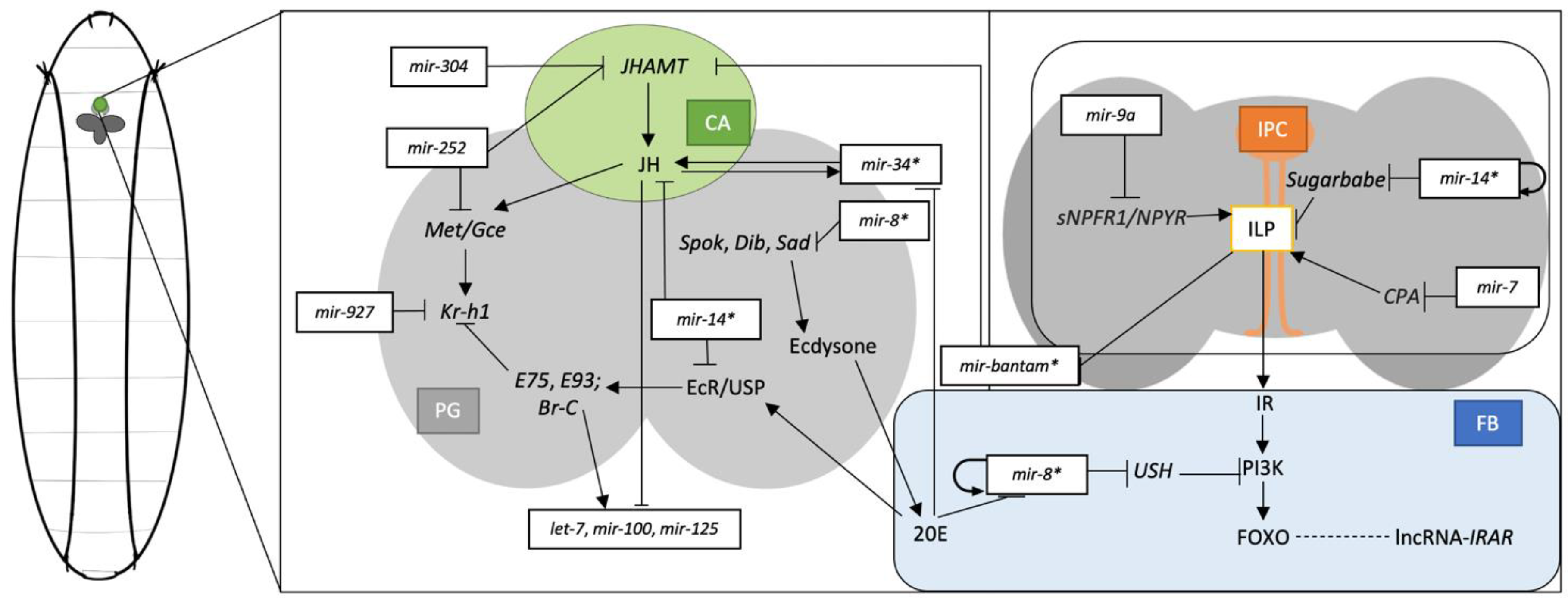
:1. Introduction
2. microRNAs in Drosophila
2.1. miRNA in Drosophila Development and Metamorphosis

| miRNA | Validated Targets | Function, Hormonal and Metabolic Regulations | References |
|---|---|---|---|
| Development and Metamorphosis | |||
| miR-6, miR-11 | rpr, grim, hid, skl | Embryonic apoptosis and CNS development | [29] |
| bantam | Akt | Dendrite arbour growth | [30] |
| miR-281, miR-311, miR-79, miR-92, miR-305, miR-131, miR-31a | N/A | Larval development | [31] |
| let-7, miR-125 | N/A | Wing development | [33] |
| miR-9a | dLMO (Beadex) | Wing development | [34] |
| miR-1, miR-2b, miR-2c, miR-13b, miR-987 | N/A | Wing development | [35] |
| miR-965 | stg, wg | Abdomen morphogenesis | [36] |
| miR-6/5/4/286/3/309 | N/A | Leg development | [37] |
| miR-277, miR-304 | mbl | Muscle development | [38] |
| Sesquiterpenoid and Ecdysteroid system | |||
| bantam | JHAMT | JH biosynthesis | [39] |
| miR-252, miR-304 | JHAMT | JH biosynthesis | [39] |
| miR-8, miR-14, miR-34, miR-278 | Met/Gce | JH signalling pathway | [39] |
| miR-927 | Kr-h1 | JH signalling pathway | [40] |
| bantam | phantom, shade, disembodied | Ecdysone biosynthesis | [41] |
| miR-14 | EcR | Ecdysone signalling pathway | [42] |
| let-7, miR-125 | dpt | Regulated by ecdysone; innate immune systems | [43] |
| miR-8 | N/A | Cell growth regulated by ecdysone; induce JH biosynthesis pathway | [44] |
| miR-252 | Abi | Cell division | [45] |
| Insulin pathway and Lipid metabolism | |||
| miR-8 | ush | Insulin signalling pathway; body size | [46] |
| miR-7 | cpa | Insulin secretion pathway | [47] |
| miR-9a | short neuropeptide F receptor 1 | Insulin signalling pathway; body size | [48] |
| miR-14 | sg | Insulin-producing pathway; fat accumulation | [49] |
| miR-305 | Dp53 | TOR regulator signalling in response to nutrient | [50] |
| miR-305 | InR, PI3K, Hairless | Notch and insulin pathways in adaptive homeostasis | [50,51] |
| miR-277 | FAO | Fatty acid metabolism; homeostasis | [52] |
| miR-14 | N/A | Fatty acid metabolism | [53] |
| miR-278 | ex | Insulin sensibility | [54] |
| Sexual Development | |||
| miR-184 | saxophone | Female germline development; nurse cell nutrient support; Egg chamber size | [55] |
| miR-318 | Tramtrack69 | Oogenesis; chorion gene amplification | [56] |
| miR-282 | rut | Egg production; apoptotic activity | [57] |
| miR-989 | N/A | Border cell migration in ovaries | [58] |
| bantam | N/A | Adult germline stem cell formation | [59] |
| bantam | dFmr1 | GSCs maintenance in ovaries | [60] |
| miR-7, miR-309, miR-278 | dap | Cell cycle regulations | [61] |
| let-7 | N/A | Sex-biased ecdysone signalling; aging in testis stem cells and fertility | [62,63] |
| miR-190 | N/A | Sexually dimorphic in male; regulating neuronal activities and lifespan | [64] |
| Lifespan and Aging | |||
| bantam, miR-1, miR-190, miR-279, miR-996 | N/A | Survival; viability | [24] |
| miR-282 | N/A | Increase lifespan | [57] |
| miR-305 | N/A | Aging; locomotor activity; abnormal protein aggregation in muscle; oxidative stress | [65] |
| miR-184, let-7 | N/A | Prolonged lifespan | [66,67] |
| miR-125 | Chinmo | Prolonged lifespan | [68] |
| miR-34 | Pcl, Su(z)12 | Chaperone expressions; healthy brain aging | [69,70] |
| miR-34 | Lst8 | Healthy brain aging | [71] |
| miR-277 | N/A | Branched-chain amino acid catabolism; growth regulation; Reduced lifespan | [72] |
| Circadian Rhythm and Photoperiod | |||
| miR-124 | BMP signalling | Circadian rhythm; rhythmic normality | [73] |
| miR-276a | tim | Circadian rhythm | [74] |
| miR-279 | unpaired | Circadian behaviour; JAK/STAT circuit | [75] |
| miR-959-964 | N/A | Rhythmic feeding; loop of feeding period control | [76] |
| miR-210 | Fasciclin 2 | Photoreceptor function | [77] |
| miR-263a, miR-263b | clk, cwo | Circadian rhythm | [78] |
| let-7 | cwo | Circadian rhythm | [79] |
2.2. miRNA in the Sesquiterpenoid and Ecdysteroid Systems
2.3. Insulin Pathway and Lipid Metabolism
2.4. miRNA in Sexual Development
2.5. miRNA in Drosophila Lifespan and Aging
2.6. miRNA in Drosophila Circadian Rhythm and Photoperiod
3. lncRNA in Drosophila
3.1. lncRNA in Development, Metamorphosis, and Ecdysteroid Hormone Systems
3.2. lncRNA in Nutrient Metabolism and Aging
3.3. lncRNA in Sexual Development
3.4. lncRNA in Drosophila Circadian Rhythm
3.5. lncRNA and miRNA Interactions
4. circRNA in Drosophila
| circRNA | Validated Targets | Function, Hormonal and Metabolic Regulations | References |
|---|---|---|---|
| sisR-4 | deadpan | Embryogenesis; positive feedback loop formation | [168,169] |
| circMbl | muscleblind | Eye and muscle development; negative feedback loop formation | [163,173] |
| Edis | castor | Neuromuscular junctions; mushroom body neuronal development; IMD pathway | [175,176] |
| circSfl | N/A | Increased female lifespan; insulin pathway; aging | [180] |
| circBoule | Hsc4, Hsp60C | Male fertility due to heat-stress | [181] |
5. Conclusions and Future Perspectives
Funding
Conflicts of Interest
References
- Truman, J.W. The Evolution of Insect Metamorphosis. Curr. Biol. 2019, 29, R1252–R1268. [Google Scholar] [CrossRef]
- Cao, J.Q.; Tong, W.S.; Yu, H.Y.; Tobe, S.S.; Bendena, W.G.; Hui, J.H.L. The Role of MicroRNAs in Drosophila Regulation of Insulin-Like Peptides and Ecdysteroid Signalling: Where Are We Now? In Advances in Insect Physiology; Elsevier: Amsterdam, The Netherlands, 2017; Volume 53, pp. 55–85. [Google Scholar] [CrossRef]
- Qu, Z.; Bendena, W.G.; Tobe, S.S.; Hui, J.H.L. Juvenile Hormone and Sesquiterpenoids in Arthropods: Biosynthesis, Signaling, and Role of MicroRNA. J. Steroid Biochem. Mol. Biol. 2018, 184, 69–76. [Google Scholar] [CrossRef] [PubMed]
- Tsang, S.S.K.; Law, S.T.S.; Li, C.; Qu, Z.; Bendena, W.G.; Tobe, S.S.; Hui, J.H.L. Diversity of Insect Sesquiterpenoid Regulation. Front. Genet. 2020, 11, 1027. [Google Scholar] [CrossRef] [PubMed]
- Truman, J.W.; Riddiford, L.M. The Evolution of Insect Metamorphosis: A Developmental and Endocrine View. Phil. Trans. R. Soc. B 2019, 374, 20190070. [Google Scholar] [CrossRef] [Green Version]
- Brandenburger, T.; Salgado Somoza, A.; Devaux, Y.; Lorenzen, J.M. Noncoding RNAs in Acute Kidney Injury. Kidney Int. 2018, 94, 870–881. [Google Scholar] [CrossRef] [PubMed]
- Cooper, G. The Cell: A Molecular Approach, 2nd ed.; Sinauer Associates: Sunderland, MA, USA, 2000. [Google Scholar]
- Briata, P.; Gherzi, R. Long Non-Coding RNA-Ribonucleoprotein Networks in the Post-Transcriptional Control of Gene Expression. ncRNA 2020, 6, 40. [Google Scholar] [CrossRef]
- Cesana, M.; Cacchiarelli, D.; Legnini, I.; Santini, T.; Sthandier, O.; Chinappi, M.; Tramontano, A.; Bozzoni, I. A Long Noncoding RNA Controls Muscle Differentiation by Functioning as a Competing Endogenous RNA. Cell 2011, 147, 358–369. [Google Scholar] [CrossRef] [Green Version]
- Yuva-Aydemir, Y.; Simkin, A.; Gascon, E.; Gao, F.-B. MicroRNA-9: Functional Evolution of a Conserved Small Regulatory RNA. RNA Biol. 2011, 8, 557–564. [Google Scholar] [CrossRef] [Green Version]
- Nong, W.; Cao, J.; Li, Y.; Qu, Z.; Sun, J.; Swale, T.; Yip, H.Y.; Qian, P.Y.; Qiu, J.-W.; Kwan, H.S.; et al. Jellyfish Genomes Reveal Distinct Homeobox Gene Clusters and Conservation of Small RNA Processing. Nat. Commun. 2020, 11, 3051. [Google Scholar] [CrossRef]
- Li, Y.; Hui, J.H.L. Small RNAs in Cnidaria: A Review. Evol. Appl. 2022. [Google Scholar] [CrossRef]
- O’Brien, J.; Hayder, H.; Zayed, Y.; Peng, C. Overview of MicroRNA Biogenesis, Mechanisms of Actions, and Circulation. Front. Endocrinol. 2018, 9, 402. [Google Scholar] [CrossRef] [PubMed] [Green Version]
- Ha, M.; Kim, V.N. Regulation of MicroRNA Biogenesis. Nat. Rev. Mol. Cell. Biol. 2014, 15, 509–524. [Google Scholar] [CrossRef] [PubMed]
- Iwasaki, S.; Kawamata, T.; Tomari, Y. Drosophila Argonaute1 and Argonaute2 Employ Distinct Mechanisms for Translational Repression. Mol. Cell 2009, 34, 58–67. [Google Scholar] [CrossRef] [Green Version]
- Jo, M.H.; Shin, S.; Jung, S.-R.; Kim, E.; Song, J.-J.; Hohng, S. Human Argonaute 2 Has Diverse Reaction Pathways on Target RNAs. Mol. Cell 2015, 59, 117–124. [Google Scholar] [CrossRef] [PubMed] [Green Version]
- Zhang, J.; Zhou, W.; Liu, Y.; Liu, T.; Li, C.; Wang, L. Oncogenic Role of MicroRNA-532-5p in Human Colorectal Cancer via Targeting of the 5’UTR of RUNX3. Oncol. Lett. 2018, 15, 7215–7220. [Google Scholar] [CrossRef] [Green Version]
- Gu, W.; Xu, Y.; Xie, X.; Wang, T.; Ko, J.-H.; Zhou, T. The Role of RNA Structure at 5′ Untranslated Region in MicroRNA-Mediated Gene Regulation. RNA 2014, 20, 1369–1375. [Google Scholar] [CrossRef] [Green Version]
- Dharap, A.; Pokrzywa, C.; Murali, S.; Pandi, G.; Vemuganti, R. MicroRNA MiR-324-3p Induces Promoter-Mediated Expression of RelA Gene. PLoS ONE 2013, 8, e79467. [Google Scholar] [CrossRef] [Green Version]
- Place, R.F.; Li, L.C.; Pookot, D.; Noonan, E.J.; Dahiya, R. MicroRNA-373 Induces Expression of Genes with Complementary Promoter Sequences. Proc. Natl. Acad. Sci. USA 2008, 105, 1608–1613. [Google Scholar] [CrossRef] [Green Version]
- Ørom, U.A.; Nielsen, F.C.; Lund, A.H. MicroRNA-10a Binds the 5′UTR of Ribosomal Protein MRNAs and Enhances Their Translation. Mol. Cell 2008, 30, 460–471. [Google Scholar] [CrossRef]
- Kozomara, A.; Birgaoanu, M.; Griffiths-Jones, S. miRBase: From MicroRNA Sequences to Function. Nucleic Acids Res. 2019, 47, D155–D162. [Google Scholar] [CrossRef]
- Soni, K.; Choudhary, A.; Patowary, A.; Singh, A.R.; Bhatia, S.; Sivasubbu, S.; Chandrasekaran, S.; Pillai, B. miR-34 Is Maternally Inherited in Drosophila Melanogaster and Danio Rerio. Nucleic Acids Res. 2013, 41, 4470–4480. [Google Scholar] [CrossRef] [PubMed] [Green Version]
- Chen, Y.W.; Song, S.; Weng, R.; Verma, P.; Kugler, J.M.; Buescher, M.; Rouam, S.; Cohen, S.M. Systematic Study of Drosophila MicroRNA Functions Using a Collection of Targeted Knockout Mutations. Dev. Cell 2014, 31, 784–800. [Google Scholar] [CrossRef] [PubMed] [Green Version]
- Tadros, W.; Goldman, A.L.; Babak, T.; Menzies, F.; Vardy, L.; Orr-Weaver, T.; Hughes, T.R.; Westwood, J.T.; Smibert, C.A.; Lipshitz, H.D. SMAUG Is a Major Regulator of Maternal mRNA Destabilization in Drosophila and Its Translation Is Activated by the PAN GU Kinase. Dev. Cell 2007, 12, 143–155. [Google Scholar] [CrossRef] [PubMed] [Green Version]
- Bushati, N.; Stark, A.; Brennecke, J.; Cohen, S.M. Temporal Reciprocity of MiRNAs and Their Targets during the Maternal-to-Zygotic Transition in Drosophila. Curr. Biol. 2008, 18, 501–506. [Google Scholar] [CrossRef] [Green Version]
- Iwasaki, Y.W.; Siomi, H. miRNA Regulatory Ecosystem in Early Development. Mol. Cell 2014, 56, 615–616. [Google Scholar] [CrossRef] [Green Version]
- Fu, S.; Nien, C.Y.; Liang, H.L.; Rushlow, C. Co-Activation of MicroRNAs by Zelda Is Essential for Early Drosophila Development. Development 2014, 141, 2108–2118. [Google Scholar] [CrossRef] [PubMed] [Green Version]
- Ge, W.; Chen, Y.W.; Weng, R.; Lim, S.F.; Buescher, M.; Zhang, R.; Cohen, S.M. Overlapping Functions of MicroRNAs in Control of Apoptosis during Drosophila Embryogenesis. Cell Death Differ. 2012, 19, 839–846. [Google Scholar] [CrossRef] [PubMed] [Green Version]
- Parrish, J.Z.; Xu, P.; Kim, C.C.; Jan, L.Y.; Jan, Y.N. The MicroRNA Bantam Functions in Epithelial Cells to Regulate Scaling Growth of Dendrite Arbors in Drosophila Sensory Neurons. Neuron 2009, 63, 788–802. [Google Scholar] [CrossRef] [Green Version]
- Enright, A.; John, B.; Gaul, U.; Tuschl, T.; Sander, C.; Marks, D. MicroRNA Targets in Drosophila. Genome Biol. 2003, 4, P8. [Google Scholar] [CrossRef]
- Belles, X. MicroRNAs and the Evolution of Insect Metamorphosis. Annu. Rev. Entomol. 2017, 62, 111–125. [Google Scholar] [CrossRef] [Green Version]
- Caygill, E.E.; Johnston, L.A. Temporal Regulation of Metamorphic Processes in Drosophila by the Let-7 and MiR-125 Heterochronic MicroRNAs. Curr. Biol. 2008, 18, 943–950. [Google Scholar] [CrossRef] [Green Version]
- Biryukova, I.; Asmar, J.; Abdesselem, H.; Heitzler, P. Drosophila Mir-9a Regulates Wing Development via Fine-Tuning Expression of the LIM Only Factor, DLMO. Dev. Biol. 2009, 327, 487–496. [Google Scholar] [CrossRef] [PubMed]
- Fulga, T.A.; McNeill, E.M.; Binari, R.; Yelick, J.; Blanche, A.; Booker, M.; Steinkraus, B.R.; Schnall-Levin, M.; Zhao, Y.; DeLuca, T.; et al. A Transgenic Resource for Conditional Competitive Inhibition of Conserved Drosophila MicroRNAs. Nat. Commun. 2015, 6, 7279. [Google Scholar] [CrossRef] [PubMed] [Green Version]
- Verma, P.; Cohen, S.M. miR-965 Controls Cell Proliferation and Migration during Tissue Morphogenesis in the Drosophila Abdomen. eLife 2015, 4, e07389. [Google Scholar] [CrossRef] [PubMed]
- Qu, Z.; Yiu, W.C.; Yip, H.Y.; Nong, W.; Yu, C.W.C.; Lee, I.H.T.; Wong, A.Y.P.; Wong, N.W.Y.; Cheung, F.K.M.; Chan, T.F.; et al. Micro-RNA Clusters Integrate Evolutionary Constraints on Expression and Target Affinities: The MiR-6/5/4/286/3/309 Cluster in Drosophila. Mol. Biol. Evol. 2020, 37, 2955–2965. [Google Scholar] [CrossRef]
- Cerro-Herreros, E.; Fernandez-Costa, J.M.; Sabater-Arcis, M.; Llamusi, B.; Artero, R. Derepressing Muscleblind Expression by MiRNA Sponges Ameliorates Myotonic Dystrophy-like Phenotypes in Drosophila. Sci. Rep. 2016, 6, 36230. [Google Scholar] [CrossRef] [Green Version]
- Qu, Z.; Bendena, W.G.; Nong, W.; Siggens, K.W.; Noriega, F.G.; Kai, Z.; Zang, Y.; Koon, A.C.; Chan, H.Y.E.; Chan, T.F.; et al. MicroRNAs Regulate the Sesquiterpenoid Hormonal Pathway in Drosophila and Other Arthropods. Proc. R. Soc. B 2017, 284, 20171827. [Google Scholar] [CrossRef] [Green Version]
- He, Q.; Zhang, Y.; Dong, W. MicroRNA miR-927 Targets the Juvenile Hormone Primary Response Gene Krüppel Homolog1 to Control Drosophila Developmental Growth. Insect. Mol. Biol. 2020, 29, 545–554. [Google Scholar] [CrossRef]
- Boulan, L.; Martín, D.; Milán, M. Bantam MiRNA Promotes Systemic Growth by Connecting Insulin Signaling and Ecdysone Production. Curr. Biol. 2013, 23, 473–478. [Google Scholar] [CrossRef] [PubMed]
- Chawla, G.; Sokol, N.S. Hormonal Activation of Let-7-C MicroRNAs via EcR Is Required for Adult Drosophila Melanogaster Morphology and Function. Development 2012, 139, 1788–1797. [Google Scholar] [CrossRef] [Green Version]
- Garbuzov, A.; Tatar, M. Hormonal Regulation of Drosophila MicroRNA Let-7 and MiR-125 That Target Innate Immunity. Fly 2010, 4, 306–311. [Google Scholar] [CrossRef] [PubMed] [Green Version]
- Zhang, J.; Wen, D.; Li, E.Y.; Palli, S.R.; Li, S.; Wang, J.; Liu, S. MicroRNA MiR-8 Promotes Cell Growth of Corpus Allatum and Juvenile Hormone Biosynthesis Independent of Insulin/IGF Signaling in Drosophila Melanogaster. Insect Biochem. Mol. Biol. 2021, 136, 103611. [Google Scholar] [CrossRef]
- Lim, D.; Lee, S.; Yun Han, J.; Choi, M.; Hong, J.; Seong, Y.; Kwon, Y.; Sik Lee, Y. Ecdysone-responsive MicroRNA-252-5p Controls the Cell Cycle by Targeting Abi in Drosophila. FASEB J. 2018, 32, 4519–4533. [Google Scholar] [CrossRef] [PubMed] [Green Version]
- Hyun, S.; Lee, J.H.; Jin, H.; Nam, J.; Namkoong, B.; Lee, G.; Chung, J.; Kim, V.N. Conserved MicroRNA MiR-8/MiR-200 and Its Target USH/FOG2 Control Growth by Regulating PI3K. Cell 2009, 139, 1096–1108. [Google Scholar] [CrossRef] [Green Version]
- Agbu, P.; Cassidy, J.J.; Braverman, J.; Jacobson, A.; Carthew, R.W. MicroRNA MiR-7 Regulates Secretion of Insulin-Like Peptides. Endocrinology 2020, 161, bqz040. [Google Scholar] [CrossRef]
- Suh, Y.S.; Bhat, S.; Hong, S.-H.; Shin, M.; Bahk, S.; Cho, K.S.; Kim, S.-W.; Lee, K.-S.; Kim, Y.-J.; Jones, W.D.; et al. Genome-Wide MicroRNA Screening Reveals That the Evolutionary Conserved MiR-9a Regulates Body Growth by Targeting SNPFR1/NPYR. Nat. Commun. 2015, 6, 7693. [Google Scholar] [CrossRef] [PubMed] [Green Version]
- Varghese, J.; Lim, S.F.; Cohen, S.M. Drosophila MiR-14 Regulates Insulin Production and Metabolism through Its Target, Sugarbabe. Genes Dev. 2010, 24, 2748–2753. [Google Scholar] [CrossRef] [Green Version]
- Barrio, L.; Dekanty, A.; Milán, M. MicroRNA-Mediated Regulation of Dp53 in the Drosophila Fat Body Contributes to Metabolic Adaptation to Nutrient Deprivation. Cell Rep. 2014, 8, 528–541. [Google Scholar] [CrossRef] [Green Version]
- Foronda, D.; Weng, R.; Verma, P.; Chen, Y.-W.; Cohen, S.M. Coordination of Insulin and Notch Pathway Activities by MicroRNA MiR-305 Mediates Adaptive Homeostasis in the Intestinal Stem Cells of the Drosophila Gut. Genes Dev. 2014, 28, 2421–2431. [Google Scholar] [CrossRef]
- Zipper, L.; Batchu, S.; Kaya, N.H.; Antonello, Z.A.; Reiff, T. The MicroRNA MiR-277 Controls Physiology and Pathology of the Adult Drosophila Midgut by Regulating the Expression of Fatty Acid β-Oxidation-Related Genes in Intestinal Stem Cells. Metabolites 2022, 12, 315. [Google Scholar] [CrossRef]
- Xu, P.; Vernooy, S.Y.; Guo, M.; Hay, B.A. The Drosophila MicroRNA Mir-14 Suppresses Cell Death and Is Required for Normal Fat Metabolism. Curr. Biol. 2003, 13, 790–795. [Google Scholar] [CrossRef] [PubMed] [Green Version]
- Teleman, A.A.; Cohen, S.M. Drosophila Lacking MicroRNA MiR-278 Are Defective in Energy Homeostasis. Genes Dev. 2006, 20, 417–422. [Google Scholar] [CrossRef] [PubMed] [Green Version]
- Iovino, N.; Pane, A.; Gaul, U. miR-184 Has Multiple Roles in Drosophila Female Germline Development. Dev. Cell 2009, 17, 123–133. [Google Scholar] [CrossRef] [PubMed] [Green Version]
- Ge, W.; Deng, Q.; Guo, T.; Hong, X.; Kugler, J.-M.; Yang, X.; Cohen, S.M. Regulation of Pattern Formation and Gene Amplification During Drosophila Oogenesis by the MiR-318 MicroRNA. Genetics 2015, 200, 255–265. [Google Scholar] [CrossRef] [PubMed] [Green Version]
- Vilmos, P.; Bujna, Á.; Szuperák, M.; Havelda, Z.; Várallyay, É.; Szabad, J.; Kucerova, L.; Somogyi, K.; Kristó, I.; Lukácsovich, T.; et al. Viability, Longevity, and Egg Production of Drosophila Melanogaster Are Regulated by the MiR-282 MicroRNA. Genetics 2013, 195, 469–480. [Google Scholar] [CrossRef] [Green Version]
- Kugler, J.M.; Chen, Y.W.; Weng, R.; Cohen, S.M. miR-989 Is Required for Border Cell Migration in the Drosophila Ovary. PLoS ONE 2013, 8, e67075. [Google Scholar] [CrossRef]
- Shcherbata, H.R.; Ward, E.J.; Fischer, K.A.; Yu, J.-Y.; Reynolds, S.H.; Chen, C.-H.; Xu, P.; Hay, B.A.; Ruohola-Baker, H. Stage-Specific Differences in the Requirements for Germline Stem Cell Maintenance in the Drosophila Ovary. Cell Stem Cell 2007, 1, 698–709. [Google Scholar] [CrossRef] [Green Version]
- Yang, Y.; Xu, S.; Xia, L.; Wang, J.; Wen, S.; Jin, P.; Chen, D. The Bantam MicroRNA Is Associated with Drosophila Fragile X Mental Retardation Protein and Regulates the Fate of Germline Stem Cells. PLoS Genet. 2009, 5, e1000444. [Google Scholar] [CrossRef]
- Yu, J.Y.; Reynolds, S.H.; Hatfield, S.D.; Shcherbata, H.R.; Fischer, K.A.; Ward, E.J.; Long, D.; Ding, Y.; Ruohola-Baker, H. Dicer-1-Dependent Dacapo Suppression Acts Downstream of Insulin Receptor in Regulating Cell Division of Drosophila Germline Stem Cells. Development 2009, 136, 1497–1507. [Google Scholar] [CrossRef]
- Fagegaltier, D.; König, A.; Gordon, A.; Lai, E.C.; Gingeras, T.R.; Hannon, G.J.; Shcherbata, H.R. A Genome-Wide Survey of Sexually Dimorphic Expression of Drosophila MiRNAs Identifies the Steroid Hormone-Induced MiRNA Let-7 as a Regulator of Sexual Identity. Genetics 2014, 198, 647–668. [Google Scholar] [CrossRef] [Green Version]
- Toledano, H.; D’Alterio, C.; Czech, B.; Levine, E.; Jones, D.L. The Let-7–Imp Axis Regulates Ageing of the Drosophila Testis Stem-Cell Niche. Nature 2012, 485, 605–610. [Google Scholar] [CrossRef] [PubMed] [Green Version]
- Fernandes, J.; Varghese, J. Sexually Dimorphic MicroRNA MiR-190 Regulates Lifespan in Male Drosophila. RNA Biol. 2022, 19, 1085–1093. [Google Scholar] [CrossRef] [PubMed]
- Ueda, M.; Sato, T.; Ohkawa, Y.; Inoue, Y.H. Identification of MiR-305, a MicroRNA That Promotes Aging, and Its Target MRNAs in Drosophila. Genes Cells 2018, 23, 80–93. [Google Scholar] [CrossRef] [PubMed] [Green Version]
- Chawla, G.; Deosthale, P.; Childress, S.; Wu, Y.; Sokol, N.S. A Let-7-to-MiR-125 MicroRNA Switch Regulates Neuronal Integrity and Lifespan in Drosophila. PLoS Genet. 2016, 12, e1006247. [Google Scholar] [CrossRef] [PubMed] [Green Version]
- Gendron, C.M.; Pletcher, S.D. Micro RNA s Mir-184 and Let-7 Alter Drosophila Metabolism and Longevity. Aging Cell 2017, 16, 1434–1438. [Google Scholar] [CrossRef] [Green Version]
- Pandey, M.; Bansal, S.; Bar, S.; Yadav, A.K.; Sokol, N.S.; Tennessen, J.M.; Kapahi, P.; Chawla, G. miR-125-Chinmo Pathway Regulates Dietary Restriction-Dependent Enhancement of Lifespan in Drosophila. eLife 2021, 10, e62621. [Google Scholar] [CrossRef]
- Liu, N.; Landreh, M.; Cao, K.; Abe, M.; Hendriks, G.-J.; Kennerdell, J.R.; Zhu, Y.; Wang, L.-S.; Bonini, N.M. The MicroRNA MiR-34 Modulates Ageing and Neurodegeneration in Drosophila. Nature 2012, 482, 519–523. [Google Scholar] [CrossRef] [Green Version]
- Kennerdell, J.R.; Liu, N.; Bonini, N.M. miR-34 Inhibits Polycomb Repressive Complex 2 to Modulate Chaperone Expression and Promote Healthy Brain Aging. Nat. Commun. 2018, 9, 4188. [Google Scholar] [CrossRef] [Green Version]
- Srinivasan, A.R.; Tran, T.T.; Bonini, N.M. Loss of MiR-34 in Drosophila Dysregulates Protein Translation and Protein Turnover in the Aging Brain. Aging Cell 2022, 21, e13559. [Google Scholar] [CrossRef]
- Esslinger, S.M.; Schwalb, B.; Helfer, S.; Michalik, K.M.; Witte, H.; Maier, K.C.; Martin, D.; Michalke, B.; Tresch, A.; Cramer, P.; et al. Drosophila MiR-277 Controls Branched-Chain Amino Acid Catabolism and Affects Lifespan. RNA Biol. 2013, 10, 1042–1056. [Google Scholar] [CrossRef] [PubMed] [Green Version]
- Garaulet, D.L.; Sun, K.; Li, W.; Wen, J.; Panzarino, A.M.; O’Neil, J.L.; Hiesinger, P.R.; Young, M.W.; Lai, E.C. miR-124 Regulates Diverse Aspects of Rhythmic Behavior in Drosophila. J. Neurosci. 2016, 36, 3414–3421. [Google Scholar] [CrossRef] [PubMed] [Green Version]
- Chen, X.; Rosbash, M. miR-276a Strengthens Drosophila Circadian Rhythms by Regulating Timeless Expression. Proc. Natl. Acad. Sci. USA 2016, 113, E2965–E2972. [Google Scholar] [CrossRef] [PubMed] [Green Version]
- Luo, W.; Sehgal, A. Regulation of Circadian Behavioral Output via a MicroRNA-JAK/STAT Circuit. Cell 2012, 148, 765–779. [Google Scholar] [CrossRef] [Green Version]
- Vodala, S.; Pescatore, S.; Rodriguez, J.; Buescher, M.; Chen, Y.W.; Weng, R.; Cohen, S.M.; Rosbash, M. The Oscillating MiRNA 959–964 Cluster Impacts Drosophila Feeding Time and Other Circadian Outputs. Cell Metab. 2012, 16, 601–612. [Google Scholar] [CrossRef] [Green Version]
- Niu, Y.; Liu, Z.; Nian, X.; Xu, X.; Zhang, Y. miR-210 Controls the Evening Phase of Circadian Locomotor Rhythms through Repression of Fasciclin 2. PLoS Genet. 2019, 15, e1007655. [Google Scholar] [CrossRef] [PubMed] [Green Version]
- Yang, M.; Lee, J.-E.; Padgett, R.W.; Edery, I. Circadian Regulation of a Limited Set of Conserved MicroRNAs in Drosophila. BMC Genom. 2008, 9, 83. [Google Scholar] [CrossRef] [Green Version]
- Chen, W.; Liu, Z.; Li, T.; Zhang, R.; Xue, Y.; Zhong, Y.; Bai, W.; Zhou, D.; Zhao, Z. Regulation of Drosophila Circadian Rhythms by MiRNA Let-7 Is Mediated by a Regulatory Cycle. Nat. Commun. 2014, 5, 5549. [Google Scholar] [CrossRef] [Green Version]
- Cheong, S.P.S.; Huang, J.; Bendena, W.G.; Tobe, S.S.; Hui, J.H.L. Evolution of Ecdysis and Metamorphosis in Arthropods: The Rise of Regulation of Juvenile Hormone. Integr. Comp. Biol. 2015, 55, 878–890. [Google Scholar] [CrossRef] [Green Version]
- Niwa, Y.S.; Niwa, R. Neural Control of Steroid Hormone Biosynthesis during Development in the Fruit Fly Drosophila Melanogaster. Genes Genet. Syst. 2014, 89, 27–34. [Google Scholar] [CrossRef]
- Nakagawa, Y.; Sonobe, H. 20-Hydroxyecdysone. In Handbook of Hormones; Elsevier: Amsterdam, The Netherlands, 2016; pp. 560–563. [Google Scholar] [CrossRef]
- Syed, M.H.; Mark, B.; Doe, C.Q. Steroid Hormone Induction of Temporal Gene Expression in Drosophila Brain Neuroblasts Generates Neuronal and Glial Diversity. eLife 2017, 6, e26287. [Google Scholar] [CrossRef]
- Williams, C.M.; Kafatos, F.C. Theoretical Aspects of the Action of Juvenile Hormone. In Insect Juvenile Hormones. Chemistry and Action; Academic Press: New York, NY, USA, 1972; pp. 29–44. [Google Scholar]
- Truman, J.W.; Riddiford, L.M. Chinmo Is the Larval Member of the Molecular Trinity That Directs Drosophila Metamorphosis. Proc. Natl. Acad. Sci. USA 2022, 119, e2201071119. [Google Scholar] [CrossRef] [PubMed]
- Lavrynenko, O.; Rodenfels, J.; Carvalho, M.; Dye, N.A.; Lafont, R.; Eaton, S.; Shevchenko, A. The Ecdysteroidome of Drosophila: Influence of Diet and Development. Development 2015, 142, dev.124982. [Google Scholar] [CrossRef] [Green Version]
- Ono, H. Ecdysone Differentially Regulates Metamorphic Timing Relative to 20-Hydroxyecdysone by Antagonizing Juvenile Hormone in Drosophila Melanogaster. Dev. Biol. 2014, 391, 32–42. [Google Scholar] [CrossRef] [PubMed] [Green Version]
- Liu, S.; Li, K.; Gao, Y.; Liu, X.; Chen, W.; Ge, W.; Feng, Q.; Palli, S.R.; Li, S. Antagonistic Actions of Juvenile Hormone and 20-Hydroxyecdysone within the Ring Gland Determine Developmental Transitions in Drosophila. Proc. Natl. Acad. Sci. USA 2017, 115, 139–144. [Google Scholar] [CrossRef] [PubMed] [Green Version]
- Belles, X.; Santos, C.G. The MEKRE93 (Methoprene Tolerant-Krüppel Homolog 1-E93) Pathway in the Regulation of Insect Metamorphosis, and the Homology of the Pupal Stage. Insect Biochem. Mol. Biol. 2014, 52, 60–68. [Google Scholar] [CrossRef]
- Huang, J.; Tian, L.; Peng, C.; Abdou, M.; Wen, D.; Wang, Y.; Li, S.; Wang, J. DPP-Mediated TGFβ Signaling Regulates Juvenile Hormone Biosynthesis by Activating the Expression of Juvenile Hormone Acid Methyltransferase. Development 2011, 138, 2283–2291. [Google Scholar] [CrossRef] [Green Version]
- Kayukawa, T.; Jouraku, A.; Ito, Y.; Shinoda, T. Molecular Mechanism Underlying Juvenile Hormone-Mediated Repression of Precocious Larval–Adult Metamorphosis. Proc. Natl. Acad. Sci. USA 2017, 114, 1057–1062. [Google Scholar] [CrossRef] [PubMed] [Green Version]
- Kayukawa, T.; Nagamine, K.; Ito, Y.; Nishita, Y.; Ishikawa, Y.; Shinoda, T. Krüppel Homolog 1 Inhibits Insect Metamorphosis via Direct Transcriptional Repression of Broad-Complex, a Pupal Specifier Gene. J. Biol. Chem. 2016, 291, 1751–1762. [Google Scholar] [CrossRef] [PubMed]
- Aravin, A.A.; Lagos-Quintana, M.; Yalcin, A.; Zavolan, M.; Marks, D.; Snyder, B.; Gaasterland, T.; Meyer, J.; Tuschl, T. The Small RNA Profile during Drosophila Melanogaster Development. Dev. Cell 2003, 5, 337–350. [Google Scholar] [CrossRef] [Green Version]
- Sempere, L.F.; Sokol, N.S.; Dubrovsky, E.B.; Berger, E.M.; Ambros, V. Temporal Regulation of MicroRNA Expression in Drosophila Melanogaster Mediated by Hormonal Signals and Broad-Complex Gene Activity. Dev. Biol. 2003, 259, 9–18. [Google Scholar] [CrossRef] [Green Version]
- Niwa, R.; Niimi, T.; Honda, N.; Yoshiyama, M.; Itoyama, K.; Kataoka, H.; Shinoda, T. Juvenile Hormone Acid O-Methyltransferase in Drosophila Melanogaster. Insect Biochem. Mol. Biol. 2008, 38, 714–720. [Google Scholar] [CrossRef] [PubMed] [Green Version]
- Pecasse, F.; Beck, Y.; Ruiz, C.; Richards, G. Krüppel-Homolog, a Stage-Specific Modulator of the Prepupal Ecdysone Response, Is Essential for Drosophila Metamorphosis. Dev. Biol. 2000, 221, 53–67. [Google Scholar] [CrossRef] [PubMed] [Green Version]
- Varghese, J.; Cohen, S.M. MicroRNA MiR-14 Acts to Modulate a Positive Autoregulatory Loop Controlling Steroid Hormone Signaling in Drosophila. Genes Dev. 2007, 21, 2277–2282. [Google Scholar] [CrossRef] [PubMed] [Green Version]
- Jin, H.; Kim, V.N.; Hyun, S. Conserved MicroRNA MiR-8 Controls Body Size in Response to Steroid Signaling in Drosophila. Genes Dev. 2012, 26, 1427–1432. [Google Scholar] [CrossRef] [Green Version]
- Wessels, H.-H.; Lebedeva, S.; Hirsekorn, A.; Wurmus, R.; Akalin, A.; Mukherjee, N.; Ohler, U. Global Identification of Functional MicroRNA-MRNA Interactions in Drosophila. Nat. Commun. 2019, 10, 1626. [Google Scholar] [CrossRef] [Green Version]
- Agarwal, V.; Subtelny, A.O.; Thiru, P.; Ulitsky, I.; Bartel, D.P. Predicting MicroRNA Targeting Efficacy in Drosophila. Genome Biol. 2018, 19, 152. [Google Scholar] [CrossRef] [Green Version]
- Lim, D.; Lee, S.; Choi, M.; Han, J.Y.; Seong, Y.; Na, D.; Kwon, Y.; Lee, Y.S. The Conserved MicroRNA MiR-8-3p Coordinates the Expression of V-ATPase Subunits to Regulate Ecdysone Biosynthesis for Drosophila Metamorphosis. FASEB J. 2020, 34, 6449–6465. [Google Scholar] [CrossRef] [Green Version]
- Bendena, W.G.; Hui, J.H.L.; Chin-Sang, I.; Tobe, S.S. Neuropeptide and MicroRNA Regulators of Juvenile Hormone Production. Gen. Comp. Endocrinol. 2020, 295, 113507. [Google Scholar] [CrossRef]
- Edgar, B.A. How Flies Get Their Size: Genetics Meets Physiology. Nat. Rev. Genet. 2006, 7, 907–916. [Google Scholar] [CrossRef]
- Böhni, R.; Riesgo-Escovar, J.; Oldham, S.; Brogiolo, W.; Stocker, H.; Andruss, B.F.; Beckingham, K.; Hafen, E. Autonomous Control of Cell and Organ Size by CHICO, a Drosophila Homolog of Vertebrate IRS1–4. Cell 1999, 97, 865–875. [Google Scholar] [CrossRef] [Green Version]
- Radimerski, T.; Montagne, J.; Rintelen, F.; Stocker, H.; van der Kaay, J.; Downes, C.P.; Hafen, E.; Thomas, G. DS6K-Regulated Cell Growth Is DPKB/DPI(3)K-Independent, but Requires DPDK1. Nat. Cell Biol. 2002, 4, 251–255. [Google Scholar] [CrossRef] [Green Version]
- Das, R.; Sebo, Z.; Pence, L.; Dobens, L.L. Drosophila Tribbles Antagonizes Insulin Signaling-Mediated Growth and Metabolism via Interactions with Akt Kinase. PLoS ONE 2014, 9, e109530. [Google Scholar] [CrossRef] [Green Version]
- Yang, H.; Li, M.; Hu, X.; Xin, T.; Zhang, S.; Zhao, G.; Xuan, T.; Li, M. MicroRNA-Dependent Roles of Drosha and Pasha in the Drosophila Larval Ovary Morphogenesis. Dev. Biol. 2016, 416, 312–323. [Google Scholar] [CrossRef] [PubMed]
- Tarikere, S.; Ylla, G.; Extavour, C.G. Distinct Gene Expression Dynamics in Germ Line and Somatic Tissue during Ovariole Morphogenesis in Drosophila Melanogaster. G3-Genes Genomes Genet. 2022, 12, jkab305. [Google Scholar] [CrossRef] [PubMed]
- Xu, S.; Poidevin, M.; Han, E.; Bi, J.; Jin, P. Circadian Rhythm-Dependent Alterations of Gene Expression in Drosophila Brain Lacking Fragile X Mental Retardation Protein. PLoS ONE 2012, 7, e37937. [Google Scholar] [CrossRef] [PubMed] [Green Version]
- Giebultowicz, J.M. Circadian Regulation of Metabolism and Healthspan in Drosophila. Free Radic. Biol. Med. 2018, 119, 62–68. [Google Scholar] [CrossRef]
- Xue, Y.; Zhang, Y. Emerging Roles for MicroRNA in the Regulation of Drosophila Circadian Clock. BMC Neurosci. 2018, 19, 1. [Google Scholar] [CrossRef] [Green Version]
- Kadener, S.; Menet, J.S.; Sugino, K.; Horwich, M.D.; Weissbein, U.; Nawathean, P.; Vagin, V.V.; Zamore, P.D.; Nelson, S.B.; Rosbash, M. A Role for MicroRNAs in the Drosophila Circadian Clock. Genes Dev. 2009, 23, 2179–2191. [Google Scholar] [CrossRef]
- Schäbler, S.; Amatobi, K.M.; Horn, M.; Rieger, D.; Helfrich-Förster, C.; Mueller, M.J.; Wegener, C.; Fekete, A. Loss of Function in the Drosophila Clock Gene Period Results in Altered Intermediary Lipid Metabolism and Increased Susceptibility to Starvation. Cell. Mol. Life Sci. 2020, 77, 4939–4956. [Google Scholar] [CrossRef] [Green Version]
- Sun, K.; Jee, D.; de Navas, L.F.; Duan, H.; Lai, E.C. Multiple In Vivo Biological Processes Are Mediated by Functionally Redundant Activities of Drosophila Mir-279 and Mir-996. PLoS Genet. 2015, 11, e1005245. [Google Scholar] [CrossRef] [Green Version]
- Pegoraro, M.; Fishman, B.; Zonato, V.; Zouganelis, G.; Francis, A.; Kyriacou, C.P.; Tauber, E. Photoperiod-Dependent Expression of MicroRNA in Drosophila. IJMS 2022, 23, 4935. [Google Scholar] [CrossRef]
- Goodwin, P.R.; Meng, A.; Moore, J.; Hobin, M.; Fulga, T.A.; Van Vactor, D.; Griffith, L.C. MicroRNAs Regulate Sleep and Sleep Homeostasis in Drosophila. Cell Rep. 2018, 23, 3776–3786. [Google Scholar] [CrossRef]
- You, S.; Fulga, T.A.; Van Vactor, D.; Jackson, F.R. Regulation of Circadian Behavior by Astroglial MicroRNAs in Drosophila. Genetics 2018, 208, 1195–1207. [Google Scholar] [CrossRef] [PubMed] [Green Version]
- Xia, X.; Fu, X.; Du, J.; Wu, B.; Zhao, X.; Zhu, J.; Zhao, Z. Regulation of Circadian Rhythm and Sleep by MiR-375-timeless Interaction in Drosophila. FASEB J. 2020, 34, 16536–16551. [Google Scholar] [CrossRef] [PubMed]
- Chen, X.; Rosbash, M. MicroRNA-92a Is a Circadian Modulator of Neuronal Excitability in Drosophila. Nat. Commun. 2017, 8, 14707. [Google Scholar] [CrossRef] [PubMed] [Green Version]
- Hobin, M.; Dorfman, K.; Adel, M.; Rivera-Rodriguez, E.J.; Kuklin, E.A.; Ma, D.; Griffith, L.C. The Drosophila MicroRNA Bantam Regulates Excitability in Adult Mushroom Body Output Neurons to Promote Early Night Sleep. iScience 2022, 25, 104874. [Google Scholar] [CrossRef]
- Wang, K.C.; Chang, H.Y. Molecular Mechanisms of Long Noncoding RNAs. Mol. R. Cell 2011, 43, 904–914. [Google Scholar] [CrossRef] [Green Version]
- Lan, W.; Li, M.; Zhao, K.; Liu, J.; Wu, F.-X.; Pan, Y.; Wang, J. LDAP: A Web Server for LncRNA-Disease Association Prediction. Bioinformatics 2016, 33, 458–460. [Google Scholar] [CrossRef]
- Li, K.; Tian, Y.; Yuan, Y.; Fan, X.; Yang, M.; He, Z.; Yang, D. Insights into the Functions of LncRNAs in Drosophila. IJMS 2019, 20, 4646. [Google Scholar] [CrossRef] [Green Version]
- Bendena, W.G.; Garbe, J.C.; Traverse, K.L.; Lakhotia, S.C.; Pardue, M.L. Multiple Inducers of the Drosophila Heat Shock Locus 93D (Hsr Omega): Inducer-Specific Patterns of the Three Transcripts. J. Cell Biol. 1989, 108, 2017–2028. [Google Scholar] [CrossRef] [Green Version]
- Pardue, M.L.; Bendena, W.G.; Fini, M.E.; Garbe, J.C.; Hogan, N.C.; Traverse, K.L. Hsr-Omega, A Novel Gene Encoded by a Drosophila Heat Shock Puff. Biol. Bull. 1990, 179, 77–86. [Google Scholar] [CrossRef] [PubMed]
- Camilleri-Robles, C.; Amador, R.; Klein, C.C.; Guigó, R.; Corominas, M.; Ruiz-Romero, M. Genomic and Functional Conservation of LncRNAs: Lessons from Flies. Mamm. Genome 2022, 33, 328–342. [Google Scholar] [CrossRef]
- Bae, E.; Calhoun, V.C.; Levine, M.; Lewis, E.B.; Drewell, R.A. Characterization of the Intergenic RNA Profile at Abdominal-A and Abdominal-B in the Drosophila Bithorax Complex. Proc. Natl. Acad. Sci. USA 2002, 99, 16847–16852. [Google Scholar] [CrossRef] [PubMed] [Green Version]
- Ríos-Barrera, L.D.; Gutiérrez-Pérez, I.; Domínguez, M.; Riesgo-Escovar, J.R. Acal Is a Long Non-Coding RNA in JNK Signaling in Epithelial Shape Changes during Drosophila Dorsal Closure. PLoS Genet. 2015, 11, e1004927. [Google Scholar] [CrossRef] [PubMed] [Green Version]
- Tse, J.; Li, T.H.; Zhang, J.; Lee, A.C.K.; Lee, I.; Qu, Z.; Lin, X.; Hui, J.; Chan, T.F. Single-Cell Atlas of the Drosophila Leg Disc Identifies a Long Non-Coding RNA in Late Development. IJMS 2022, 23, 6796. [Google Scholar] [CrossRef] [PubMed]
- Valanne, S.; Salminen, T.S.; Järvelä-Stölting, M.; Vesala, L.; Rämet, M. Immune-Inducible Non-Coding RNA Molecule LincRNA-IBIN Connects Immunity and Metabolism in Drosophila Melanogaster. PLoS Pathog. 2019, 15, e1007504. [Google Scholar] [CrossRef] [Green Version]
- Chen, J.; Huang, Y.; Qi, G. LncRNA-IRAR-mediated Regulation of Insulin Receptor Transcripts in Drosophila Melanogaster during Nutritional Stress. Insect Mol. Biol. 2022, 31, 261–272. [Google Scholar] [CrossRef]
- Chen, M.J.M.; Chen, L.K.; Lai, Y.S.; Lin, Y.Y.; Wu, D.C.; Tung, Y.A.; Liu, K.Y.; Shih, H.T.; Chen, Y.J.; Lin, Y.L.; et al. Integrating RNA-Seq and ChIP-Seq Data to Characterize Long Non-Coding RNAs in Drosophila Melanogaster. BMC Genom. 2016, 17, 220. [Google Scholar] [CrossRef]
- Maeda, R.K.; Sitnik, J.L.; Frei, Y.; Prince, E.; Gligorov, D.; Wolfner, M.F.; Karch, F. The LncRNA Male-Specific Abdominal Plays a Critical Role in Drosophila Accessory Gland Development and Male Fertility. PLoS Genet. 2018, 14, e1007519. [Google Scholar] [CrossRef] [Green Version]
- Meller, V.H.; Rattner, B.P. The RoX Genes Encode Redundant Male-Specific Lethal Transcripts Required for Targeting of the MSL Complex. EMBO J. 2002, 21, 1084–1091. [Google Scholar] [CrossRef] [Green Version]
- Deng, X.; Meller, V.H. RoX RNAs Are Required for Increased Expression of X-Linked Genes in Drosophila Melanogaster Males. Genetics 2006, 174, 1859–1866. [Google Scholar] [CrossRef] [PubMed] [Green Version]
- Wen, K.; Yang, L.; Xiong, T.; Di, C.; Ma, D.; Wu, M.; Xue, Z.; Zhang, X.; Long, L.; Zhang, W.; et al. Critical Roles of Long Noncoding RNAs in Drosophila Spermatogenesis. Genome Res. 2016, 26, 1233–1244. [Google Scholar] [CrossRef] [PubMed] [Green Version]
- Bouska, M.J.; Bai, H. Long Noncoding RNA Regulation of Spermatogenesis via the Spectrin Cytoskeleton in Drosophila. G3-Genes Genomes Genet. 2021, 11, jkab080. [Google Scholar] [CrossRef] [PubMed]
- Jenny, A.; Hachet, O.; Závorszky, P.; Cyrklaff, A.; Weston, M.D.J.; Johnston, D.S.; Erdélyi, M.; Ephrussi, A. A Translation-Independent Role of Oskar RNA in Early Drosophila Oogenesis. Development 2006, 133, 2827–2833. [Google Scholar] [CrossRef] [PubMed] [Green Version]
- Soshnev, A.A.; Ishimoto, H.; McAllister, B.F.; Li, X.; Wehling, M.D.; Kitamoto, T.; Geyer, P.K. A Conserved Long Noncoding RNA Affects Sleep Behavior in Drosophila. Genetics 2011, 189, 455–468. [Google Scholar] [CrossRef] [PubMed] [Green Version]
- Hermann, A.; Kosman, D.; McGinnis, W.; Tour, E. The Expression of Drosophila Melanogaster Hox Gene Ultrabithorax Is Not Overtly Regulated by the Intronic Long Noncoding RNA LncRNA:PS4 in a Wild-Type Genetic Background. G3-Genes Genomes Genet. 2022, 12, jkab374. [Google Scholar] [CrossRef]
- Chen, B.; Zhang, Y.; Zhang, X.; Jia, S.; Chen, S.; Kang, L. Genome-Wide Identification and Developmental Expression Profiling of Long Noncoding RNAs during Drosophila Metamorphosis. Sci. Rep. 2016, 6, 23330. [Google Scholar] [CrossRef] [Green Version]
- Stoiber, M.; Celniker, S.; Cherbas, L.; Brown, B.; Cherbas, P. Diverse Hormone Response Networks in 41 Independent Drosophila Cell Lines. G3-Genes Genomes Genet. 2016, 6, 683–694. [Google Scholar] [CrossRef]
- Yang, D.; Lian, T.; Tu, J.; Gaur, U.; Mao, X.; Fan, X.; Li, D.; Li, Y.; Yang, M. LncRNA Mediated Regulation of Aging Pathways in Drosophila Melanogaster during Dietary Restriction. Aging 2016, 8, 2182–2203. [Google Scholar] [CrossRef] [Green Version]
- Nyberg, K.G.; Machado, C.A. Comparative Expression Dynamics of Intergenic Long Noncoding RNAs in the Genus Drosophila. Genome Biol. Evol. 2016, 8, 1839–1858. [Google Scholar] [CrossRef] [Green Version]
- Immarigeon, C.; Frei, Y.; Delbare, S.Y.N.; Gligorov, D.; Machado Almeida, P.; Grey, J.; Fabbro, L.; Nagoshi, E.; Billeter, J.-C.; Wolfner, M.F.; et al. Identification of a Micropeptide and Multiple Secondary Cell Genes That Modulate Drosophila Male Reproductive Success. Proc. Natl. Acad. Sci. USA 2021, 118, e2001897118. [Google Scholar] [CrossRef] [PubMed]
- Meller, V.H.; Wu, K.H.; Roman, G.; Kuroda, M.I.; Davis, R.L. RoX1 RNA Paints the X Chromosome of Male Drosophila and Is Regulated by the Dosage Compensation System. Cell 1997, 88, 445–457. [Google Scholar] [CrossRef] [PubMed] [Green Version]
- Kingston, E.R.; Blodgett, L.W.; Bartel, D.P. Endogenous Transcripts Direct MicroRNA Degradation in Drosophila, and This Targeted Degradation Is Required for Proper Embryonic Development. Mol. Cell 2022, 82, 3872–3884.e9. [Google Scholar] [CrossRef]
- Moure, U.A.E.; Tan, T.; Sha, L.; Lu, X.; Shao, Z.; Yang, G.; Wang, Y.; Cui, H. Advances in the Immune Regulatory Role of Non-Coding RNAs (MiRNAs and LncRNAs) in Insect-Pathogen Interactions. Front. Immunol. 2022, 13, 856457. [Google Scholar] [CrossRef]
- Wang, Y.; Wang, Z. Efficient Backsplicing Produces Translatable Circular MRNAs. RNA 2015, 21, 172–179. [Google Scholar] [CrossRef] [PubMed] [Green Version]
- Cai, H.; Li, Y.; Niringiyumukiza, J.D.; Su, P.; Xiang, W. Circular RNA Involvement in Aging: An Emerging Player with Great Potential. Mech. Ageing Dev. 2019, 178, 16–24. [Google Scholar] [CrossRef]
- Xiao, M.S.; Ai, Y.; Wilusz, J.E. Biogenesis and Functions of Circular RNAs Come into Focus. Trends Cell Biol. 2020, 30, 226–240. [Google Scholar] [CrossRef]
- Wang, S.; Xiao, F.; Li, J.; Fan, X.; He, Z.; Yan, T.; Yang, M.; Yang, D. Circular RNAs Involved in the Regulation of the Age-Related Pathways. IJMS 2022, 23, 10443. [Google Scholar] [CrossRef]
- Meng, X.; Li, X.; Zhang, P.; Wang, J.; Zhou, Y.; Chen, M. Circular RNA: An Emerging Key Player in RNA World. Brief. Bioinform. 2016, 18, bbw045. [Google Scholar] [CrossRef]
- Guria, A.; Sharma, P.; Natesan, S.; Pandi, G. Circular RNAs—The Road Less Traveled. Front. Mol. Biosci. 2020, 6, 146. [Google Scholar] [CrossRef] [Green Version]
- Patop, I.L.; Wüst, S.; Kadener, S. Past, Present, and Future of CircRNA s. EMBO J 2019, 38, e100836. [Google Scholar] [CrossRef] [PubMed]
- Zhou, M.; Xiao, M.-S.; Li, Z.; Huang, C. New Progresses of Circular RNA Biology: From Nuclear Export to Degradation. RNA Biol. 2021, 18, 1365–1373. [Google Scholar] [CrossRef] [PubMed]
- Wilusz, J.E. Circular RNAs: Unexpected Outputs of Many Protein-Coding Genes. RNA Biol. 2016, 14, 1007–1017. [Google Scholar] [CrossRef] [Green Version]
- Huang, A.; Zheng, H.; Wu, Z.; Chen, M.; Huang, Y. Circular RNA-Protein Interactions: Functions, Mechanisms, and Identification. Theranostics 2020, 10, 3503–3517. [Google Scholar] [CrossRef] [PubMed]
- Wang, P.L.; Bao, Y.; Yee, M.-C.; Barrett, S.P.; Hogan, G.J.; Olsen, M.N.; Dinneny, J.R.; Brown, P.O.; Salzman, J. Circular RNA Is Expressed across the Eukaryotic Tree of Life. PLoS ONE 2014, 9, e90859. [Google Scholar] [CrossRef] [PubMed] [Green Version]
- Salzman, J.; Chen, R.E.; Olsen, M.N.; Wang, P.L.; Brown, P.O. Cell-Type Specific Features of Circular RNA Expression. PLoS Genet. 2013, 9, e1003777. [Google Scholar] [CrossRef]
- Rybak-Wolf, A.; Stottmeister, C.; Glažar, P.; Jens, M.; Pino, N.; Giusti, S.; Hanan, M.; Behm, M.; Bartok, O.; Ashwal-Fluss, R.; et al. Circular RNAs in the Mammalian Brain Are Highly Abundant, Conserved, and Dynamically Expressed. Mol. Cell 2015, 58, 870–885. [Google Scholar] [CrossRef] [Green Version]
- Zhang, X.-O.; Dong, R.; Zhang, Y.; Zhang, J.-L.; Luo, Z.; Zhang, J.; Chen, L.-L.; Yang, L. Diverse Alternative Back-Splicing and Alternative Splicing Landscape of Circular RNAs. Genome Res. 2016, 26, 1277–1287. [Google Scholar] [CrossRef]
- Kramer, M.C.; Liang, D.; Tatomer, D.C.; Gold, B.; March, Z.M.; Cherry, S.; Wilusz, J.E. Combinatorial Control of Drosophila Circular RNA Expression by Intronic Repeats, HnRNPs, and SR Proteins. Genes Dev. 2015, 29, 2168–2182. [Google Scholar] [CrossRef] [Green Version]
- Ashwal-Fluss, R.; Meyer, M.; Pamudurti, N.R.; Ivanov, A.; Bartok, O.; Hanan, M.; Evantal, N.; Memczak, S.; Rajewsky, N.; Kadener, S. CircRNA Biogenesis Competes with Pre-MRNA Splicing. Mol. Cell 2014, 56, 55–66. [Google Scholar] [CrossRef] [Green Version]
- Jia, R.; Xiao, M.-S.; Li, Z.; Shan, G.; Huang, C. Defining an Evolutionarily Conserved Role of GW182 in Circular RNA Degradation. Cell Discov. 2019, 5, 45. [Google Scholar] [CrossRef] [PubMed] [Green Version]
- Huang, C.; Liang, D.; Tatomer, D.C.; Wilusz, J.E. A Length-Dependent Evolutionarily Conserved Pathway Controls Nuclear Export of Circular RNAs. Genes Dev. 2018, 32, 639–644. [Google Scholar] [CrossRef] [PubMed] [Green Version]
- Chen, L.; Wang, Y.; Lin, J.; Song, Z.; Wang, Q.; Zhao, W.; Wang, Y.; Xiu, X.; Deng, Y.; Li, X.; et al. Exportin 4 Depletion Leads to Nuclear Accumulation of a Subset of Circular RNAs. Nat. Commun. 2022, 13, 5769. [Google Scholar] [CrossRef] [PubMed]
- Li, Z.; Kearse, M.G.; Huang, C. The Nuclear Export of Circular RNAs Is Primarily Defined by Their Length. RNA Biol. 2019, 16, 1–4. [Google Scholar] [CrossRef] [Green Version]
- Pek, J.W.; Osman, I.; Tay, M.L.-I.; Zheng, R.T. Stable Intronic Sequence RNAs Have Possible Regulatory Roles in Drosophila Melanogaster. J. Cell Biol. 2015, 211, 243–251. [Google Scholar] [CrossRef] [Green Version]
- Tay, M.L.I.; Pek, J.W. Maternally Inherited Stable Intronic Sequence RNA Triggers a Self-Reinforcing Feedback Loop during Development. Curr. Biol. 2017, 27, 1062–1067. [Google Scholar] [CrossRef] [Green Version]
- Houseley, J.M.; Garcia-Casado, Z.; Pascual, M.; Paricio, N.; O’Dell, K.M.C.; Monckton, D.G.; Artero, R.D. Noncanonical RNAs From Transcripts of the Drosophila Muscleblind Gene. J. Hered. 2006, 97, 253–260. [Google Scholar] [CrossRef] [Green Version]
- Irion, U. Drosophila Muscleblind Codes for Proteins with One and Two Tandem Zinc Finger Motifs. PLoS ONE 2012, 7, e34248. [Google Scholar] [CrossRef]
- Pamudurti, N.R.; Bartok, O.; Jens, M.; Ashwal-Fluss, R.; Stottmeister, C.; Ruhe, L.; Hanan, M.; Wyler, E.; Perez-Hernandez, D.; Ramberger, E.; et al. Translation of CircRNAs. Mol. Cell 2017, 66, 9–21.e7. [Google Scholar] [CrossRef] [Green Version]
- Pamudurti, N.R.; Patop, I.L.; Krishnamoorthy, A.; Bartok, O.; Maya, R.; Lerner, N.; Ashwall-Fluss, R.; Konakondla, J.V.V.; Beatus, T.; Kadener, S. CircMbl Functions in Cis and in Trans to Regulate Gene Expression and Physiology in a Tissue-Specific Fashion. Cell Rep. 2022, 39, 110740. [Google Scholar] [CrossRef]
- Xiong, X.-P.; Liang, W.; Liu, W.; Xu, S.; Li, J.-L.; Tito, A.; Situ, J.; Martinez, D.; Wu, C.; Perera, R.J.; et al. The Circular RNA Edis Regulates Neurodevelopment and Innate Immunity. PLoS Genet. 2022, 18, e1010429. [Google Scholar] [CrossRef]
- Liu, W.; Liang, W.; Xiong, X.-P.; Li, J.-L.; Zhou, R. A Circular RNA Edis-Relish-Castor Axis Regulates Neuronal Development in Drosophila. PLoS Genet. 2022, 18, e1010433. [Google Scholar] [CrossRef] [PubMed]
- Westholm, J.O.; Miura, P.; Olson, S.; Shenker, S.; Joseph, B.; Sanfilippo, P.; Celniker, S.E.; Graveley, B.R.; Lai, E.C. Genome-Wide Analysis of Drosophila Circular RNAs Reveals Their Structural and Sequence Properties and Age-Dependent Neural Accumulation. Cell Rep. 2014, 9, 1966–1980. [Google Scholar] [CrossRef] [PubMed] [Green Version]
- Hall, H.; Medina, P.; Cooper, D.A.; Escobedo, S.E.; Rounds, J.; Brennan, K.J.; Vincent, C.; Miura, P.; Doerge, R.; Weake, V.M. Transcriptome Profiling of Aging Drosophila Photoreceptors Reveals Gene Expression Trends That Correlate with Visual Senescence. BMC Genom. 2017, 18, 894. [Google Scholar] [CrossRef] [PubMed] [Green Version]
- Stegeman, R.; Hall, H.; Escobedo, S.E.; Chang, H.C.; Weake, V.M. Proper Splicing Contributes to Visual Function in the Aging Drosophila Eye. Aging Cell 2018, 17, e12817. [Google Scholar] [CrossRef] [Green Version]
- Weigelt, C.M.; Sehgal, R.; Tain, L.S.; Cheng, J.; Eßer, J.; Pahl, A.; Dieterich, C.; Grönke, S.; Partridge, L. An Insulin-Sensitive Circular RNA That Regulates Lifespan in Drosophila. Mol. Cell 2020, 79, 268–279.e5. [Google Scholar] [CrossRef]
- Gao, L.; Chang, S.; Xia, W.; Wang, X.; Zhang, C.; Cheng, L.; Liu, X.; Chen, L.; Shi, Q.; Huang, J.; et al. Circular RNAs from BOULE Play Conserved Roles in Protection against Stress-Induced Fertility Decline. Sci. Adv. 2020, 6, eabb7426. [Google Scholar] [CrossRef] [PubMed]
- Montigny, A.; Tavormina, P.; Duboe, C.; San Clémente, H.; Aguilar, M.; Valenti, P.; Lauressergues, D.; Combier, J.-P.; Plaza, S. Drosophila Primary MicroRNA-8 Encodes a MicroRNA-Encoded Peptide Acting in Parallel of MiR-8. Genome Biol. 2021, 22, 118. [Google Scholar] [CrossRef]
- Dozier, C.; Montigny, A.; Viladrich, M.; Culerrier, R.; Combier, J.-P.; Besson, A.; Plaza, S. Small ORFs as New Regulators of Pri-MiRNAs and MiRNAs Expression in Human and Drosophila. IJMS 2022, 23, 5764. [Google Scholar] [CrossRef] [PubMed]
- Hartford, C.C.R.; Lal, A. When Long Noncoding Becomes Protein Coding. Mol. Cell Biol. 2020, 40, e00528-19. [Google Scholar] [CrossRef]
| lncRNA | Validated Targets | Function, Hormonal and Metabolic Regulations | References |
|---|---|---|---|
| Development, metamorphosis, and ecdysteroid hormone system | |||
| iab | Ubx, Abd-A, Abd-B | Spatiotemporal expression pattern in development; abdomen segmentation | [127] |
| acal | N/A | JNK signalling pathway; dorsal closure | [128] |
| CR33938 | N/A | Leg development | [129] |
| Nutrient metabolism and aging | |||
| IBIN | N/A | Enhanced carbohydrate metabolism and reduced amino acid metabolism gene clusters | [130] |
| IRAR | IR | Nutrient-sensitive expression differences | [131] |
| Sexual development | |||
| msa | N/A | Accessory gland development in males; cell morphology and male fertility | [132] |
| iab-8 | Hox genes | Male and female fertility | [133] |
| roX1, roX2 | MSL protein complex | Increased X-linked gene transcription; hyperactive X chromosome in males | [134,135] |
| CR44455/6 | N/A | Male fertility and late spermatogenesis | [136] |
| CR45362 | α-Spectrin | Spermatid nuclear bundling | [137] |
| Oskar | mRNA-Oskar | Female oogenesis | [138] |
| Circadian rhythm | |||
| yar | N/A | Circadian rhythm; sleep | [139] |
Disclaimer/Publisher’s Note: The statements, opinions and data contained in all publications are solely those of the individual author(s) and contributor(s) and not of MDPI and/or the editor(s). MDPI and/or the editor(s) disclaim responsibility for any injury to people or property resulting from any ideas, methods, instructions or products referred to in the content. |
© 2023 by the authors. Licensee MDPI, Basel, Switzerland. This article is an open access article distributed under the terms and conditions of the Creative Commons Attribution (CC BY) license (https://creativecommons.org/licenses/by/4.0/).
Share and Cite
Chan, K.-K.; Chan, T.-F.; Bendena, W.; Hui, J.H.L. Noncoding RNA Regulation of Hormonal and Metabolic Systems in the Fruit Fly Drosophila. Metabolites 2023, 13, 152. https://doi.org/10.3390/metabo13020152
Chan K-K, Chan T-F, Bendena W, Hui JHL. Noncoding RNA Regulation of Hormonal and Metabolic Systems in the Fruit Fly Drosophila. Metabolites. 2023; 13(2):152. https://doi.org/10.3390/metabo13020152
Chicago/Turabian StyleChan, Ki-Kei, Ting-Fung Chan, William Bendena, and Jerome H. L. Hui. 2023. "Noncoding RNA Regulation of Hormonal and Metabolic Systems in the Fruit Fly Drosophila" Metabolites 13, no. 2: 152. https://doi.org/10.3390/metabo13020152