Article

Efficient SABRE-SHEATH Hyperpolarization of Potent Branched-Chain-Amino-Acid Metabolic Probe [1-13C]ketoisocaproate

1
Department of Chemistry, Integrative Biosciences (Ibio), Karmanos Cancer Institute (KCI), Wayne State University, Detroit, MI 48202, USA
2
Department of Chemistry, Department of Physics, Joint UNC-CH & NC State Department of Biomedical Engineering, North Carolina State University, Raleigh, NC 27695, USA
3
School of Chemical & Biomolecular Sciences and Materials Technology Center, Southern Illinois University, Carbondale, IL 62901, USA
4
Russian Academy of Sciences, Leninskiy Prospekt 14, 119991 Moscow, Russia
*
Authors to whom correspondence should be addressed.
Metabolites 2023, 13(2), 200; https://doi.org/10.3390/metabo13020200
Submission received: 30 December 2022 / Revised: 22 January 2023 / Accepted: 23 January 2023 / Published: 29 January 2023

Abstract

:
Efficient 13C hyperpolarization of ketoisocaproate is demonstrated in natural isotopic abundance and [1-13C]enriched forms via SABRE-SHEATH (Signal Amplification By Reversible Exchange in SHield Enables Alignment Transfer to Heteronuclei). Parahydrogen, as the source of nuclear spin order, and ketoisocaproate undergo simultaneous chemical exchange with an Ir-IMes-based hexacoordinate complex in CD3OD. SABRE-SHEATH enables spontaneous polarization transfer from parahydrogen-derived hydrides to the 13C nucleus of transiently bound ketoisocaproate. 13C polarization values of up to 18% are achieved at the 1-13C site in 1 min in the liquid state at 30 mM substrate concentration. The efficient polarization build-up becomes possible due to favorable relaxation dynamics. Specifically, the exponential build-up time constant (14.3 ± 0.6 s) is substantially lower than the corresponding polarization decay time constant (22.8 ± 1.2 s) at the optimum polarization transfer field (0.4 microtesla) and temperature (10 °C). The experiments with natural abundance ketoisocaproate revealed polarization level on the 13C-2 site of less than 1%—i.e., one order of magnitude lower than that of the 1-13C site—which is only partially due to more-efficient relaxation dynamics in sub-microtesla fields. We rationalize the overall much lower 13C-2 polarization efficiency in part by less favorable catalyst-binding dynamics of the C-2 site. Pilot SABRE experiments at pH 4.0 (acidified sample) versus pH 6.1 (unaltered sodium [1-13C]ketoisocaproate) reveal substantial modulation of SABRE-SHEATH processes by pH, warranting future systematic pH titration studies of ketoisocaproate, as well as other structurally similar ketocarboxylate motifs including pyruvate and alpha-ketoglutarate, with the overarching goal of maximizing 13C polarization levels in these potent molecular probes. Finally, we also report on the pilot post-mortem use of HP [1-13C]ketoisocaproate in a euthanized mouse, demonstrating that SABRE-hyperpolarized 13C contrast agents hold promise for future metabolic studies.

1. Introduction

NMR has enabled a wide range of applications, one of the most important of which is MRI. Conventional biomedical MRI relies on the nuclear spin polarization (P) of protons (typically of water and fat) for visualization of organ morphology, structure, and function. In clinical MRI scanners, P is low (PH = 10−5 or 0.001%), which significantly limits the overall sensitivity. As a consequence, only compounds at a high physiological concentration such as water protons (ca. 100 M) [1] can be detected easily. Imaging of lower-concentration species is challenging, and only possible at the expense of spatial and temporal resolution [2].
NMR hyperpolarization enables transient P increases, up to order unity [1,3,4,5] (i.e., 100%). Hyperpolarized (HP) exogenous contrast agents can be administered via inhalation or injection [1,6,7,8,9,10,11]. The hyperpolarized signals are increased by 4–5 orders of magnitude [12], and enable the use of a relatively small bolus of HP contrast agent [13,14] for enhanced MRI. For example, a human dose of HP [1-13C]pyruvate only requires ~1 g [15], and a mouse dose only ~1–3 mg [16]. The use of 13C-hyperpolarized contrast agents, instead of 1H agents, has garnered increasing attention because the 13C nucleus (despite being less MR-sensitive than 1H) offers several advantages [17,18]. First, the 13C nucleus of carboxylate moieties can retain the HP state for minutes in vivo [17], in sharp contrast to 1H, where polarization decays in seconds. Second, 13C has a negligible background signal [17]. Third, carbon chains form the backbone of biomolecules, and thus, the 13C nucleus can be engineered into a versatile and sensitive biomolecular probe [17]. Fourth, carboxylate moieties in particular offer a wide range of 13C chemical shift dispersion [17], which is useful for differentiating downstream metabolic products of injected HP 13C molecular probes [17]. These combined advantages allow for real-time metabolic imaging of exogenous 13C HP contrast agents [19,20].
[1-13C]pyruvate is currently the most developed contrast agent due to its central role in cellular energy pathways [17,18,21]. It is now under evaluation in over 30 clinical trials according to clinicaltrials.gov. Other HP 13C metabolic probes are being developed to selectively image corresponding metabolic pathways, which may be upregulated in a wide range of diseases—most notably in cancer [18].
[1-13C]ketoisocaproate is structurally similar to [1-13C]pyruvate. Both of them are α-ketocarboxylates with ketoisocaproate having a longer side chain compared to [1-13C]pyruvate: -CH(CH3)2 instead of –CH3. This structural difference has a profound impact on the biochemical utilization of HP [1-13C]ketoisocaproate in vivo. The pioneering work by Karlsson and co-workers demonstrated the biochemical conversion of injected HP [1-13C]ketoisocaproate to HP [1-13C]leucine [22]. This transformation is catalyzed by the enzyme, branched-chain amino acid transferase (BCAT) [22]. BCAT is a marker for cancer metastasis and a target of the proto-oncogene c-myc [22]. Karlsson and co-workers demonstrated that metabolic conversion is greatly elevated in murine lymphoma (EL4) metastatic cells compared to surrounding tissues and other tumor cell types (more than 7-fold difference) [22]. In addition to the utility for molecular imaging of cancer demonstrated by Karlsson and co-workers, Butt and co-workers have also demonstrated that HP [1-13C]ketoisocaproate may be a promising HP contrast agent for evaluation of cerebral BCAT activity, which is dysregulated in neurodegenerative disease [23,24]. These pioneering studies [22,23] have utilized the dissolution dynamic nuclear polarization (d-DNP) hyperpolarization technique to produce HP [1-13C]ketoisocaproate for in vivo studies.
The d-DNP technique requires the use of cryogenic equipment and high-field magnets to hyperpolarize substrate molecules, resulting in high cost and low throughput of d-DNP instrumentation [12,25,26,27]. Recently, Tickner and coworkers demonstrated that [1-13C]ketoisocaproate can be hyperpolarized via SABRE-SHEATH (Signal Amplification By Reversible Exchange in SHield Enables Alignment Transfer to Heteronuclei) [28]. SABRE-SHEATH [29,30] relies on the simultaneous exchange of parahydrogen (p-H2) and a to-be-hyperpolarized substrate with a coordinating metal complex [31] in the presence of a sub-microtesla magnetic field [32]. The use of an IrIMes polarization transfer catalyst [33] with co-ligating DMSO (Scheme 1) leads to the formation of the SABRE-active complex 3b that enables coordination of a α-ketocarboxylate in the equatorial plane [34,35]. During the transient coordination of ketoisocaproate, the spin order of p-H2-derived hydrides is spontaneously transferred to the 13C nucleus [28]. The equatorial positions of the IrIMes catalyst [33] shown in Scheme 1 are exchangeable, leading to the release of HP ketoisocaproate. Multiple cycles of substrate exchange led to a build-up of “free” (i.e., not catalyst-bound) HP ketoisocaproate. Unlike in 3b, in 3a the ketoisocaproate substrate ligates via an axial position, rendering ketoisocaproate virtually non-exchangeable on the time scale of the SABRE-SHEATH NMR experiment (at least several seconds to a minute or more), Scheme 1. Tickner and co-workers demonstrated P13C of up to 0.8% for [1-13C]ketoisocaproate [28].
Here, we report on several key advances of SABRE-SHEATH hyperpolarization of [1-13C]ketoisocaproate to significantly improve hyperpolarization levels. First, through optimization of temperature, and the use of higher p-H2 pressure in our p-H2 bubbling apparatus, we demonstrate the feasibility of P13C of over 18% [1-13C]ketoisocaproate, representing a >20-fold P13C increase over the pioneering report [28]. Second, we have also investigated the feasibility of SABRE-SHEATH hyperpolarization of natural abundance (1.1% 13C) ketoisocaproate, which allows us to study 13C polarization of C-1 and C-2 independently. These studies reveal dramatically reduced levels of 13C polarization of the C-2 site compared to C-1, which is in part due to less favorable relaxation and in part due to less efficient J-coupling and less favorable exchange rates. Third, this work reports on the influence of pH, which reveals a remarkable modulation of the SABRE-SHEATH polarization chemistry. Fourth, the substantially higher SABRE-SHEATH P13C level reported here enabled dynamic magnetic resonance spectroscopy (MRS) in a mouse post-mortem: HP [1-13C]ketoisocaproate was injected in a recently euthanized animal to demonstrate that HP [1-13C]ketoisocaproate can be detected inside an animal using a 0.35 T clinical MRI scanner. Combined with recent developments describing bio-compatible formulations of 13C contrast agents polarized by SABRE-SHEATH [36], these results pave the way to future in vivo studies.

2. Materials and Methods

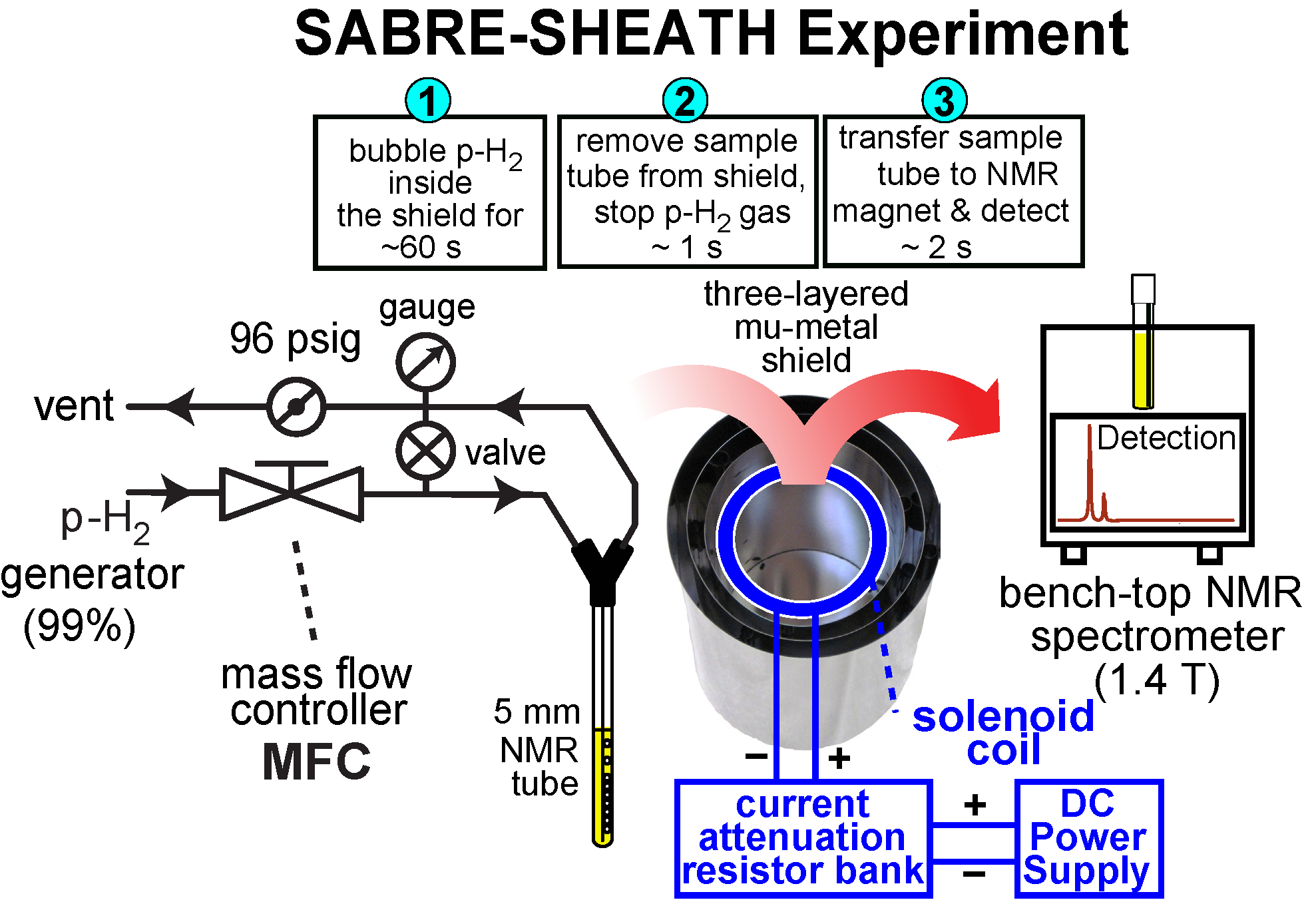
2.1. Hyperpolarizer Setup

The hyperpolarizer apparatus contains a three-layered mu-metal shield equipped with a degaussing coil and circuit [37]. This setup (to be described in full detail elsewhere) is also equipped with a solenoid magnet inside the inner mu-metal in series with an attenuating resistor bank and 5 VDC power supply to provide precise control of the magnetic field inside the shield, Scheme 2 [38]. The field was monitored by a fluxgate magnetometer, and p-H2 gas flow was metered by a mass flow controller (MFC). Temperature control of the sample was maintained using a water bath, Scheme 2 [38]. The p-H2 over-pressure (over atmospheric value) is achieved using a 96 psig valve to provide ~8 bar of total p-H2 pressure inside the sample tube, Scheme 2. Access to p-H2 is provided via a p-H2 storage and distribution setup described in detail previously [39]. Full details including the bill of materials (BOM) for the hyperpolarizer setup will be presented in the near future in a separate publication.

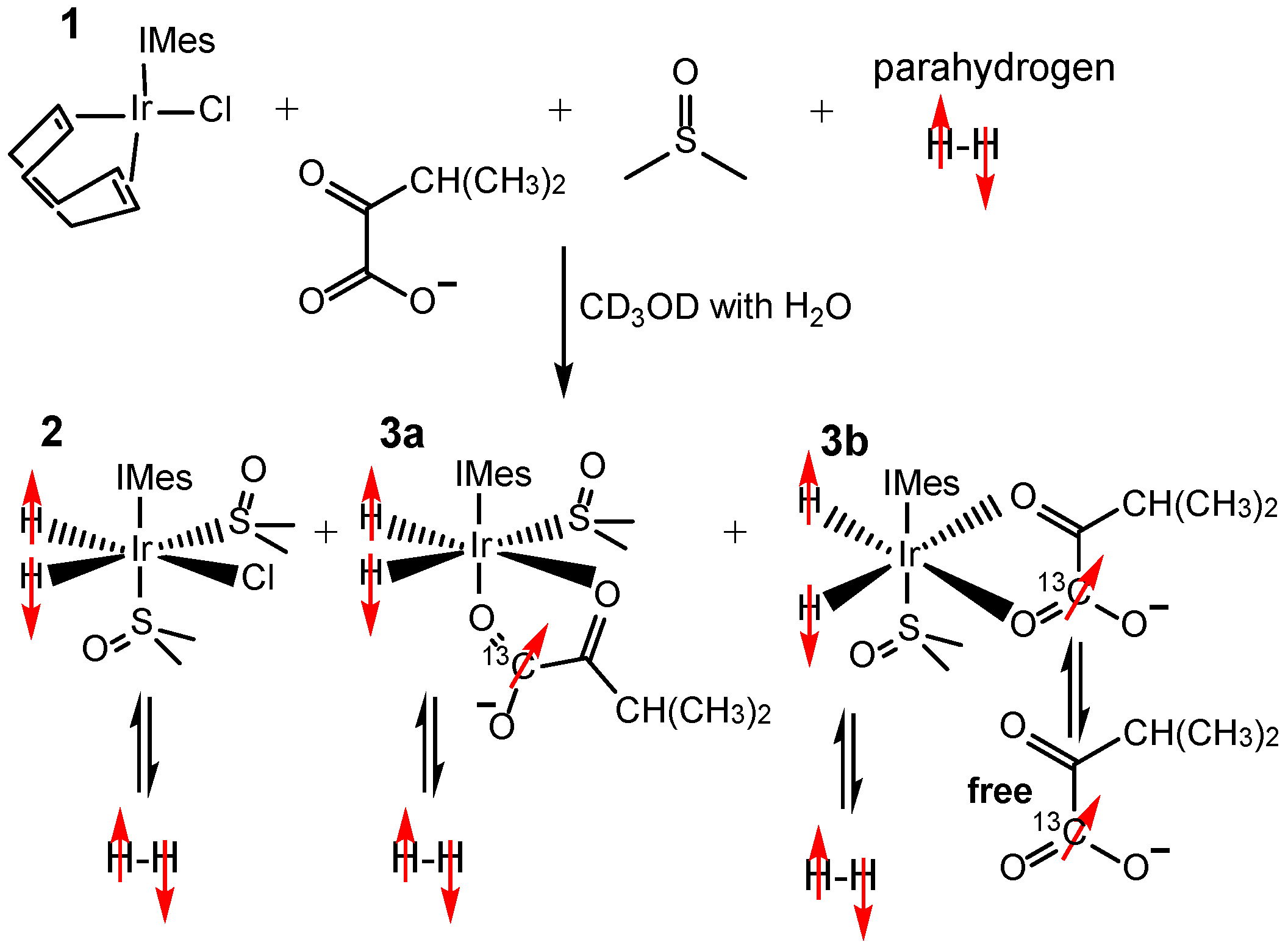
2.2. Sample Preparation for SABRE Hyperpolarization

All experiments employed the standard SABRE pre-catalyst for SABRE [IrCl(COD)(IMes)] (IMes = 1,3-bis(2,4,6-trimethylphenyl)imidazol-2-ylidene; COD = cyclooctadiene; species 1, Scheme 1) [33,41,42], which was synthesized as described previously [43]. For SABRE experiments, a solution containing 5 mM SABRE pre-catalyst 1, 30 mM sodium ketoisocaproate, 20 mM dimethylsulfoxide (DMSO, AC348445000, Fisher Scientific, Waltham, MA, USA) in CD3OD (DLM-24-25, Cambridge Isotope Laboratories Inc., Tewksbury, MA, USA) was prepared. Both natural abundance sodium ketoisocaproate (K0629, Sigma-Aldrich) or sodium [1-13C]ketoisocaproate (99% 13C, 487716, Sigma-Aldrich, St. Louis, MO, USA) were used as received. A 0.6-mL aliquot of the prepared solution was transferred into a medium-wall 5 mm NMR tube (Wilmad-LabGlass, P/N 504-PP-9), which was equipped with ¼” outer-diameter (OD) and 3/16” inner diameter (ID) Teflon tube jacket extension (50–80 mm long, McMaster Carr, Elmhurst, IL, USA, P/N 5239K12). This Teflon tubing allows connecting the NMR tube to the push-to-connect wye connector (McMaster Carr, Elmhurst, IL, USA, P/N 5779K262) of the p-H2 bubbling manifold, Scheme 2 [37].
Then, the sample was immediately purged with ultra-high-purity argon gas (>99.9999%) to remove any trapped air by bubbling pure argon through the solution for at least 1 min and capped with a precision rubber cap (Sigma-Aldrich, St. Louis, MO, USA, P/N Z554014-100EA). The prepared sample was stored at room temperature for 0.2–2 h prior to SABRE hyperpolarization studies. Next, the tube was connected to the hyperpolarizer setup via a ¼”-push-to-connect adapter, Scheme 2. Bubbling of p-H2 through the sample was established via 1/16” OD Teflon tubing (1/32” inner diameter (ID), McMaster Carr, Elmhurst, IL, USA, P/N 5239K23), Scheme 2. The bubbling tube was equipped with an optional Teflon tube insert (0.9 mm OD, 0.5 mm ID); this thinner catheter minimized susceptibility-induced magnetic field gradients, and therefore, improves the field homogeneity (and subsequently full width at half maximum (FWHM) and sensitivity) of HP molecules’ detection for SABRE studies. Note the narrower catheter also delivers smaller p-H2 bubbles potentially increasing surface-to-volume ratio for gas exchange. Closing or opening the valve shown in Scheme 2 results in p-H2 bubbling through the sample or cessation of the bubbling, respectively. Each sample was activated for at least ten minutes prior to SABRE studies with ultra-high-purity (>99.999%) p-H2 (>98% para-state [44,45]) at a flow rate of 70 standard cubic centimeters (sccm), Scheme 2. The sample activation leads to the removal of the COD moiety via hydrogenation [46] and formation of the hexa-coordinate species as shown in Scheme 1.

2.3. NMR Signal Detection

Following cessation of p-H2 bubbling, the sample containing the HP material was transferred to a 1.4 T benchtop NMR spectrometer (transit time: ~2 s), where the 1H or 13C NMR spectra were recorded, Scheme 2. The corresponding NMR signals were integrated using Prospa software provided by the spectrometer vendor. The integrated values were either employed directly to analyze the polarization trends (e.g., build-up, decay, etc.), or these values were converted to 13C signal enhancement (ε13C) and P13C values in accord with Equations (1) and (2) below.

2.4. 13C NMR Signal Enhancement (ε13C) and Polarization (P13C) Calculations

The level of signal enhancement was calculated using the concentration-based signal-referencing equation proposed by Shchepin et al. by integrating the data obtained from the 1.4 T benchtop Magritek Spinsolve NMR spectrometer [47], using the following equation.
ε 13 C = S H P S R E F × C R E F C H P × A R E F A H P
where SREF and SHP denote the NMR signals for a neat thermally polarized reference sample ([1-13C]acetic acid) and HP ketoisocaproate samples, respectively. CREF and CHP represent the concentrations of the thermally polarized [1-13C]acetic acid signal–reference sample (17.5 M) and the concentration of the HP ketoisocaproate sample (30 mM). The AREF and AHP ratio (which corrects for differences in the effective cross-sectional areas of the HP and reference samples) was taken to be approximately 1.53 as described in previous studies [48,49] (note that the slight difference of 1.53 (this report) compared to 1.70 (previous studies) is to do narrower p-H2 bubbling catheter employed in this study). In some cases, we have calculated the ε13C for “free” and the sum of 3a + 3b species using 25 mM and 5 mM concentrations of the “free” species and the pre-catalyst, respectively, and using the integrated signal intensities of the corresponding 13C resonances.
The percentage P13C was calculated using the equation below by multiplying the equilibrium 13C spin polarization (1.2 × 10⁻⁴%) at 1.4 T and 298 K by the corresponding ε13C.
P 13 C = ε 13 C × 1.2 × 10 4 %

2.5. 13C Polarization Build-Up and T1 Relaxation Measurements

In each 13C polarization build-up experiment, the p-H2 bubbling time at the optimal polarization transfer field (e.g., ~0.42 µT) is varied systematically until a steady P13C level is achieved. A separate experiment is employed to acquire each data point to comprise an entire build-up curve for such an experiment, e.g., Figure 1d. For 13C T1 relaxation measurements, p-H2 was bubbled for 60 s to achieve the steady-state P13C at 0.42 µT. Next, the p-H2 flow was stopped, and the sample was either kept at the same field or transferred to a different magnetic field of interest (e.g., the Earth’s magnetic field of approximately 50 µT or 1.4 T field of the NMR spectrometer). Then, the sample was allowed to depolarize for a variable period of time in a systematic fashion. As with the build-up curves, a separate experiment is employed to acquire each data point comprising a given T1 decay curve, e.g., Figure 1d,e. For each experimental series, a mono-exponential build-up or decay function was fit to the experimental P13C build-up or decay data using Origin Pro software (OriginLab, Northampton, MA, USA).

2.6. pH Modulation Study

All experiments (unless noted otherwise) were performed using the given sample composition without any pH adjustment. In some cases (as noted in the text), sample pH dependence was modified by adding 50 µL of 1 M HCl (Fisher Scientific, Waltham, MA, USA, P/N A144-212) solution in CD3OD to 0.6-mL sample aliquot in CD3OD. This addition reduced the pH value of the prepared sample from the initial value of 6.1 (unaltered sample) to 4.0 (acidified sample). The pH adjustment was performed before sample activation with p-H2 bubbling, and the pH was tested by a portable pH meter (P/N SX811-MS, APERA Instruments, Columbus, OH, USA).

2.7. Post-Mortem Studies Using HP [1-13C]ketoisocaproate Injection in a Euthanized Mouse

The rationale for the pilot injection of HP [1-13C]ketoisocaproate into a freshly euthanized mouse post-mortem was two-fold: our first goal was to demonstrate quantitative delivery of hyperpolarization payload into an animal placed inside an MRI scanner. Our second goal was to investigate the 13C T1 relaxation of an injected solution under (nearly) physiologically relevant conditions. The overarching goal is to validate all necessary steps prior to future utilization of HP contrast media prepared via SABRE-SHEATH in an alcohol solvent followed by reconstitution into aqueous media using a recently demonstrated approach [36]. The experimental setup consists of a replica SABRE-SHEATH hyperpolarizer (Scheme 2), and a custom-made transmit-receive circuit for 13C NMR spectroscopy. The circuit consists of a solenoid made from 24 American Wire Gauge (AWG) (0.8 Ω resistance, 72.8 µH inductance, 3.50 cm length, 27 mm ID, and 31 mm OD). This solenoid was tuned to the 13C Larmor frequency in a parallel circuit with a 1-120 pF non-magnetic trimmer capacitor (CT). The circuit was matched to 50 Ω impedance using a non-magnetic trimmer capacitor CM. The final circuit tuning and matching were performed inside a 0.35 T homogeneous MRI magnet (Pica, Time Medical, Hong Kong) using a Kea2 NMR spectrometer (Magritek, Wellington, New Zealand) equipped with a 200 W Tomco amplifier.
Prior to experiments with the post-mortem animal model, the NMR coil was calibrated, and a reference signal from thermally polarized neat [1-13C]acetic acid (0.0732 moles) was recorded using a 120 μs excitation RF pulse, corresponding to a tipping angle (α) of 86°. For the post-mortem experiments, a freshly euthanized 12-week-old C57BL/6 male mouse [50] (~euthanized 1h before the contrast agent injection) received two injections of HP [1-13C]ketoisocaproate. For each injection, a fresh batch of HP [1-13C]ketoisocaproate was produced using the SABRE-SHEATH polarizer, the HP liquid (~0.6 mL) was transferred into a plastic vial and loaded into a plastic syringe (1 mL) and injected intraperitoneally (i.p.) into a euthanized mouse via a 20-gauge metal needle. Approximately 0.3 mL of HP liquid was injected containing 9 × 10⁻⁶ moles of HP [1-13C]ketoisocaproate. The total time between the HP agent production (i.e., cessation of p-H2 flow) and the 13C MRS detection was ~18 s. Dynamic 13C MRS was performed with 2 s temporal resolution (32 total NMR spectra recorded) using α = 16.5° (23 μs). Other acquisition parameters included spectral width (SW) = 5 kHz, number of acquisition points = 2048, and acquisition time (AQ) = 409.6 ms. Once the first dynamic run was completed, another batch of HP [1-13C]ketoisocaproate was produced, and the injection protocol was repeated. The time delay between the start of the first and the second runs was approximately 5 min. Because the reference sample positioning (in the center of the solenoid) was similar to that of the injection location with respect to the detection coil (and because of the lack of circulation and metabolism in the euthanized animal), the following molar quantity-based approach was employed (the above-described concentration-based approach was unsuitable due to uncertainly to HP concentration after injection) to compute the 13C signal enhancement of the injected contrast agent of the first acquisition during dynamic MRS run:
ε 13 C = S H P S R E F × N R E F N H P × sin ( α ) R E F sin ( α ) H P
where NREF and NHP represent the molar quantity of the thermally polarized [1-13C]acetic acid signal–reference sample (0.0732 moles) and the molar quantity of HP [1-13C]ketoisocaproate sample (9 × 10⁻⁶ moles); and sin(α)REF/sin(α)HP accounts for the difference in the excitation pulse angle in signal reference and HP detection (3.4 in our case). The determined 13C signal enhancements were multiplied by the thermal P13C (3.08 × 10⁻⁵%) at 0.35 T (resonance frequency of 3.81860 MHz) and 298 K:
P 13 C = ε 13 C × 3.08 × 10 5 %
Following each injection of HP [1-13C]ketoisocaproate, the recorded 13C signal decayed due to T1 relaxation and the application of RF excitation pulses. No metabolism was anticipated, which would otherwise manifest in the appearance of additional HP resonances. To account for 13C depolarization due to RF excitation, the signal intensities were corrected as follows: SHP(i) = SHP(i)/[cos(α)(i−1)], where i is the acquisition number ranging from 1 to 32. After such compensation for P13C loss due to RF pulses, the data were fitted to a model of mono-exponential decay to determine the 13C T1.

3. Results and Discussion

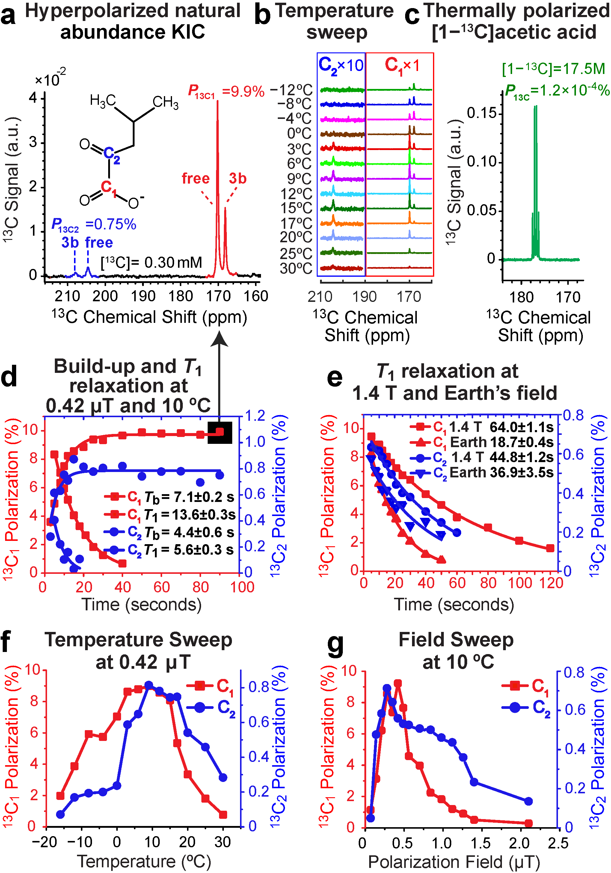
3.1. 13C SABRE-SHEATH of Natural Abundance Ketoisocaproate

Recently, we have demonstrated that 13C-hyperpolarized molecules can be comprehensively and efficiently studied at the natural abundance level of the 13C isotope (1.1%) using a bench-top NMR spectrometer [51], including SABRE-SHEATH-hyperpolarized compounds [39]. Such studies become possible because the detection sensitivity of lower field instruments can be nearly as good as that of the higher field spectrometers in the context of NMR hyperpolarization because the nuclear spin polarization of the HP state is not endowed by the field of the NMR spectrometer employed for the detection [52]. Studying SABRE-SHEATH hyperpolarization at natural 13C abundance effectively allows studying the P13C polarization dynamics of each carbon independently because the probability of having two 13C nuclei next to each other is negligible.
For natural abundance ketoisocaproate (KIC), P13C1 of 9.9% and P13C2 of 0.80% were observed for C1 and C2 sites, respectively, Figure 1a,f. In line with the previous SABRE studies with structurally similar pyruvate and α-ketoglutarate [39,53], the exchange rate between the “free” and 3b species depends on the temperature, which is evident from the ratio of the relative intensities of the HP 13C “free” and 3b peaks of the C1 and C2 sites, Figure 1b. At elevated temperatures, the “free” HP resonance dominates the spectrum because fast exchange (on the time scale of the SABRE experiment) leads to effective averaging of the HP magnetization over “free” and 3b species, which are present in solution in approximately a 5:1 ratio. On the other hand, at temperatures of −4 °C and below, the spectra are dominated by the catalyst-bound 3b resonance, clearly indicating that exchange is slow on the time scale of the SABRE experiment, and the bulk of the HP magnetization is retained on the 3b catalyst-bound species. Moreover, performing a temperature sweep experiment (shown in Figure 1f) reveals a polarization maximum at 6–8 °C for both C1 and C2 resonances. The magnetic field sweep of C1 and C2 resonances follows a slightly different trend, with P13C maxima around 0.4 µT and 0.3 µT, respectively (Figure 1g). This optimal field is similar to the previously reported value for the structurally similar pyruvate [38,54] and α-ketoglutarate [39]. This observation is rationalized by the overall similar complex structures of keto-carboxylates with comparable values of spin–spin couplings relevant to the polarization transfer from p-H2-derived hydrides to the target 13C nucleus.
The P13C build-up time of 7.1 ± 0.2 s corresponds to a build-up rate of ~0.14 s−1, which is substantially faster than the corresponding spin relaxation rate of ~0.07 s−1 (T1 = 13.6 ± 0.3 s) for the C1 site at 0.4 µT, Figure 1d. Because the spin relaxation rate is much slower than the build-up rate, it is kinetically possible to reach relatively high polarization values, e.g., P13C of 10%. Furthermore, the long 13C1 T1 values of 64.0 ± 1.1 s and 18.7 ± 0.4 s at 1.4 T and the Earth’s magnetic field (Figure 1e) demonstrate good potential for the use of the C1 site as a polarization storage site for future applications. The corresponding 13C relaxation dynamics for the C2 site at 0.4 µT are less favorable, Figure 1d, with the Tb and T1 values, reduced to just 4.4 ± 0.6 s and 5.6 ± 0.3 s, respectively. These less favorable relaxation dynamics likely contribute to overall reduced P13C values for the C2 site, yet differences in hydride target J-coupling and differences in the exchange processes may also contribute.
The ratio of maximum polarization values for the C1 and C2 resonances was 12:1, whereas this ratio was 12:3 for pyruvate, and 12:0 for α-ketoglutarate [39]. This observation of C2 polarization efficiency ranking (pyruvate > KIC > α-ketoglutarate) correlates well with the decrease in the corresponding side chain (-CH3 < -CH(CH3)2 < -CH2-CH2-COO). We hypothesize that having a longer side chain can be partially responsible for less favorable dynamics during the SABRE-SHEATH hyperpolarization process. It is also anticipated that the C2 site has stronger spin–spin interactions with the chain protons, which can potentially act as relaxation sinks at low (i.e., microtesla) magnetic fields [55], thereby resulting in effectively shorter 13C T1 values and lower P13C values as compared to the C1 site.

3.2. 13C SABRE-SHEATH of [1-13C]ketoisocaproate

As expected, the comparative studies with isotopically enriched [1-13C]ketoisocaproate ([1-13C]KIC) revealed overall the same trends, Figure 2a–g, as those observed for the natural abundance isotopologue, Figure 1. For example, the temperature sweeps (Figure 2b,g), and the BT sweep (Figure 2f) match those for the natural abundance compound, Figure 1.
The detailed quantitative comparison also revealed small differences between the two isotopologues. The maximum P13C value was 18.3%, Figure 2a, while Tb and T1 decay values at 0.42 µT were 14.3 ± 0.6 s and 22.8 ± 1.2 s, respectively, Figure 2d. Moreover, the 13C T1 values at 1.4 T (74.1 ± 0.7 s) and the Earth’s field (31.1 ± 0.5 s) were markedly higher for [1-13C]KIC (Figure 2e) compared to those for the natural abundance variant vs. 64.0 ± 1.1 s at 1.4 T and 18.7 ± 0.4 s at the Earth’s field (Figure 1e). We hypothesize that these differences could be potentially attributed to different impurities or other variations in the ketocarboxylate samples—including pH. More detailed studies are certainly warranted to shed more light on the differences in the performance and polarization dynamics of 13C-labeled and natural abundance ketoisocaproate samples.

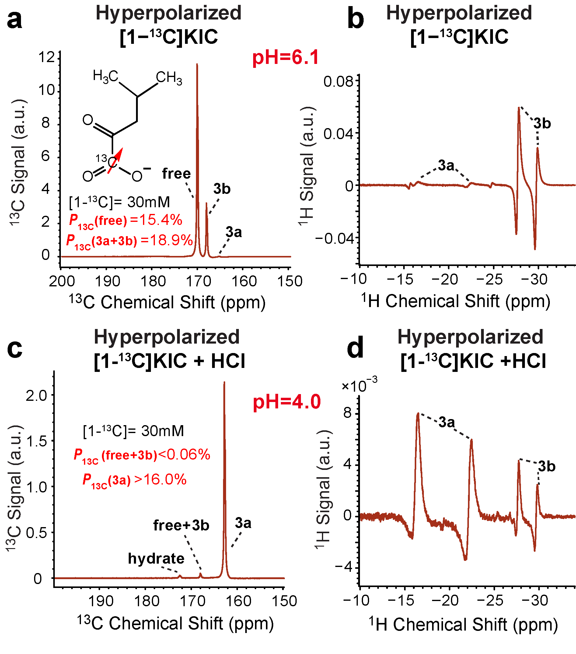
3.3. Effect of pH on 13C SABRE-SHEATH of [1-13C]ketoisocaproate

Prompted by the differences in SABRE-SHEATH efficiency between KIC and [1-13C]KIC (see Section 3.2 above for details), a pilot pH-spiking study was performed.
For this pilot study, we added 50 µL of 1 M HCl solution in CD3OD (Section 2.6) to the [1-13C]KIC (before activation with p-H2), which resulted in a pH shift from 6.1 (unaltered solution) to pH 4.0 (acidified sample). At an initial pH of 6.1, we observed P13C 15.4% and 18.9% for the “free” and (3a + 3b) species, respectively (Figure 3a). Surprisingly, upon lowering the pH to 4.0, HP species 3a dominates the 13C spectrum shown in Figure 3c. This finding is additionally supported by 1H NMR spectroscopy of the hydride spectral region: Figure 3b shows the spectrum of HP [1-13C]KIC dominated by 3b (as expected before the sample acidification), whereas the 1H spectrum of the acidified sample is dominated by 3a. Due to spectral overlap between 3b and “free” resonances in Figure 3c (and by extension ambiguity of the 3a concentration), P13C of 3a was estimated to be in excess of 16.0%. We conclude that pH has a profound effect on the 3a/3b ratio. At lower pH, the equilibrium is shifted to 3a, which does not exchange with “free” species on the time scale of the SABRE-SHEATH experiment (Scheme 1), resulting in no appreciable polarization of the “free” species. Moreover, the acidified sample also exhibited the appearance of the HP 13C resonance of the KIC hydrate. Despite this remarkable near-complete disappearance of the HP 13C “free” species, the polarization transfer remained active in the context of chemical exchange with p-H2—as is evident from the high P13C values of 3a.
Prompted by these initial findings, an additional temperature sweep study was performed on a separate sample before and after sample acidification with 50 µL of 1 M HCl solution in CD3OD, Figure 4.
The non-acidified sample exhibited similar P13C values (Figure 4a) to those shown in Figure 3a, demonstrating the overall reproducibility of our sample preparation and hyperpolarization approach. The temperature sweep shown in Figure 4b was also similar to the one presented in Figure 2b. The detailed analysis of the temperature sweep shown in Figure 4c clearly demonstrates that “free” species reaches a HP signal maximum at ~10 °C, whereas the maximum for 3a is at −10 °C, and the maximum for 3b is at ~0 °C. The temperature sweep performed after sample acidification (Figure 4d,e) reveals that pH also has a profound effect on the maximum temperature positions for the individual HP species. For example, the temperature maximum for 3a species is shifted to 4 °C. Moreover, the temperature maximum for 3b + “free” (which could not be differentiated due to spectral overlap) is shifted to −8 °C (Figure 4f), which is substantially lower than any of the individual 3b or “free” maxima before the sample acidification (0 °C and 10 °C, respectively, Figure 4c). Therefore, it is concluded that pH modulates not only the relative ratios of 3a and 3b (and by extension the “free”) HP 13C resonances, but it also affects the exchange rates of the substrate with the 3a and 3b catalyst species. This finding is important as pH can be potentially employed as a tuning tool to optimize the overall efficiency of the SABRE-SHEATH polarization process. Future detailed studies of this effect are certainly warranted.

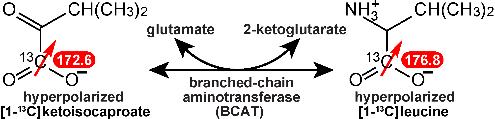
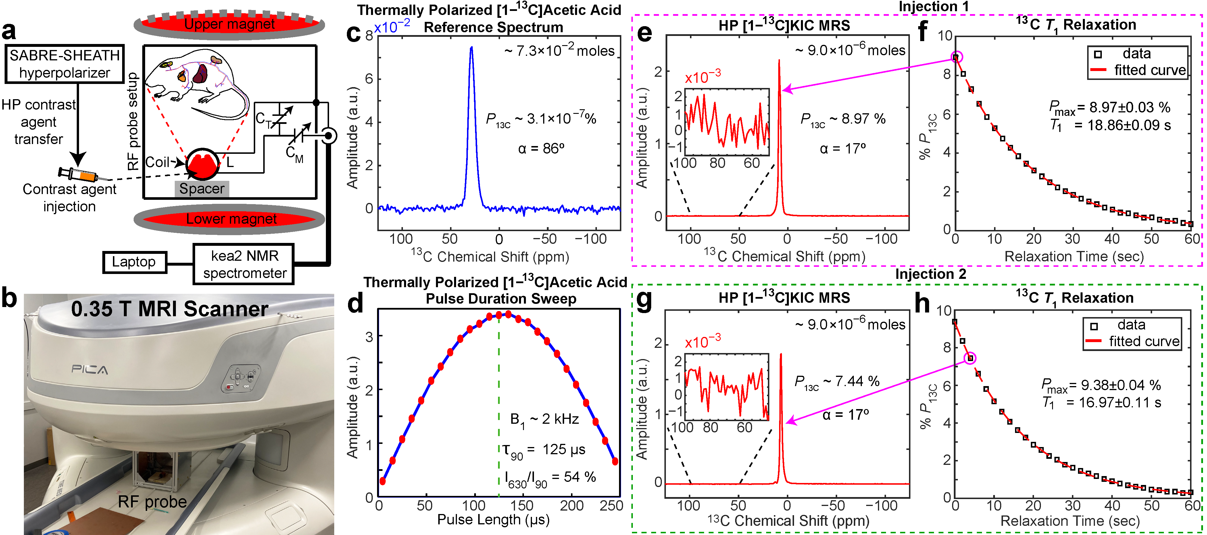
3.4. Pilot Post-Mortem 13C Dynamic Magnetic Resonance Spectroscopy Using HP [1-13C]ketoisocaproate Injection in a Euthanized Mouse

Encouraged by the overall good hyperpolarization potency of HP [1-13C]KIC, pilot i.p. injection of HP [1-13C]KIC was performed in a freshly euthanized mouse. Our rationale for this experiment was to demonstrate the feasibility of successful injection and detection of HP [1-13C]KIC under biologically relevant conditions. To the best of our knowledge, no in vivo demonstration of any HP 13C biomolecule has been published using SABRE hyperpolarization technology. Given that the HP solutions were prepared in CD3OD, we performed two injections in a freshly euthanized animal, Figure 5.
Before the post-mortem injection, the 13C volume detection coil (Figure 5a,b) was optimized using 13C detection of a thermally polarized signal–reference phantom (Figure 5c) to determine the optimum RF excitation pulse (Figure 5d). The optimum τ90 excitation pulse length was 125 μs, corresponding to a B1 strength of 2 kHz, with good B1 homogeneity (I630/I90 = 54%).
Once HP [1-13C]KIC was injected into the euthanized animal, the 13C detection using 17° excitation pulses started immediately, and dynamic whole body 13C MRS was recorded every 2.0 s. The first scan of the first injection is shown in Figure 5e, and the third scan of the second injection is shown in Figure 5g. These 13C spectra reveal excellent signal-to-noise ratios (SNRs) in excess of 2000, which can be potentially further boosted by a factor of 3–5 using a singular value decomposition (SVD) approach demonstrated previously [56]. The initial level of P13C detected inside the animal was 9.0–9.4%, demonstrating good shot-to-shot reproducibility of our HP contrast agent production and administration procedures.
The dynamic 13C MRS revealed the T1 decay of hyperpolarization with mono-exponential rate constants of 18.9 ± 0.1 s (Figure 5f) and 17.0 ± 0.1 s (Figure 5g) for the first and the second injections, respectively. These values are substantially shorter than the corresponding in vitro 13C T1 value of 74.1 ± 0.7 s (Figure 2e) at 1.4 T. Moreover, the post-mortem 13C T1 value is substantially shorter than the 13C T1 in vivo value of structurally similar [1-13C]pyruvate (~67 s at 3 T [57]). We rationalize this observation potentially through non-specific interactions between HP [1-13C]KIC, residual methanol, the catalyst, and the biological environment. It should be noted that the recent progress with the production of bio-compatible formulations of HP solutions [36] prepared by SABRE may potentially mitigate the 13C T1 reduction challenge. Combined with recent advances in the field of SABRE for purification of HP solutions [58,59,60,61] and the use of heterogeneous [62,63] and aqueous [46,64,65,66,67,68,69] SABRE catalysis, the findings reported here bode well for the future in vivo translation of SABRE-hyperpolarized HP [1-13C]KIC.

4. Biomedical Outlook

The next logical future steps of the fundamental work presented here in the context of biomedical applications should focus on the validation of SABRE-hyperpolarized [1-13C]KIC in animal models of cancer with unregulated BCAT metabolic pathways [22,23]. We envision the feasibility of this approach because the polarization level demonstrated here (P13C of 18%) is comparable to the in vivo studies that employed the d-DNP hyperpolarization technique. Transitioning to clinical studies using HP [1-13C]KIC would require increasing the production scale to that of HP [1-13C]pyruvate, which has not been demonstrated yet, representing yet another milestone that must be accomplished in the future.
From the experimental standpoint, the in vivo metabolism of HP [1-13C]KIC leads to the production of HP [1-13C]leucine, which are well separated spectroscopically: 172.6 ppm and 176.8 ppm, respectively, Scheme 3 [70]. It should also be noted that substantially less HP [1-13C]leucine is being produced from HP [1-13C]KIC compared to that of HP [1-13C]pyruvate in many healthy tissues due to the inherently lower metabolic flux of this pathway compared to lactate dehydrogenase (LDH). This difference offers an advantage for clinical applications of a much greater specificity of the HP [1-13C]KIC metabolic probe compared to HP [1-13C]pyruvate. Indeed, virtually no background signal from intestine tissue was detected compared to murine lymphoma (EL4) tumors [22].
Moreover, previous studies have also revealed that BCAT metabolism may be elevated in some but certainly not in all types and sub-types of solid tumors [22,70,71]. For example, Billingsley and co-workers concluded that metabolic activity in two prostate cancer cell lines (PC3 and DU145) was remarkably low, noting that “the low levels of BCAT activity in the examined prostate cancer cell lines may limit the future application of hyperpolarized [1-13C]KIC in the assessment of the disease”. On the other hand, Shu and workers [71] detected high metabolic signal from HP [1-13C]KIC in R98 glioma, and Karlsson and co-workers have detected metabolic signal from HP [1-13C]KIC in EL4 lymphoma [22]. Furthermore, Butt and co-workers demonstrated the feasibility of employing HP [1-13C]KIC for studying cerebral metabolism in a healthy brain [23]. All in all, the pilot small-scale pre-clinical studies employing HP [1-13C]KIC as a metabolic probe concluded that HP [1-13C]KIC may be an excellent specific probe of BCAT metabolism associated with c-Myc signaling and metastases (moreover, because c-Myc is potentially druggable [72], we envision that HP [1-13C]KIC could be a potentially useful metabolic probe for next-generation precision drug development). Therefore, while HP [1-13C]pyruvate is an excellent non-specific cancer metabolic probe, we envision that HP [1-13C]KIC can provide synergistic information regarding additional profiling in solid tumors. Another area of clinical imaging may involve brain imaging applications—we speculate that HP [1-13C]KIC could be potentially useful in the area of brain diseases beyond cancer, including neurodegenerative diseases and aging. Finally, we note that unlike HP [1-13C]pyruvate, which is now under evaluation in over 30 clinical trials according to clinicaltrials.gov, clinical studies with HP [1-13C]KIC have not been reported yet to the best of our knowledge.

5. Conclusions

We have reported the efficient hyperpolarization of KIC and [1-13C]KIC and demonstrated P13C levels of up to 18% with 13C T1 values of up to 74.1 ± 0.7 s. This high degree of polarization is enabled by favorable 13C relaxation dynamics in microtesla magnetic fields. It is anticipated that P13C can be increased further through the use of a higher p-H2 pressure [73] and flow rates using more advanced instrumentation. When using an NMR-tube-based experimental setup, a sufficiently high dose of HP contrast agent was produced for future in vivo studies, as demonstrated by pilot experiments in a freshly euthanized mouse. The pilot pH modulation studies revealed remarkable dependence of SABRE hyperpolarization chemistry and dynamics on pH, indicating that pH modulation could be employed as a tuning tool for SABRE-SHEATH hyperpolarization of KIC and other carboxylates, including most notably, pyruvate and α-ketoglutarate.

Author Contributions

Conceptualization, I.A. and E.Y.C.; methodology, I.A., E.Y.C., M.S. and S.N.; software, M.R.H.C.; validation, L.S., M.S. and M.R.H.C.; formal analysis, I.A. and M.R.H.C.; investigation, I.A., M.R.H.C., S.N., C.O., F.A. and L.S.; resources, B.M.G., M.S., T.T. and E.Y.C.; data curation, E.Y.C., B.M.G. and M.S.; writing—original draft preparation, I.A., M.R.H.C. and E.Y.C.; writing—review and editing, I.A., B.M.G., M.S. and E.Y.C.; visualization, I.A. and M.R.H.C.; supervision, E.Y.C. and M.S.; project administration, E.Y.C.; funding acquisition, B.M.G., T.T., M.S. and E.Y.C. All authors have read and agreed to the published version of the manuscript.

Funding

This research was funded by the NSF under grants CHE-1904780 and CHE-1905341, and NIBIB R01EB029829. L.K. was further supported by NIH (5T32GM142519-02). S.N. and E.Y.C. thank Wayne State University Thomas C. Rumble University Graduate Fellowship. The content is solely the responsibility of the authors and does not necessarily represent the official views of the National Institutes of Health. T.T. also acknowledges funding from the US Department of Energy under DE-SC0023334.

Institutional Review Board Statement

The animal employed in this study was euthanized according to the animal protocol 20-01-1712 approved by IACUC of Wayne State University.

Informed Consent Statement

Not applicable.

Data Availability Statement

NMR spectroscopic data are available from co-authors upon reasonable request. Data is not publicly available due to privacy.

Acknowledgments

We thank Mohammad S. H. Kabir for stimulating discussions, and Oleg G. Salnikov for the suggestion to implement a 0.9 mm Teflon catheter insert to improve the hyperpolarizer setup.

Conflicts of Interest

BMG, EYC declare stake ownership in XeUS Technologies, LTD. TT holds stock in Vizma Life Sciences LLC. The terms of this arrangement have been reviewed and approved by NC State University in accordance with its policy on objectivity in research. The funders had no role in the design of the study; in the collection, analyses, or interpretation of data; in the writing of the manuscript; or in the decision to publish the results.

References

  1. Eills, J.; Budker, D.; Cavagnero, S.; Chekmenev, E.Y.; Elliott, S.J.; Jannin, S.; Lesage, A.; Matysik, J.; Meersmann, T.; Prisner, T.; et al. Spin Hyperpolarization in Modern Magnetic Resonance. Chem. Rev. 2023. [Google Scholar] [CrossRef] [PubMed]
  2. Dorrius, M.D.; Pijnappel, R.M.; Jansen-van der Weide, M.C.; Jansen, L.; Kappert, P.; Oudkerk, M.; Sijens, P.E. Determination of Choline Concentration in Breast Lesions: Quantitative Multivoxel Proton MR Spectroscopy as a Promising Noninvasive Assessment Tool to Exclude Benign Lesions. Radiology 2011, 259, 695–703. [Google Scholar] [CrossRef] [PubMed] [Green Version]
  3. Nikolaou, P.; Goodson, B.M.; Chekmenev, E.Y. NMR Hyperpolarization Techniques for Biomedicine. Chem. Eur. J. 2015, 21, 3156–3166. [Google Scholar] [CrossRef] [PubMed] [Green Version]
  4. Kovtunov, K.V.; Pokochueva, E.V.; Salnikov, O.G.; Cousin, S.; Kurzbach, D.; Vuichoud, B.; Jannin, S.; Chekmenev, E.Y.; Goodson, B.M.; Barskiy, D.A.; et al. Hyperpolarized NMR: D-DNP, PHIP, and SABRE. Chem. Asian J. 2018, 13, 1857–1871. [Google Scholar] [CrossRef] [PubMed] [Green Version]
  5. Goodson, B.M.; Kidd, B.; Hövener, J.-B.; Schröder, L.; Theis, T.; Whiting, N.; Chekmenev, E.Y. Nuclear Magnetic Resonance Spectroscopy | Hyperpolarization for Sensitivity Enhancement. In Encyclopedia of Analytical Science, 3rd ed.; Worsfold, P., Poole, C., Townshend, A., Miró, M., Eds.; Academic Press: Oxford, UK, 2019; pp. 168–181. [Google Scholar]
  6. Goodson, B.M. Nuclear magnetic resonance of laser-polarized noble gases in molecules, materials, and organisms. J. Magn. Reson. 2002, 155, 157–216. [Google Scholar] [CrossRef] [PubMed] [Green Version]
  7. Mugler, J.P.; Altes, T.A. Hyperpolarized 129Xe MRI of the human lung. J. Magn. Reson. Imaging 2013, 37, 313–331. [Google Scholar] [CrossRef] [PubMed] [Green Version]
  8. Golman, K.; Axelsson, O.; Johannesson, H.; Mansson, S.; Olofsson, C.; Petersson, J.S. Parahydrogen-induced polarization in imaging: Subsecond C-13 angiography. Magn. Reson. Med. 2001, 46, 1–5. [Google Scholar] [CrossRef] [PubMed]
  9. Witte, C.; Schroder, L. NMR of hyperpolarised probes. NMR Biomed. 2013, 26, 788–802. [Google Scholar] [CrossRef]
  10. Goodson, B.M.; Whiting, N.; Coffey, A.M.; Nikolaou, P.; Shi, F.; Gust, B.M.; Gemeinhardt, M.E.; Shchepin, R.V.; Skinner, J.G.; Birchall, J.R.; et al. Hyperpolarization Methods for MRS. Emagres 2015, 4, 797–810. [Google Scholar]
  11. Hövener, J.-B.; Pravdivtsev, A.N.; Kidd, B.; Bowers, C.R.; Glöggler, S.; Kovtunov, K.V.; Plaumann, M.; Katz-Brull, R.; Buckenmaier, K.; Jerschow, A.; et al. Parahydrogen-based Hyperpolarization for Biomedicine. Angew. Chem. Int. Ed. 2018, 57, 11140–11162. [Google Scholar] [CrossRef]
  12. Ardenkjaer-Larsen, J.H.; Fridlund, B.; Gram, A.; Hansson, G.; Hansson, L.; Lerche, M.H.; Servin, R.; Thaning, M.; Golman, K. Increase in signal-to-noise ratio of > 10,000 times in liquid-state NMR. Proc. Natl. Acad. Sci. USA 2003, 100, 10158–10163. [Google Scholar] [CrossRef] [PubMed]
  13. Golman, K.; Ardenkjær-Larsen, J.H.; Petersson, J.S.; Månsson, S.; Leunbach, I. Molecular imaging with endogenous substances. Proc. Natl. Acad. Sci. USA 2003, 100, 10435–10439. [Google Scholar] [CrossRef] [PubMed] [Green Version]
  14. Merritt, M.; Harrison, C.; Storey, C.; Jeffrey, F.; Sherry, A.; Malloy, C. Hyperpolarized C-13 allows a direct measure of flux through a single enzyme-catalyzed step by NMR. Proc. Natl. Acad. Sci. USA 2007, 104, 19773–19777. [Google Scholar] [CrossRef] [PubMed] [Green Version]
  15. Nelson, S.J.; Kurhanewicz, J.; Vigneron, D.B.; Larson, P.E.Z.; Harzstark, A.L.; Ferrone, M.; van Criekinge, M.; Chang, J.W.; Bok, R.; Park, I.; et al. Metabolic Imaging of Patients with Prostate Cancer Using Hyperpolarized 1-C-13 Pyruvate. Sci. Transl. Med. 2013, 5, 198ra108. [Google Scholar] [CrossRef] [Green Version]
  16. Day, S.E.; Kettunen, M.I.; Gallagher, F.A.; Hu, D.E.; Lerche, M.; Wolber, J.; Golman, K.; Ardenkjaer-Larsen, J.H.; Brindle, K.M. Detecting tumor response to treatment using hyperpolarized C-13 magnetic resonance imaging and spectroscopy. Nat. Med. 2007, 13, 1382–1387. [Google Scholar] [CrossRef]
  17. Kurhanewicz, J.; Vigneron, D.B.; Brindle, K.; Chekmenev, E.Y.; Comment, A.; Cunningham, C.H.; DeBerardinis, R.J.; Green, G.G.; Leach, M.O.; Rajan, S.S.; et al. Analysis of Cancer Metabolism by Imaging Hyperpolarized Nuclei: Prospects for Translation to Clinical Research. Neoplasia 2011, 13, 81–97. [Google Scholar] [CrossRef] [Green Version]
  18. Kurhanewicz, J.; Vigneron, D.B.; Ardenkjaer-Larsen, J.H.; Bankson, J.A.; Brindle, K.; Cunningham, C.H.; Gallagher, F.A.; Keshari, K.R.; Kjaer, A.; Laustsen, C.; et al. Hyperpolarized 13C MRI: Path to Clinical Translation in Oncology. Neoplasia 2019, 21, 1–16. [Google Scholar] [CrossRef]
  19. Golman, K.; in’t Zandt, R.; Thaning, M. Real-time metabolic imaging. Proc. Natl. Acad. Sci. USA 2006, 103, 11270–11275. [Google Scholar] [CrossRef] [Green Version]
  20. Chung, B.T.; Chen, H.-Y.; Gordon, J.; Mammoli, D.; Sriram, R.; Autry, A.W.; Le Page, L.M.; Chaumeil, M.M.; Shin, P.; Slater, J.; et al. First hyperpolarized [2-13C]pyruvate MR studies of human brain metabolism. J. Magn. Reson. 2019, 309, 106617. [Google Scholar] [CrossRef]
  21. Brindle, K.M. Imaging Metabolism with Hyperpolarized 13C-Labeled Cell Substrates. J. Am. Chem. Soc. 2015, 137, 6418–6427. [Google Scholar] [CrossRef]
  22. Karlsson, M.; Jensen, P.R.; in’t Zandt, R.; Gisselsson, A.; Hansson, G.; Duus, J.Ø.; Meier, S.; Lerche, M.H. Imaging of branched chain amino acid metabolism in tumors with hyperpolarized 13C ketoisocaproate. Int. J. Cancer 2010, 127, 729–736. [Google Scholar] [CrossRef] [PubMed]
  23. Butt, S.A.; Søgaard, L.V.; Magnusson, P.O.; Lauritzen, M.H.; Laustsen, C.; Åkeson, P.; Ardenkjær-Larsen, J.H. Imaging cerebral 2-ketoisocaproate metabolism with hyperpolarized 13C magnetic resonance spectroscopic imaging. J. Cereb. Blood Flow Metab. 2012, 32, 1508–1514. [Google Scholar] [CrossRef] [PubMed] [Green Version]
  24. Comment, A.; Merritt, M.E. Hyperpolarized Magnetic Resonance as a Sensitive Detector of Metabolic Function. Biochemistry 2014, 53, 7333–7357. [Google Scholar] [CrossRef] [Green Version]
  25. Ardenkjaer-Larsen, J.H.; Leach, A.M.; Clarke, N.; Urbahn, J.; Anderson, D.; Skloss, T.W. Dynamic Nuclear Polarization Polarizer for Sterile Use Intent. NMR Biomed. 2011, 24, 927–932. [Google Scholar] [CrossRef] [PubMed]
  26. Ardenkjaer-Larsen, J.H. On the present and future of dissolution-DNP. J. Magn. Reson. 2016, 264, 3–12. [Google Scholar] [CrossRef] [PubMed] [Green Version]
  27. Ardenkjaer-Larsen, J.H.; Bowen, S.; Petersen, J.R.; Rybalko, O.; Vinding, M.S.; Ullisch, M.; Nielsen, N.C. Cryogen-free dissolution dynamic nuclear polarization polarizer operating at 3.35 T, 6.70 T, and 10.1 T. Magn. Reson. Med. 2019, 81, 2184–2194. [Google Scholar] [CrossRef] [PubMed] [Green Version]
  28. Tickner, B.J.; Ahwal, F.; Whitwood, A.C.; Duckett, S.B. Reversible Hyperpolarization of Ketoisocaproate Using Sulfoxide-containing Polarization Transfer Catalysts. ChemPhysChem 2021, 22, 13–17. [Google Scholar] [CrossRef]
  29. Theis, T.; Truong, M.L.; Coffey, A.M.; Shchepin, R.V.; Waddell, K.W.; Shi, F.; Goodson, B.M.; Warren, W.S.; Chekmenev, E.Y. Microtesla SABRE Enables 10% Nitrogen-15 Nuclear Spin Polarization. J. Am. Chem. Soc. 2015, 137, 1404–1407. [Google Scholar] [CrossRef] [PubMed]
  30. Truong, M.L.; Theis, T.; Coffey, A.M.; Shchepin, R.V.; Waddell, K.W.; Shi, F.; Goodson, B.M.; Warren, W.S.; Chekmenev, E.Y. 15N Hyperpolarization By Reversible Exchange Using SABRE-SHEATH. J. Phys. Chem. C 2015, 119, 8786–8797. [Google Scholar] [CrossRef] [Green Version]
  31. Adams, R.W.; Aguilar, J.A.; Atkinson, K.D.; Cowley, M.J.; Elliott, P.I.P.; Duckett, S.B.; Green, G.G.R.; Khazal, I.G.; Lopez-Serrano, J.; Williamson, D.C. Reversible Interactions with para-Hydrogen Enhance NMR Sensitivity by Polarization Transfer. Science 2009, 323, 1708–1711. [Google Scholar] [CrossRef] [Green Version]
  32. Barskiy, D.A.; Shchepin, R.V.; Tanner, C.P.N.; Colell, J.F.P.; Goodson, B.M.; Theis, T.; Warren, W.S.; Chekmenev, E.Y. The Absence of Quadrupolar Nuclei Facilitates Efficient 13C Hyperpolarization via Reversible Exchange with Parahydrogen. ChemPhysChem 2017, 18, 1493–1498. [Google Scholar] [CrossRef] [PubMed]
  33. Cowley, M.J.; Adams, R.W.; Atkinson, K.D.; Cockett, M.C.R.; Duckett, S.B.; Green, G.G.R.; Lohman, J.A.B.; Kerssebaum, R.; Kilgour, D.; Mewis, R.E. Iridium N-Heterocyclic Carbene Complexes as Efficient Catalysts for Magnetization Transfer from para-Hydrogen. J. Am. Chem. Soc. 2011, 133, 6134–6137. [Google Scholar] [CrossRef] [PubMed] [Green Version]
  34. Iali, W.; Roy, S.S.; Tickner, B.J.; Ahwal, F.; Kennerley, A.J.; Duckett, S.B. Hyperpolarising Pyruvate through Signal Amplification by Reversible Exchange (SABRE). Angew. Chem. Int. Ed. 2019, 58, 10271–10275. [Google Scholar] [CrossRef] [Green Version]
  35. Tickner, B.J.; Semenova, O.; Iali, W.; Rayner, P.J.; Whitwood, A.C.; Duckett, S.B. Optimisation of pyruvate hyperpolarisation using SABRE by tuning the active magnetisation transfer catalyst. Catal. Sci. Technol. 2020, 10, 1343–1355. [Google Scholar] [CrossRef] [PubMed]
  36. Schmidt, A.B.; de Maissin, H.; Adelabu, I.; Nantogma, S.; Ettedgui, J.; TomHon, P.; Goodson, B.M.; Theis, T.; Chekmenev, E.Y. Catalyst-Free Aqueous Hyperpolarized [1-13C]Pyruvate Obtained by Re-Dissolution Signal Amplification by Reversible Exchange. ACS Sensors 2022, 7, 3430–3439. [Google Scholar] [CrossRef]
  37. Joalland, B.; Nantogma, S.; Chowdhury, M.R.H.; Nikolaou, P.; Chekmenev , E.Y. Magnetic Shielding of Parahydrogen Hyperpolarization Experiments for the Masses. Magn. Reson. Chem. 2021, 59, 1180–1186. [Google Scholar] [CrossRef] [PubMed]
  38. Adelabu, I.; TomHon, P.; Kabir, M.S.H.; Nantogma, S.; Abdulmojeed, M.; Mandzhieva, I.; Ettedgui, J.; Swenson, R.E.; Krishna, M.C.; Theis, T.; et al. Order-Unity 13C Nuclear Polarization of [1-13C]Pyruvate in Seconds and the Interplay of Water and SABRE Enhancement. ChemPhysChem 2022, 23, 131–136. [Google Scholar] [CrossRef]
  39. Adelabu, I.; Ettedgui, J.; Joshi, S.M.; Nantogma, S.; Chowdhury, M.R.H.; McBride, S.; Theis, T.; Sabbasani, V.R.; Chandrasekhar, M.; Sail, D.; et al. Rapid 13C Hyperpolarization of the TCA Cycle Intermediate α-Ketoglutarate via SABRE-SHEATH. Anal. Chem. 2022, 94, 13422–13431. [Google Scholar] [CrossRef]
  40. Shchepin, R.V.; Jaigirdar, L.; Chekmenev, E.Y. Spin-Lattice Relaxation of Hyperpolarized Metronidazole in Signal Amplification by Reversible Exchange in Micro-Tesla Fields. J. Phys. Chem. C 2018, 122, 4984–4996. [Google Scholar] [CrossRef]
  41. Vazquez-Serrano, L.D.; Owens, B.T.; Buriak, J.M. The search for new hydrogenation catalyst motifs based on N-heterocyclic carbene ligands. Inorg. Chim. Acta 2006, 359, 2786–2797. [Google Scholar] [CrossRef]
  42. Rayner, P.J.; Duckett, S.B. Signal Amplification by Reversible Exchange (SABRE): From Discovery to Diagnosis. Angew. Chem. Int. Ed. 2018, 57, 6742–6753. [Google Scholar] [CrossRef] [PubMed]
  43. Barskiy, D.A.; Kovtunov, K.V.; Koptyug, I.V.; He, P.; Groome, K.A.; Best, Q.A.; Shi, F.; Goodson, B.M.; Shchepin, R.V.; Coffey, A.M.; et al. The feasibility of formation and kinetics of NMR Signal Amplification by Reversible Exchange (SABRE) at high magnetic field (9.4 T). J. Am. Chem. Soc. 2014, 136, 3322–3325. [Google Scholar] [CrossRef]
  44. Nantogma, S.; Joalland, B.; Wilkens, K.; Chekmenev, E.Y. Clinical-Scale Production of Nearly Pure (>98.5%) Parahydrogen and Quantification by Benchtop NMR Spectroscopy. Anal. Chem. 2021, 93, 3594–3601. [Google Scholar] [CrossRef] [PubMed]
  45. Feng, B.; Coffey, A.M.; Colon, R.D.; Chekmenev, E.Y.; Waddell, K.W. A pulsed injection parahydrogen generator and techniques for quantifying enrichment. J. Magn. Reson. 2012, 214, 258–262. [Google Scholar] [CrossRef] [PubMed] [Green Version]
  46. Truong, M.L.; Shi, F.; He, P.; Yuan, B.; Plunkett, K.N.; Coffey, A.M.; Shchepin, R.V.; Barskiy, D.A.; Kovtunov, K.V.; Koptyug, I.V.; et al. Irreversible Catalyst Activation Enables Hyperpolarization and Water Solubility for NMR Signal Amplification by Reversible Exchange. J. Phys. Chem. B 2014, 18, 13882–13889. [Google Scholar] [CrossRef] [Green Version]
  47. Shchepin, R.V.; Birchall, J.R.; Chukanov, N.V.; Kovtunov, K.V.; Koptyug, I.V.; Theis, T.; Warren, W.S.; Gelovani, J.G.; Goodson, B.M.; Shokouhi, S.; et al. Hyperpolarizing Concentrated Metronidazole 15NO2 Group Over Six Chemical Bonds with More Than 15% Polarization and 20 Minute Lifetime. Chem. Eur. J. 2019, 25, 8829–8836. [Google Scholar] [CrossRef]
  48. Shchepin, R.V.; Jaigirdar, L.; Theis, T.; Warren, W.S.; Goodson, B.M.; Chekmenev, E.Y. Spin Relays Enable Efficient Long-Range Heteronuclear Signal Amplification by Reversible Exchange. J. Phys. Chem. C 2017, 121, 28425–28434. [Google Scholar] [CrossRef]
  49. Salnikov, O.G.; Chukanov, N.V.; Svyatova, A.; Trofimov, I.A.; Kabir, M.S.H.; Gelovani, J.G.; Kovtunov, K.V.; Koptyug, I.V.; Chekmenev, E.Y. 15N NMR Hyperpolarization of Radiosensitizing Antibiotic Nimorazole via Reversible Parahydrogen Exchange in Microtesla Magnetic Fields. Angew. Chem. Int. Ed. 2021, 60, 2406–2413. [Google Scholar] [CrossRef]
  50. Debarba, L.K.; Mulka, A.; Lima, J.B.M.; Didyuk, O.; Fakhoury, P.; Koshko, L.; Awada, A.A.; Zhang, K.; Klueh, U.; Sadagurski, M. Acarbose protects from central and peripheral metabolic imbalance induced by benzene exposure. Brain Behav. Immun. 2020, 89, 87–99. [Google Scholar] [CrossRef]
  51. Joalland, B.; Chekmenev, E.Y. Scanning Nuclear Spin Level Anticrossings by Constant-Adiabaticity Magnetic Field Sweeping of Parahydrogen-Induced 13C Polarization. J. Phys. Chem. Lett. 2022, 13, 1925–1930. [Google Scholar] [CrossRef]
  52. Coffey, A.M.; Truong, M.L.; Chekmenev, E.Y. Low-field MRI can be more sensitive than high-field MRI. J. Magn. Reson. 2013, 237, 169–174. [Google Scholar] [CrossRef] [PubMed]
  53. Chapman, B.; Joalland, B.; Meersman, C.; Ettedgui, J.; Swenson, R.E.; Krishna, M.C.; Nikolaou, P.; Kovtunov, K.V.; Salnikov, O.G.; Koptyug, I.V.; et al. Low-Cost High-Pressure Clinical-Scale 50% Parahydrogen Generator Using Liquid Nitrogen at 77 K. Anal. Chem. 2021, 93, 8476–8483. [Google Scholar] [CrossRef] [PubMed]
  54. TomHon, P.; Abdulmojeed, M.; Adelabu, I.; Nantogma, S.; Kabir, M.S.H.; Lehmkuhl, S.; Chekmenev, E.Y.; Theis, T. Temperature Cycling Enables Efficient 13C SABRE-SHEATH Hyperpolarization and Imaging of [1-13C]-Pyruvate. J. Am. Chem. Soc. 2022, 144, 282–287. [Google Scholar] [CrossRef]
  55. Gemeinhardt, M.E.; Limbach, M.N.; Gebhardt, T.R.; Eriksson, C.W.; Eriksson, S.L.; Lindale, J.R.; Goodson, E.A.; Warren, W.S.; Chekmenev, E.Y.; Goodson, B.M. “Direct” 13C Hyperpolarization of 13C-Acetate by MicroTesla NMR Signal Amplification by Reversible Exchange (SABRE). Angew. Chem. Int. Ed. 2020, 59, 418–423. [Google Scholar] [CrossRef] [Green Version]
  56. Coffey, A.M.; Feldman, M.A.; Shchepin, R.V.; Barskiy, D.A.; Truong, M.L.; Pham, W.; Chekmenev, E.Y. High-resolution hyperpolarized in vivo metabolic 13C spectroscopy at low magnetic field (48.7 mT) following murine tail-vein injection. J. Magn. Reson. 2017, 281, 246–252. [Google Scholar] [CrossRef] [PubMed]
  57. Wang, Z.J.; Ohliger, M.A.; Larson, P.E.Z.; Gordon, J.W.; Bok, R.A.; Slater, J.; Villanueva-Meyer, J.E.; Hess, C.P.; Kurhanewicz, J.; Vigneron, D.B. Hyperpolarized 13C MRI: State of the Art and Future Directions. Radiology 2019, 291, 273–284. [Google Scholar] [CrossRef]
  58. Mewis, R.E.; Fekete, M.; Green, G.G.R.; Whitwood, A.C.; Duckett, S.B. Deactivation of signal amplification by reversible exchange catalysis, progress towards in vivo application. Chem. Comm. 2015, 51, 9857–9859. [Google Scholar] [CrossRef]
  59. Kidd, B.E.; Gesiorski, J.L.; Gemeinhardt, M.E.; Shchepin, R.V.; Kovtunov, K.V.; Koptyug, I.V.; Chekmenev, E.Y.; Goodson, B.M. Facile Removal of Homogeneous SABRE Catalysts for Purifying Hyperpolarized Metronidazole, a Potential Hypoxia Sensor. J. Phys. Chem. C 2018, 122, 16848–16852. [Google Scholar] [CrossRef] [PubMed]
  60. Barskiy, D.A.; Ke, L.A.; Li, X.; Stevenson, V.; Widarman, N.; Zhang, H.; Truxal, A.; Pines, A. Rapid Catalyst Capture Enables Metal-Free para-Hydrogen-Based Hyperpolarized Contrast Agents. J. Phys. Chem. Lett. 2018, 9, 2721–2724. [Google Scholar] [CrossRef] [PubMed]
  61. Manoharan, A.; Rayner, P.; Iali, W.; Burns, M.; Perry, V.; Duckett, S. Achieving Biocompatible SABRE: An invitro Cytotoxicity Study. ChemMedChem 2018, 13, 352–359. [Google Scholar] [CrossRef] [Green Version]
  62. Shi, F.; Coffey, A.M.; Waddell, K.W.; Chekmenev, E.Y.; Goodson, B.M. Heterogeneous Solution NMR Signal Amplification by Reversible Exchange. Angew. Chem. Int. Ed. 2014, 53, 7495–7498. [Google Scholar] [CrossRef] [PubMed]
  63. Kovtunov, K.V.; Kovtunova, L.M.; Gemeinhardt, M.E.; Bukhtiyarov, A.V.; Gesiorski, J.; Bukhtiyarov, V.I.; Chekmenev, E.Y.; Koptyug, I.V.; Goodson, B.M. Heterogeneous Microtesla SABRE Enhancement of 15N NMR Signals. Angew. Chem. Int. Ed. 2017, 56, 10433–10437. [Google Scholar] [CrossRef] [PubMed]
  64. Spannring, P.; Reile, I.; Emondts, M.; Schleker, P.P.M.; Hermkens, N.K.J.; van der Zwaluw, N.G.J.; van Weerdenburg, B.J.A.; Tinnemans, P.; Tessari, M.; Blümich, B.; et al. A New Ir-NHC Catalyst for Signal Amplification by Reversible Exchange in D2O. Chem. Eur. J. 2016, 22, 9277–9282. [Google Scholar] [CrossRef] [Green Version]
  65. Colell, J.F.P.; Emondts, M.; Logan, A.W.J.; Shen, K.; Bae, J.; Shchepin, R.V.; Ortiz, G.X.; Spannring, P.; Wang, Q.; Malcolmson, S.J.; et al. Direct Hyperpolarization of Nitrogen-15 in Aqueous Media with Parahydrogen in Reversible Exchange. J. Am. Chem. Soc. 2017, 139, 7761–7767. [Google Scholar] [CrossRef] [PubMed] [Green Version]
  66. Shi, F.; He, P.; Best, Q.A.; Groome, K.; Truong, M.L.; Coffey, A.M.; Zimay, G.; Shchepin, R.V.; Waddell, K.W.; Chekmenev, E.Y.; et al. Aqueous NMR Signal Enhancement by Reversible Exchange in a Single Step Using Water-Soluble Catalysts. J. Phys. Chem. C 2016, 120, 12149–12156. [Google Scholar] [CrossRef] [PubMed]
  67. Iali, W.; Olaru, A.; Green, G.; Duckett, S. Achieving High Levels of NMR-Hyperpolarization in Aqueous Media with Minimal Catalyst Contamination Using SABRE. Chem. Eur. J. 2017, 23, 10491–10495. [Google Scholar] [CrossRef] [PubMed] [Green Version]
  68. Fekete, M.; Gibard, C.; Dear, G.J.; Green, G.G.R.; Hooper, A.J.J.; Roberts, A.D.; Cisnetti, F.; Duckett, S.B. Utilisation of water soluble iridium catalysts for Signal Amplification by Reversible Exchange. Dalton Trans. 2015, 44, 7870–7880. [Google Scholar] [CrossRef]
  69. Hövener, J.-B.; Schwaderlapp, N.; Borowiak, R.; Lickert, T.; Duckett, S.B.; Mewis, R.E.; Adams, R.W.; Burns, M.J.; Highton, L.A.R.; Green, G.G.R.; et al. Toward Biocompatible Nuclear Hyperpolarization Using Signal Amplification by Reversible Exchange: Quantitative in Situ Spectroscopy and High-Field Imaging. Anal. Chem. 2014, 86, 1767–1774. [Google Scholar] [CrossRef]
  70. Billingsley, K.L.; Park, J.M.; Josan, S.; Hurd, R.; Mayer, D.; Spielman-Sun, E.; Nishimura, D.G.; Brooks, J.D.; Spielman, D. The feasibility of assessing branched-chain amino acid metabolism in cellular models of prostate cancer with hyperpolarized [1-13C]-ketoisocaproate. Magn. Reson. Imaging 2014, 32, 791–795. [Google Scholar] [CrossRef] [Green Version]
  71. Suh, E.H.; Hackett, E.P.; Wynn, R.M.; Chuang, D.T.; Zhang, B.; Luo, W.; Sherry, A.D.; Park, J.M. In vivo assessment of increased oxidation of branched-chain amino acids in glioblastoma. Sci. Rep. 2019, 9, 340. [Google Scholar] [CrossRef] [PubMed] [Green Version]
  72. Llombart, V.; Mansour, M.R. Therapeutic targeting of “undruggable” MYC. eBioMedicine 2022, 75, 103756. [Google Scholar] [CrossRef] [PubMed]
  73. Nantogma, S.; Eriksson, S.L.; Adelabu, I.; Mandzhieva, I.; Browning, A.; TomHon, P.; Warren, W.S.; Theis, T.; Goodson, B.M.; Chekmenev, E.Y. Interplay of Near-Zero-Field Dephasing, Rephasing, and Relaxation Dynamics and [1-13C]Pyruvate Polarization Transfer Efficiency in Pulsed SABRE-SHEATH. J. Phys. Chem. A 2022, 126, 9114–9123. [Google Scholar] [CrossRef] [PubMed]
Scheme 1. Chemistry of activation of pre-catalyst (1) and formation of complexes 2, 3a, and 3b. Adapted from previous assignments by Duckett and co-workers [28].
Metabolites 13 00200 sch001
Scheme 2. Experimental schematic of the SABRE-SHEATH hyperpolarizer equipped with a benchtop 1.4 T NMR spectrometer (SpinSolve Carbon, Magritek, Wellington, New Zealand). Parts of this scheme are adapted with permission from Ref. [40]. Copyright 2018 American Chemical Society.
Metabolites 13 00200 sch002
Figure 1. 13C SABRE-SHEATH studies of natural abundance ketoisocaproate (KIC). (a) 13C spectrum of HP KIC. (b) Stacked variable-temperature 13C spectra of KIC showing the interplay between HP complex 3b and the “free” resonances for C1 and C2 sites as a function of temperature. (c) Corresponding 13C spectrum of the thermally polarized neat [1-13C]acetic acid reference sample. (d) P13C build-up and T1 relaxation at BT = 0.42 μT and 10 °C. (e) T1 at 1.4 T and the Earth’s magnetic field; (f) P13C temperature sweep at BT = 0.42 μT; (g) P13C polarization transfer field (BT) sweep at 10 °C. C1 and C2 carbon positions are color-coded by red and blue, respectively.
Metabolites 13 00200 g001
Figure 2. 13C SABRE-SHEATH studies of [1-13C]ketoisocaproate ([1-13C]KIC). (a) 13C spectrum of HP [1-13C]KIC. (b) Stacked variable-temperature 13C spectra showing the interplay between HP complex 3a, 3b and the “free” resonances for the C1 site as a function of temperature. (c) Corresponding 13C spectrum of thermally polarized neat [1-13C]acetic acid reference sample. (d) P13C build-up and T1 relaxation at BT = 0.42 μT and 10 °C. (e) T1 at 1.4 T and the Earth’s magnetic field. (f) P13C temperature sweep at BT = 0.42 μT. (g) P13C polarization transfer field (BT) sweep at 10 °C.
Metabolites 13 00200 g002
Figure 3. (a) 13C spectrum of HP [1-13C]KIC before adding HCl. (b) Hydride region of the corresponding 1H spectrum from the HP [1-13C]KIC sample before adding HCl. (c) 13C spectrum of HP [1-13C]KIC after adding HCl; (d) Hydride region of the corresponding 1H spectrum from the HP [1-13C]KIC sample after adding HCl. Note that vertical scales are not comparable.
Metabolites 13 00200 g003
Figure 4. (a) 13C spectrum of HP [1-13C]KIC before adding HCl. (b) stacked variable-temperature 13C spectra of [1-13C]KIC before adding acid, showing the interplay between HP complexes 3a, 3b and the “free” HP resonances as a function of temperature during the SABRE-SHEATH hyperpolarization process. (c) Corresponding temperature sweep projections of HP 3a, 3b, and the “free” HP resonances from display b. (d) 13C spectrum of HP [1-13C]KIC after adding HCl. (e) Stacked variable-temperature 13C spectra of [1-13C]KIC after adding acid, showing the interplay between HP complex 3a, 3b, and the “free” HP resonances. (f) Corresponding temperature sweep projections of HP 3a, (3b + “free”), and the hydrate HP resonances from display e.
Metabolites 13 00200 g004
Figure 5. Post-mortem studies employing i.p. injection of HP [1-13C]KIC. (a) Experimental setup schematic including the circuit diagram of the whole mouse body 13C NMR coil used in the work. (b) A photograph of the experimental 0.35 T MRI scanner setup. (c) 13C NMR spectrum from a thermally polarized reference sample of neat [1-13C]acetic acid. (d) 13C RF pulse calibration curve obtained using the thermally polarized reference sample. (e) First scan of the 13C dynamic MRS run following i.p. injection #1 of HP [1-13C]KIC into the euthanized mouse. (f) Corresponding 13C T1 decay of the HP signal during dynamic MRS scan #1. (g) Third scan of the 13C dynamic MRS run following i.p. injection #2 of HP [1-13C]KIC into the euthanized mouse. (h) Corresponding 13C T1 decay of the HP signal during dynamic MRS scan #2. All T1 values have been corrected for the effect of excitation RF pulses.
Metabolites 13 00200 g005
Scheme 3. In vivo metabolism of HP [1-13C]ketoisocaproate to HP [1-13C]leucine. 13C chemical shift values are provided in ppm from Reference # [70].
Metabolites 13 00200 sch003
Disclaimer/Publisher’s Note: The statements, opinions and data contained in all publications are solely those of the individual author(s) and contributor(s) and not of MDPI and/or the editor(s). MDPI and/or the editor(s) disclaim responsibility for any injury to people or property resulting from any ideas, methods, instructions or products referred to in the content.

