Abstract
The total alcohol extract obtained from the aerial parts of R. stricta and fractions of the liquid–liquid fractionation process were tested against picornavirus-causing foot-and-mouth disease (FMD) based on the traditional use of the plant in Saudi Arabia. The most active petroleum ether soluble fraction was subjected to chromatographic purification, and nine compounds were isolated, identified using various chemical and spectroscopic methods, and tested for their anti-viral potential. The new ester identified as α-Amyrin 3-(3′R-hydroxy)-hexadecanoate (1) was the most active compound with 51% inhibition of the viral growth and was given the name Rhazyin A. Compounds with ursane skeleton were more active than those with lupane skeleton except in the case of the acid derivatives where betulenic acid showed 26.1% inhibition against the viral growth, while ursolic acid showed only 16.6% inhibition. Moreover, molecular docking analysis using a glide extra-precision module was utilized for investigating the possible molecular interactions accounting for anti-viral activity against picornavirus of the nine isolated compounds. Molecular docking studies revealed a strong binding of the discovered hits within the active site of FMDV 3Cpro. Compound 1 showed the lowest docking score within the nine isolated compounds comparable to the two known anti-viral drugs; glycyrrhizic acid and ribavirin. The results of this research will provide lead candidates from natural origin with potential safety and efficacy compared to the synthetic ones with lower production costs for managing FMVD.
1. Introduction
Rhazya stricta family Apocynacea is a small shrub that grows commonly in the Arabian Peninsula and the Indian subcontinent [1]. R. stricta is known in Arabic as “Harmal”. The plant is popular in traditional medicine in many Asian and Middle Eastern countries [2]. In Saudi Arabia, the leaves of R. stricta are used as a vermifuge and purgative as well as a treatment for mange [3]. R. stricta is used in UAE traditional medicine as an antidiabetic, antihelminthic, anti-inflammatory, skin infections, and stomach disorders [4,5], while in Oman, the leaves of R. stricta are used as an antipyretic [6]. In India, the plant is used for the treatment of chronic rheumatism, sore throat, and general debility [7]. Different parts of R. stricta are used in Pakistan as a tonic, to cure sore throat, diabetes, constipation, and intestinal and skin diseases [8,9].
The earliest record of probable foot-and-mouth disease (FMD) in cattle was made by Hieronymus Fracastorius in Venice, Italy, in 1514 [10]. Foot-and-mouth disease (FMD) is caused by single-stranded positive-sense RNA picornavirus belonging to the genus Aphthovirus within the family Picornaviridae [11]. FMD is a highly contagious disease that can cause acute and prolonged, asymptomatic, but persistent infection [12]. Susceptible animals are mainly cloven-hoofed animals such as sheep, goats, and cattle [10,11]. FMD virus has seven major serotypes given the symbols: A, O, C, Asia1 and SAT1, SAT2, and SAT3 [13]. The most prominent clinical symptoms of FMD include salivation, loss of body and vesicular lesions on the feet, tongue, and teats, along with fever [14]. The rupture of the vesicles results in marked painful swelling of the coronary band leading to lameness, severe mastitis, and abortions [15]. Due to its serious effects on the livestock industry and international trade in animals and animal products, the Office International des Epizooties (OIE) classified the disease into the A list of infectious diseases of animals [16]. There is no specific treatment for FMD. The conventional method of treating infected animals mainly involves the use of antibiotics, flunixin meglumine, and mild disinfectants [15,17].
In the current study, phytochemical investigation directed by the anti-viral effect against picornavirus was inspired by the common practice in Saudi Arabia among the sheep and cattle herders to use the plant during the outbreak of foot-and-mouth disease. Although we could not find any scientific reference citing this practice, we carried out a detailed study to verify this claim and identify the active components, if any, responsible for this action. The isolated compounds were identified by a combination of spectral and chemical methods. The isolated compounds were further docked against FMDV viral protease, namely 3Cpro, a necessary target for viral replication. The 3C protease of FMDV (FMDV 3Cpro) is a chymotrypsin-like cysteine protease, which is one of the most highly conserved proteins among all picornaviruses, including FMDV. This enzyme plays a crucial role in the viral life cycle by cleaving the picornavirus polyprotein into functional mature structural and non-structural proteins [18]. Docking analysis was carried out to investigate the best pose of chosen ligands with the objective protein to gain molecular perception of the inhibitor’s mechanism of binding.
2. Materials and Methods
2.1. General
Infrared (IR) spectra were recorded on an FT-IR spectrophotometer (Perkin Elmer, Waltham, MA, USA) in the form of KBr pallets. 1H, 13C-NMR, and 2D-NMR data were collected on a Bruker UltraShield Plus 500 MHz spectrometer (Fällanden, Switzerland) located at the NMR Unite, College of Pharmacy, Prince Sattam Bin Abdulaziz University. The instrument operates at 500 MHz for protons and 125 MHz for carbon atoms, respectively. Chemical shift values were reported in δ (ppm) relative to the residual solvent peaks. Coupling constants (J) were reported in Hertz (Hz). The 2D-NMR experiments (COSY, HSQC, HMBC, H2BC, NOESY, and/or ROESY) were performed utilizing the standard Bruker program. HRMS were determined by direct injection using the Thermo Scientific UPLC RS Ultimate 3000-Q Exactive hybrid quadrupole-Orbitrap mass spectrometer that combines the high-performance quadrupole precursor selection with high resolution and accurate-mass (HR/AM) Orbitrap™ detection (Thermo Fisher Scientific, Waltham, MA, USA). Direct infusion of isocratic elution Acetonitrile/Methanol (70:30) with 0.1% formic acid was used for flushing the samples. Run time was 1 min using nitrogen as auxiliary gas with a flow rate of 5 µL/min. A scan range from 160 to 1500 m/z was used. Resolving power was adjusted to 70,000 at m/z 200. Detection was in both positive and negative modes separately. Calibration was done using Thermo Scientific Pierce™ LTQ Velos ESI Positive Ion Calibration Solution including Caffeine, Met-Arg-Phe-Ala (MRFA), Ultramark 1621, n-Butyl-amine components, and Pierce™ LTQ Velos ESI Negative Ion Calibration Solution including sodium dodecyl sulfate (SDS), sodium taurocholate, Ultramark 1621 components. The capillary temperature was set at 320 °C and the capillary voltage at 4.2 Kv. Freeze drying was conducted using a Millroch freeze drier model LD85 (Millroch, Kingston, NY, USA). MPLC was done using a Buchi medium pressure system composed of Buchi pump module C-605 controlled by Buchi control unit C-620 equipped with Buchi fraction collector C-660. The column eluate was detected by Buchi UV photometer C-640. Column 15/460-044032 was used, and the system was operated by Sepacore control chromatography software. Sephadex LH20 (Sigma–Aldrich, Burlington, MA, USA) and silica gel 60/230–400 mesh (EM Science) were used for column chromatography. TLC was done using silica gel 60 F254 (Merck). Centrifugal preparative TLC (CPTLC) using a 4 mm silica gel P254 disc was performed on a Chromatotron (Harrison Research Inc., Union, NJ, USA, model 7924).
2.2. Plant Materials
The plants of Rhazya stricta Decne, family Apocynacea, were collected in January 2019 from the Al-Kharj region South of Riyadh, 24.20450° N, 47.23455° E, Saudi Arabia. The plant was authenticated by Dr. Mohammad Atiqur Rahman, a taxonomist at MAP-PRC, College of Pharmacy, King Saud University, Riyadh, Saudi Arabia. A voucher specimen (#16723) was kept at the herbarium of this center [19].
2.3. Extraction and Fractionation
The air-dried powdered aerial part (1750 g) was extracted with 95% ethanol (4 × 10 L) at room temperature. The extract was evaporated under reduced pressure using a rotary vacuum evaporator to produce 150.92 g of the total extract (RST). The total extract was suspended in ethanol/H2O mixture (2:1) and subjected to liquid–liquid fractionation using petroleum ether, CHCl3, and EtOAc to yield 22.10 g of the petroleum ether soluble fraction (RSP), 46.6 g of the CHCl3 soluble fraction (RSC), 17.11 g of the EtOAc soluble fraction (RSE), and 62.11 g of the aqueous soluble fraction (RSH).
2.4. Chromatographic Purification
Twenty grams of the petroleum ether soluble fraction were subjected to chromatographic purification on a silica gel column (400 g, 5 cm i.d.), eluting with hexane followed by hexane/EtOAc mixtures in a gradient system. Fractions of 150 mL were collected and screened with TLC, and similar fractions were pooled to produce eight fractions A–H.
Fraction B (3.3 g) eluted with 5% EtOAc in hexane was repurified on a silica gel column (150 g, 2.5 cm i.d.) and hexane/EtOAc mixtures in a gradient system. Fractions of 20 mL each were collected and screened by TLC, and similar fractions were collected. Fractions eluted with 5% EtOAc in hexane (500 mg) were subjected to CPTLC using a 4 mm silica gel P254 disc and hexane/acetone mixture 9:1 as a mobile phase to obtain 40 mg of 1 and 200 mg of 2. Fractions eluted with 10% EtOAc (435 mg) afforded 120 mg of 3.
Fraction C (2.3 g) eluted with 10% EtOAc in hexane was purified over RP18 MPLC (45 cm × 1 cm id) eluting with 30% H2O in MeOH with increasing MeOH contents in a gradient system till 100% MeOH. Fractions of 15 mL were collected, and similar fractions by TLC were combined. Fractions 46–55 (187 mg) afforded 83 mg of 4, while fractions 59–67 (110 mg) afforded 43 mg of 5 on crystallization from MeOH.
Fraction D (640 mg) eluted with 15% EtOAc in hexane was crystallized from MeOH to afford 236 mg of 6.
Fraction D (640 mg) eluted with 15% EtOAc in hexane was further purified over a flash silica gel column (30 g, 1 cm i.d.), eluting with 10% EtOAc in hexane. Fractions of 15 mL were collected, and similar fractions were combined to afford 64 mg of 7.
Fraction G (2.1 g) eluted with 25% EtOAc in hexane was further purified over a flash silica gel column (100 g, 2.5 cm i.d.) to afford 195 mg of 8 and 58 mg of 9 upon crystallization from MeOH.
Due to the complex 1H, 13C-NMR spectra of 1 and 2 and the overlapping of signals, it was necessary to carry out some simple chemical reactions followed by spectroscopic measurements to provide undoubtful evidence for structure elucidation.
2.4.1. Acetylation of 1 and 2
Five mg from 1 and 2 were separately dissolved in 0.5 mL pyridine, 200 μL of acetic anhydride was added, and the mixture was kept in the dark for 24 h. The mixtures were dried under nitrogen to afford chromatographically homogenous products 1a and 2a.
2.4.2. Alkaline Hydrolysis of 1 and 2
Solutions of 10 mg of 1 and 20 mg of 2 in 1 mL of MeOH were stirred with 1 mL of methanolic 0.1 N NaOH for 8 h at room temperature. The mixtures were diluted with 10 mL 0.1 N HCl and extracted with CHCl3 (3 × 10 mL). The CHCl3 layers were evaporated under reduced pressure and purified over silica gel columns (10 g, 0.5 cm i.d.) eluting with hexane and hexane/EtOAc 95:5. Fractions of 5 mL were collected, and similar fractions were combined to afford 4 mg of 4 and 3 mg 1b from the hydrolytic products of 1. The hydrolysis of 2 afforded 11 mg of 5 and 8 mg of 1b.
2.4.3. Acetylation of 1b
Two mg of 1b were dissolved in 100 μL pyridine, and 50 μL of acetic anhydride was added, and the mixture was kept in the dark for 24 h. The reaction mixture was dried under nitrogen to afford chromatographically homogenous products 1bAc.
2.5. Anti-Viral Assay
The anti-viral assay was carried out in the Military Veterinary Hospital, Cairo, Egypt.
2.5.1. Preparations of the Total Extract, Fractions, and Pure Isolates for the In Vitro Assay
The RST, RSP, RSC, RSE, RSH, as well as compounds 1–9, were dissolved in DMSO to obtain 10 mg/mL solutions and filtered using sterile Cobetter syringes with 0.2 µm pore size. The dilution was made using the used culture medium Modified Eagle Medium (MEM).
2.5.2. Determination of Samples Cytotoxicity on BHK (Baby Hamster Kidney) Cells
Micro titer plates 96-wells were seeded with the BHK cells. After the confluent sheet was formed, the growth medium was decanted, and the monolayers of the cells were washed twice with wash media. Different concentrations from the tested samples were prepared, and double-fold dilutions from each sample were made in Modified Eagle Medium (MEM). From each dilution, 100 μL was added to wells in triplicate. Three wells in each plate received a maintenance medium and were kept as a control. The plate was incubated at 37 °C and examined frequently for up to two days. Cells were checked for any physical signs of toxicity, e.g., partial or complete loss of the monolayer, rounding, shrinkage, or cell granulation. A solution of 3-(4,5-dimethylthiazol-2-yl)-2,5-diphenyltetrazolium bromide (MTT) was prepared in 5 mg/mL in Phosphate-buffered saline (PBS) (BIO BASIC CANADA INC). From the MTT solution, 20 μL was added to each well. Plates were placed on a shaking table at 150 rpm for 5 min to thoroughly mix the MTT into the media and incubated at 37 °C under a 5% CO2 environment for 1–5 h to allow the MTT to be metabolized. The formed formazan was dissolved in 200 μL DMSO after removal of the growth media and shacked at 150 rpm for 5 min to thoroughly mix the formazan into the solvent. The optical density was measured at 560 nm and subtract background at 620 nm. Optical density should be directly correlated with cell quantity.
2.5.3. MTT Assay Protocol
In 96-well plates, 200 μL media containing 10,000 cells were seeded, leaving three wells empty as blank controls. The plates were incubated at 37 °C, 5% CO2 overnight, to allow the cells to attach to the wells. Equal volumes of non-lethal dilution of the tested sample and the picornavirus suspension in sterile double distilled water were incubated for one hour. From this mixture, 100 μL was added to each well, shacked at 150 rpm for 5 min, then the plates were incubated at 37 °C, 5% CO2 for one day to allow the virus to take effect. To each well, 20 μL of MTT solution was added, shacked at 150 rpm for 5 min, and incubated at 37 °C under a 5% CO2 environment for 1–5 h. The formed formazan was dissolved in 200 μL DMSO after removal of the growth media and shacked at 150 rpm for 5 min to thoroughly mix the formazan into the solvent. The optical density was measured at 560 nm and subtract background at 620 nm.
2.6. Molecular Docking Analysis
2.6.1. Preparation of Ligand Structures
The nine isolated compounds were further promoted to molecular docking study to investigate their molecular binding mechanism with the target enzyme. The SDF format of the structure was imported to Schrödinger Maestro 10.2 software package (LLC, New York, NY, USA). To construct the 3D structure and search for alternative conformers, the Lig Prep 2.3 module (Lig Prep, version 2.3, 2015, Schrödinger, Cambridge, MA, USA) was used to perform energy minimization of ligand structures. In order to geometrically optimize each ligand structure and generate tautomers, the OPLS (OPLS 3, Schrödinger, New York, NY, USA) force field was used. Epik was utilized to produce all of the ionization states.
2.6.2. Retrieval and Preparation of Target Protein Structure
Preparation of the target protein structure was carried out with the help of Schrödinger software. To determine protein–ligand binding and interactions, the studied FMDV 3Cpro structure was modeled using the published three-dimensional (3D) structure of a wide-type 3Cpro [18,20]. The X-ray crystallographic structure of the FMDV 3C protease (PDB: 2WV4) was retrieved from RCSB Protein Data Bank (http://www.rcsb.org/pdb, accessed on 14 April 2023).
The crystal structure of the target protein was downloaded as a PDB file, then prepared and optimized by minimizing the energy utilizing the protein preparation wizard (OPLS 3 force field) module executed in Schrödinger suit. Hydrogen bonds and bond order were assigned after the protein optimization was performed. At pH 7, zero-order bonds to metals and disulfide bonds were also constructed. Additionally, the water molecules were eliminated. For grid box generation, the residues involved in the interactions with the incorporated peptide were utilized to build the grid.
2.6.3. In Silico Molecular Docking
The Glide 10.2 extra-precision module (Glide, version 10.2, 2015, Schrödinger, New York, NY, USA) was used to dock the reduced and refined compounds from the Lig Prep file in extra-precision (XP) mode, with default settings set. The empirical scoring function of the Glide-Dock program was used to create modeling scores. The 2D and 3D ligand-target protein interactions as ion pairs, hydrogen bonds, and hydrophobic interactions have been demonstrated in the Maestro interface to investigate their most preferred binding modes [21,22].
3. Results
3.1. Compounds Characterization
α-Amyrin 3-(3′R-hydroxy)-hexadecanoate (Rhazyin A) (1): White powder; IR (KBr) ν max/cm−1 1729 (C0), 3489 (OH); 1H and 13C NMR see Table 1 and Table 2; HRESIMS [M+Na]+ m/z: 703.6001 (calcd for C46H80O3+Na, 703.6005).

Table 1.
Selected 1H-NMR data (δ ppm, J in parentheses in Hz) in C6D6 of compounds 1, 1a, 1b, 2, and 2a.

Table 2.
13C-NMR data (δ ppm) of compounds 1, 1a, 1b, 2 and 2a.
3′-Acetyl α-Amyrin 3-(3′R-hydroxy)-hexadecanoate (1a): 1H and 13C NMR see Table 1 and Table 2; HRESIMS [M+Na]+ m/z: 745.6100 (calcd for C48H82O4+Na, 745.6111).
3-Hydroxy-hexadecanoic acid (1b): 1H and 13C NMR see Table 1 and Table 2; HRESIMS [M−1]+ m/z: 271.2277 (calcd for C16H32O3-H, 271.2273).
3-Acetyl-hexadecanoic acid ester (1bAc): HRESIMS [M−1]+ m/z 313.2388 (calcd for C18H33O4-H, 313.2379).
3.2. Anti-viral Assay
3.2.1. Determination of Samples Cytotoxicity on BHK Cells
The maximum non-toxic concentrations [MNTC] were determined for the total extract, fractions, and pure compounds to be used in further biological studies. The results are presented in Table 3.

Table 3.
Determination of total extract and fractions of R. stricta cytotoxicity [MNTC] on BHK cell *.
3.2.2. MTT Assay Protocol
The maximum non-toxic concentrations [MNTC] of the total extract, fractions, and pure compounds were used to determine the anti-viral activity against the picornavirus that causes FMD. The results are presented in Table 4.

Table 4.
Effect of the total extract, fractions, and pure compounds of R. stricta on picornavirus causing FMD *.
3.3. Molecular Docking Analysis
Molecular docking analysis of the nine isolated compounds was investigated in an attempt to develop new natural products that can inhibit FMDV replication by targeting a viral protease, namely 3Cpro. The docking scores and interaction modes were compared with two anti-viral drugs; natural triterpene; glycyrrhizic acid, and a synthetic drug; ribavirin (Table 5).

Table 5.
Free binding energies of the isolated compounds and two reference anti-viral drugs were obtained through molecular docking with FMVD 3Cpro crystal structure (2WV4), in addition to types of binding interactions between ligands and critical amino acid residues.
4. Discussion
Many of the currently used drugs were discovered from plants based on their traditional uses. For example, the antimalarial drug artemisinin was discovered from the plant sweet wormwood plant Artemisia annua L. used in Traditional Chinese Medicine for the treatment of malaria [23]. It is a common practice in Saudi Arabia among sheep and cattle herders to use R. stricta “Harmal” during the outbreak of foot-and-mouth disease to save their animals. Although we could not find any scientific reference citing this practice, we carried out a detailed study to verify this claim and identify the active components, if any, responsible for this action.
The total ethanol extract (RST) and the resulting fractions from liquid–liquid fractionation: petroleum ether (RSP), CHCl3 (RSC), EtOAc (RSE), and aqueous fractions (RSH) were all tested for their toxicity on the BHK host cells used to grow the virus. RSE fraction was the most non-toxic fraction with 250 μg/mL MNTC followed by the RSP fraction with 125 μg/mL. The RSC fraction showed the highest toxicity with 7.812 μg/mL MNTC. The experimentally obtained MNTC was used to challenge the picornavirus growth to ensure that the effect is not due to the death of the host cells. The RSP fraction expressed the highest anti-viral activity with 40.1% inhibition of viral growth. Although the RSC and RSH were highly toxic on the BHK host cells, their corresponding MNTC were inactive against the viral growth. Consequently, the active RSP fraction was subjected to comprehensive chromatographic purification to isolate and identify the active secondary metabolites.
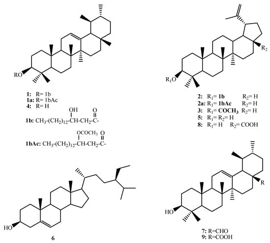
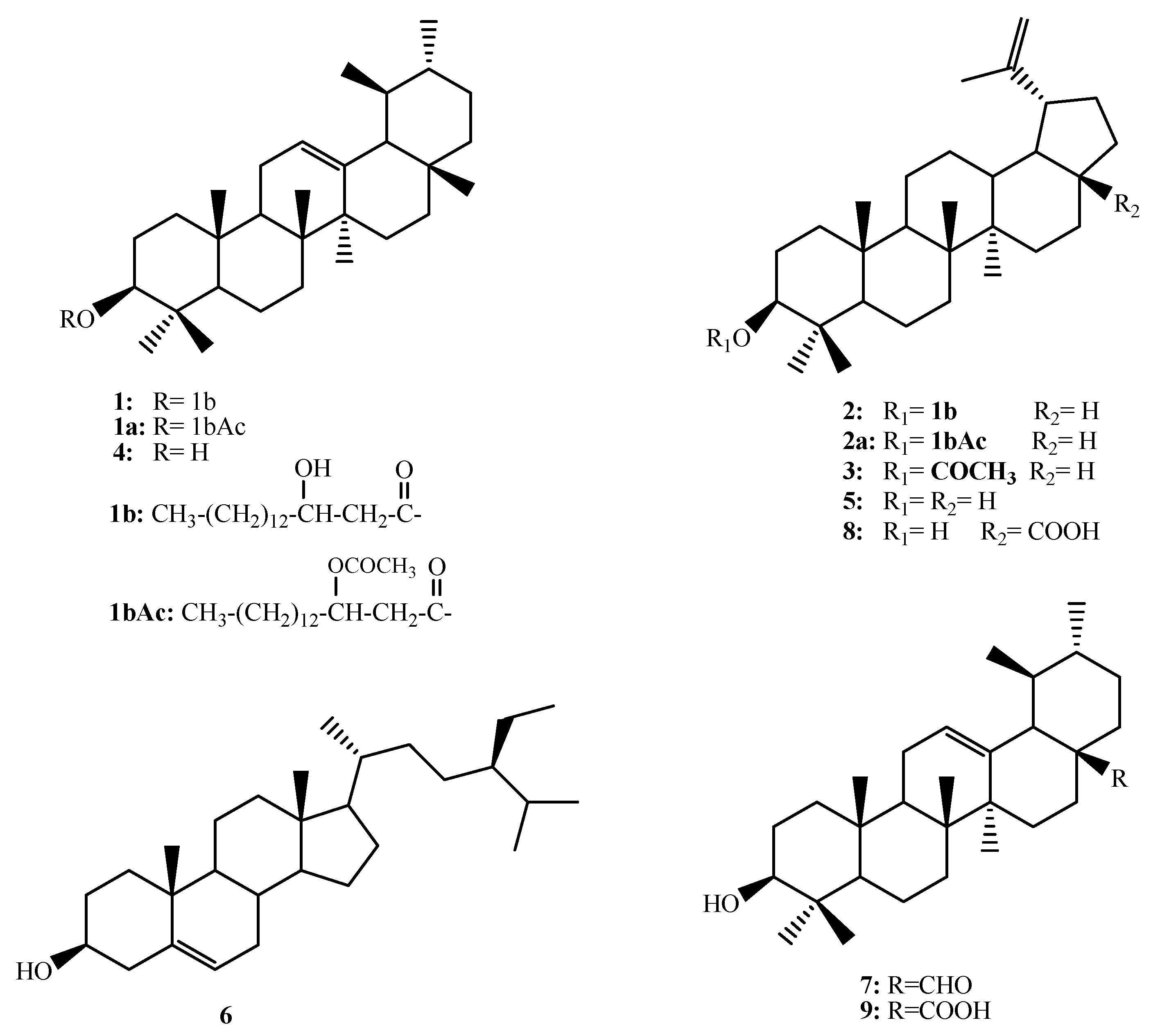
Compound 1 showed a complex overlapped 1H NMR spectrum (Figures S1 and S2, Table 1). Few resolved signals could be assigned, including one broad singlet at δH 5.16 ppm correlated with the carbon signal at δC 124.63 ppm (Figures S3 and S6, Table 2) assigned for =CH. The pattern of the carbon signals in the 13C NMR and DEPT135 experiments indicated a triterpenoidal skeleton. The data of the triterpenoid moiety were closely similar to those reported for α-amyrin [24,25]. However, the H-3 proton appeared as a double doublet with J = 4.5 and 11.7 Hz and was downfield shifted to δH 4.46 ppm compared with the chemical shift values of H-3 in triterpenes (Figure S2). The corresponding C-3 shifted to δC 80.82 ppm (Figures S3 and S6). Both values for H-3 and C-3 were diagnostic for the acylated hydroxyl group at C-3. Due to the overlapping of the proton and carbon spectra in the aliphatic region, it was expected that the aceylating moiety would be a long-chain fatty acid. The multiplet at δH 3.78 ppm (Figure S2) and the CHOH signal at δC 70.68 ppm (Figures S4 and S6) indicated the presence of a hydroxyl group on the fatty acid part. The presence of a free hydroxyl group in compound 1 was proven by acetylation with acetic anhydride in pyridine to give 1a. The 1H and 13C NMR spectra (Figures S8–S13, Table 1 and Table 2) of 1a pointed out one acetyl group at δH 1.75, δC 20.52, and 169.37 ppm. The carbonyl ester was shifted to δC 169.57 ppm. The CH-O proton signal expressed a downfield shift to δH 5.46 ppm as a result of the acetylation process. The CH-O proton showed a COSY correlation with the resolved CH2 protons at δH 2.44 and 2.56 ppm (Figure S14). These protons were correlated in the HSQC experiment (Figure S15) to the methylene carbon signal at δC 39.79 ppm and showed 2-bonds HMBC correlations (Figure S16) with the ester carbonyl at δC 169.57 and the hydroxyl bearing CH at δC 70.60 ppm giving the hydroxyl group position 3′ on the fatty acid skeleton and the CH2 position 2′. This assignment was further supported by the two-way H2BC correlations (Figure S18) between the C-2′ CH2 and the C-3′ CH-O. To obtain further clear spectroscopic evidence for the structure of compound 1, it was subjected to alkaline hydrolysis using 0.1 N alcoholic NaOH to hydrolyze the ester group. Hydrolytic products were purified on a silica gel column to obtain the two components of 1. The triterpene part spectral data were similar to compound 4 and identified as α-amyrin [24,25] by comparison of the obtained spectral data with the literature values. The other hydrolytic product was 1b, representing the esterifying fatty acid. The spectral data of 1b were in complete agreement with the 3-hydroxy long-chain fatty acid. Similar correlations were observed in COSY, HMBC, and H2BC between CH2 at C-2 and CHOH at C-3 (Figures S21–S32). The length of the fatty acid carbon chain was obtained from the HRESIMS of the fatty acid 1b, the acetylation product 1bAc, 1, and 1a (Figures S7, S12, S33, and S34). HRESIMS of 1b showed [M−1]+ at m/e 271.2277 for the molecular formula C16H32O3-H (Figure S33), while the mono-acetate derivative showed [M−1]+ at m/z 313.2388 for C18H33O4-H (Figure S34). These data enable the identification of the esterifying fatty acid as 3-hydroxyhexadecanoic acid (3-hydroxypalmitic acid). The spectrum of 1 showed [M+Na]+ at m/z 703.5995 for C46H80O3+Na (Figure S7), while that of 1a showed [M+Na]+ at m/z 745.6100 for C48H82O4+Na (Figure S20) all enable the identification of 1 as the new ester α-amyrin 3-(3′-hydroxy)-hexadecanoate given the trivial name Rhazyin A (Figure 1).

Figure 1.
Chemical strucrures of 1–9.
A similar treatment of 2 enables the identification of the acid part as 3-hydroxyhexadecanoic acid (3-hydroxypalmitic acid). The triterpene skeleton was identified as lupeol based on the comparison of the recorded data with the literature values [26,27]. Compound 2 was previously reported from Alecrim–Propolis (Brazilian green propolis) and was given the name procrim A (Figure 1) [28].
The known compounds were identified as lupeol acetate (3) [27,29], α-amyrin (4) [20,21], lupeol (5) [27,29], β-sitosterol (6) [29], ursaldehyde (7) [30], betulenic acid (8) [31] and ursolic acid (9) [30] based on the comparison with the literature data (Figure 1).
Molecular docking of phytochemicals on appropriate drug target(s) of new diseases provides clues about which plants can be targeted for biological activity screening [32]. The 3C protease of FMDV (FMDV 3Cpro) is a chymotrypsin-like cysteine protease [33,34], which is one of the most highly conserved proteins among all picornaviruses, including FMDV. This enzyme plays a crucial role in the viral life cycle by cleaving the picornavirus polyprotein into functional mature structural and non-structural proteins [35]. FMDV 3Cpro processes 10 of 13 cleavage sites on the polyprotein, making this enzyme an attractive target for anti-viral drugs [18]. Docking analysis was carried out to investigate the best pose of chosen ligands with the objective protein to gain molecular perception of the inhibitor’s mechanism of binding. Molecular docking analysis using a glide extra-precision module was utilized for investigating the possible molecular interactions accounting for anti-viral activity against picornavirus of the nine isolated compounds. To determine protein–ligand binding and interactions, the studied FMDV 3Cpro structure was modeled using the published three-dimensional (3D) structure of a wide-type 3Cpro [20]. The FMDV 3Cpro structure was generated based on the deduced amino acid sequence of FMDV by homology modeling with the PDB ID: 2WV4.pdb using the SWISS-MODEL [36]. The nine isolated compounds and the reference drugs; the natural anti-viral triterpene, glycyrrhizic acid [37], and the anti-viral synthetic drugs, ribavirin [38], were rated based on their extra precision docking scores. As a result of the in silico docking experiments, the screened compounds were arranged based on their docking scores (Table 5). The molecular docking studies revealed a strong binding of the discovered hits within the active site of FMDV 3Cpro. The highest active compound shown by docking XP g score was Rhazyin A (α-Amyrin 3-(3′R-hydroxy)-hexadecanoate).
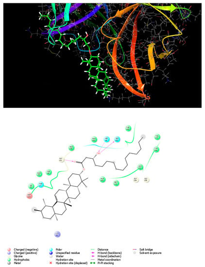
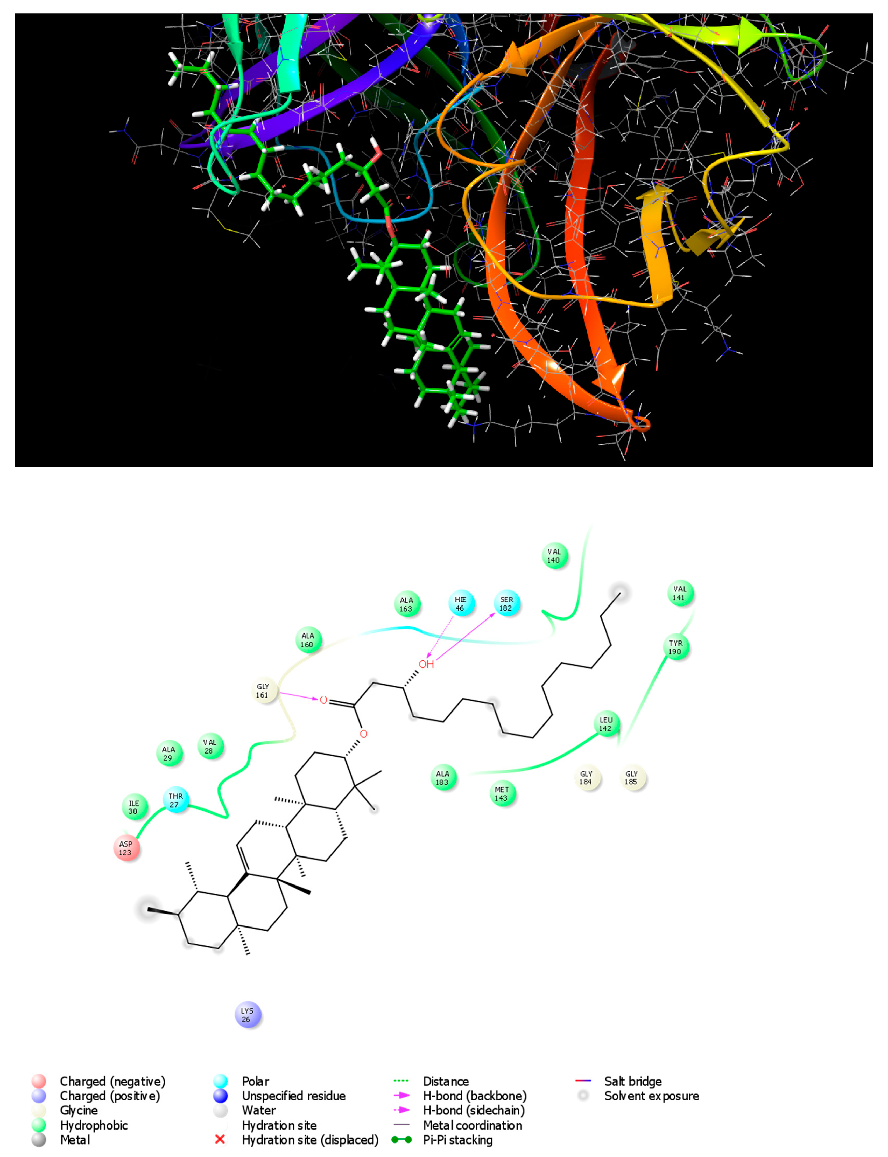
Docking results revealed that Rhazyin A had the lowest binding energy (−5.048 kcal mol−1) and most favorable docking poses, followed by procrim A (−4.76 kcal mol−1) and then Lupeol acetate (−3.986 kcal mol−1) as shown in (Table 5). The top recognized molecule, Rhazyin A, interactions with the enzyme are depicted in Figure 2. Rhazyin A established three hydrogen bonds with GLY161, SER182, and HIE46 residues, which are important for enzyme-tight binding. Additionally, numerous hydrophobic interactions with VAL28, ALA29, ILE30, VAL140, VAL141, LEU142, MET143, ALA160, ALA163, ALA183, and TYR190 residues and also polar interactions with THR27, HIE46, and SER182 residues in a manner comparable to the binding modalities seen in the previously investigated enzyme substrates ensuring successful docking [18]. Compound 1 (Rhazyin A) showed the lowest docking score within the nine isolated compounds comparable to the two known anti-viral drugs, glycyrrhizic acid and ribavirin. The reference drugs, glycyrrhizic acid and ribavirin interactions with the enzyme, were depicted in S1 and S2, respectively. The molecular docking confirmed the in vitro results, where Rhazyin A showed the most activity with 51% inhibition. To our knowledge, this is the first in silico study on the anti-FMDV-3Cpro activity of R. stricta.

Figure 2.
3D and 2D interaction diagrams of FMDV 3Cpro with Rhazyin A.
Different classes of triterpenes such as cycloartane, dammarane, lupane, oleanane, and ursane expressed promising anti-viral activity against dengue virus (DENV), human immunodeficiency viruses (HIV), herpes simplex virus (HSV), hepatitis virus (HV), influenza virus (IV), porcine epidemic diarrhea virus (PEDV), and respiratory syncytial virus (RSV) with selectivity index (SI)/Therapeutic index (TI) > 10 [37].
The MNTC of each of the isolated compounds 1–9 were tested for the possible inhibitory effect against picornavirus growth. Compound 1 with ursane skeleton was the most active with about 51% viral inhibitory effect, followed by 2. Both compounds share the presence of the long-chain alkanoic acid moiety. The acetate ester 3 was as active as 2, indicating that the esterification of the C-3 OH of these compounds potentiates the anti-viral activity. α-Amyrin was more active than lupeol; however, both compounds expressed weak activity. However, the member with lupane skeleton “betulenic acid” was more active than its analog with ursane skeleton “ursolic acid”. β-sitosterol was as active as ursaldehyde with ursane type triterepenoidal skeleton.
5. Conclusions
Biologically directed phytochemical study of Rhazya stricta against picornavirus causing foot-and-mouth disease revealed that the petroleum ether soluble fraction (RSP) was the most active and the least toxic fraction to the host cells necessary for the viral growth. Extensive chromatographic purification of the RSP fraction resulted in the isolation of nine triterpenoid and steroidal compounds. The structures of the compounds were identified by various spectroscopic and chemical methods. Among the isolates, a new alkanoic acid ester Rhazyin A (α-Amyrin 3-(3′R-hydroxy)-hexadecanoate) was the most active in the anti-viral assay with 51% inhibition of the viral growth using the MNTC. The obtained results indicated that the ester moiety at C-3 is essential for potentiating the anti-viral activity. These findings were supported by molecular docking analysis where Rhazyin A showed the most stable conformation among the nine isolated compounds within the active site of FMDV 3Cpro. Molecular docking enabled us to predict potential interactions between the screened phytochemicals and the disease target. The results of this research suggest a potential role of medicinal plants in the management of FMVD infection and provide lead candidates from natural origin with potential safety and efficacy compared to the synthetic ones with lower production costs for managing FMVD.
Supplementary Materials
The following supporting information can be downloaded at: https://www.mdpi.com/article/10.3390/metabo13060750/s1, Figures S1–S61: Spectral data of compounds 1 and 2; Tables S1 and S2: Cytotoxicity data of extracts and oure comounds.
Author Contributions
Conceptualization, M.S.A.-K., G.A.S., H.Y.E. and H.H.Z.; methodology, M.S.A.-K., G.A.S. and M.H.A.; software, H.Y.E., R.S.I. and H.H.Z.; formal analysis F.S.A., A.F.A., H.Y.E., R.S.I. and H.H.Z.; investigation, M.S.A.-K., F.S.A., A.F.A. and M.H.A.; resources, F.S.A., M.H.A., R.S.I. and H.H.Z.; data curation, H.Y.E., M.H.A., R.S.I. and H.H.Z.; writing—original draft preparation, F.S.A., A.F.A. and H.H.Z.; writing—review and editing, M.S.A.-K., G.A.S., H.Y.E. and H.H.Z.; visualization, G.A.S., H.Y.E., M.H.A., R.S.I. and H.H.Z.; supervision, M.S.A.-K., G.A.S. and H.Y.E.; project administration, M.S.A.-K. All authors have read and agreed to the published version of the manuscript.
Funding
This study is supported via funding from Prince Sattam Bin Abdulaziz University project number (PSAU/2023/R/1444).
Institutional Review Board Statement
Not applicable.
Informed Consent Statement
Not applicable.
Data Availability Statement
No new data were created or analyzed in this study. Data sharing is not applicable to this article.
Acknowledgments
The authors express their thanks to Prince Sattam Bin Abdulaziz University for supporting this study via funding from project number (PSAU/2023/R/1444). The authors would like to thank the Military Veterinary Hospital, Cairo, Egypt, for carrying out the anti-viral assay.
Conflicts of Interest
The authors declare no conflict of interest.
References
- Bukhari, N.A.; Al-Otaibi, R.A.; Ibhrahim, M.M. Phytochemical and taxonomic evaluation of Rhazya stricta in Saudi Arabia. Saudi J. Biol. Sci. 2017, 24, 1513–1521. [Google Scholar] [CrossRef]
- Gilani, S.A.; Kikuchi, A.; Shinwari, Z.K.; Khattak, Z.I.; Watanabe, K.N. Phytochemical, pharmacological and ethnobotanical studies of Rhazya stricta Decne. Phytother. Res. 2007, 21, 301–307. [Google Scholar] [CrossRef]
- Yahya, M.A. Saudi Plants: A Phytochemical and Biological Approach; King Abdulaziz City for Science and Technology: Riyah, Saudi Arabia, 1990. [Google Scholar]
- Bashir, A.K.; Abdalla, A.A.; Hassan, E.S.; Wasfi, I.A.; Amiri, M.A.; Crabb, T.A. Alkaloids with antimicrobial activity from the root of Rhazya stricta Decn. growing in United Arab Emirates. Arab. Gulf J. Sci. Res. 1994, 12, 119–131. [Google Scholar]
- Tanira, M.; Ali, B.; Bashir, A.; Chandranath, I. Some pharmacologic and toxicologic studies on rhazya stricta decne in rats, mice and rabbits. Gen. Pharmacol. Vasc. Syst. 1996, 27, 1261–1267. [Google Scholar] [CrossRef]
- Read, M. Plants of Dhofar (The Southern Region of Oman, Traditional Economic and Medicinal Uses) Anthony G. Miller and Miranda Morris The Office of the Adviser for Conservation of the Environment, Diwan of Royal Court, Sultanate of Oman. 1988, 361pp., HB. In UK, available from Holmes McDougall Ltd., Allander House, 137–141 Leith Walk, Edinburgh EH6 8NS. £37.50 inc postage. Oryx 2009, 24, 232. [Google Scholar]
- Dymock, W.; Warden, C.J.H.; Hooper, D. Pharmacographia Indica; Kegan Paul, Trench, Trubner & Co.: London, UK, 1890. [Google Scholar]
- Ahmad, H.; Bhatti, G.R.; Latif, A. Medicinal flora of the thar desert: An overview of problems and their feasible solutions. Zonas Áridas 2006, 8, 73–84. [Google Scholar]
- Ahmed, M. Checklist of Medicinal Flora of Tehsil Isakhel, District Mianwali-Pakistan. Ethnobot. Leafl. 2006, 2006, 10. [Google Scholar]
- Jamal, S.M.; Belsham, G.J. Foot-and-mouth disease: Past, present and future. Vet. Res. 2013, 44, 116. [Google Scholar] [CrossRef]
- Grubman, M.J.; Baxt, B. Foot-and-mouth disease. Clin. Microbiol. Rev. 2004, 17, 465–493. [Google Scholar] [CrossRef]
- Knowles, N.J.; Samuel, A.R. Molecular epidemiology of foot-and-mouth disease virus. Virus Res. 2003, 91, 65–80. [Google Scholar] [CrossRef]
- Kahn, C.N.; Line, S. The Mercks Veterinary Manual, 9th ed.; Merck and Company Incorporated: White House Station, NJ, USA, 2005. [Google Scholar]
- Alexandersen, S.; Kitching, R.P.; Mansley, L.M.; Donaldson, A.I. Clinical and laboratory investigations of five outbreaks of foot-and-mouth disease during the 2001 epidemic in the United Kingdom. Vet. Rec. 2003, 152, 489–496. [Google Scholar] [CrossRef] [PubMed]
- Radostitis, O.M.; Gay, C.C.; Blood, D.C.; Hinchcliff, K.W. Veterinary Medicine: A Textbook of the Diseases of Cattle, Sheep, Pigs, Goats and Horses, 9th ed.; W.B. Saunders Company: Sarasota, FL, USA; Harcourt Publishers: London, UK, 2000. [Google Scholar]
- Knight-Jones, T.J.; Rushton, J. The economic impacts of foot and mouth disease-what are they, how big are they and where do they occur? Prev. Vet. Med. 2013, 112, 161–173. [Google Scholar] [CrossRef] [PubMed]
- Gakuya, D.W.; Mulei, C.M.; Wekesa, S.B. Use of ethnoveterinary remedies in the management of foot and mouth disease lesions in a dairy herd. Afr. J. Tradit. Complement. Altern. Med. 2011, 8, 165–169. [Google Scholar] [CrossRef] [PubMed]
- Theerawatanasirikul, S.; Lueangaramkul, V.; Thangthamniyom, N.; Chankeeree, P.; Semkum, P.; Lekcharoensuk, P. Andrographolide and Deoxyandrographolide Inhibit Protease and IFN-Antagonist Activities of Foot-and-Mouth Disease Virus 3Cpro. Animals 2022, 12, 1995. [Google Scholar] [CrossRef]
- Abdel-Kader, M.S.; Rehman, N.U.; Aldosari, A.F.; Almutib, F.S.; Al Muwinea, A.I.; Saeedan, A.S. Bronchodilator Secondary Metabolites from Rhazya stricta Decne Aerial Parts. Separations 2022, 9, 412. [Google Scholar] [CrossRef]
- Theerawatanasirikul, S.; Lekcharoensuk, P. Virtual Screening of Natural Compounds Targeting Proteases of Coronaviruses and Picornaviruses. In Methods in Pharmacology and Toxicology; Roy, K., Ed.; Springer: New York, NY, USA, 2021. [Google Scholar]
- Khairy, A.; Hammoda, H.M.; Celik, I.; Zaatout, H.H.; Ibrahim, R.S. Discovery of potential natural dihydroorotate dehydrogenase inhibitors and their synergism with brequinar via integrated molecular docking, dynamic simulations and in vitro approach. Sci. Rep. 2022, 12, 19037. [Google Scholar] [CrossRef]
- Reham, S.; El-Mezayen, I.N.S.; Khairy, A.; Zaatout, H.H.; Hammoda, H.M.; Metwally, A.M. Biologically-Guided Isolation of Natural Lead Antithyroid Drug from Medicago sativa L. Sprouts and Its Toxic Profile in Comparison with Propylthiouracil. J. Food Drug Anal. 2020, 28, 407–448. [Google Scholar]
- Wang, J.; Xu, C.; Wong, Y.; Li, Y.; Liao, F.; Jiang, T.; Tu, Y. Artemisinin, the Magic Drug Discovered from Traditional Chinese Medicine. Engineering 2019, 5, 32–39. [Google Scholar] [CrossRef]
- Jin, Q.; Ko, H.J.; Chang, Y.-S.; Woo, E.-R. Chemical constituents from the aerial parts of Aster yomena. Nat. Prod. Sci. 2013, 19, 269–274. [Google Scholar]
- Dekebo, A.; Dagne, E.; Gautun, O.; Aasen, A. Triterpenes from the resin of Boswellia neglecta. Bull. Chem. Soc. Ethiop. 2002, 16, 87–90. [Google Scholar] [CrossRef]
- Abdullahi, S.; Musa, A.; Abdullahi, M.; Sule, M.; Sani, M. Isolation of Lupeol from the Stem-bark of Lonchocarpus sericeus (Papilionaceae). Sch. Acad. J. Biosci. 2013, 1, 18–19. [Google Scholar]
- Ben nejma, A.; Besbes, M.; Guérineau, V.; Touboul, D.; Ben jannet, H.; Hamza, M. Isolation and structure elucidation of acetylcholinesterase lipophilic lupeol derivatives inhibitors from the latex of the Tunisian Periploca laevigata. Arab. J. Chem. 2017, 10, S2767–S2772. [Google Scholar] [CrossRef]
- Furukawa, S.; Takagi, N.; Ikeda, T.; Ono, M.; Nafady, A.M.; Nohara, T.; Sugimoto, H.; Doi, S.; Yamada, H. Two novel long-chain alkanoic acid esters of lupeol from alecrim-propolis. Chem. Pharm. Bull. (Tokyo) 2002, 50, 439–440. [Google Scholar] [CrossRef] [PubMed]
- Ododo, M.M.; Choudhury, M.K.; Dekebo, A.H. Structure elucidation of β-sitosterol with antibacterial activity from the root bark of Malva parviflora. SpringerPlus 2016, 5, 1210. [Google Scholar] [CrossRef]
- Kim, D.-H.; Han, K.-M.; Chung, I.-S.; Kim, D.-K.; Kim, S.-H.; Kwon, B.-M.; Jeong, T.-S.; Park, M.-H.; Ahn, E.-M.; Baek, N.-I. Triterpenoids from the Flower of Campsis grandiflora K. Schum. as Human Acyl-CoA: Cholesterol Acyltransferase Inhibitors. Arch. Pharm. Res. 2005, 28, 550–556. [Google Scholar] [CrossRef]
- Noviany, N.; Osman, H. Structure Elucidation of Betulinic Acid from Sesbania grandiflora Root. J. Phys. J. Phys. Conf. Ser. 2021, 1751, 012090. [Google Scholar] [CrossRef]
- Mason, P.W.; Grubman, M.J.; Baxt, B. Molecular basis of pathogenesis of FMDV. Virus Res. 2003, 91, 9–32. [Google Scholar] [CrossRef]
- Gao, Y.; Sun, S.-Q.; Guo, H.-C. Biological function of Foot-and-mouth disease virus non-structural proteins and non-coding elements. Virol. J. 2016, 13, 107. [Google Scholar] [CrossRef]
- Birtley, J.R.; Knox, S.R.; Jaulent, A.M.; Brick, P.; Leatherbarrow, R.J.; Curry, S. Crystal structure of foot-and-mouth disease virus 3C protease. New insights into catalytic mechanism and cleavage specificity. J. Biol. Chem. 2005, 280, 11520–11527. [Google Scholar] [CrossRef]
- Asiamah, I.; Obiri, S.A.; Tamekloe, W.; Armah, F.A.; Borquaye, L.S. Applications of molecular docking in natural products-based drug discovery. Sci. Afr. 2023, 20, e01593. [Google Scholar] [CrossRef]
- Waterhouse, A.; Bertoni, M.; Bienert, S.; Studer, G.; Tauriello, G.; Gumienny, R.; Heer, F.T.; De Beer, T.A.P.; Rempfer, C.; Bordoli, L.; et al. SWISS-MODEL: Homology modelling of protein structures and complexes. Nucleic Acids Res. 2018, 46, W296–W303. [Google Scholar] [CrossRef] [PubMed]
- Darshani, P.; Sen Sarma, S.; Srivastava, A.K.; Baishya, R.; Kumar, D. Anti-viral triterpenes: A review. Phytochem. Rev. 2022, 21, 1761–1842. [Google Scholar] [CrossRef] [PubMed]
- Wassel, M.M.S.; Gamal El-Din, W.M.; Ahmed Ragab, A.; Elhag Ali, G.A.M.; Ammar, Y.A. Antiviral Activity of Adamantane-Pyrazole Derivatives Against Foot and Mouth Disease Virus Infection in vivo and in vitro with Molecular Docking Study. J. Appl. Vet. Sci. 2020, 5, 37–46. [Google Scholar]
Disclaimer/Publisher’s Note: The statements, opinions and data contained in all publications are solely those of the individual author(s) and contributor(s) and not of MDPI and/or the editor(s). MDPI and/or the editor(s) disclaim responsibility for any injury to people or property resulting from any ideas, methods, instructions or products referred to in the content. |
© 2023 by the authors. Licensee MDPI, Basel, Switzerland. This article is an open access article distributed under the terms and conditions of the Creative Commons Attribution (CC BY) license (https://creativecommons.org/licenses/by/4.0/).


