4-Aminobenzoic Acid Derivatives: Converting Folate Precursor to Antimicrobial and Cytotoxic Agents

 ,
,  , , ,
, , ,
Abstract
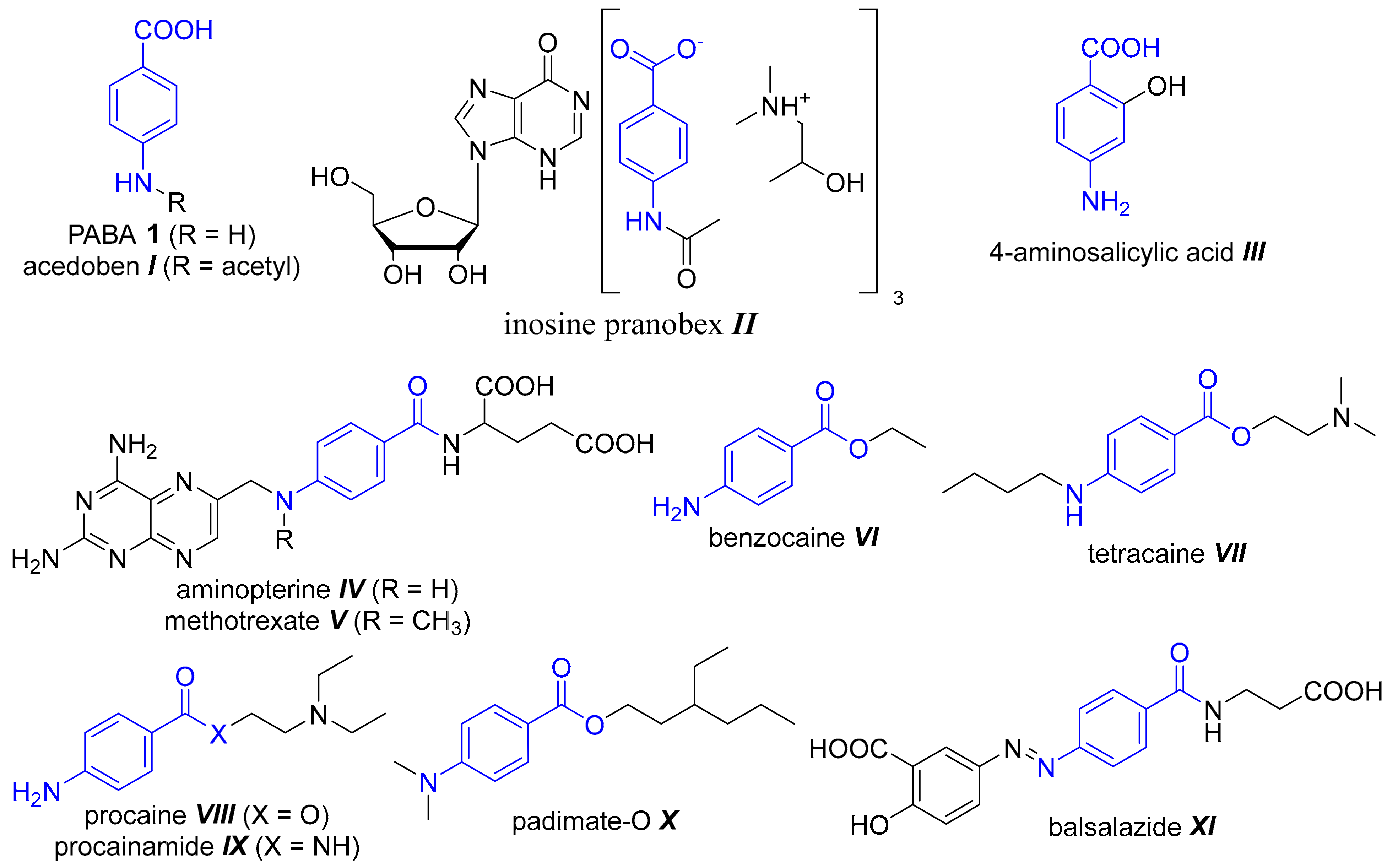
:1. Introduction
2. Materials and Methods
2.1. Chemistry
2.1.1. General
2.1.2. Synthesis of PABA-Derived Schiff Bases 1a–1t
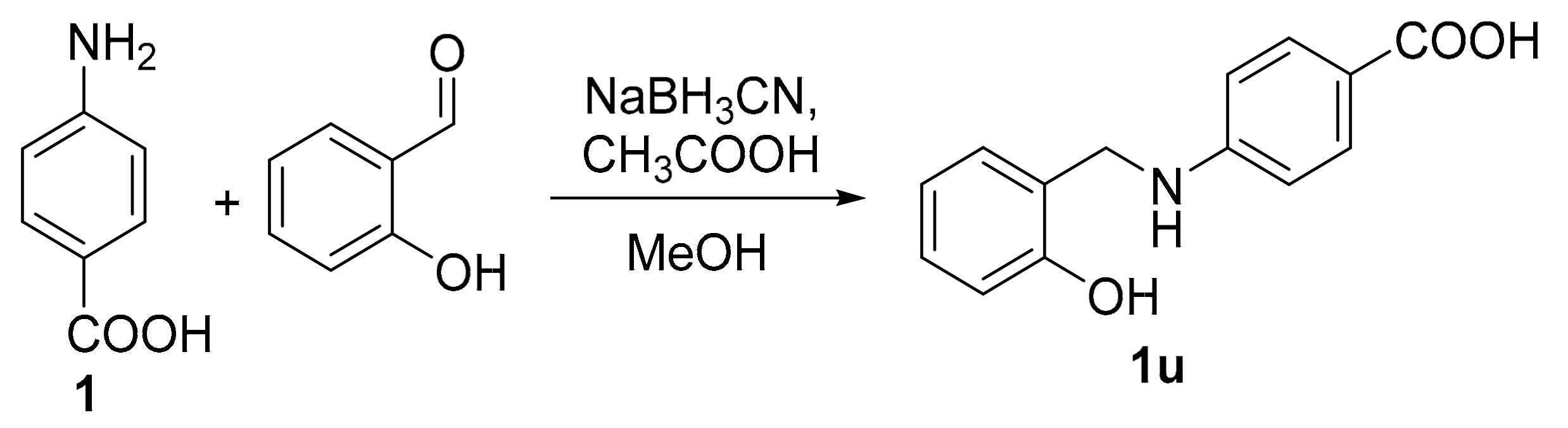
2.1.3. Synthesis of 1u
2.2. Pharmacology
2.2.1. Antibacterial Activity
2.2.2. Antimycobacterial Activity
2.2.3. Antifungal Activity
2.2.4. Cytotoxicity
3. Results and Discussion
3.1. Chemistry
3.2. Antibacterial Activity
- The presence of 5-F (1b), 5-t-Bu (1j) and chlorine at the positions 4 and 6 of the salicylic ring (1k, 1l) did not improve activity of 1a.
- Nitro (1f) and hydroxy group (1i) with opposite electron properties led to similar results—inhibition of multiple Gram-positive strains including MRSA, but at high concentrations (≥ 250 µM).
- The 5-substitution by a single halogen (Cl 1c, Br 1d, I 1e) improves the inhibition of S. epidermidis; in the case of 1e MRSA, too (MIC from 62.5 µM).
- Electron-donation groups (CH3 1g, OCH3 1h) together with 3-Cl (1m) produced inhibition of only S. epidermidis, but with MIC from 250 µM.
- 5-Nitrofurylidene PABA derivative 1r inhibited all three staphylococcal strains (MIC 62.5–125 µM).
- The Schiff bases derived from 3,5-dihalogenosalicylaldehydes (1n–1q) represent the most successful modification of PABA with MIC values of ≥ 15.62 µM, especially if the particular halogens are different (1o, 1p). These latter compounds containing one iodine atom also demonstrated activity against three Gram-negative strains (E. coli, K. pneumoniae).
3.3. Antimycobacterial Activity
3.4. Antifungal Activity
- Substituted salicylidene or 5-nitrofurfurylidene moiety is indispensable: unsubstituted salicylaldehyde (1a), its reduced analogue 1u, 2-methoxybenzaldehyde (1s) and 4-hydroxybenzaldehyde (1t) share no antimycotic potency.
- 5-F (1b) and 5-alkyls (1g, 1j) are not translated into any antifungal action.
- The presence of NO2 (1f), OH (1i), OCH3 (1h) and 4-Cl (1l) substituents provides active compounds but with higher MIC values (≥ 250 µM).
- Focusing on isomeric 6-Cl (1k), 5-Cl (1c), 4-Cl (1l) and 3-Cl (1m) derivatives, the increase of the activity is as follows: 4-Cl (MIC ≥ 250 µM) < 5-Cl ≤ 6-Cl < 3-Cl (MIC ≥ 31.25 µM). Interestingly, the introduction of second chlorine atom (3,5-Cl2) did not improve the activity (1n; MIC ≥ 125 µM).
- The activity of single halogen-substituted derivatives (5-F 1b, 5-Cl 1c, 5-Br 1d, 5-I 1e) is increasing with the atomic mass/lipophilicity/atomic radius of the halogens (5-F > 500 µM; 5-I ≥ 31.25 µM).
- 3,5-Dihalogenosalicylidene imines (1n–1q) were identified as the most active derivatives providing both low MIC (≥ 7.81 µM) and broad-spectrum of the activity. The presence of at least one iodine atom results in the highest antifungal properties (1o and 1p).
3.5. Cytotoxicity
4. Conclusions
Author Contributions
Funding
Conflicts of Interest
References
- Patel, H.M.; Bhardwaj, V.; Sharma, P.; Noolvi, M.N.; Lohan, S.; Bansal, S.; Sharma, A. Quinoxaline-PABA bipartite hybrid derivatization approach: Design and search for antimicrobial agents. J. Mol. Struct. 2019, 1184, 562–568. [Google Scholar] [CrossRef]
- Akberova, S.I. New Biological Properties of p-Aminobenzoic Acid. Biol. Bull. 2002, 29, 390–393. [Google Scholar] [CrossRef]
- Kadhum, W.R.; Oshizaka, T.; Ichiro, H.; Todo, H.; Sugibayashi, K. Usefulness of liquid-crystal oral formulations to enhance the bioavailability and skin tissue targeting of p-amino benzoic acid as a model compound. Eur. J. Pharm. Sci. 2016, 88, 282–290. [Google Scholar] [CrossRef] [PubMed] [Green Version]
- Crisan, M.E.; Bourosh, P.; Maffei, M.E.; Forni, A.; Pieraccini, S.; Sironi, M.; Chumakov, Y.M. Synthesis, Crystal Structure and Biological Activity of 2-Hydroxyethylammonium Salt of p-Aminobenzoic Acid. PLoS One 2014, 9, e101892. [Google Scholar] [CrossRef] [PubMed] [Green Version]
- Sowinska, M.; Morawiak, M.; Bochyńska-Czyż, M.; Lipkowski, A.W.; Ziemińska, E.; Zabłocka, B.; Urbanczyk-Lipkowska, Z. Molecular Antioxidant Properties and In Vitro Cell Toxicity of the p-Aminobenzoic Acid (PABA) Functionalized Peptide Dendrimers. Biomolecules 2019, 9, 89. [Google Scholar] [CrossRef] [PubMed] [Green Version]
- Richards, R.M.; Xing, D.K.; King, T.P. Activity of p-aminobenzoic acid compared with other organic acids against selected bacteria. J. Appl. Bacteriol. 1995, 78, 209–215. [Google Scholar] [CrossRef]
- Markitantova, Y.V.; Akberova, S.I.; Ryabtseva, A.A.; Stroeva, O.G. Effect of para-Aminobenzoic Acid on Apoptosis Processes in the Adult Rat Conjunctiva and Corneal Epithelium in vivo after Hypobaric Hypoxia. Biol. Bull. Russ. Acad. Sci. 2018, 45, 226–234. [Google Scholar] [CrossRef]
- Chang, T.-Y.; Hu, M.-L. Concentrations and lipid peroxidation in tissues and toxicity of para-aminobenzoic acid fed to rats in drinking water. J. Nutr. Biochem. 1996, 7, 408–413. [Google Scholar] [CrossRef]
- Nortje, C.; Jansen van Rensburg, P.; Cooke, C.; Erasmus, E. The simultaneous detection and quantification of p-aminobenzoic acid and its phase 2 biotransformation metabolites in human urine using LC–MS/MS. Bioanalysis 2015, 7, 1211–1224. [Google Scholar] [CrossRef]
- Švarcová, M.; Krátký, M.; Vinšová, J. Investigation of potential inhibitors of chorismate-utilizing enzymes. Curr. Med. Chem. 2015, 22, 1383–1399. [Google Scholar] [CrossRef]
- Yun, M.-K.; Wu, Y.; Li, Z.; Zhao, Y.; Waddell, M.B.; Ferreira, A.M.; Lee, R.E.; Bashford, D.; White, S.W. Catalysis and Sulfa Drug Resistance in Dihydropteroate Synthase: Crystal structures reveal the catalytic mechanism of DHPS and the structural basis of sulfa drug action and resistance. Science 2012, 335, 1110–1114. [Google Scholar] [CrossRef] [PubMed] [Green Version]
- Kluczyk, A.; Popek, T.; Kiyota, T.; de Macedo, P.; Stefanowicz, P.; Lazar, C.; Konishi, Y. Drug evolution: p-aminobenzoic acid as a building block. Curr. Med. Chem. 2002, 9, 1871–1892. [Google Scholar] [CrossRef] [PubMed]
- Beran, J.; Šalapová, E.; Špajdel, M. Inosine pranobex is safe and effective for the treatment of subjects with confirmed acute respiratory viral infections: Analysis and subgroup analysis from a Phase 4, randomised, placebo-controlled, double-blind study. BMC Infect. Dis. 2016, 16, 648. [Google Scholar] [CrossRef] [PubMed] [Green Version]
- Zheng, J.; Rubin, E.J.; Bifani, P.; Mathys, V.; Lim, V.; Au, M.; Jang, J.; Nam, J.; Dick, T.; Walker, J.R.; et al. para-Aminosalicylic acid is a prodrug targeting dihydrofolate reductase in Mycobacterium tuberculosis. J. Biol. Chem. 2013, 288, 23447–23456. [Google Scholar] [CrossRef] [PubMed] [Green Version]
- Marc, G.; Oniga, S.; Pirnau, A.; Duma, M.; Vlase, L.; Oniga, O. Rational Synthesis of Some New para-Aminobenzoic Acid Hybrids with Thiazolidin-2,4-diones with Antimicrobial Properties. ADMET and molecular docking evaluation. Rev. Chim. (Buchar.) 2019, 70, 769–775. [Google Scholar]
- Scherman, M.S.; North, E.J.; Jones, V.; Hess, T.N.; Grzegorzewicz, A.E.; Kasagami, T.; Kim, I.H.; Merzlikin, O.; Lenaerts, A.J.; Lee, R.E.; et al. Screening a library of 1600 adamantyl ureas for anti-Mycobacterium tuberculosis activity in vitro and for better physical chemical properties for bioavailability. Bioorg. Med. Chem. 2012, 20, 3255–3262. [Google Scholar] [CrossRef] [PubMed] [Green Version]
- Mureşan-Pop, M.; Kacsó, I.; Martin, F.; Simon, S.; Ştefan, R.; Bratu, I. Ambazone salt with p-aminobenzoic acid. J. Therm. Anal. Calorim. 2015, 120, 905–912. [Google Scholar] [CrossRef]
- Farooq, M.; Al Marhoon, Z.M.; Taha, N.A.; Baabbad, A.A.; Al-Wadaan, M.A.; El-Faham, A. Synthesis of Novel Class of N-Alkyl-isatin-3-iminobenzoic Acid Derivatives and Their Biological Activity in Zebrafish Embryos and Human Cancer Cell Lines. Biol. Pharm. Bull. 2018, 41, 350–359. [Google Scholar] [CrossRef] [Green Version]
- Krátký, M.; Vinšová, J.; Volková, M.; Buchta, V.; Trejtnar, F.; Stolaříková, J. Antimicrobial activity of sulfonamides containing 5-chloro-2-hydroxybenzaldehyde and 5-chloro-2-hydroxybenzoic acid scaffold. Eur. J. Med. Chem. 2012, 50, 433–440. [Google Scholar] [CrossRef]
- Krátký, M.; Dzurková, M.; Janoušek, J.; Konečná, K.; Trejtnar, F.; Stolaříková, J.; Vinšová, J. Sulfadiazine Salicylaldehyde-Based Schiff Bases: Synthesis, Antimicrobial Activity and Cytotoxicity. Molecules 2017, 22, 1573. [Google Scholar] [CrossRef]
- Krátký, M.; Bősze, S.; Baranyai, Z.; Stolaříková, J.; Vinšová, J. Synthesis and biological evolution of hydrazones derived from 4-(trifluoromethyl)benzohydrazide. Bioorg. Med. Chem. Lett. 2017, 27, 5185–5189. [Google Scholar] [CrossRef] [PubMed]
- Krátký, M.; Vinšová, J.; Stolaříková, J. Antimicrobial activity of rhodanine-3-acetic acid derivatives. Bioorg. Med. Chem. 2017, 25, 1839–1845. [Google Scholar] [CrossRef]
- Shrivastava, S.K.; Srivastava, P.; Upendra, T.V.R.; Tripathi, P.N.; Sinha, S.K. Design, synthesis and evaluation of some N-methylenebenzenamine derivatives as selective acetylcholinesterase (AChE) inhibitor and antioxidant to enhance learning and memory. Bioorg. Med. Chem. 2017, 25, 1471–1480. [Google Scholar] [CrossRef] [PubMed]
- Shetty, M.M.; Sadanandam, Y.S.; Sattur, P.B. Synthese und Pharmakologie von substituierten N-Benzyliden-Derivaten. Arch. Pharm. 1979, 312, 721–726. [Google Scholar] [CrossRef] [PubMed]
- Brewster, C.M.; Millam, L.H. Phototropic and Thermotropic Anils from 5-Bromosalicylaldehyde. J. Am. Chem. Soc. 1933, 55, 763–766. [Google Scholar] [CrossRef]
- Mayadeo, M.S.; Dhakappa, V.P. Determination of stability constants of N-[2-hydroxy-5-methylbenzylidene]-4-carboxyaniline complexes with Y3+, La3+, Pr3+, Nd3+, Sm3+, Gd3+ and Dy3+. J. Indian Chem. Soc. 1981, 58, 1010. [Google Scholar]
- Nasr, G.; Cristian, A.; Barboiu, M.; Vullo, D.; Winum, J.-Y.; Supuran, C.T. Carbonic anhydrase inhibitors. Inhibition of human cytosolic isoforms I and II with (reduced) Schiff’s bases incorporating sulfonamide, carboxylate and carboxymethyl moieties. Bioorg. Med. Chem. 2014, 22, 2867–2874. [Google Scholar] [CrossRef]
- UNIVERSITÀ DEGLI STUDI DI SALERNO; Longo, P.; Saturnino, C.; Arra, C.; Palma, G.; Mariconda, A.; Sinicropi, M.S.; Puoci, F.; Iacopetta, D. Hydroxybenzene derivatives having a N-aryl substitute imino group and use thereof in the treatment of solid tumours. Patent WO2018/116158; A1, 28 June 2018. [Google Scholar]
- Cronenberger, L.; Gaige, T.; Pacheco, H.; Pillon, D. Nouvelles bases de Schiff dérivées des aldehydes dihalogéno-3,5 salicyliques et possédant des propriétés antifongiques et antibactériennes. Chim. Ther. 1968, 3, 87–99. [Google Scholar]
- Ujiie, T. Experimental Anticancer Studies. XXVIII. Anticancer Activity of Some Nitrofuran Derivatives. Chem. Pharm. Bull. 1966, 14, 461–466. [Google Scholar] [CrossRef] [Green Version]
- Silku, P.; Özkinali, S.; Öztürk, Z.; Asan, A.; Köse, D.A. Synthesis of novel Schiff Bases containing acryloyl moiety and the investigation of spectroscopic and electrochemical properties. J. Mol. Struct. 2016, 1116, 72–83. [Google Scholar] [CrossRef]
- Bhunia, M.; Hota, P.K.; Vijaykumar, G.; Adhikari, D.; Mandal, S.K. A Highly Efficient Base-Metal Catalyst: Chemoselective Reduction of Imines to Amines Using An Abnormal-NHC–Fe(0) Complex. Organometallics 2016, 35, 2930–2937. [Google Scholar] [CrossRef]
- EUCAST DISCUSSION DOCUMENT. E.Dis 5.1. Determination of minimum inhibitory concentrations (MICs) of antibacterial agents by broth dilution. Clin. Microbiol. Infect. 2003, 9, 1–7.
- Krátký, M.; Vinšová, J.; Novotná, E.; Mandíková, J.; Trejtnar, F.; Stolaříková, J. Antibacterial Activity of Salicylanilide 4-(Trifluoromethyl)benzoates. Molecules 2013, 18, 3674–3688. [Google Scholar] [CrossRef] [PubMed] [Green Version]
- EUCAST DEFINITIVE DOCUMENT EDEF 7.3.1. Method for the Determination of Broth Dilution Minimum Inhibitory Concentrations of Antifungal Agents for Yeasts. 2017, pp. 1–21. Available online: http://www.eucast.org/fileadmin/src/media/PDFs/EUCAST_files/AFST/Files/EUCAST_E_Def_7_3_1_Yeast_testing__definitive.pdf (accessed on 20 October 2019).
- EUCAST DEFINITIVE DOCUMENT E.DEF 9.3.1. Method for the Determination of Broth Dilution Minimum Inhibitory Concentrations of Antifungal Agents for Conidia Forming Moulds. 2017, pp. 1–23. Available online: http://www.eucast.org/fileadmin/src/media/PDFs/EUCAST_files/AFST/Files/EUCAST_E_Def_9_3_1_Mould_testing__definitive.pdf (accessed on 20 October 2019).
- Ordonez, A.A.; Weinstein, E.A.; Bambarger, L.E.; Saini, V.; Chang, Y.S.; DeMarco, V.P.; Klunk, M.H.; Urbanowski, M.E.; Moulton, K.L.; Murawski, A.M.; et al. A Systematic Approach for Developing Bacteria-Specific Imaging Tracers. J. Nucl. Med. 2017, 58, 144–150. [Google Scholar] [CrossRef] [Green Version]
- Elsaman, T.; Mohamed, M.S.; Mohamed, M.A. Current development of 5-nitrofuran-2-yl derivatives as antitubercular agents. Bioorg. Chem. 2019, 88, 102969. [Google Scholar] [CrossRef]
- Laborda, P.; Zhao, Y.; Ling, J.; Hou, R.; Liu, F. Production of Antifungal p-Aminobenzoic Acid in Lysobacter antibioticus OH13. J. Agric. Food Chem. 2018, 66, 630–636. [Google Scholar] [CrossRef]
- Meir, Z.; Osherov, N. Vitamin Biosynthesis as an Antifungal Target. J. Fungi 2018, 4, 72. [Google Scholar] [CrossRef] [Green Version]
- Iliev, I.; Kontrec, D.; Detcheva, R.; Georgieva, M.; Balacheva, A.; Galić, N.; Pajpanova, T. Cancer cell growth inhibition by aroylhydrazone derivatives. Biotechnol. Biotec. Eq. 2019, 33, 756–763. [Google Scholar] [CrossRef] [Green Version]
- Gerets, H.H.J.; Tilmant, K.; Gerin, B.; Chanteux, H.; Depelchin, B.O.; Dhalluin, S.; Atienzar, F.A. Characterization of primary human hepatocytes, HepG2 cells, and HepaRG cells at the mRNA level and CYP activity in response to inducers and their predictivity for the detection of human hepatotoxins. Cell Biol. Toxicol. 2012, 28, 69–87. [Google Scholar] [CrossRef] [Green Version]
- Kamalian, L.; Chadwick, A.E.; Bayliss, M.; French, N.S.; Monshouwer, M.; Snoeys, J.; Park, B.K. The utility of HepG2 cells to identify direct mitochondrial dysfunction in the absence of cell death. Toxicol. In Vitro 2015, 29, 732–740. [Google Scholar] [CrossRef] [Green Version]
- Hradilová, L.; Poláková, M.; Dvořáková, B.; Hajdúch, M.; Petruš, L. Synthesis and cytotoxicity of some d-mannose click conjugates with aminobenzoic acid derivatives. Carbohydr. Res. 2012, 361, 1–6. [Google Scholar] [CrossRef] [PubMed]


 | ||||||||||||||||||
|---|---|---|---|---|---|---|---|---|---|---|---|---|---|---|---|---|---|---|
| Code | R1 | R2 | MIC [µM] | |||||||||||||||
| SA | MRSA | SE | EF | EC | KP | KP-E | PA | |||||||||||
| 24 h | 48 h | 24 h | 48 h | 24 h | 48 h | 24 h | 48 h | 24 h | 48 h | 24 h | 48 h | 24 h | 48 h | 24 h | 48 h | |||
| 1a | H | H | >500 | >500 | >500 | >500 | >500 | >500 | >500 | >500 | >500 | >500 | >500 | >500 | >500 | >500 | >500 | >500 |
| 1b | 5-F | H | >500 | >500 | >500 | >500 | >500 | >500 | >500 | >500 | >500 | >500 | >500 | >500 | >500 | >500 | >500 | >500 |
| 1c | 5-Cl | H | >500 | >500 | >500 | >500 | 62.5 | 62.5 | >500 | >500 | >500 | >500 | >500 | >500 | >500 | >500 | >500 | >500 |
| 1d | 5-Br | H | >250 | >250 | >250 | >250 | 62.5 | 62.5 | >250 | >250 | >250 | >250 | >250 | >250 | >250 | >250 | >250 | >250 |
| 1e | 5-I | H | >250 | >250 | 250 | 250 | 62.5 | 62.5 | >250 | >250 | >250 | >250 | >250 | >250 | >250 | >250 | >250 | >250 |
| 1f | 5-NO2 | H | 500 | 500 | 500 | 500 | 250 | 250 | 500 | 500 | >500 | >500 | >500 | >500 | >500 | >500 | >500 | >500 |
| 1g | 5-CH3 | H | >500 | >500 | >500 | >500 | 250 | 250 | >500 | >500 | >500 | >500 | >500 | >500 | >500 | >500 | >500 | >500 |
| 1h | 5-OCH3 | H | >500 | >500 | >500 | >500 | 500 | 500 | >500 | >500 | >500 | >500 | >500 | >500 | >500 | >500 | >500 | >500 |
| 1i | 5-OH | H | 500 | 500 | 250 | 250 | 250 | 250 | 500 | 500 | >500 | >500 | 500 | 500 | 500 | 500 | 500 | 500 |
| 1j | 5-tert-Bu | H | >500 | >500 | >500 | >500 | >500 | >500 | >500 | >500 | >500 | >500 | >500 | >500 | >500 | >500 | >500 | >500 |
| 1k | 6-Cl | H | >500 | >500 | >500 | >500 | >500 | >500 | >500 | >500 | >500 | >500 | >500 | >500 | >500 | >500 | >500 | >500 |
| 1l | 4-Cl | H | >500 | >500 | >500 | >500 | >500 | >500 | >500 | >500 | >500 | >500 | >500 | >500 | >500 | >500 | >500 | >500 |
| 1m | H | Cl | >500 | >500 | >500 | >500 | 500 | 500 | >500 | >500 | >500 | >500 | >500 | >500 | >500 | >500 | >500 | >500 |
| 1n | 5-Cl | Cl | 125 | 125 | 250 | 250 | 250 | 250 | >500 | >500 | >500 | >500 | >500 | >500 | >500 | >500 | >500 | >500 |
| 1o | 5-Cl | Br | 15.62 | 15.62 | 31.25 | 31.25 | 15.62 | 31.25 | 62.5 | 62.5 | 500 | 500 | 500 | 500 | 500 | 500 | >500 | >500 |
| 1p | 5-Cl | I | 15.62 | 15.62 | 15.62 | 15.62 | 31.25 | 31.25 | 62.5 | 62.5 | 62.5 | 62.5 | 125 | 125 | 125 | 125 | >500 | >500 |
| 1q | 5-I | I | 62.5 | 62.5 | 62.5 | 62.5 | 62.5 | 62.5 | 500 | 500 | 500 | 500 | 500 | 500 | 500 | 500 | >500 | >500 |
| 1r | - | - | 62.5 | 62.5 | 62.5 | 62.5 | 125 | 125 | >500 | >500 | >500 | >500 | 500 | 500 | 500 | 500 | 500 | 500 |
| 1s | - | - | >500 | >500 | >500 | >500 | >500 | >500 | >500 | >500 | >500 | >500 | >500 | >500 | >500 | >500 | >500 | >500 |
| 1t | - | - | >500 | >500 | >500 | >500 | >500 | >500 | >500 | >500 | >500 | >500 | >500 | >500 | >500 | >500 | >500 | >500 |
| 1u | - | - | >500 | >500 | >500 | >500 | >500 | >500 | >500 | >500 | >500 | >500 | >500 | >500 | >500 | >500 | >500 | >500 |
| PABA 1 | - | - | >500 | >500 | >500 | >500 | >500 | >500 | >500 | >500 | >500 | >500 | >500 | >500 | >500 | >500 | >500 | >500 |
| BAC | - | - | 7.81 | 15.62 | 15.62 | 15.62 | 15.62 | 31.25 | 15.62 | 62.5 | >500 | >500 | >500 | >500 | >500 | >500 | >500 | >500 |
| NEO | - | - | ND | ND | ND | ND | ND | ND | ND | ND | 3.9 | 3.9 | 1.95 | 1.95 | 3.9 | 3.9 | 7.81 | 15.62 |
| Code | R | R2 | MIC [µM] | |||||||||
|---|---|---|---|---|---|---|---|---|---|---|---|---|
| Mtb. 331/88 | M. avium 330/88 | M. kansasii 235/80 | M. kansasii 6509/96 | |||||||||
| 14 d | 21 d | 14 d | 21 d | 7 d | 14 d | 21 d | 7 d | 14 d | 21 d | |||
| 1a | H | H | >1000 | >1000 | >1000 | >1000 | 1000 | >1000 | >1000 | 1000 | 1000 | 1000 |
| 1b | 5-F | H | 500 | 500 | >1000 | >1000 | 250 | 500 | 500 | 500 | 500 | 500 |
| 1c | 5-Cl | H | 250 | 500 | 1000 | >1000 | 250 | 250 | 500 | 500 | 500 | 1000 |
| 1d | 5-Br | H | 250 | 500 | 500 | 500 | 250 | 250 | 500 | 500 | 500 | 500 |
| 1e | 5-I | H | 250 | 250 | 500 | 500 | 250 | 250 | 250 | 250 | 250 | 500 |
| 1f | 5-NO2 | H | 250 | 500 | 1000 | >1000 | 500 | 500 | 500 | 500 | 500 | 500 |
| 1g | 5-CH3 | H | 500 | 500 | >1000 | >1000 | 500 | 500 | 500 | 500 | 500 | 1000 |
| 1h | 5-OCH3 | H | 250 | 500 | 1000 | 1000 | 500 | 500 | 500 | 500 | 500 | 500 |
| 1i | 5-OH | H | 500 | 1000 | 1000 | >1000 | 500 | 500 | 1000 | 500 | 1000 | 1000 |
| 1j | 5-tert-Bu | H | 250 | 250 | 1000 | >1000 | 125 | 125 | 250 | 250 | 250 | 500 |
| 1k | 6-Cl | H | 250 | 500 | 500 | 1000 | 250 | 500 | 500 | 250 | 250 | 500 |
| 1l | 4-Cl | H | 250 | 500 | 1000 | 1000 | 250 | 500 | 500 | 250 | 250 | 500 |
| 1m | H | Cl | 500 | 500 | 1000 | 1000 | 500 | 500 | 500 | 250 | 500 | 500 |
| 1n | 5-Cl | Cl | 500 | 500 | 1000 | 1000 | 500 | 500 | 500 | 250 | 500 | 500 |
| 1o | 5-Cl | Br | 250 | 500 | 500 | 500 | 250 | 500 | 500 | 500 | 500 | 500 |
| 1p | 5-Cl | I | 250 | 250 | 500 | 1000 | 250 | 500 | 500 | 250 | 250 | 500 |
| 1q | 5-I | I | 250 | 250 | 500 | 500 | 250 | 500 | 500 | 250 | 250 | 250 |
| 1r | - | - | 62.5 | 125 | 250 | 500 | 62.5 | 125 | 250 | 125 | 125 | 250 |
| 1s | - | - | 1000 | 1000 | >1000 | >1000 | 1000 | 1000 | >1000 | 1000 | 1000 | 1000 |
| 1t | - | - | 1000 | 1000 | 1000 | >1000 | 1000 | 1000 | >1000 | 1000 | 1000 | 1000 |
| 1u | - | - | 1000 | 1000 | >1000 | >1000 | 1000 | 1000 | 1000 | 1000 | 1000 | 1000 |
| PABA 1 | - | - | >1000 | >1000 | >1000 | >1000 | 1000 | >1000 | >1000 | 1000 | 1000 | 1000 |
| INH | - | - | 0.5 | 1 | >250 | >250 | >250 | >250 | >250 | 8 | 8 | 8 |
| Code | R1 | R2 | MIC [µM] | |||||||||||||||
|---|---|---|---|---|---|---|---|---|---|---|---|---|---|---|---|---|---|---|
| CA | CT | CK | CG | TA | AF | LC | TI | |||||||||||
| 24 h | 48 h | 24 h | 48 h | 24 h | 48 h | 24 h | 48 h | 24 h | 48 h | 24 h | 48 h | 24 h | 48 h | 72 h | 120 h | |||
| 1a | H | H | >500 | >500 | >500 | >500 | >500 | >500 | >500 | >500 | >500 | >500 | >500 | >500 | >500 | >500 | >500 | >500 |
| 1b | 5-F | H | >500 | >500 | >500 | >500 | >500 | >500 | >500 | >500 | >500 | >500 | >500 | >500 | >500 | >500 | >500 | >500 |
| 1c | 5-Cl | H | >125 | >125 | 125 | 125 | 125 | 125 | 125 | 125 | >125 | >125 | >125 | >125 | >125 | >125 | 125 | 125 |
| 1d | 5-Br | H | 62.5 | 62.5 | 125 | 125 | 125 | 125 | 62.5 | 62.5 | 125 | 125 | 250 | 250 | >250 | >250 | 62.5 | 62.5 |
| 1e | 5-I | H | 62.5 | 62.5 | 125 | 125 | 62.5 | 62.5 | 31.25 | 31.25 | 62.5 | 62.5 | 125 | 125 | >125 | >125 | 31.25 | 31.25 |
| 1f | 5-NO2 | H | 500 | 500 | >500 | >500 | >500 | >500 | >500 | >500 | 500 | 500 | >500 | >500 | 500 | 500 | 500 | 500 |
| 1g | 5-CH3 | H | >125 | >125 | >125 | >125 | >125 | >125 | >125 | >125 | >125 | >125 | >125 | >125 | >125 | >125 | >125 | >125 |
| 1h | 5-OCH3 | H | 250 | 250 | 500 | 500 | 500 | 500 | 250 | 250 | 500 | 500 | >500 | >500 | >500 | >500 | >500 | >500 |
| 1i | 5-OH | H | 500 | 500 | >500 | >500 | >500 | >500 | 250 | 250 | >500 | >500 | >500 | >500 | >500 | >500 | >500 | >500 |
| 1j | 5-tert-Bu | H | >500 | >500 | >500 | >500 | >500 | >500 | >500 | >500 | >500 | >500 | >500 | >500 | >500 | >500 | >500 | >500 |
| 1k | 6-Cl | H | 125 | 125 | 62.5 | 125 | 125 | 250 | 125 | 125 | 125 | >125 | >125 | >125 | >125 | >125 | 125 | 125 |
| 1l | 4-Cl | H | 250 | >500 | 500 | >500 | 500 | >500 | 500 | >500 | 500 | >500 | 250 | >500 | >500 | >500 | 500 | >500 |
| 1m | H | Cl | 31.25 | 31.25 | 62.5 | 62.5 | 250 | 250 | 62.5 | 62.5 | 125 | 125 | 250 | 250 | 500 | 500 | 125 | 125 |
| 1n | 5-Cl | Cl | 125 | 125 | 250 | 250 | 125 | 125 | 125 | 125 | 125 | 125 | 500 | 500 | 500 | 500 | 250 | 250 |
| 1o | 5-Cl | Br | 31.25 | 31.25 | 62.5 | 62.5 | 62.5 | 62.5 | 15.62 | 15.62 | 31.25 | 31.25 | 250 | 250 | 500 | 500 | 62.5 | 62.5 |
| 1p | 5-Cl | I | 7.81 | 7.81 | 31.25 | 31.25 | 15.62 | 15.62 | 7.81 | 7.81 | 7.81 | 7.81 | 125 | 125 | 250 | 250 | 7.81 | 7.81 |
| 1q | 5-I | I | 7.81 | 7.81 | 15.62 | 15.62 | 31.25 | 31.25 | 7.81 | 7.81 | 15.62 | 15.62 | 125 | 125 | 250 | 250 | 7.81 | 7.81 |
| 1r | - | - | 250 | 250 | 250 | 250 | 62.5 | 62.5 | 250 | 250 | 500 | 500 | >500 | >500 | >500 | >500 | 250 | 250 |
| 1s | - | - | >500 | >500 | >500 | >500 | >500 | >500 | >500 | >500 | >500 | >500 | >500 | >500 | >500 | >500 | >500 | >500 |
| 1t | - | - | >500 | >500 | >500 | >500 | >500 | >500 | >500 | >500 | >500 | >500 | >500 | >500 | >500 | >500 | >500 | >500 |
| 1u | - | - | >500 | >500 | >500 | >500 | >500 | >500 | >500 | >500 | >500 | >500 | >500 | >500 | >500 | >500 | >500 | >500 |
| PABA 1 | - | - | >500 | >500 | >500 | >500 | >500 | >500 | >500 | >500 | >500 | >500 | >500 | >500 | >500 | >500 | >500 | >500 |
| FLU | - | - | 0.24 | 0.24 | >500 | >500 | 125 | 250 | 31.25 | 500 | 250 | 500 | >500 | >500 | >500 | >500 | 7.81 | 125 |
| Code | R1 | R2 | IC50 [µM] | Range of Concentrations Tested |
|---|---|---|---|---|
| 1a | H | H | >1500 * | 1–1500 |
| 1b | 5-F | H | 497.6 | 1–500 |
| 1c | 5-Cl | H | 142.9 | 1–500 |
| 1d | 5-Br | H | 178.7 | 1–500 |
| 1e | 5-I | H | 113.4 | 1–500 |
| 1f | 5-NO2 | H | 243.8 | 1–500 |
| 1g | 5-CH3 | H | 956.4 | 1–1500 |
| 1h | 5-CH3O | H | 530.4 | 1–1500 |
| 1i | 5-OH | H | ND | 1–1500 |
| 1j | 5-tert-Bu | H | 80.8 | 1–500 |
| 1k | 6-Cl | H | 279.6 | 1–500 |
| 1l | 4-Cl | H | 263.9 | 1–500 |
| 1m | H | Cl | 247.4 | 1–500 |
| 1n | 5-Cl | Cl | 98.7 | 1–500 |
| 1o | 5-Cl | Br | 85.7 | 1–500 |
| 1p | 5-Cl | I | 53.4 | 1–500 |
| 1q | 5-I | I | 57.8 | 1–500 |
| 1r | - | - | 15.0 | 1–500 |
| 1s | - | - | 1090.0 | 1–1500 |
| 1t | - | - | >1500 * | 1–1500 |
| 1u | - | - | >500 * | 1–500 |
| Tamoxifen | - | - | 19.6 | 1–500 |
| Cisplatin | - | - | 21.3 | 1–500 |
| PABA 1 | - | - | >1500 * | 1–1500 |
© 2019 by the authors. Licensee MDPI, Basel, Switzerland. This article is an open access article distributed under the terms and conditions of the Creative Commons Attribution (CC BY) license (http://creativecommons.org/licenses/by/4.0/).
Share and Cite
Krátký, M.; Konečná, K.; Janoušek, J.; Brablíková, M.; Janďourek, O.; Trejtnar, F.; Stolaříková, J.; Vinšová, J. 4-Aminobenzoic Acid Derivatives: Converting Folate Precursor to Antimicrobial and Cytotoxic Agents. Biomolecules 2020, 10, 9. https://doi.org/10.3390/biom10010009
Krátký M, Konečná K, Janoušek J, Brablíková M, Janďourek O, Trejtnar F, Stolaříková J, Vinšová J. 4-Aminobenzoic Acid Derivatives: Converting Folate Precursor to Antimicrobial and Cytotoxic Agents. Biomolecules. 2020; 10(1):9. https://doi.org/10.3390/biom10010009
Chicago/Turabian StyleKrátký, Martin, Klára Konečná, Jiří Janoušek, Michaela Brablíková, Ondřej Janďourek, František Trejtnar, Jiřina Stolaříková, and Jarmila Vinšová. 2020. "4-Aminobenzoic Acid Derivatives: Converting Folate Precursor to Antimicrobial and Cytotoxic Agents" Biomolecules 10, no. 1: 9. https://doi.org/10.3390/biom10010009