
- Article
21 Pages
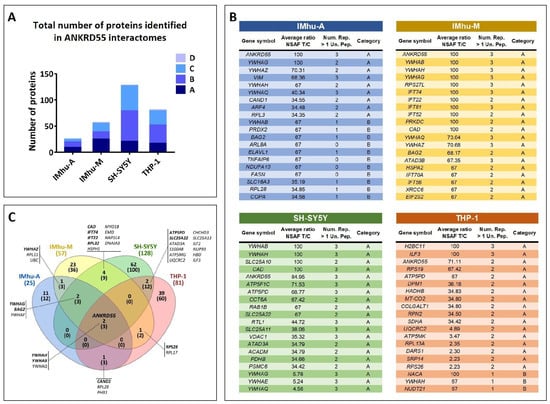
Ankyrin repeat domain-containing protein 55 (ANKRD55) is genetically associated with multiple sclerosis, but its cellular function remains incompletely defined. To investigate its potential role in neuroinflammatory pathways, we mapped ANKRD55-interacting proteins in neuroblastoma, astrocytic, microglial, and monocytic cell lines by affinity purification–mass spectrometry. Across all four cell lines, 14-3-3 isoforms were the most reproducible partners, whereas the IMhu-M microglial interactome uniquely contained eight intraflagellar transport complex B (IFT-B) components. In visible immunoprecipitation assays, IFT46–IFT56 was the smallest tested IFT-B subassembly sufficient to support detectable association with ANKRD55. Confocal microscopy localized endogenous ANKRD55 to pericentriolar material and basal bodies, but not to primary cilia, and identified partial association with P-body markers. These data distinguish a conserved 14-3-3 interaction core from cell-restricted centrosomal/IFT-B and RNP-associated ANKRD55 pools.
Biomolecules
6 November 2026
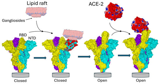
![NTD-ganglioside-induced conformational change in the Spike trimer. The three subunits of the Spike trimer are colored cyan, yellow, and purple. Each NTD is located at the periphery of the trimer. Following the binding of one NTD to a lipid raft enriched in gangliosides, a conformational change propagates throughout the involved subunit (the one in cyan in this example) until it reaches the neighboring subunit (colored yellow). Areas of the NTD whose conformation has been modified after interaction with the raft are indicated in red. Note that the conformational rearrangements affect distant domains of the NTD, including the contact zone with the neighboring subunit. This triggers the unmasking of the RBD of the yellow subunit, now able to bind the ACE-2 receptor. Overall, this process is functionally reminiscent of interfacial activation [60]. Trimeric structures in the closed pre-fusion conformation were constructed with Swiss-PdbViewer [61] by homology with a reference model (pdb: 6VSB) [62]. ACE2 and the spike trimer in the open conformation were from pdb 7DK3 [37]. Protein structures were generated with Molegro Molecular Viewer 2.5 (MMV, http://molexus.io/molegro-molecular-viewer/ accessed on 3 March 2025).](https://mdpi-res.com/biomolecules/biomolecules-16-00111/article_deploy/html/images/biomolecules-16-00111-g001-550.jpg)

