Article

A Novel Approach to Bacterial Expression and Purification of Myristoylated Forms of Neuronal Calcium Sensor Proteins

by
Vasiliy I. Vladimirov
1,
Viktoriia E. Baksheeva
2,
Irina V. Mikhailova
1,3,
Ramis G. Ismailov
4,
Ekaterina A. Litus
4,
Natalia K. Tikhomirova
2,
Aliya A. Nazipova
4,
Sergei E. Permyakov
4,
Evgeni Yu. Zernii
2,5 and
Dmitry V. Zinchenko
1,*
1
Laboratory of pharmacokinetics, Department of Biological Testing, Branch of Shemyakin and Ovchinnikov Institute of Bioorganic Chemistry of the Russian Academy of Sciences in Puschino, Pushchino, 142290 Moscow Region, Russia
2
Department of Cell Signaling, Belozersky Institute of Physico-Chemical Biology, Lomonosov Moscow State University, 119992 Moscow, Russia
3
Faculty of BioMedPharmTechnological, Pushchino State Institute of Natural Sciences, Pushchino, 142290 Moscow Region, Russia
4
Laboratory of New Methods in Biology, Institute for Biological Instrumentation of the Russian Academy of Sciences, Federal Research Center “Pushchino Scientific Center for Biological Research of the Russian Academy of Sciences”, Pushchino, 142290 Moscow Region, Russia
5
Institute of Molecular Medicine, Sechenov First Moscow State Medical University, 119991 Moscow, Russia
*
Author to whom correspondence should be addressed.
Biomolecules 2020, 10(7), 1025; https://doi.org/10.3390/biom10071025
Submission received: 23 June 2020 / Revised: 5 July 2020 / Accepted: 7 July 2020 / Published: 10 July 2020
(This article belongs to the Special Issue Metal Binding Proteins 2020)

Abstract

:
N-terminal myristoylation is a common co-and post-translational modification of numerous eukaryotic and viral proteins, which affects their interaction with lipids and partner proteins, thereby modulating various cellular processes. Among those are neuronal calcium sensor (NCS) proteins, mediating transduction of calcium signals in a wide range of regulatory cascades, including reception, neurotransmission, neuronal growth and survival. The details of NCSs functioning are of special interest due to their involvement in the progression of ophthalmological and neurodegenerative diseases and their role in cancer. The well-established procedures for preparation of native-like myristoylated forms of recombinant NCSs via their bacterial co-expression with N-myristoyl transferase from Saccharomyces cerevisiae often yield a mixture of the myristoylated and non-myristoylated forms. Here, we report a novel approach to preparation of several NCSs, including recoverin, GCAP1, GCAP2, neurocalcin δ and NCS-1, ensuring their nearly complete N-myristoylation. The optimized bacterial expression and myristoylation of the NCSs is followed by a set of procedures for separation of their myristoylated and non-myristoylated forms using a combination of hydrophobic interaction chromatography steps. We demonstrate that the refolded and further purified myristoylated NCS-1 maintains its Ca2+-binding ability and stability of tertiary structure. The developed approach is generally suited for preparation of other myristoylated proteins.

1. Introduction

Neuronal calcium sensors (NCSs) are EF-hand Ca2+-binding proteins responsible for transduction of calcium signals into a wide range of neuronal responses by regulating activity of effector enzymes and other target proteins (for review, see [1]). Due to these properties, NCSs are involved in a number of processes crucial for neuronal function, including reception, neurotransmission, synaptic plasticity, neuronal growth and survival [2,3,4,5,6,7]. Growing evidence indicates that NCSs are involved in the pathogenesis of neurological and neurodegenerative disorders as well as different types of cancer [8,9,10,11]. In addition, NCSs were recognized as redox-sensitive proteins, which may underlie their pathophysiological activity in oxidative stress-related disorders [12,13,14].
NCSs protein family includes 14 members characterized by different distribution in the nervous system. Thus, recoverin and guanylate cyclase-activating proteins (GCAPs) are mostly specific for photoreceptor cells of the retina; visinin-like proteins (VILIPs) are expressed throughout the cerebral cortex, the midbrain and the cerebellum, as well as in retinal and olfactory neurons [15,16,17,18]; neurocalcin δ (NCALD) is found in most neurons of the central nervous system, in the retina and the adrenal gland [19,20]; Kv channel-interacting proteins (KChIPs) are characterized by varied localization throughout the brain, and are also present in the heart muscle and male reproductive system [21]; neuronal calcium sensor-1 (NCS-1) is expressed ubiquitously in the central and peripheral nervous systems, including the innervation of the gastrointestinal tract and endocrine glands, in mast cells of the blood, as well as in smooth muscle and in the developing heart [15,22,23,24,25,26]. NCSs possess common domain architecture consisting of N-terminal and C-terminal domains, each containing two structurally coupled EF-hand motifs. Among these motifs, only EF2 and EF3 (recoverin), EF3 and EF4 (KChIP1) or EF2, EF3 and EF4 (other KChIPs, GCAPs, VILIPs, NCS-1) are capable of binding calcium or magnesium, whereas EF1 is always non-functional due to the absence of certain amino acids responsible for metal ion coordination. Due to specific structural features, such as different organization of Ca2+-binding loops and the presence of specific regulatory elements (for instance, C-terminal segment [27,28,29]), each NCS binds calcium within its own narrow range of the cation concentration, which allows these proteins to respond to different Ca2+-signals and play the role of Ca2+-buffer in cells [30]. Alongside calcium and magnesium, NCSs are capable of binding zinc, which is suggested to be important for their normal and pathological activity [31,32].
N-terminal myristoylation is an indispensable modification of the majority of NCS proteins [33]. It is believed to occur co-translationally (after cleavage of the initiator methionine by methionyl aminopeptidase), but may also represent post-translational modification, accomplished after proteolytic cleavage of the N-terminal glycine residue [34]. Basically, myristoyl group is responsible for targeting NCS proteins to different membranes, thereby governing their intracellular localization and compartmentalization with multiple binding partners. Most of the NCSs bind to phospholipid membranes via the so-called Ca2+-myristoyl switch mechanism, which is best characterized in case of recoverin [35]. In the absence of Ca2+, myristoyl group is buried inside a hydrophobic pocket in the recoverin molecule. Sequential binding of two calcium ions to EF3 and EF2 of the protein leads to extrusion of the myristoyl group, thereby anchoring the protein in the lipid bilayer. In recoverin, the functioning of the Ca2+-myristoyl switch and subsequent membrane binding are affected by a number of different factors, such as proper structure and conformation of its unique N-terminal and C-terminal segments, electrostatic contacts between the protein and the membrane, phosphatidylserine content and potential of the membrane as well as the presence of zinc and redox state of the medium [12,31,36,37,38,39]. NCALD [40], VILIP1 [41] and VILIP3 [42] possess a similar Ca2+-myristoyl switch mechanism, whereas NCS-1 has its myristoyl group permanently exposed in the presence of membranes [28,43,44,45] and its membrane localization is predominantly governed by the interaction of the protein with specific phospholipids in the bilayer [46]. In contrast to the other NCSs, in GCAP1 the myristoyl group is not involved in membrane binding. Instead, it participates in a Ca2+-dependent activator-to-inhibitor transition of the protein with respect to its targets, retinal guanylate cyslases, through the so-called Ca2+-myristoyl tug mechanism [47,48]. In general, the role of myristoyl group in NCSs is not limited to membrane anchoring. Thus, it was found to contribute to overall structural stability of these proteins, thereby affecting their critical functional features, such as calcium sensitivity and target recognition [48,49,50,51,52]. It is suggested that in Ca2+-free NCSs intramolecular interactions between the myristoyl group and the binding pocket residues shape these homologous proteins into different structures, which therefore expose different sets of hydrophobic residues in response to calcium binding. Apparently, this mechanism provides for the specificity of NCSs association with different physiological targets, thereby diversifying their intracellular activities [53].
Given the critical importance of the myristoyl group for proper folding and functioning of NCSs, it is clear that any in vitro study involving these proteins requires preparations of their myristoylated forms. While the production of intrinsically myristoylated NCSs in animal cell lines is characterized by low yields, their generation in bacteria presents a number of technical problems. Since N-myristoyl transferases are absent in Escherichia coli, myristoylation of NCSs is normally achieved by their co-expression with the respective enzyme from Saccharomyces cerevisiae (NMT1p) [33]. Unfortunately, NMT1p exhibits different specificity with respect to substrate sequence as compared to the mammalian enzyme, resulting in unstable and sometimes low acylation of NCS proteins in bacteria [54]. In some cases, this problem can be solved by point mutations, which makes myristoylation signal more appropriate for NMT1p. For instance, mutation D6S was demonstrated to enhance myristoylation of GCAP1 [55]. Nevertheless, although myristoylation level of bacteria-expressed recoverin, VILIP1, VILIP3 and neurocalcin δ can reach 80–95% of the total protein [56,57,58,59], in case of GCAPs this value is about 50–80% [60], whereas for NCS-1, it does not exceed 60% [43,61]. On the other hand, the separation of myristoylated and non-myristoylated forms of NCSs is a complicated task as currently it is only achieved by reverse phase high-performance liquid chromatography (HPLC) that may result in irreversible denaturation of the target proteins and is not suitable for obtaining their preparative quantities. An attempt to solve this problem was made in case of NCS-1 by providing it with C-terminal 6×His-tag, which increased solubility of the protein and allowed for chromatographic separation of its myristoylated and non-myristoylated forms [62]. Yet, any approach involving introduction of 6×His-tag in NCSs is questionable, since modification of their N-terminus is impossible due to myristoylation, whereas modification of C-terminus might affect the function of these proteins [27,28,29,63,64].
With this in mind, in this study we aimed at developing a novel approach for obtaining native-like myristoylated forms of various NCSs, including recoverin, GCAP1, GCAP2, NCALD and NCS-1. To this end, we analyzed and optimized their expression and myristoylation in bacteria and adopted a simple method for separation of their myristoylated and non-myristoylated forms by a combination of hydrophobic chromatography steps performed under non-denaturing conditions. In addition, we examined feasibility of insoluble expression, refolding and purification procedures for obtaining myristoylated NCS-1 with intact structural and functional properties. We propose that general approach suggested in this study may be considered upon developing novel efficient techniques for expression and purification of different proteins bearing myristoyl group.

2. Materials and Methods

2.1. Materials

Chromatographic resins Phenyl Sepharose, Butyl Sepharose and CNBr-activated Sepharose 4B were from GE Lifesciences (Marlborough, MA, USA); Butyl Toyopearl was from Tosoh Bioscience (Griesheim, Germany). The SDS-PAGE molecular weight standards and bicinchoninic acid (BCA) protein assay kit were from Thermo Fisher Scientific (Rockford, IL, USA). Other reagents were obtained from Sigma-Aldrich (St. Louis, MO, USA), Merck (Darmstadt, Germany), Fluka (Buchs, Switzerland), PanReac AppliChem ITW Reagents (Darmstadt, Germany), Serva (Heidelberg, Germany) and Amresco (Solon, OH, USA) and were at least analytical grade. All buffers and other solutions were prepared using ultrapure water.

2.2. Antibodies

Rabbit polyclonal antibodies against recoverin, NCS-1, GCAP1 and GCAP2 were prepared previously [28,65,66]. Polyclonal anti-NCALD antibodies were produced by rabbit immunization. The generated antibodies were purified from serum using a resin with immobilized antigen [67]. To obtain the resin, recombinant NCALD was conjugated to CNBr-activated Sepharose 4B (GE Lifesciences) according to the manufacturer’s instructions. The antibody titers were monitored by Western blotting.

2.3. Genetic Constructs

Plasmid vectors for expression of recoverin (in pET-11d) and NCS-1 (in pET-22b) genes were obtained in previous studies [28,68]. The genetic constructs encoding GCAP1 and GCAP2 (in pET-11a) were kindly provided by Prof. Karl-Wilhelm Koch (University of Oldenburg, Oldenburg, Germany) [55]. Bovine NCALD gene was obtained by reverse transcription and PCR amplification using total mRNA purified from Bos taurus brain tissue. The resulting amplicon was subcloned to pET-22b(+) expression vector for Escherichia coli using endonucleases sites HindIII and NdeI by the standard cloning techniques.

2.4. Expression Protocol

Expression of NCS proteins was performed in Escherichia coli strains BL-21(DE-3) Star, or BL-21(DE-3) Codon Plus RIL. The cells transformed with NCS-expressing vectors were cultivated on Petri dishes with agarized LB medium (10 g/L NaCl, 10 g/L triptone, 5 g/L yeast extract, 1.5% agar) containing 50 μg/mL ampicillin. To obtain myristoylated proteins, the cells were co-transformed with plasmid pBB131 bearing NMT1p gene [69] and cultivated in the same medium in the presence of 50 μg/mL ampicillin and 10 μg/mL of kanamycin. The resulting strains were stored by freezing freshly grown culture (OD595 = 1.0) in 15% glycerol at –70 °C.
For expression of NCS proteins, 50 mL of LB medium (10 g/L NaCl, 10 g/L triptone, 5 g/L yeast extract) containing 0.1% glucose was inoculated with a single colony from a fresh dish prepared by overnight cultivation of cells from a frozen glycerol stock. The cells were cultivated to OD595 = 1.0 at 37 °C with continuous shaking (250 rpm) for 6–8 h and kept overnight at +4 °C. The next morning, 5 mL of the cell suspension was added to 500 mL of glucose-free pH-buffered LB medium (10 g/L NaCl, 10 g/L triptone, 5 g/L yeast extract, 50 mM Na2HPO4, pH 8.0) containing the respective antibiotics. In the case of obtaining myristoylated NCSs, the cells were grown to OD595 = 1.0 and myristic acid (50 mg/mL stock in 96% ethanol) was added to final concentration of 20 mg/L. At OD595 = 1.5–2, IPTG was added to final concentration of 0.5 mM and the shaking was continued for additional 4–5 h (250 rpm, 37 °C).

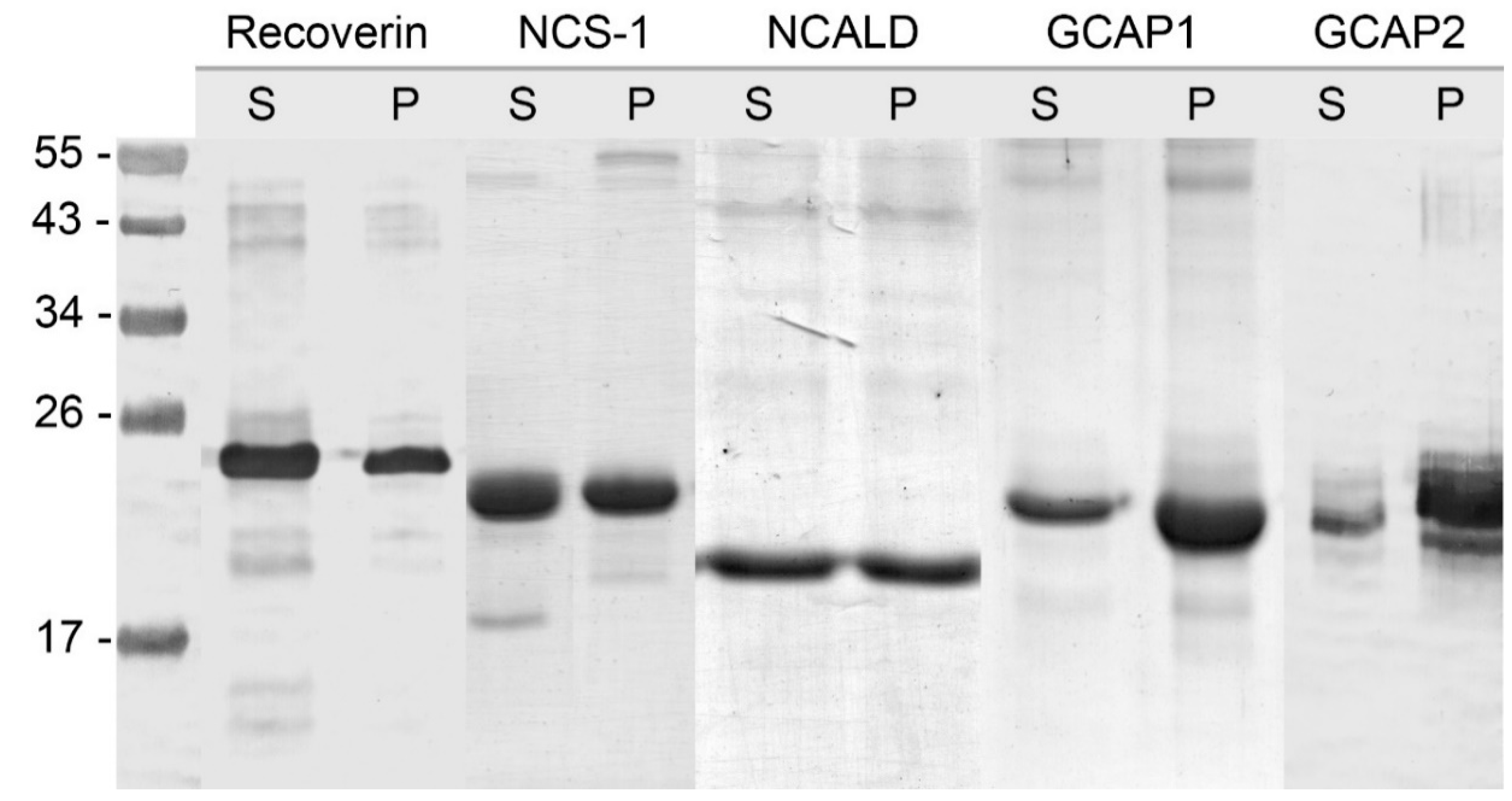
2.5. Extraction of Recoverin, NCALD and NCS-1 as Soluble Proteins

After the expression, the cells expressing recoverin, NCALD or NCS-1 were centrifuged at 6000× g for 10 min, the supernatant was discarded, and the cells were resuspended in lysis buffer containing 50 mM Tris-HCl (pH 7.5), 5 mM MgCl2, 1 mM EDTA, 0.1 mM PMSF and 3 mM DTT (25–50 mL for 500 mL of the cell culture). The lysates were frozen at -20 °C, thawed at room temperature, and kept on ice with lysozyme (25–50 µg/mL) for 20–40 min with stirring. The resulting suspension was centrifuged for 20 min at 12,000× g. If necessary, the pellet was subjected to another freeze/thaw cycles to extract additional 10–20% of the protein.

2.6. Extraction from Inclusion Bodies as Insoluble Protein and Renaturation of NCS-1, GCAP1 and GCAP2

After the extraction of soluble NCS-1, the cell pellets containing residual insoluble protein were resuspended in lysis buffer (2–5 mL for 500 mL of cell culture) and the suspension was added, drop by drop, to dissolution buffer (25 mL for 500 mL of cell culture) containing 20 mM Tris-HCl (pH 7.5), 100 mM NaCl, 1 mM EGTA, 2 mM MgCl2, 6 M urea and 1 mM DTT. GCAPs-containing pellets were treated similarly except for dissolution buffer contained 7M guanidine hydrochloride instead of urea. The mixture was kept dissolving overnight at 4 °C with gentle stirring and then dialyzed against 10 mM Tris-HCl-buffer (pH 7.5), 100 mM NaCl, 1 mM EGTA, 2 mM MgCl2, 1 mM DTT (1000–10000 w/w) two times for 3 h with the buffer exchange. The resulting fraction was centrifuged for 30 min at 20,000× g, and the supernatant was used for chromatographic purification.

2.7. Purification of Myristoylated Recoverin, NCALD and NCS-1 by Hydrophobic Chromatography

For purification of recoverin and NCALD, calcium chloride was added dropwise to the cellular extracts to reach the final concentration of 3 mM (on ice). The resulting fraction was centrifuged for 30 min at 20,000× g, and the supernatant was loaded onto Phenyl Sepharose column (GE Lifesciences), equilibrated with 20 mM Tris-HCl-buffer (pH 7.5), 2 mM CaCl2, 2 mM MgCl2 and 1 mM DTT. The elution was performed using (1) 20 mM Tris-HCl-buffer (pH 7.5), 2 mM MgCl2, 1 mM DTT and 1 mM EGTA and (2) deionized water. The bulk of the myristoylated proteins was eluted in step (1).
For purification of NCS-1, the respective cellular extract was mixed with 1 M NaCl (to reduce protein aggregation) and calcium chloride was added as described above. After the centrifugation (30 min at 20,000× g), the mixture was loaded onto Phenyl Sepharose column, equilibrated with 20 mM Tris-HCl-buffer (pH 7.5), 2 mM CaCl2, 2 mM MgCl2, 1 mM DTT. The elution was performed in three steps using the following conditions: (1) 20 mM Tris-HCl-buffer (pH 7.5), 1 mM EGTA, 2 mM MgCl2 1 mM DTT and 200 mM NaCl; (2) 20 mM Tris-HCl (pH 7.5), 1 mM EGTA, 2 mM MgCl2 and 1 mM DTT; (3) deionized water. The bulk of the myristoylated protein was eluted in steps (2) and (3).
To accomplish final separation of myristoylated from of NCS-1, the fractions obtained in the steps (2) and/or (3) were mixed with NaCl to reach the final concentration of 1 M and loaded onto Butyl Sepharose column, equilibrated with 20 mM Tris-HCl-buffer (pH 7.5), 1 mM EGTA, 2 mM MgCl2 and 1 mM DTT, containing 1M NaCl. The target protein was eluted by reducing NaCl concentration in the same buffer using linear or step gradient. If necessary, myristoylated recoverin was purified from the EGTA fraction by the same procedure. The approximate gradient conditions required for elution of myristoylated NCS-1 and recoverin are indicated in Table 2. If degree of NCSs myristoylation was still below 90%, the chromatography on Butyl Sepharose column was repeated.

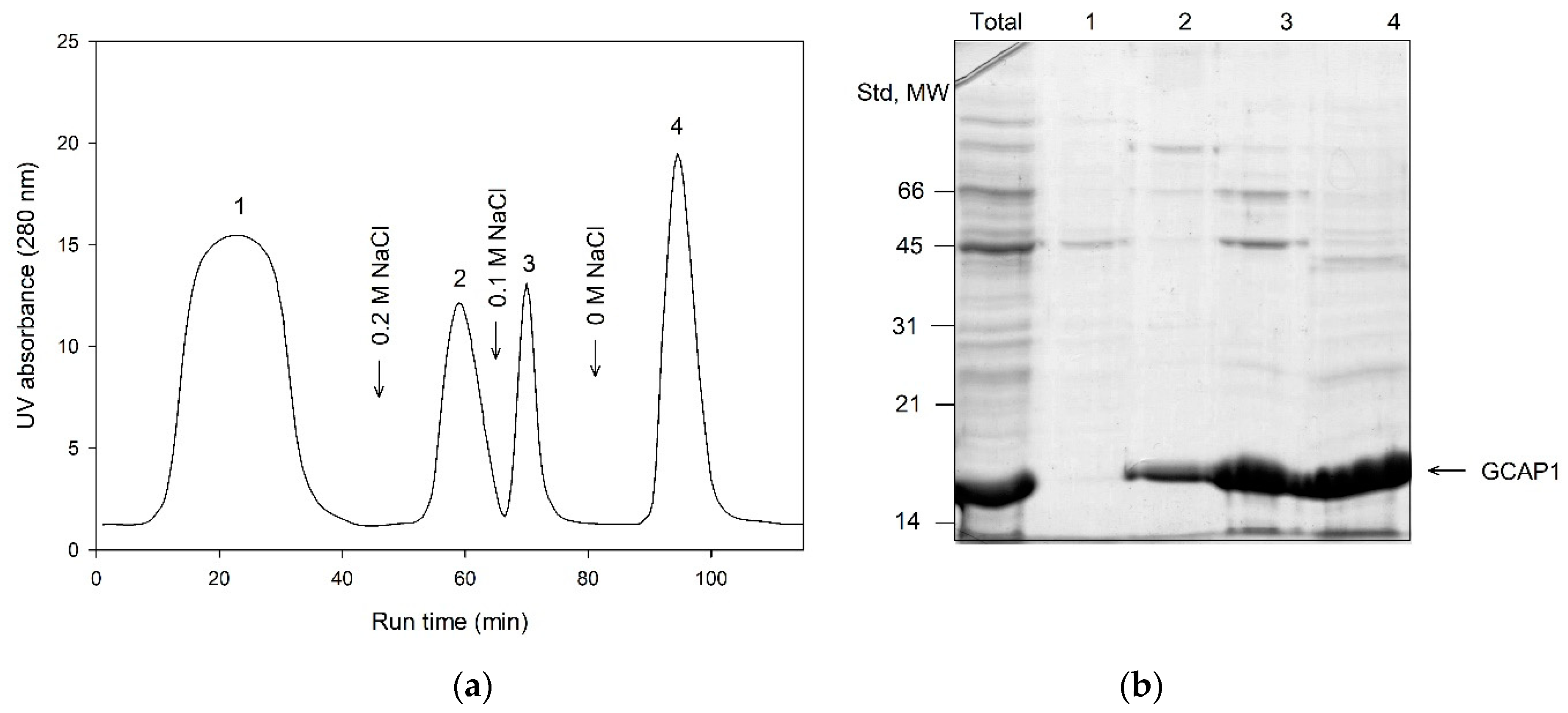
2.8. Purification of Myristoylated GCAPs by Hydrophobic Chromatography

The fractions containing renaturated GCAP1 and GCAP2 were mixed on ice with NaCl to reach its final concentration of 1 M, and the resulting mixture was loaded onto Toyopearl Butyl column equilibrated with 20 mM Tris-HCl-buffer (pH 7.5), 1 mM EGTA, 2 mM MgCl2, 1 mM DTT and 1M NaCl. The elution was performed in three steps using the following conditions: (1) 20 mM Tris-HCl-buffer (pH 7.5), 1 mM EGTA, 2 mM MgCl2 1 mM DTT and 200 mM NaCl; (2) 20 mM Tris-HCl-buffer (pH 7.5), 1 mM EGTA, 2 mM MgCl2 1 mM DTT and 100 mM NaCl; (3) 20 mM Tris-HCl (pH 7.5), 1 mM EGTA, 2 mM MgCl2 and 1 mM DTT. The bulk of the myristoylated proteins was eluted in step (2) and (3).
If necessary, the final separation of myristoylated forms of GCAPs was achieved using Butyl Sepharose chromatography. To this end, the fractions obtained in the step (2) and (3) were mixed with NaCl to reach the final concentration of 1 M and loaded onto Butyl Sepharose column, equilibrated with 20 mM Tris-HCl-buffer (pH 7.5), 1 mM EGTA, 2 mM MgCl2 and 1 mM DTT, containing 1M NaCl. The target protein was eluted by reducing NaCl concentration in the same buffer using linear or step gradient. The approximate gradient conditions required for elution of myristoylated NCS-1 and recoverin are indicated in Table 2.

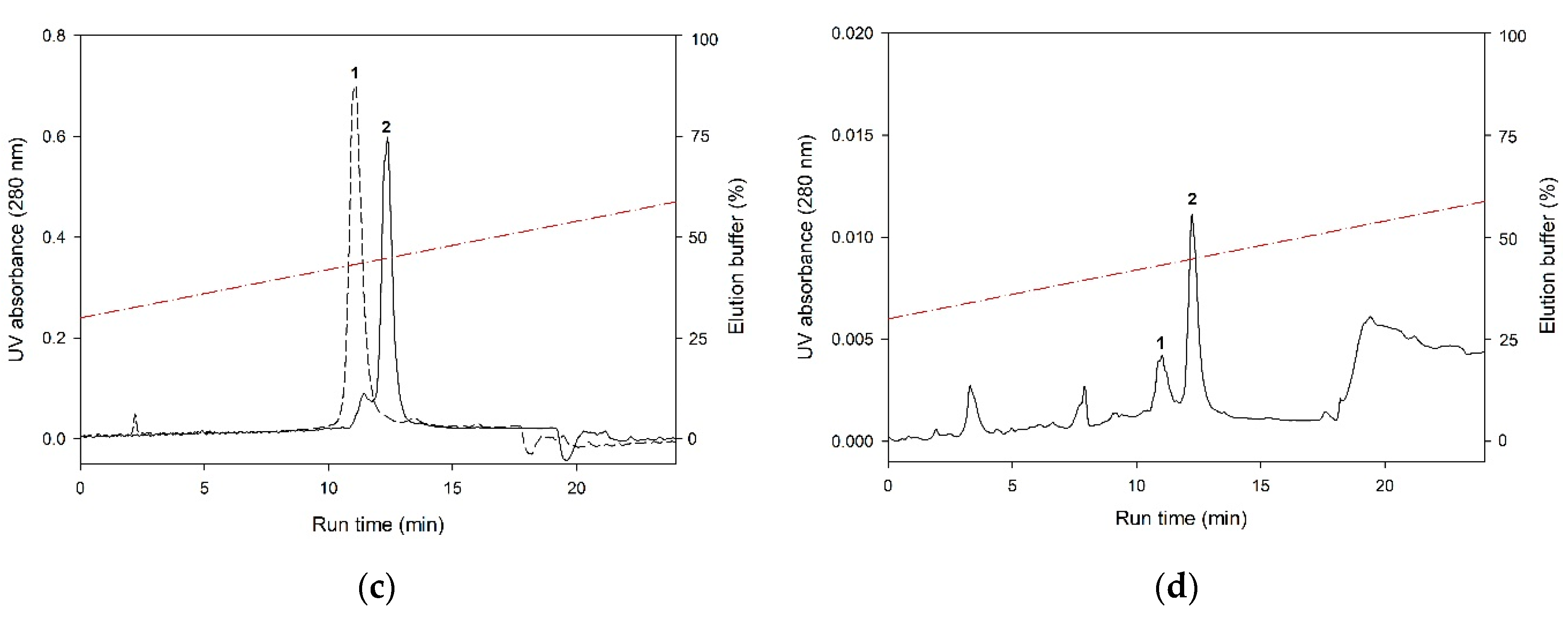
2.9. HPLC Detection of The Myristoylated Protein Forms

The degree of NCSs myristoylation was determined by analytical HPLC (Waters Breeze) using 3.9 × 150 mm Luna C18 reversed-phase column (Phenomenex, Torrance, CA, USA)) in acetonitrile-water system [27]. First, 20 µL samples containing ~0.1–1 mg/mL of the target proteins were mixed with 50 µL of 100% acetonitrile, incubated with shaking for 30 min and centrifugated 8000× g. Then, 20 µL of supernatant were loaded onto the column equilibrated with water solution containing 2% acetonitrile and 0.1% TFA. The separation of myristoylated and non-myristoylated forms of recoverin, GCAP1 and GCAP2 was accomplished using linear gradient from 0 to 95% acetonitrile in water solution containing 0.1% TFA in the course of 30 min. In the case of NCS-1, linear gradient from 30 to 60% acetonitrile in the same solution was employed. The protein detection was performed by measuring absorbance at 280 and 215 nm. The purified non-myristoylated NCS proteins (obtained in the absence of NMT1p) were used as control.

2.10. Mass Spectrometry Analysis

Prior to analysis, recoverin and NCS-1 were dialyzed against ultrapure distilled water and mixed with acetonitrile (50%) and formic acid (5 mM). The resulting samples (100 μL) containing 5 μg/mL proteins were applied by direct injection. The mass spectrum was recorded in the positive mode from 600 to 1400 m/z (scanning speed 125 amu/s, detector voltage 1.5 kV) and a constant nitrogen flow of 1.5 l/min using Shimadzu LCMS-2010EV single quadrupole mass spectrometer (Shimadzu Co., Kyoto, Japan) with an electrospray ionization. The flow rate was 40 μL/min. The device was pre-calibrated using horse heart myoglobin.

2.11. Fluorescence Measurements

Fluorescence emission spectra of NCS-1, recoverin and GCAP2 (4.8 μM) were measured at different temperatures using a Cary Eclipse spectrofluorimeter (Varian Inc., Mulgrave Victoria, Australia) equipped with a Peltier-controlled cell holder, mainly as previously described [28,29,32]. The measurements were carried out in 10 mM HEPES-KOH, 100 mM KCl, pH 7.6 buffer, in the presence of 1 mM CaCl2 or 1 mM EDTA. Excitation wavelength was 280 nm. All spectra were corrected for spectral sensitivity of the instrument and described by a log-normal function [70] using LogNormal software (IBI RAS, Pushchino, Russia).

2.12. Analytical Procedures

SDS-PAGE and Western blotting was performed using the standard procedures [71,72]. Concentration of proteins was measured spectrophotometrically using previously calculated extinction coefficients at 280 nm of 24,075 M−1cm−1 (recoverin) [73], 21,430 M−1cm−1 (NCS-1) [28], 28,378 M−1cm−1 (GCAP1), 37,512 M−1cm−1 (GCAP2) [74] and 20,000 M−1cm−1 (NCALD) [75]. In some cases, protein concentration was verified using BCA protein assay.

3. Results

3.1. Analysis of Myristoylation and Solubility of NCS Proteins Expressed in Bacterial Cells

Given that previous studies report contradictory information regarding solubility and myristoylation levels of recombinant NCSs [43,55,59,61,62,76], we firstly reproduced and verified these data. Five NCSs, namely recoverin, NCALD, GCAP1, GCAP2 and NCS-1, were expressed in bacteria according to the standard protocol and distribution of their myristoylated and non-myristoylated forms between soluble and insoluble fractions was studied. According to Western blotting data, NCSs significantly differed in terms of solubility in bacteria (Figure 1).
Thus, recoverin was expressed mostly as a soluble protein and GCAPs were detected predominantly in the membrane fraction, whereas NCS-1 and NCALD were almost equally distributed between the supernatant and the pellet. To assess myristoylation levels of the expressed NCSs, their soluble and insoluble fractions were additionally examined by analytical HPLC. Prior to the analysis, the insoluble fractions were treated with guanidine hydrochloride. The chromatographic separation of myristoylated and non-myristoylated forms of NCSs based on differences in their hydrophobicity was performed using high-resolution reversed-phase column (C18). In our experiments, the best fractionation was achieved using water-acetonitrile gradient (0–90% over the course of 30 min) in the presence of 0.1% TFA. Under these conditions, recoverin (Figure 2A), GCAP1, GCAP2 and NCALD exhibited similar elution profiles, where non-myristoylated form was eluted first. In case of NCS-1, efficient peak resolution was achieved employing more slopping gradient (30–90% over the course of 30 min) (Figure 2C). According to the HPLC data, the myristoylated forms of recoverin (Figure 2B) and NCALD were predominantly present in the soluble fractions (Table 1). Thus, we confirmed that these proteins can be purified directly from the cellular extracts. Meanwhile, substantial fractions of myristoylated NCS-1 (Figure 2D) and especially GCAPs were found in the insoluble fractions (Table 1).
This does not necessarily point to their accumulation in the inclusion bodies, as both proteins are known to exhibit high membrane affinity regardless of the presence of calcium [43,77]. Nevertheless, their purification required additional treatment of the pellets, such as using sonication, or otherwise these proteins could be extracted with chaotropic agents, i.e., in denatured form. Since NCSs are recognized as redox-sensitive proteins [12,13,14], the sonication procedure was considered undesirable as it could cause thiol oxidation and alterations in structure and function of these proteins. We concluded that purification of GCAPs and NCS-1 may involve their renaturation after extraction from bacterial pellets as insoluble proteins. In case of NCS-1, the fraction containing renatured protein could be mixed with its soluble extracts, thereby increasing the total yield of the myristoylated form.

3.2. Optimization of Myristoylation of NCS-1 in Bacterial Cells

In our experiments, NCS-1 exhibited generally low level of myristoylation: about 50–60% of the total expressed protein lacked this modification. Therefore, we next modified the cultivation procedure in attempt to optimize expression and myristoylation of NCS-1 in bacteria. In particular, we varied conditions of the cell growth, such as pH, temperature and duration, as well as time points for induction and addition of myristic acid and analyzed the resulting products by means of analytical HPLC. The most pronounced benefit was observed upon alkalization of the cell growth medium: the incubation of the cells in LB medium buffered at pH 8.0 did not negatively affect NCS-1 expression, but increased its myristoylation up to 60–70%. It should be noted that the doubling time for bacterial cells cultured at pH 8.0 increased about 1.5-fold as compared to their cultivation at pH 7.0 (standard LB medium).

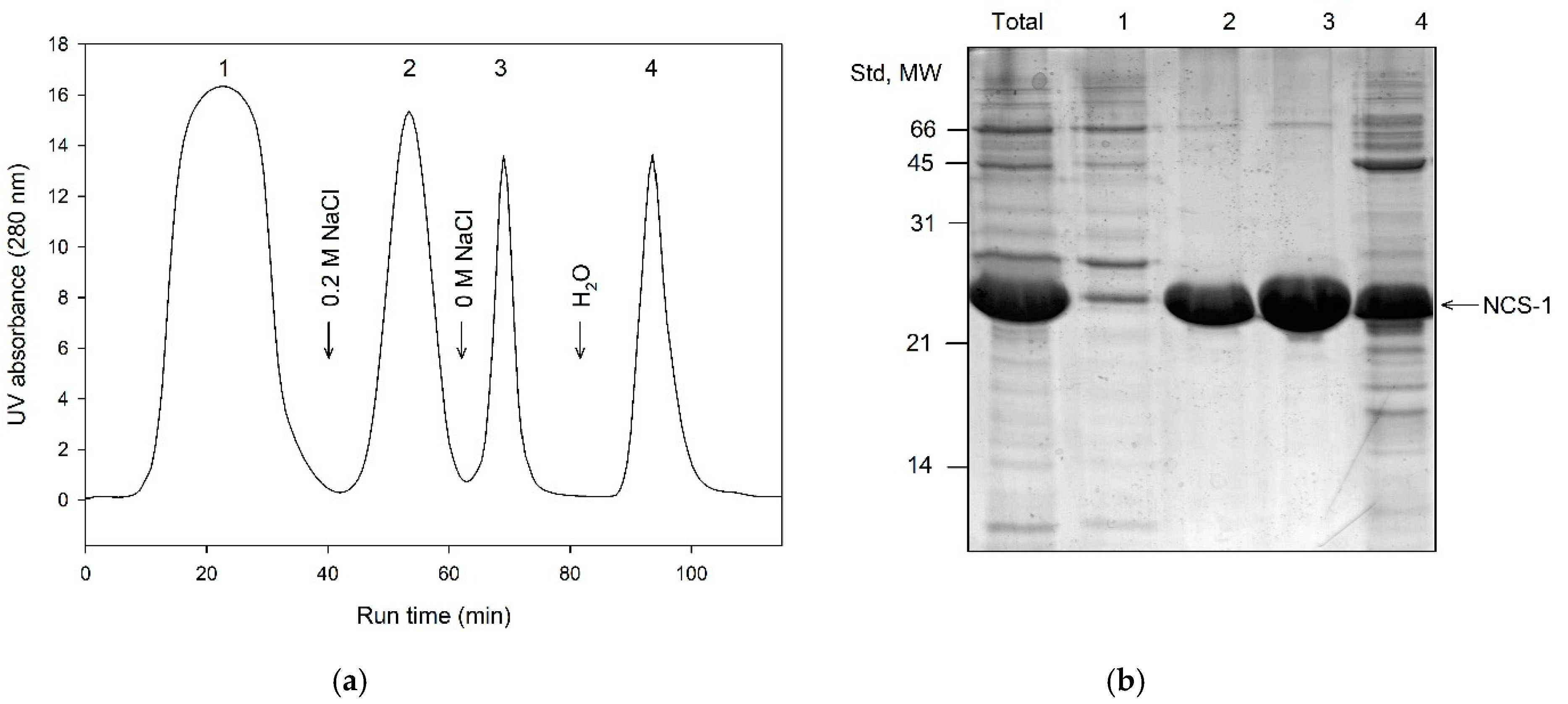
3.3. Primary Purification of Myristoylated Recoverin, NCALD and NCS-1: Phenyl Sepharose Chromatography

The well-accepted primary step for purification of recoverin, NCALD and NCS-1 is Ca2+/affinity chromatography on Phenyl Sepharose [14,19,33]. The procedure is based on ability of the NCS proteins to reversibly expose their hydrophobic target-recognizing site (hydrophobic pocket), in response to binding/chelation of the calcium ions [35]. Considering that myristoylation can differently affect overall hydrophobicity of NCSs, we analyzed distribution of their myristoylated and non-myristoylated forms in Phenyl Sepharose chromatography fractions. Protein extracts containing recoverin, NCALD or NCS-1 (obtained from soluble fraction or from insoluble fraction after refolding) were loaded onto the column equilibrated with low ionic strength/high calcium buffer and eluted with (1) the same buffer containing calcium chelator EGTA and (2) deionized water. The resulting fractions were analyzed for the myristoylation rate of the contained protein using analytical HPLC (Table 1). In case of recoverin, the treatment of the column with EGTA-containing buffer resulted in release of the relatively pure protein with degree of myristoylation of 90–95%, whereas for NCALD, this procedure yielded protein myristoylated by 97%. In contrast, in case of NCS-1, the respective fraction contained NCS-1 myristoylated only by 5–25%, whereas the rest of the modified protein (40–60% of myristoylation) was found in the water fraction. Given that NCS-1 was structurally unstable in water and the respective fraction contained multiple contaminations, the procedure of Phenyl Sepharose chromatography of NCS-1 was modified as follows (Figure 3). The protein was loaded onto the column in high ionic strength/high calcium, washed with high ionic strength/high magnesium and eluted by gradual reduction of the ionic strength in the presence of EGTA and Mg2+ (Figure 3). Under these conditions, the most of the non-myristoylated NCS-1 was eluted at 200 mM NaCl, whereas the treatment with salt-free Mg2+/EGTA-containing buffer yielded a large fraction of the protein with myristoylation level of 40–60%. Overall, we demonstrated that a combination of Ca2+/affinity and hydrophobic interaction chromatography on Phenyl Sepharose can be used as first step for separation of myristoylated and non-myristoylated forms of recoverin, NCALD and NCS-1 yielding fractions enriched in their myristoylated forms.

3.4. Primary Purification of Myristoylated GCAP1 and GCAP2: Toyopearl Butyl Chromatography

In contrast to the other NCSs, myristoylated GCAPs were mainly expressed in bacteria as insoluble proteins. Nevertheless, both proteins exhibited incomplete myristoylation in bacterial cells (approximately 80%, Table 1), which still necessitated separation of their myristoylated forms. Our preliminary experiments revealed that this separation can be accomplished after refolding and it occurred more efficiently when GCAPs were free from contamination of bacterial proteins. To accomplish such primary purification, we employed, for the first time, hydrophobic interaction chromatography on Toyopearl Butyl (Figure 4). Both GCAPs were bound to this resin under high ionic strength conditions (1 M NaCl) in EGTA-containing buffer and eluted by reverse salt gradient in the same buffer at 200 mM NaCl, yielding relatively pure proteins. Myristoylation level of the resulting products remained at 75–80%, indicating that myristoylation forms of GCAPs were completely preserved during this purification step. It should be emphasized that the described protocol was valid only when GCAPs maintained their Ca2+-free conformation during the chromatographic process. Notably, most of the commercially available sodium chloride reagents contain admixtures of calcium chloride, yielding approximately 1–100 µM Ca2+ in 1 M NaCl solution. Thus, the presence of background concentration of calcium chelator, such as 250 µM EGTA, is strictly required for proper purification of GCAPs (as well as NCS-1, see above), as they exhibit submicromolar affinities to calcium [32,78,79,80].

3.5. Final Separation of Myristoylated Forms of NCS Proteins: Butyl Sepharose Chromatography

The primary steps of purification described above yielded relatively pure NCSs enriched in myristoylated forms. To complete the purification procedure, we next searched for universally valid method for total removing of the admixture of the non-myristoylated forms from the NCSs preparations. Analysis of different hydrophobic resins revealed that Butyl Sepharose was the most suitable for this purpose. Indeed, all NCSs bound to this resin at high ionic strength (1 M NaCl) in EGTA/Mg2+-containing buffer and their non-myristoylated and myristoylated forms were fractionated by reducing salt concentration (for NCALD, this step is normally not required due to high degree of myristoylation of the protein obtained after Phenyl Sepharose chromatography). The best separation was achieved by employing step gradient rather than linear gradient of NaCl concentration (Figure 5, Table 1). The approximate gradient conditions favoring elution of non-myristoylated and myristoylated forms of recoverin, NCS-1, GCAP1 and GCAP2 are summarized in Table 2. Importantly, the exact NaCl concentration required for the elution of myristoylated form of each of the NCSs depended on the degree of its myristoylation in the loading fraction. Therefore, if possible, this parameter should be controlled (i.e., by analytical HPLC) during the purification.
It should be noted that the introduction of Butyl Sepharose chromatography not only allowed separation of myristoylated forms of NCS proteins, but also lead to their more efficient purification from other admixtures. For instance, this step resulted in separation of NCS-1 from its proteolyzed fragment (18 kDa), commonly found in different preparations of the protein [81]. In the case of recoverin purification, Butyl Sepharose step efficiently substituted anion-exchange chromatography. MonoQ resin is normally used for ion-exchange chromatography in the final step of recoverin purification procedure, yielding protein preparation free of admixtures absorbing at 260 nm (nucleic acids).

3.6. Characterization of The Purified Myristoylated NCS Proteins

To verify myristoylation level and structural integrity of NCS proteins obtained using the described new protocols, the following additional studies were performed. Firstly, LC/ESI-MS analysis was employed to check molecular weights of these proteins. For the analysis, we selected two NCSs, NCS-1 and recoverin, both obtained from soluble bacterial extracts by a combination of Phenyl Sepharose (primary purification) and Butyl Sepharose (final separation) chromatography. In this set of experiments, we also compared myristoylation of two variants of NCS-1, namely obtained from soluble and insoluble/refolding fractions. Preparations of non-myristoylated NCS-1 and recoverin expressed in bacteria in the absence of NMT1p and purified using the standard methods [82] were used as a control. It was found that average molecular weights of non-myristoylated and myristoylated (both soluble and refolded) forms of NCS-1 equaled to 21,746 and 21,957 Da, respectively. In turn, molecular weights of the corresponding recoverin forms were detected as 23,201 and 23,411 Da (Table 1). Considering the calculated molecular weights of NCS-1 (21,747.53 Da) and recoverin (23,202.22 Da) polypeptides lacking N-terminal methionine and the average mass change due to myristoylation (210 Da), the myristoylated forms of both proteins were identical to their native variants. Given that myristoylation involves N-terminal glycine [34], these full-size proteins represented NCS-1 and recoverin with myristoyl group attached to G2. It should be emphasized that, according to mass spectrometry, the obtained preparations of myristoylated NCS-1 and recoverin were free of non-myristoylated proteins.
Secondly, we analyzed structural stability and Ca2+-binding ability of the purified NCS samples by examination of thermal stabilities of their Ca2+-free and Ca2+-bound states. Temperature dependencies of tryptophan fluorescence spectrum maximum position for myristoylated recoverin, GCAP2 and NCS-1, purified from either soluble or insoluble fractions, are shown in Figure 6. The myristoylated recoverin sample demonstrated a Ca2+-induced red shift of the fluorescence spectrum (consistent with exposure of the emitting Trp residue(s)), and a cooperative thermal transition (Table 1), which shifted towards higher temperatures upon Ca2+ binding (Figure 6A), in accord with previous observations [14,27,29,39,64]. Similarly, Ca2+ binding induces red shift in GCAP2 fluorescence spectrum, but fluorescence technique did not reveal cooperative thermal transitions in this case (Figure 6B) [29]. The NCS-1 samples exhibited similar thermal denaturation profiles (Figure 6C) characteristic for this protein [28,32]: Ca2+ binding is accompanied by a blue shift of the fluorescence spectrum (indicates burial of the emitting Trp residue(s)) and a pronounced stabilization of its tertiary structure (Table 1). Hence, the refolding of myristoylated NCS-1 did not affect stability of its tertiary structure and Ca2+-binding ability. Overall, the myristoylated NCS samples prepared in the present study preserved their native tertiary structure and Ca2+-binding ability.

4. Discussion

In this study, we report a novel approach for purification of nearly completely myristoylated forms of NCS proteins belonging to four out of five subgroups of the family, namely recoverin (subgroup ‘recoverins’), NCALD (subgroup ‘visinin-like proteins’), GCAP1/GCAP2 (subgroup ‘GCAPs’) and NCS-1 (subgroup ‘frequenins’) [83]. In particular, we optimized expression and myristoylation of these proteins in bacteria and developed a set of new procedures for separation of their myristoylated and non-myristoylated forms by a combination of hydrophobic interaction chromatography steps (Figure 7). Indeed, these forms differ in their hydrophobic properties because the presence of a myristoyl group not only makes its own contribution as a non-polar moiety (such as under the denaturing conditions of reverse phase HPLC), but significantly perturbs non-polar sites of the folded protein [53]. Notably, hydrophobic properties of NCSs display Ca2+-dependence and vary significantly among these proteins. Functionally, it can be observed from the different behavior of NCSs with respect to membrane binding and target recognition. In the current work, we employed all these features as the basis for developing optimized purification approach, the hallmark of which is that the separation of myristoylated and non-myristoylated forms of NCSs is conducted under non-denaturing conditions, thereby fully maintaining their structure. The method for production of myristoylated NCSs can be represented by a simple protocol consisting of four major steps, namely expression in bacterial cells, extraction, primary purification and final separation (Figure 2). All these steps differed among the NCS proteins as they were fine tuned in such a way as to maximally preserve and accumulate the respective structurally distinct myristoylated forms.
Generally, our approach improved purification of all myristoylated NCSs as compared to previously published methods. However, in case of each NCS, the benefit of these modifications was different. Thus, in case of recoverin and NCALD our modifications only moderately contributed the overall efficiency of the purification. The distinctive features of these proteins is that (1) they are well myristoylated in bacteria, (2) their myristoylated forms are extracted as soluble proteins and (3) both their non-myristoylated and myristoylated forms interact with Phenyl Sepharose in the presence of calcium, but the myristoylated form is eluted separately from (prior to) the non-myristoylated form upon chelating the cation. Such behavior can be explained using the example of recoverin based on ANS/Bis-ANS fluorescence probing of its non-polar properties [84]. Thus, (1) surface hydrophobicity of recoverin is significantly increased upon binding calcium and (2) non-myristoylated Ca2+-free recoverin is more hydrophobic than myristoylated Ca2+-free recoverin. Consistently, in our experiments the bulk of non-myristoylated Ca2+-free recoverin was eluted from Phenyl Sepharose only upon its washing with deionized water. In the aggregate, these features make it possible to purify recoverin (and NCALD) containing 90–95% of myristoylated form in a single step (Ca2+/affinity chromatography on Phenyl Sepharose) yielding relatively pure products. Yet, it is often necessary to have high-purity myristoylated proteins, which can be obtained by our Butyl Sepharose chromatographic procedure giving almost 100%-myristoylated form.
In contrast to recoverin and NCALD, GCAPs do not possess a functional Ca2+-myristoyl switch, and their myristoyl group is sequestered inside the protein globule, which makes the structure of their Ca2+-free and Ca2+-bound conformers less different (at least in case of GCAP1) [50,79]. Consistently, these proteins do not change or even decrease their surface hydrophobicity upon calcium binding [29,85] and Ca2+/affinity chromatography on Phenyl Sepharose is not applicable for their purification. Nevertheless, both GCAPs are generally very hydrophobic [29,85], and therefore, can be purified using Ca2+-independent hydrophobic interaction chromatography. This approach was employed in previous studies for GCAP1 purification: after the gel filtration step the protein was purified on Butyl Sepharose column without using salt gradient [86]. Alternatively, the gel filtration step was complemented by anion-exchange chromatography [60]. In our opinion, these variants have one significant limitation, namely, they do not separate myristoylated and non-myristoylated forms of GCAPs and the content of the myristoylated form is not being controlled during the purification. Indeed, in our experiments GCAPs were characterized by initially lower level of myristoylation in cells than recoverin and NCALD, but the bulk of their myristoylated form accumulated in insoluble fraction and was thereby separated. Yet, the content of myristoylated GCAPs in this fraction is still approximately 80% and, according to our observations, their level can be less in bacterial cells, which necessitates separation of the acylated form. In the current study, we resolved this problem by using hydrophobic Butyl Sepharose chromatography and fine-tuned ionic strength gradient. Since fractionation of the myristoylated and non-myristoylated forms after refolding occurred more efficiently when they were free from contamination of bacterial proteins, we also introduced a primary purification step employing Toyopearl Butyl. The use of this variant of hydrophobic chromatography instead of conventional gel filtration was justified by the fact that the latter has a number of limitations, such as low amount of the loaded sample per chromatography round, as well as dilution and loss of the target protein. Overall, our procedure allowed obtaining preparative amounts of GCAPs with degree of myristoylation of 96–98%.
However, the most significant achievement of our approach can be recognized in case of isolation of myristoylated NCS-1, which was initially the most problematic in this regard. Previous studies reported several procedures for the production of this protein, but neither of them yielded its highly myristoylated native-like form. In early works, NCS-1 has been purified using a two-step method originally developed for recoverin and NCALD [82], which included Ca2+/affinity chromatography on Phenyl Sepharose or similar resins as a single step [61] or with subsequent purification using ionic exchange chromatography or gel filtration [52,87]. However, the myristoylation level of the protein obtained with this method did not exceed 58–60% [43,61]. In general, Ca2+-dependent hydrophobic chromatography is hardly suitable for isolation of myristoylated NCS-1. Indeed, apo-from of this protein is unstable and, according to ANS/Bis-ANS probing, even more hydrophobic than its Ca2+-bound form. In turn, Mg2+-bound/Ca2+-free NCS-1 is less hydrophobic than Ca2+-bound NCS-1, but the difference between non-polar properties of these forms [28,32] is not sufficient to ensure complete elution of the former from Phenyl Sepharose upon calcium chelation. More recently, an alternative method of NCS-1 purification was suggested, which involved using N-terminally myristoylated NCS-1 expressed with C-terminal 6×His-tag [62]. Remarkably, the authors managed to separate its myristoylated and non-myristoylated forms using Ni-NTA resin and demonstrated that the tagged protein retains its overall structure and general ability to bind Ca2+or phospholipids. Yet, multiple studies conducted by our [27,28,29,63,64] and other [88,89] laboratories indicate that the structure and conformation of C-terminal segment of NCSs are critical for ensuring their precise Ca2+-sensitivity and specific regulatory activity. Thus, the introduction of 6×His-tag in C-terminus of NCS-1 may affect position of its flexible C-terminal segment and, consequently, specific functional properties of the protein.
In the current study, we propose a novel approach for obtaining untagged NCS-1 characterized by high degree of myristoylation (>95%). In particular, we made the following observations. (1) The level of NCS-1 myristoylation in bacterial cells is relatively low as compared to other NCSs, but it can be enhanced by increasing pH of the growth medium from 7 to 8. (2) Although some of the myristoylated NCS-1 is transferred into the bacterial extract, the substantial part of this form remains in insoluble fraction, but it can be extracted using chaotropic agent and purified after refolding with maintaining structure and Ca2+-binding properties of the protein. (3) Myristoylated NCS-1 demonstrates partially Ca2+-irreversible interaction with Phenyl Sepharose, but it can be separated from non-myristoylated form by loading the sample on the resin under high calcium conditions and elution by gradual reduction of the ionic strength in the presence of calcium chelator and magnesium. (4) Myristoylated and non-myristoylated forms of NCS-1 can be efficiently separated in the presence of Mg2+ by means of Butyl Sepharose chromatography using step gradient of ionic strength.
Generally, the factors underlying the initially low degree of NCS-1 myristoylation in bacterial cells and the revealed alkalization-induced boost of the myristoylation remain scarce. NCS-1 contains a somewhat suboptimal NMT1p-binding site with a charged residue (lysine) in position 3, which was suggested to be unfavorable for yeast NMT binding, as compared to the mammalian enzyme [54,90]. However, virtually the same site is present, for instance, in NCALD, which exhibits a much higher degree of myristoylation (Table 1). Although alkalization of the bacterial medium induces an abrupt jump of intracellular pH, the latter undergoes relatively fast restoration [91]. Yet, it is suggested that bacteria may respond to alkaline stress by moderate increase in cytoplasmic pH and activation of enzymes with optimum activity at high pH levels [92]. Among these enzymes can be NMT1p, which doubles its activity with an increase in pH from 7 to 8 [93].
The revealed novel behavior of NCS-1 in hydrophobic chromatography is in agreement with previous evaluations of non-polar properties of its forms using ANS/Bis-ANS [52,78]. Thus, non-myristoylated apo-NCS-1 exhibited more pronounced hydrophobicity than myristoylated apo-NCS-1 resembling recoverin in this respect [84]. Meanwhile, in the presence of Mg2+ this trend became reverted as both forms demonstrated decrease in non-polar properties, but in case of non-myristoylated NCS-1 this decrease was much more pronounced. Taken together, these effects account for efficient resolution of NCS-1 forms during both types of hydrophobic interaction chromatography demonstrated in this study.

5. Conclusions

In this study, we suggested a novel approach for obtaining various NCSs, including recoverin, GCAP1, GCAP2, neurocalcin δ and NCS-1, ensuring their nearly complete N-myristoylation. In particular, we optimized bacterial expression and myristoylation of the NCSs and developed a set of procedures for separation of their myristoylated and non-myristoylated forms using a combination of hydrophobic interaction chromatography steps. The obtained preparations of NCS proteins with native-like modification are required for conducting proper structural and functional studies in vitro. We can suggest that general structural principles underlying the proposed approach may be common for a wide range of proteins bearing myristoyl group [94], and therefore, can be applicable for their efficient isolation.

Author Contributions

Conceptualization, V.I.V., D.V.Z. and E.Y.Z.; methodology, V.I.V.; I.V.M., N.K.T. and V.E.B.; software, S.E.P.; validation, R.G.I., A.A.N. and E.A.L.; formal analysis, S.E.P., E.A.L. and E.Y.Z.; investigation, V.I.V., V.E.B. and I.V.M.; resources, A.A.N. and V.I.V.; data curation, E.Y.Z. and D.V.Z.; writing—original draft preparation, V.I.V. and D.V.Z. writing—review and editing, V.E.B., S.E.P. and E.Y.Z.; visualization, V.I.V. and I.V.M.; supervision, D.V.Z. and E.Y.Z.; project administration, D.V.Z.; funding acquisition, E.Y.Z. All authors have read and agreed to the published version of the manuscript.

Funding

This study was supported by the Russian Foundation for Basic Research (Grant No. 18-04-01250).

Conflicts of Interest

The authors declare no conflict of interest.

Abbreviations

NCSneuronal calcium sensor;
NCS-1neuronal calcium sensor-1;
NMT1pyeast N-myristoyltransferase;
HPLChigh performance liquid chromatography;
FPLCfast protein liquid chromatography;
GCAP1guanylate activating protein 1;
GCAP2guanylate activating protein 2;
NCALDneurocalcin δ;
DTT1,4-dithiothreitol;
EGTAethylene glycol-bis-N,N,N′,N′-tetraacetic acid;
Bis-ANS4,4’-Dianilino-1,1’-Binaphthyl-5,5’- disulfonic acid, dipotassium salt;
LC/ESI-MSliquid chromatography electrospray ionization tandem mass spectrometric;
BCAbicinchoninic acid assay;
TFAtrifluoroacetic acid;
ANS8-anilino-1-naphthalenesulfonic acid;
LBlysogeny broth;
SDS PAGEsodium dodecyl sulfate-polyacrylamide gel electrophoresis.

References

  1. Burgoyne, R.D.; Helassa, N.; McCue, H.V.; Haynes, L.P. Calcium Sensors in Neuronal Function and Dysfunction. Cold Spring Harb. Perspect. Biol. 2019, 11. [Google Scholar] [CrossRef] [PubMed]
  2. Hilfiker, S. Neuronal calcium sensor-1: A multifunctional regulator of secretion. Biochem. Soc. Trans. 2003, 31, 828–832. [Google Scholar] [CrossRef]
  3. Nakamura, T.Y.; Nakao, S.; Wakabayashi, S. Emerging Roles of Neuronal Ca(2+) Sensor-1 in Cardiac and Neuronal Tissues: A Mini Review. Front. Mol. Neurosci. 2019, 12, 56. [Google Scholar] [CrossRef] [PubMed] [Green Version]
  4. Zang, J.; Neuhauss, S.C.F. The Binding Properties and Physiological Functions of Recoverin. Front. Mol. Neurosci. 2018, 11, 473. [Google Scholar] [CrossRef] [PubMed]
  5. Stephen, R.; Filipek, S.; Palczewski, K.; Sousa, M.C. Ca2+ -dependent regulation of phototransduction. Photochem. Photobiol. 2008, 84, 903–910. [Google Scholar] [CrossRef] [Green Version]
  6. Braunewell, K.H.; Klein-Szanto, A.J. Visinin-like proteins (VSNLs): Interaction partners and emerging functions in signal transduction of a subfamily of neuronal Ca2+ -sensor proteins. Cell Tissue Res. 2009, 335, 301–316. [Google Scholar] [CrossRef] [PubMed]
  7. Bähring, R. Kv channel-interacting proteins as neuronal and non-neuronal calcium sensors. Channels 2018, 12, 187–200. [Google Scholar] [CrossRef] [Green Version]
  8. Dell’Orco, D.; Koch, K.W.; Kreutz, M.R.; Naranjo, J.R.; Schwaller, B. Editorial: Neuronal Calcium Sensors in Health and Disease. Front. Mol. Neurosci. 2019, 12, 278. [Google Scholar] [CrossRef]
  9. Bazhin, A.V.; Schadendorf, D.; Philippov, P.P.; Eichmüller, S.B. Recoverin as a cancer-retina antigen. Cancer Immunol. Immunother. CII 2007, 56, 110–116. [Google Scholar] [CrossRef]
  10. Boeckel, G.R.; Ehrlich, B.E. NCS-1 is a regulator of calcium signaling in health and disease. Biochim. Et Biophys. Acta. Mol. Cell Res. 2018, 1865, 1660–1667. [Google Scholar] [CrossRef]
  11. Groblewska, M.; Muszyński, P.; Wojtulewska-Supron, A.; Kulczyńska-Przybik, A.; Mroczko, B. The Role of Visinin-Like Protein-1 in the Pathophysiology of Alzheimer’s Disease. J. Alzheimer’s Dis. JAD 2015, 47, 17–32. [Google Scholar] [CrossRef] [PubMed]
  12. Permyakov, S.E.; Nazipova, A.A.; Denesyuk, A.I.; Bakunts, A.G.; Zinchenko, D.V.; Lipkin, V.M.; Uversky, V.N.; Permyakov, E.A. Recoverin as a redox-sensitive protein. J. Proteome Res. 2007, 6, 1855–1863. [Google Scholar] [CrossRef]
  13. Liebl, M.P.; Kaya, A.M.; Tenzer, S.; Mittenzwei, R.; Koziollek-Drechsler, I.; Schild, H.; Moosmann, B.; Behl, C.; Clement, A.M. Dimerization of visinin-like protein 1 is regulated by oxidative stress and calcium and is a pathological hallmark of amyotrophic lateral sclerosis. Free Radic. Biol. Med. 2014, 72, 41–54. [Google Scholar] [CrossRef] [PubMed] [Green Version]
  14. Zernii, E.Y.; Nazipova, A.A.; Nemashkalova, E.L.; Kazakov, A.S.; Gancharova, O.S.; Serebryakova, M.V.; Tikhomirova, N.K.; Baksheeva, V.E.; Vladimirov, V.I.; Zinchenko, D.V.; et al. Light-Induced Thiol Oxidation of Recoverin Affects Rhodopsin Desensitization. Front. Mol. Neurosci. 2018, 11, 474. [Google Scholar] [CrossRef] [PubMed]
  15. De Raad, S.; Comte, M.; Nef, P.; Lenz, S.E.; Gundelfinger, E.D.; Cox, J.A. Distribution pattern of three neural calcium-binding proteins (NCS-1, VILIP and recoverin) in chicken, bovine and rat retina. Histochem. J. 1995, 27, 524–535. [Google Scholar] [CrossRef] [PubMed]
  16. Bernstein, H.-G.; Baumann, B.; Danos, P.; Diekmann, S.; Bogerts, B.; Gundelfinger, E.D.; Braunewell, K.H. Regional and cellular distribution of neural visinin-like protein immunoreactivities (VILIP-1 and VILIP-3) in human brain. J. Neurocytol. 1999, 28, 655–662. [Google Scholar] [CrossRef]
  17. Boekhoff, I.; Braunewell, K.H.; Andreini, I.; Breer, H.; Gundelfinger, E. The calcium-binding protein VILIP in olfactory neurons: Regulation of second messenger signaling. Eur. J. Cell Biol. 1997, 72, 151–158. [Google Scholar]
  18. Spilker, C.; Richter, K.; Smalla, K.H.; Manahan-Vaughan, D.; Gundelfinger, E.; Braunewell, K.H. The neuronal EF-hand calcium-binding protein visinin-like protein-3 is expressed in cerebellar Purkinje cells and shows a calcium-dependent membrane association. Neuroscience 2000, 96, 121–129. [Google Scholar] [CrossRef]
  19. Krishnan, A.; Venkataraman, V.; Fik-Rymarkiewicz, E.; Duda, T.; Sharma, R.K. Structural, biochemical, and functional characterization of the calcium sensor neurocalcin δ in the inner retinal neurons and its linkage with the rod outer segment membrane guanylate cyclase transduction system. Biochemistry 2004, 43, 2708–2723. [Google Scholar] [CrossRef]
  20. Nakano, A.; Terasawa, M.; Watanabe, M.; Okazaki, K.; Inoue, S.; Kato, M.; Nimura, Y.; Usuda, N.; Morita, T.; Hidaka, H. Distinct regional localization of neurocalcin, a Ca2+-binding protein, in the bovine adrenal gland. J. Endocrinol. 1993, 138, 283–NP. [Google Scholar] [CrossRef]
  21. An, W.F.; Bowlby, M.R.; Betty, M.; Cao, J.; Ling, H.P.; Mendoza, G.; Hinson, J.W.; Mattsson, K.I.; Strassle, B.W.; Trimmer, J.S.; et al. Modulation of A-type potassium channels by a family of calcium sensors. Nature 2000, 403, 553–556. [Google Scholar] [CrossRef] [PubMed]
  22. McFerran, B.W.; Graham, M.E.; Burgoyne, R.D. Neuronal Ca2+ sensor 1, the mammalian homologue of frequenin, is expressed in chromaffin and PC12 cells and regulates neurosecretion from dense-core granules. J. Biol. Chem. 1998, 273, 22768–22772. [Google Scholar] [CrossRef] [PubMed] [Green Version]
  23. Nakamura, T.Y.; Jeromin, A.; Mikoshiba, K.; Wakabayashi, S. Neuronal calcium sensor 1 promotes immature heart function and hypertrophy by enhancing Ca2+ signals. Circ. Res. 2011, 109, 512–523. [Google Scholar] [CrossRef] [PubMed] [Green Version]
  24. Martone, M.E.; Edelmann, V.M.; Ellisman, M.H.; Nef, P. Cellular and subcellular distribution of the calcium-binding protein NCS-1 in the central nervous system of the rat. Cell Tissue Res. 1999, 295, 395–407. [Google Scholar] [CrossRef] [PubMed]
  25. Koh, P.O.; Undie, A.S.; Kabbani, N.; Levenson, R.; Goldman-Rakic, P.S.; Lidow, M.S. Up-regulation of neuronal calcium sensor 1 (NCS-1) in the prefrontal cortex of schizophrenic and bipolar patients. Proc. Natl. Acad. Sci. USA 2003, 100, 313–317. [Google Scholar] [CrossRef] [PubMed] [Green Version]
  26. Gierke, P.; Zhao, C.; Brackmann, M.; Linke, B.; Heinemann, U.; Braunewell, K.H. Expression analysis of members of the neuronal calcium sensor protein family: Combining bioinformatics and Western blot analysis. Biochem. Biophys. Res. Commun. 2004, 323, 38–43. [Google Scholar] [CrossRef] [PubMed]
  27. Zernii, E.Y.; Komolov, K.E.; Permyakov, S.E.; Kolpakova, T.; Dell’orco, D.; Poetzsch, A.; Knyazeva, E.L.; Grigoriev, I.I.; Permyakov, E.A.; Senin, I.I.; et al. Involvement of the recoverin C-terminal segment in recognition of the target enzyme rhodopsin kinase. Biochem. J. 2011, 435, 441–450. [Google Scholar] [CrossRef] [Green Version]
  28. Baksheeva, V.E.; Nazipova, A.A.; Zinchenko, D.V.; Serebryakova, M.V.; Senin, I.I.; Permyakov, S.E.; Philippov, P.P.; Li, Y.; Zamyatnin, A.A.; Zernii, E.Y.; et al. Ca2+ -myristoyl switch in neuronal calcium sensor-1: A role of C-terminal segment. CNS Neurol. Disord. Drug Targets 2015, 14, 437–451. [Google Scholar] [CrossRef] [Green Version]
  29. Zernii, E.Y.; Grigoriev, I.I.; Nazipova, A.A.; Scholten, A.; Kolpakova, T.V.; Zinchenko, D.V.; Kazakov, A.S.; Senin, I.I.; Permyakov, S.E.; Dell’Orco, D.; et al. Regulatory function of the C-terminal segment of guanylate cyclase-activating protein 2. Biochim. Et Biophys. Acta 2015, 1854, 1325–1337. [Google Scholar] [CrossRef]
  30. Burgoyne, R.D. Neuronal calcium sensor proteins: Generating diversity in neuronal Ca2+ signalling. Nat. Rev. Neurosci. 2007, 8, 182–193. [Google Scholar] [CrossRef] [Green Version]
  31. Permyakov, S.E.; Cherskaya, A.M.; Wasserman, L.A.; Khokhlova, T.I.; Senin, I.I.; Zargarov, A.A.; Zinchenko, D.V.; Zernii, E.Y.; Lipkin, V.M.; Philippov, P.P.; et al. Recoverin is a zinc-binding protein. J. Proteome Res. 2003, 2, 51–57. [Google Scholar] [CrossRef] [PubMed]
  32. Tsvetkov, P.O.; Roman, A.Y.; Baksheeva, V.E.; Nazipova, A.A.; Shevelyova, M.P.; Vladimirov, V.I.; Buyanova, M.F.; Zinchenko, D.V.; Zamyatnin, A.A., Jr.; Devred, F.; et al. Functional Status of Neuronal Calcium Sensor-1 Is Modulated by Zinc Binding. Front. Mol. Neurosci. 2018, 11, 459. [Google Scholar] [CrossRef] [PubMed] [Green Version]
  33. O’Callaghan, D.W.; Burgoyne, R.D. Role of myristoylation in the intracellular targeting of neuronal calcium sensor (NCS) proteins. Biochem. Soc. Trans. 2003, 31, 963–965. [Google Scholar] [CrossRef]
  34. Martin, D.D.; Beauchamp, E.; Berthiaume, L.G. Post-translational myristoylation: Fat matters in cellular life and death. Biochimie 2011, 93, 18–31. [Google Scholar] [CrossRef]
  35. Ames, J.B.; Ishima, R.; Tanaka, T.; Gordon, J.I.; Stryer, L.; Ikura, M. Molecular mechanics of calcium-myristoyl switches. Nature 1997, 389, 198–202. [Google Scholar] [CrossRef]
  36. Dizhoor, A.M.; Chen, C.K.; Olshevskaya, E.; Sinelnikova, V.V.; Phillipov, P.; Hurley, J.B. Role of the acylated amino terminus of recoverin in Ca (2+)-dependent membrane interaction. Science 1993, 259, 829–832. [Google Scholar] [CrossRef] [PubMed]
  37. Calvez, P.; Schmidt, T.F.; Cantin, L.; Klinker, K.; Salesse, C. Phosphatidylserine allows observation of the calcium–myristoyl switch of recoverin and its preferential binding. J. Am. Chem. Soc. 2016, 138, 13533–13540. [Google Scholar] [CrossRef] [PubMed]
  38. Valentine, K.G.; Mesleh, M.F.; Opella, S.J.; Ikura, M.; Ames, J.B. Structure, topology, and dynamics of myristoylated recoverin bound to phospholipid bilayers. Biochemistry 2003, 42, 6333–6340. [Google Scholar] [CrossRef]
  39. Permyakov, S.E.; Zernii, E.Y.; Knyazeva, E.L.; Denesyuk, A.I.; Nazipova, A.A.; Kolpakova, T.V.; Zinchenko, D.V.; Philippov, P.P.; Permyakov, E.A.; Senin, I.I. Oxidation mimicking substitution of conservative cysteine in recoverin suppresses its membrane association. Amino Acids 2012, 42, 1435–1442. [Google Scholar] [CrossRef]
  40. Vijay-Kumar, S.; Kumar, V.D. Crystal structure of recombinant bovine neurocalcin. Nat. Struct. Biol. 1999, 6, 80–88. [Google Scholar] [CrossRef]
  41. Spilker, C.; Dresbach, T.; Braunewell, K.H. Reversible translocation and activity-dependent localization of the calcium–myristoyl switch protein VILIP-1 to different membrane compartments in living hippocampal neurons. J. Neurosci. 2002, 22, 7331–7339. [Google Scholar] [CrossRef] [Green Version]
  42. Spilker, C.; Braunewell, K.H. Calcium–myristoyl switch, subcellular localization, and calcium-dependent translocation of the neuronal calcium sensor protein VILIP-3, and comparison with VILIP-1 in hippocampal neurons. Mol. Cell. Neurosci. 2003, 24, 766–778. [Google Scholar] [CrossRef]
  43. McFerran, B.W.; Weiss, J.L.; Burgoyne, R.D. Neuronal Ca2+ Sensor 1. Characterization of the myristoylated protein, its cellular effects in permeabilized adrenal chromaffin cells, Ca2+-independent membrane association, and interaction with binding proteins, suggesting a role in rapid Ca2+ signal transduction. J. Biol. Chem. 1999, 274, 30258–30265. [Google Scholar] [PubMed] [Green Version]
  44. O’Callaghan, D.W.; Burgoyne, R.D. Identification of residues that determine the absence of a Ca2+/myristoyl switch in neuronal calcium sensor-1. J. Biol. Chem. 2004, 279, 14347–14354. [Google Scholar] [CrossRef] [PubMed] [Green Version]
  45. Strahl, T.; Huttner, I.G.; Lusin, J.D.; Osawa, M.; King, D.; Thorner, J.; Ames, J.B. Structural insights into activation of phosphatidylinositol 4-kinase (Pik1) by yeast frequenin (Frq1). J. Biol. Chem. 2007, 282, 30949–30959. [Google Scholar] [CrossRef] [Green Version]
  46. Baksheeva, V.E.; Nemashkalova, E.L.; Firsov, A.M.; Zalevsky, A.O.; Vladimirov, V.I.; Tikhomirova, N.K.; Philippov, P.P.; Zamyatnin, A.A., Jr.; Zinchenko, D.V.; Antonenko, Y.N.; et al. Membrane Binding of Neuronal Calcium Sensor-1: Highly Specific Interaction with Phosphatidylinositol-3-Phosphate. Biomolecules 2020, 10, 164. [Google Scholar] [CrossRef] [Green Version]
  47. Peshenko, I.V.; Olshevskaya, E.V.; Lim, S.; Ames, J.B.; Dizhoor, A.M. Calcium-myristoyl Tug is a new mechanism for intramolecular tuning of calcium sensitivity and target enzyme interaction for guanylyl cyclase-activating protein 1: Dynamic connection between N-fatty acyl group and EF-hand controls calcium sensitivity. J. Biol. Chem. 2012, 287, 13972–13984. [Google Scholar] [CrossRef] [Green Version]
  48. Koch, K.W.; Dell’orco, D. A calcium-relay mechanism in vertebrate phototransduction. ACS Chem. Neurosci. 2013, 4, 909–917. [Google Scholar] [CrossRef] [Green Version]
  49. Calvert, P.D.; Klenchin, V.A.; Bownds, M.D. Rhodopsin Kinase Inhibition by Recoverin Function of recoverin myristoylation. J. Biol. Chem. 1995, 270, 24127–24129. [Google Scholar] [CrossRef] [Green Version]
  50. Lim, S.; Peshenko, I.; Dizhoor, A.; Ames, J.B. Effects of Ca2+, Mg2+, and myristoylation on guanylyl cyclase activating protein 1 structure and stability. Biochemistry 2009, 48, 850–862. [Google Scholar] [CrossRef] [Green Version]
  51. Muralidhar, D.; Jobby, M.K.; Krishnan, K.; Annapurna, V.; Chary, K.V.; Jeromin, A.; Sharma, Y. Equilibrium Unfolding of Neuronal Calcium Sensor-1. N-terminal myristoylation influences unfolding and reduces protein stiffening in the presence of calcium. J. Biol. Chem. 2005, 280, 15569–15578. [Google Scholar] [CrossRef] [Green Version]
  52. Jeromin, A.; Muralidhar, D.; Parameswaran, M.N.; Roder, J.; Fairwell, T.; Scarlata, S.; Dowal, L.; Mustafi, S.M.; Chary, K.V.; Sharma, Y. N-terminal myristoylation regulates calcium-induced conformational changes in neuronal calcium sensor-1. J. Biol. Chem. 2004, 279, 27158–27167. [Google Scholar] [CrossRef] [PubMed] [Green Version]
  53. Ames, J.; Lim, S.; Ikura, M. Molecular structure and target recognition of neuronal calcium sensor proteins. Front. Mol. Neurosci. 2012, 5, 10. [Google Scholar] [CrossRef] [PubMed] [Green Version]
  54. Rocque, W.J.; McWherter, C.; Wood, D.; Gordon, J. A comparative analysis of the kinetic mechanism and peptide substrate specificity of human and Saccharomyces cerevisiae myristoyl-CoA: Protein N-myristoyltransferase. J. Biol. Chem. 1993, 268, 9964–9971. [Google Scholar]
  55. Hwang, J.Y.; Koch, K.W. Calcium-and myristoyl-dependent properties of guanylate cyclase-activating protein-1 and protein-2. Biochemistry 2002, 41, 13021–13028. [Google Scholar] [CrossRef] [PubMed]
  56. Desmeules, P.; Penney, S.É.; Salesse, C. Single-step purification of myristoylated and nonmyristoylated recoverin and substrate dependence of myristoylation level. Anal. Biochem. 2006, 349, 25–32. [Google Scholar] [CrossRef]
  57. Li, C.; Pan, W.; Braunewell, K.H.; Ames, J.B. Structural analysis of Mg2+ and Ca2+ binding, myristoylation, and dimerization of the neuronal calcium sensor and visinin-like protein 1 (VILIP-1). J. Biol. Chem. 2011, 286, 6354–6366. [Google Scholar] [CrossRef] [Green Version]
  58. Li, C.; Lim, S.; Braunewell, K.H.; Ames, J.B. Structure and Calcium Binding Properties of a Neuronal Calcium-Myristoyl Switch Protein, Visinin-Like Protein 3. PLoS ONE 2016, 11, e0165921. [Google Scholar] [CrossRef] [Green Version]
  59. Hoareau, E.; Belley, N.; Klinker, K.; Desbat, B.; Boisselier, É. Characterization of neurocalcin delta membrane binding by biophysical methods. Colloids Surf. B Biointerfaces 2019, 174, 291–299. [Google Scholar] [CrossRef]
  60. Hwang, J.Y.; Koch, K.W. The myristoylation of the neuronal Ca2+-sensors guanylate cyclase-activating protein 1 and 2. Biochim. Et Biophys. Acta (BBA)-Proteins Proteom. 2002, 1600, 111–117. [Google Scholar] [CrossRef]
  61. Fisher, J.R.; Sharma, Y.; Iuliano, S.; Piccioti, R.A.; Krylov, D.; Hurley, J.; Roder, J.; Jeromin, A. Purification of myristoylated and nonmyristoylated neuronal calcium sensor-1 using single-step hydrophobic interaction chromatography. Protein Expr. Purif. 2000, 20, 66–72. [Google Scholar] [CrossRef] [PubMed]
  62. De Cotiis, D.A.; Woll, M.P.; Fox, T.E.; Hill, R.B.; Levenson, R.; Flanagan, J.M. Optimized expression and purification of myristoylated human neuronal calcium sensor 1 in E. coli. Protein Expr. Purif. 2008, 61, 103–112. [Google Scholar] [CrossRef] [PubMed] [Green Version]
  63. Pandalaneni, S.; Karuppiah, V.; Saleem, M.; Haynes, L.P.; Burgoyne, R.D.; Mayans, O.; Derrick, J.P.; Lian, L.Y. Neuronal Calcium Sensor-1 Binds the D2 Dopamine Receptor and G-protein-coupled Receptor Kinase 1 (GRK1) Peptides Using Different Modes of Interactions. J. Biol. Chem. 2015, 290, 18744–18756. [Google Scholar] [CrossRef] [PubMed] [Green Version]
  64. Weiergraber, O.H.; Senin, I.I.; Zernii, E.Y.; Churumova, V.A.; Kovaleva, N.A.; Nazipova, A.A.; Permyakov, S.E.; Permyakov, E.A.; Philippov, P.P.; Granzin, J.; et al. Tuning of a neuronal calcium sensor. J. Biol. Chem. 2006, 281, 37594–37602. [Google Scholar] [CrossRef] [PubMed] [Green Version]
  65. Senin, I.I.; Tikhomirova, N.K.; Churumova, V.A.; Grigoriev, I.I.; Kolpakova, T.A.; Zinchenko, D.V.; Philippov, P.P.; Zernii, E.Y. Amino acid sequences of two immune-dominant epitopes of recoverin are involved in Ca2+/recoverin-dependent inhibition of phosphorylation of rhodopsin. Biochem. Biokhimiia 2011, 76, 332–338. [Google Scholar] [CrossRef]
  66. Vladimirov, V.I.; Zernii, E.Y.; Baksheeva, V.E.; Wimberg, H.; Kazakov, A.S.; Tikhomirova, N.K.; Nemashkalova, E.L.; Mitkevich, V.A.; Zamyatnin, A.A., Jr.; Lipkin, V.M.; et al. Photoreceptor calcium sensor proteins in detergent-resistant membrane rafts are regulated via binding to caveolin-1. Cell Calcium 2018, 73, 55–69. [Google Scholar] [CrossRef]
  67. Zernii, E.Y.; Tikhomirova, N.K.; Philippov, P.P.; Senin, I.I. Detection of annexin IV in bovine retinal rods. Biochem. Biokhimiia 2003, 68, 129–160. [Google Scholar] [CrossRef]
  68. Senin, I.I.; Zargarov, A.A.; Alekseev, A.M.; Gorodovikova, E.N.; Lipkin, V.M.; Philippov, P.P. N-myristoylation of recoverin enhances its efficiency as an inhibitor of rhodopsin kinase. FEBS Lett. 1995, 376, 87–90. [Google Scholar] [CrossRef] [Green Version]
  69. Duronio, R.J.; Olins, P.O.; Gordon, J.I. Method for protein N-myristoylation. U.S. Patent 5,504,008, 2 April 1996. [Google Scholar]
  70. Burstein, E.A.; Emelyanenko, V.I. Log-normal description of fluorescence spectra of organic fluorophores. Photochem. Photobiol. 1996, 64, 316–320. [Google Scholar] [CrossRef]
  71. Laemmli, U.K. Cleavage of structural proteins during the assembly of the head of bacteriophage T4. Nature 1970, 227, 680–685. [Google Scholar] [CrossRef]
  72. Towbin, H.; Staehelin, T.; Gordon, J. Electrophoretic transfer of proteins from polyacrylamide gels to nitrocellulose sheets: Procedure and some applications. Proc. Natl. Acad. Sci. USA 1979, 76, 4350–4354. [Google Scholar] [CrossRef] [PubMed] [Green Version]
  73. Zernii, E.Y.; Nazipova, A.A.; Gancharova, O.S.; Kazakov, A.S.; Serebryakova, M.V.; Zinchenko, D.V.; Tikhomirova, N.K.; Senin, I.I.; Philippov, P.P.; Permyakov, E.A.; et al. Light-induced disulfide dimerization of recoverin under ex vivo and in vivo conditions. Free Radic. Biol. Med. 2015, 83, 283–295. [Google Scholar] [CrossRef] [PubMed]
  74. Hwang, J.Y.; Lange, C.; Helten, A.; Hoppner-Heitmann, D.; Duda, T.; Sharma, R.K.; Koch, K.W. Regulatory modes of rod outer segment membrane guanylate cyclase differ in catalytic efficiency and Ca(2+)-sensitivity. Eur. J. Biochem. 2003, 270, 3814–3821. [Google Scholar] [CrossRef] [PubMed]
  75. Ladant, D. Calcium and membrane binding properties of bovine neurocalcin expressed in Escherichia coli. J. Biol. Chem. 1995, 270, 3179–3185. [Google Scholar] [PubMed]
  76. Ames, B.; Ikura, M. Structure and membrane-targeting mechanism of retinal Ca 2+-binding proteins, recoverin and GCAP-2. In Photoreceptors and Calcium; Springer: Boston, MA, USA, 2020; pp. 333–348. [Google Scholar]
  77. Lim, S.; Dizhoor, A.; Ames, J. Structural diversity of neuronal calcium sensor proteins and insights for activation of retinal guanylyl cyclase by GCAP1. Front. Mol. Neurosci. 2014, 7, 19. [Google Scholar] [CrossRef] [Green Version]
  78. Aravind, P.; Chandra, K.; Reddy, P.P.; Jeromin, A.; Chary, K.V.; Sharma, Y. Regulatory and structural EF-hand motifs of neuronal calcium sensor-1: Mg 2+ modulates Ca 2+ binding, Ca 2+ -induced conformational changes, and equilibrium unfolding transitions. J. Mol. Biol. 2008, 376, 1100–1115. [Google Scholar] [CrossRef]
  79. Olshevskaya, E.V.; Hughes, R.E.; Hurley, J.B.; Dizhoor, A.M. Calcium binding, but not a calcium-myristoyl switch, controls the ability of guanylyl cyclase-activating protein GCAP-2 to regulate photoreceptor guanylyl cyclase. J. Biol. Chem. 1997, 272, 14327–14333. [Google Scholar] [CrossRef] [Green Version]
  80. Dell’Orco, D.; Behnen, P.; Linse, S.; Koch, K.-W. Calcium binding, structural stability and guanylate cyclase activation in GCAP1 variants associated with human cone dystrophy. Cell. Mol. Life Sci. 2010, 67, 973–984. [Google Scholar] [CrossRef]
  81. Wang, B.; Boeckel, G.R.; Huynh, L.; Nguyen, L.; Cao, W.; De La Cruz, E.M.; Kaftan, E.J.; Ehrlich, B.E. Neuronal Calcium Sensor 1 Has Two Variants with Distinct Calcium Binding Characteristics. PLoS ONE 2016, 11, e0161414. [Google Scholar] [CrossRef]
  82. Zozulya, S.; Ladant, D.; Stryer, L. Expression and characterization of calcium-myristoyl switch proteins. In Methods in enzymology; Academic Press: New York, NY, USA, 1995; Volume 250, pp. 383–393. [Google Scholar]
  83. Burgoyne, R.D.; Weiss, J.L. The neuronal calcium sensor family of Ca2+-binding proteins. Biochem. J. 2001, 353, 1–12. [Google Scholar] [CrossRef]
  84. Hughes, R.E.; Brzovic, P.S.; Klevit, R.E.; Hurley, J.B. Calcium-dependent solvation of the myristoyl group of recoverin. Biochemistry 1995, 34, 11410–11416. [Google Scholar] [CrossRef] [PubMed]
  85. Gorczyca, W.A.; Kobialka, M.; Kuropatwa, M.; Kurowska, E. Ca2+ differently affects hydrophobic properties of guanylyl cyclase-activating proteins (GCAPs) and recoverin. Acta Biochim. Pol. 2003, 50, 367–376. [Google Scholar] [CrossRef] [PubMed] [Green Version]
  86. Peshenko, I.V.; Dizhoor, A.M. Ca2+ and Mg2+ binding properties of GCAP-1. Evidence that Mg2+-bound form is the physiological activator of photoreceptor guanylyl cyclase. J. Biol. Chem. 2006, 281, 23830–23841. [Google Scholar] [CrossRef] [PubMed] [Green Version]
  87. Cox, J.A.; Durussel, I.; Comte, M.; Nef, S.; Nef, P.; Lenz, S.E.; Gundelfinger, E.D. Cation binding and conformational changes in VILIP and NCS-1, two neuron-specific calcium-binding proteins. J. Biol. Chem. 1994, 269, 32807–32813. [Google Scholar]
  88. Handley, M.T.; Lian, L.Y.; Haynes, L.P.; Burgoyne, R.D. Structural and functional deficits in a neuronal calcium sensor-1 mutant identified in a case of autistic spectrum disorder. PLoS ONE 2010, 5, e10534. [Google Scholar] [CrossRef] [PubMed] [Green Version]
  89. Heidarsson, P.O.; Bjerrum-Bohr, I.J.; Jensen, G.A.; Pongs, O.; Finn, B.E.; Poulsen, F.M.; Kragelund, B.B. The C-terminal tail of human neuronal calcium sensor 1 regulates the conformational stability of the Ca2+-activated state. J. Mol. Biol. 2012, 417, 51–64. [Google Scholar] [CrossRef] [PubMed]
  90. Maurer-Stroh, S.; Eisenhaber, B.; Eisenhaber, F. N-terminal N-myristoylation of proteins: Refinement of the sequence motif and its taxon-specific differences. J. Mol. Biol. 2002, 317, 523–540. [Google Scholar] [CrossRef] [PubMed]
  91. Zilberstein, D.; Agmon, V.; Schuldiner, S.; Padan, E. Escherichia coli intracellular pH, membrane potential, and cell growth. J. Bacteriol. 1984, 158, 246–252. [Google Scholar] [CrossRef] [PubMed] [Green Version]
  92. Saito, H.; Kobayashi, H. Bacterial responses to alkaline stress. Sci. Prog. 2003, 86, 271–282. [Google Scholar] [CrossRef]
  93. Towler, D.A.; Adams, S.P.; Eubanks, S.R.; Towery, D.S.; Jackson-Machelski, E.; Glaser, L.; Gordon, J.I. Purification and characterization of yeast myristoyl CoA:protein N-myristoyltransferase. Proc. Natl. Acad. Sci. USA 1987, 84, 2708–2712. [Google Scholar] [CrossRef] [Green Version]
  94. Thinon, E.; Serwa, R.A.; Broncel, M.; Brannigan, J.A.; Brassat, U.; Wright, M.H.; Heal, W.P.; Wilkinson, A.J.; Mann, D.J.; Tate, E.W. Global profiling of co-and post-translationally N-myristoylated proteomes in human cells. Nat. Commun. 2014, 5, 1–13. [Google Scholar] [CrossRef] [PubMed] [Green Version]
Figure 1. Solubility of neuronal calcium sensor (NCS) proteins expressed in bacteria. Western blotting of recoverin, NCS-1, neurocalcin δ (NCALD), guanylate cyclase activator protein 1 (GCAP1) and GCAP2 in soluble (‘S‘) and pellet (‘P‘) fractions obtained upon lysis of bacterial cells followed by centrifugation. The numbers in left-hand column indicate the molecular masses of protein standards in kDa.
Biomolecules 10 01025 g001
Figure 2. Distribution of recombinant recoverin and NCS-1 between soluble and insoluble fractions obtained upon lysis of bacterial cells expressing these proteins. Representative profiles of reverse phase HPLC of soluble fraction of bacterial cells expressing recoverin (b) and insoluble fraction of bacterial cells expressing NCS-1 (d). For comparison, HPLC profiles of the purified recoverin (a) and NCS-1 (c) are demonstrated. Non-myristoylated and myristoylated forms of both proteins are denoted as 1 and 2, respectively. UV absorbance at 280 nm is showed as a function of run time. Dashed red lines represent 0–95% (a,b) or 30–60% (c,d) acetonitrile gradient in water solution containing 0.1% TFA.
Biomolecules 10 01025 g002aBiomolecules 10 01025 g002b
Figure 3. Primary purification of NCS-1 using Ca2+-dependent hydrophobic interaction chromatography on Phenyl Sepharose. (a) Representative chromatographic profile. The numbers 1–4 denote fractions obtained under the following conditions: 2 mM CaCl2, 2 mM MgCl2, 1 M NaCl and pH 8.0 (flow throw; 1); 2 mM EGTA, 2 mM MgCl2, 200 mM NaCl and pH 8.0 (2); 2 mM EGTA, 2 mM MgCl2 and pH 8.0 (3), deionized H2O (4). (b) SDS-PAGE of the fractions 14. The first track contains a total protein extract loaded onto the column. The positions of molecular weight standards (in kDa) are denoted in the left-hand column.
Biomolecules 10 01025 g003
Figure 4. Primary purification of GCAP1 using hydrophobic interaction chromatography on Toyopearl Butyl. (a) Representative chromatographic profile. The numbers 1–4 denote fractions obtained under the following conditions: 1 M NaCl, 2 mM MgCl2 and pH 8.0 (flow throw; 1); 200 mM NaCl, 2 mM MgCl2 and pH 8.0 (2); 100 mM NaCl, 2 mM MgCl2 and pH 8.0 (3), 2 mM MgCl2 and pH 8.0 (4). (b) SDS-PAGE of the fractions 14. The first track contains total protein extract loaded onto the column. The positions of molecular weight standards (in kDa) are denoted in the left-hand column.
Biomolecules 10 01025 g004
Figure 5. Final separation of myristoylated form of NCS-1 using hydrophobic interaction chromatography on Butyl Sepharose. (a) Representative chromatographic profiles obtained using linear gradient of NaCl. The numbers 1–4 denote fractions obtained under the following conditions: 800–600 mM NaCl, 2 mM MgCl2 and pH 8.0 (1); 600–300 mM NaCl, 2 mM MgCl2 and pH 8.0 (2), 300–100 mM NaCl, 2 mM MgCl2 and pH 8.0 (3); 100–0 mM NaCl, 2 mM MgCl2 and pH 8.0 (4). (b) Representative chromatographic profiles obtained using step gradient of NaCl. The numbers 5–8 denote fractions obtained under the following conditions: 400 mM NaCl, 2 mM MgCl2 and pH 8.0 (5); 300 mM NaCl, 2 mM MgCl2 and pH 8.0 (6), 200 mM NaCl, 2 mM MgCl2 and pH 8.0 (7); 2 mM MgCl2 and pH 8.0 (8). (c,d) SDS-PAGE of the fractions 14 (c) and 58 (d). The positions of molecular weight standards (in kDa) are denoted in the left-hand columns. The content of myristoylated NCS-1 contained in each fraction is indicated in Table 2.
Biomolecules 10 01025 g005
Figure 6. Thermal denaturation of apo- (1 mM EGTA, open circles) and Ca2+-bound (1 mM CaCl2, solid circles) forms of the purified myristoylated NCS proteins (4.8 μM), monitored by their tryptophan fluorescence (λmax, fluorescence spectrum maximum position): recoverin (a), GCAP2 (b) and NCS-1 purified from either soluble or insoluble fractions (c). Excitation wavelength was 280 nm.
Biomolecules 10 01025 g006
Figure 7. A summary of the purification steps for myristoylated forms of NCS proteins.
Biomolecules 10 01025 g007
Table 1. Quantitative characteristics of NCS expression and purification. Primary purification steps: Phenyl Sepharose for NCS-1, NCALD and recoverin; Butyl Toyopearl for GCAPs. Final separation steps: Butyl Sepharose for NCS-1, GCAP1 and GCAP2.
ProteinExpressionPrimary PurificationFinal SeparationMolecular Weight, kDa ± SD *Mid-Transition Temperature, °C **
SupernatantPellet
RecoverinContent, mg11030706523411.0 ± 1.766.5
Purity, %70309698
Degree of myristoylation, %80–909090–9599
NCALDContent, mg90256050--
Purity, %65359799
Degree of myristoylation, %90909799
GCAP1Content, mg25501510--
Purity, %50658595
Degree of myristoylation, %08075–8098
GCAP2Content, mg15351210-no cooperative transition
Purity, %0608594
Degree of myristoylation, %08075–8096
NCS-1Content, mg8060421521957.0 ± 1.443.6
Purity, %70508097
Degree of myristoylation, %208040–6098
* According to LC/ESI-MS data. ** According to monitoring of temperature dependencies of tryptophan fluorescence spectrum maximum position of the apo-form.
Table 2. Approximate content of myristoylated NCSs in the fractions from Butyl Sepharose chromatography upon use of step gradient of NaCl.
ProteinDegree of Myristoylation, %
Loading Fraction400 mM NaCl300 mM NaCl200 mM NaCl100 mM NaCl0 mM NaCl
Recoverin96--105099
NCALD97--105099
NCS-1501030708596
GCAP-175-2158598
GCAP-280-5209098

