Review

Insight on the Role of Leptin: A Bridge from Obesity to Breast Cancer

1
Department of Clinical Medicine and Surgery, University of Naples Federico II, 80131 Naples, Italy
2
Department of Translational Medicine, University of Naples Federico II, 80131 Naples, Italy
*
Author to whom correspondence should be addressed.
Biomolecules 2022, 12(10), 1394; https://doi.org/10.3390/biom12101394
Submission received: 18 August 2022 / Revised: 21 September 2022 / Accepted: 25 September 2022 / Published: 29 September 2022

Abstract

:
Leptin is a peptide hormone, mainly known for its role as a mediator of adipose tissue endocrine functions, such as appetite control and energy homeostasis. In addition, leptin signaling is involved in several physiological processes as modulation of innate and adaptive immune responses and regulation of sex hormone levels. When adipose tissue expands, an imbalance of adipokines secretion may occur and increasing leptin levels contribute to promoting a chronic inflammatory state, which is largely acknowledged as a hallmark of cancer. Indeed, upon binding its receptor (LEPR), leptin activates several oncogenic pathways, such as JAK/STAT, MAPK, and PI3K/AKT, and seems to affect cancer immune response by inducing a proinflammatory immune polarization and eventually enhancing T-cell exhaustion. In particular, obesity-associated hyperleptinemia has been related to breast cancer risk development, although the underlying mechanism is yet to be completely clarified and needs to be deemed in light of multiple variables, such as menopausal state and immune response. The aim of this review is to provide an overview of the potential role of leptin as a bridge between obesity and breast cancer and to establish the physio-pathological basis of the linkage between these major health concerns in order to identify appropriate and novel therapeutic strategies to adopt in daily clinical practice.

1. Introduction

Breast cancer (BC) represents the most frequently diagnosed tumor in women worldwide, with an estimated incidence of 2.3 million new cases in 2021 [1]. During the past decades, BC incidence has markedly increased, reflecting both the improved detection strategy and the rising prevalence of modifiable environmental risk factors, especially in developed western countries [2]. Indeed, lifestyle habits such as alcohol intake, physical activity, body fatness, and weight gain have been largely reported to impact cancer risk development [3]. In particular, obesity, nowadays considered a global epidemic, is a well-known predisposition state for several pathologic conditions. According to the World Health Organization (WHO), obesity prevalence has nearly tripled since 1975: 650 million people, aged 18 or over, are affected by obesity, and it is estimated to hit 1 in 5 adults by 2025 [4]. Furthermore, the correlation between cancer development and obesity has been widely accepted, although the underlying physio-pathological mechanisms have not been completely established yet [5].
The adipose tissue represents the body’s largest endocrine organ and maintains homeostasis through the secretion of multiple cytokines, chemokines, and growth factors [6]. Lean adipose tissue activity is normally balanced between proinflammatory and anti-inflammatory mediators. Eventually, this balance may be lost as a result of the adipose tissue expansion associated with obesity [7]. Adipocytes’ hyperplasia and hypertrophy provoke activation of adipose tissue macrophages (ATMs), which leads to recruitment of inflammatory cells, as mast cells, and CD8-positive T lymphocytes, through the secretion of chemokines, such as monocyte chemoattractant protein-1 (MCP-1) [8]. Therefore, the hypertrophic adipocytes are surrounded by ATMs, thus resulting in the formation of typical crown-like structures (CLS) [9,10,11], considered a hallmark of adipose tissue microenvironment proinflammatory reprogramming. Indeed, these structures can regulate immune cell infiltration by releasing free fatty acids and have been observed in many BC adipose tissues, confirming the well-acknowledged correlation between cancer and chronic inflammation [12]. Mammary adipose cells may also directly contribute to cancer progression by providing protumorigenic signals to cancer cells [13,14,15]. Furthermore, the immune cell response, as well as the inflammatory state, is directed and sustained by a hormone secreted by the adipocytes, known as leptins, whose role is hereafter further analyzed in order to define the connection between BC development and obesity-related inflammation.

2. Leptin Structure and Function

Leptin (LEP) is a 16 kD peptide hormone encoded by the obese (OB) gene located on chromosome 7q32.1, primarily synthesized and secreted by white adipocytes. LEP is composed of 167 amino acids and structurally presents homology sequences with the four helical cytokine subfamily that includes granulocyte colony-stimulating factor (G-CSF), interleukin-4/-5/-10 (IL-4/-5/-10), and erythropoietin (EPO) [16]. Although LEP is primarily synthesized and secreted by adipocytes, gastric, and colonic mucosa, the anterior pituitary gland, placenta, skeletal muscle, bone marrow, and lymphoid tissue play a role in its production [17,18]. LEP is known to regulate satiety by indicating the presence of sufficient energy reserves in the lateral hypothalamic area [19]. Indeed, LEP levels reflect the adipose reserve, increasing with caloric intake, and induce negative feedback on the arcuate nucleus to suppress appetite. Serum leptin levels have been correlated with adipose tissue mass, being higher in obese people than in normal-weight individuals [20].
Indeed, obese-related hyperleptinemia has been associated with leptin resistance and desensitization of leptin’s hypothalamic receptors, which leads to loss of central satiety inhibition [21].
LEP functions are not limited to the hypothalamus and do not concern the maintenance of energy homeostasis [22] exclusively. Indeed, LEP is known to take part in several physiological processes such as the regulation of bone metabolism, the onset of puberty, modulation of T cells activity, stress response, maintenance of respiratory function, and blood pressure [23]. LEP interacts with a specific leptin receptor (LEPR), highly homologous to the sequences of the receptors for class I cytokines [24]. Several isoforms (OB-Ra, OB-Rb, OB-Rc, OB-Rd, OB-Re, and OB-Rf) of LEPR have been identified, resulting from alternative splicing of the OB-R gene. LEPR isoforms share the extracellular C-terminal leptin binding domain, while they differ in the intracellular N-terminal domain (Figure 1) [25]. The OB-Ra, OB-Rb, OB-Rc, OB-Rd, and OB-Rf isoforms present at the N-terminal domain of the BOX-1 motif, which is critical for JAK2 binding, thus leading to the activation of both the phosphatidylinositol 3-kinase/v-Akt (PI3K/AKT) pathway, via phosphorylation of the adapter protein insulin receptor substrate (IRS) and the mitogen-activated protein kinase (MAPK) pathway. OB-Rb is the main isoform responsible for the homeostatic metabolic function of LEP. OB-Rb has an N-terminal domain of 302 amino acids with an additional conservative matching motif (BOX-2), essential for the Janus kinase/signal transducer and activator of transcription (JAK/STAT) signaling pathway activation [26] (Figure 1). Ultimately, OB-Re is a soluble leptin receptor (SLE), which binds circulating LEP and regulates LEP plasma bioavailability and functionality [27] (Figure 1).

3. The Role of Leptin in Breast Cancer

Accumulating evidence suggests a role for LEP in cancer development and progression by interacting with several oncogenic pathways and shaping the immune microenvironment [28].
The characterization of LEP role in BC carcinogenesis is controversial and strictly linked to the adipose endocrine function. Several studies have revealed a correlation between serum leptin levels and BC risk, suggesting that LEP may have an independent role in BC carcinogenesis [29]. A meta-analysis of 43 studies suggested a positive correlation between increased serum LEP levels and BC development. This analysis further showed that higher LEP serum levels were more commonly detected among BC patients with BMI > 25 and lymph node invasion, suggesting a potential role in promoting BC metastasis [30]. Moreover, the association of LEP levels and BC risk, beyond BMI, is strongly dependent on menopausal status. Indeed, a systematic review conducted on 35 studies identified LEP as a potential biomarker for cancer risk assessment in postmenopausal obese or overweight women, but no significant association was found in premenopausal women [31]. Interestingly, a multicenter study conducted on more than 750,000 premenopausal women reported the paradoxical effect of BMI on BC risk related to menopausal status [32]. Indeed, the analysis showed an increased risk of BC in premenopausal underweight women as compared to those who presented a BMI > 25.0. Particularly, a 4.2-fold increased risk was detected among women aged 18 to 24 years and with a BMI < 17.0 versus ≥35.0, with the association being stronger for estrogen-receptor (ER) and/or progesterone-receptor (PR) positive BC [32]. This risk imbalance, which is known to be influenced by early life events occurred before first childbirth as the peak height growth velocity, the age at menarche, adolescent physical activity, diet, and alcohol intake [33,34], has been related to estrogen serum levels, normally higher in premenopausal women. Indeed, estrogen synthesis in the premenopausal state is primarily limited to the ovaries, whereas in postmenopausal women, adipose tissue represents the main source of estrogen production through the androgens’ conversion promoted by the aromatase enzymatic complex [35]. Thus, in obese premenopausal women, the adipose tissue expansion induces negative feedback on the hypothalamic-pituitary axis, suppressing ovarian function by decreasing gonadotrophin-releasing hormone secretion (GnRH), and inhibiting ovarian estradiol production [36]. Furthermore, the hormonal deregulation eventually leads to menstrual disorders commonly associated with obesity, such as amenorrhea, impairing ovulatory cycles, and decreasing breast cell mitotic rate, which is known to be higher during the luteal phase of the menstrual cycle under estrogen and progesterone stimulation [37]. Preclinical studies have tried to shed light on the physio-pathological linkage between BC and BMI in premenopausal women. Indeed, in mouse models of 40 days of age, the administration of 17β-estradiol and progesterone has been associated with high mammary gland differentiation, providing protection against carcinogenesis subsequent to N-nitroso-N-methylurea exposure [38]. Moreover, estrogen signaling, starting early during childhood, may increase the tumor suppressor gene’s expression as BRCA1, promoting breast cell differentiation and DNA damage repair [39]. Notably, sex hormone levels may be affected by LEP activity and its binding with the long isoform receptor. In particular, LEP has been demonstrated to enhance aromatase expression in MCF-7 breast cancer cells [40], and to be strictly involved in estrogen receptor (ER) activity modulation, through the existing crosstalk between LEP and ERα [41]. In the xenografts model, indeed, it has been shown that the levels of serum estradiol double after 13 days of LEP exposure [42], whereas in MCF-7 breast cancer cell lines, estradiol administration has enhanced LEP and OB-Rb expression [43].
In addition, LEP appears to stimulate ligand-independent ER activation through the up-regulation of the mediator subunit 1 (Med1), a key coactivator transcriptional factor, via inhibition of miR-205, and increasing nuclear translocation of phosphorylated ER, thus resulting in ER transactivation and the expression of ER regulated genes. Furthermore, it has been demonstrated that Med1 may contribute to the onset of tamoxifen resistance in breast cancer cells [44] and high Med1 expression correlates with poor prognosis in tamoxifen-treated patients [45].
Interestingly, in addition to its involvement in ER+ BC, LEP seems to have a role in the signaling pathway transduction of HER2-positive and triple-negative BCs. Indeed, a crosstalk has been demonstrated between LEP and HER2 receptor, through HER2-OB-R physical interaction. In particular, LEP has been shown to promote HER2 transactivation and phosphorylation in either HER2 high (the expression on immunohistochemistry, IHC 3+) or low (IHC 1+/2+) expressing BC cell lines (BT-474, SK-BR-3, MCF-7, and ZR-75-1), resulting in BC cells proliferation [46]. Moreover, in triple-negative BC cell lines (MDA-MB-231, MDA-MB-468, and HCC-1806), a crosstalk between LEP and IGF-1 signaling has been demonstrated to promote the transactivation of the epidermal growth factor receptor (EGFR), which may enhance tumor cell invasion and migration [47]. Furthermore, LEP in mouse mammary cancer cells models (4T1, EMT6, and MMT) appeared to promote angiogenesis through VEGF up-regulation via canonic (JAK/STAT, MAPK, PI3K) and non-canonical (PKC, JN) signaling cascade [48]. Indeed, activated transcription factors such as HIFα and NFkβ, induced by these signaling pathways, enhance VEGF transcription, which is known to be critical for blood vessel formation from pre-existing vasculature [48]. In addition, LEP promotes VEGFR2 Tyr1175 phosphorylation resulting in endothelial cells growth and motility via COX-2 expression induced by MAPK and PI3K/AKT signaling activation [49].
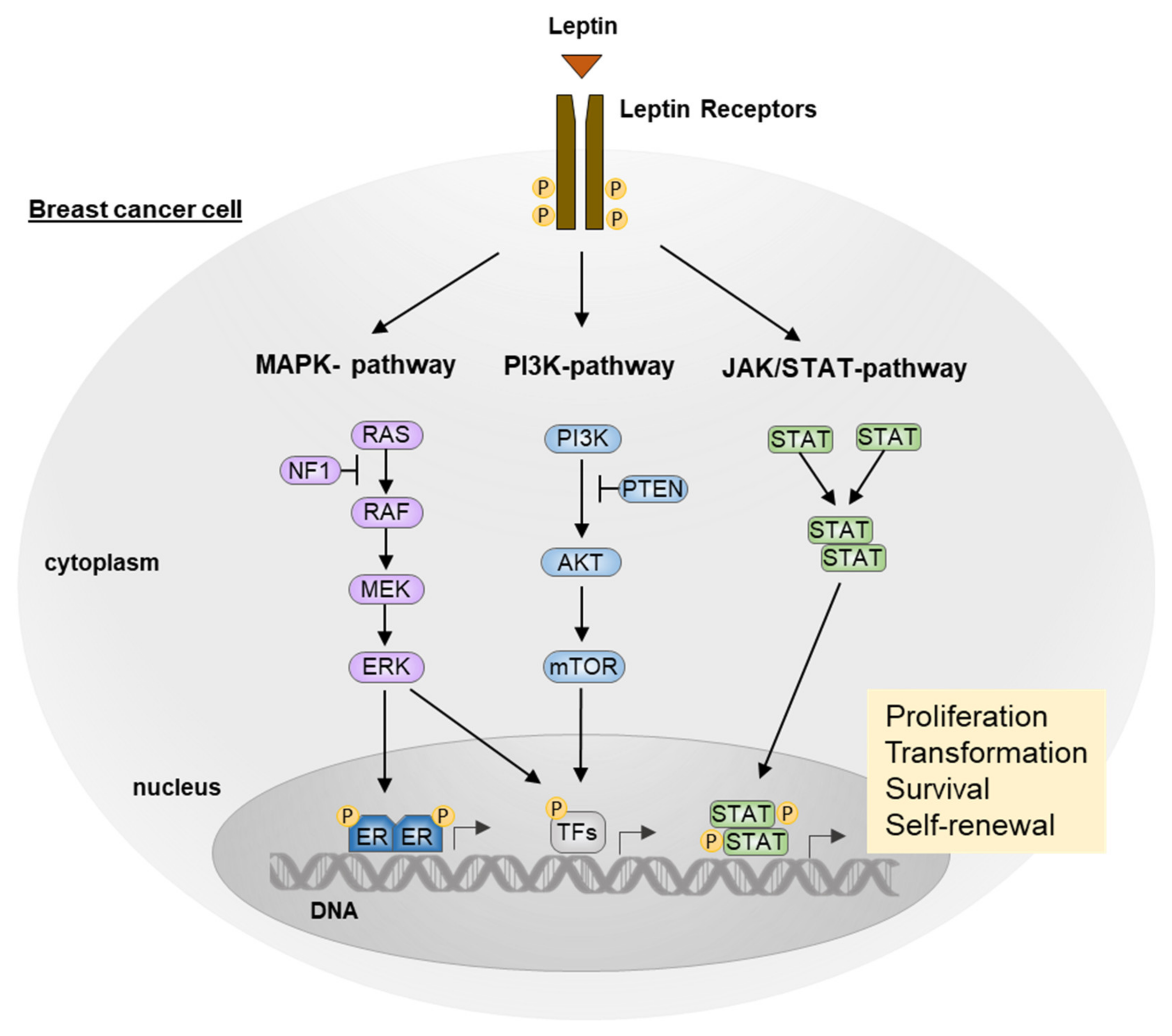
Hereby, we discuss in detail the oncogenic pathways that LEP, upon binding to its receptor LEPR is able to activate, thus promoting cell proliferation, migration, and angiogenesis, largely acknowledged as hallmarks of cancer [50] (Figure 2).

3.1. JAK/STAT Pathway

LEP binds the cytokine receptor homology site (CRH2) in the extracellular domain of LEPR. This interaction leads to receptor dimerization according to the cytokine receptor superfamily binding model [51], and the conformational change induces the cascade signaling transduction. BOX1 and BOX2 motifs in the N-terminal domain of the OB-Rb receptor lead to JAK2 activation, and the LEPR-JAK2 complex enables the phosphorylation of tyrosine residues Tyr986, Tyr1076, and Tyr1141 in the intracellular receptor domain. The phosphorylation of residues Tyr1076 and Tyr1138 enables STAT5 binding and activation, whereas phosphorylated Tyr1141 residues induce STAT3 dimerization and activation [52]. Thus, STAT3 dimers translocate in the nucleus to enhance the signaling through transcription factors such as c-jun, erg-1, and c-myc, which are involved in cancer cell proliferation [53]. Furthermore, it has been reported that LEP in breast cancer models, via JAK2/STAT3 pathway, promotes cell cycle progression by increasing the expression of cdk2 and cyclin D1 levels, thus inducing hyperphosphorylation and subsequent inactivation of the cell cycle inhibitor Rb [54]. Interestingly, in MCF-7 breast cancer cells, LEP has been demonstrated to be able to sustain, in a dose-dependent manner, the cancer cell immortalization by upregulating the expression of Human Telomerase Reverse Transcriptase (hTERT) via STAT3 dimerization [55]. Moreover, STAT3 phosphorylation promoted by LEP enhances carnitine palmitoyl transferase 1B (CPT1B) expression, which encodes the key enzyme for fatty acid β-oxidation (FAO). Thus, in BC cells derived models, FAO induced by LEP signaling seems to play a critical role in BC stem cell (BCSC) self-renewal that is associated with chemoresistance [56].

3.2. MAPK Pathway

As a result of JAK2 activation, phospho-Tyr986 residual motifs bind the Src homology 2 domain (SH2) of protein tyrosine phosphatase-2 (SHP2), which via the Growth factor receptor-bound protein 2 (GRB2) adaptor protein, leads to the ERK1/2 and JNK activation [57]. ERK nuclear translocation induces the expression of target genes, which are known to stimulate growth and cell proliferation, such as c-fos and egr-1 [57]. Furthermore, through the MAPK pathway activation, LEP promotes the growth of breast cancer in nude mice and induces the proliferation and migration of the MCF-7 BC cell line [58]. In addition, ERK1/2 activation, as well as p53 downregulation, has been demonstrated to induce aromatase expression in breast adipose stromal cells [58] and to promote functional activation of ER in the MCF-7 BC cell line [59].

3.3. PI3K/AKT Pathway

LEPR-JAK2 complex can induce IRS-1 phosphorylation, which by binding p85, the PI3K regulatory subunit, relieves p110, the catalytic subunit, thus allowing AKT activation [60]; therefore, in MCF-7 breast cancer cell lines, PI3K/AKT pathway induced by leptin has been demonstrated to promote epithelial–mesenchymal transition (EMT), which is known to facilitate cancer cells invasion, and the development of a plasticity phenotype, via IL-8 secretion [61] and pyruvate kinase M2 (PKM2) activation [62]. Furthermore, PI3K/AKT LEP-dependent signaling enables the upregulation of Acyl-CoA: cholesterol acyltransferase 2 (ACAT2) mRNA and protein expression in ER-positive BC cell lines (MCF-7, T47D, and BT474), facilitating cell migration and invasion by increasing cholesterol esterification [63].

4. Leptin and Immune System

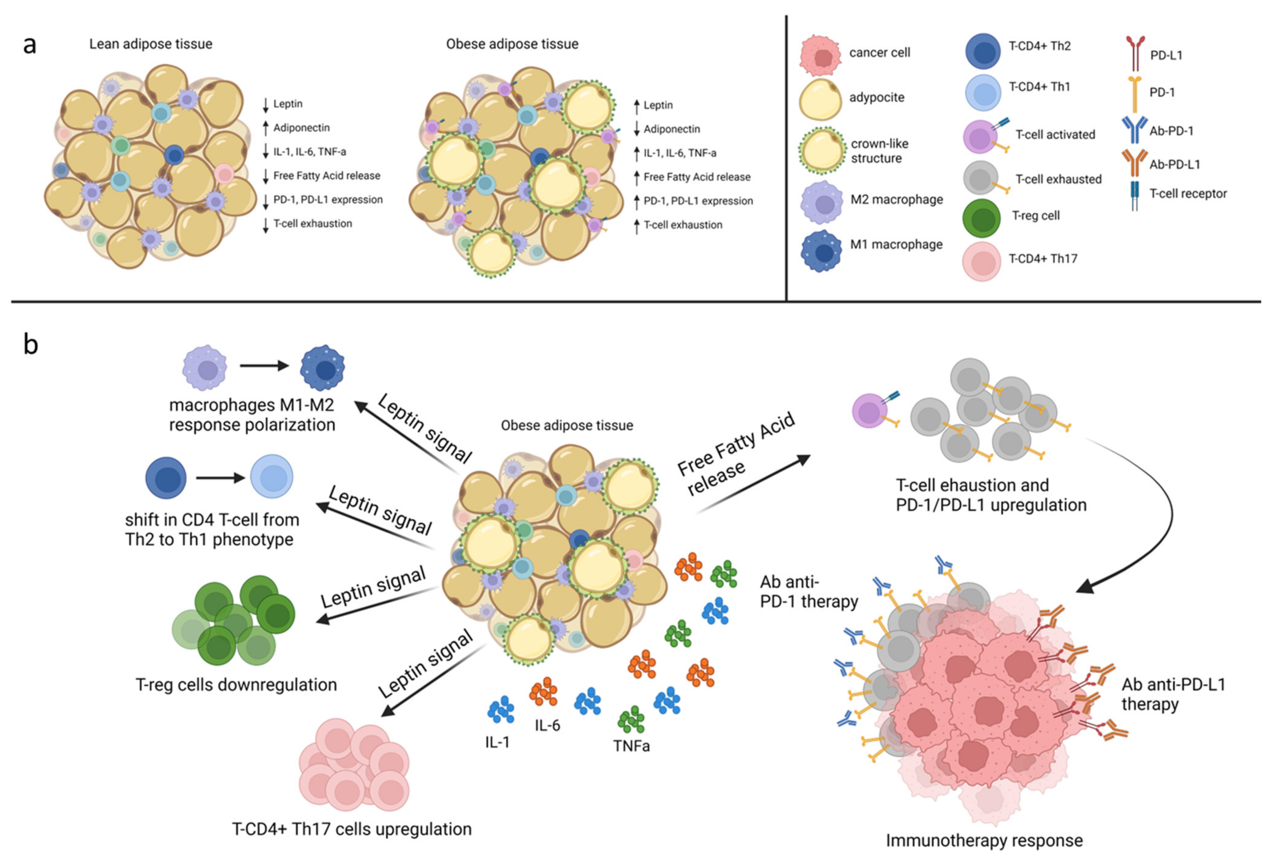
Adipose tissue expansion is known to be strongly associated with a chronic inflammatory state, resulting in a phenotypic change in adipose resident immune cells and in an imbalance of adipokines secretion (Figure 3). Indeed, as adipocyte surface area increases, a reduced thymopoiesis and T-cell progenitor pool proliferation may occur because of thymic involution, known as thymic aging, promoted by adipocyte infiltration [64]. Moreover, LEP signaling markedly increased in obese patients, resulting in several implications on T-cell function and proliferation [65]. In particular, LEP improves CD4 T-cell activation beyond the major histocompatibility complex (MHC) and induces a shift in CD4 T-cell from a Th2 towards Th1 phenotype by stimulating IL-2, IL-12, and IFN-γ expression [66,67].
Moreover, LEP has been demonstrated to influence CD4+ Th17 and CD4+ Treg differentiation. Indeed, in LEP-deficient mice (ob-ob), the exogenous hormone administration showed to enhance CD4+ Th17 upregulation, while reducing Treg proliferation [68,69]. Furthermore, in a study conducted on 30 obese and 13 lean patients, plasma LEP levels, as well as BMI, resulted in being inversely correlated with Treg cells circulating numbers in the obese group, whereas no correlation was observed in the control group [70]. In addition, LEP levels were observed to be positively associated with CD4+ Th17 circulating percentage in patients diagnosed with autoimmune disease as Hashimoto’s thyroiditis [71] and negatively with CD4+ CD25+ regulatory cells observed in relapsing-remitting multiple sclerosis (RRMS) patients [72].
LEP activity also exerts an impact on the innate immune response. Indeed, LEP promotes IL-1, IL-6, and TNFα secretion by monocytes and induces an M1 pro-inflammatory macrophage response polarization through mast cells (MCs) signaling [73]. Indeed, in ob-ob mice models, MCs resident in white adipose tissue showed to drive an anti-inflammatory macrophage phenotype (M2), whereas MCs depletion has been proven to facilitate weight gain and obesity [73].
Additionally, the knockdown of LEPR in ER-positive MCF-7 and in triple-negative MDA-MB-231 BC cells (ObR sh) was demonstrated to hamper macrophage recruitment in the tumor microenvironment (TME) and influence immune response. In particular, in mice models injected with MCF-7 and MDA-MB-231 ObR sh clones, a reduced tumor-associated macrophage (TAMs) infiltration within xenograft tumors was observed. Moreover, in line with the decreased macrophage recruitment, a lower expression on TAMs surface of programmed cell death protein 1 (PD-1), commonly associated with cancer immune escape, was detected [74].
This obesity-related “meta-inflammatory” state [75,76] can eventually impair antitumor immunity: in addition to increasing Treg differentiation and promoting M1 proinflammatory macrophages polarization, it can also act by exhausting immune response through low inflammatory chronic stimulation of Toll-like receptors (TLRs) promoted by lipolysis and secretion of FFAs [77].
The immune regulation promoted by LEP in the inflamed adipose tissue may influence BC carcinogenesis and treatment response. Indeed, in a study conducted on 87 breast cancer patients treated with neoadjuvant chemotherapy, LEP and LEPR overexpression, were found in patients with higher levels of tumor-infiltrating lymphocytes (TILs). This association was observed mainly in patients with HER2+ BC (21.5% vs. 9%; p = 0.015) and, among those with high levels of LEPR, in patients who achieved a complete pathological response compared to those with residual disease (26.6% vs. 12.5%; p = 0.005) whereas this imbalance was not observed in patients with the LEPR not overexpressed [78].
Furthermore, a retrospective analysis conducted on 445 TNBC patients treated with neoadjuvant chemotherapy evaluated the response rate and TILs percentage according to BMI. High TILs were significantly associated with pathological complete response (pCR) in lean patients (OR = 4.24, 95% CI = 2.10 to 8.56; p < 0.001) but not in overweight and obese patients (OR = 1.48, 95% CI = 0.75 to 2.91; p = 0.26) [79].
Interestingly, despite facilitating cancer immune evasion, obesity-related T cell exhaustion, occurring as a result of thymic aging and chronic antigenic stimulation [65,76], leads to the compensatory increase in PD-1/PD-L1 expression on immune cells, providing a potential target for immune-checkpoint inhibitors [80].
Indeed, the tumorigenic immune dysfunction appeared to be reversed by anti-PD-1/PD-L1 treatment in a retrospective analysis conducted on 976 advanced cancer patients stratified according to BMI. The analysis included 635 patients diagnosed with NSCLC, 183 patients with melanoma, 135 patients with renal cell carcinoma, and 23 with other tumors [81]. In particular, overall response rate (ORR) and time to treatment failure (TTF) were found to be significantly higher in overweight/obese patients (BMI ≥ 25.0), compared to non-overweight (p < 0.0001), as well as median progression-free survival (PFS) being longer in the first group (11.7 months vs. 3.7 months; HR= 0.46 p < 0.0001). Furthermore, these results are consistent with those observed in a retrospective analysis which included patients (n = 2046) diagnosed with metastatic melanoma stratified according to BMI and treated with targeted therapy, immunotherapy, or chemotherapy. Indeed, obesity compared with normal BMI was associated with improved progression-free survival (PFS) and overall survival (OS) in the cohort of patients treated with immunotherapy and target therapy [82].
Despite the absence of available data on obese BC patients, in obese female mice fed with an obesogenic high-fat diet, injected with BC E0771 cells and treated with anti-PD-1 therapy, it has been demonstrated a significant decrease in tumor volume, a reduced cell proliferation and a concomitant elevation of CD8+ T cells, CD4+ Th1 cells and dendritic cells [83].

5. Leptin Signaling as a Potential Target for Therapeutic Intervention

As LEP signaling transduction is a well-known carcinogenesis promoting factor, diverse therapeutic agents have been designed to modulate the LEP cascade. Great research efforts focused on the development of LEPR antagonists able to interfere with LEP binding activity. The OB-R antagonist Allo-aca peptide, an analog of OB-R leptin binding site III, has shown promising activity in delaying BC progression and extending survival in TNBC mice models; however, its use was associated with an accelerated weight gain [84]. The peptide LDFI (Leu-Asp-Phe-Ile), a selective LEPR antagonist, was developed based on leptin binding site I and demonstrated to inhibit the growth and motility of both ER-positive MCF-7 and ER-negative SKBR3 breast cancer cell lines. Notably, the PEGylated isoform of LDFI (PEG-LDFI) showed comparable antitumor efficacy in xenograft models without affecting energy balance, since no changes in body weight were observed [85]. In addition, the peptide LDFI recently showed to reduce cell-to-cell communication by decreasing LEP-dependent exosome biosynthesis in MCF-7 and triple-negative MDA-MB-23 breast cancer models [86]. Honokiol (HNK) is a bioactive polyphenol from magnolia grandiflora, acting as a leptin antagonist. HNK is demonstrated to reduce LEP-induced EMT by targeting the STAT3/Zeb1/E-cadherin axis [87]. However, despite the attractive results from several preclinical studies, no clinical trials evaluating LEP antagonists in patients with BC have been conducted to date.
Alternative strategies to target LEP-associated signaling have been explored. The 1,25(OH)2D3 showed to downregulate the human telomerase reverse transcriptase (hTERT) activation promoted by LEP in both breast and ovarian cancer models [88]. Since LEP induces CPT1B expression and FAO activity in BC stem cells to ensure self-renewal and chemoresistance, FAO inhibitors such as perhexiline were able to restore chemosensitivity in BC cells [56]. LEP signaling also enhances aromatase expression [43], impairing tamoxifen treatment efficacy [47,48]. Therefore, aromatase inhibitors (AIs) based regimen exhibits the major beneficial effects on the LEP target cascade. Finally, PD-L1/PD1 upregulation enhanced by the obese-related low chronic inflammatory state could be exploited to improve the immune checkpoint inhibitors treatment activity, especially in patients diagnosed with TNBC, which is known to be the most immunogenic breast cancer subtype.

6. Conclusions

Leptin is a peptide hormone known to play a critical role in the meta-inflammatory state observed in obesity. Many mechanisms appear to be involved in the correlation between obese-related LEP activity and breast cancer carcinogenesis. LEP signaling cascade promotes breast cancer cell proliferation, migration, and invasion through the activation of MAPK, PI3K/AKT, JAK2/STAT pathways, and EMT, irrespective of BC subtypes, primarily in post-menopausal women. Moreover, LEP is involved in immune activity exhaustion associated with low chronic inflammation in obese cancer patients, which could be employed to improve the response to immune checkpoint inhibitors. Novel therapeutic strategies, such as LEP or LEPR antagonist, are not currently under development, although they would represent a promising step toward a more personalized treatment approach for breast cancer patients.

Author Contributions

Conceptualization, R.B., F.N. and C.D.A.; methodology, C.D.A., V.F., M.G., G.A., S.D.P., L.F.; investigation, R.B., G.P., S.P., P.D.P., A.S.; resources, C.D.A., S.D.P., G.A., M.G., P.F., L.F., A.S.; writing—original draft preparation, R.B., F.N.; writing—review and editing, C.D.A., S.D.P., G.A., M.G., P.F., L.F., A.S., G.P., V.F., P.D.P., S.P.; visualization, R.B., F.N.; supervision, C.D.A.; funding acquisition, P.F. All authors have read and agreed to the published version of the manuscript.

Funding

This research received no external funding.

Conflicts of Interest

C.D.A. reports personal fees from Roche, AstraZeneca, Lilly, GSK, Novartis, Seagen, and Pfizer, Advisory Board for Roche, AstraZeneca, Lilly, GSK, Novartis, Seagen, and Pfizer, support for attending meetings and/or travel Roche, AstraZeneca, Lilly, GSK, Novartis, Celgene, and Pfizer, grants from Novartis; Mario Giuliano reports consulting or advisory Role from Lilly, Seagen, Roche, MSD, Gilead, Novartis and Pfizer, Speaker’s Bureau for Lilly, Seagen, AstraZeneca, Novartis, and Pfizer, support for attending meetings and/or travel Novartis and Pfizer, Roche, grants from Novartis; Grazia Arpino reports personal fees from Novartis, during the conduct of the study; personal fees from Lilly, grants and personal fees from Roche, grants, personal fees and non-financial support from Pfizer, grants, personal fees and non-financial support from AstraZeneca, personal fees from Daichi, outside the submitted work. Sabino De Placido receives honoraria from Roche, Novartis, Pfizer, Celgene, Eli Lilly, AstraZeneca, Clovis, Seagen, Daiichi Sankyo, and MSD; Alberto Servetto received honoraria from Eli Lilly, MSD and Janssen; received support for attending meetings from Bristol-Myers Squibb. Luigi Formisano received a research grant from AIRC (My First Grant MFGA2018-21505). All other authors declared no conflict of interests.

References

  1. Sung, H.; Ferlay, J.; Siegel, R.L.; Laversanne, M.; Soerjomataram, I.; Jemal, A.; Bray, F. Global Cancer Statistics 2020: GLOBOCAN Estimates of Incidence and Mortality Worldwide for 36 Cancers in 185 Countries. CA Cancer J. Clin. 2021, 71, 209–249. [Google Scholar] [CrossRef] [PubMed]
  2. Lei, S.; Zheng, R.; Zhang, S.; Wang, S.; Chen, R.; Sun, K.; Zeng, H.; Zhou, J.; Wei, W. Global patterns of breast cancer incidence and mortality: A population-based cancer registry data analysis from 2000 to 2020. Cancer Commun. 2021, 41, 1183–1194. [Google Scholar] [CrossRef] [PubMed]
  3. Wu, X.; Wang, W.; Zhang, D.; Zhu, F. Nutritional status, lifestyle habits and cancer mortality: A population-based prospective cohort study. Eur. J. Nutr. 2021, 61, 1343–1352. [Google Scholar] [CrossRef] [PubMed]
  4. World Health Organization. WHO Discussion Paper: Draft Recommendations for the Prevention and Management of Obesity over the Life Course, Including Potential Targets. Available online: https://www.who.int/publications/m/item/who-discussion-paper-draft-recommendations-for-the-prevention-and-management-of-obesity-over-the-life-course-including-potential-targets (accessed on 19 August 2021).
  5. Kim, D.-S.; Scherer, P.E. Obesity, Diabetes, and Increased Cancer Progression. Diabetes Metab. J. 2021, 45, 799–812. [Google Scholar] [CrossRef]
  6. Coelho, M.; Oliveira, T.; Fernandes, R. Biochemistry of adipose tissue: An endocrine organ. Arch. Med. Sci. 2013, 9, 191–200. [Google Scholar] [CrossRef]
  7. Makki, K.; Froguel, P.; Wolowczuk, I. Adipose tissue in obesity-related inflammation and insulin resistance: Cells, cytokines, and chemokines. Int. Sch. Res. Not. 2013, 2013, 139239. [Google Scholar] [CrossRef]
  8. Iyengar, N.M.; Gucalp, A.; Dannenberg, A.J.; Hudis, C.A. Obesity and Cancer Mechanisms: Tumor Microenvironment and Inflammation. J. Clin. Oncol. 2016, 34, 4270–4276. [Google Scholar] [CrossRef]
  9. Surmi, B.K.; Hasty, A.H. Macrophage infiltration into adipose tissue: Initiation, propagation and remodeling. Future Lipidol. 2008, 3, 545–556. [Google Scholar] [CrossRef]
  10. Cinti, S.; Mitchell, G.; Barbatelli, G.; Murano, I.; Ceresi, E.; Faloia, E.; Wang, S.; Fortier, M.; Greenberg, A.S.; Obin, M.S. Adipocyte death defines macrophage localization and function in adipose tissue of obese mice and humans. J. Lipid Res. 2005, 46, 2347–2355. [Google Scholar] [CrossRef]
  11. Murano, I.; Barbatelli, G.; Parisani, V.; Latini, C.; Muzzonigro, G.; Castellucci, M.; Cinti, S. Dead adipocytes, detected as crown-like structures, are prevalent in visceral fat depots of genetically obese mice. J. Lipid Res. 2008, 49, 1562–1568. [Google Scholar] [CrossRef] [Green Version]
  12. Morris, P.G.; Hudis, C.A.; Giri, D.; Morrow, M.; Falcone, D.J.; Zhou, X.K.; Du, B.; Brogi, E.; Crawford, C.B.; Kopelovich, L.; et al. Inflammation and Increased Aromatase Expression Occur in the Breast Tissue of Obese Women with Breast Cancer. Cancer Prev. Res. 2011, 4, 1021–1029. [Google Scholar] [CrossRef]
  13. D’Esposito, V.; Ambrosio, M.R.; Giuliano, M.; Cabaro, S.; Miele, C.; Beguinot, F.; Formisano, P. Mammary Adipose Tissue Control of Breast Cancer Progression: Impact of Obesity and Diabetes. Front. Oncol. 2020, 10, 1554. [Google Scholar] [CrossRef]
  14. Ambrosio, M.R.; D’Esposito, V.; Costa, V.; Liguoro, D.; Collina, F.; Cantile, M.; Prevete, N.; Passaro, C.; Mosca, G.; De Laurentiis, M.; et al. Glucose impairs tamoxifen responsiveness modulating connective tissue growth factor in breast cancer cells. Oncotarget 2017, 8, 109000–109017. [Google Scholar] [CrossRef]
  15. D’Esposito, V.; Liguoro, D.; Ambrosio, M.R.; Collina, F.; Cantile, M.; Spinelli, R.; Raciti, G.A.; Miele, C.; Valentino, R.; Campiglia, P.; et al. Adipose microenvironment promotes triple negative breast cancer cell invasiveness and dissemination by producing CCL5. Oncotarget 2016, 7, 24495–24509. [Google Scholar] [CrossRef]
  16. Ouyang, S.; He, F. Phylogeny of a Growth Hormone-Like Cytokine Superfamily Based upon 3D Structure. J. Mol. Evol. 2003, 56, 131–136. [Google Scholar] [CrossRef]
  17. Sánchez-Jiménez, F.; Pérez, A.P.; de la Cruz-Merino, L.; Sánchez-Margalet, V. Obesity and Breast Cancer: Role of Leptin. Front. Oncol. 2019, 9, 596. [Google Scholar] [CrossRef]
  18. Trayhurn, P.; Bing, C. Appetite and energy balance signals from adipocytes. Philos. Trans. R. Soc. B Biol. Sci. 2006, 361, 1237–1249. [Google Scholar] [CrossRef]
  19. Flak, J.N.; Myers, M.G. Minireview: CNS Mechanisms of Leptin Action. Mol. Endocrinol. 2016, 30, 3–12. [Google Scholar] [CrossRef]
  20. Farr, O.M.; Gavrieli, A.; Mantzoros, C.S. Leptin Applications in 2015: What Have We Learned about Leptin and Obesity? Curr. Opin. Endocrinol. Diabetes Obes. 2015, 22, 353–359. [Google Scholar] [CrossRef]
  21. Zhao, S.; Zhu, Y.; Schultz, R.D.; Li, N.; He, Z.; Zhang, Z.; Caron, A.; Zhu, Q.; Sun, K.; Xiong, W.; et al. Partial Leptin Reduction as an Insulin Sensitization and Weight Loss Strategy. Cell Metab. 2019, 30, 706–719.e6. [Google Scholar] [CrossRef]
  22. Sahu, A. Minireview: A Hypothalamic Role in Energy Balance with Special Emphasis on Leptin. Endocrinology 2004, 145, 2613–2620. [Google Scholar] [CrossRef] [PubMed]
  23. Ramos-Lobo, A.M.; Donato, J., Jr. The role of leptin in health and disease. Temperature 2017, 4, 258–291. [Google Scholar] [CrossRef] [PubMed]
  24. Mantzoros, C.S.; Magkos, F.; Brinkoetter, M.; Sienkiewicz, E.; Dardeno, T.A.; Kim, S.-Y.; Hamnvik, O.-P.; Koniaris, A. Leptin in human physiology and pathophysiology. Am. J. Physiol. Metab. 2011, 301, E567–E584. [Google Scholar] [CrossRef] [PubMed]
  25. Tartaglia, L.A.; Dembski, M.; Weng, X.; Deng, N.; Culpepper, J.; Devos, R.; Richards, G.J.; Campfield, L.A.; Clark, F.T.; Deeds, J.; et al. Identification and expression cloning of a leptin receptor, OB-R. Cell 1995, 83, 1263–1271. [Google Scholar] [CrossRef]
  26. Kowalski, T.J.; Liu, S.-M.; Leibel, R.L.; Chua, S.C. Transgenic Complementation of Leptin-Receptor Deficiency. I. Rescue of the Obesity/Diabetes Phenotype of LEPR-Null Mice Expressing a LEPR-B Transgene. Diabetes 2001, 50, 425–435. [Google Scholar] [CrossRef]
  27. Lammertab, A.; Kiess, W.; Bottner, A.; Glasowab, A.; Kratzsch, J. Soluble Leptin Receptor Represents the Main Leptin Binding Activity in Human Blood. Biochem. Biophys. Res. Commun. 2001, 283, 982–988. [Google Scholar] [CrossRef]
  28. Jiménez-Cortegana, C.; López-Saavedra, A.; Sánchez-Jiménez, F.; Pérez-Pérez, A.; Castiñeiras, J.; Virizuela-Echaburu, J.A.; de la Cruz-Merino, L.; Sánchez-Margalet, V. Leptin, Both Bad and Good Actor in Cancer. Biomolecules 2021, 11, 913. [Google Scholar] [CrossRef]
  29. Wu, M.-H.; Chou, Y.-C.; Chou, W.-Y.; Hsu, G.-C.; Chu, C.-H.; Yu, C.-P.; Yu, J.-C.; Sun, C.-A. Circulating levels of leptin, adiposity and breast cancer risk. Br. J. Cancer 2009, 100, 578–582. [Google Scholar] [CrossRef]
  30. Gu, L.; Wang, C.-D.; Cao, C.; Cai, L.-R.; Li, D.-H.; Zheng, Y.-Z. Association of serum leptin with breast cancer. Medicine 2019, 98, e14094. [Google Scholar] [CrossRef]
  31. Pan, H.; Deng, L.-L.; Cui, J.-Q.; Shi, L.; Yang, Y.-C.; Luo, J.-H.; Qin, D.; Wang, L. Association between Serum Leptin Levels and Breast Cancer Risk: An Updated Systematic Review and Meta-Analysis. Medicine 2018, 97, e11345. [Google Scholar] [CrossRef]
  32. The Premenopausal Breast Cancer Collaborative Group; Schoemaker, M.J.; Nichols, H.B.; Wright, L.B.; Brook, M.N.; Jones, M.E.; O’Brien, K.M.; Adami, H.-O.; Baglietto, L.; Bernstein, L.; et al. Association of Body Mass Index and Age With Subsequent Breast Cancer Risk in Premenopausal Women. JAMA Oncol. 2018, 4, e181771. [Google Scholar] [CrossRef]
  33. Colditz, G.; Frazier, A.L. Models of breast cancer show that risk is set by events of early life: Prevention efforts must shift focus. Cancer Epidemiol. Biomark. Prev. 1995, 4, 567–571. [Google Scholar]
  34. Colditz, G.A.; Bohlke, K. Priorities for the primary prevention of breast cancer. CA: A Cancer J. Clin. 2014, 64, 186–194. [Google Scholar] [CrossRef]
  35. Miller, W. Aromatase and the breast: Regulation and clinical aspects. Maturitas 2006, 54, 335–341. [Google Scholar] [CrossRef]
  36. Dowsett, M.; Folkerd, E. Reduced progesterone levels explain the reduced risk of breast cancer in obese premenopausal women: A new hypothesis. Breast Cancer Res. Treat. 2015, 149, 1–4. [Google Scholar] [CrossRef]
  37. Key, T.; Pike, M. The role of oestrogens and progestagens in the epidemiology and prevention of breast cancer. Eur. J. Cancer Clin. Oncol. 1988, 24, 29–43. [Google Scholar] [CrossRef]
  38. Grubbs, C.J.; Farnell, D.R.; Hill, D.L.; McDonough, K.C. Chemoprevention of N-nitroso-N-methylurea-induced mammary cancers by pretreatment with 17 beta-estradiol and progesterone. J. Natl. Cancer Inst. 1985, 74, 927–931. [Google Scholar]
  39. Hilakivi-Clarke, L. Estrogens, BRCA1, and Breast Cancer. Cancer Res. 2000, 60, 4993–5001. [Google Scholar]
  40. Catalano, S.; Marsico, S.; Giordano, C.; Mauro, L.; Rizza, P.; Panno, M.L.; Andò, S. Leptin Enhances, via AP-1, Expression of Aromatase in the MCF-7 Cell Line. J. Biol. Chem. 2003, 278, 28668–28676. [Google Scholar] [CrossRef]
  41. Andò, S.; Gelsomino, L.; Panza, S.; Giordano, C.; Bonofiglio, D.; Barone, I.; Catalano, S. Obesity, Leptin and Breast Cancer: Epidemiological Evidence and Proposed Mechanisms. Cancers 2019, 11, 62. [Google Scholar] [CrossRef]
  42. Mauro, L.; Catalano, S.; Bossi, G.; Pellegrino, M.; Barone, I.; Morales, S.; Giordano, C.; Bartella, V.; Casaburi, I.; Andò, S. Evidences that Leptin Up-regulates E-Cadherin Expression in Breast Cancer: Effects on Tumor Growth and Progression. Cancer Res. 2007, 67, 3412–3421. [Google Scholar] [CrossRef]
  43. Garofalo, C.; Koda, M.; Cascio, S.; Sulkowska, M.; Kanczuga-Koda, L.; Golaszewska, J.; Russo, A.; Sulkowski, S.; Surmacz, E. Increased Expression of Leptin and the Leptin Receptor as a Marker of Breast Cancer Progression: Possible Role of Obesity-Related Stimuli. Clin. Cancer Res. 2006, 12, 1447–1453. [Google Scholar] [CrossRef]
  44. Nagalingam, A.; Siddharth, S.; Parida, S.; Muniraj, N.; Avtanski, D.; Kuppusamy, P.; Elsey, J.; Arbiser, J.L.; Győrffy, B.; Sharma, D. Hyperleptinemia in obese state renders luminal breast cancers refractory to tamoxifen by coordinating a crosstalk between Med1, miR205 and ErbB. NPJ Breast Cancer 2021, 7, 1–13. [Google Scholar] [CrossRef]
  45. Chen, X.; Zha, X.; Chen, W.; Zhu, T.; Qiu, J.; Røe, O.D.; Li, J.; Wang, Z.; Yin, Y. Leptin attenuates the anti-estrogen effect of tamoxifen in breast cancer. Biomed. Pharmacother. 2013, 67, 22–30. [Google Scholar] [CrossRef] [PubMed]
  46. Fiorio, E.; Mercanti, A.; Terrasi, M.; Micciolo, R.; Remo, A.; Auriemma, A.; Molino, A.; Parolin, V.; Di Stefano, B.; Bonetti, F.; et al. Leptin/HER2 crosstalk in breast cancer: In vitro study and preliminary in vivoanalysis. BMC Cancer 2008, 8, 305. [Google Scholar] [CrossRef] [PubMed]
  47. Saxena, N.K.; Taliaferro-Smith, L.; Knight, B.B.; Merlin, D.; Anania, F.A.; O’Regan, R.M.; Sharma, D. Bidirectional Crosstalk between Leptin and Insulin-like Growth Factor-I Signaling Promotes Invasion and Migration of Breast Cancer Cells via Transactivation of Epidermal Growth Factor Receptor. Cancer Res. 2008, 68, 9712–9722. [Google Scholar] [CrossRef] [PubMed]
  48. Gonzalez-Perez, R.R.; Xu, Y.; Guo, S.; Watters, A.; Zhou, W.; Leibovich, S.J. Leptin upregulates VEGF in breast cancer via canonic and non-canonical signalling pathways and NFκB/HIF-1α activation. Cell. Signal. 2010, 22, 1350–1362. [Google Scholar] [CrossRef] [PubMed]
  49. Garonna, E.; Botham, K.M.; Birdsey, G.M.; Randi, A.M.; Gonzalez-Perez, R.R.; Wheeler-Jones, C.P.D. Vascular Endothelial Growth Factor Receptor-2 Couples Cyclo-Oxygenase-2 with Pro-Angiogenic Actions of Leptin on Human Endothelial Cells. PLoS ONE 2011, 6, e18823. [Google Scholar] [CrossRef]
  50. Gonzalez, R.R.; Cherfils, S.; Escobar, M.; Yoo, J.H.; Carino, C.; Styer, A.K.; Sullivan, B.T.; Sakamoto, H.; Olawaiye, A.; Serikawa, T.; et al. Leptin Signaling Promotes the Growth of Mammary Tumors and Increases the Expression of Vascular Endothelial Growth Factor (VEGF) and Its Receptor Type Two (VEGF-R2). J. Biol. Chem. 2006, 281, 26320–26328. [Google Scholar] [CrossRef]
  51. Mancour, L.V.; Daghestani, H.N.; Dutta, S.; Westfield, G.H.; Schilling, J.; Oleskie, A.N.; Herbstman, J.F.; Chou, S.Z.; Skiniotis, G. Ligand-Induced Architecture of the Leptin Receptor Signaling Complex. Mol. Cell 2012, 48, 655–661. [Google Scholar] [CrossRef]
  52. Vaisse, C.; Halaas, J.L.; Horvath, C.M.; Darnell, J.E.; Stoffel, M.; Friedman, J.M. Leptin activation of Stat3 in the hypothalamus of wild–type and ob/ob mice but not db/db mice. Nat. Genet. 1996, 14, 95–97. [Google Scholar] [CrossRef]
  53. Banks, A.S.; Davis, S.M.; Bates, S.H.; Myers, M.G. Activation of Downstream Signals by the Long Form of the Leptin Receptor. J. Biol. Chem. 2000, 275, 14563–14572. [Google Scholar] [CrossRef]
  54. Saxena, N.K.; Vertino, P.M.; Anania, F.A.; Sharma, D. Leptin-induced Growth Stimulation of Breast Cancer Cells Involves Recruitment of Histone Acetyltransferases and Mediator Complex to CYCLIN D1 Promoter via Activation of Stat3. J. Biol. Chem. 2007, 282, 13316–13325. [Google Scholar] [CrossRef] [Green Version]
  55. Ren, H.; Zhao, T.; Wang, X.; Gao, C.; Wang, J.; Yu, M.; Hao, J. Leptin upregulates telomerase activity and transcription of human telomerase reverse transcriptase in MCF-7 breast cancer cells. Biochem. Biophys. Res. Commun. 2010, 394, 59–63. [Google Scholar] [CrossRef]
  56. Wang, T.; Fahrmann, J.F.; Lee, H.; Li, Y.-J.; Tripathi, S.C.; Yue, C.; Zhang, C.; Lifshitz, V.; Song, J.; Yuan, Y.; et al. JAK/STAT3-Regulated Fatty Acid β-Oxidation Is Critical for Breast Cancer Stem Cell Self-Renewal and Chemoresistance. Cell Metab. 2018, 27, 136–150.e5. [Google Scholar] [CrossRef]
  57. Bjørbæk, C.; Buchholz, R.M.; Davis, S.M.; Bates, S.H.; Pierroz, D.D.; Gu, H.; Neel, B.G.; Myers, M.G.; Flier, J.S. Divergent Roles of SHP-2 in ERK Activation by Leptin Receptors. J. Biol. Chem. 2001, 276, 4747–4755. [Google Scholar] [CrossRef]
  58. Yuan, H.-J.; Sun, K.-W.; Yu, K. Leptin promotes the proliferation and migration of human breast cancer through the extracellular-signal regulated kinase pathway. Mol. Med. Rep. 2013, 9, 350–354. [Google Scholar] [CrossRef]
  59. Catalano, S.; Mauro, L.; Marsico, S.; Giordano, C.; Rizza, P.; Rago, V.; Montanaro, D.; Maggiolini, M.; Panno, M.L.; Andó, S. Leptin Induces, via ERK1/ERK2 Signal, Functional Activation of Estrogen Receptor α in MCF-7 Cells. J. Biol. Chem. 2004, 279, 19908–19915. [Google Scholar] [CrossRef]
  60. Gorgisen, G.; Gulacar, I.M.; Ozes, O.N. The role of insulin receptor substrate (IRS) proteins in oncogenic transformation. Cell. Mol. Biol. 2017, 63, 1–5. [Google Scholar] [CrossRef]
  61. Wang, L.; Tang, C.; Cao, H.; Li, K.; Pang, X.; Zhong, L.; Dang, W.; Tang, H.; Huang, Y.; Wei, L.; et al. Activation of IL-8 via PI3K/Akt-dependent pathway is involved in leptin-mediated epithelial-mesenchymal transition in human breast cancer cells. Cancer Biol. Ther. 2015, 16, 1220–1230. [Google Scholar] [CrossRef]
  62. Wei, L.; Li, K.; Pang, X.; Guo, B.; Su, M.; Huang, Y.; Wang, N.; Ji, F.; Zhong, C.; Yang, J.; et al. Leptin promotes epithelial-mesenchymal transition of breast cancer via the upregulation of pyruvate kinase M2. J. Exp. Clin. Cancer Res. 2016, 35, 1–10. [Google Scholar] [CrossRef] [PubMed]
  63. Huang, Y.; Jin, Q.; Su, M.; Ji, F.; Wang, N.; Zhong, C.; Jiang, Y.; Liu, Y.; Zhang, Z.; Yang, J.; et al. Leptin promotes the migration and invasion of breast cancer cells by upregulating ACAT2. Cell. Oncol. 2017, 40, 537–547. [Google Scholar] [CrossRef] [PubMed]
  64. Yang, H.; Youm, Y.-H.; Vandanmagsar, B.; Rood, J.; Kumar, K.G.; Butler, A.; Dixit, V.D. Obesity accelerates thymic aging. Blood 2009, 114, 3803–3812. [Google Scholar] [CrossRef] [PubMed] [Green Version]
  65. Naylor, C.; Petri, W.A., Jr. Leptin Regulation of Immune Responses. Trends Mol. Med. 2016, 22, 88–98. [Google Scholar] [CrossRef]
  66. Batra, A.; Okur, B.; Glauben, R.; Erben, U.; Ihbe, J.; Stroh, T.; Fedke, I.; Chang, H.-D.; Zeitz, M.; Siegmund, B. Leptin: A Critical Regulator of CD4+ T-cell Polarization in Vitro and in Vivo. Endocrinology 2010, 151, 56–62. [Google Scholar] [CrossRef]
  67. Carbone, F.; La Rocca, C.; Matarese, G. Immunological functions of leptin and adiponectin. Biochimie 2012, 94, 2082–2088. [Google Scholar] [CrossRef]
  68. Liu, Y.; Yu, Y.; Matarese, G.; La Cava, A. Cutting Edge: Fasting-Induced Hypoleptinemia Expands Functional Regulatory T Cells in Systemic Lupus Erythematosus. J. Immunol. 2012, 188, 2070–2073. [Google Scholar] [CrossRef]
  69. Reis, B.S.; Lee, K.; Fanok, M.H.; Mascaraque, C.; Amoury, M.; Cohn, L.B.; Rogoz, A.; Dallner, O.S.; Moraes-Vieira, P.M.; Domingos, A.I.; et al. Leptin Receptor Signaling in T Cells Is Required for Th17 Differentiation. J. Immunol. 2015, 194, 5253–5260. [Google Scholar] [CrossRef]
  70. Wagner, N.-M.; Brandhorst, G.; Czepluch, F.; Lankeit, M.; Eberle, C.; Herzberg, S.; Faustin, V.; Riggert, J.; Oellerich, M.; Hasenfuss, G.; et al. Circulating regulatory T cells are reduced in obesity and may identify subjects at increased metabolic and cardiovascular risk. Obesity 2012, 21, 461–468. [Google Scholar] [CrossRef]
  71. Wang, S.; Baidoo, S.E.; Liu, Y.; Zhu, C.; Tian, J.; Ma, J.; Tong, J.; Chen, J.; Tang, X.; Xu, H.; et al. T cell-derived leptin contributes to increased frequency of T helper type 17 cells in female patients with Hashimoto’s thyroiditis. Clin. Exp. Immunol. 2012, 171, 63–68. [Google Scholar] [CrossRef]
  72. Matarese, G.; Carrieri, P.B.; La Cava, A.; Perna, F.; Sanna, V.; De Rosa, V.; Aufiero, D.; Fontana, S.; Zappacosta, S. Leptin increase in multiple sclerosis associates with reduced number of CD4 + CD25 + regulatory T cells. Proc. Natl. Acad. Sci. USA 2005, 102, 5150–5155. [Google Scholar] [CrossRef]
  73. Zhou, Y.; Yu, X.; Chen, H.; Sjöberg, S.; Roux, J.; Zhang, L.; Ivoulsou, A.-H.; Bensaid, F.; Liu, C.-L.; Liu, J.; et al. Leptin Deficiency Shifts Mast Cells toward Anti-Inflammatory Actions and Protects Mice from Obesity and Diabetes by Polarizing M2 Macrophages. Cell Metab. 2015, 22, 1045–1058. [Google Scholar] [CrossRef]
  74. Gelsomino, L.; Naimo, G.D.; Malivindi, R.; Augimeri, G.; Panza, S.; Giordano, C.; Barone, I.; Bonofiglio, D.; Mauro, L.; Catalano, S.; et al. Knockdown of Leptin Receptor Affects Macrophage Phenotype in the Tumor Microenvironment Inhibiting Breast Cancer Growth and Progression. Cancers 2020, 12, 2078. [Google Scholar] [CrossRef]
  75. Naik, A.; Monjazeb, A.M.; Decock, J. The Obesity Paradox in Cancer, Tumor Immunology, and Immunotherapy: Potential Therapeutic Implications in Triple Negative Breast Cancer. Front. Immunol. 2019, 10, 1940. [Google Scholar] [CrossRef]
  76. Mirsoian, A.; Murphy, W.J. Obesity and cancer immunotherapy toxicity. Immunotherapy 2015, 7, 319–322. [Google Scholar] [CrossRef]
  77. Reilly, S.M.; Saltiel, A.R. Adapting to obesity with adipose tissue inflammation. Nat. Rev. Endocrinol. 2017, 13, 633–643. [Google Scholar] [CrossRef]
  78. García-Estevez, L.; González-Martínez, S.; Moreno-Bueno, G. The Leptin Axis and Its Association With the Adaptive Immune System in Breast Cancer. Front. Immunol. 2021, 12, 784823. [Google Scholar] [CrossRef]
  79. Floris, G.; Richard, F.; Hamy, A.-S.; Jongen, L.; Wildiers, H.; Ardui, J.; Punie, K.; Smeets, A.; Berteloot, P.; Vergote, I.; et al. Body Mass Index and Tumor-Infiltrating Lymphocytes in Triple-Negative Breast Cancer. JNCI J. Natl. Cancer Inst. 2020, 113, 146–153. [Google Scholar] [CrossRef]
  80. Wang, Z.; Aguilar, E.G.; Luna, J.I.; Dunai, C.; Khuat, L.T.; Le, C.; Mirsoian, A.; Minnar, C.M.; Stoffel, K.M.; Sturgill, I.R.; et al. Paradoxical effects of obesity on T cell function during tumor progression and PD-1 checkpoint blockade. Nat. Med. 2019, 25, 141–151. [Google Scholar] [CrossRef]
  81. Cortellini, A.; Bersanelli, M.; Buti, S.; Cannita, K.; Santini, D.; Perrone, F.; Giusti, R.; Tiseo, M.; Michiara, M.; Di Marino, P.; et al. A multicenter study of body mass index in cancer patients treated with anti-PD-1/PD-L1 immune checkpoint inhibitors: When overweight becomes favorable. J. Immunother. Cancer 2019, 7, 57. [Google Scholar] [CrossRef]
  82. McQuade, J.L.; Daniel, C.R.; Hess, K.R.; Mak, C.; Wang, D.Y.; Rai, R.R.; Park, J.J.; Haydu, L.E.; Spencer, C.; Wongchenko, M.; et al. Association of body-mass index and outcomes in patients with metastatic melanoma treated with targeted therapy, immunotherapy, or chemotherapy: A retrospective, multicohort analysis. Lancet Oncol. 2018, 19, 310–322. [Google Scholar] [CrossRef]
  83. Pingili, A.K.; Chaib, M.; Sipe, L.M.; Miller, E.J.; Teng, B.; Sharma, R.; Yarbro, J.R.; Asemota, S.; Al Abdallah, Q.; Mims, T.S.; et al. Immune checkpoint blockade reprograms systemic immune landscape and tumor microenvironment in obesity-associated breast cancer. Cell Rep. 2021, 35, 109285. [Google Scholar] [CrossRef] [PubMed]
  84. Otvos, L.; Surmacz, E. Targeting the leptin receptor: A potential new mode of treatment for breast cancer. Expert Rev. Anticancer Ther. 2011, 11, 1147–1150. [Google Scholar] [CrossRef] [PubMed]
  85. Catalano, S.; Leggio, A.; Barone, I.; De Marco, R.; Gelsomino, L.; Campana, A.; Malivindi, R.; Panza, S.; Giordano, C.; Liguori, A.; et al. A novel leptin antagonist peptide inhibits breast cancer growth in vitro and in vivo. J. Cell. Mol. Med. 2015, 19, 1122–1132. [Google Scholar] [CrossRef] [PubMed]
  86. Giordano, C.; Gelsomino, L.; Barone, I.; Panza, S.; Augimeri, G.; Bonofiglio, D.; Rovito, D.; Naimo, G.D.; Leggio, A.; Catalano, S.; et al. Leptin Modulates Exosome Biogenesis in Breast Cancer Cells: An Additional Mechanism in Cell-to-Cell Communication. J. Clin. Med. 2019, 8, 1027. [Google Scholar] [CrossRef] [PubMed]
  87. Avtanski, D.B.; Nagalingam, A.; Bonner, M.Y.; Arbiser, J.L.; Saxena, N.K.; Sharma, D. Honokiol activates LKB1-miR-34a axis and antagonizes the oncogenic actions of leptin in breast cancer. Oncotarget 2015, 6, 29947–29962. [Google Scholar] [CrossRef] [PubMed]
  88. Kasiappan, R.; Sun, Y.; Lungchukiet, P.; Quarni, W.; Zhang, X.; Bai, W. Vitamin D Suppresses Leptin Stimulation of Cancer Growth through microRNA. Cancer Res. 2014, 74, 6194–6204. [Google Scholar] [CrossRef] [PubMed] [Green Version]
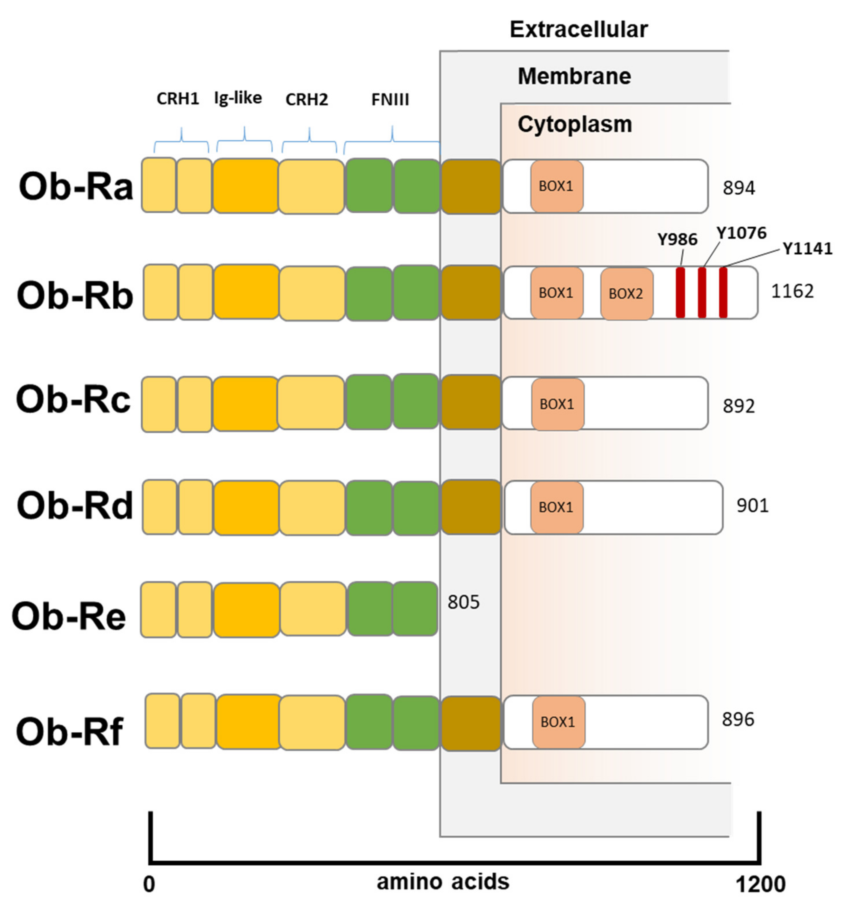
Figure 1. The Structure of Leptin Receptors. All leptin receptor isoforms share a binding C-terminal extracellular domain consisting of 2 CRH regions separated by an IG-like domain and followed by 2 FNIII domains. The intracellular N-terminal domain presents a BOX-1 motif, which is critical for JAK-2 binding. OB-Rb receptor is the only isoform to present a BOX-2 motif, which facilitates the activation of JAK2-STAT transduction pathway. Additionally, OB-Rb presents 3tyrosine residues (Tyr986, Tyr1076, and Tyr1141) whose phosphorylation enables STAT3-5 activation. OB-Re receptor is a soluble isoform, which binds circulating LEP and regulates LEP plasma bioavailability and functionality, not being able to transduce any downstream signal. CHR, cytokine receptor homology; IG-like, immunoglobulin-like; FNIII, fibronectin type III.
Biomolecules 12 01394 g001
Figure 2. Leptin-induced oncogenic signaling pathways in breast cancer. Schematic representation of key intracellular signaling pathways associated with activation of leptin receptors upon binging with leptin in breast cancer cells. AKT, protein kinases; ER, estrogen receptor; PI3K, phosphatidylinositol 3-kinase; Ras, Rat sarcoma subfamily of GTPases; mTOR, mammalian target of rapamycin; MEK, mitogen-activated protein kinase; RAF, RAF kinase; JAK/STAT: Janus kinases/signal transducers and activators of transcription; PTEN, phosphatase and tensin homolog; TFs, transcription factors.
Biomolecules 12 01394 g002
Figure 3. Obesity-related immune landscape. (a). Expansion of fat adipose tissue, compared to lean adipose tissue, is associated with a microenvironment proinflammatory state characterized by an imbalance of adipokines secretion and a systemic immune response reprogramming. Adipocytes’ hyperplasia and hypertrophy provoke adipose tissue macrophages (ATMs) activation resulting in the formation of typical crown-like structures (CLS). (b). Leptin increasing signal stimulates macrophages M1-M2 response polarization, induces a shift in CD4 T-cell from a Th2 towards Th1 phenotype, promotes T-reg cells down-regulation and T-CD4+ Th17 up-regulation. Free fatty acid (FFAs) release and antigenic chronic stimulation lead to T cell exhaustion and consequently PD-1 and PD-L1 upregulation, resulting in cancer cell immune escape. Eventually, PD-1/PD-L1 hyperexpression represents a paradoxical potential target for immune-checkpoint inhibitors. Figure created in BioRender (https://biorender.com/ (accessed on 15 September 2022)).
Biomolecules 12 01394 g003
Publisher’s Note: MDPI stays neutral with regard to jurisdictional claims in published maps and institutional affiliations.