Review

A Systematic Review of Common and Brain-Disease-Specific RNA Editing Alterations Providing Novel Insights into Neurological and Neurodegenerative Disease Manifestations

by
Korina Karagianni
1,†,
Spyros Pettas
1,†,
Georgia Christoforidou
1,
Eirini Kanata
2,
Nikolaos Bekas
1,
Konstantinos Xanthopoulos
2,
Dimitra Dafou
1,*,‡ and
Theodoros Sklaviadis
2,‡
1
Department of Genetics, Development, and Molecular Biology, School of Biology, Aristotle University of Thessaloniki, 541 24 Thessaloniki, Greece
2
Neurodegenerative Diseases Research Group, Department of Pharmacy, School of Health Sciences, Aristotle University of Thessaloniki, 541 24 Thessaloniki, Greece
*
Author to whom correspondence should be addressed.
These authors contributed equally to this work.
Biomolecules 2022, 12(3), 465; https://doi.org/10.3390/biom12030465
Submission received: 1 February 2022 / Revised: 8 March 2022 / Accepted: 15 March 2022 / Published: 17 March 2022
(This article belongs to the Topic Cell Signaling Pathways)

Abstract

:
RNA editing contributes to transcriptome diversification through RNA modifications in relation to genome-encoded information (RNA–DNA differences, RDDs). The deamination of Adenosine (A) to Inosine (I) or Cytidine (C) to Uridine (U) is the most common type of mammalian RNA editing. It occurs as a nuclear co- and/or post-transcriptional event catalyzed by ADARs (Adenosine deaminases acting on RNA) and APOBECs (apolipoprotein B mRNA editing enzyme catalytic polypeptide-like genes). RNA editing may modify the structure, stability, and processing of a transcript. This review focuses on RNA editing in psychiatric, neurological, neurodegenerative (NDs), and autoimmune brain disorders in humans and rodent models. We discuss targeted studies that focus on RNA editing in specific neuron-enriched transcripts with well-established functions in neuronal activity, and transcriptome-wide studies, enabled by recent technological advances. We provide comparative editome analyses between human disease and corresponding animal models. Data suggest RNA editing to be an emerging mechanism in disease development, displaying common and disease-specific patterns. Commonly edited RNAs represent potential disease-associated targets for therapeutic and diagnostic values. Currently available data are primarily descriptive, calling for additional research to expand global editing profiles and to provide disease mechanistic insights. The potential use of RNA editing events as disease biomarkers and available tools for RNA editing identification, classification, ranking, and functional characterization that are being developed will enable comprehensive analyses for a better understanding of disease(s) pathogenesis and potential cures.

1. Introduction

RNA editing refers to an epigenetic mechanism that contributes to transcriptome diversification through the introduction of alterations in RNA species relative to the corresponding genome-encoded information (RNA–DNA differences, RDDs) [1].
The major type of RNA editing in mammals is the chemical modification (deamination) of Adenosine (A) to Inosine (I) or Cytidine (C) to Uridine (U), resulting in A-I or C-U substitutions. It occurs as a nuclear and cytoplasmic co- and/or post-transcriptional event and is catalyzed by the ADAR (Adenosine deaminases acting on RNA) and APOBEC (apolipoprotein B mRNA editing enzyme catalytic polypeptide-like) enzyme families [2] (Figure 1A,B).
Depending on the RNA species undergoing editing and the edited position(s) within a transcript, RNA editing may result in an altered structure, stability, and transcript processing; strong evidence suggests that RNA editing is a major epigenetic gene expression regulator, acting both independently and in conjunction with RNA interference pathways, the latter of which may also be subjected to the dependent regulation of RNA editing. Furthermore, protein recoding (altered amino acid encoding) and immature stop codon introduction may occur as a result of non-synonymous changes introduced by RNA editing in coding regions (Figure 1C).
RNA editing is highly regulated in a tissue- [3], developmental- [4], and cell-type- [5] dependent manner. It is involved in the fine-tuning of cellular responses to hormonal and nutritional stimuli [6] and to cellular environment alterations, including hypoxic conditions [2,7,8,9], suggesting a significant role in sustaining cellular homeostasis.
As a result, several studies have associated RNA editing with disease conditions. Type I neurofibromatosis is associated with a dysfunctional protein resulting from the introduction of a premature stop codon in the NF1 mRNA due to false C-U editing [10]; random editing by both ADAR1 and ADAR2, as well as mutations in the APOBEC1 gene have been reported in several cancer cases [11] and ADAR mutations were identified as causative for the Aicardi–Goutières Syndrome (AGS), an inherited encephalopathy that affects newborn infants and results in severe mental and physical disability [12].
Among other tissues, the brain displays the highest editing levels in both mice and humans [13,14] and shows a gradual increase in both mRNA and miRNA editing during its development [15,16]. Neurons and astrocytes represent the most-edited brain cell types [17]. RNA editing is altered by neuronal activity [18]. A-I editing has been associated with the trafficking and assembly of kaniate [19] and GABA(A) receptors [20], modulation of Ca(v)1.3 channels activation [21,22] and regulation of Nova1 stability [23], a neuron-specific RNA-binding protein involved in alternative splicing [24]. Similar to other diseases, it is expected that brain disorders are associated with RNA editing alterations [25].
In this review, we focus on the role of RNA editing alterations in psychiatric, neurological, neurodegenerative (NDs) and autoimmune brain disorders in human cases and in related mouse models. We provide a comprehensive presentation of relevant data supporting RNA editing contribution in these disorders. We include studies focusing on editing events identified in selected, disease-associated targets, as well as recently published transcriptome-wide studies and discuss common editing patterns among different brain disorders. Furthermore, we attempt to identify commonly edited targets and affected cellular processes among human and mouse brain disorders, suggestive of common underlying molecular mechanisms and indicative of animal models’ utilization reliability. Subsequently, we discuss the significance of extending transcriptome-wide RNA editing studies at pre-clinical disease stages in relevant disease models, aiming to identify RNA editing alterations that may contribute to driver pathogenetic events, that could provide novel candidate intervention targets. Finally, we discuss future perspectives and challenges in the RNA editing study field, focusing on CNS disorders.

2. Materials and Methods

2.1. Literature Search Strategy

Five independent reviewers conducted a systematic literature search on PubMed for suitable research published between 1995 and 2021, following preset methods and intending to identify studies related to RNA editing in brain disorders. The search terms were: “RNA editing” OR “RNA alterations” AND “neurological disorders” OR “neurodegenerative diseases” OR “neurodegenerative disorders” OR “neurodegeneration” OR “psychiatric disorders”.

2.2. Eligibility Criteria

Studies were considered eligible if they met the following requirements: (1) Studies in humans and rodent models examining RNA editing events in relation to psychiatric, neurological, neurodegenerative, and autoimmune brain disorders; (2) genetically determined neurological and neurodegenerative disorders, influenced by environmental factors or caused by an injury; (3) studies in brain and/or spinal cord tissue material from either postmortem human material or animal disease models.
Exclusion criteria were as follows: (1) non-original publications, such as letters to the editor, opinions, reviews, case reports, protocols, conference or meeting abstracts, comments, or meta-analyses; (2) studies unrelated to the subject; (3) studies with insufficient or unqualified data.

2.3. Statistical Analysis and Data Extraction

Only articles written in English were considered. In addition, duplicate publications were excluded. The following information was gathered from all the studies that were included: basic information (first author, publication year, and research country), disorder, species, brain area, study type, targets, methodology, validation method, differentially expressed targets compared to controls.

2.4. Registration for Studies

The studies were evaluated using the criteria specified by the PRISMA (preferential reporting items for systematic review and meta-analysis) guidelines [26].

3. Results

The term ‘Neurological Disorder’ refers to a wide range of disorders concerning the nervous system, which are characterized by a variety of symptoms ranging from mild to severe and are either accompanied by neurodegeneration or not. Neurological disorders may be either inherited with genetic predisposition, associated with environmental factors or may occur as a result of injury. Neurological/neurodegenerative disorders, such as epilepsy, autism, Alzheimer’s disease (AD), Parkinson’s disease (PD), Huntington’s disease (HD), prion diseases, amyotrophic lateral sclerosis (ALS) and multiple sclerosis (MS), can be clustered together because they all involve the malfunction or damaging of the brain, spinal cord, and nerves. Nervous system degeneration affects neuronal communication resulting in behavior and emotional state disruptions, which also represent hallmarks of psychiatric disorders, such as schizophrenia (SCZ), chronic social defeat stress (CSDS), depression and suicide. Thus, despite their difference, neurological/neurodegenerative and psychiatric disorders display some degree of overlapping aspects and possibly common or similar pathogenetic mechanisms.
These disorders have long attracted the interest of the scientific community due to a dramatic increase in cases and their economical as well as social impact. Studies on the effect of RNA editing within these disorders’ contexts first began in the 1990s. Table 1 and Table 2 provide a summary of relevant studies conducted in brain and/or spinal cord post-mortem human autopsy or from experimental disease animal models.
As shown in Table 1 and Table 2, most of these studies focused on the analysis of selected RNA editing events, frequently resulting in the recoding of neuron-enriched transcripts with established roles in neuronal function. Indeed, studied targets included A-I-edited transcripts encoding neurotransmitter receptors, such as the AMPA and kainate ionotropic glutamate receptor subunits encoded by the Gria2, Gria3 and Gria4 and by the Grik1 and Grik2 transcripts, respectively; the serotonin receptor, encoded by the 5-HT2C transcript; and the KCNA1 encoded alpha subunit of the Kv1.1 potassium channel, which all have established roles in neuronal excitability [15].
Among these, the Gria2 encoded subunit of the AMPA glutamate receptor seems to be the most significant, as the introduction of the Q/R recoding site in ADAR2-deficient mice prevents animal lethality [58], which occurs as a result of neurotoxicity due to increased Ca++ permeability. Given the prominent role of this recoding event, it is not surprising that Gria2 was selected as a study target in the context of several brain disorders, including HD, AD, ALS, SCZ and has been detected as under-edited in these disease conditions (Table 1 and Table 2).
Indeed, several studies indicated that reduced RNA editing at the GluA2 Q/R site increases the influx of Ca++, leading to excitotoxicity, and a negative effect on motor neurons in ALS [59]. Increased Ca++ influx leads to calpain activation. Calpain acts as a protease, cleaving TDP-43 into aggregation-prone fragments. These fragments accumulate progressively due to the constant calpain activation caused by elevated intracellular levels of Ca++, resulting in the mislocalization and aggregation of TDP-43 in the cytoplasm, a key characteristic of ALS [60,61]. This is also confirmed by a study with ALS patients, in which all ADAR2-deficient motor neurons demonstrated TDP-43 mislocalization [62].
Moreover, it was demonstrated that RNA editing plays a pivotal role in Ca++ permeability of AMPA receptor ion channels in epilepsy and it was suggested that GluA2 Q/R RNA editing is associated with seizure susceptibility [58,63]. Highly increased levels of intracellular Ca++ can be lethal, leading to fatal epilepsy in a mouse model expressing the unedited GluA2 [63]. ADAR2 deficiency also appears to induce seizures during the early postnatal stages in mice [58]. Additionally, conditional mouse mutants, in which GluA2 editing was inactivated postnatally in selected forebrain regions, showed synaptic changes implying a susceptibility to seizure manifestation [64]. This suggests that the epileptic phenotype of mouse models with GluA2 Q/R-editing deficits may be a direct consequence of altered AMPA receptor properties in the adult brain.
In addition, RNA editing events at the R/G site of Gria2, 3 and 4 transcripts have also been shown to modulate the speed of inactivation following depolarization. It has been suggested that reduced editing at the Gria2 R/G site may compensate for glutamate over-stimulation, thus providing a compensatory and protective effect against neuronal death. On the other hand, increased editing at this site, reported in the epileptic hippocampus [29], was suggested to result in the overactivation of the receptor and to promote chronic Ca++ overload, thus contributing to the progression of epilepsy.
Apart from the connection of RNA editing at the Gria2-encoded subunit of the AMPA glutamate receptor with several brain disorders, the GluK1 and GluK2 subunits of kainite receptors (KA) also possess the Q/R RNA editing site, which also affects Ca++ permeability. Therefore, it was suggested that altered RNA editing in these sites might also contribute to epileptogenesis. KA-induced epilepsy leads to increased GluK1 Q/R site editing following seizures in the hippocampus in a rat model [65]. Additionally, GluK1 and GluK2 Q/R RNA editing was found to be elevated in the hippocampus and temporal cortex of epileptic patients [28]. In addition, increased GluK2 Q/R RNA editing was accompanied with ADAR2 upregulation in the hippocampus of patients with refractory epilepsy [27].
Another RNA editing event that seems to be connected to epileptogenesis is the I/V site of the potassium channel Kv1.1. This ADAR2 catalyzed isoleucine (I) to valine (V) editing takes place in the pore domain of the channel, resulting in a rapid recovery from fast inactivation. This editing event increases fourfold in the entorhinal cortex of chronic epileptic rats compared to their healthy counterparts [64]. Mutations in the gene that encodes Kv1.1 (KCNA1) that hinder the Kv1.1 I/V site editing are also hypothesized to be responsible for the epileptic events observed in patients suffering from episodic ataxia type 1 (EA1) [66].
Moreover, given that abnormalities in serotonergic transmission are strongly associated with mood and psychiatric disorders, it is plausible that the serotonin receptor 5-HT2C transcript has been selected as a study target in depression and suicide studies, as well as in autism, SCZ and AD. The 5-HT2C transcript is known to undergo A-I editing at five sites (A–E) within the region involved in G-protein coupling. Editing reduces the receptors’ G-protein coupling and thus its function. The differential representation of 5-HT2C isoforms resulting from the combination of editing at positions A-E was reported in these disorders and associated with disease progression. Additionally, reduced editing reported in the KCNA1 transcript in epilepsy was inversely correlated with disease duration [30].
Apart from neuronal transcripts, a few studies have focused on glial targets. PDE8A is expressed mainly in oligodendrocytes (Brain RNA-seq, https://www.brainrnaseq.org/, (accessed on 31 January 2022)) and encodes a phosphodiesterase involved in signal transduction affecting inflammatory responses, memory and cognition. PDE8A was studied in terms of RNA editing alterations in depression and suicide. Similar to 5-HT2C, several editing sites were reported in PDE8A and differential representation of the corresponding edited transcripts was detected in depression and suicide, providing novel targets for further functional characterization [42]. Editing alterations have also been reported in EAAT, another glia-enriched transcript, expressed mainly in astrocytes. This editing alteration, detected in AD, occurs in intron 7 and affects intron retention, as shown by in vitro functional evidence [48].
The previously mentioned, strictly focused, studies were extended by studies focusing on transcripts involved in known disease-related molecular pathways [43,44,53], providing better insights into the contribution of RNA editing in central nervous system (CNS) diseases.
Advances in high-throughput transcriptomic studies, achieved through the development of efficient experimental technologies and robust bioinformatics approaches for data analysis, significantly contributed to RNA editing research. Indeed, several ADAR- and APOBEC-mediated editing events were reported in different organisms, and corresponding databases (RADAR, rigorously annotated database of A-to-I RNA editing [67], DARNED [68], and REDIportal [69] have been created).
These studies have shown that, among the thousands of RNA editing events identified, only a small proportion was found within coding regions, and an even smaller number of them resulted in non-synonumous changes; in contrast, the majority of editing events is detected in non-coding regions, such as intergenic and intronic regions [70], 3′ UTRs [13,69,70,71,72,73,74] and 5′ UTRs, where repetitive elements such as the Alu/SINE elements are found [75]. This observation is particularly interesting, as it suggests functional roles of RNA editing within non-coding regions and allows for the extension of relevant studies beyond the handful of non-synonymous, recoding events studied so far. This is expected to contribute to the identification of novel disease-associated targets, with extensions to disease pathogenesis, diagnosis and/or therapeutics.
Transcriptome-wide analyses allow for the unbiased identification of disease-related changes and were recently employed for RNA editing studies in health and disease conditions. As of now, few transcriptome-wide studies on RNA editing alterations within the context of neurodegenerative/neurological and psychiatric disorders have been conducted. These include human diseases (autism [45], schizophrenia [35], genetic-C9orf72 ALS [51], Alzheimer’s disease [55]), in vivo models of epilepsy [31], and prion diseases [57]. Interestingly, in most of these studies, reduced global editing was associated with disease, highlighting a ‘protective’ role of RNA editing against disease progression; most importantly, these studies will allow for the identification and subsequent functional characterization of novel disease-associated targets.
A link between RNA hypoediting and the neurodevelopmental condition of autism has been recently revealed through a transcriptome-wide study [45]. The authors highlighted RNA editing downregulation in transcripts involved in synaptic functions and in targets corresponding to ASD susceptibility genes. Furthermore, they identified a small number of ASD-related transcripts displaying concurrent and differential editing and gene expression alterations (e.g., KCND2, GRIK2), thus providing some functional molecular links between RNA editing alterations and ASD. Interestingly, significantly overlapped patterns of editome alterations were observed in autism spectrum disorder (ASD) and fragile X syndrome (FXS), suggesting RNA editing as a link between ASD and FXS. FXS is caused by the silencing (methylation) of the FMR1 gene that is often present in features of ASD and accounts for about 5% of ASD cases [76].
Another transcriptome-wide analysis aimed to identify differentially edited sites in the hippocampus of a mouse model with temporal lobe epilepsy relative to healthy controls [31]. The authors identified 256 differentially edited sites, residing in 87 transcripts, including epilepsy-related targets. The differential editing in these sites was positively correlated with the occurrence of seizures and epilepsy. Importantly, a subset of differentially edited targets identified in the epileptic mouse model were also found to undergo RNA editing in human epileptic hippocampi, further suggesting their relevance to epilepsy [31].
Finally, a systematic transcriptome-wide evaluation in a large dataset from the human cortex and cerebellum has uncovered a significant connection between RNA editing and Alzheimer’s disease. Through a correlation analysis and RNA editing ranking, the study highlights several RNA editing events highly associated with AD (e.g., mitochondria-associated targets, including SOD2, MCUR1, PFKP and transcripts involved in fatty acid and lipid metabolism, such as HSDL2). In addition, by utilizing multi-omics (transcriptomics, proteomics and neuropathological traits) integration approaches, the authors highlight a negative correlation of RNA editing in the 3′UTR of SYT11 with SYT11 protein levels, and a correlation of RNA editing at the 3′-UTR of ORAI2 with aggregation of hyperphosphorylated tau and with neuritic amyloid plaque burden, suggesting functional molecular links between RNA editing alterations and AD [55].
Remarkably, a comparative analysis of these studies’ data unraveled common, differentially edited sites and transcripts among different human brain disorders (Figure 2). Indeed, 26 editing events (chr1:155853123, chr1:160112452, chr1:86119319, chr10:15119402, chr10:82192352, chr12:121089976, chr12:51324192, chr12:72096123, chr14:101322726, chr14:74525531, chr15:55668134, chr15:90618580, chr16:75479250, chr19:23542087, chr19:59095532, chr2:206986949, chr22:24968618, chr3:14986471, chr3:169807487, chr3:179115692, chr5:150647846, chr7:130629894, chr8:38829925, chr9:115233907, chrX:73421922, and chrX:73445571; coordinates given relative to hg19 Reference Genome) are commonly reported as being differentially edited relative to corresponding controls in schizophrenia, genetic ALS (C9orf72) and autism (Figure 2A). Interestingly, when the same comparison entailed differentially edited transcripts, a 7-fold increase in common differentially edited targets (187) was observed (Figure 2B). These data suggest that different brain disorders display both common (common sites and targets) and disease-specific (individual) RNA editing signatures, possibly involved in disease-associated molecular processes. In support to this notion, a pathway analysis identified the enrichment of these common targets in molecular processes associated with synaptic transmission, the response to hypoxic conditions, endosome/lysosome transport, apoptosis and cytoskeleton rearrangements (Figure 2C), commonly occurring in these disease conditions [77,78,79,80,81].
Despite being highly informative, studies of human postmortem CNS tissue are hampered due to limited availability of autopsy material. Studies of in vivo animal disease models may compensate for this limitation. However, animal disease models usually fail to fully recapitulate the whole range of human disease aspects. This is relevant in RNA editing studies, as species-specific differences may affect editing enzymes targeting and function, thus stressing that a direct extrapolation of mouse-to-human RNA editing events may not always be possible. However, if RNA editing has functional effects on disease progression, differentially edited transcripts would at least partially overlap and, most importantly, converge to common disease-associated pathways between human and mice.
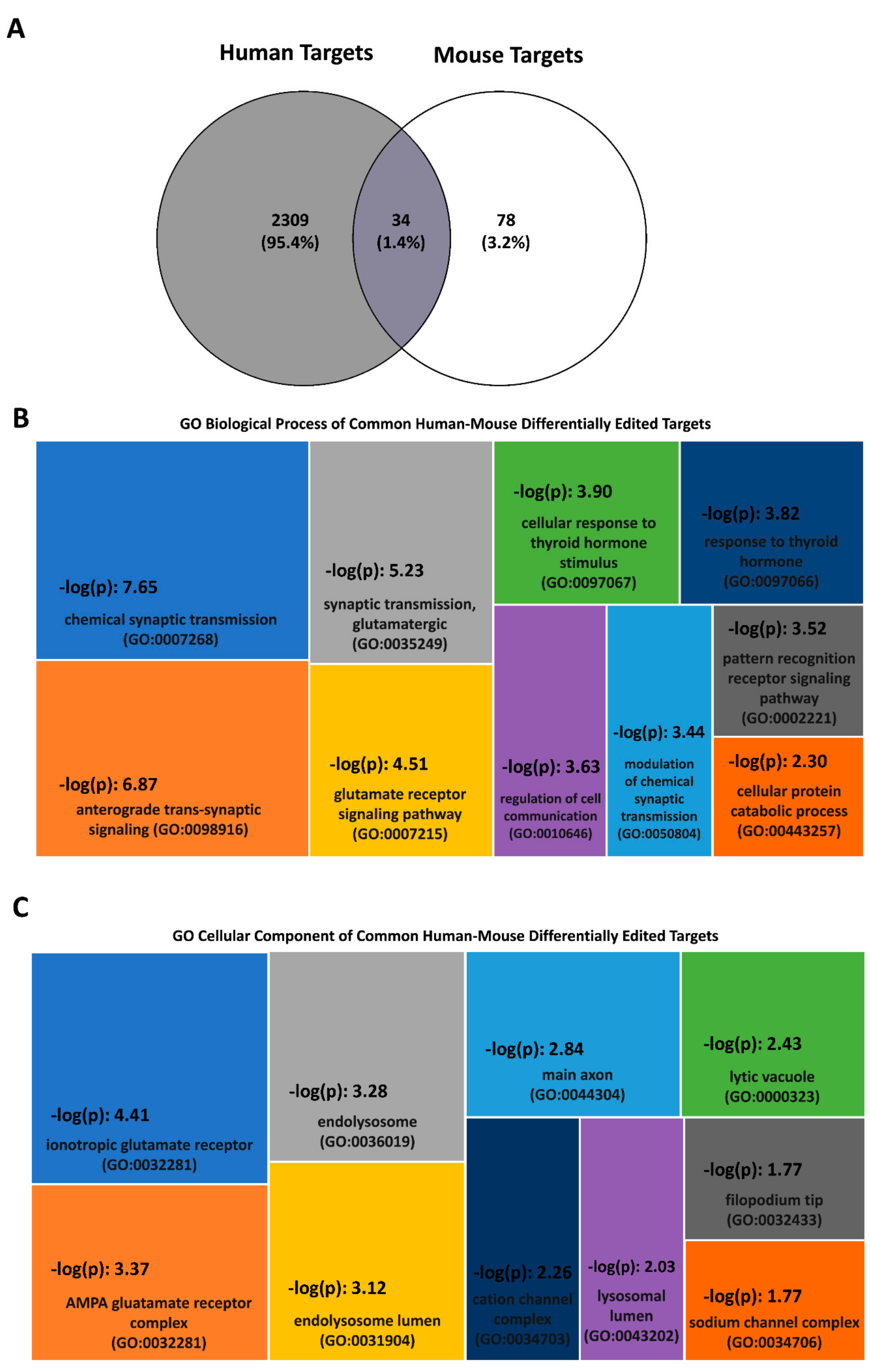
Indeed, a comparative analysis of differentially edited targets detected in at least one of the human and mouse neurological/neurodegenerative and psychiatric disorders studied (data obtained from [31,35,43,51,57]) identified overlaps between species (Figure 3A). Specifically, 34 transcripts were reported as similarly differentially edited in at least one of the studied disorders in both human and mice (Abi2, Agbl4, Ankrd28, Arf3, Bri3bp, Cds2, Copa, Cpe, Ctsb, Ctss, Dnajc18, Fam107a, Gabra3, Gnl3l, Gria2, Gria4, Grik2, Hspa4l, Itm2b, Mbp, Mdga2, Meg3, Nova1, Nup155, Paqr8, Rragd, Samd8, Scn1b, Slc1a2, Slc35e1, Smim14, Sparcl1, Spock2, Tcp11l1). Interestingly, these targets are enriched in biological processes related to synaptic transmission and signaling, the regulation of cell communication, pattern recognition receptor signaling, response to thyroid signaling and protein catabolism (Figure 3B), thus confirming the involvement of RNA editing in common disease-related processes. Synaptic processes, representing the major molecular hallmark of CNS disorders, are the most (affected) represented functions. Moreover, a cellular component analysis revealed enrichment in the terms ‘main axon’, ‘glutamate receptor’, ‘cation’, ‘sodium channel complexes’, ‘lytic vacuoles’, ‘endo/lysosome lumen’, and ‘filipodium tip’, thus providing more detailed insights into RNA editing contribution to disease (Figure 3C).
These data highlight common disease-related processes in human and mice and support the utility of RNA editing studies in murine disease models, provided that species-specific differences possibly affecting editing patterns. However, an experimental cross-validation of novel differentially edited targets should be performed in human autopsy tissue.
Of note, all of the above processes, determined at end-point (human) or significantly advanced (mice) disease stages, possibly reflect common end-stage disease consequences rather than disease promoting events. When similar studies would be conducted at earlier disease stages will significantly aid towards the identification of disease driver/promoting RNA editing events. Thus, pre-clinical studies utilizing animal models are highly needed. To our knowledge, there is currently only one published study reporting pre-clinical disease alterations of RNA editing patterns [57]. This study reported differentially edited transcripts potentially involved in early synaptic dysfunction in Prion diseases.

4. Discussion

4.1. The Diagnostic/Prognostic Potential of RNA Editing in Neurological/Neurodegenerative and Psychiatric Disorders

Altered RNA editing profiles within Alu regions in the brain of individuals with glioblastoma were suggested as a prognostic factor in a gender-dependent manner and decreases in global A-I editing profiles were reported as an efficient patient stratification approach in this disease [82]. Moreover, the loss of editing in GABRA3 was associated with an aggressive phenotype of glioma, promoting migration and invasion [82].
Peripheral blood analysis from healthy individuals, detected more than 1000 RNA editing sites [83]. Moreover, increased A-I editing within the AluSx+ sequence, which is in the Ctss 3′UTR and affects Ctss expression, was reported in peripheral blood mononuclear cells of rheumatoid arthritis (RA) patients [84]. The same authors also identified altered editing as a response to drug treatment [84]. Furthermore, a recent review discusses the possibility of exploiting RNA editing biomarkers for drug development against suicidal ideation [85].
The above data suggest that RNA editing profile studies in body fluids may enhance disease diagnosis as well as patient responses to treatments. Indeed, a recent study reports characteristic PDE8A RNA editing patterns in the blood of depressed patients and suicide attempters with major depression, suggesting its diagnostic potential [86]. In addition, a recent review of biomarkers for ALS and other NDs suggested altered RNA editing efficiencies in ADAR2-dependent sites as candidate biomarkers [87]. Furthermore, a recent transcriptome-wide RNA editing analysis performed on peripheral blood from a mutli-ethnic group of AD patients and corresponding control samples, identified differentially edited targets converging in endocytic and inflammatory pathways [88]. This further supports the diagnostic potential of RNA editing studies of body fluids. However, even though RNA editing may hold a promising diagnostic and prognostic potential, additional studies are required.

4.2. Future Perspectives and Challenges in the RNA Editing Research Field

The existing literature indicates RNA editing as an emerging mechanism that contributes to disease progression and highlights common and disease-specific editing patterns.
The advent of transcriptome-wide studies has enabled a comprehensive profiling of RNA editing alterations in health and disease conditions. Further studies focusing on different disorders are expected to extend the so-far-limited data on transcriptome-wide and descriptive studies of RNA editing. A significant challenge in this field refers to the accurate identification of true RNA editing events, which is based on comparative analyses of RNA-seq data aligned against the corresponding reference genome for the detection of RNA:DNA differences (RDDs). Both experimental setup and data analysis pipelines affect the accuracy and efficiency of identifying RNA editing events. Accurate alignment in the reference genome was identified as a critical step influencing the correct calling of RNA editing, and several alignment approaches were reported (some of them reviewed in [89]). False-positive RDDs detection is an inherent pitfall of RNA editing analysis approaches, and extensive research has aimed to develop improved data analysis pipelines that incorporate several filtering steps to limit false discovery rates. These filtering steps refer to stringent quality-control criteria of base quality and read depth; the exclusion of reads mapping to multiple genomic loci, pseudogenes and known SNPs; and the application of statistical analyses to evaluate editing calling efficiency and estimate false discovery rates. VarScan [90], RediTools [14,91], GIREMI [92], SPRINT [93], JACUSA [94] and RNAEditor [95] are some of the currently available RNA editing calling tools, of which the efficiency of some has recently been evaluated using both simulated and real data sets [89].
Importantly, and considering that RNA editing is tissue- and cell-type-specific, it is plausible to assume that refined studies focusing on specific cell sub-types or even on single-cell analyses, will help to unravel cell-type-specific, disease-related differences that may be undetectable in bulk tissue analyses. In this direction, few recent studies have reported cell-type-specific editing patterns in the human brain [96], murine macrophages and dendritic cells [73].
Further improvements of existing pipelines to allow for a more efficient analysis of editing events residing in long non-coding RNAs [97], and the development of algorithms for identification of RNA editing in miRNAs [98], have also been reported. Following accurate RNA editing identification, powerful statistical analysis approaches are required for the classification and ranking of RNA editing events, and for the identification of affected pathways and networks, which would allow for the selection of the most potent editing events for further functional analyses. Integrative approaches entailing multi-omics, neuropathological and cognitive status data significantly contribute to this direction. In addition, robust predictions of the functional effects of RNA editing on gene expression, protein recoding, miRNA binding and alternative splicing is highly desired. Advanced bioinformatics tools for such functional predictions applied to large-scale data, as recently reported for AD [99], will significantly contribute towards this direction.
Functional studies to delineate factors leading to altered editing and the estimation of RNA editing effects are also required. For the study of the functional effects of RNA editing, appropriate in vivo (e.g., editing deficient animal models [71,73]) and in vitro systems [100] are used. Further, novel, recently developed experimental approaches allowing site-directed A-I [101,102,103] and C-U [104,105] RNA editing modification may be utilized for functional RNA editing studies; however, further optimization is required. These functional studies should also consider the additional complication of evaluating different editing-related isoforms occurring as the result of the concomitant occurrence of several editing events in proximity within a selected target. Another important aspect in the field of RNA editing in relation to disease condition(s) refers to the delineation of RNA editing modulatory mechanisms driving or resulting in the observed RNA editing aberrations. To this end, several studies have attempted to correlate RNA editing alterations with the expression levels of the main RNA-editing mediating enzymes. The increased expression of APOBEC transcripts was reported in epilectic mice compared to controls, while ADAR1 and ADAR2 were identified as downregulated [31]. In addition, a recently published study, entailing many AD cases and controls, reported reduced ADAR2 expression in AD [55]. Interestingly, the same study identified increased ADAR3 expression; ADAR3 represents a potential RNA editing inhibitor, which blocks ADAR1 or ADAR2 catalytic activity through heterodimerization with either ADAR1 or ADAR2. In contrast, no significant alterations in ADAR1 or ADAR2 expression levels were detected in autism and C9orf72 ALS [45,51]. However, other RNA editing modulatory mechanisms have been suggested in these disorders. In the autism-related study, the authors provided experimental evidence on protein–protein interactions between FMRP-ADAR1, FMRP-ADAR2 and FXR1P-ADAR1, which directly affect RNA editing at selected transcripts, thus assigning RNA editing modulatory effects to FMRP (enhancer) and FXR1P (repressor) [45]. Moreover, widespread RNA editing alterations reported in familial ALS cases were associated with ADAR2 mislocalization, as evidenced by studies in postmortem tissues of ALS patients with C9orf72 mutations, as well as in vitro and in vivo disease models [51]. These studies highlight some of the molecular mechanisms resulting in altered RNA editing in different disease contexts. By utilizing advanced bioinformatics and technological approaches, additional RNA modulatory mechanisms are expected to be discovered and validated, thus contributing to a better understanding of molecular links between altered RNA editing and disease.

5. Conclusions

In summary, RNA editing studies provide significant insights in terms of CNS disease-related molecular mechanisms, suggesting a protective role in disease progression, which could be exploited in subsequent diagnostic/prognostic and even therapeutic approaches. The currently available data are mostly descriptive and further studies are required to extend the global editing profiles for different CNS disorders. Comprehensive analyses through the development of more efficient tools for RNA editing identification, classification, ranking, and functional characterization are being developed and are expected to allow for a better understanding of the role of RNA editing in these disorders.

Author Contributions

K.K., S.P., G.C., E.K., N.B., K.X.: conducted research on the literature and wrote the manuscript; D.D., T.S.: designed and wrote the manuscript. All authors have read and agreed to the published version of the manuscript.

Funding

This work was funded by (a) the Action RESEARCH—CREATE—INNOVATE, co-funded by the European Regional Development Fund of the European Union and National Recourses through the OP Competitiveness, Entrepreneurship and Innovation (EPANEK) (project code: T1EDK-03884) and (b) the Hellenic Foundation for Research and Innovation (H.F.R.I) under the “2nd Call for H.F.R.I Research Projects to support Post-Doctoral Researchers” (project number: 1146).

Conflicts of Interest

The authors declare no conflict of interest.

References

  1. Farajollahi, S.; Maas, S. Molecular diversity through RNA editing: A balancing act. Trends Genet. 2010, 26, 221–230. [Google Scholar] [CrossRef] [Green Version]
  2. Licht, K.; Jantsch, M.F. Rapid and dynamic transcriptome regulation by RNA editing and RNA modifications. J. Cell Biol. 2016, 213, 15–22. [Google Scholar] [CrossRef] [PubMed] [Green Version]
  3. Huntley, M.A.; Lou, M.; Goldstein, L.D.; Lawrence, M.; Dijkgraaf, G.J.P.; Kaminker, J.S.; Gentleman, R. Complex regulation of ADAR-mediated RNA-editing across tissues. BMC Genom. 2016, 17, 61. [Google Scholar] [CrossRef] [PubMed] [Green Version]
  4. Daniel, C.; Lagergren, J.; Öhman, M. RNA editing of non-coding RNA and its role in gene regulation. Biochimie 2015, 117, 22–27. [Google Scholar] [CrossRef] [PubMed]
  5. Deffit, S.N.; Hundley, H.A. To edit or not to edit: Regulation of ADAR editing specificity and efficiency. Wiley Interdiscip. Rev. RNA 2016, 7, 113–127. [Google Scholar] [CrossRef]
  6. Blanc, V.; Davidson, N.O. APOBEC-1-mediated RNA editing. Wiley Interdiscip Rev. Syst. Biol. Med. 2010, 2, 594–602. [Google Scholar] [CrossRef] [Green Version]
  7. Sharma, S.; Patnaik, S.K.; Taggart, R.T.; Kannisto, E.D.; Enriquez, S.M.; Gollnick, P.; Baysal, B.E. APOBEC3A cytidine deaminase induces RNA editing in monocytes and macrophages. Nat. Commun. 2015, 6, 6881. [Google Scholar] [CrossRef]
  8. Sharma, S.; Wang, J.; Alqassim, E.; Portwood, S.; Gomez, E.C.; Maguire, O.; Basse, P.H.; Wang, E.S.; Segal, B.H.; Baysal, B.E. Mitochondrial hypoxic stress induces widespread RNA editing by APOBEC3G in natural killer cells. Genome Biol. 2019, 20, 37. [Google Scholar] [CrossRef] [Green Version]
  9. Nevo-Caspi, Y.; Amariglio, N.; Rechavi, G.; Paret, G. A-to-I RNA Editing is Induced upon Hypoxia. Shock 2011, 35, 585–589. [Google Scholar] [CrossRef]
  10. Wedekind, J.E.; Dance, G.S.; Sowden, M.; Smith, H.C. Messenger RNA editing in mammals: New members of the APOBEC family seeking roles in the family business. Trends Genet. 2003, 19, 207–216. [Google Scholar] [CrossRef]
  11. Roberts, S.A.; Lawrence, M.S.; Klimczak, L.J.; Grimm, S.A.; Fargo, D.; Stojanov, P.; Kiezun, A.; Kryukov, G.; Carter, S.L.; Saksena, G.; et al. An APOBEC cytidine deaminase mutagenesis pattern is widespread in human cancers. Nat. Genet. 2013, 45, 970–976. [Google Scholar] [CrossRef] [PubMed]
  12. Rice, G.I.; Kasher, P.R.; Forte, G.M.A.; Mannion, N.M.; Greenwood, S.M.; Szynkiewicz, M.; Dickerson, J.E.; Bhaskar, S.S.; Zampini, M.; Briggs, T.A.; et al. Mutations in ADAR1 cause AGS with Type 1 IFN signature. Nat. Genet. 2012, 44, 1243–1248. [Google Scholar] [CrossRef] [PubMed] [Green Version]
  13. Danecek, P.; Nellaker, C.; McIntyre, R.E.; Buendia-Buendia, J.E.; Bumpstead, S.; Ponting, C.P.; Flint, J.; Durbin, R.; Keane, T.M.; Adams, D.J. High levels of RNA-editing site conservation amongst 15 laboratory mouse strains. Genome Biol. 2012, 13, 26. [Google Scholar] [CrossRef] [PubMed] [Green Version]
  14. Picardi, E.; Manzari, C.; Mastropasqua, F.; Aiello, I.; D’Erchia, A.M.; Pesole, G. Profiling RNA editing in human tissues: Towards the inosinome Atlas. Sci. Rep. 2015, 5, 14941. [Google Scholar] [CrossRef] [Green Version]
  15. Behm, M.; Öhman, M. RNA Editing: A Contributor to Neuronal Dynamics in the Mammalian Brain. Trends Genet. 2016, 32, 165–175. [Google Scholar] [CrossRef]
  16. Ekdahl, Y.; Farahani, H.S.; Behm, M.; Lagergren, J.; Öhman, M. A-to-I editing of microRNAs in the mammalian brain increases during development. Genome Res. 2012, 22, 1477–1487. [Google Scholar] [CrossRef]
  17. Li, J.B.; Church, G.M. Deciphering the functions and regulation of brain-enriched A-to-I RNA editing. Nat. Neurosci. 2013, 16, 1518–1522. [Google Scholar] [CrossRef] [Green Version]
  18. Sanjana, N.E.; Levanon, E.Y.; Hueske, E.A.; Ambrose, J.M.; Li, J.B. Activity-Dependent A-to-I RNA Editing in Rat Cortical Neurons. Genetics 2012, 192, 281–287. [Google Scholar] [CrossRef] [Green Version]
  19. Ball, S.M.; Atlason, P.T.; Shittu-Balogun, O.O.; Molnár, E. Assembly and intracellular distribution of kainate receptors is determined by RNA editing and subunit composition. J. Neurochem. 2010, 114, 1805–1818. [Google Scholar] [CrossRef]
  20. Daniel, C.; Wahlstedt, H.; Ohlson, J.; Björk, P.; Öhman, M. Adenosine-to-Inosine RNA Editing Affects Trafficking of the γ-Aminobutyric Acid Type A (GABAA) Receptor. J. Biol. Chem. 2011, 286, 2031–2040. [Google Scholar] [CrossRef] [Green Version]
  21. Huang, H.; Tan, B.Z.; Shen, Y.; Tao, J.; Jiang, F.; Sung, Y.Y.; Ng, C.K.; Raida, M.; Köhr, G.; Higuchi, M.; et al. RNA Editing of the IQ Domain in Cav1.3 Channels Modulates Their Ca2+-Dependent Inactivation. Neuron 2012, 73, 304–316. [Google Scholar] [CrossRef] [PubMed] [Green Version]
  22. Bazzazi, H.; Johny, M.B.; Adams, P.J.; Soong, T.W.; Yue, D.T. Continuously Tunable Ca2+ Regulation of RNA-Edited CaV1.3 Channels. Cell Rep. 2013, 5, 367–377. [Google Scholar] [CrossRef] [PubMed] [Green Version]
  23. Irimia, M.; Denuc, A.; Ferrán, J.L.; Pernaute, B.; Puelles, L.; Roy, S.W.; Garcia-Fernàndez, J.; Marfany, G. Evolutionarily conserved A-to-I editing increases protein stability of the alternative splicing factorNova1. RNA Biol. 2012, 9, 12–21. [Google Scholar] [CrossRef] [PubMed] [Green Version]
  24. Jensen, K.B.; Dredge, B.K.; Stefani, G.; Zhong, R.; Buckanovich, R.J.; Okano, H.J.; Yang, Y.Y.; Darnell, R.B. Nova-1 Regulates Neuron-Specific Alternative Splicing and Is Essential for Neuronal Viability. Neuron 2000, 25, 359–371. [Google Scholar] [CrossRef] [Green Version]
  25. Singh, M. Dysregulated A to I RNA editing and non-coding RNAs in neurodegeneration. Front. Genet. 2013, 3, 326. [Google Scholar] [CrossRef] [Green Version]
  26. Page, M.J.; McKenzie, J.E.; Bossuyt, P.M.; Boutron, I.; Hoffmann, T.C.; Mulrow, C.D.; Shamseer, L.; Tetzlaff, J.M.; Akl, E.A.; Brennan, S.E.; et al. The PRISMA 2020 statement: An updated guideline for reporting systematic reviews. Int. J. Surg. 2021, 88, 105906. [Google Scholar] [CrossRef]
  27. Grigorenko, E.V.; Bell, W.L.; Glazier, S.; Pons, T.; Deadwyler, S. Editing status at the Q/R site of the GluR2 and GluR6 glutamate receptor subunits in the surgically excised hippocampus of patients with refractory epilepsy. Neuroreport 1998, 9, 2219–2224. [Google Scholar] [CrossRef]
  28. Kortenbruck, G.; Berger, E.; Speckmann, E.-J.; Musshoff, U. RNA Editing at the Q/R Site for the Glutamate Receptor Subunits GLUR2, GLUR5, and GLUR6 in Hippocampus and Temporal Cortex from Epileptic Patients. Neurobiol. Dis. 2001, 8, 459–468. [Google Scholar] [CrossRef] [Green Version]
  29. Vollmar, W.; Gloger, J.; Berger, E.; Kortenbruck, G.; Köhling, R.; Speckmann, E.-J.; Musshoff, U. RNA editing (R/G site) and flip–flop splicing of the AMPA receptor subunit GluR2 in nervous tissue of epilepsy patients. Neurobiol. Dis. 2004, 15, 371–379. [Google Scholar] [CrossRef]
  30. Krestel, H.; Raffel, S.; von Lehe, M.; Jagella, C.; Moskau-Hartmann, S.; Becker, A.; Elger, C.E.; Seeburg, P.H.; Nirkko, A. Differences between RNA and DNA due to RNA editing in temporal lobe epilepsy. Neurobiol. Dis. 2013, 56, 66–73. [Google Scholar] [CrossRef]
  31. Srivastava, P.K.; Bagnati, M.; Delahaye-Duriez, A.; Ko, J.-H.; Rotival, M.; Langley, S.R.; Shkura, K.; Mazzuferi, M.; Danis, B.; Van Eyll, J.; et al. Genome-wide analysis of differential RNA editing in epilepsy. Genome Res. 2017, 27, 440–450. [Google Scholar] [CrossRef] [PubMed] [Green Version]
  32. Akbarian, S.; Smith, M.A.; Jones, E.G. Editing for an AMPA receptor subunit RNA in prefrontal cortex and striatum in Alzheimer’s disease, Huntington’s disease and schizophrenia. Brain Res. 1995, 699, 297–304. [Google Scholar] [CrossRef]
  33. Sodhi, M.S.; Burnet, P.W.J.; Makoff, A.J.; Kerwin, R.W.; Harrison, P.J. RNA editing of the 5-HT2C receptor is reduced in schizophrenia. Mol. Psychiatry 2001, 6, 373–379. [Google Scholar] [CrossRef] [Green Version]
  34. Dracheva, S.; Elhakem, S.L.; Marcus, S.M.; Siever, L.J.; McGurk, S.R.; Haroutunian, V. RNA editing and alternative splicing of human serotonin 2C receptor in schizophrenia. J. Neurochem. 2003, 87, 1402–1412. [Google Scholar] [CrossRef] [PubMed]
  35. Breen, M.S.; Dobbyn, A.; Li, Q.; Roussos, P.; Hoffman, G.E.; Stahl, E.; Chess, A.; Sklar, P.; Li, J.B.; Devlin, B.; et al. Global landscape and genetic regulation of RNA editing in cortical samples from individuals with schizophrenia. Nat. Neurosci. 2019, 22, 1402–1412. [Google Scholar] [CrossRef]
  36. Niswender, C.M.; Herrick-Davis, K.; Dilley, G.E.; Meltzer, H.Y.; Overholser, J.C.; Stockmeier, C.A.; Emeson, R.B.; Sanders-Bush, E. RNA Editing of the Human Serotonin 5-HT2C Receptor Alterations in Suicide and Implications for Serotonergic Pharmacotherapy. Neuropsychopharmacology 2001, 24, 478–491. [Google Scholar] [CrossRef]
  37. Lyddon, R.; Dwork, A.J.; Keddache, M.; Siever, L.J.; Dracheva, S. Serotonin 2c receptor RNA editing in major depression and suicide. World J. Biol. Psychiatry 2013, 14, 590–601. [Google Scholar] [CrossRef] [Green Version]
  38. Di Narzo, A.F.; Kozlenkov, A.; Roussos, P.; Hao, K.; Hurd, Y.; Lewis, D.A.; Sibille, E.; Siever, L.J.; Koonin, E.; Dracheva, S. A unique gene expression signature associated with serotonin 2C receptor RNA editing in the prefrontal cortex and altered in suicide. Hum. Mol. Genet. 2014, 23, 4801–4813. [Google Scholar] [CrossRef] [Green Version]
  39. Weissmann, D.; Van Der Laan, S.; Underwood, M.D.; Salvetat, N.; Cavarec, L.; Vincent, L.; Molina, F.; Mann, J.J.; Arango, V.; Pujol, J.F. Region-specific alterations of A-to-I RNA editing of serotonin 2c receptor in the cortex of suicides with major depression. Transl. Psychiatry 2016, 6, e878. [Google Scholar] [CrossRef]
  40. Gurevich, I.; Tamir, H.; Arango, V.; Dwork, A.J.; Mann, J.J.; Schmauss, C. Altered Editing of Serotonin 2C Receptor Pre-mRNA in the Prefrontal Cortex of Depressed Suicide Victims. Neuron 2002, 34, 349–356. [Google Scholar] [CrossRef] [Green Version]
  41. Iwamoto, K.; Kato, T. RNA editing of serotonin 2C receptor in human postmortem brains of major mental disorders. Neurosci. Lett. 2003, 346, 169–172. [Google Scholar] [CrossRef]
  42. Chimienti, F.; Cavarec, L.; Vincent, L.; Salvetat, N.; Arango, V.; Underwood, M.D.; Mann, J.J.; Pujol, J.-F.; Weissmann, D. Brain region-specific alterations of RNA editing in PDE8A mRNA in suicide decedents. Transl. Psychiatry 2019, 9, 91. [Google Scholar] [CrossRef] [PubMed]
  43. Dick, A.L.W.; Khermesh, K.; Paul, E.; Stamp, F.; Levanon, E.Y.; Chen, A. Adenosine-to-Inosine RNA Editing within Corticolimbic Brain Regions Is Regulated in Response to Chronic Social Defeat Stress in Mice. Front. Psychiatry 2019, 10, 277. [Google Scholar] [CrossRef] [PubMed] [Green Version]
  44. Eran, A.; Li, J.B.; Vatalaro, K.; McCarthy, J.; Rahimov, F.; Collins, C.; Markianos, K.; Margulies, D.M.; Brown, E.N.; Calvo, S.E.; et al. Comparative RNA editing in autistic and neurotypical cerebella. Mol. Psychiatry 2013, 18, 1041–1048. [Google Scholar] [CrossRef] [Green Version]
  45. Tran, S.S.; Jun, H.-I.; Bahn, J.H.; Azghadi, A.; Ramaswami, G.; Van Nostrand, E.L.; Nguyen, T.B.; Hsiao, Y.-H.E.; Lee, C.; Pratt, G.A.; et al. Widespread RNA editing dysregulation in brains from autistic individuals. Nat. Neurosci. 2019, 22, 25–36. [Google Scholar] [CrossRef]
  46. Takuma, H.; Kwak, S.; Yoshizawa, T.; Kanazawa, I. Reduction of GluR2 RNA editing, a molecular change that increases calcium influx through AMPA receptors, selective in the spinal ventral gray of patients with amyotrophic lateral sclerosis. Ann. Neurol. 1999, 46, 806–815. [Google Scholar] [CrossRef]
  47. Kawahara, Y.; Ito, K.; Sun, H.; Aizawa, H.; Kanazawa, I.; Kwak, S. RNA editing and death of motor neurons. Nature 2004, 427, 801. [Google Scholar] [CrossRef]
  48. Flomen, R.; Makoff, A. Increased RNA editing in EAAT2 pre-mRNA from amyotrophic lateral sclerosis patients: Involvement of a cryptic polyadenylation site. Neurosci. Lett. 2011, 497, 139–143. [Google Scholar] [CrossRef]
  49. Hideyama, T.; Yamashita, T.; Aizawa, H.; Tsuji, S.; Kakita, A.; Takahashi, H.; Kwak, S. Profound downregulation of the RNA editing enzyme ADAR2 in ALS spinal motor neurons. Neurobiol. Dis. 2012, 45, 1121–1128. [Google Scholar] [CrossRef]
  50. D’Erchia, A.M.; Gallo, A.; Manzari, C.; Raho, S.; Horner, D.S.; Chiara, M.; Valletti, A.; Aiello, I.; Mastropasqua, F.; Ciaccia, L.; et al. Massive transcriptome sequencing of human spinal cord tissues provides new insights into motor neuron degeneration in ALS. Sci. Rep. 2017, 7, 10046. [Google Scholar] [CrossRef]
  51. Moore, S.; Alsop, E.; Lorenzini, I.; Starr, A.; Rabichow, B.E.; Mendez, E.; Levy, J.L.; Burciu, C.; Reiman, R.; Chew, J.; et al. ADAR2 mislocalization and widespread RNA editing aberrations in C9orf72-mediated ALS/FTD. Acta Neuropathol. 2019, 138, 49–65. [Google Scholar] [CrossRef] [PubMed]
  52. Gaisler-Salomon, I.; Kravitz, E.; Feiler, Y.; Safran, M.; Biegon, A.; Amariglio, N.; Rechavi, G. Hippocampus-specific deficiency in RNA editing of GluA2 in Alzheimer’s disease. Neurobiol. Aging 2014, 35, 1785–1791. [Google Scholar] [CrossRef] [PubMed]
  53. Khermesh, K.; D’Erchia, A.M.; Barak, M.; Annese, A.; Wachtel, C.; Levanon, E.Y.; Picardi, E.; Eisenberg, E. Reduced levels of protein recoding by A-to-I RNA editing in Alzheimer’s disease. RNA 2016, 22, 290–302. [Google Scholar] [CrossRef] [PubMed] [Green Version]
  54. Annese, A.; Manzari, C.; Lionetti, C.; Picardi, E.; Horner, D.S.; Chiara, M.; Caratozzolo, M.F.; Tullo, A.; Fosso, B.; Pesole, G.; et al. Whole transcriptome profiling of Late-Onset Alzheimer’s Disease patients provides insights into the molecular changes involved in the disease. Sci. Rep. 2018, 8, 4282. [Google Scholar] [CrossRef] [PubMed] [Green Version]
  55. Ma, Y.; Dammer, E.B.; Felsky, D.; Duong, D.M.; Klein, H.-U.; White, C.C.; Zhou, M.; Logsdon, B.A.; McCabe, C.; Xu, J.; et al. Atlas of RNA editing events affecting protein expression in aged and Alzheimer’s disease human brain tissue. Nat. Commun. 2021, 12, 7035. [Google Scholar] [CrossRef]
  56. Kiesel, P.; Bodemer, W.; Gibson, T.; Zischler, H.; Kaup, F.-J. Prion infected rhesus monkeys to study differential transcription of Alu DNA elements and editing of Alu transcripts in neuronal cells and blood cells. J. Med. Primatol. 2012, 41, 176–182. [Google Scholar] [CrossRef]
  57. Kanata, E.; Llorens, F.; Dafou, D.; Dimitriadis, A.; Thüne, K.; Xanthopoulos, K.; Bekas, N.; Espinosa, J.C.; Schmitz, M.; Marín-Moreno, A.; et al. RNA editing alterations define manifestation of prion diseases. Proc. Natl. Acad. Sci. USA 2019, 116, 19727–19735. [Google Scholar] [CrossRef] [Green Version]
  58. Higuchi, M.; Maas, S.; Single, F.N.; Hartner, J.C.; Rozov, A.; Burnashev, N.; Feldmeyer, D.; Sprengel, R.; Seeburg, P.H. Point mutation in an AMPA receptor gene rescues lethality in mice deficient in the RNA-editing enzyme ADAR2. Nature 2000, 406, 78–81. [Google Scholar] [CrossRef]
  59. Hosaka, T.; Tsuji, H.; Kwak, S. RNA Editing: A New Therapeutic Target in Amyotrophic Lateral Sclerosis and Other Neurological Diseases. Int. J. Mol. Sci. 2021, 22, 10958. [Google Scholar] [CrossRef]
  60. Yamashita, T.; Hideyama, T.; Hachiga, K.; Teramoto, S.; Takano, J.; Iwata, N.; Saido, T.C.; Kwak, S. A role for calpain-dependent cleavage of TDP-43 in amyotrophic lateral sclerosis pathology. Nat. Commun. 2012, 3, 1307. [Google Scholar] [CrossRef] [Green Version]
  61. Yamashita, T.; Kwak, S. The molecular link between inefficient GluA2 Q/R site-RNA editing and TDP-43 pathology in motor neurons of sporadic amyotrophic lateral sclerosis patients. Brain Res. 2014, 1584, 28–38. [Google Scholar] [CrossRef] [PubMed] [Green Version]
  62. Aizawa, H.; Sawada, J.; Hideyama, T.; Yamashita, T.; Katayama, T.; Hasebe, N.; Kimura, T.; Yahara, O.; Kwak, S. TDP-43 pathology in sporadic ALS occurs in motor neurons lacking the RNA editing enzyme ADAR2. Acta Neuropathol. 2010, 120, 75–84. [Google Scholar] [CrossRef] [PubMed]
  63. Brusa, R.; Zimmermann, F.; Koh, D.S.; Feldmeyer, D.; Gass, P.; Seeburg, P.H.; Sprengel, R. Early-Onset Epilepsy and Postnatal Lethality Associated with an Editing-Deficient GluR-B Allele in Mice. Science 1995, 270, 1677–1680. [Google Scholar] [CrossRef] [PubMed]
  64. Streit, A.K.; Derst, C.; Wegner, S.; Heinemann, U.; Zahn, R.K.; Decher, N. RNA editing of Kv1.1 channels may account for reduced ictogenic potential of 4-aminopyridine in chronic epileptic rats. Epilepsia 2011, 52, 645–648. [Google Scholar] [CrossRef]
  65. Bernard, A.; Ferhat, L.; Dessi, F.; Charton, G.; Represa, A.; Ben-Ari, Y.; Khrestchatisky, M. Q/R editing of the rat GluR5 and GluR6 kainate receptors in vivo and in vitro: Evidence for independent developmental, pathological and cellular regulation. Eur. J. Neurosci. 1999, 11, 604–616. [Google Scholar] [CrossRef]
  66. Ferrick-Kiddie, E.A.; Rosenthal, J.J.C.; Ayers, G.D.; Emeson, R.B. Mutations underlying Episodic Ataxia type-1 antagonize Kv1.1 RNA editing. Sci. Rep. 2017, 7, 41095. [Google Scholar] [CrossRef] [Green Version]
  67. Ramaswami, G.; Li, J.B. RADAR: A rigorously annotated database of A-to-I RNA editing. Nucleic Acids Res. 2014, 42, D109–D113. [Google Scholar] [CrossRef] [Green Version]
  68. Kiran, A.; Baranov, P.V. DARNED: A DAtabase of RNa EDiting in humans. Bioinformatics 2010, 26, 1772–1776. [Google Scholar] [CrossRef] [PubMed] [Green Version]
  69. Picardi, E.; D’Erchia, A.M.; Lo Giudice, C.; Pesole, G. REDIportal: A comprehensive database of A-to-I RNA editing events in humans. Nucleic Acids Res. 2017, 45, D750–D757. [Google Scholar] [CrossRef] [Green Version]
  70. Hwang, T.; Park, C.-K.; Leung, A.K.; Gao, Y.; Hyde, T.M.; Kleinman, J.E.; Rajpurohit, A.; Tao, R.; Shin, J.H.; Weinberger, D.R. Dynamic regulation of RNA editing in human brain development and disease. Nat. Neurosci. 2016, 19, 1093–1099. [Google Scholar] [CrossRef]
  71. Cole, D.C.; Chung, Y.; Gagnidze, K.; Hajdarovic, K.H.; Rayon-Estrada, V.; Harjanto, D.; Bigio, B.; Gal-Toth, J.; Milner, T.A.; McEwen, B.S.; et al. Loss of APOBEC1 RNA-editing function in microglia exacerbates age-related CNS pathophysiology. Proc. Natl. Acad. Sci. USA 2017, 114, 13272–13277. [Google Scholar] [CrossRef] [Green Version]
  72. Rayon-Estrada, V.; Harjanto, D.; Hamilton, C.E.; Berchiche, Y.A.; Gantman, E.C.; Sakmar, T.P.; Bulloch, K.; Gagnidze, K.; Harroch, S.; McEwen, B.S.; et al. Epitranscriptomic profiling across cell types reveals associations between APOBEC1-mediated RNA editing, gene expression outcomes, and cellular function. Proc. Natl. Acad. Sci. USA 2017, 114, 13296–13301. [Google Scholar] [CrossRef] [PubMed] [Green Version]
  73. Harjanto, D.; Papamarkou, T.; Oates, C.J.; Rayon-Estrada, V.; Papavasiliou, F.N.; Papavasiliou, A. RNA editing generates cellular subsets with diverse sequence within populations. Nat. Commun. 2016, 7, 12145. [Google Scholar] [CrossRef] [PubMed] [Green Version]
  74. Rosenberg, B.R.; Hamilton, C.E.; Mwangi, M.M.; Dewell, S.; Papavasiliou, F.N. Transcriptome-wide sequencing reveals numerous APOBEC1 mRNA-editing targets in transcript 3′ UTRs. Nat. Struct. Mol. Biol. 2011, 18, 230–236. [Google Scholar] [CrossRef] [PubMed] [Green Version]
  75. Deininger, P.L. Alu elements: Know the SINEs. Genome Biol. 2011, 12, 236. [Google Scholar] [CrossRef] [PubMed] [Green Version]
  76. O’Donnell, W.T.; Warren, S.T. A Decade of Molecular Studies of Fragile X Syndrome. Annu. Rev. Neurosci. 2002, 25, 315–338. [Google Scholar] [CrossRef] [PubMed] [Green Version]
  77. Tang, G.; Gudsnuk, K.; Kuo, S.-H.; Cotrina, M.L.; Rosoklija, G.; Sosunov, A.; Sonders, M.S.; Kanter, E.; Castagna, C.; Yamamoto, A.; et al. Loss of mTOR-Dependent Macroautophagy Causes Autistic-like Synaptic Pruning Deficits. Neuron 2014, 83, 1131–1143. [Google Scholar] [CrossRef] [PubMed] [Green Version]
  78. Won, H.; Mah, W.; Kim, E. Autism spectrum disorder causes, mechanisms, and treatments: Focus on neuronal synapses. Front. Mol. Neurosci. 2013, 6, 19. [Google Scholar] [CrossRef] [Green Version]
  79. Starr, A.; Sattler, R. Synaptic dysfunction and altered excitability in C9ORF72 ALS/FTD. Brain Res. 2018, 1693, 98–108. [Google Scholar] [CrossRef]
  80. Schneider, J.L.; Miller, A.M.; Woesner, M.E. Autophagy and Schizophrenia: A Closer Look at How Dysregulation of Neuronal Cell Homeostasis Influences the Pathogenesis of Schizophrenia. Einstein J. Biol. Med. 2016, 31, 34–39. [Google Scholar] [CrossRef] [Green Version]
  81. Perera, R.M.; Zoncu, R. The Lysosome as a Regulatory Hub. Annu. Rev. Cell Dev. Biol. 2016, 32, 223–253. [Google Scholar] [CrossRef] [PubMed] [Green Version]
  82. Silvestris, D.A.; Picardi, E.; Cesarini, V.; Fosso, B.; Mangraviti, N.; Massimi, L.; Martini, M.; Pesole, G.; Locatelli, F.; Gallo, A. Dynamic inosinome profiles reveal novel patient stratification and gender-specific differences in glioblastoma. Genome Biol. 2019, 20, 33. [Google Scholar] [CrossRef] [PubMed]
  83. Giacopuzzi, E.; Gennarelli, M.; Sacco, C.; Filippini, A.; Mingardi, J.; Magri, C.; Barbon, A. Genome-wide analysis of consistently RNA edited sites in human blood reveals interactions with mRNA processing genes and suggests correlations with cell types and biological variables. BMC Genom. 2018, 19, 963. [Google Scholar] [CrossRef] [PubMed] [Green Version]
  84. Vlachogiannis, N.; Gatsiou, A.; Silvestris, D.A.; Stamatelopoulos, K.; Tektonidou, M.G.; Gallo, A.; Sfikakis, P.P.; Stellos, K. Increased adenosine-to-inosine RNA editing in rheumatoid arthritis. J. Autoimmun. 2020, 106, 102329. [Google Scholar] [CrossRef]
  85. van der Laan, S.; Salvetat, N.; Weissmann, D.; Molina, F. Emerging RNA editing biomarkers will foster drug development. Drug Discov. Today 2017, 22, 1056–1063. [Google Scholar] [CrossRef]
  86. Salvetat, N.; Chimienti, F.; Cayzac, C.; Dubuc, B.; Checa-Robles, F.; Dupre, P.; Mereuze, S.; Patel, V.; Genty, C.; Lang, J.-P.; et al. Phosphodiesterase 8A to discriminate in blood samples depressed patients and suicide attempters from healthy controls based on A-to-I RNA editing modifications. Transl. Psychiatry 2021, 11, 255. [Google Scholar] [CrossRef]
  87. Hosaka, T.; Yamashita, T.; Tamaoka, A.; Kwak, S. Extracellular RNAs as Biomarkers of Sporadic Amyotrophic Lateral Sclerosis and Other Neurodegenerative Diseases. Int. J. Mol. Sci. 2019, 20, 3148. [Google Scholar] [CrossRef] [Green Version]
  88. Gardner, O.K.; Wang, L.; Van Booven, D.; Whitehead, P.L.; Hamilton-Nelson, K.L.; Adams, L.D.; Starks, T.D.; Hofmann, N.K.; Vance, J.M.; Cuccaro, M.L.; et al. RNA editing alterations in a multi-ethnic Alzheimer disease cohort converge on immune and endocytic molecular pathways. Hum. Mol. Genet. 2019, 28, 3053–3061. [Google Scholar] [CrossRef]
  89. Diroma, M.A.; Ciaccia, L.; Pesole, G.; Picardi, E. Elucidating the editome: Bioinformatics approaches for RNA editing detection. Brief. Bioinform. 2019, 20, 436–447. [Google Scholar] [CrossRef]
  90. Koboldt, D.C.; Chen, K.; Wylie, T.; Larson, D.E.; McLellan, M.D.; Mardis, E.R.; Weinstock, G.M.; Wilson, R.K.; Ding, L. VarScan: Variant detection in massively parallel sequencing of individual and pooled samples. Bioinformatics 2009, 25, 2283–2285. [Google Scholar] [CrossRef] [Green Version]
  91. Picardi, E.; Pesole, G. REDItools: High-throughput RNA editing detection made easy. Bioinformatics 2013, 29, 1813–1814. [Google Scholar] [CrossRef] [PubMed] [Green Version]
  92. Zhang, Q.; Xiao, X. Genome sequence–independent identification of RNA editing sites. Nat. Methods 2015, 12, 347–350. [Google Scholar] [CrossRef] [PubMed] [Green Version]
  93. Zhang, F.; Lu, Y.; Yan, S.; Xing, Q.; Tian, W. SPRINT: An SNP-free toolkit for identifying RNA editing sites. Bioinformatics 2017, 33, 3538–3548. [Google Scholar] [CrossRef] [PubMed]
  94. Piechotta, M.; Wyler, E.; Ohler, U.; Landthaler, M.; Dieterich, C. JACUSA: Site-specific identification of RNA editing events from replicate sequencing data. BMC Bioinform. 2017, 18, 7. [Google Scholar] [CrossRef] [Green Version]
  95. John, D.; Weirick, T.; Dimmeler, S.; Uchida, S. RNAEditor: Easy detection of RNA editing events and the introduction of editing islands. Brief. Bioinform. 2017, 18, 993–1001. [Google Scholar] [CrossRef]
  96. Picardi, E.; Horner, D.S.; Pesole, G. Single-cell transcriptomics reveals specific RNA editing signatures in the human brain. RNA 2017, 23, 860–865. [Google Scholar] [CrossRef] [Green Version]
  97. Picardi, E.; D’Erchia, A.M.; Gallo, A.; Montalvo, A.; Pesole, G. Uncovering RNA Editing Sites in Long Non-Coding RNAs. Front. Bioeng. Biotechnol. 2014, 49, 12. [Google Scholar] [CrossRef]
  98. Zheng, Y.; Jinyan, L.; Song, R.; Wang, S.; Li, T.; Zhang, X.; Chen, K.; Li, T.; Li, J. Accurate detection for a wide range of mutation and editing sites of microRNAs from small RNA high-throughput sequencing profiles. Nucleic Acids Res. 2016, 44, e123. [Google Scholar] [CrossRef]
  99. Wu, S.; Yang, M.; Kim, P.; Zhou, X. ADeditome provides the genomic landscape of A-to-I RNA editing in Alzheimer’s disease. Brief. Bioinform. 2021, 22, bbaa384. [Google Scholar] [CrossRef]
  100. Schaffer, A.A.; Kopel, E.; Hendel, A.; Picardi, E.; Levanon, E.Y.; Eisenberg, E. The cell line A-to-I RNA editing catalogue. Nucleic Acids Res. 2020, 48, 5849–5858. [Google Scholar] [CrossRef]
  101. Merkle, T.; Merz, S.; Reautschnig, P.; Blaha, A.; Li, Q.; Vogel, P.; Wettengel, J.; Li, J.B.; Stafforst, T. Precise RNA editing by recruiting endogenous ADARs with antisense oligonucleotides. Nat. Biotechnol. 2019, 37, 133–138. [Google Scholar] [CrossRef] [PubMed]
  102. Qu, L.; Yi, Z.; Zhu, S.; Wang, C.; Cao, Z.; Zhou, Z.; Yuan, P.; Yu, Y.; Tian, F.; Liu, Z.; et al. Programmable RNA editing by recruiting endogenous ADAR using engineered RNAs. Nat. Biotechnol. 2019, 37, 1059–1069. [Google Scholar] [CrossRef] [PubMed]
  103. Rauch, S.; He, E.; Srienc, M.; Zhou, H.; Zhang, Z.; Dickinson, B.C. Programmable RNA-Guided RNA Effector Proteins Built from Human Parts. Cell 2019, 178, 122–134.e12. [Google Scholar] [CrossRef] [PubMed]
  104. Abudayyeh, O.O.; Gootenberg, J.S.; Franklin, B.; Koob, J.; Kellner, M.J.; Ladha, A.; Joung, J.; Kirchgatterer, P.; Cox, D.B.T.; Zhang, F. A cytosine deaminase for programmable single-base RNA editing. Science 2019, 365, 382–386. [Google Scholar] [CrossRef] [PubMed]
  105. Huang, X.; Lv, J.; Li, Y.; Mao, S.; Li, Z.; Jing, Z.; Sun, Y.; Zhang, X.; Shen, S.; Wang, X.; et al. Programmable C-to-U RNA editing using the human APOBEC 3A deaminase. EMBO J. 2020, 39, e104741. [Google Scholar] [CrossRef] [PubMed]
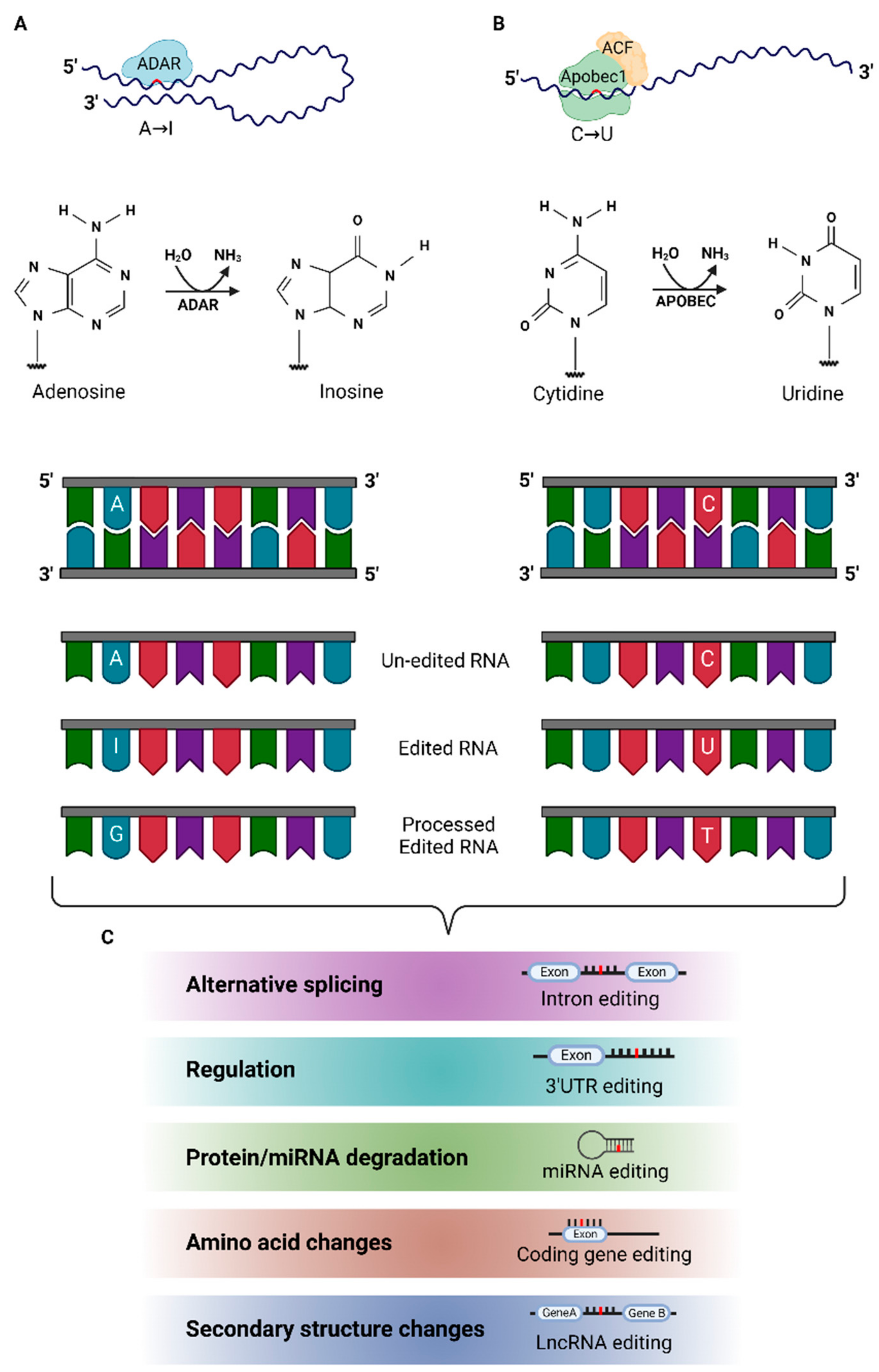
Figure 1. ADAR and APOBEC enzyme members catalyze RNA editing in mammals leading to epi-transcriptomic alterations. (A) The mammalian ADAR family comprises three members: ADAR1, ADAR2 and ADAR3. The first two are catalytically active and widely expressed; ADAR3 is expressed exclusively in the brain, has no proven catalytic activity, and is suggested to act as an ADAR1 and ADAR2 regulator. ADARs catalyze A-I editing in the form of homo- and/or heterodimers, without the requirement of other co-factors. Complementary or partially double-stranded RNAs may be used as ADARs’ substrates. Any dsRNA ≥ 20 bp, including protein-coding exons in pre-mRNAs, repetitive sequence elements, as well as microRNA (miRNA) precursor transcripts, may be ADAR substrates. ADARs deaminate Adenosine (A) to Inosine (I). The cellular transcriptional and translational machinery recognizes Inosine (I) as Guanine (G); thus, processed ADAR edited transcripts display a G at the edited site (A-I-G editing). (B) APOBEC1 is the main C-U editing enzyme in mammals. APOBEC1-mediated editing is highly specific and requires the formation of the editosome, a protein complex that comprises an enzyme homodimer, an essential co-factor (A1CF or RBM47) and auxiliary proteins that regulate enzymatic activity. APOBEC1 targets are ssRNAs and display specific sequence elements, corresponding to the mooring sequence (an 11 nt consensus sequence located downstream the C undergoing deamination, required for A1CF binding) and an AU-rich ‘efficiency region’, located upstream of the edited residue. APOBECs deaminate Cytidine (C) to Uridine (U). The cellular transcriptional and translational machinery recognizes Uridine (U) as Thymine (T); thus, processed APOBEC edited transcripts display a T at the edited site (C-U-T editing). (C) RNA editing events may occur at several sites within a transcript and affect stability, processing, and function of the edited target. Editing in intronic regions or close to splice junction sites may cause the alternative splicing of the edited transcripts. RNA editing events within non-coding regions (5′UTR, 3′UTR) may affect transcript stability and regulation. Editing events within a miRNA seed sequence may re-direct miRNA targeting and cause degradation. Introduction of non-synonymous changes within a coding region of a transcript results in amino acid alterations (protein recoding, stop codon introduction) and may also affect a transcript’s secondary structure and alter its interactions with RNA-binding proteins (RBPs). LncRNA editing can lead to changes in secondary structure affecting its regulatory functions. Figure created with BioRender.com (accessed on 31 January 2022).
Biomolecules 12 00465 g001
Figure 2. Differential editing patterns in different human neuropsychiatric, autoimmune/neurodegenerative and neurological brain disorders compared to healthy individuals. (A) Venn diagrams depicting differentially edited sites identified in schizophrenia (SCZ), genetic ALS (ALS c9orf72) and autism. The intersections represent editing events identified as differentially edited in more than one condition. Twenty-six editing sites have been commonly identified as differentially edited in all three conditions. (Β) Venn diagrams depict differentially edited transcripts (targets) in the same human disorders as in A. The intersections represent editing targets identified as differentially edited in more than one condition. Please note that a higher number of transcripts (187) are differentially edited in all three conditions compared to common, individual editing events (intersection in A), suggesting disease-associated editing that converges in common targets. (C) Gene ontology (GO) analysis of the differentially edited targets in all three human brain disorders (n = 187). Enrichment values are given as –log (p) values (higher –log (p) values indicate greater statistical significance). Differentially edited transcripts are involved in molecular processes associated with hypoxia, synaptic transmission, endosome/lysosome function, cytoskeleton, apoptosis, protein and RNA processing, all of which have been reported as deregulated in these disorders. Differential editing data (p < 0.05) reported in the studies of [35,45,51] were utilized for the analysis. Reported data correspond to different brain regions.
Biomolecules 12 00465 g002
Figure 3. Differentially edited targets in human neuropsychiatric, autoimmune/neurodegenerative, neurological disorders [35,45,51] and in neurological/neurodegenerative disease mouse models [31,43,57] compared to healthy controls. (A) Venn diagram depicting differentially edited transcripts identified in humans and mice compared to healthy controls. The intersection represents editing targets commonly identified as DE in human and mouse brain disorders. (B,C) Gene ontology (GO) analysis of the differentially edited targets in human and mouse brain disorders (n = 34). Enrichment values are given as –log (p) values (higher –log (p) values indicate greater statistical significance). (B) Biological process enrichment. Common DE transcripts are involved in molecular processes associated with synaptic transmission and signaling, regulation of cell communication, pattern recognition receptor signaling, response to thyroid signaling and protein catabolism. (C) Cellular component enrichment. Main axon, glutamate receptor, cation and sodium channel complexes, lytic vacuoles and specifically endo/lysosome lumen, as well as filipodium tip, emerge as cellular component terms enriched in DE transcripts in both human and mouse brain disorders.
Biomolecules 12 00465 g003
Table 1. Studies of RNA editing alterations in neurological and psychiatric disorders, utilizing either human postmortem or murine mouse models central nervous system (CNS) material. DE: differentially edited, SCZ: schizophrenia, CSDS: chronic social defeat stress, HPC: hippocampus, TC: temporal cortex, FC: frontal cortex, ACC: anterior cingulate cortex, DLPFC: dorsolateral prefrontal cortex, PFC: prefrontal cortex, BLA: basolateral amygdala, CB: cerebellum, RFLPS: restriction fragment length polymorphism, NGS: next-generation sequencing, CE-SSCP: capillary electrophoresis single-stranded conformational polymorphism, DHPLC: denaturing high performance liquid chromatography, mmPCR_seq: microfluidics multiplex polymerase chain reaction and deep sequencing, ns: non-significant.
DisorderSpecies/Brain RegionStudy Type/Target(s)Methodology/Validation MethodDE Targets/Trend Relative to ControlsRemarksRef.
EpilepsyHuman/HPCFocused/Grik2RFLPsGrik2: ↑ Q/R siteSuggested as a compensatory mechanism[27]
Human/TCFocused/Grik1 and Grik2RFLPsGrik1: ↑ Q/R site Grik2: ↑ Q/R siteSuggested as a compensatory mechanism[28]
Human/HPCFocused/Gria2RFLPsGria2: ↑ R/G siteSuggested as contributor in disease pathogenesis[29]
Human/HPCFocused/Gria2-4 Grik1-2, KCNA1, 5-HT2CSanger sequencingKCNA1: ↓ I/V siteInversely associated with disease duration[30]
Mouse/HPCTranscriptome-wide/whole transcriptomeNGS/Sanger sequencing19 DE targets experimentally validated, ↑ Grik2, Ctsb, Rpa, Sparc, Slc1a2, Ovca2, ↓ Ncl, Wipi2, Klhl24, Hspa4l, Slc37a3, Cyfip2DE targets involved in disease related pathways[31]
SCZHuman/PFCFocused/Gria2RFLPsGria2: ↓ Q/R sitePossible contributor to disease pathogenesis[32]
Human/FCFocused/5-HT2CCloning and Sanger sequencing5-HT2C: ↓ site B↑ Unedited isoform[33]
Human/PFCFocused/5-HT2CCloning and Sanger sequencing5-HT2C: ns site differencesTrend: ↑ unedited isoform[34]
Human/ACC/DLPFCTranscriptome-wide/whole transcriptomeNGS meta-analysis/independent (validation) cohort in silico analysis>100 DE sites per region, DE overlap between brain regions (n = 29)↑ Global editing, DE targets involved in disease associated pathways[35]
SuicideHuman/PFCFocused/5-HT2CPrimer extension5-HT2C: site A↑ Site A[36]
Human/PFCFocused/5-HT2CTargeted NGS/cloning and Sanger sequencing5-HT2C isoforms due to editing↑ ABCD isoform (hypoactive)[37]
Human/PFCFocused/5-HT2CTargeted NGS/validation cohort analysis5-HT2C edited isoforms, ↑ ABCD isoformABCD isoform associated with gene expression alterations[38]
Human/ACC/DLPFFocused/5-HT2CCE-SSCP5-HT2C edited isoforms, ACC: ↑ A, ABDE, ↓ D/DLPFC: ↑ ABRegion-specific differential representation[39]
Depression and suicideHuman/PFCFocused/5-HT2CCloning and Sanger sequencing5-HT2C: ↑ site C’ (Ε), ↓ site D [40]
Human/PFCFocused/5-HT2CPrimer extension and DHPLC/Sanger sequencing5-HT2C: sites A and D nsTrend, Depr.: ↑ D, Suicide: ↑ A[41]
Human/ACC/DLPFCFocused/PDE8ACE-SSCPPDE8A edited isoforms, ACC: ↑ ABCEF, ABC, ABEFG, BFG ↓ B, ABDE/DLPFC: ↑ ABEFG, BCEG, ↓ ABF, BEGRegion specific differential representation[42]
Human/whole bloodFocused/PDE8ATargeted NGSPDE8A: ↓ sites B, C, E and D, F ns
↓ Isoforms B, BC, BD, BE, BF
Similar patterns with the brain of suicide decedents[42]
CSDSMouse/PFC/BLAFocused/recoding in neuronal function related transcripts (551 sites)Targeted NGS (mmPCR_seq)PFC: ↑ Commd2, Rsad1, Iqgap1, Klf16, Nova1 ↓ Wipi2, Zfp81, Rn45s, Rwdd2b, Dagla, BLA: ↑ Htr2c (C, D site), Gabra3, Tcp11l1, Qpctl ↓ Zfp324, Copa, Gria4, Fubp3, Nova1Region-specific DE[43]
AutismHuman/CBFocused/synaptic transcripts (10 targets)Pyrosequencing/validation: Padlock probes and NGS (5 targets)Gria 4: ↑ R/G site, Grik2 and 5-HT2C edited isoforms differential representationGria4 editing associated with differential splicing isoform usage[44]
Human/TC, FC, CBTranscriptome-wide/whole transcriptomeNGS/2nd cohort meta-analysis/cloning and Sanger sequencing↑ Ctsb, Neat1 ↓ Gsk3b, Nova1, Grik1, FAM213A, Dennd3↓ Global editing[45]
↓ denotes reduced levels of editing, ↑ denotes increased levels of editing.
Table 2. Studies on RNA editing alterations in neurodegenerative disorders utilizing either human postmortem or murine mouse models central nervous system (CNS) material. DE: differentially edited, ALS: amyotrophic lateral sclerosis, HD: Huntington’s disease, AD: Alzheimer’s disease, vCJD: variant Creutzfeldt–Jakob disease, sCJD: sporadic Creutzfeldt–Jakob disease, SC: spinal cord, FC: frontal cortex, CB: cerebellum, ACC: anterior cingulate cortex, DLPFC: dorsolateral prefrontal cortex, PCC: posterior cingulate cortex, aPFC: anterior prefrontal cortex, pSTG: posterior superior temporal gyrus, IFGo: pars opercularis of the inferior frontal gyrus, FFG: fusiform gyrus, TC: temporal cortex, PFC: prefrontal cortex, HPC: hippocampus, RFLPS: restriction fragment length polymorphism, NGS: next-generation sequencing, hiPSC-MNs: human induced pluripotent stem cell-derived motor neurons, mmPCR_seq: microfluidics multiplex polymerase chain reaction and deep sequencing, ns: non-significant.
DisorderSpecies/Brain RegionStudy Type/Target(s)Methodology/
Validation Method
DE Targets/Trend Relative to ControlsRemarksRef.
ALSHuman/SCFocused/Gria2RFLPs/Sanger sequencingGria2: ↓ Q/R site [46]
Human/neurons $Focused/Gria2RFLPsGria2: ↓ Q/R siteNo editing changes in Purkinje cells[47]
Human/SC and motor cortexFocused/EAAT2 (astroglial glutamate transporter)Cloning and Sanger sequencingEAAT2: ↑ intron7Alternative polyadenylation and intron 7 retention transcripts (in vitro functional evidence)[48]
Human/SC neurons $Focused/Gria2RFLPsGria2: ↓ Q/R site [49]
Human/SCTranscriptome-wide/focus on database listed A-I editing sitesNGSGria2: ↓ Q/R site ns trendLow sample number, n = 5–6/group[50]
ALS (C9orf72)Human/SC, motor cortex, FC, CBTranscriptome-wide/whole transcriptomeNGS/ADAR1 and/or ADAR2 deficient hiPSC-MNs cells and cells with aberrant ADAR2 localization1526 DE transcriptsNo changes in global editing, region-specific hypo- and hyper-edited patterns[51]
HDHuman/striatumFocused/Gria2RFLPsGria2: ↓ Q/R site [32]
ADHuman/PFCFocused/Gria2RFLPsGria2: ↓ Q/R site [32]
Human/HPCFocused/Gria2Sanger sequencing/primer extensionGria2: ↓ Q/R site [52]
Human/HPC, temporal and frontal lobeFocused/recoding in synaptic transcripts (72 targets, 118 sites)Targeted NGS (mmPCR_seq)↓ 5-HT2C receptor isoforms, HPC: ↓ Cacna1d, Ddx58, Fbxl6, Fis1, Flj43663, Gria3, Gria4, Igfbp7, Kcna1, Meg3, Narf, Nova1, Ptpn14, Unc80 ↑ Copa Temporal lobe: ↓ Ccni, Fbxl6, Flj43663, Gria2, Gria4, Grik1, Grik2, Meg3, Mfn1, Tme63b, Unc80 ↑ Narf/Frontal lobe: ↓ Mfn1, Grik2, Meg3, Gria2, Unc80, Ddx58↓ Recoding[53]
Human/HPCTranscriptome-wideNGS11 DE targets, ↓ Gria2, Gria3, Gria4, Grik1, Grik2 ↑ Blcap, Copa, Vn1r1, Znf235, Znf397, Znf582↓ Recoding[54]
Human/ACC/DLPFC/PCC/aPFC/pSTG/IFGo/FFG/CB/TCTranscriptome-wide/focus on database listed A-I editing sitesNGS↓ Editing in SYT11, MCUR1, SOD2, ORAI2, HSDL2, PFKP, and GPRC5BDLPFC Samples: ↓ ADAR1 ↑ ADAR3 expression in AD cases[55]
Prion diseasessCJD and vCJD Rhesus monkeys/CBFocused/AluCloning and Sanger sequencing↓ Alu editingStrain specific differences[56]
sCJD Mouse/CortexTranscriptome-wideNGS and Sanger sequencing3 DE targets experimentally validated, Mouse pre-clinical: ↓ Sidt2, ↑ Fkrp/Mouse clinical: ↑ Rragd↓ Global editing, Human cross-validation: ↓ Paqr8, ↑ Ctss, Rrgad[57]
$ microdissected, ↓ denotes reduced levels of editing, denotes increased levels of editing.
Publisher’s Note: MDPI stays neutral with regard to jurisdictional claims in published maps and institutional affiliations.