Article

Albumin Thiolation and Oxidative Stress Status in Patients with Aortic Valve Stenosis

1
GVM Care and Research, Maria Cecilia Hospital, 48033 Cotignola, Italy
2
Dipartimento di Scienze Mediche e Chirurgiche, Alma Mater Studiorum, Università di Bologna, 40126 Bologna, Italy
3
Unit of Functional Proteomics, Metabolomics, and Network Analysis, Centro Cardiologico Monzino IRCCS, 20138 Milano, Italy
4
Department of Pharmaceutical Sciences, University of Milan, 20133 Milano, Italy
*
Author to whom correspondence should be addressed.
These authors contributed equally to this work.
Biomolecules 2023, 13(12), 1713; https://doi.org/10.3390/biom13121713
Submission received: 27 September 2023 / Revised: 8 November 2023 / Accepted: 24 November 2023 / Published: 28 November 2023

Abstract

:
Recent evidence indicates that reactive oxygen species play an important causative role in the onset and progression of valvular diseases. Here, we analyzed the oxidative modifications of albumin (HSA) occurring on Cysteine 34 and the antioxidant capacity of the serum in 44 patients with severe aortic stenosis (36 patients underwent aortic valve replacement and 8 underwent a second aortic valve substitution due to a degenerated bioprosthetic valve), and in 10 healthy donors (controls). Before surgical intervention, patients showed an increase in the oxidized form of albumin (HSA-Cys), a decrease in the native reduced form (HSA-SH), and a significant reduction in serum free sulfhydryl groups and in the total serum antioxidant activity. Patients undergoing a second valve replacement showed levels of HSA-Cys, free sulfhydryl groups, and total antioxidant activity similar to those of controls. In vitro incubation of whole blood with aspirin (ASA) significantly increased the free sulfhydryl groups, suggesting that the in vivo treatment with ASA may contribute to reducing oxidative stress. We also found that N-acetylcysteine and its amide derivative were able to regenerate HSA-SH. In conclusion, the systemic oxidative stress reflected by high levels of HSA-Cys is increased in patients with aortic valve stenosis. Thiol–disulfide breaking agents regenerate HSA-SH, thus paving the way to the use these compounds to mitigate the oxidative stress occurring in the disease.

1. Introduction

Aortic valve stenosis is a serious and complex condition that affects millions of patients worldwide and is associated with high morbidity and mortality [1]. Its prevalence is increasing as the population ages, with an incidence of more than 10% in individuals older than 65 years of age in US and European populations [2,3].
Due to the absence of pharmacological therapies available for preventing, treating, or slowing the development of aortic valve stenosis, understanding the mechanisms underlying the onset and progression of the disease is crucial for identifying novel therapeutic targets. Indeed, the treatment of severe valvular disease is based on surgical intervention (TAVR, transcatheter aortic valve replacement, or TAVI, transcatheter aortic valve implantation) in which, with a minimally invasive procedure, a dysfunctional valve is replaced by an artificial valve, such as a mechanical valve or bioprosthetic valve [4]; currently, around 300,000 artificial valves are implanted every year, a number that is set to increase further [5].
Recent findings have highlighted an important causative role for reactive oxygen species (ROS)-mediated oxidative stress in the onset and progression of valvular disease, which can contribute to the differentiation of valve interstitial cells into myofibroblasts and osteoblasts [6]. Indeed, oxidative stress, a condition derived from an imbalance between ROS production and ROS degradation, is involved in several pathological processes, such as inflammation, lipid infiltration, and calcification, all aspects that participate in cardiovascular disease, including aortic valve disease [7]. In addition, ROS induce the oxidation of several molecules, redox gene overexpression, and DNA fragmentation, all of which contribute to myocardial cell damage [8]. Proteins are important targets of ROS, which can irreversibly damage them with deleterious consequences on their function [9].
In addition to the irreversible oxidative damage, the protein function can be altered by the reversible oxidation of the thiol group of the cysteine residues, so much so that proteins containing thiol groups may be potential biomarkers of oxidative stress [10]. Among these, albumin (HSA), through the free sulfhydryl group present on cysteine residue in position 34 (Cys34), exerts antioxidant activity and protects from systemic oxidative stress [11,12,13]. Therefore, HSA represents the major and predominant antioxidant in plasma whose relative contribution was reported to range from 40% to 70% of the total antioxidant capacity. Cys34 plays the most important role in the antioxidant activity of HSA against the tested oxidant species, namely O2−●, NO, HO, HOCl, and H2O2. Furthermore, in vitro, HSA is an effective detoxifying nucleophilic agent of α-β unsaturated aldehydes generated from lipid peroxidation, including 4-hydroxy-trans-2-nonenal and other reactive carbonyls, such as glyoxal and methylglyoxal [14]. Considering its abundance, HSA and its corresponding oxidized forms represent valid and robust biomarkers of systemic oxidative stress and are predictors of the onset and development of some oxidation-based diseases. In healthy subjects, 70–80% of Cys34 present in HSA is in the free sulfhydryl form (mercaptoalbumin, HSA-SH), with the rest being oxidized to higher oxidation states where the thiol is modified in a reversible (non-mercaptolbumin 1, HNA-1) or irreversible (HNA-2) manner [15,16]. In HNA-1, accounting for 20–30%, the reduced thiol group (–SH) forms a disulfide bond with low molecular weight thiols, such as cysteine, cysteinylglycine, homocysteine, and glutathione, whilst, in HNA-2, which accounts for approximately 2–5%, the oxidation of SH is responsible for forming reaction products, such as sulfinic and sulfonic acid derivatives. Based on the principle that HSA-SH is primarily converted to cysteinylated HSA (HSA-Cys) to maintain redox balance, the HSA proteoforms may provide a useful measure of the body’s ability to maintain redox balance [17]. Accordingly, we have recently shown that patients affected by heart failure, a pathological condition characterized by inflammation and oxidative stress, had elevated plasma levels of HSA-Cys, the oxidized form of HSA, with a simultaneous decrease in the native reduced form of HSA (HSA-SH) [18]. In vitro experiments also showed that HSA-Cys does not protect the cells from the cytotoxic effect induced by H2O2, indicating that oxidation impairs the protective function of HSA [18]. Furthermore, the redox state of HSA directly influences platelet functions [19].
Because recent evidence has emerged of an important causative role for ROS-mediated oxidative stress in the pathophysiology of valve diseases [6], in this study, we evaluated the HSA oxidative status in patients with aortic valve stenosis undergoing aortic valve replacement, and in patients who have undergone a first replacement and then require a second valve replacement due to a degenerated bioprosthetic valve, compared to healthy subjects. Furthermore, since patients requiring a second surgery are in pharmacological treatment with aspirin (ASA), we tested in vitro the capability of ASA and thiol–disulfide breaking agents to affect the levels of the free sulfhydryl groups.

2. Materials and Methods

2.1. Study Population

Forty-four patients undergoing valve replacement for calcific severe aortic stenosis at Maria Cecilia Hospital, Cotignola (RA), Italy, were enrolled between September 2018 and May 2022. Of these, 36 patients had an aortic valve replacement and 8 underwent a second aortic valve substitution by surgical aortic valve replacement. Patients must have met all of the following inclusion criteria: age > 65, calcific severe aortic valve stenosis, hospitalization to perform aortic valve replacement or redo aortic valve replacement for a degenerated bioprosthetic valve, and patient able to understand the trial and to sign the informed consent. The exclusion criteria included the following: rheumatic valve disease, infective endocarditis, end-stage kidney disease, hematological disorders, severe cognitive impairment (Short Portable Mental Status Questionnaire, SPMSQ < 4), life expectancy < 12 months for non-cardiac causes, hepatic dysfunction (bilirubin > 20 mmol/L, prothrombin > 2.0 ratio), severe renal failure (glomerular filtration rate < 30 mL/min/1.73 m2), and bioprosthetic degeneration caused by endocarditis. The functional data (left ventricular ejection fraction and aortic gradient) were assessed 24 h before the surgical procedure.
In comparison, 10 healthy donors (controls) with neither a history of cardiovascular disease nor inflammatory disorders and not taking cardiovascular therapy were recruited as the control group. A blood sample was taken before surgery; serum was obtained from blood clotted for 2 h at 37 °C and subsequently centrifuged at 1500× g for 15 min. Serum samples were stored at −80 °C until analysis. The study was approved by the institutional Ethics Committee, and it was performed in accordance with the principles of the Declaration of Helsinki. All the study participants provided written informed consent at the time of enrollment.

2.2. Albumin Analysis by LC–Mass Spectrometry

The composition of HSA proteoforms was assessed in serum samples obtained from patients and controls. Although several studies have evaluated the HSA redox status in serum [11], during the clotting process, the cell activation might affect the oxidative modification of HSA, leading to an overestimation of the results. To assess possible ex vivo oxidation in serum samples, we performed appropriate experiments comparing the HSA-Cys levels in serum and plasma obtained from the same subjects. As shown in Supplemental Figure S1, only a modest difference in HSA-Cys levels was detected between the two different compartments. We therefore analyzed the serum samples, because they were available from all subjects.
The HSA proteoforms were evaluated as previously described [20,21] with a direct infusion of the sample into a Xevo TQ-S micro triple quadrupole mass spectrometer coupled with the ACQUITY UPLC® M-Class system (Waters Corporation, Milford, CT, USA). The samples centrifuged at 3000× g for 10 min at 4 °C were diluted (400-fold) with a solution of 30% acetonitrile and 0.1% formic acid. The samples were centrifuged at 14,000× g at 4 °C for 10 min, and then 2 µL were injected in a full loop mode at 5 µL/min. The spectra were acquired with the following parameters: positive ESI mode; mass range 1100–1350 m/z; MS scan in multi-channel acquisition with 1 s scan time; capillary voltage 3 kV; cone 90 V; desolvation temperature 350 °C; source temperature 150 °C. The data were processed for deconvolution with the MaxEnt 1TM function on the MassLynx software version V4.1 (Waters Corporation, Milford, CT, USA). Intensities of HSA proteoforms (native reduced HSA, HSA-SH; cysteinylated HSA, HSA-Cys; glycated HSA, HSA-Gly) were used to calculate the relative abundances as previously described [21]. Of note, the method does not us allow to measure the HNA2 proteoform, which represents a minor fraction of the HSA modifications (~2–5%). The mass of HSA was calculated by a deconvolution algorithm, which calculates the centroid of the mass distribution instead of the monoisotopic mass of the protein. For all the tested samples, a coefficient of variation of 7.0 ± 1.1% (mean ± SEM) was calculated for the percentages of HSA-Cys.

2.3. Quantification of Free Sulfhydryl Groups in Serum from Patients and Controls

Free sulfhydryl groups were detected in serum samples obtained from all patients with aortic valve stenosis before valve replacement and healthy subjects using Ellman’s reagent, as previously described [22] with some modifications. Briefly, 25 µL of samples and 5 µL of Ellman’s reagent [4 mg 5,5′-dithio-bis-(2-nitrobenzoic acid)/mL in the reaction buffer (0.1 M sodium phosphate, pH 8.0 containing 1 mM EDTA)] were added to 250 µL of reaction buffer. Ellman’s reagent reacts with free sulfhydryl groups of proteins, generating the yellow-colored product 2-nitro-5-thiobenzoic acid. After 15 min at room temperature, the absorbance of each sample was measured at 412 nm. The concentration of free sulfhydryl groups was calculated using the molar extinction coefficient 14,150 M−1cm−1 after subtracting the background for each sample.

2.4. In Vitro Treatment of Plasma with Aspirin

Free sulfhydryl groups were also detected in plasma samples obtained from healthy donors in vitro treated with or without ASA. For these experiments, 10 mL of venous blood were collected in Vacutainer® tubes containing EDTA and incubated for 120 min at 37 °C with slow shaking in the presence or the absence of 100 µM ASA. After centrifugation at 1500× g for 15 min, plasma was collected, and free sulfhydryl groups were detected as described above.

2.5. In Vitro Treatment of Serum from Patients with N-Acetylcysteine and N-Acetylcysteine Amide AD4/NACA

Serum obtained from 6 patients with high levels of HSA-Cys before valve replacement was treated in vitro with vehicle (ammonium formate buffer pH 3.5), N-acetylcysteine (NAC, 0.6 mM), or N-acetylcysteine amide (AD4/NACA, 0.6 mM) for 1 h at 37 °C with stirring (300 rpm). HSA proteoforms (HSA-Cys, HSA-Gly, and HSA-SH) as well as the free sulfhydryl groups wre quantified as described above.

2.6. Measurement of the Antioxidant Activity

Serum obtained from 6 patients with high levels of HSA-Cys before valve replacement previously treated in vitro with vehicle (ammonium formate buffer pH 3.5) or N-acetylcysteine (NAC, 0.6 mM), or N-acetylcysteine amide (AD4/NACA, 0.6 mM) was treated with 2′,7′-dichlorodihydrofluorescin diacetate (DCFH-DA; Sigma Aldrich S.r.l., Milan, Italy) as a probe and 2,2′-Azobis (2-amidinopropane) dihydrochloride (AAPH, Sigma Aldrich S.r.l., Milan, Italy) as the radical generator. DCFH was obtained by basic hydrolysis of DCFH-DA, i.e., by mixing 500 µL of 1 mM DCFH-DA with 2 mL 0.01 M NaOH at 4 °C. After 20 min, the mixture was neutralized with 2 mL 0.01 M HCl. Samples (10 µL) were analyzed in triplicate in a 96-well black plate (Corning Incorporated Costar, Euroclone S.p.A., Milan, Italy) with the addition of 25 µL PBS, 1 µL AAPH (1 mM final concentration), and 5 µL DCFH (10 µM final concentration). The oxidation of DCFH to 2′,7′-dichlorofluorescin (DCF) was assessed by setting the excitation at λ 485 nm and emission at λ 535 nm, at 37 °C.

2.7. Statistical Analysis

Continuous variables were reported as median and interquartile range (IQR) and compared with the Mann–Whitney test; categorical variables were reported as absolute number (n) and frequencies (%) and compared with the chi-squared test or Fisher exact test as appropriate. All analyses were performed with STATA 17.0 SE (StataCorp LLC, TX, USA); p-values < 0.05 were considered statistically significant.
Comparisons between the two groups were performed using paired Student’s t-tests. Comparisons among three groups were performed using ANOVA and Tukey’s or Dunnett’s post hoc tests, as indicated. Pearson correlation analysis was performed to test the relationship between serum HSA-Cys and free sulfhydryl groups.
All tests were 2-sided. A p ≤ 0.05 was considered statistically significant.

3. Results

3.1. Characteristics of the Study Participants

The study was performed on 44 patients with severe aortic valve stenosis, 36 of whom underwent aortic valve replacement and 8 of whom underwent a second aortic valve replacement. Participants had a median age of 74 years, with 21 women and 23 men; 88% had hypertension, 22% had diabetes, 70% had dyslipidemia, 9% had an oncological history, 16% had coronary artery disease, and 5% had previous coronary artery bypass graft (CABG). Moreover, 57% were in pharmacological treatment with statins and 14% were being treated with oral anticoagulants (Table 1).

3.2. Albumin Proteoforms in the Study Population

Total HSA and the different HSA proteoforms, namely HSA-Cys, HSA-Gly, and HSA-SH, were identified by the MS-based method in serum of patients with aortic valve stenosis undergoing aortic valve replacement, in patients who had already undergone valve replacement and required a second operation, and in controls. Representative chromatograms of the HSA isoforms in patients and controls are shown in Figure 1.
The analysis of the HSA proteoforms shows that patients undergoing the first valve replacement have higher levels of HSA-Cys, the oxidized form of HSA, compared to patients undergoing the second surgery due to a degenerated bioprosthetic valve, and controls (Table 2 and Figure 2A). In addition, patients undergoing the first valve replacement have significantly higher levels of HSA-Gly, a modified form of HSA, due to the non-enzymatic addition of reducing sugars and/or their reactive degradation products to the protein, than those of patients undergoing the second surgery and controls (Table 2 and Figure 2B). In contrast, a marked reduction in the native form HSA-SH in patients undergoing the first valve replacement compared to patients undergoing the second surgery and controls was also detected (Table 2 and Figure 2C). No statistical difference was observed in total HSA levels between the control subjects and patients.

3.3. Levels of Free Sulfhydryl Groups in Serum from Patients and Controls

Analysis of serum free sulfhydryl groups performed in the study population shows a negative correlation between the serum levels of sulfhydryl groups and HSA-Cys (Figure 3A).
The levels of sulfhydryl groups in patients with aortic valve stenosis before the first surgical valve replacement were significantly lower compared to control subjects (Figure 3B).
In contrast, the levels of sulfhydryl groups in patients requiring a second valve replacement due to a degenerated bioprosthetic valve were similar to those detected in control subjects (299 ± 12 µM and 309 ± 16 µM, for patients and controls respectively; p = 0.105).

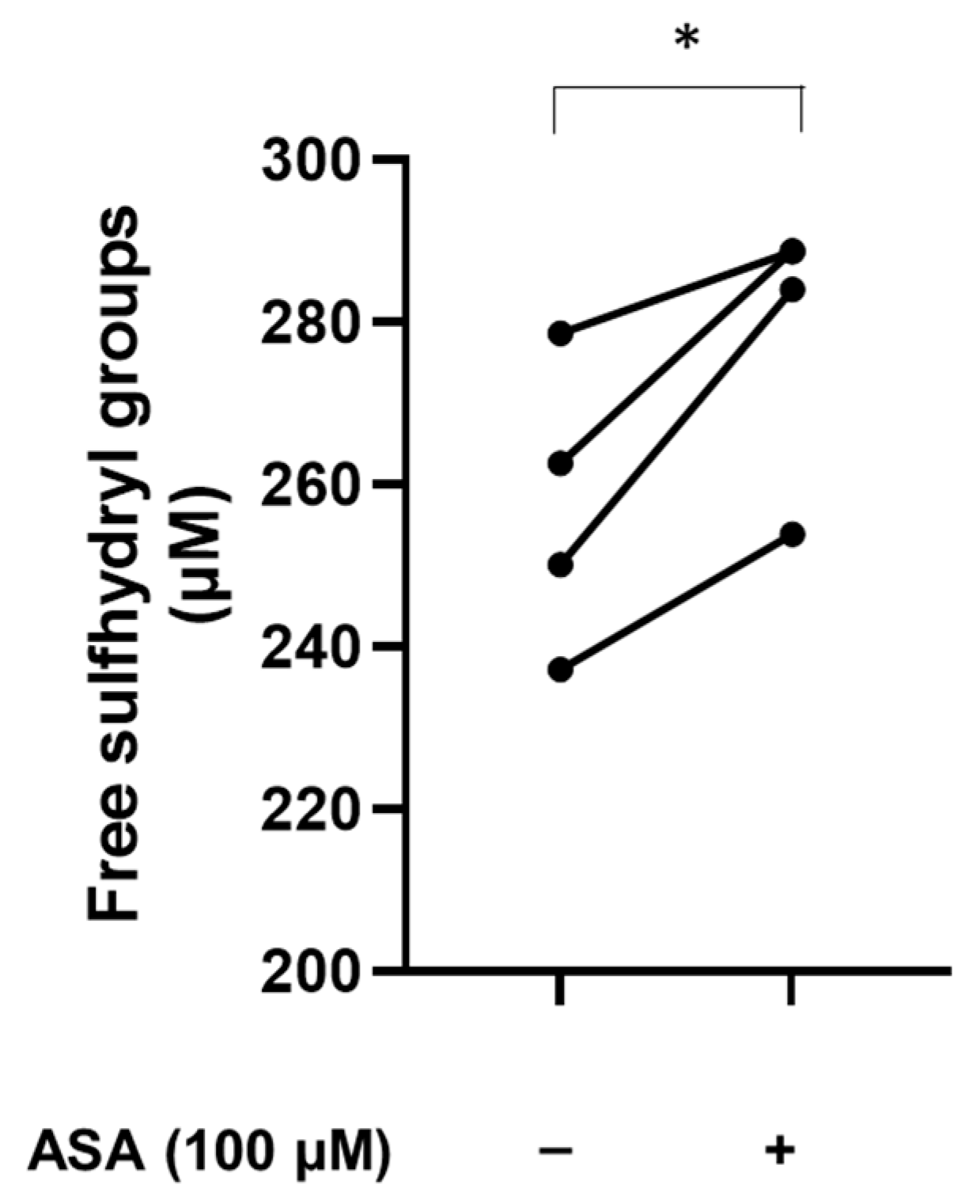
3.4. Aspirin Increases the Levels of Free Sulfhydryl Groups

To assess whether ASA can affect the levels of free sulfhydryl groups in plasma proteins, blood obtained from healthy donors was treated in vitro with or without 100 µM ASA. As shown in Figure 4, sulfhydryl groups show a marked increase in plasma proteins after in vitro treatment with ASA, suggesting that the pharmacological treatment with ASA can affect the levels of free sulfhydryl groups and the oxidation status of plasma proteins.

3.5. N-Acetylcysteine and N-Acetylcysteine Amide (AD4/NACA) Reduce the Serum Levels of Cysteinylated Proteoform of Albumin

The serum obtained from patients with aortic valve stenosis before valve replacement was treated in vitro with NAC (0.6 mM) or AD4/NACA (0.6 mM). Results showed a marked reduction in the levels of the HSA-Cys proteoform, associated with a significant increase in the levels of HSA-SH (Figure 5A,B).

3.6. N-Acetylcysteine and N-Acetylcysteine Amide (AD4/NACA) Increase the Levels of Free Sulfhydryl Groups in Serum

The serum samples obtained from patients with aortic valve stenosis before valve replacement was treated in vitro with NAC (0.6 mM) or AD4/NACA (0.6 mM) and subsequently analyzed for the measurement of the free sulfhydryl groups using Ellman’s reagent. A significant increase was detected with both these compounds, suggesting their ability to reduce oxidative stress and maintain redox homeostasis through the regeneration of free sulfhydryl groups of serum proteins (Figure 6).

3.7. N-Acetylcysteine and N-Acetylcysteine Amide (AD4/NACA) Increase the Total Antioxidant Activity of Serum from Patients

The antioxidant activity was evaluated in serum samples of patients before the first valve replacement treated in vitro with NAC or AD4/NACA. After the addition of the radical generator AAPH, a lag time of ~50 min was detected. Afterward, the propagation phase started, and the radical generation was monitored for up to 180 min. In vehicle-treated samples, a gradual increase in radical generation was detected. In contrast, the treatment with NAC or AD4/NACA markedly prevented this radical generation (Figure 7A,B).

4. Discussion

In this paper, we showed for the first time that patients with severe aortic valve stenosis present high levels of serum HSA-Cys, the oxidized form of HSA, with a dramatic decrease in the native reduced proteoform HSA-SH compared to control subjects and patients undergoing a second valve substitution due to a degenerated bioprosthetic valve. Furthermore, the increase in HSA-Cys was paralleled by significant impairment of the total serum antioxidant activity and a decrease in the levels of free sulfhydryl groups, an index of the antioxidant capacity. Indeed, total sulfhydryl groups, and especially protein sulfhydryl groups, are considered to be the main circulating antioxidant defense, and an imbalance between the free sulfhydryl groups and disulfides contributes to the pathogenesis of several oxidative stress-mediated diseases [23]. In particular, free sulfhydryl groups are mainly found in HSA, the most abundant circulating protein, giving it important antioxidant properties, so that changes in the redox status of HSA are associated with an alteration in its beneficial antioxidant properties [24]. Of interest, in its oxidized form, circulating HSA may act as a carrier of low molecular weight thiols, which could be released into the subendothelial space to aggravate the oxidative stress of the diseased tissue [25].
We also found that the levels of HSA-Cys in patients undergoing a second surgery due to a degenerated bioprosthetic valve are similar to control subjects. Thus, we hypothesize that the removal of the pathological valve is expected to stop the feedback maladaptive process, which includes oxidative stress. The increase in the cysteinylation of serum HSA reflects a condition of systemic oxidative stress, which could be amplified by mechanosensitive signaling pathways that convert mechanical forces experienced by valve leaflets and circulating cells into biochemical signals, thus providing a positive feedback loop that accelerates the progression of valvular disease in the advanced stages [26,27,28,29]. Indeed, it has been demonstrated that the pathological stretching of the valve tissue promotes maladaptive tissue remodeling by activating cell signaling, which adversely impacts the valve tissue properties, feeding back to further deteriorate valve function and propagate valve cell pathological responses [30]. In this process, the mechanosensitive pathways lead to the activation of circulating monocytes and platelets, inducing endothelial-mesenchymal transition, upregulation of proinflammatory and oxidant pathways, and promoting the valvular interstitial cells differentiation (Figure 8) [31,32]. These observations suggest a positive feedback mechanism by which the increased leaflet stiffness and altered shear stress conditions of early calcific aortic valve disease perpetuate and accelerate inflammation and oxidative stress, leading to the progression of valvular sclerosis and calcification [26,33,34].
Indeed, in a recent cohort study that evaluated systemic lipid peroxidation and the oxidative modifications of plasma proteins by measuring 2,4-dinitrophenylhydrazine, elevated oxidative stress was observed to correlate with the severity of aortic stenosis [35]. Levels of oxidative stress are also correlated with impaired systemic fibrinolysis, suggesting that the oxidative stress in calcific aortic valve disease patients might not simply reflect a localized phenomenon [35]. Results obtained from animal models also support a putative role for systemic inflammation in calcific aortic valve disease [36].
In this context, it is not surprising that valve replacement led to a decrease in cysteinylation of HSA to normal levels. Similarly, high levels of circulating inflammatory mediators associated with severe aortic stenosis are reduced with valve replacement. Indeed, transcatheter aortic valve replacement improves circulating protein composition to reverse myofibroblast activation in fibrotic valves and cardiac tissues, as shown in a proteomic analysis of patient sera [37]. Moreover, at 3 months after TAVR and 6 months post-surgical aortic valve replacement, there are significant reductions in circulating intermediate phenotype monocytes, which, being recruited at a later stage of inflammation, have a predominant role in inducing T-cell proliferation [38].
Furthermore, in accordance with the current guidelines, all our patients who have undergone a first valve replacement were in pharmacological treatment with ASA [39]. Therefore, we cannot exclude that ASA treatment, by regenerating the free sulfhydryl groups, contributes to a reduction in the levels of HSA-Cys. Indeed, besides its antiplatelet activity, the treatment with ASA is associated with a significant reduction in cardiovascular events and a slowdown in the progression of atherosclerosis [40]. In addition, it has been reported that ASA exhibits radical scavenging and antioxidant properties [41,42,43,44]. In particular, ASA administration protects low-density lipoproteins from oxidative modifications [45], the vascular wall from oxygen radical damage [46], and prevents protein oxidation by acetylation of amino groups of lysine residues [47] or scavenging hydroxyl radicals [48]. In addition, ASA reduced ROS production and oxidative stress in low-density lipoproteins-stimulated endothelial cells by downregulation of Nox4 and inducible nitric oxide synthase [49]. More recently, ASA administered for two months to healthy subjects significantly reduced the total oxidative status and the levels of oxidized low-density lipoproteins [50]. Several antioxidant effects of ASA may be attributed to an activation of gene transcription. For example, ASA increases the expression and the activity of heme-oxygenase-1, an important protein involved in the defense against oxidative tissue injury by several mechanisms [51]. In accordance with this, the enhanced heme-oxygenase-1 activity induced by ASA has also been documented in preclinical studies [44]. Additionally, ASA exerts antioxidant properties through the inhibition of NF-kB transcription factor, thus reducing ROS levels [41,52,53]. Of note, an in vivo study has shown that the treatment of healthy subjects with enteric-coated ASA for two weeks significantly increased the antioxidant capacity of plasma [54] and treatment with 160 mg of ASA until one day before the surgery markedly reduced the oxidative stress in patients scheduled for coronary heart bypass graft [55]. All these heterogeneous antioxidant properties of ASA are evidenced in several in vitro and in vivo studies and suggest that it plays a role at different levels.
Finally, we demonstrated, at least in vitro, that the antioxidant compounds NAC and AD4/NACA [56,57] completely regenerate HSA-Cys to native reduced HSA, thus confirming our previous findings showing that albumin Cys34 can be regenerated in vitro by the dietary supplement NAC through a thiol–disulfide breaking mechanism [58,59], which leads to a full recovery of the HSA antioxidant [19,58]. The regenerative effects of NAC were subsequently verified in ex vivo conditions following both oral and intravenous administration [59]. A single-blinded, placebo-controlled crossover study was conducted on hypertensive individuals who were randomized into two groups. Participants received either NAC (600 mg/day) or a placebo, administered orally and intravenously, which both resulted in a significant decrease in HSA-Cys levels compared to the initial conditions (T0), with the maximal effect observed after 60 min. This reduction in HSA-Cys was accompanied by a simultaneous increase in native HSA-SH levels. Additionally, the total antioxidant activity of plasma significantly increased following NAC infusion compared to the placebo, suggesting that the restoration of Cys34 content may modulate oxidative stress in vivo and potentially have implications in diseases related to oxidative damage [59].
We acknowledge the limitations of this study. The results should be considered as preliminary due to the small size of the subject cohort and require validation with a larger sample size. Additionally, due to the limited sample size, we cannot completely rule out the impact of ongoing therapies. Lastly, the data is limited to subjects attending the local clinic, thus representing a specific geographical area.

5. Conclusions

This study contributes to highlighting the potential role of HSA oxidative modifications as a marker of oxidative stress in patients with aortic valve stenosis, a multifactorial process, including chemical, mechanical, and immunological factors. Although the contribution of each mechanism remains poorly understood, it represents an active field of investigation [32,60,61]. In this study, we showed that patients with a severe valvular disease that required surgical intervention with replacement of the aortic valve had higher oxidative stress compared to control subjects evidenced by an increase in HSA-Cys, the oxidized form of HSA, and a concomitant decrease in the reduced form (HSA-SH). Moreover, this group of patients showed a significant reduction in the free sulfhydryl groups in serum, an index of the antioxidant capacity of serum. Furthermore, patients who have already undergone valve replacement and are waiting for a second substitution due to a degenerated bioprosthetic valve had an oxidative stress status similar to control subjects, with similar HSA proteoform frequency and levels of free sulfhydryl groups in serum. As this second group of patients was in pharmacological treatment with ASA, an antithrombotic drug with antioxidant properties, we cannot exclude an additional contribution of ASA in the reduction in oxidative stress by regenerating the free sulfhydryl groups, which likely led to the decrease in HSA-Cys. We therefore found for the first time that HSA-Cys is increased in aortic valve stenosis representing both a stable and precise marker of oxidative stress as well as a therapeutic marker. Indeed, we show that HSA-Cys can be regenerated to native HSA by NAC and AD4/NACA, which fully replenishes its antioxidant activity, thus representing a potential treatment to reduce the oxidative stress burden in valvular disease. As well as being a biomarker of oxidative stress, HSA-SH also functions in the redox control of extracellular spaces and oxidative-based disease onset. Oxidative stress is a key driver of these processes, and inhibiting ROS is an important avenue to explore new therapies. Indeed, aortic valve stenosis can be potentially treated by targeting oxidative stress, which is a promising therapeutic approach. However, while animal and in vitro studies have shown some success in reducing oxidative stress in the context of aortic valve stenosis, clinical trials have not yet yielded similar benefits [6]. So far, the regeneration of the functional HSA-SH as a therapeutic target could represent a promising approach in oxidative-based diseases, including aortic valve stenosis.

Supplementary Materials

The following supporting information can be downloaded at: https://www.mdpi.com/article/10.3390/biom13121713/s1, Figure S1: Levels of HSA-Cys in serum and plasma of controls and valvular disease patients.

Author Contributions

Conceptualization, C.B. and E.T. (Elena Tremoli); methodology, S.E., C.S., E.T. (Elena Tenti), E.M., M.M., A.G. (Anna Gaspardo), S.G., E.G. and A.G. (Arianna Greco); validation, S.E. and M.M.; formal analysis, S.E., M.M., E.G., S.G., A.G. (Anna Gaspardo) and A.G. (Arianna Greco); investigation, C.S., E.T. (Elena Tenti), E.M., S.E., M.M., E.G., S.G, A.G. (Anna Gaspardo) and A.G. (Arianna Greco); resources, C.B.; data curation, S.E. and A.G. (Anna Gaspardo); writing—original draft preparation, S.E. and C.B.; writing—review and editing, C.S., E.T. (Elena Tenti), E.M., S.E., M.M., A.G. (Anna Gaspardo), S.G., E.G., A.G. (Arianna Greco), G.A., E.T. (Elena Tremoli) and C.B.; visualization, S.E. and C.B.; supervision, C.B., G.A. and E.T. (Elena Tremoli); project administration, C.B.; funding acquisition, C.B. All authors have read and agreed to the published version of the manuscript.

Funding

This work was supported by the Italian Ministry of Health, Italy (Ricerca Corrente).

Institutional Review Board Statement

The study was conducted in accordance with the Declaration of Helsinki and was approved by the Ethics Committee of Vast Area of Romagna (CEROM). Protocol code 7043/2018 I.5/185, date of approval 19 September 2018.

Informed Consent Statement

Informed consent was obtained from all subjects involved in the study.

Data Availability Statement

Data collected in the study will be made available using the data repository Zenodo (https://zenodo.org, accessed on 23 November 2023) with restricted access upon request to [email protected]. Any remaining information can be obtained from the corresponding author upon reasonable request.

Acknowledgments

The authors thank Daphne Atlas (Department of Biological Chemistry, Institute of Life Sciences, The Hebrew University of Jerusalem) for providing the AD4/NACA compound.

Conflicts of Interest

The authors declare no conflict of interest.

References

  1. Aluru, J.S.; Barsouk, A.; Saginala, K.; Rawla, P.; Barsouk, A. Valvular Heart Disease Epidemiology. Med. Sci. 2022, 10, 32. [Google Scholar] [CrossRef] [PubMed]
  2. Virani, S.S.; Alonso, A.; Benjamin, E.J.; Bittencourt, M.S.; Callaway, C.W.; Carson, A.P.; Chamberlain, A.M.; Chang, A.R.; Cheng, S.; Delling, F.N.; et al. Heart Disease and Stroke Statistics-2020 Update: A Report from the American Heart Association. Circulation 2020, 141, e139–e596. [Google Scholar] [CrossRef] [PubMed]
  3. Alfieri, O.; Vahanian, A. The year in cardiology 2016: Valvular heart disease. Eur. Heart J. 2017, 38, 628–633. [Google Scholar] [CrossRef] [PubMed]
  4. Vahanian, A.; Beyersdorf, F.; Praz, F.; Milojevic, M.; Baldus, S.; Bauersachs, J.; Capodanno, D.; Conradi, L.; De Bonis, M.; De Paulis, R.; et al. 2021 ESC/EACTS Guidelines for the management of valvular heart disease. Eur. J. Cardiothorac. Surg. 2021, 60, 727–800. [Google Scholar] [CrossRef]
  5. Sun, J.C.; Davidson, M.J.; Lamy, A.; Eikelboom, J.W. Antithrombotic management of patients with prosthetic heart valves: Current evidence and future trends. Lancet 2009, 374, 565–576. [Google Scholar] [CrossRef]
  6. Greenberg, H.Z.E.; Zhao, G.; Shah, A.M.; Zhang, M. Role of oxidative stress in calcific aortic valve disease and its therapeutic implications. Cardiovasc. Res. 2022, 118, 1433–1451. [Google Scholar] [CrossRef]
  7. Phua, K.; Chew, N.W.; Kong, W.K.; Tan, R.S.; Ye, L.; Poh, K.K. The mechanistic pathways of oxidative stress in aortic stenosis and clinical implications. Theranostics 2022, 12, 5189–5203. [Google Scholar] [CrossRef]
  8. Kayama, Y.; Raaz, U.; Jagger, A.; Adam, M.; Schellinger, I.N.; Sakamoto, M.; Suzuki, H.; Toyama, K.; Spin, J.M.; Tsao, P.S. Diabetic Cardiovascular Disease Induced by Oxidative Stress. Int. J. Mol. Sci. 2015, 16, 25234–25263. [Google Scholar] [CrossRef]
  9. Davies, M.J. Protein oxidation and peroxidation. Biochem. J. 2016, 473, 805–825. [Google Scholar] [CrossRef]
  10. Oliveira, P.V.S.; Laurindo, F.R.M. Implications of plasma thiol redox in disease. Clin. Sci. 2018, 132, 1257–1280. [Google Scholar] [CrossRef]
  11. Tabata, F.; Wada, Y.; Kawakami, S.; Miyaji, K. Serum Albumin Redox States: More Than Oxidative Stress Biomarker. Antioxidants 2021, 10, 503. [Google Scholar] [CrossRef] [PubMed]
  12. Turell, L.; Radi, R.; Alvarez, B. The thiol pool in human plasma: The central contribution of albumin to redox processes. Free Radic. Biol. Med. 2013, 65, 244–253. [Google Scholar] [CrossRef] [PubMed]
  13. Steglich, M.; Lombide, R.; Lopez, I.; Portela, M.; Flo, M.; Marin, M.; Alvarez, B.; Turell, L. Expression, purification and initial characterization of human serum albumin domain I and its cysteine 34. PLoS ONE 2020, 15, e0240580. [Google Scholar] [CrossRef] [PubMed]
  14. Zoanni, B.; Brioschi, M.; Mallia, A.; Gianazza, E.; Eligini, S.; Carini, M.; Aldini, G.; Banfi, C. Novel insights about albumin in cardiovascular diseases: Focus on heart failure. Mass Spectrom. Rev. 2023, 42, 1113–1128. [Google Scholar] [CrossRef] [PubMed]
  15. Oettl, K.; Stauber, R.E. Physiological and pathological changes in the redox state of human serum albumin critically influence its binding properties. Br. J. Pharmacol. 2007, 151, 580–590. [Google Scholar] [CrossRef] [PubMed]
  16. Nakashima, F.; Shibata, T.; Uchida, K. A unique mechanism for thiolation of serum albumins by disulphide molecules. J. Biochem. 2020, 167, 165–171. [Google Scholar] [CrossRef] [PubMed]
  17. Nakashima, F.; Shibata, T.; Kamiya, K.; Yoshitake, J.; Kikuchi, R.; Matsushita, T.; Ishii, I.; Gimenez-Bastida, J.A.; Schneider, C.; Uchida, K. Structural and functional insights into S-thiolation of human serum albumins. Sci. Rep. 2018, 8, 932. [Google Scholar] [CrossRef] [PubMed]
  18. Brioschi, M.; Gianazza, E.; Mallia, A.; Zoanni, B.; Altomare, A.; Martinez Fernandez, A.; Agostoni, P.; Aldini, G.; Banfi, C. S-Thiolation Targets Albumin in Heart Failure. Antioxidants 2020, 9, 763. [Google Scholar] [CrossRef]
  19. Eligini, S.; Porro, B.; Aldini, G.; Colli, S.; Banfi, C. N-Acetylcysteine Inhibits Platelet Function through the Regeneration of the Non-Oxidative Form of Albumin. Antioxidants 2022, 11, 445. [Google Scholar] [CrossRef]
  20. Martinez Fernandez, A.; Regazzoni, L.; Brioschi, M.; Gianazza, E.; Agostoni, P.; Aldini, G.; Banfi, C. Pro-oxidant and pro-inflammatory effects of glycated albumin on cardiomyocytes. Free Radic. Biol. Med. 2019, 144, 245–255. [Google Scholar] [CrossRef]
  21. Regazzoni, L.; Del Vecchio, L.; Altomare, A.; Yeum, K.J.; Cusi, D.; Locatelli, F.; Carini, M.; Aldini, G. Human serum albumin cysteinylation is increased in end stage renal disease patients and reduced by hemodialysis: Mass spectrometry studies. Free Radic. Res. 2013, 47, 172–180. [Google Scholar] [CrossRef] [PubMed]
  22. Eligini, S.; Munno, M.; Atlas, D.; Banfi, C. N-acetylcysteine Amide AD4/NACA and Thioredoxin Mimetic Peptides Inhibit Platelet Aggregation and Protect against Oxidative Stress. Antioxidants 2023, 12, 1395. [Google Scholar] [CrossRef] [PubMed]
  23. Erel, O.; Neselioglu, S. A novel and automated assay for thiol/disulphide homeostasis. Clin. Biochem. 2014, 47, 326–332. [Google Scholar] [CrossRef] [PubMed]
  24. Belinskaia, D.A.; Voronina, P.A.; Shmurak, V.I.; Jenkins, R.O.; Goncharov, N.V. Serum Albumin in Health and Disease: Esterase, Antioxidant, Transporting and Signaling Properties. Int. J. Mol. Sci. 2021, 22, 10318. [Google Scholar] [CrossRef] [PubMed]
  25. Sastre-Oliva, T.; Corbacho-Alonso, N.; Albo-Escalona, D.; Lopez, J.A.; Lopez-Almodovar, L.F.; Vazquez, J.; Padial, L.R.; Mourino-Alvarez, L.; Barderas, M.G. The Influence of Coronary Artery Disease in the Development of Aortic Stenosis and the Importance of the Albumin Redox State. Antioxidants 2022, 11, 317. [Google Scholar] [CrossRef]
  26. Dayawansa, N.H.; Baratchi, S.; Peter, K. Uncoupling the Vicious Cycle of Mechanical Stress and Inflammation in Calcific Aortic Valve Disease. Front. Cardiovasc. Med. 2022, 9, 783543. [Google Scholar] [CrossRef]
  27. Lee, S.H.; Choi, J.H. Involvement of Immune Cell Network in Aortic Valve Stenosis: Communication between Valvular Interstitial Cells and Immune Cells. Immune Netw. 2016, 16, 26–32. [Google Scholar] [CrossRef]
  28. Shu, L.; Yuan, Z.; Li, F.; Cai, Z. Oxidative stress and valvular endothelial cells in aortic valve calcification. Biomed. Pharmacother. 2023, 163, 114775. [Google Scholar] [CrossRef]
  29. Tanase, D.M.; Valasciuc, E.; Gosav, E.M.; Floria, M.; Costea, C.F.; Dima, N.; Tudorancea, I.; Maranduca, M.A.; Serban, I.L. Contribution of Oxidative Stress (OS) in Calcific Aortic Valve Disease (CAVD): From Pathophysiology to Therapeutic Targets. Cells 2022, 11, 2663. [Google Scholar] [CrossRef]
  30. Gould, S.T.; Srigunapalan, S.; Simmons, C.A.; Anseth, K.S. Hemodynamic and cellular response feedback in calcific aortic valve disease. Circ. Res. 2013, 113, 186–197. [Google Scholar] [CrossRef]
  31. Rutkovskiy, A.; Malashicheva, A.; Sullivan, G.; Bogdanova, M.; Kostareva, A.; Stenslokken, K.O.; Fiane, A.; Vaage, J. Valve Interstitial Cells: The Key to Understanding the Pathophysiology of Heart Valve Calcification. J. Am. Heart Assoc. 2017, 6, e006339. [Google Scholar] [CrossRef]
  32. Goody, P.R.; Hosen, M.R.; Christmann, D.; Niepmann, S.T.; Zietzer, A.; Adam, M.; Bonner, F.; Zimmer, S.; Nickenig, G.; Jansen, F. Aortic Valve Stenosis: From Basic Mechanisms to Novel Therapeutic Targets. Arter. Thromb. Vasc. Biol. 2020, 40, 885–900. [Google Scholar] [CrossRef] [PubMed]
  33. Serrano, C.V., Jr.; de Mattos, F.R.; Pitta, F.G.; Nomura, C.H.; de Lemos, J.; Ramires, J.A.F.; Kalil-Filho, R. Association between Neutrophil-Lymphocyte and Platelet-Lymphocyte Ratios and Coronary Artery Calcification Score among Asymptomatic Patients: Data from a Cross-Sectional Study. Mediat. Inflamm. 2019, 2019, 6513847. [Google Scholar] [CrossRef] [PubMed]
  34. Li, C.; Ying, Q.; Su, X.; Li, T. Development and application of reverse transcription loop-mediated isothermal amplification for detecting live Shewanella putrefaciens in preserved fish sample. J. Food Sci. 2012, 77, M226–M230. [Google Scholar] [CrossRef] [PubMed]
  35. Siudut, J.; Natorska, J.; Wypasek, E.; Wiewiorka, L.; Ostrowska-Kaim, E.; Wisniowska-Smialek, S.; Plens, K.; Legutko, J.; Undas, A. Impaired Fibrinolysis in Patients with Isolated Aortic Stenosis is Associated with Enhanced Oxidative Stress. J. Clin. Med. 2020, 9, 2002. [Google Scholar] [CrossRef] [PubMed]
  36. van Broekhoven, A.; Krijnen, P.A.J.; Fuijkschot, W.W.; Morrison, M.C.; Zethof, I.P.A.; van Wieringen, W.N.; Smulders, Y.M.; Niessen, H.W.M.; Vonk, A.B.A. Short-term LPS induces aortic valve thickening in ApoE*3Leiden mice. Eur. J. Clin. Investig. 2019, 49, e13121. [Google Scholar] [CrossRef]
  37. Aguado, B.A.; Schuetze, K.B.; Grim, J.C.; Walker, C.J.; Cox, A.C.; Ceccato, T.L.; Tan, A.C.; Sucharov, C.C.; Leinwand, L.A.; Taylor, M.R.G.; et al. Transcatheter aortic valve replacements alter circulating serum factors to mediate myofibroblast deactivation. Sci. Transl. Med. 2019, 11, eaav3233. [Google Scholar] [CrossRef]
  38. Hewing, B.; Ellerbroek, R.; Au, S.C.; Stangl, V.; Dreger, H.; Laule, M.; Grubitzsch, H.; Knebel, F.; Baumann, G.; Ludwig, A.; et al. Levels of Circulating Intermediate Monocytes Decrease after Aortic Valve Replacement in Patients with Severe Aortic Stenosis. Thromb. Haemost. 2017, 117, 2346–2355. [Google Scholar] [CrossRef]
  39. Singh, M.; Sporn, Z.A.; Schaff, H.V.; Pellikka, P.A. ACC/AHA Versus ESC Guidelines on Prosthetic Heart Valve Management: JACC Guideline Comparison. J. Am. Coll. Cardiol. 2019, 73, 1707–1718. [Google Scholar] [CrossRef]
  40. Hedin, U.; Matic, L.P. Recent advances in therapeutic targeting of inflammation in atherosclerosis. J. Vasc. Surg. 2019, 69, 944–951. [Google Scholar] [CrossRef]
  41. Chen, C.M.; Tung, Y.T.; Wei, C.H.; Lee, P.Y.; Chen, W. Anti-Inflammatory and Reactive Oxygen Species Suppression through Aspirin Pretreatment to Treat Hyperoxia-Induced Acute Lung Injury in NF-kappaB-Luciferase Inducible Transgenic Mice. Antioxidants 2020, 9, 429. [Google Scholar] [CrossRef]
  42. Ayyadevara, S.; Bharill, P.; Dandapat, A.; Hu, C.; Khaidakov, M.; Mitra, S.; Shmookler Reis, R.J.; Mehta, J.L. Aspirin inhibits oxidant stress, reduces age-associated functional declines, and extends lifespan of Caenorhabditis elegans. Antioxid. Redox Signal 2013, 18, 481–490. [Google Scholar] [CrossRef]
  43. Paseban, M.; Niazmand, S. The Comparison of Antioxidant Effect of Aspirin, Metformin, Atorvastatin and Captopril Co-administration in the Heart and Kidney Tissues of Diabetic Rats. Iran. J. Pharm. Res. 2021, 20, 27–39. [Google Scholar] [CrossRef]
  44. Falco, L.; Tessitore, V.; Ciccarelli, G.; Malvezzi, M.; D’Andrea, A.; Imbalzano, E.; Golino, P.; Russo, V. Antioxidant Properties of Oral Antithrombotic Therapies in Atherosclerotic Disease and Atrial Fibrillation. Antioxidants 2023, 12, 1185. [Google Scholar] [CrossRef]
  45. Steer, K.A.; Wallace, T.M.; Bolton, C.H.; Hartog, M. Aspirin protects low density lipoprotein from oxidative modification. Heart 1997, 77, 333–337. [Google Scholar] [CrossRef]
  46. Wu, R.; Lamontagne, D.; de Champlain, J. Antioxidative properties of acetylsalicylic Acid on vascular tissues from normotensive and spontaneously hypertensive rats. Circulation 2002, 105, 387–392. [Google Scholar] [CrossRef]
  47. Betts, W.H.; Whitehouse, M.W.; Cleland, L.G.; Vernon-Roberts, B. In vitro antioxidant properties of potential biotransformation products of salicylate, sulphasalazine and amidopyrine. J. Free Radic. Biol. Med. 1985, 1, 273–280. [Google Scholar] [CrossRef]
  48. Pinckard, R.N.; Hawkins, D.; Farr, R.S. In vitro acetylation of plasma proteins, enzymes and DNA by aspirin. Nature 1968, 219, 68–69. [Google Scholar] [CrossRef]
  49. Chen, B.; Zhao, J.; Zhang, S.; Wu, W.; Qi, R. Aspirin inhibits the production of reactive oxygen species by downregulating Nox4 and inducible nitric oxide synthase in human endothelial cells exposed to oxidized low-density lipoprotein. J. Cardiovasc. Pharmacol. 2012, 59, 405–412. [Google Scholar] [CrossRef]
  50. Kurban, S.; Mehmetoglu, I. Effects of acetylsalicylic acid on serum paraoxonase activity, Ox-LDL, coenzyme Q10 and other oxidative stress markers in healthy volunteers. Clin. Biochem. 2010, 43, 287–290. [Google Scholar] [CrossRef]
  51. Jian, Z.; Tang, L.; Yi, X.; Liu, B.; Zhang, Q.; Zhu, G.; Wang, G.; Gao, T.; Li, C. Aspirin induces Nrf2-mediated transcriptional activation of haem oxygenase-1 in protection of human melanocytes from H2 O2 -induced oxidative stress. J. Cell. Mol. Med. 2016, 20, 1307–1318. [Google Scholar] [CrossRef]
  52. Jorda, A.; Aldasoro, M.; Aldasoro, C.; Guerra-Ojeda, S.; Iradi, A.; Vila, J.M.; Campos-Campos, J.; Valles, S.L. Action of low doses of Aspirin in Inflammation and Oxidative Stress induced by abeta(1-42) on Astrocytes in primary culture. Int. J. Med. Sci. 2020, 17, 834–843. [Google Scholar] [CrossRef]
  53. Yang, J.J.; Li, P.; Wang, F.; Liang, W.J.; Ma, H.; Chen, Y.; Ma, Z.M.; Li, Q.Z.; Peng, Q.S.; Zhang, Y.; et al. Activation of activator protein 2 alpha by aspirin alleviates atherosclerotic plaque growth and instability in vivo. Oncotarget 2016, 7, 52729–52739. [Google Scholar] [CrossRef]
  54. Ristimae, T.; Zilmer, M.; Zilmer, K.; Kairane, C.; Kullisaar, T.; Teesalu, R. Effect of low-dose aspirin on the markers of oxidative stress. Cardiovasc. Drugs Ther. 1999, 13, 485–490. [Google Scholar] [CrossRef]
  55. Berg, K.; Langaas, M.; Ericsson, M.; Pleym, H.; Basu, S.; Nordrum, I.S.; Vitale, N.; Haaverstad, R. Acetylsalicylic acid treatment until surgery reduces oxidative stress and inflammation in patients undergoing coronary artery bypass grafting. Eur. J. Cardiothorac. Surg. 2013, 43, 1154–1163. [Google Scholar] [CrossRef]
  56. Offen, D.; Gilgun-Sherki, Y.; Barhum, Y.; Benhar, M.; Grinberg, L.; Reich, R.; Melamed, E.; Atlas, D. A low molecular weight copper chelator crosses the blood-brain barrier and attenuates experimental autoimmune encephalomyelitis. J. Neurochem. 2004, 89, 1241–1251. [Google Scholar] [CrossRef]
  57. Grinberg, L.; Fibach, E.; Amer, J.; Atlas, D. N-acetylcysteine amide, a novel cell-permeating thiol, restores cellular glutathione and protects human red blood cells from oxidative stress. Free Radic. Biol. Med. 2005, 38, 136–145. [Google Scholar] [CrossRef]
  58. Altomare, A.; Baron, G.; Brioschi, M.; Longoni, M.; Butti, R.; Valvassori, E.; Tremoli, E.; Carini, M.; Agostoni, P.; Vistoli, G.; et al. N-Acetyl-Cysteine Regenerates Albumin Cys34 by a Thiol-Disulfide Breaking Mechanism: An Explanation of Its Extracellular Antioxidant Activity. Antioxidants 2020, 9, 367. [Google Scholar] [CrossRef]
  59. Altomare, A.A.; Brioschi, M.; Eligini, S.; Bonomi, A.; Zoanni, B.; Iezzi, A.; Jemos, C.; Porro, B.; D’Alessandra, Y.; Guarino, A.; et al. N-Acetylcysteine Regenerates In Vivo Mercaptoalbumin. Antioxidants 2022, 11, 1758. [Google Scholar] [CrossRef]
  60. Velho, T.R.; Pereira, R.M.; Fernandes, F.; Guerra, N.C.; Ferreira, R.; Nobre, A. Bioprosthetic Aortic Valve Degeneration: A Review from a Basic Science Perspective. Braz. J. Cardiovasc. Surg. 2022, 37, 239–250. [Google Scholar] [CrossRef]
  61. Yetkin, E.; Waltenberger, J. Molecular and cellular mechanisms of aortic stenosis. Int. J. Cardiol. 2009, 135, 4–13. [Google Scholar] [CrossRef]
Figure 1. Representative chromatograms of different HSA proteoforms. Serum was obtained from (A) patients with aortic valve stenosis before valve replacement, (B) patients undergoing a second valve replacement, and (C) controls.
Biomolecules 13 01713 g001
Figure 2. HSA proteoforms in patients and control subjects. Percentages of (A) HSA-Cys, (B) HSA-Gly, (C) HSA-SH were measured in serum obtained from patients with valvular disease before valve replacement (Valv repl, n = 36), patients requiring a second valve replacement to a degenerated bioprosthetic valve (Redo valv repl, n = 8), and healthy donors (Controls n = 10) by LC–mass spectrometry. Data are expressed as mean ± SEM. * p < 0.05; **** p < 0.0001 by one-way ANOVA followed by Tukey’s post hoc test.
Biomolecules 13 01713 g002
Figure 3. Correlation between HSA-Cys and free sulfhydryl groups and levels of free sulfhydryl groups in patients and control subjects. (A) HSA-Cys was measured in serum of all participants to the study by LC–mass spectrometry. Free sulfhydryl groups were detected in serum using Ellman’s reagent. (B) Free sulfhydryl groups detected in serum obtained from patients with valvular disease before valve replacement (Valv repl, n = 36), and healthy donors (Controls n = 10). Data are expressed as mean ± SEM. * p < 0.05.
Biomolecules 13 01713 g003
Figure 4. Analysis of free sulfhydryl groups in plasma treated with ASA. Blood obtained from healthy donors was incubated in vitro at 37 °C for 120 min in the presence or the absence of ASA. After centrifugation, plasma was collected, and free sulfhydryl groups were measured using Ellman’s reagent. n = 4. * p < 0.05 by paired Student’s t-test.
Biomolecules 13 01713 g004
Figure 5. HSA proteoforms in serum of patients with aortic valve stenosis after in vitro treatment with NAC or its amide derivative AD4/NACA. Serum obtained from patients before valve replacement was incubated at 37 °C for 60 min in the presence of NAC or AD4/NACA. Levels of (A) HSA-Cys and (B) HSA-SH were measured by LC–mass spectrometry. n = 6. **** p < 0.0001 by one-way ANOVA followed by Dunnett’s post hoc test.
Biomolecules 13 01713 g005
Figure 6. Analysis of serum free sulfhydryl groups after in vitro treatment with NAC or its amide derivative AD4/NACA. Serum of patients before valve replacement was incubated at 37 °C for 60 min in the presence of NAC or AD4/NACA. Free sulfhydryl groups were measured using Ellman’s reagent. n = 6. * p < 0.05; *** p < 0.001 by ANOVA followed by Dunnett’s post hoc test.
Biomolecules 13 01713 g006
Figure 7. Antioxidant activity of serum after in vitro treatment with NAC or its amide derivative AD4/NACA. The serum of patients before valve replacement was incubated at 37 °C for 60 min in the presence of NAC or AD4/NACA. Antioxidant activity was detected by DCF fluorescence. (A) A representative curve of fluorescence of DCF (λex = 485 nm, λem = 532 nm) in the presence of AAPH as a generator of radical species. Data are expressed as arbitrary units of fluorescence (AFU) of DCF. (B) Effect of NAC and AD4/NACA on the oxidation of DCFH. The fluorescence of DCF was measured after 180 min after the start of the reaction. Data are expressed as % of fluorescence. n = 6. **** p < 0.0001 by ANOVA followed by Dunnett’s post hoc test.
Biomolecules 13 01713 g007
Figure 8. Feedback mechanisms that might perpetuate oxidative stress in patients with valvular disease. After the initiation of valvular inflammation and stenosis, mechanosensitive feedback pathways may provide the coupling link that promotes further valvular interstitial cell differentiation, valvular inflammation, and oxidative stress.
Biomolecules 13 01713 g008
Table 1. Characteristics of patients enrolled in the study.
Aortic Valve
Replacement
Redo Aortic Valve
Replacement
p
N368/
Male gender18 (50.0)5 (62.5)0.701
Age (years)78.0 (8.5)70.5 (14)0.100
Weight (Kg)75.5 (12.5)70.0 (43)0.604
BMI (Kg/m2)27.5 (3.7)29.2 (12.2)0.784
Hemoglobin (g/dL)12.7 (2.1)12.8 (3.8)0.346
Platelets (×109/L)196 (80)157.5 (89)0.041
Neutrophils (×109/L)/6.4 (53.8)/
Lymphocytes (×109/L)/2.0 (17.6)/
Creatinine (mg/dL)0.91 (0.40)1.04 (0.36)0.640
LDL (mg/dL)71 (38)92.5 (40)0.439
Hypertension (n, %)33 (91.7)6 (75.0)0.219
Dyslipidemia (n, %)27 (75.0)5 (62.5)0.663
Diabetes (n, %)7 (19.4)2 (25.0)0.659
Coronary artery disease (n, %)6 (16.7)1 (12.5)1.000
LVEF (%)61 (11.0)55 (32.0)0.692
Peak aortic gradient (mmHg)72 (32)75 (27)0.737
Mean aortic gradient (mmHg)43 (15)48 (25)0.608
Previous coronary artery bypass graft (n, %)1 (2.8)1 (12.5)0.334
Oncological history (n, %)3 (8.3)1 (12.5)1.000
Novel oral anticoagulants (n, %)6 (16.7)0 (0.0)0.053
Statins (n, %)21 (58.3)4 (50.0)0.710
Antidiabetic drugs (n, %)7 (19.4)2 (25.0)0.659
Antihypertensive drugs (n, %)32 (88.9)5 (63.0)0.100
Years since first surgery/8.7 (6.1)/
Continuous variables were reported as median and IQR, and categorical variables were reported as absolute number (n) and frequency (%). p value was calculated using the Mann–Whitney test for continuous variables, and the chi-squared test or Fisher exact test as appropriate for categorical variables.
Table 2. HSA proteoforms distribution.
Albumin
Proteoforms (%)
Aortic Valve
Replacement
Redo
Aortic Valve
Replacement
Controlsp Value
Valv Repl vs. Redo Valv Repl
p Value
Valv Repl vs. Controls
n36810//
% Cys (PA)32.0 (14.4)15.7 (3.1)14.3 (4.2)<0.001<0.0001
% Gly (PA)6.1 (1.8)5.1 (0.8)5.6 (1.5)0.0120.136
% SH (PA)61.0 (17.0)79.9 (4.0)80.1 (3.9)<0.001<0.0001
HSA total (PA)6.58 × 10⁹ (26.2 × 10⁹)4.54 × 10⁹ (3 × 10⁹)6.0 × 10⁹ (18.3 × 10⁹)0.3310.564
Data are expressed as median and IQR of PA (peak area).
Disclaimer/Publisher’s Note: The statements, opinions and data contained in all publications are solely those of the individual author(s) and contributor(s) and not of MDPI and/or the editor(s). MDPI and/or the editor(s) disclaim responsibility for any injury to people or property resulting from any ideas, methods, instructions or products referred to in the content.

