Brief Report

Normal β-Cell Glut2 Expression Is not Required for Regulating Glucose-Stimulated Insulin Secretion and Systemic Glucose Homeostasis in Mice

by
Siresha Bathina
1,2,
Tumininu S. Faniyan
1,
Lauren Bainbridge
1,
Autumn Davis
1 and
Kavaljit H. Chhabra
1,3,*
1
Department of Medicine, Division of Endocrinology, Diabetes and Metabolism, School of Medicine and Dentistry, University of Rochester, Rochester, NY 14642, USA
2
Department of Medicine, Division of Endocrinology, Diabetes and Metabolism, Baylor College of Medicine, Houston, TX 77030, USA
3
Department of Pharmacology and Physiology, University of Rochester Medical Center, Rochester, NY 14642, USA
*
Author to whom correspondence should be addressed.
Biomolecules 2023, 13(3), 540; https://doi.org/10.3390/biom13030540
Submission received: 24 February 2023 / Revised: 9 March 2023 / Accepted: 11 March 2023 / Published: 16 March 2023
(This article belongs to the Section Biomacromolecules: Carbohydrates)

Abstract

:
Objective: Glucose transporter 2 (GLUT2) is expressed in the pancreatic β-cell, intestine, liver, and kidney in mice. Although GLUT2 is considered as a major regulator of insulin secretion, in vivo contribution of β-cell Glut2 to glucose-stimulated insulin secretion and systemic glucose homeostasis is undefined. Therefore, the main objective of this study is to determine the role of β-cell Glut2 in regulating insulin secretion and blood glucose levels in mice. Methods: We produced mice in which we can knock down Glut2 at a desired time specifically in β-cells (β-Glut2 KD) by crossing Glut2LoxP/LoxP mice with Ins1CreERT2 mouse strain and using the Cre-Lox recombination technique. We measured fasting blood glucose levels, glucose tolerance, and glucose-stimulated insulin secretion in the β-Glut2 KD mice. We used qRT-PCR and immunofluorescence to validate the deficiency of β-cell Glut2 in β-Glut2 KD mice. Results: We report that both male and female β-Glut2 KD mice have normal glucose-stimulated insulin secretion. Moreover, the β-Glut2 KD mice exhibit normal fasting blood glucose levels and glucose tolerance. The β-Glut2 KD mice have upregulated GLUT1 in islets. Conclusions: Our findings demonstrate that normal β-cell Glut2 expression is not essential for regulating glucose-stimulated insulin secretion and systemic glucose homeostasis in mice. Therefore, the currently assumed role of β-cell GLUT2 in regulating insulin secretion and blood glucose levels needs to be recalibrated. This will allow an opportunity to determine the contribution of other β-cell glucose transporters or factors whose normal expression may be necessary for mediating glucose stimulated insulin secretion.

1. Introduction

Glucose transporter 2 (GLUT2) is a major transporter of glucose in the β-cells, liver, intestine, and kidney in mice. GLUT2 deficiency in humans causes Fanconi–Bickel syndrome [1], which is characterized by glycogen accumulation, glycosuria, impaired glucose and galactose tolerance, and nephropathy. Loss of Glut2 in the intestine in mice improves glucose tolerance [2]. Deficiency of hepatic GLUT2 contributes to progressive impairment of glucose regulation in mice [3]. Moreover, we recently reported the contribution of renal GLUT2 to systemic glucose homeostasis [4,5,6]. Loss of renal Glut2 protects mice from streptozocin induced hyperglycemia and diet induced obesity [4].
Global Glut2 knockout mice [7] exhibit hypoinsulinemia, hyperglycemia, and die within three weeks after birth. In contrast, humans with GLUT2 deficiency show good prognosis [8], although they may experience transient neonatal diabetes [9,10]. Humans and rodents have differential expression of β-cell GLUT2 [11]. GLUT2 is considered as a major glucose transporter in rodent β-cells [12], but this is not true for human β-cells [11]. Whether normal β-cell Glut2 expression is required for regulating glucose-stimulated insulin secretion and systemic glucose regulation in adult mice is unknown.
In this study, we determined the contribution of β-cell Glut2 to glucose stimulated insulin secretion and systemic glucose homeostasis in mice. We used an inducible Cre-Lox system to produce β-cell GLUT2 deficiency at a desired time in adult mice. This strategy minimized potential developmental defects that would otherwise affect the phenotype in mice because of absence of Glut2 during prenatal and perinatal periods.

2. Materials and Methods

2.1. Animal Care and Generation of Mice

All mouse procedures were approved by the Institutional Animal Care and Use Committee at the University of Rochester and were performed according to the US Public Health Service guidelines for the humane care and use of experimental animals. The reporting in the manuscript follows the recommendations in the ARRIVE guidelines. The mice were housed in ventilated cages under controlled temperature (~23 °C) and photoperiod (12 h light/dark cycle, lights on from 06:00 h to 18:00 h) conditions with free access to Hydropac water (Lab Products, Seaford, DE, USA) and regular laboratory chow (5010, LabDiet, Arden Hills, MN, USA). The mice were randomly assigned to different experimental groups. We used two mouse lines with their littermate controls in this study: Glut2LoxP/LoxP and Ins1CreERT2 mouse strains [4,13].
We generated Ins1CreERT2;Glut2loxP/loxP mice and their littermate controls by first crossing the Glut2loxP/loxP mice [4] with Ins1CreERT2 mouse line [13] (026802, The Jackson Laboratory, Bar Harbor, ME, USA) followed by a second generation of intercrossing between Ins1CreERT2;Glut2loxP/+ and Glut2loxP/loxP mice. To knockdown β-cell Glut2 in Ins1CreERT2;Glut2loxP/loxP mice, we injected them with tamoxifen (100 mg/kg dissolved first in 1 part 100% v/v ethanol by incubating the mixture at 60 °C for 20 min followed by addition of 9 parts of sesame oil, T5648, and S3547, Sigma, St. Louis, MO, USA) once daily for five consecutive days. The control mice were also injected with tamoxifen using the same protocol.

2.2. Islet Isolation Procedure

We isolated islets for measuring mRNA levels of Glut2 to confirm its deficiency in β-cell Glut2 knockdown mice. We inflated the mouse pancreas by injecting 2 mL of Liberase TL (Roche 05401020001, 1.3 Wünsch units/mL in RPMI 1640 medium) using the bile duct, as described previously [14,15]. We collected the inflated pancreata and incubated them at 37 °C in a water bath for 14 min to digest the tissue. We then followed published protocols [14,15] to separate and purify the islets. The isolated islets were immediately frozen in liquid nitrogen and stored at −80 °C in a freezer until the mRNA assay.

2.3. Oral Glucose Tolerance Test

After fasting mice for 6 h (08:00–14:00 h), their tails were slightly nicked laterally using a razor blade, and a drop of blood was used to measure baseline (0 min) blood glucose with AlphaTRAK 2 glucometer. Then, a fixed dose of glucose (100 mg/mouse in 250–300 µL water, G8270; Sigma) was administered into each mouse by oral gavage (18 g needle, FNS-18-2, Kent Scientific Corporation, Torrington, CT, USA) and blood glucose levels were determined at 15, 30, 60, and 120 min after the oral glucose administration.

2.4. Glucose Stimulated Insulin Secretion

To determine glucose-stimulated insulin secretion, we fasted mice for 6 h (08:00–14:00 h) and collected tail blood using heparinized capillary tubes (22-362-566, Fisherbrand) at baseline (0 min) and 20 min after glucose administration (100 mg glucose dissolved in 250–300 µL water, oral gavage). The blood was centrifuged for 20 min at 2000× g speed and 4 °C to separate plasma. Plasma insulin levels were measured using ELISA (90080; Crystal Chem, Elk Grove Village, IL, USA).

2.5. RT-qPCR

We used Aurum Total RNA Fatty and Fibrous Tissue Kit (7326830, Bio-rad, Hercules, CA, USA) to extract total RNA. Five hundred ng total RNA and random hexamer primers (1708891, iScript cDNA synthesis kit, Bio-Rad) were used to generate cDNA. RT-qPCR was performed using a StepOne Real Time PCR System (Applied Biosystems) and SYBR green master mix (1725124, Bio-Rad). We used the following primers, Glut2 (Slc2a2): 5′-GAA GGA ACT CAG TAC AGC AGT G-3′ and 5′-TCA TCC ACA TTC AGT ACA GGA C-3′; Glut1 (Slc2a1): 5′-GTG GTG AGT GTG GAT G-3′ and 5′-AGT TCG GCT ATA ACA CTG GTG-3′; Hprt: 5′-AAC AAA GTC TGG CCT GTA TCC-3′ and 5′-CCC CAA AAT GGT TAA GGT TGC-3′. All primers were used at a final concentration of 500 nM. The relative quantity of each mRNA was calculated from standard curves and normalized to the internal control Hprt, and then normalized to the mean of corresponding controls.

2.6. Immunohistochemistry

After euthanizing the mice, their pancreata were collected and fixed in 10% formalin before embedding them in paraffin. The samples were cut into 5µm sections using a microtome and placed onto slides. Before staining the sections, they were deparaffinized and rehydrated using xylene (3 min incubation, twice), ethanol (3 min incubation each with 1:1 with xylene followed by 100%, 70%, and 50% ethanol in water), and water (5 min under running tap water to rinse off ethanol). Antigen retrieval was carried out by incubating the sections in sodium citrate buffer (pH 6.0) in water bath at 95 °C for 25 min. Nonspecific staining was blocked by subsequent incubation with 10% goat serum in tris-buffered saline with 0.1% Tween 20 detergent (TBST) for 60 min at room temperature. The tissue sections were then processed for GLUT1, GLUT2, or insulin staining using previously validated antibodies [4,5]: rabbit anti-GLUT1 (1:250 dilution in TBST, ab652 or ab115730, Abcam, Waltham, MA, USA), rabbit anti-GLUT2 (1:250 dilution in TBST, 600-401-GN3, Rockland Immunochemicals, Pottstown, PA, USA), or mouse anti-insulin (1:100 dilution in TBST, sc-8033, Santa Cruz Biotechnology, Inc., Dallas, TX, USA) incubated at 4 °C overnight. The next day, the sections were washed three times (10 min/wash) in TBST and then incubated with secondary goat anti-rabbit or anti-mouse antibody conjugated to Alexa Fluor 488 or 647 as appropriate (ab150077, ab150115; Abcam; 1:1000 dilution in TBST) for 2 h at room temperature. After this incubation, the sections were washed three times (10 min/wash) in TBST and counterstained with DAPI (D9542; Sigma; 1 μg/mL) for 1 min. This was followed by a quick wash with TBST again before the sections were air-dried and coverslipped using ProLong Antifade mounting medium (P36930; Molecular Probes, Waltham, MA, USA). Images were captured using Keyence fluorescence microscope BZ-X800. We quantified the images using the NIH ImageJ software, version 1.53n (https://imagej.nih.gov/ij/download.html, accessed on 19 April 2022 through 9 June 2022).

2.7. Statistical Analyses

We included 6 to 12 mice per group in this study based on our previously published reports [4,5]. All data are presented as mean ± SEM and were analyzed by Two-tailed Student’s paired or unpaired t test, one-way ANOVA or two-way ANOVA followed by a Bonferroni post hoc multiple comparison test when appropriate. All analyses were performed using Prism 8.0 (GraphPad, San Diego, CA, USA) and differences were considered statistically significant at p value < 0.05.

3. Results

3.1. Generation and Validation of β-cell Glut2 Knockdown Mice

We generated Ins1CreERT2;Glut2LoxP/LoxP mice and their littermate controls by crossing our Glut2LoxP/LoxP mouse line [4] with Ins1CreERT2 mouse strain [13] (Figure 1A). This Ins1CreERT2 mouse line is validated for tamoxifen inducible Cre-Lox recombination selectively in β-cells [13]. We injected the Ins1CreERT2;Glut2LoxP/LoxP mice with tamoxifen (100 mg/kg, once daily for five days) to produce β-cell Glut2 knockdown (β-Glut2 KD) mice. We measured mRNA and protein levels of islet or β-cell GLUT2 using qRT-PCR and immunofluorescence, respectively, one week after the last injection of tamoxifen or after completion of the study. We observed that β-Glut2 KD mice have about 15% GLUT2 in islets or β-cells compared to their littermate controls (Figure 1B,C), demonstrating the deficiency of Glut2 and therefore, validating the mouse model. As we did not observe a complete knockout of β-cell Glut2 in our mouse model describe here, we used the term knockdown for the level of Glut2 deficiency achieved in the model.

3.2. β-Cell Glut2 Knockdown Mice Have Normal Glucose-Stimulated Insulin Secretion

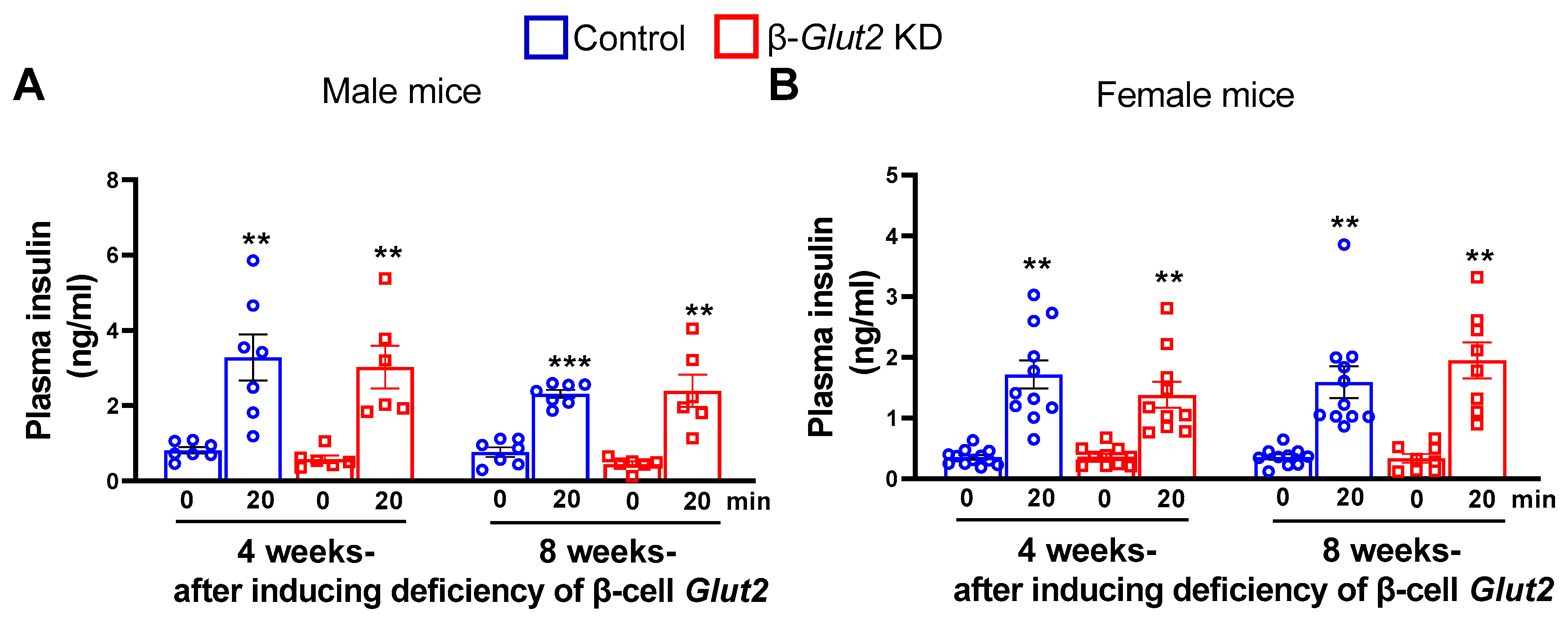
Although GLUT2 is considered as a major regulator of glucose-stimulated insulin secretion, it is unknown whether normal expression of β-cell GLUT2 is necessary for regulating this phenomenon. To address this question, we measured glucose-stimulated insulin secretion four and eight weeks after inducing β-cell GLUT2 deficiency in 8-week old male and female β-Glut2 KD mice. We observed that fasting plasma insulin levels and insulin secretion in response to glucose administration in the Glut2 knockdown mice were similar to that observed in their littermate control mice (Figure 2). The variability in the extent of β-cell Glut2 knockdown achieved in β-Glut2 KD mice could have contributed to the observed wide range of glucose-stimulated insulin secretion. Nevertheless, these results demonstrate that normal expression of β-cell Glut2 is not essential for regulating glucose-stimulated insulin secretion in mice.

3.3. β-Cell Glut2 Knockdown Mice Have Normal Glucose Tolerance

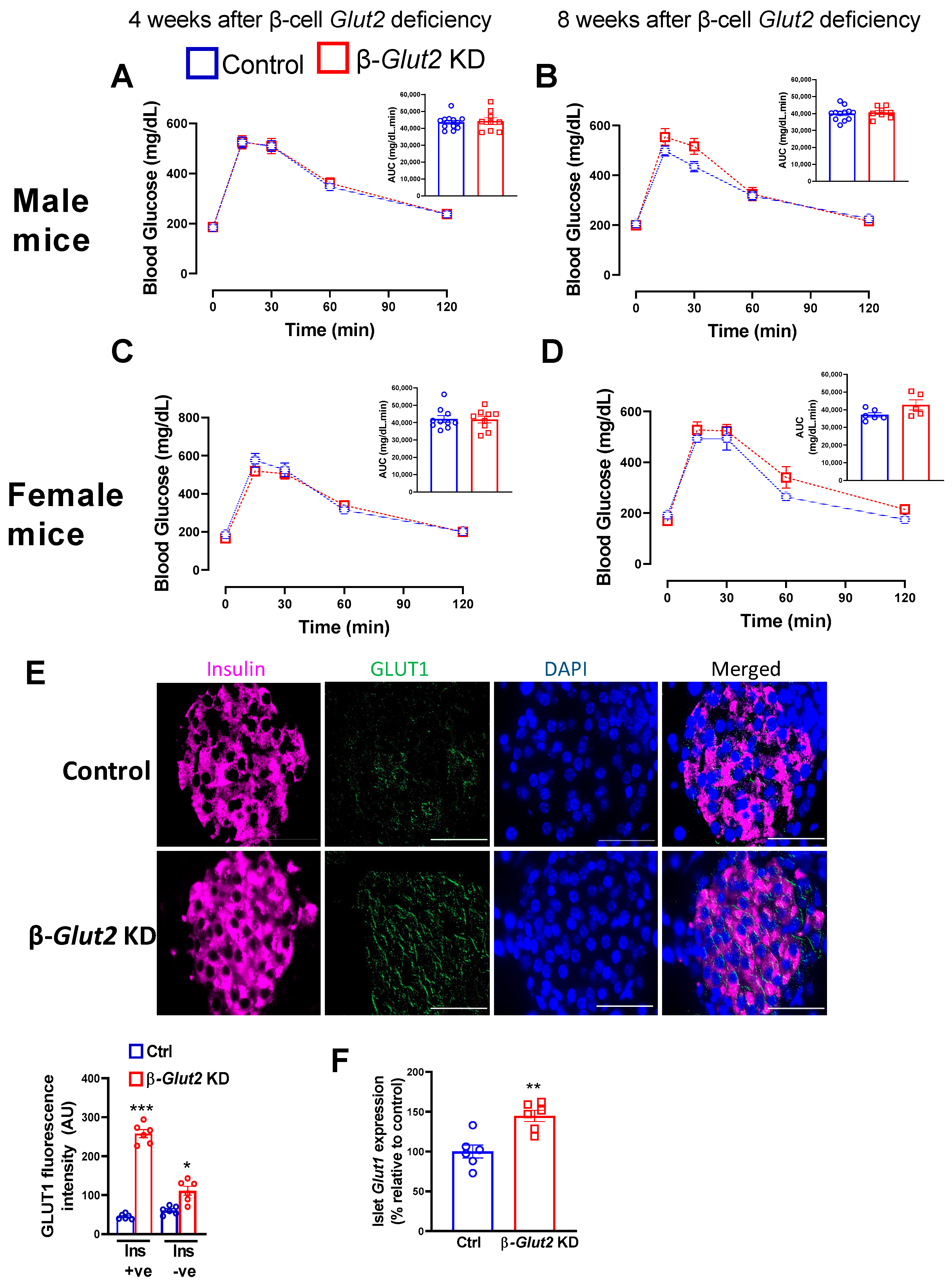
As global Glut2 knockout mice display impaired glucose homeostasis [7], we determined whether deficiency of β-cell Glut2 in adult mice recapitulates this defect. We performed oral glucose tolerance tests four and eight weeks after inducing β-cell GLUT2 deficiency in 8-week old male and female β-Glut2 KD mice. The mice have normal glucose tolerance (Figure 3A–D) despite the deficiency of β-cell Glut2. Moreover, β-Glut2 KD mice have 6 h fasting (Female, 169.8 ± 7 vs. 166.3 ± 8.6; Male, 189.5 ± 9 vs. 190 ± 4 mg/dL, Control vs. β-Glut2 KD mice, n = 6) and random non-fasting (Female, 206 ± 14 vs. 213 ± 11; Male, 217 ± 9 vs. 223 ± 16 mg/dL, Control vs. β-Glut2 KD mice, n = 6) blood glucose levels similar to that observed in their littermate control mice. As some reports have suggested the contribution of other glucose transporters such as GLUT1 to mediating glucose transport and metabolism in absence of GLUT2 [16,17], we measured protein levels and gene expression of islet Glut1 using immunofluorescence and qRT-PCR in female mice. We found that islet Glut1 was increased in female β-Glut2 KD mice compared to their littermate controls (Figure 3E,F). This increase was predominantly in insulin-positive cells (β-cells) as quantified using ImageJ software (Figure 3E). Overall, these findings indicate that normal level of β-cell GLUT2 is not necessary for regulating systemic glucose homeostasis.

4. Discussion

In this study, we determined the contribution of β-cell Glut2 to glucose stimulated insulin secretion and systemic glucose homeostasis in adult mice. We observed that β-cell Glut2 knockdown mice have similar insulin secretion as their littermate controls in response to glucose administration. Moreover, the knockdown mice show normal fasting blood glucose levels and glucose tolerance compared to their littermate control mice. These findings demonstrate that normal expression of β-cell GLUT2 is not necessary for regulating systemic glucose homeostasis in adult mice.
GLUT2 is regarded as a major regulator of insulin secretion. For example, almost all textbooks [18] and reviews [12] on this topic depict β-cell GLUT2 as a carrier through which glucose is transported into β-cells to trigger subsequent events and accomplish glucose stimulated insulin secretion. This knowledge is based on results obtained from either global Glut2 knockout mice [7] or in vitro studies using isolated islets [19]. As Glut2 is absent during prenatal and perinatal periods in global Glut2 knockout mice, some of their phenotype could be attributed to developmental defects arising from the Glut2 deficiency. For example, during the pancreas development, β-cells are produced from Glut2-expressing cells [20], which may influence insulin secretion in the global Glut2 knockout mice. To determine the contribution of β-cell Glut2 to glucose-stimulated insulin secretion and glucose homeostasis independently of potential developmental defects, we induced knockdown of β-cell Glut2 in adult mice using the Cre-lox system.
Our findings here demonstrate that deficiency of β-cell GLUT2 does not affect glucose-stimulated insulin secretion and blood glucose levels. In contrast, loss of hepatic Glut2 progressively impairs glucose homeostasis in mice [3]. Moreover, deficiency of Glut2 in the intestine [2] or kidney [4] improves glucose tolerance in mice by decreasing absorption or increasing excretion of glucose, respectively. These findings indicate that glycosuria observed in global Glut2 knockout mice is attributed to loss of renal Glut2. Hypoinsulinemia and short life span observed in global Glut2 knockout mice could be due to developmental defects or secondary to lack of glucose absorption and profound loss of glucose in urine. It is important to note that transgenic re-expression of β-cell Glut2 (at embryonic stage) is sufficient to normalize fed blood glucose levels and glucose stimulated insulin secretion in otherwise global Glut2 knockout mice [16]. In humans, genetic GLUT2 deficiency causes Fanconi–Bickel syndrome, occurrence of which is rare. Unlike the Glut2 knockout mice, overall prognosis of this syndrome in humans is good [8] with normal fertility [21,22]. The syndrome may manifest temporary neonatal diabetes [9,10,23]. Altogether, normal level of β-cell GLUT2 is not essential for regulating glucose-stimulated insulin secretion and systemic glucose homeostasis in mice.
Some studies have reported that β-cell GLUT1 can maintain glucose transport and metabolism in absence of β-cell GLUT2. For example, β-cell GLUT1 is sufficient to reverse defects observed in global Glut2 knockout mice or in vitro β-cells [16,17]. Our present study supports these findings as we observed an increase in islet GLUT1 in the β-Glut2 KD mice, which may be adequate for maintaining normal glucose transport and metabolism including glucose-stimulated insulin secretion despite the deficiency of β-cell Glut2. This speculation is corroborated by a previous report [24] suggesting that β-cell GLUT2 is not necessary for regulating glucose homeostasis in mice. These findings indicate that other glucose transporters compensate for the deficiency of β-cell GLUT2. The cross-talk between the transporters may be explained by transcription factors such as HNF1α, which are involved in regulating the gene expression of such transporters. For example, we recently reported that renal GLUT2 deficiency downregulates renal SGLT2 via HNF1α [4].
This study has some limitations. Given that GLUT2 is a high capacity low affinity glucose transporter, it is possible that remnant β-cell Glut2 in the β-cell Glut2 knockdown mice may be sufficient to maintain normal systemic glucose homeostasis and glucose stimulated insulin secretion. We used the term knockdown instead of knockout because about 15% β-cell Glut2 is still present after the Cre-Lox recombination in our mouse model described here. We did not determine the consequence of β-cell GLUT2 deficiency on glucose metabolism or glycolysis in β-cells. Although we observed that islet GLUT1 is upregulated in β-Glut2 KD mice, it is unclear whether this increase is responsible for maintaining normal glucose stimulated insulin secretion and glucose homeostasis in these mice. Moreover, other glucose transporters such as SGLT1 or SGLT2 are not expressed in β-cells per previous reports [25,26], therefore we did not determine their contribution to glucose homeostasis in this present study. We measured the effects of β-cell Glut2 deficiency on glucose homeostasis until eight weeks after inducing the Glut2 deficiency. Therefore, secondary effects on glucose homeostasis arising beyond eight weeks after the knockdown of β-cell Glut2 cannot be excluded.
In summary, β-cell GLUT2 deficiency in mice does not impair their glucose stimulated insulin secretion and blood glucose levels. Therefore, the theory of β-cell GLUT2-mediated insulin secretion needs to be revisited to further appreciate the necessity of normal expression of other factors or transporters that are required for glucose-stimulated insulin secretion.

Author Contributions

S.B. designed and performed experiments including microscopy, analyzed results, prepared graphs and figures, contributed to writing and editing the manuscript. T.S.F. performed experiments including genotyping and qRT-PCR, and analyzed results. L.B. and A.D. genotyped mice and/or performed qRT-PCR experiments. K.H.C. conceived and supervised the study, designed and performed experiments, analyzed results, prepared graphs and figures, wrote and edited the manuscript. K.H.C. is the guarantor of this work. All authors have read and agreed to the published version of the manuscript.

Funding

National Institutes of Health grant DK124619 to K.H.C. Start-up funds and pilot research award, Department of Medicine, University of Rochester, NY to K.H.C. The Del Monte Institute for Neuroscience Pilot Research Award, University of Rochester, to K.H.C. University Research Award, Office of the Vice President for Research, University of Rochester, NY to K.H.C.

Institutional Review Board Statement

The animal study protocol was approved by the University Committee on Animal Resources atthe University of Rochester (UCAR-2018-010, approved on 17 August 2018) for studies involving animals.

Informed Consent Statement

Not applicable.

Data Availability Statement

All data are available in the main text. The reagents and mouse model used in this study are available via material transfer agreement addressed to the corresponding author.

Acknowledgments

We thank V. Kaye Thomas and Julie Zhang, URMC Center for Advanced Light Microscopy and Nanoscopy, for help with microscopy.

Conflicts of Interest

The authors declare no conflict of interest.

References

  1. Santer, R.; Schneppenheim, R.; Dombrowski, A.; Götze, H.; Steinmann, B.; Schaub, J. Mutations in GLUT2, the gene for the liver-type glucose transporter, in patients with Fanconi-Bickel syndrome. Nat. Genet. 1997, 17, 324–326. [Google Scholar] [CrossRef]
  2. Schmitt, C.C.; Aranias, T.; Viel, T.; Chateau, D.; Le Gall, M.; Waligora-Dupriet, A.-J.; Melchior, C.; Rouxel, O.; Kapel, N.; Gourcerol, G.; et al. Intestinal invalidation of the glucose transporter GLUT2 delays tissue distribution of glucose and reveals an unexpected role in gut homeostasis. Mol. Metab. 2017, 6, 61–72. [Google Scholar] [CrossRef]
  3. Seyer, P.; Vallois, D.; Poitry-Yamate, C.; Schütz, F.; Metref, S.; Tarussio, D.; Maechler, P.; Staels, B.; Lanz, B.; Grueter, R.; et al. Hepatic glucose sensing is required to preserve β cell glucose competence. J. Clin. Investig. 2013, 123, 1662–1676. [Google Scholar] [CrossRef] [PubMed] [Green Version]
  4. de Souza Cordeiro, L.M.; Bainbridge, L.; Devisetty, N.; McDougal, D.H.; Peters, D.J.M.; Chhabra, K.H. Loss of function of renal Glut2 reverses hyperglycaemia and normalises body weight in mouse models of diabetes and obesity. Diabetologia 2022, 65, 1032–1047. [Google Scholar] [CrossRef]
  5. de Souza Cordeiro, L.M.; Elsheikh, A.; Devisetty, N.; Morgan, D.A.; Ebert, S.N.; Rahmouni, K.; Chhabra, K.H. Hypothalamic MC4R regulates glucose homeostasis through adrenaline-mediated control of glucose reabsorption via renal GLUT2 in mice. Diabetologia 2021, 64, 181–194. [Google Scholar] [CrossRef]
  6. Chhabra, K.H.; Adams, J.M.; Fagel, B.; Lam, D.D.; Qi, N.; Rubinstein, M.; Low, M.J. Hypothalamic POMC Deficiency Improves Glucose Tolerance Despite Insulin Resistance by Increasing Glycosuria. Diabetes 2016, 65, 660–672. [Google Scholar] [CrossRef] [Green Version]
  7. Guillam, M.-T.; Hümmler, E.; Schaerer, E.; Wu, J.Y.; Birnbaum, M.J.; Beermann, F.; Schmidt, A.; Dériaz, N.; Thorens, B. Early diabetes and abnormal postnatal pancreatic islet development in mice lacking Glut-2. Nat. Genet. 1997, 17, 327–330. [Google Scholar] [CrossRef] [PubMed]
  8. Gupta, N.; Nambam, B.; Weinstein, D.A.; Shoemaker, L.R. Late Diagnosis of Fanconi-Bickel Syndrome: Challenges With the Diagnosis and Literature Review. J. Inborn Errors Metab. Screen. 2016, 4, 2326409816679430. [Google Scholar] [CrossRef] [Green Version]
  9. Sansbury, F.H.; Flanagan, S.E.; Houghton, J.A.L.; Shuixian Shen, F.L.; Al-Senani, A.M.S.; Habeb, A.M.; Abdullah, M.; Kariminejad, A.; Ellard, S.; Hattersley, A.T. SLC2A2 mutations can cause neonatal diabetes, suggesting GLUT2 may have a role in human insulin secretion. Diabetologia 2012, 55, 2381–2385. [Google Scholar] [CrossRef] [Green Version]
  10. Setoodeh, A.; Rabbani, A. Transient neonatal diabetes as a presentation of Fanconi-Bickel Syndrome. Acta Med. Iran. 2012, 50, 836–838. [Google Scholar]
  11. De Vos, A.; Heimberg, H.; Quartier, E.; Huypens, P.; Bouwens, L.; Pipeleers, D.; Schuit, F. Human and rat beta cells differ in glucose transporter but not in glucokinase gene expression. J. Clin. Investig. 1995, 96, 2489–2495. [Google Scholar] [CrossRef] [PubMed] [Green Version]
  12. Thorens, B. GLUT2, glucose sensing and glucose homeostasis. Diabetologia 2015, 58, 221–232. [Google Scholar] [CrossRef] [Green Version]
  13. Thorens, B.; Tarussio, D.; Maestro, M.A.; Rovira, M.; Heikkilä, E.; Ferrer, J. Ins1(Cre) knock-in mice for beta cell-specific gene recombination. Diabetologia 2015, 58, 558–565. [Google Scholar] [CrossRef] [PubMed] [Green Version]
  14. Chhabra, K.H.; Xia, H.; Pedersen, K.B.; Speth, R.C.; Lazartigues, E. Pancreatic angiotensin-converting enzyme 2 improves glycemia in angiotensin II-infused mice. Am. J. Physiol. Endocrinol. Metab. 2013, 304, E874–E884. [Google Scholar] [CrossRef] [Green Version]
  15. Zmuda, E.J.; Powell, C.A.; Hai, T. A method for murine islet isolation and subcapsular kidney transplantation. J. Vis. Exp. 2011, e2096. [Google Scholar] [CrossRef] [Green Version]
  16. Thorens, B.; Guillam, M.-T.; Beermann, F.; Burcelin, R.; Jaquet, M. Transgenic Reexpression of GLUT1 or GLUT2 in Pancreatic β Cells Rescues GLUT2-null Mice from Early Death and Restores Normal Glucose-stimulated Insulin Secretion. J. Biol. Chem. 2000, 275, 23751–23758. [Google Scholar] [CrossRef] [Green Version]
  17. Liang, Y.; Cushman, S.M.; Whitesell, R.R.; Matschinsky, F.M. GLUT1 is adequate for glucose uptake in GLUT2-deficient insulin-releasing beta-cells. Horm. Metab. Res. 1997, 29, 255–260. [Google Scholar] [CrossRef]
  18. Hall, J.E.; Hall, M.E. Guyton and Hall Textbook of Medical Physiology; Elsevier, Inc.: Amsterdam, The Netherlands, 2020. [Google Scholar]
  19. Guillam, M.T.; Dupraz, P.; Thorens, B. Glucose uptake, utilization, and signaling in GLUT2-null islets. Diabetes 2000, 49, 1485–1491. [Google Scholar] [CrossRef] [Green Version]
  20. Pang, K.; Mukonoweshuro, C.; Wong, G.G. Beta cells arise from glucose transporter type 2 (Glut2)-expressing epithelial cells of the developing rat pancreas. Proc. Natl. Acad. Sci. USA 1994, 91, 9559–9563. [Google Scholar] [CrossRef] [Green Version]
  21. Pena, L.; Charrow, J. Fanconi-Bickel syndrome: Report of life history and successful pregnancy in an affected patient. Am. J. Med. Genet. A 2011, 155, 415–417. [Google Scholar] [CrossRef] [PubMed]
  22. von Schnakenburg, C.; Santer, R. Fanconi–Bickel syndrome and fertility. Am. J. Med. Genet. Part. A 2011, 155, 2607. [Google Scholar] [CrossRef] [PubMed]
  23. Khandelwal, P.; Sinha, A.; Jain, V.; Houghton, J.; Hari, P.; Bagga, A. Fanconi syndrome and neonatal diabetes: Phenotypic heterogeneity in patients with GLUT2 defects. CEN Case Rep. 2018, 7, 1–4. [Google Scholar] [CrossRef] [Green Version]
  24. Tal, M.; Wu, Y.J.; Leiser, M.; Surana, M.; Lodish, H.; Fleischer, N.; Weir, G.; Efrat, S. [Val12] HRAS downregulates GLUT2 in beta cells of transgenic mice without affecting glucose homeostasis. Proc. Natl. Acad. Sci. USA 1992, 89, 5744–5748. [Google Scholar] [CrossRef] [PubMed] [Green Version]
  25. Chae, H.; Augustin, R.; Gatineau, E.; Mayoux, E.; Bensellam, M.; Antoine, N.; Khattab, F.; Lai, B.-K.; Brusa, D.; Stierstorfer, B.; et al. SGLT2 is not expressed in pancreatic α- and β-cells, and its inhibition does not directly affect glucagon and insulin secretion in rodents and humans. Mol. Metab. 2020, 42, 101071. [Google Scholar] [CrossRef]
  26. Suga, T.; Kikuchi, O.; Kobayashi, M.; Matsui, S.; Yokota-Hashimoto, H.; Wada, E.; Kohno, D.; Sasaki, T.; Takeuchi, K.; Kakizaki, S.; et al. SGLT1 in pancreatic α cells regulates glucagon secretion in mice, possibly explaining the distinct effects of SGLT2 inhibitors on plasma glucagon levels. Mol. Metab. 2019, 19, 1–12. [Google Scholar] [CrossRef] [PubMed]
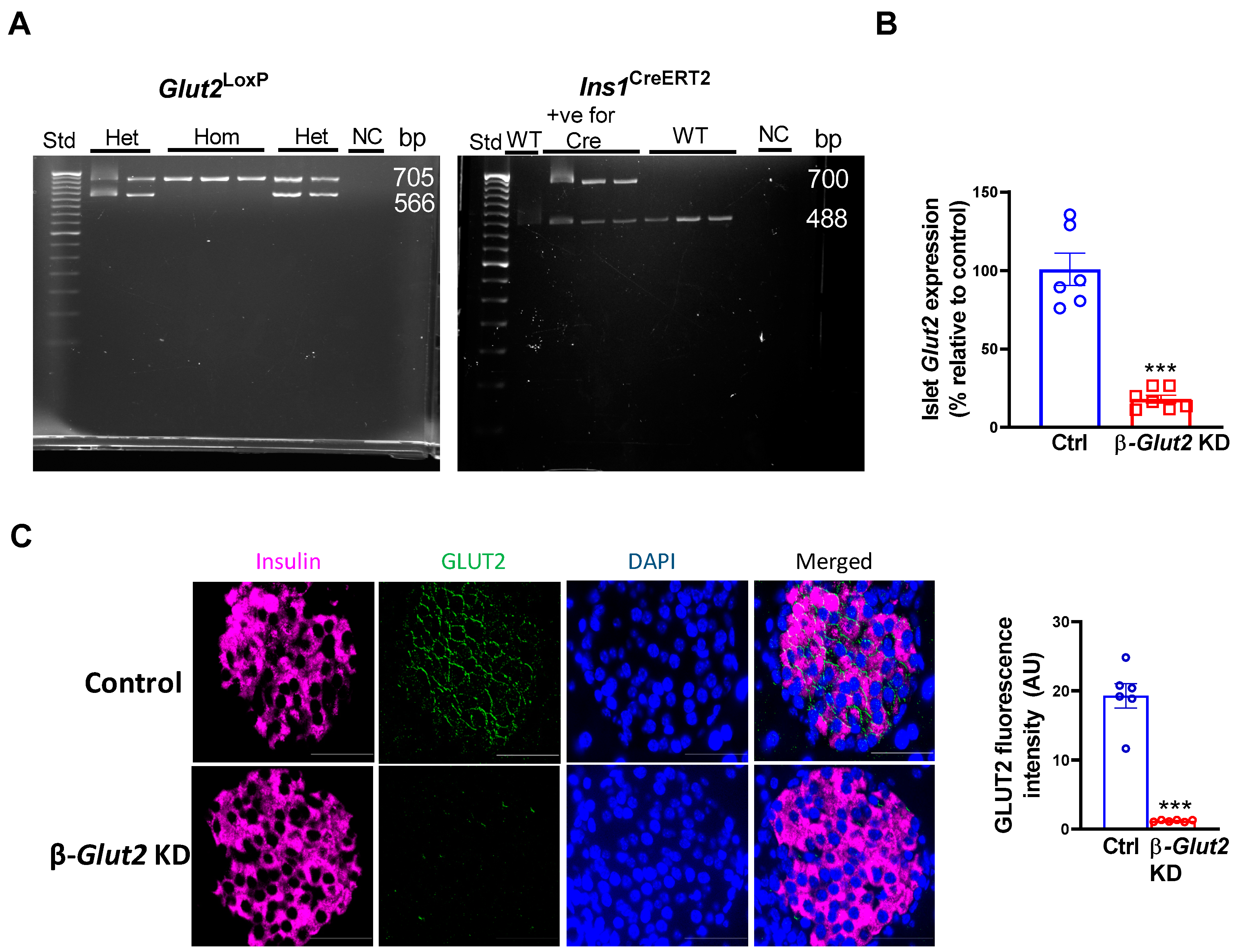
Figure 1. Generation and validation of β-cell Glut2 knockdown (β-Glut2 KD) mice. Representative genotypes of mice used in this study (A). Het, heterozygous; Hom, homozygous for Glut2loxP. Std, standard DNA ladder; NC, negative control; bp, base pair. Results from qRT-PCR showing knockdown of islet Glut2 in 8 weeks old male β-Glut2 KD mice one week after inducing β-cell Glut2 deficiency, n = 6 or 7 (B). Immunofluorescence demonstrating deficiency of β-cell GLUT2 in male β-Glut2 KD mice, n = 6 or 7 (C). Three islets per section and 3 sections per mouse were quantified (C). Scale bar is 50 µm. Ctrl, control Glut2loxP/loxP mice; AU, arbitrary unit. Student’s unpaired t-test was used for comparisons, *** p < 0.001. Error bars are mean ± SEM.
Biomolecules 13 00540 g001
Figure 2. β-cell Glut2 knockdown (β-Glut2 KD) mice have normal glucose-stimulated insulin secretion. Fasting (6 h) plasma insulin levels (0 min) and glucose-stimulated insulin secretion (20 min) in male, n = 6 or 7 (A) and female, n = 8, 10, or 11 (B) mice 4 and 8 weeks after inducing β-cell Glut2 deficiency. The mice were 8–10 weeks old when β-cell Glut2 deficiency was induced. Repeated measures two-way ANOVA followed by Bonferroni’s multiple comparison test were used for comparisons. ** p < 0.01, *** p < 0.001 vs. their corresponding baseline (0 min) groups. Error bars are mean ± SEM.
Biomolecules 13 00540 g002
Figure 3. β-cell Glut2 knockdown (β-Glut2 KD) mice exhibit normal glucose tolerance. Results obtained from oral glucose tolerance tests in male, n = 9 or 12 (A,B) and female, n = 6–10 (C,D) mice 4 (A,C) and 8 (B,D) weeks after inducing β-cell Glut2 deficiency. Area under the curve (AUC) is shown in inset bar graphs. The mice were 8–10 weeks old when β-cell Glut2 deficiency was induced. Immunofluorescence, n = 6 (E) and qRT-PCR, n = 6 (F) demonstrating upregulation of islet Glut1 in female β-Glut2 KD mice after completion of the study. 3 islets per section and 3 sections per mouse were quantified (E). Ins +ve and −ve, quantification of GLUT1 fluorescence in insulin positive and negative staining. Scale bar is 50 µm. Control, Glut2loxP/loxP mice; AU, arbitrary unit. Student’s unpaired t-test was used for comparisons, * p < 0.05, ** p < 0.01, *** p < 0.001. Error bars are mean ± SEM.
Biomolecules 13 00540 g003
Disclaimer/Publisher’s Note: The statements, opinions and data contained in all publications are solely those of the individual author(s) and contributor(s) and not of MDPI and/or the editor(s). MDPI and/or the editor(s) disclaim responsibility for any injury to people or property resulting from any ideas, methods, instructions or products referred to in the content.