Article

New Anti-Hypoxic Metabolites from Co-Culture of Marine-Derived Fungi Aspergillus carneus KMM 4638 and Amphichorda sp. KMM 4639

by
Elena B. Belousova
1,
Olesya I. Zhuravleva
1,2,
Ekaterina A. Yurchenko
1,*,
Galina K. Oleynikova
1,
Alexandr S. Antonov
1,
Natalya N. Kirichuk
1,
Viktoria E. Chausova
1,
Yuliya V. Khudyakova
1,
Alexander S. Menshov
1,
Roman S. Popov
1,
Ekaterina S. Menchinskaya
1,
Evgeny A. Pislyagin
1,
Valery V. Mikhailov
1,* and
Anton N. Yurchenko
1,*
1
G.B. Elyakov Pacific Institute of Bioorganic Chemistry, Far Eastern Branch of the Russian Academy of Sciences, Prospect 100-Letiya Vladivostoka, 159, Vladivostok 690022, Russia
2
Institute of High Technologies and Advanced Materials, Far Eastern Federal University, 10 Ajax Bay, Russky Island, Vladivostok 690922, Russia
*
Authors to whom correspondence should be addressed.
Biomolecules 2023, 13(5), 741; https://doi.org/10.3390/biom13050741
Submission received: 28 February 2023 / Revised: 20 April 2023 / Accepted: 21 April 2023 / Published: 25 April 2023
(This article belongs to the Special Issue Marine Natural Compounds with Biomedical Potential 2.0)

Abstract

:
The KMM 4639 strain was identified as Amphichorda sp. based on two molecular genetic markers: ITS and β-tubulin regions. Chemical investigation of co-culture marine-derived fungi Amphichorda sp. KMM 4639 and Aspergillus carneus KMM 4638 led to the identification of five new quinazolinone alkaloids felicarnezolines A–E (15), a new highly oxygenated chromene derivative oxirapentyn M (6) and five previously reported related compounds. Their structures were established using spectroscopic methods and by comparison with related known compounds. The isolated compounds showed low cytotoxicity against human prostate and breast cancer cells but felicarnezoline B (2) protected rat cardiomyocytes H9c2 and human neuroblastoma SH-SY5Y cells against CoCl2-induced damage.

1. Introduction

An in-depth study of the organisms stored in bioresource collections can move us towards achieving one of the United Nations Sustainable Development Goals of 2015, aimed at improving human health [1]. For microorganisms (especially fungi), new results can be achieved using the OSMAC approach, including the co-cultivation of different strains. Their influence on each other can force them to produce new compounds with biological properties, which may also be of practical importance [2].
Quinazoline alkaloids with a pyrazino[2,1-b]quinazoline-3,6-dione moiety are not rare metabolites for a number of Aspergillus and Penicillium species [3,4]. The pyrazinequinazoline core is unchanged in the majority of fungal metabolites (Figure 1). Usually, substituents originate from amino acid residues, which form the framework, and are located only at the C-3 and C-14 positions. The rare exceptions are carnequinazolines B and C from Aspergillus carneus Blochwitz which include hydroxy groups in the benzene ring [5] and scedapins A-E from Scedosporium apiospermum Sacc. ex Castell. & Chalm. without nitrogen between C-1 and C-3 in a piperazine ring [6].
Piperazine-containing quinazolines demonstrated a wide spectrum of biological activity. For instance, fumiquinazoline C was reported as an inhibitor against α-glucosidase [7] and fumiquinazoline Q is a promising drug candidate for cardiovascular disease treatment [8]. Scedapin C exhibited significant antiviral activity against the hepatitis C virus [6]. Polonimides A–C showed low cytotoxic activity against epithelial human cells [9] and fumigatosides E demonstrated significant antifungal activity [10].
Recently a series of twelve drimane sesquiterpenes were produced in response to the addition of a marine fungus Amphichorda sp. KMM 4638 (earlier identified and published as Beauveria felina (D.C.) J.W. Carmich. and Isaria felina (D.C.) Fr.) to the 7-day-old culture of a marine-derived fungus Aspergillus carneus KMM 4639 [11]. Further investigation of metabolites from this co-culture made it possible to isolate five new quinazolinone alkaloid felicarnezolines A–E (15), new highly oxygenated chromene derivative oxirapentyn M (6) and five known metabolites oxirapentyn B (7) [12], cinereain (8) [13], carneamide A (9) [5], aspergillicin A (10) [14], isaridin E (11) [15], earlier isolated from axenic cultures of Amphichorda sp. KMM 4638 and Aspergillus carneus KMM 4639.
In this work, we report on the isolation and structure elucidation of fungal co-culture-derived metabolites as well as the investigation of their cytotoxicity against human prostate PC-3 and breast cancer MCF-7 cells. Moreover, the effects of compounds at non-toxic concentrations against cobalt (II) chloride-induced damage of rat cardiomyocytes H9c2 and human neuroblastoma SH-SY5Y cells are determined.

2. Materials and Methods

2.1. General Experimental Procedures

Optical rotations were measured on a Perkin–Elmer 343 polarimeter (Perkin Elmer, Waltham, MA, USA). UV spectra were recorded on a Shimadzu UV-1601PC spectrometer (Shimadzu Corporation, Kyoto, Japan) in methanol. CD spectra were measured with a Chirascan-Plus CD spectrometer (Applied Photophysics Ltd., Leatherhead, UK) in methanol. NMR spectra were recorded in CDCl3, acetone-d6 and DMSO-d6, on a Bruker DPX-300 (Bruker BioSpin GmbH, Rheinstetten, Germany), a Bruker Avance III-500 (Bruker BioSpin GmbH, Rheinstetten, Germany) and a Bruker Avance III-700 (Bruker BioSpin GmbH, Rheinstetten, Germany) spectrometers, using TMS as an internal standard. HRESIMS spectra were measured on a Maxis impact mass spectrometer (Bruker Daltonics GmbH, Rheinstetten, Germany). Microscopic examination and photography of fungal cultures were performed with an Olympus CX41 microscope equipped with an Olympus SC30 digital camera. Detailed examination of ornamentation of the fungal conidia was performed by scanning electron microscopy (SEM) EVO 40.
Low-pressure liquid column chromatography was performed using Si gel (50/100 μm, Imid Ltd., Krasnodar, Russia) and Gel ODS-A (12 nm, S—75 um, YMC Co., Ishikawa, Japan). Plates precoated with Si gel (5–17 μm, 4.5 × 6.0 cm, Imid Ltd., Krasnodar, Russia) and Si gel 60 RP-18 F254S (20 × 20 cm, Merck KGaA, Darmstadt, Germany) were used for thin-layer chromatography. Preparative HPLC was carried out on an Agilent 1100 chromatograph (Agilent Technologies, Santa Clara, CA, USA) with an Agilent 1100 refractometer (Agilent Technologies, Santa Clara, CA, USA) and a Shimadzu LC-20 chromatograph (Shimadzu USA Manufacturing, Canby, OR, USA) with a Shimadzu RID-20A refractometer (Shimadzu Corporation, Kyoto, Japan) using YMC ODS-AM (YMC Co., Ishikawa, Japan) (5 µm, 10 × 250 mm), YMC ODS-AM (YMC Co., Ishikawa, Japan) (5 µm, 4.6 × 250 mm) and Hydro-RP (Phenomenex, Torrance, CA, USA) (4 μm, 250 × 10 mm) columns.

2.2. Fungal Strains

The A. carneus fungal strain was isolated from superficial mycobiota of the brown alga Laminaria sachalinensis (Miyabe) collected on Kunashir Island and was identified based on morphological evaluation by Dr. Mikhail V. Pivkin from the Pacific Institute of Bioorganic Chemistry (PIBOC). The strain is stored in the Collection of Marine Microorganisms, PIBOC, Vladivostok, Russia, under the code KMM 4638.
The Amphichorda sp. fungal strain was isolated from marine sediments collected at a depth of 10 m (Van Phong Bay, the South China Sea, Vietnam) during the 34th expedition of r/v “Akademik Oparin” and was identified based on morphological evaluation by Dr. Natalya N. Kirichuk from the Pacific Institute of Bioorganic Chemistry (PIBOC). The strain is stored in the Collection of Marine Microorganisms, PIBOC, Vladivostok, Russia, under the code KMM 4639.

2.3. DNA Extraction and Amplification

The cultures used for the molecular studies were grown on malt extract agar under 25 °C for 7 d. Genomic DNA was isolated from fungal mycelium grown on MEA (malt extract agar) at 25 °C for 7 days, using the MagJET Plant Genomic DNA Kit (Thermo Fisher Scientific, Waltham, MA, USA), according to the manufacturer’s protocol. PCR was conducted using GoTaq Flexi DNA Polymerase (Promega, Madison, WI, USA). For amplification of the internal transcribed spacer region (ITS) were used the primer pair ITSpr1 (5′-GCGTTGATATACGTCCCTGCC-3′) and ITSpr9 (5′-CCTTGGTCCGTGTTTCAAGA-3′) [16]. The reaction profile was 95 °C for 300 s, 35 cycles of 94 °C for 20 s, 60 °C for 20 s, and 72 °C for 90 s, and finally 72 °C for 300 s. For amplification of the partial beta/β-tubulin gene region the primer pair Bt-2a and Bt-2b was used [17]. The reaction profile was 95 °C for 300 s, 35 cycles of 94 °C for 20 s, 60 °C for 20 s, and 72 °C for 60 s, and finally 72 °C for 300 s. The amplified ITS and partial beta/β-tubulin genes were purified with the ExoSAP-IT™ PCR Product Cleanup Reagent (Thermo Fisher Scientific, Waltham, MA, USA). Sequencing was bidirectionally performed with the same primers on an Applied Biosystems SeqStudio Genetic Analyzer (Thermo Fisher Scientific, Waltham, MA, USA) using the Big Dye Terminator reagent kit, version 3.1. Gene sequences were deposited in GenBank under accession numbers OQ344667 for the ITS gene region and OQ418107 for the partial β-tubulin gene region (Table 1).

2.4. Phylogenetic Analysis

The ITS gene and partial β-tubulin gene sequences were aligned by MEGA X software version 11.0.9 [18] using the Clustal W algorithm. The available homologs were searched in the GenBank database (http://ncbi.nlm.nih.gov, accessed on 8 February 2023) using the BLASTN algorithm (http://www.ncbi.nlm.nih.gov/BLAST, accessed on 8 February 2023). Phylogenetic analysis was conducted using MEGA X software version 11.0.9 [18]. Phylogenetic trees were constructed on model-tested alignments according to the maximum likelihood algorithm. The topologies of the trees were evaluated by 1000 bootstrap replicates.

2.5. Cultivation of Fungus

The fungi Aspergillus carneus and Amphichorda sp. were cultivated separately at 22 °C for 7 days in Erlenmeyer flasks (500 mL) each containing 20 g of rice, 20 mg of yeast extract, 10 mg of KH2PO4, and 40 mL of natural seawater. After that, Amphichorda sp. mycelium was inoculated into 20 flasks with Aspergillus carneus culture. Then fungal cultures were co-cultivated for 14 days.

2.6. Extraction and Isolation

At the end of the incubation period, the mycelia and medium were homogenized and extracted with EtOAc (1 L). The obtained extract was concentrated to dryness. The residue (17.5 g) was dissolved in H2O−EtOH (4:1) (100 mL) and extracted with n-hexane (0.2 L × 3), EtOAc (0.2 L × 3) and BuOH (0.2 L × 3). After evaporation of the EtOAc layer, the residual material (5.5 g) was passed through a silica column (3 × 14 cm), which was eluted first with n-hexane (200 mL), then by a step gradient from 5% to 50% EtOAc in n-hexane (total volume 20 L). Fractions of 250 mL were collected and combined on the basis of TLC (Si gel, toluene–isopropanol 6:1 and 3:1, v/v).
The n-hexane–EtOAc (90:10) eluate (1.2 g) was separated on a Gel ODS-A column (1.5 × 8 cm), which was eluted by a step gradient from 40% to 80% CH3OH in H2O (total volume 1 L), to yield subfractions I and II. Subfraction I (60% CH3OH, 150 mg) was purified by RP HPLC on a Hydro-RP column eluted with CH3CN-H2O (60:40) to yield individual compound 8 (2.4 mg) and fraction I-1 (62 mg). Fraction I-1 was purified by RP HPLC on a Hydro-RP column eluted with CH3CN-H2O (40:60) to yield 1 (1.3 mg) and 2 (1.7 mg). Subfraction II (80% CH3OH, 53 mg) was purified by RP HPLC on a Hydro-RP column eluted with CH3OH-H2O (80:20) and then with CH3CN-H2O (70:30) to yield individual compound 5 (2.0 mg).
The n-hexane-EtOAc (80:20, 646 mg) fraction was separated on a Gel ODS-A column (1.5 × 8 cm), which was eluted by a step gradient from 40% to 80% CH3OH in H2O (total volume 1 L) to yield subfraction III. Subfraction III (60% CH3OH, 205 mg) was purified by RP HPLC on a Hydro-RP column eluting with CH3OH-H2O (80:20) to yield fractions III-1 (125.5 mg) and III-2 (16.2). Fraction III-1 was purified by RP HPLC on a YMC-Pack Pro C-18 column eluted with CH3CN-H2O (40:60) and then with CH3OH-H2O (10:90) to yield 3 (0.7 mg) and 4 (10.0 mg). Fraction III-2 was purified by RP HPLC on a Hydro-RP column eluted with CH3CN-H2O (55:45) to yield 6 (1.5 mg) and 7 (2.1 mg).
The n-hexane-EtOAc (70:30, 1.0 g) fraction was separated on a Gel ODS-A column (1.5 × 8 cm), which was eluted by a step gradient from 40% to 80% CH3OH in H2O (total volume 1 L) to yield subfractions IV and V. Subfraction IV (60% CH3OH, 282 mg) was purified by RP HPLC on a Hydro-RP column eluting with CH3OH-H2O (70:30) and then on a YMC ODS-A column eluted with CH3CN-H2O (40:60) to yield 11 (5.0 mg). Subfraction V (80% CH3OH, 168 mg) was purified by RP HPLC on a Hydro-RP column eluted with CH3CN-H2O (55:45) to yield 9 (16.8 mg) and 10 (17.3 mg).

2.7. Spectral Data

Felicarnezoline A (1): amorphous solids; [ α ] D 20 −24.7 (c 0.08, MeOH); CD (c 2.9 × 10⁻⁴, CH3OH), λmax (∆ε) 193 (+22.10), 208 (−15.20), 224 (+17.49), 267 (+6.52), 307 (−6.85) nm, see Supplementary Figure S56; UV (CH3OH) λmax (log ε) 307 (3.82), 273 (3.39), 211 (4.39) nm, see Supplementary Figure S57; 1H and 13C NMR data, see Table 1, Supplementary Figures S1–S6; HRESIMS m/z 270.0891 [M − H] (calcd. for C14H12N3O3, 270.0884, Δ −2.5 ppm), 294.0852 [M + Na]+ (calcd. for C14H13N3O3Na, 294.0849, Δ −1.0 ppm).
Felicarnezoline B (2): amorphous solids; [ α ] D 20 −35.4 (c 0.07, MeOH); CD (c 4.2 × 10⁻⁴, CH3OH), λmax (∆ε) 196 (+14.1), 213 (−1.99), 239 (+2.78), 261 (+2.92), 287 (−2.57) nm, see Supplementary Figure S58; UV (CH3OH) λmax (log ε) 360 (3.83), 327 (3.62), 317 (3.66), 298 (3.60), 252 (4.11), 241 (4.08), 214 (4.36) nm, see Supplementary Figure S59; 1H and 13C NMR data, see Supplementary Figures S7–S13; HRESIMS m/z 286.0842 [M − H] (calcd. for C14H12N3O4, 286.0833, Δ −3.2 ppm), 310.0802 [M + Na]+ (calcd. for C14H13N3O4Na, 310.0798, Δ −1.2 ppm).
Felicarnezoline C (3): amorphous solids; 1H NMR data, see Supplementary Figure S14; HRESIMS m/z 334.1530 [M + Na]+ (calcd. for C18H21N3O2, 334.1526, Δ −1.2 ppm).
Felicarnezoline D (4): amorphous solids; [ α ] D 20 −20.0 (c 0.07, MeOH); CD (c 8.0 × 10⁻⁷, CH3OH), λmax (∆ε) 197 (+30.1), 230 (−1.94), 259 (+2.05), 278 (−0.32), 304 (+4.12) nm, see Supplementary Figure S60; UV (CH3OH) λmax (log ε) 343 (4.21), 234 (4.54), 214 (4.62), 197 (4.60), nm, see Supplementary Figure S61; 1H NMR data, see Supplementary Figure S15; HRESIMS m/z 350.1471 [M + Na]+ (calcd. for C18H23N3O3Na, 350.1475, Δ 1.1 ppm).
Felicarnezoline E (5): amorphous solids; 1H NMR data, see Supplementary Figure S16; HRESIMS m/z 352.1628 [M + Na]+ (calcd. for C18H23N3O3Na, 352.1632, Δ 1.1 ppm).
Oxirapentyn M (6): amorphous solids; [ α ] D 20 −71.8° (c 0.04, MeOH); CD (c 2.0 × 10⁻³, CH3OH), λmax (∆ε) 220 (−0.23), 295 (−0.04) nm, see Supplementary Figure S62; UV (CH3OH) λmax (log ε) 225 (3.74) nm, see Supplementary Figure S63; 1H and 13C NMR data, see Table 2, Supplementary Figures S17–S23; HRESIMS m/z 335.1492 [M − H] (calcd. for C18H23O6, 335.1492, Δ 2.5 ppm), 359.1453 [M + Na]+ (calcd. for C18H24O6Na, 359.1453, Δ 3.3 ppm).

2.8. Stereo Configuration Analysis of Amino Acids in Compounds 1, 2, 5 and 6

The compounds (0.6 mg of each) were placed in glass ampoules and dissolved in 6 N HCl (1.2 mL). Solutions in ampoules were frozen in liquid nitrogen, then vacuumed, sealed, and heated at 105 °C for 24 h. Then, the cooled reaction mixture was diluted with distilled water and concentrated in vacuo. The obtained hydrolysates of compounds 15 and standard amino acid Val of the L- and D-configurations (0.2 mg each) were dissolved in 0.1 mL of distilled water, then 0.4 mL of 1M NaHCO3 and 0.2 mL of a 1% solution of Marfey’s reagent in acetone were added. The reaction mixtures were kept at 37 °C for 75 min and 0.05 mL of 1M HCl was added. Then, obtained L-FDDA derivatives were analyzed by HPLC-UV in a gradient from 25% to 65% of MeCN in H2O (0.1% TFA) over 40 min at 20 °C using the YMC C-18 Pro column.

2.9. Cell Lines and Culture Conditions

The human prostate cancer PC-3, human breast cancer MCF-7, and human neuroblastoma SH-SY5Y cells were purchased from ATCC (Manassas, VA, USA). Rat cardiomyocyte H9c2 cells were kindly provided by Prof. Dr. Gunhild von Amsberg from Martini-Klinik Prostate Cancer Center, University Hospital Hamburg-Eppendorf, Hamburg, Germany.
PC-3, MCF-7, SH-SY5Y and H9c2 cells were cultured in DMEM medium (Biolot, St. Petersburg, Russia) containing 10% fetal bovine serum (Biolot, St. Petersburg, Russia) and 1% penicillin/streptomycin (Biolot, St. Petersburg, Russia) at 37 °C in a humidified atmosphere with 5% (v/v) CO2.
Initially, the cells were incubated in culture flasks until subconfluent (~80%). For testing, the cells were seeded at concentrations of 5 × 10³ cells/well (PC-3, MCF-7, SH-SY5Y cells) or 3 × 10³ cells/well (H9c2 cells), and experiments were started after 24 h.

2.10. In Vitro MTT-Based Cytotoxicity Assay

The in vitro cytotoxicity of individual substances was determined by the MTT method (3-(4,5-dimethylthiazol-2-yl)-2,5-diphenyltetrazolium bromide), according to the manufacturer’s instructions (Sigma-Aldrich, St. Louis, MO, USA).
Investigated compounds were dissolved in DMSO at a concentration of 10 mM. This solution was used to obtain the required concentration of compounds in the cell suspension so that the concentration of DMSO in the cell suspension did not exceed 1%.
The cells were treated with the investigated compounds for 24 h or 48 h, and MTT reagent was added to each well of the plate. The vehicle with DMSO at a concentration of 1% was used as a control. The absorbance of formed formazan was measured at λ = 570 nm using a Multiskan FC microplate photometer (Thermo Scientific, Waltham, MA, USA) and expressed in optical units (o.u.). The results are presented as % of viable cells relative to the vehicle, and 50% inhibition concentration of cell viability (IC50) was calculated.

2.11. CoCl2-Mimic Hypoxia Modeling

The SH-SY5Y and H9c2 cells were treated with a dH2O-solution of CoCl2 at a concentration of 500 µM for 1 h. Then, compounds at a concentration of 10 µM were added for 23 h (SH-SY5Y cells) or 47 h (H9c2 cells). The viability of the SH-SY5Y and H9c2 cells was measured by an MTT assay as described above.

2.12. Reactive Oxygen Species (ROS) Level Assay

The SH-SY5Y and H9c2 cells were treated with a dH2O-solution of CoCl2 at a concentration of 500 µM for 1 h. Then, compounds at a concentration of 10 µM were added for 3 h. The non-treated cells were used as a control. The 20 μL of 2,7-dichlorodihydrofluorescein diacetate solution (H2DCFDA, Molecular Probes, Eugene, OR, USA) was added to each well (10 μM, final concentration) and the plate was incubated for an additional 10 min at 37 °C. The intensity of dichlorofluorescein fluorescence was measured with a PHERAstar FS plate reader (BMG Labtech, Ortenberg, Germany) at λex = 485 nm and λem = 518 nm. The data were processed by MARS Data Analysis v. 3.01R2 (BMG Labtech, Germany). The results were presented as relative fluorescence units.

2.13. Superoxide Dismutase Activity Detection

The SH-SY5Y and H9c2 cells were seeded in 6-well plates for 24 h. The dH2O-solution of CoCl2 at a concentration of 500 µM was added for 1 h and then compounds at a concentration of 10 µM were added for 3 h. The non-treated cells were used as a control.
The cells were washed with PBS twice, collected in 1.5 mL tubes and lysed with RIPA buffer (Sigma-Aldrich, St. Louis, MO, USA). Then the cells were centrifuged at 14,000 per min (Eppendorf, Framingham, MA, USA) and the supernatant was used. The reaction mixture contains 1 mL of 26.9 µM EDTA, 1 mL of 4.04 µM tetrazolium nitroblue tetrazolium chloride (Dia-M, Novosibirsk, Russia), 1 mL of 65 µM 5-methylphenazinium methyl sulfate (Dia-M, Novosibirsk, Russia) and 26 mL PBS. The supernatant, 1 mM NADH and reaction mixture were added to a 96-well plate as 1:1:28 for 10 min in dark and the reaction was stopped by light. The mixture with RIPA buffer instead of supernatant was used as a control. The mixture without NADH was used as a background. The optical density of reaction mixtures was measured at λ = 540 nm using a Multiskan FC microplate photometer (Thermo Scientific, Waltham, MA, USA).
The total protein concentration of each probe was measured by Bradford assay.
The activity of superoxide dismutase (Asod, u/mg) was mind as inhibition of nitroblue tetrazolium reduction (T, %):
T (%) = (OD control − OD test)/OD control × 100%
and calculated per total protein content.
The total protein content was measured by Bradford assay. Bovine serum albumin was used to build a calibration curve.

2.14. Statistical Data Evaluation

All the data were obtained in three independent replicates, and the calculated values were expressed as a mean ± standard error mean (SEM). A Student’s t-test was performed using SigmaPlot 14.0 (Systat Software Inc., San Jose, CA, USA) to determine the statistical significance. The differences were considered statistically significant at p < 0.05.

3. Results

3.1. Molecular Identification of the KMM 4639 Fungal Strain

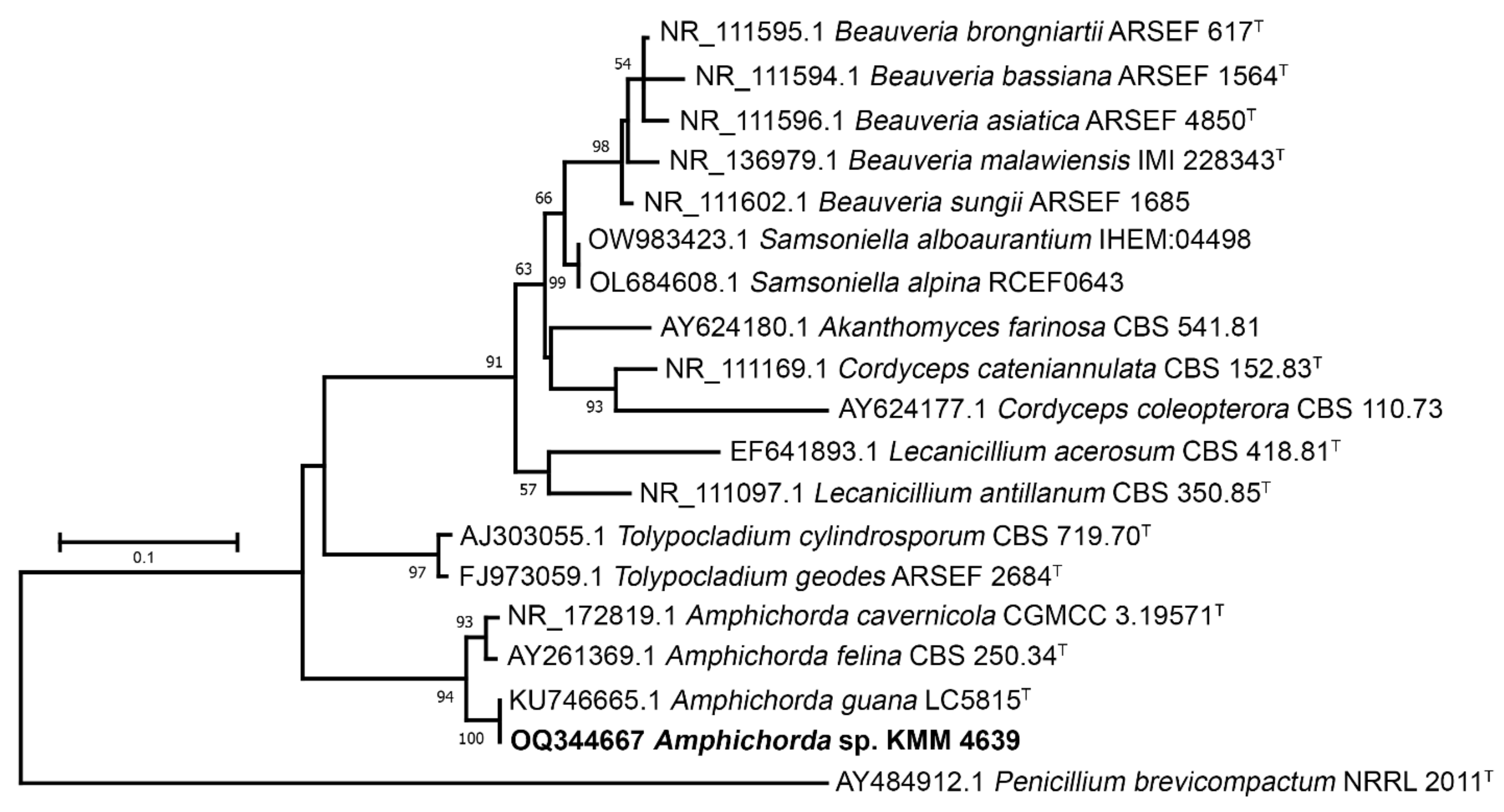
The KMM 4639 strain was identified using two molecular markers: ITS and β-tubulin regions. A 1300 bp fragment of the ITS gene region and a 350 bp fragment of the β-tubulin gene was successfully amplified. BLAST search results indicated that the sequences were 98% (for β-tubulin) and 100% (for ITS) identical to the sequences of the ex-type strain Amphichorda guana Z.F. Zhang, F. Liu & L. Cai (CGMCC 3.17908). Phylogenetic ML trees constructed on the basis of the ITS gene (Figure 2) and partial β-tubulin gene sequences (Figure 3) clearly showed that the strain KMM 4639 clusters with ex-type strain Amphichorda guana CGMCC 3.17908 within the family Cordycipitaceae.
Within these studies, the strain KMM 4639 was identified as Amphichorda sp. Given the percentage of similarity in tubulin, it is necessary to use additional molecular markers to confirm the species affiliation of the strain.

3.2. Isolated Compouns from Co-Culture

The fungi Aspergillus carneus and Amphichorda sp. were cultivated separately for 7 days in a solid rice medium. After that, Amphichorda sp. mycelium was inoculated into flasks with Aspergillus carneus culture. Then fungal cultures were co-cultivated for 14 days. The EtOAc extract of the mycelium of co-culture was purified by a combination of Si gel and ODS-AM column chromatography and reversed phase HPLC to yield compounds 112 (Figure 4).

3.3. Structure Characterization of New Compounds

The molecular formula of 1 was determined to be C14H13N3O3 by an HRESIMS peak at m/z 272.1034 [M + H]+ and was in accordance with 13C NMR data. The 1H and 13C NMR (Table 1), DEPT and HSQC spectra showed the signals of one amide proton (δH 8.48), 1,2-disubstitute benzene-ring (δH 7.69, 7.91, 8.06 and 8.36), two methyl (δH 0.92 and 1.23) and two methine (δH 2.47 and 5.57) groups, three sp2-quaternary carbon signals (δC 121.9, 139.2 and 146.0) along with three amide carbonyls (δC 156.5, 159.7 and 165.2).
The chemical shift values of C-1, C-4–C-14 carbon atoms in the 13C NMR spectrum closely resemble those of carnequinazoline A [5] indicating a quinazoline moiety in 1. The correlations H-14/H-15/H3-16(H3-17) observed in the 1H-1H COSY spectrum and HMBC correlations from H-16 (δH 0.92) to C-14 (δC 61.7) and from H-14 (δH 5.57) to C-1 (δC 165.3), C-4 (δC 139.2), C-12 (δC 159.7), C-15 (δC 33.7) and C-17 (δC 19.0) revealed the location of an isopropyl group at C-14 in 1. The presence in the 13C NMR spectrum of compound 1 of an additional signal of the amide carbon atom and the absence of signals of the 2-methylpropylidene group compared to carnequinazoline A suggested that this side group at C-3 was oxidized to carbonyl, which corresponds to the molecular formula of the compound.
The absolute configurations of C-14 stereocenter in 1 were established by Marfey’s method [19] as R. Analysis of the L-FDAA derivative of the amino acid residue obtained by acid hydrolysis of compound 1 showed it to be derivative of D-Val standard sample (Figures S38 and S39, Supplementary Material). Compound 1 was named felicarnezoline A.
The molecular formula of 2 was determined to be C14H13N3O4 from an HRESIMS peak at m/z 288.0983 [M + H]+ and was in accordance with 13C NMR data (Table 2). The 1H and 13C NMR data for 2 were in good agreement with those for felicarnezoline A (1) with the exception of proton and carbon signals of the benzene ring. The molecular mass difference of 16 mass units between 1 and 2, characteristic of the 1,2,3-trisubstituted benzene ring proton multiplicity and HMBC correlations (Figure 5) from H-8 (δH 7.40) to C-6 (δC 134.4), C-7 (δC 153.6) and C-10 (δC 117.7), from H-9 (δH 7.58) to C-6, C-7 and C-11 (δC 121.9) indicated the location of the hydroxy group at C-7.
The absolute configurations of C-14 stereocenter in 2 were established by Marfey’s method as R. Analysis of L-FDAA derivative of the amino acid residue obtained by acid hydrolysis of compound 1 showed it to be derivative of D-Val standard sample (Figures S40 and S41, Supplementary Material). Compound 2 was named felicarnezoline B.
The NMR data of 3, 4 and 5 corresponded to the signals of known carnequinazolines A, B [5] and dihydrocinereain [20], respectively. The presence of the D-valine in the structures of compounds 1 and 2 lets us suggest this amino acid in structures 3, 4, and 5 instead of L-valine in known related compounds. To prove the absolute stereochemistry of 3, 4, and 5 by Marfey’s method their acid hydrolysis was carried out and L-FDAA derivatives of the amino acids were obtained. Thus, the presence of D-valine in structures 3, 4, and 5 was established (Figures S42–S47, Supplementary Material).
We hypothesize that D-valine may also be present in compounds 8, 10 and 11 instead of L-valine, as previously described for cinereain, aspergillicin A [14] and isaridin E. However, due to the insufficient amount of these compounds, the use of Marfey’s method turned out to be impossible to determine the configuration of the amino acids included in the structures of these compounds.
The HRESIMS of 6 showed the quasimolecular ion at m/z 359.1453 [M + Na]+. These data, coupled with 13C NMR spectral data (DEPT), established the molecular formula of all compounds as C18H24O6. A close inspection of 1H and 13C NMR data of 6 by DEPT and HSQC (Table 3) indicated the presence of four methyl (δH 2.11, δC 20.8, δH 1.40, δC 22.0 и δH 1.90, δC 23.7, δH 1.19, δC 25.4), two methylene (δH 1.57, 2.55 δC 32.3, δH 5.22, 5.30 δC 122.1) and six methine groups (δH 2.90, δC 37.5, δH 3.08, δC 64.3, δH 4.17, δC 67.8, δH 4.09, δC 68.2, δH 3.84, δC 72.4, δH 4.90, δC 73.8), five of them oxygen-bearing, one sp2- (δC 126.5) and three sp3 (δC 76.3, 88.9 and 90.3) oxygen-bearing carbons, three sp2 (δC 106.0, 122.7, 145.4) and two sp3C 57.9, 74.3) quaternary carbons along with one carboxy group (δC 170.3).
The general features of 1H and 13C NMR spectra of 6 indicated that the compound belongs to the family of oxirapentyns, highly oxidized polyketides previously isolated from Amphichorda sp. KMM 4639.
A comparison of the NMR spectra of 6 with those of oxirapentyn F [21] revealed some similarities, including three methyl, two methylene and one acetoxy groups. The HMBC correlations (Figure 5) from H-4′a (δH 5.22) to C-2′ (δC 84.9) and C-5′ (δC 23.7), from H3-5′ (δH 1.90) to C-2′, C-3′ (δC 126.5) and C-4′ (δC 122.1), from H-6 (δH 4.17) to C-1′ (δC 87.1) revealed the presence of a 3-methyl-3-buten-1-ynyl side chain in 6. The 13C NMR spectrum of 6 showed signals at δC 57.9 and 64.3, which are the characteristic signals of epoxide carbons. The 1H NMR spectrum indicated the presence of one epoxy proton at δH 3.08. These data together with the HMBC correlations from H2-3 (δH 1.57, 2.55) and H-9 (δH 4.09) to C-4 (δC 57.9) and C-5 (δC 64.3) indicated the location of an epoxy group at C-4–C-5. The 1H–1H COSY correlations of H-6(OH)/H-7/H-8(OH)/H-9 indicated the presence of hydroxy groups at C-6 and C-8 in 6.
The key correlations observed in ROESY spectrum H-2/H-3α, H-5/H-3β and H-9/H3-11, H-3α and 1H-1H coupling constants suggested the α-orientation of epoxide and hydroxy group at C-6 and β-orientation of acetate and hydroxy group at C-8 in 6. The compound was named oxirapentyn M. Due to the shortage of the pure sample, Mosher’s method was not feasible to study the absolute configuration of oxirapentyn M.

3.4. Cytotoxic Activity of Isolated Compounds

The cytotoxic effects of the compounds 15 and 711 against human prostate cancer PC-3, breast cancer MCF-7 and neuroblastoma SH-SY5Y cells as well as rat normal cardiomyocytes H9c2 were investigated and presented in Table 4. Compound 6 was isolated in an insufficient amount and was not tested.
The investigated compounds did not show significant cytotoxic activity toward both cancer and normal cells. Only compound 4 had weak toxicity for MCF-7 and SH-SY5Y cells and prolonged treatment of the cells with this one did not result in stronger cytotoxicity.

3.5. Effects of Isolated Compounds in CoCl2-Mimic Hypoxia

The cytoprotective effects of isolated compounds were investigated in cobalt (II) chloride (CoCl2)-mimic hypoxic model using neuronal SH-SY5Y cells and normal cardiomyocytes H9c2.
The viability of CoCl2-treated SH-SY5Y cells dramatically decreased and was only 27.8% in comparison with non-treated cells (Figure 6a). Felicarnezoline B (2) statistically significantly increased the viability of CoCl2-treated SH-SY5Y cells by 72.6%. The effect of compounds 1, 5 and 11 was observed but was not statistically significant.
The viability of CoCl2-treated cardiomyocytes H9c2 was 37.8% only in comparison with non-treated cells (Figure 6b). Felicarnezoline B (2) statistically significantly increased the viability of CoCl2-treated H9c2 cells by 19.1%. The effect of other compounds was not statistically significant.
Treating with CoCl2 induces oxidative stress in cells and the protective effect of compounds may be caused by their antioxidant properties. To detect this, the effects of active compounds 1, 2, 5 and 11 on reactive oxygen species (ROS) level in CoCl2-treated cells was investigated (Figure 7).
The ROS level in SH-SY5Y cells treated with CoCl2 for 4 h was increased by 49.6% (Figure 7a). Compounds 2 and 5 statistically significantly decreased the ROS level in these cells by 18.8% and 11.0%, respectively. The ROS level in CoCl2-treated H9c2 cells increases by 32.6% after 4 h of treatment (Figure 7b). Compounds 2 and 5 statistically diminished the ROS level in these cells by 25.7% and 18.6%, respectively.
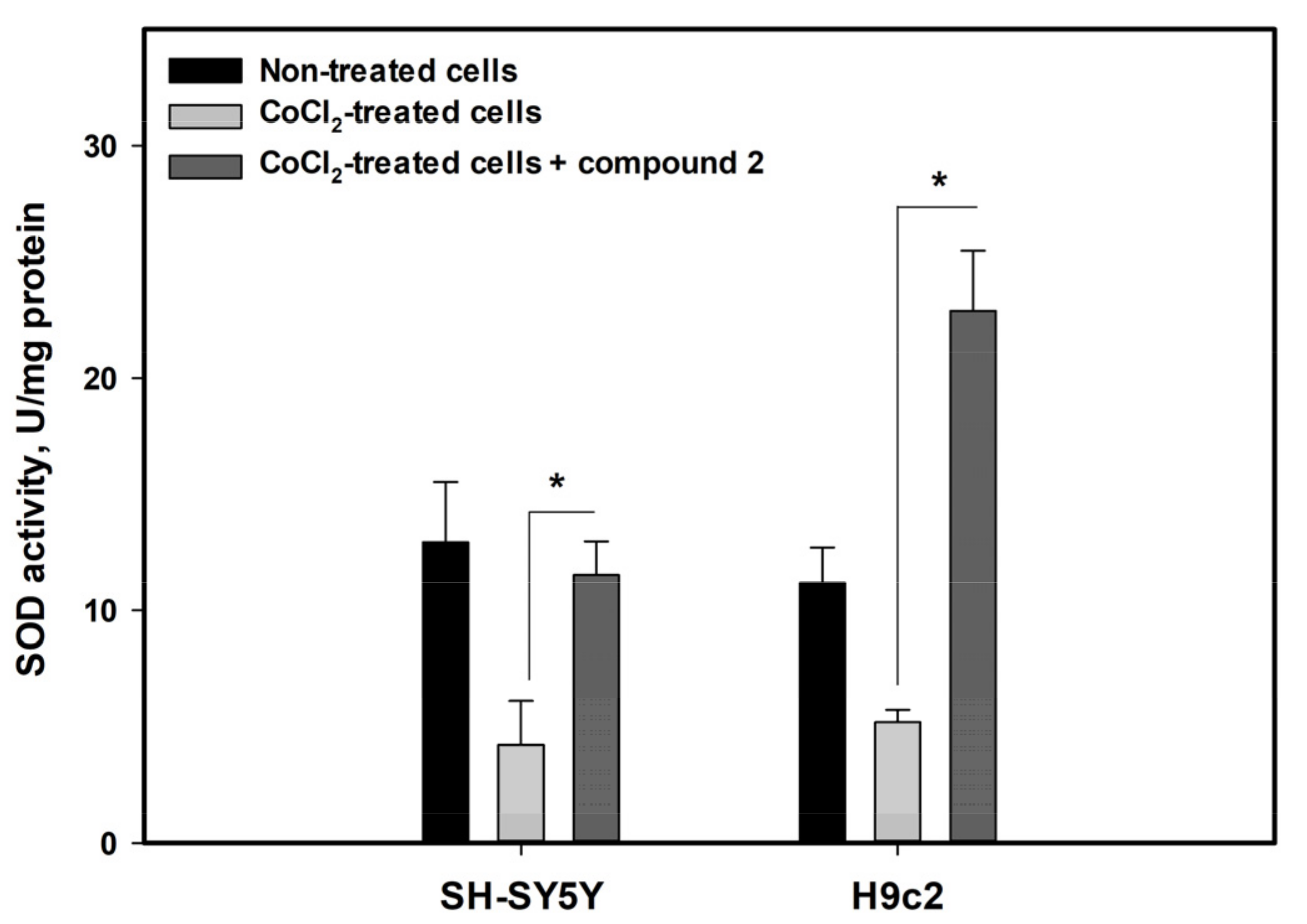
In addition, the effect of felicarnezoline B (2) on the activity of the superoxide dismutase (SOD) antioxidant enzyme was investigated to verify its influence on the intracellular antioxidant system in CoCl2-treated cells (Figure 8).
The activity of SOD in SH-SY5Y and H9c2 cells treated with CoCl2 for 4 h was dramatically diminished by 67.4% and 53.5%, respectively. The incubation with felicarnezoline B (2) resulted in a significant increase in SOD activity in both cases.

4. Discussions

Previously, we described the isolation of drimane sesquiterpenoids from a co-culture of Aspergillus carneus KMM 4638 and Amphichorda sp. KMM 4639, some of which had a pronounced cytotoxic activity and inhibited the cell cycle of human breast cancer MCF-7 cells [11]. Now we have managed to isolate new substances with cytoprotective properties, and it is obvious that co-cultivation led to their production (Table 5, Figure 9).
Earlier, carnequinazolines A-C were isolated from Aspergillus carneus KMM 4638 [5]. These compounds have L-valine, at the same time new alkaloids have D-valine in their structures. In addition, felicarnezolines A (1) and B (2) contain a 1,3-diketopiperazine moiety in their structure, which is unique for quinazoline alkaloids and may be biosynthesized by the action of oxidative enzymes of Amphichorda sp.
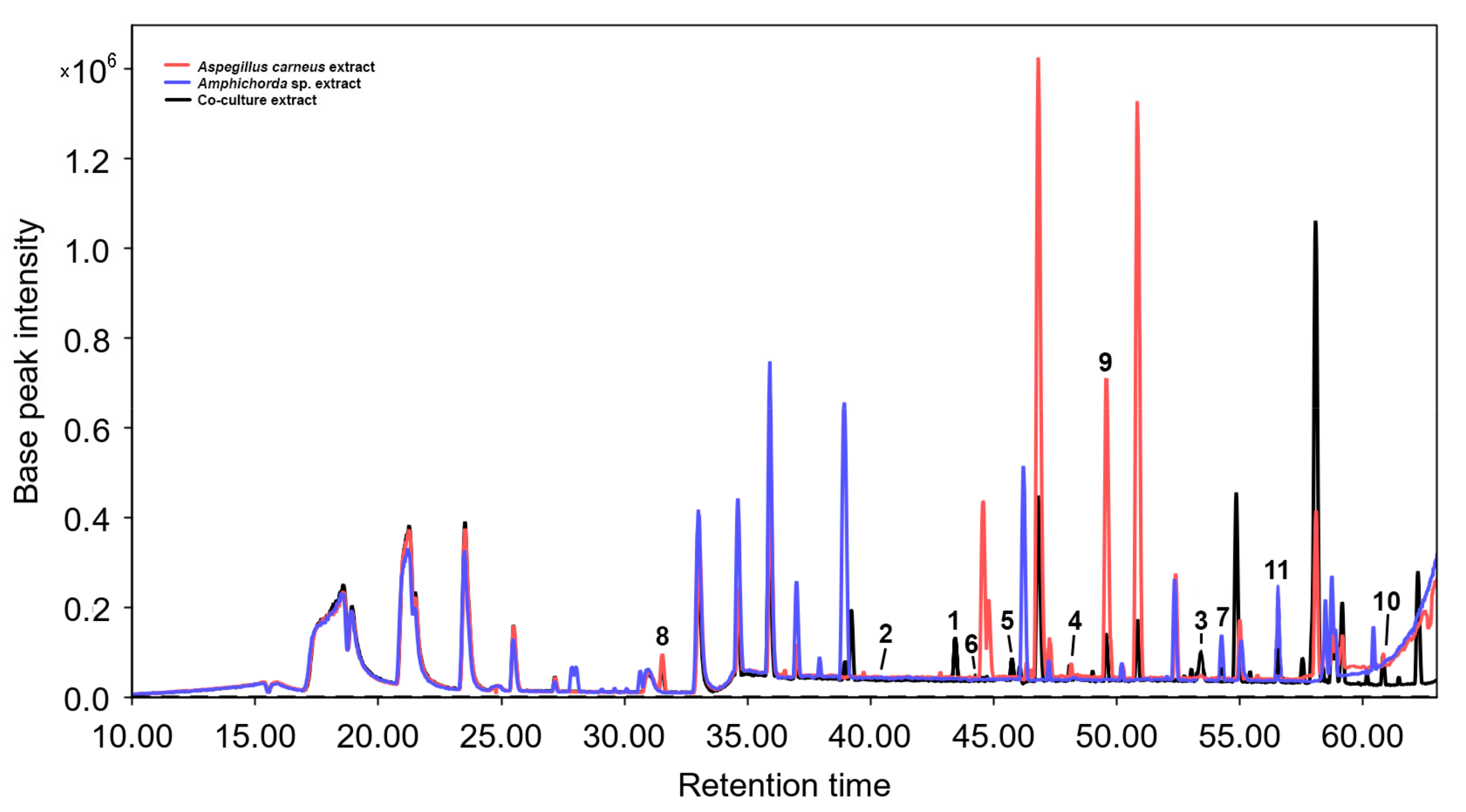
It should be noted that “undetected” compounds may still be present in the extracts in amounts beyond detection. This is partly confirmed by the results of HPLC-MS (Figure 9). Moreover, a detailed evaluation of the obtained results of Marfey’s analysis revealed that we cannot exactly prove the absence of L-valine impurity in the hydrolyzate of samples of compounds 14. The chromatograms of these samples show small peaks with an RT close to L-valine. The ratio of peak areas assigned to D- and L-valine is approximately 4:1. Thus, we can deal with a mixture of enantiomers in the case of these compounds. At the same time, the chromatogram of the FDAA-derivatives of the hydrolyzate of compound 5 does not contain visible peaks with the RT of L-valine; therefore, we can accurately state the absence of an impurity of the “original” stereoisomer (dihydrocinereain).
Stereo conversion of amino acids is not unusual in microorganisms, including fungi. As a rule, this occurs either under the action of amino acid racemase or amino acid oxidase, followed by reductive amination [22]. For example, L-alanine racemase has been described in the fungus Amphichorda felina (D.C.) Fr., which produces cyclosporin C (contains D-alanine in the structure) [23]. Unfortunately, no relevant data about fungal valine racemase or valine oxidase could be found. Finally, epimerization of L-valine can occur as part of a dipeptide precursor of 15, similar to the proposed mechanism for the penicillin producing Acremonium chrysogenum (Thirum. & Sukapure) W. Gams [24]. Thus, the presence of D-valine in compounds 15 instead of L-valine in related compounds from Aspergillus carneus KMM 4638 monoculture is most likely the result of the action of the fungus Amphichorda sp. KMM 4639. However, the proof of this assumption should be the subject of further detailed studies.
The KMM 4639 strain of the Amphichorda sp. fungus is very interesting due to the high production of oxidated secondary metabolites [21] and the co-cultivation of various fungal strain stable resulted in the isolation of new biologically active compounds.
It should be noted that the earlier fungal strain KMM 4639 was misidentified using morphological features and published as Isaria felina and Beauveria felina. These species as well as the genus Amphichorda belong to the Cordycipitaceae family and may have similar metabolism [25,26,27]. So, a number of depsipeptides isolated earlier by us from the KMM 4639 fungal strain were isolated from Amphichorda guana fungus [25] as well as Beauveria felina [27] and Isaria sp. [15]. However, the ability of this strain to produce highly oxygenated metabolites is not typical for this group of fungi, which requires further research for a more complete realization of its biotechnological potential.
Under the highly competitive conditions of densely populated microbial communities, marine fungi are forced to produce secondary metabolites both for aggression against competitors and for defense against them [28]. The first purpose is served by various substances with cytotoxic activity, the second one is provided, first of all, by antioxidants. However, in most cases, conclusions about antioxidant properties are made by the presence of DPPH-scavenging activity in a cell-free test [29]. Obviously, the manifestation of these radical-scavenging properties in a living system can be very limited and the investigation of the antioxidant properties of compounds in in vitro conditions is necessary. Earlier the antioxidant effects of fungal echinulin-related indoldiketopiperazine and desoxyisoaustamide alkaloids were investigated in cell models of oxidative stress induced with neurotoxins [30,31]. Now we used the cobalt (II) chloride solution for modeling hypoxia in two different cell lines.
The CoCl2-induced hypoxia-mimic in vitro model is widely used to search for cytoprotective compounds, despite some limitations [32]. Similar to real oxygen deprivation, treating with CoCl2 induces oxidative stress and mitochondrial DNA damage [33]. Various investigations found that chronic low oxygen level or hypoxia/reoxygenation conditions results in a significant increase in ROS level in H9c2 cells [34,35]. It is considered that the decrease in the activity of the mitochondrial electron transport chain during hypoxia slows down electron transfer, increasing the likelihood of an undesired electron transition to molecular oxygen, which produces a highly efficient reactive superoxide anion (O2−) [36]. However, some literature data confirm that CoCl2 caused an increase in ROS level in H9c2 cells while 1% O2 hypoxia resulted in a decrease in ROS level, but the SOD activity was decreased in both cases [37]. In addition, the activation of cellular antioxidant machinery can protect the cell from CoCl2 -caused damage similar to oxygen deprivation induced cell death [38,39].
In our experiments, the CoCl2 treatment decreases human neuroblastoma SH-SY5Y cells and rat H9c2 cardiomyocytes viability and was accompanied by diminished SOD activity and increased intracellular ROS level. The cytoprotective effect of the new alkaloid felicarnezoline B against CoCl2-induced damage is obviously related to SOD activity enhancement. SOD is one of the components of the first line of the antioxidant defense system [40] and may be induced via Keap-1/Nrf2- or NF-κB-dependent pathways as well as more specifically the specificity protein (Sp)-1, CCAAT-Enhancer-Binding Proteins (C/EBP), and the activator proteins (AP)-1 and-2, which exert similar effects on the regulation of SOD genes [41]. So, the influence of felicarnezoline B (2) on the cellular antioxidant system in hypoxia-mimic conditions is unknown now and is interesting for future investigation.

5. Conclusions

As a result of the mixed cultivation of two microfilamentous fungi, Aspergillus carneus KMM 4638 and Amphichorda sp. KMM 4639, five new alkaloids and one new chromene derivative were obtained. Felicarnezoline B has shown a good protective effect in hypoxia-mimic conditions via antioxidant pathways.

Supplementary Materials

The following supporting information can be downloaded at: https://www.mdpi.com/article/10.3390/biom13050741/s1, Figures S1–S37: NMR spectra of compounds 111; Figures S38–S57: HPLC profile of FDAA-derivatives of compounds 15; Figures S58–S61: MS spectrum of compounds 1, 2, 4, 6; Figures S62, S64, S66, S68: CD spectra of compounds 1, 2, 4, 6; Figures S63, S65, S67, S69: UV spectra of compounds 1, 2, 4, 6.

Author Contributions

Conceptualization, A.N.Y.; Data curation, O.I.Z.; Funding acquisition, V.V.M.; Investigation, E.B.B., O.I.Z., E.A.Y., G.K.O., A.S.A., N.N.K., V.E.C., Y.V.K., A.S.M., R.S.P., E.S.M. and E.A.P.; Project administration, V.V.M.; Resources, A.N.Y.; Supervision, A.N.Y.; Validation, E.A.Y. and A.N.Y.; Visualization, E.B.B., E.A.Y. and V.E.C.; Writing—original draft, E.B.B., O.I.Z., E.A.Y., N.N.K. and V.E.C.; Writing—review & editing, V.V.M. and A.N.Y. All authors have read and agreed to the published version of the manuscript.

Funding

This research was funded by a grant from the Ministry of Science and Higher Education of Russian Federation 15.BRK.21.0004 (Contract No. 075-15-2021-1052).

Institutional Review Board Statement

Not applicable.

Informed Consent Statement

Not applicable.

Data Availability Statement

Not applicable.

Acknowledgments

The study was carried out using the Collective Facilities Center “Collection of Marine Microorganisms PIBOC FEB RAS” and on the equipment of the Collective Facilities Center, “The Far Eastern Center for Structural Molecular Research (NMR/MS) PIBOC FEB RAS”.

Conflicts of Interest

The authors declare no conflict of interest. The funders had no role in the design of the study; in the collection, analyses, or interpretation of data; in the writing of the manuscript; or in the decision to publish the results.

References

  1. Carlsen, L.; Bruggemann, R. The 17 United Nations’ sustainable development goals: A status by 2020. Int. J. Sustain. Dev. World Ecol. 2022, 29, 219–229. [Google Scholar] [CrossRef]
  2. Knowles, S.L.; Raja, H.A.; Roberts, C.D.; Oberlies, N.H. Fungal-fungal co-culture: A primer for generating chemical diversity. Nat. Prod. Rep. 2022, 1557–1573. [Google Scholar] [CrossRef]
  3. Xu, K.; Yuan, X.L.; Li, C.; Li, X.D. Recent discovery of heterocyclic alkaloids from marine-derived Aspergillus species. Mar. Drugs 2020, 18, 54. [Google Scholar] [CrossRef] [PubMed]
  4. Zhang, P.; Wei, Q.; Yuan, X.; Xu, K. Newly reported alkaloids produced by marine-derived Penicillium species (covering 2014–2018). Bioorg. Chem. 2020, 99, 103840. [Google Scholar] [CrossRef] [PubMed]
  5. Zhuravleva, O.I.; Afiyatullov, S.S.; Denisenko, V.A.; Ermakova, S.P.; Slinkina, N.N.; Dmitrenok, P.S.; Kim, N.Y. Secondary metabolites from a marine-derived fungus Aspergillus carneus Blochwitz. Phytochemistry 2012, 80, 123–131. [Google Scholar] [CrossRef] [PubMed]
  6. Huang, L.H.; Xu, M.Y.; Li, H.J.; Li, J.Q.; Chen, Y.X.; Ma, W.Z.; Li, Y.P.; Xu, J.; Yang, D.P.; Lan, W.J. Amino Acid-Directed Strategy for Inducing the Marine-Derived Fungus Scedosporium apiospermum F41-1 to Maximize Alkaloid Diversity. Org. Lett. 2017, 19, 4888–4891. [Google Scholar] [CrossRef]
  7. Tuan, C.D.; Hung, N.V.; Minh, L.T.H.; Lien, H.T.H.; Chae, J.W.; Yun, H.Y.; Kim, Y.H.; Cuong, P.V.; Huong, D.T.M. A New Indole Glucoside and Other Constituents from the Sea Cucumber-Derived Aspergillus fumigatus M580 and Their Biological Activities. Rec. Nat. Prod. 2022, 16, 633–638. [Google Scholar] [CrossRef]
  8. Fan, Y.Q.; Li, P.H.; Chao, Y.X.; Chen, H.; Du, N.; He, Q.X.; Liu, K.C. Alkaloids with cardiovascular effects from the marine-derived fungus Penicillium expansum Y32. Mar. Drugs 2015, 13, 6489–6504. [Google Scholar] [CrossRef]
  9. Guo, X.C.; Zhang, Y.H.; Gao, W.B.; Pan, L.; Zhu, H.J.; Cao, F. Absolute configurations and chitinase inhibitions of quinazoline-containing diketopiperazines from the marine-derived fungus penicillium polonicum. Mar. Drugs 2020, 18, 479. [Google Scholar] [CrossRef]
  10. Limbadri, S.; Luo, X.; Lin, X.; Liao, S.; Wang, J.; Zhou, X.; Yang, B.; Liu, Y. Bioactive novel indole alkaloids and steroids from deep sea-derived fungus aspergillus fumigatus SCSIO 41012. Molecules 2018, 23, 2379. [Google Scholar] [CrossRef]
  11. Zhuravleva, O.I.; Belousova, E.B.; Oleinikova, G.K.; Antonov, A.S.; Khudyakova, Y.V.; Rasin, A.B.; Popov, R.S.; Menchinskaya, E.S.; Trinh, P.T.H.; Yurchenko, A.N.; et al. Cytotoxic Drimane-Type Sesquiterpenes from Co-Culture of the Marine-Derived Fungi Aspergillus carneus KMM 4638 and Beauveria felina (=Isaria felina) KMM 4639. Mar. Drugs 2022, 20, 584. [Google Scholar] [CrossRef]
  12. Smetanina, O.F.; Yurchenko, A.N.; Afiyatullov, S.S.; Kalinovsky, A.I.; Pushilin, M.A.; Khudyakova, Y.V.; Slinkina, N.N.; Ermakova, S.P.; Yurchenko, E.A. Oxirapentyns B–D produced by a marine sediment-derived fungus Isaria felina (DC.) Fr. Phytochem. Lett. 2012, 5, 165–169. [Google Scholar] [CrossRef]
  13. Cutler, H.G.; Arrendale, R.F.; Cole, P.D.; Springer, J.P.; Arison, B.H.; Roberts, R.G. Cinereain: A novel metabolite with plant growth regulating properties from Botrytis cinerea. Agric. Biol. Chem. 1988, 52, 1725–1733. [Google Scholar] [CrossRef]
  14. Capon, R.J.; Skene, C.; Stewart, M.; Ford, J.; O’Hair, R.A.J.; Williams, L.; Lacey, E.; Gill, J.H.; Heiland, K.; Friedel, T. Aspergillicins A–E: Five novel depsipeptides from the marine-derived fungus Aspergillus carneus. OBC Org. Biomol. Chem. 2003, 1, 1856–1862. [Google Scholar] [CrossRef]
  15. Sabareesh, V.; Ranganayaki, R.S.; Raghothama, S.; Bopanna, M.P.; Balaram, H.; Srinivasan, M.C.; Balaram, P. Identification and characterization of a library of microheterogeneous cyclohexadepsipeptides from the fungus Isaria. J. Nat. Prod. 2007, 70, 715–729. [Google Scholar] [CrossRef] [PubMed]
  16. Scholin, C.A.; Herzog, M.; Sogin, M.; Anderson, D.M. Identification of group- and strain-specific genetic markers for globally distributed Alexandrium (Dinophyceae). II. Sequence analysis of a fragment of the LSU rRNA gene. J. Phycol. 1994, 30, 999–1011. [Google Scholar] [CrossRef]
  17. Glass, N.L.; Donaldson, G.C. Development of primer sets designed for use with the PCR to amplify conserved genes from filamentous ascomycetes. Appl. Environ. Microbiol. 1995, 61, 1323–1330. [Google Scholar] [CrossRef]
  18. Kumar, S.; Stecher, G.; Li, M.; Knyaz, C.; Tamura, K. MEGA X: Molecular evolutionary genetics analysis across computing platforms. Mol. Biol. Evol. 2018, 35, 1547. [Google Scholar] [CrossRef] [PubMed]
  19. Fujii, K.; Shimoya, T.; Ikai, Y.; Oka, H.; Harada, K.-I. Further application of advanced Marfey's method for determination of absolute configuration of primary amino compound. Tetrahedron Lett. 1998, 39, 2579–2582. [Google Scholar] [CrossRef]
  20. Zhuravleva, O.I.; Afiyatullov, S.S.; Yurchenko, E.A.; Denisenko, V.A.; Kirichuk, N.N.; Dmitrenok, P.S. New Metabolites from the Algal Associated Marine-derived Fungus Aspergillus carneus. Nat. Prod. Commun. 2013, 8, 1071–1074. [Google Scholar] [CrossRef]
  21. Yurchenko, A.N.; Smetanina, O.F.; Kalinovsky, A.I.; Pushilin, M.A.; Glazunov, V.P.; Khudyakova, Y.V.; Kirichuk, N.N.; Ermakova, S.P.; Dyshlovoy, S.A.; Yurchenko, E.A.; et al. Oxirapentyns F-K from the marine-sediment-derived fungus Isaria felina KMM 4639. J. Nat. Prod. 2014, 77, 1321–1328. [Google Scholar] [CrossRef]
  22. Friedman, M. Chemistry, Nutrition, and Microbiology of D-Amino Acids. J. Agric. Food Chem. 1999, 47, 3457–3479. [Google Scholar] [CrossRef]
  23. Xu, L.; Li, Y.; Biggins, J.B.; Bowman, B.R.; Verdine, G.L.; Gloer, J.B.; Alspaugh, J.A.; Bills, G.F. Identification of cyclosporin C from Amphichorda felina using a Cryptococcus neoformans differential temperature sensitivity assay. Appl. Microbiol. Biotechnol. 2018, 102, 2337–2350. [Google Scholar] [CrossRef] [PubMed]
  24. Kallow, W.; Neuhof, T.; Arezi, B.; Jungblut, P.; von Döhren, H. Penicillin biosynthesis: Intermediates of biosynthesis of δ-l-α-aminoadipyl-l-cysteinyl-d-valine formed by ACV synthetase from Acremonium chrysogenum. FEBS Lett. 1997, 414, 74–78. [Google Scholar] [CrossRef]
  25. Liang, M.; Lyu, H.N.; Ma, Z.Y.; Li, E.W.; Cai, L.; Yin, W.B. Genomics-driven discovery of a new cyclodepsipeptide from the guanophilic fungus Amphichorda guana. Org. Biomol. Chem. 2021, 19, 1960–1964. [Google Scholar] [CrossRef]
  26. Jiang, M.; Wu, Z.; Wu, Q.; Yin, H.; Guo, H.; Yuan, S.; Liu, Z.; Chen, S.; Liu, L. Amphichoterpenoids A–C, unprecedented picoline-derived meroterpenoids from the ascidian-derived fungus Amphichorda felina SYSU-MS7908. Chin. Chem. Lett. 2021, 32, 1893–1896. [Google Scholar] [CrossRef]
  27. Du, F.Y.; Zhang, P.; Li, X.M.; Li, C.S.; Cui, C.M.; Wang, B.G. Cyclohexadepsipeptides of the isaridin class from the marine-derived fungus Beauveria felina EN-135. J. Nat. Prod. 2014, 77, 1164–1169. [Google Scholar] [CrossRef] [PubMed]
  28. Raffa, N.; Keller, N.P. A call to arms: Mustering secondary metabolites for success and survival of an opportunistic pathogen. PLoS Pathog. 2019, 15, e1007606. [Google Scholar] [CrossRef] [PubMed]
  29. Vitale, G.A.; Coppola, D.; Palma Esposito, F.; Buonocore, C.; Ausuri, J.; Tortorella, E.; de Pascale, D. Antioxidant Molecules from Marine Fungi: Methodologies and Perspectives. Antioxidants 2020, 9, 1183. [Google Scholar] [CrossRef] [PubMed]
  30. Smetanina, O.F.; Yurchenko, A.N.; Girich, E.V.; Trinh, P.T.H.; Antonov, A.S.; Dyshlovoy, S.A.; von Amsberg, G.; Kim, N.Y.; Chingizova, E.A.; Pislyagin, E.A.; et al. Biologically Active Echinulin-Related Indolediketopiperazines from the Marine Sediment-Derived Fungus Aspergillus niveoglaucus. Molecules 2020, 25, 61. [Google Scholar] [CrossRef] [PubMed]
  31. Zhuravleva, O.I.; Antonov, A.S.; Trang, V.T.D.; Pivkin, M.V.; Khudyakova, Y.V.; Denisenko, V.A.; Popov, R.S.; Kim, N.Y.; Yurchenko, E.A.; Gerasimenko, A.V.; et al. New Deoxyisoaustamide Derivatives from the Coral-Derived Fungus Penicillium dimorphosporum KMM 4689. Mar. Drugs 2021, 19, 553. [Google Scholar] [CrossRef] [PubMed]
  32. Tripathi, V.K.; Subramaniyan, S.A.; Hwang, I. Molecular and Cellular Response of Co-cultured Cells toward Cobalt Chloride (CoCl2)-Induced Hypoxia. ACS Omega 2019, 4, 20882–20893. [Google Scholar] [CrossRef]
  33. Muñoz-Sánchez, J.; Chánez-Cárdenas, M.E. The use of cobalt chloride as a chemical hypoxia model. J. Appl. Toxicol. 2019, 39, 556–570. [Google Scholar] [CrossRef]
  34. Bao, M.; Huang, W.; Zhao, Y.; Fang, X.; Zhang, Y.; Gao, F.; Huang, D.; Wang, B.; Shi, G. Verapamil Alleviates Myocardial Ischemia/Reperfusion Injury by Attenuating Oxidative Stress via Activation of SIRT1. Front. Pharmacol. 2022, 13. [Google Scholar] [CrossRef] [PubMed]
  35. Jin, H.J.; Li, C.G. Tanshinone IIA and cryptotanshinone prevent mitochondrial dysfunction in hypoxia-induced H9c2 Cells: Association to mitochondrial ROS, intracellular nitric oxide, and calcium levels. Evid.-Based Complement. Altern. Med. 2013, 2013, 610694. [Google Scholar] [CrossRef]
  36. Thomas, L.W.; Ashcroft, M. Exploring the molecular interface between hypoxia-inducible factor signalling and mitochondria. Cell. Mol. Life Sci. 2019, 76, 1759–1777. [Google Scholar] [CrossRef] [PubMed]
  37. Osuru, H.P.; Lavallee, M.; Thiele, R.H. Molecular and Cellular Response of the Myocardium (H9C2 Cells) Towards Hypoxia and HIF-1α Inhibition. Front. Cardiovasc. Med. 2022, 9, 711421. [Google Scholar] [CrossRef]
  38. Baskaran, R.; Kalaiselvi, P.; Huang, C.-Y.; Padma, V.V. Neferine, a bisbenzylisoquinoline alkaloid, offers protection against cobalt chloride-mediated hypoxia-induced oxidative stress in muscle cells. Integr. Med. Res. 2015, 4, 231–241. [Google Scholar] [CrossRef]
  39. Baskaran, R.; Poornima, P.; Priya, L.B.; Huang, C.-Y.; Padma, V.V. Neferine prevents autophagy induced by hypoxia through activation of Akt/mTOR pathway and Nrf2 in muscle cells. Biomed. Pharmacother. 2016, 83, 1407–1413. [Google Scholar] [CrossRef]
  40. Ighodaro, O.M.; Akinloye, O.A. First line defence antioxidants-superoxide dismutase (SOD), catalase (CAT) and glutathione peroxidase (GPX): Their fundamental role in the entire antioxidant defence grid. Alex. J. Med. 2018, 54, 287–293. [Google Scholar] [CrossRef]
  41. Rosa, A.C.; Corsi, D.; Cavi, N.; Bruni, N.; Dosio, F. Superoxide Dismutase Administration: A Review of Proposed Human Uses. Molecules 2021, 26, 1844. [Google Scholar] [CrossRef] [PubMed]
Figure 1. General framework of pyrazinequinazoline alkaloids.
Biomolecules 13 00741 g001
Figure 2. ML tree based on ITS gene sequences showing phylogenetic position of the strain KMM 4639 within family Cordycipitaceae. Bootstrap values (%) of 1000 replications. Nodes with confidence values greater than 50% are indicated. The scale bars represent 0.1 substitutions per site. T—ex-type strain.
Biomolecules 13 00741 g002
Figure 3. ML tree based on partial β-tubulin gene sequences showing phylogenetic position of the strain KMM 4639. Bootstrap values (%) of 1000 replications. Nodes with confidence values greater than 50% are indicated. The scale bars represent 0.1 substitutions per site. T—ex-type strain.
Biomolecules 13 00741 g003
Figure 4. Chemical structures of 111.
Biomolecules 13 00741 g004
Figure 5. Key COSY (bold lines) and HMBC (arrows) correlations of 2 and 6.
Biomolecules 13 00741 g005
Figure 6. Influence of the compounds on viability of CoCl2-treated (a) SH-SY5Y and (b) H9c2 cells. The viability of nontreated (control) cells were 100.3 ± 1.2%. All experiments were carried out in three independent replicates and the data are presented as a mean ± SEM. * indicated the statistically significant differences (p < 0.05).
Biomolecules 13 00741 g006
Figure 7. Influence of the compounds on ROS level in CoCl2-treated (a) SH-SY5Y and (b) H9c2 cells. All experiments were carried out in three independent replicates and the data are presented as a mean ± SEM. * indicated the statistically significant differences (p < 0.05).
Biomolecules 13 00741 g007
Figure 8. Influence of felicarnezoline B (2) on superoxide dismutase activity in CoCl2-treated cells. All experiments were carried out in three independent replicates and the data are presented as a mean ± SEM. * indicated the statistically significant differences.
Biomolecules 13 00741 g008
Figure 9. HPLC MS chromatogram of extracts of Aspergillus carneus and Amphichorda sp. monocultures as well as their co-culture. The numbers correspond to the numbers of the isolated compounds.
Biomolecules 13 00741 g009
Table 1. Strains used in phylogenetic analysis and GenBank accession numbers of ITS and β-tubulin molecular data.
TaxonCollection NumberGenBank Accession Numbers
ITSβ-Tubulin
Akanthomyces farinosaCBS 541.81AY624180AY624218
Amphichorda cavernicolaCGMCC 3.19571TNR 172819
Amphichorda guanaCGMCC 3.17908TKU746665KU746757
Amphichorda felinaCBS 250.34TAY261369
Amphichorda sp.KMM 4639OQ344667OQ418107
Tolypocladium cylindrosporumCBS 719.70TAJ303055
Tolypocladium geodesARSEF 2684TFJ973059
Beauveria malawiensisIMI 228343TNR_136979
Beauveria sungiiARSEF 1685TNR_111602
Beauveria asiaticaARSEF 4850TNR_111596
Beauveria brongniartiiARSEF 617TNR_111595
Beauveria bassianaARSEF 1564TNR_111594
Cordyceps cateniannulataCBS 152.83TNR_111169KY574462
Cordyceps coleopteroraCBS 110.73AY624177AY624216
Lecanicillium acerosumCBS 418.81TEF641893
Lecanicillium antillanumCBS 350.85TNR_111097MG993038
Penicillium brevicompactumNRRL 2011TAY484912DQ645784
Samsoniella alboaurantiumIHEM:04498OW983423
Samsoniella alpinaRCEF0643OL684608
T—ex-type strain.
Table 2. 1H and 13C NMR data for compounds 1 and 2.
Position1 a2 b
δC, TypeδH, Mult, J in HzδC, TypeδH, Mult, J in Hz
1165.2, C 165.1, C
2 8.48, brs 8.54, s
3156.5, C 156.0, C
4139.2, C 138.0, C
5
6146.0, C 134.3, C
7129.8, CH8.06, d (8.2)153.6, C
8135.5, CH7.91, td (8.1, 1.6)119.1, CH7.40, d (8.0)
9130.1, CH7.69, td (7.9, 1.1)131.5, CH7.58, t (8.0)
10127.2, CH8.36, dd (8.0, 1.2)117.7, CH7.81, d (8.0)
11121.9, C 121.9, C
12159.7, C 159.3, C
13
1461.7, CH5.57, dd (4.2, 0.8)61.8, CH5.55, d (4.3)
1533.7, CH2.27, m33.6, CH2.46, m
1616.9, CH30.92, d (6.9)16.8, CH30.93, d (6.9)
1719.0, CH31.23, d (6.9)19.0, CH31.23, d (6.9)
a chemical shifts were measured in CDCl3 at 500.13 MHz for 1H and 125.77 MHz for 13C. b chemical shifts were measured in CDCl3 at 700.13 MHz for 1H and 176.04 MHz for 13C.
Table 3. 1H and 13C NMR data for compound 6.
Position6 a
δC, TypeδH, Mult, J in Hz
174.3, C
273.8, CH4.90 t (3.2)
332.3, CH2α: 2.55, dd (14.3, 2.8)
β: 1.57, dd (14.3, 3.4)
457.9, C
564.3, CH3.08, s
667.8, CH4.17, dd (9.9, 3.2)
737.5, CH2.90, dd (10.2, 2.0)
872.4, CH3.84, dt (10.4, 2.5)
968.2, CH4.09, d (2.9)
1025.4, CH31.19, s
1122.0, CH31.40, s
1′87.1, C
2′84.9, C
3′126.5, C
4′122.1, CH25.30, brs
5.22, m
5′23.7, CH31.90, brs
1″170.3, C
2″20.8, CH32.11, s
6-OHOH2.21, d (3.6)
8-OHOH2.31, d (10.4)
a chemical shifts were measured in CDCl3 at 500.13 MHz for 1H and 125.77 MHz for 13C.
Table 4. The cytotoxic activity of compounds 15 and 711.
CompoundCell Lines
PC-3MCF-7SH-SY5YH9c2
IC50, µM
1>100>100>100>100
2>100>100>100>100
3>10092.5 ± 3.1>100>100
4>10068.7 ± 1.672.9 ± 2.8>100
583.8 ± 5.586.3 ± 2.3>100>100
7>100>100>100>100
8>100>10093.8 ± 3.8>100
9>100>100>100>100
10>100>100>100>100
11>100>100>100>100
The concentration of half maximum effect (IC50) is presented as a mean ± standard error of mean (SEM). All experiments were carried out in three independent replicates.
Table 5. Metabolites of co-culture of Aspergillus carneus KMM 4638 and Amphichorda sp. KMM 4639.
CompoundSource
Amphichorda sp. KMM 4639Aspergillus carneus KMM 4638Co-Culture
1+
2+
3+
4+
5+
6+
7+ [12]+
8+
9+ [5]+
10+ [14]+
11+ [15]+
“+”—detected in the extract, “–”—not detected in the extract.
Disclaimer/Publisher’s Note: The statements, opinions and data contained in all publications are solely those of the individual author(s) and contributor(s) and not of MDPI and/or the editor(s). MDPI and/or the editor(s) disclaim responsibility for any injury to people or property resulting from any ideas, methods, instructions or products referred to in the content.