Article

Adenosine A2A Receptor Up-Regulation Pre-Dates Deficits of Synaptic Plasticity and of Memory in Mice Exposed to Aβ1–42 to Model Early Alzheimer’s Disease

1
CNC-Center for Neuroscience and Cell Biology, University of Coimbra, 3004-504 Coimbra, Portugal
2
Faculty of Medicine, University of Coimbra, 3000-370 Coimbra, Portugal
*
Author to whom correspondence should be addressed.
Biomolecules 2023, 13(8), 1173; https://doi.org/10.3390/biom13081173
Submission received: 16 June 2023 / Revised: 19 July 2023 / Accepted: 26 July 2023 / Published: 28 July 2023
(This article belongs to the Special Issue Biomarkers and Risk Factors for Neurodegenerative Disease)

Abstract

:
The intracerebroventricular (icv) injection of amyloid peptides (Aβ) models Alzheimer’s disease (AD) in mice, as typified by the onset within 15 days of deficits of memory and of hippocampal long-term potentiation (LTP) that are prevented by the blockade of adenosine A2A receptors (A2AR). Since A2AR overfunction is sufficient to trigger memory deficits, we tested if A2AR were upregulated in hippocampal synapses before the onset of memory deficits to support the hypothesis that A2AR overfunction could be a trigger of AD. Six to eight days after Aβ-icv injection, mice displayed no alterations of hippocampal dependent memory; however, they presented an increased excitability of hippocampal synapses, a slight increase in LTP magnitude in Schaffer fiber-CA1 pyramid synapses and an increased density of A2AR in hippocampal synapses. A2AR blockade with SCH58261 (50 nM) normalized excitability and LTP in hippocampal slices from mice sacrificed 7–8 days after Aβ-icv injection. Fifteen days after Aβ-icv injection, mice displayed evident deficits of hippocampal-dependent memory deterioration, with reduced hippocampal CA1 LTP but no hyperexcitability and a sustained increase in synaptic A2AR, which blockade restored LTP magnitude. This shows that the upregulation of synaptic A2AR precedes the onset of deterioration of memory and of hippocampal synaptic plasticity, supporting the hypothesis that the overfunction of synaptic A2AR could be a trigger of memory deterioration in AD.

1. Introduction

Alzheimer’s disease (AD) is the most common memory pathology. It is neurochemically characterized by an abnormal production of amyloid peptides (Aβ) and an hyperphosphorylation of microtubule-associated tau protein [1]. One of the earliest alterations in AD evolution is a dysfunction and loss of synapses [2,3,4]—in particular, of glutamatergic synapses [5,6]—with aberrant patterns of synaptic plasticity in prominently afflicted regions, namely, in the hippocampus (e.g., [7,8]). However, the mechanisms triggering this synaptotoxicity, already present in mild cognitive impairment [9,10,11], considered a prodrome of AD, are still unknown.
The adenosine modulation system has emerged in recent years as a candidate target to alleviate the burden of AD. Adenosine is released in an activity-dependent manner [12,13] and is responsible for assisting in the encoding of information salience in neuronal networks through a combined action of inhibitory A1 receptors (A1R) and facilitatory A2A receptors (A2AR) (reviewed in [14]). Both A1R and A2AR are mainly located in excitatory synapses in the cerebral and limbic cortices [15,16,17]. In particular, A2AR increase glutamate release [18,19] and bolster NMDA receptor function in the hippocampus [20,21,22] to selectively control synaptic plasticity (e.g., [23,24]) as well as synaptotoxicity (e.g., [24,25,26]). Notably, the pharmacological, genetic or optogenetic overfunction of A2AR is actually sufficient to deteriorate behavioral performance in tests of spatial reference memory [22,27,28,29], and, conversely, the pharmacological or genetic blockade of A2AR afford a general neuroprotection against brain damage (reviewed in [14,30,31]) and prevent memory dysfunction in different animal models of AD [8,22,25,32,33,34]. This is in notable agreement with human epidemiological findings showing that the regular intake of caffeinated coffee (caffeine is a selective antagonist of central adenosine receptors [35]) attenuates memory deficits and the onset of AD (e.g., [36,37,38]) and that A2AR polymorphisms are associated with the incidence of AD [39,40]. Importantly, in different models of neurodegeneration, A2AR undergo a gain of function, typified by a parallel upregulation of cortical A2AR (e.g., [8,22,24,25,26,29,33]) and an increased formation of ATP-derived adenosine within cortical synapses [41,42,43,44,45], which is selectively directed at the activation of A2AR [20,41,42,43,44,45]. Furthermore, in animal models of temporal lobe epilepsy [42] and of Parkinson’s disease [45], A2AR overfunction predates alterations of synaptic plasticity and of behavior that are characteristic of these diseases, prompting the hypothesis that A2AR upregulation might actually be a trigger of brain dysfunction resulting from aberrant synaptic plasticity [46].
We took advantage of a model of early AD based on the intracerebroventricular (icv) administration of a suspension of soluble β-amyloid peptides monomers and oligomers (Aβ1–42) to test if hippocampal A2AR upregulation was a precocious event, i.e., pre-dating hippocampal-dependent memory deficits. Previous studies have shown that the selective A2AR antagonist SCH58261 prevents Aβ-induced dysfunction of synaptic plasticity and of memory [25,41], and that there is time gap of at least 14 days between the icv administration of Aβ1–42 and the establishment of memory dysfunction [25]. This was exploited to test if A2AR density was increased in hippocampal synapses before the onset of memory dysfunction and if this was coupled to adaptive compensatory alterations of synaptic transmission and plasticity in the AD presymptomatic period.

2. Materials and Methods

2.1. Animals

We used adult C57bl/6j mice, 3 months of age (24.2 ± 1.7 g) and of both sexes (20 females and 12 males for the data presented in Figure 1 and Figure 2; 14 females and 18 males in the groups treated with the A2AR antagonist in Figure 3, with similar number of mice from each sex in each of the two groups), obtained from Charles River (Barcelona, Spain). We did not carry out a de novo power analysis, since we relied on our previous experience of the variability associated with the model used and drugs tested to pre-define the sample size as 8 mice per group in the behavioral and electrophysiology experiments and a minimum of 6 mice per group in the binding experiments in order to be able to detect 20% changes with 95% confidence. Mice were housed under controlled temperature (23 ± 1 °C) and a 12 h light/dark cycle, with free access to food and water. Mice were handled following European Community guidelines (EU Directive 2010/63/EU) and the Portuguese law on animal care (1005/92), and all procedures were approved by the Ethical Committee of the Center for Neuroscience and Cell Biology of Coimbra (ORBEA-128/2015).

2.2. Intracerebroventricular Injection of Aβ to Model AD

The Aβ1–42 peptide fragment was purchased from Bachem (Bubendorf, Germany) and dissolved in water to obtain a solution mostly composed of small-molecular-weight Aβ oligomers [25,47] with a final concentration of 2.25 mg/mL. Mice were subjected to stereotaxic surgery for unilateral intracerebroventricular (icv) injection alternatively in the right or left hemisphere (dorsoventral: −2.00 mm; anteroposterior: −0.58 mm; lateral: ±1.13 mm) of Aβ (single dose of 2 nmol of Aβ1–42 in 4 µL) or vehicle (water, which caused no behavioral or neurochemical effects, similarly to the administration of scrambled Aβ42–1 (see [25])) under anesthesia with avertin, as previously performed [6,25,45]. This dose of Aβ1–42 translates into levels of 5–30 pmol Aβ1–42 within the hippocampus, causing synaptic alterations and selective reference memory dysfunction after 14 days without evidence of cellular damage [25], thus constituting a model of early AD, which has been previously used by different groups (e.g., [25,48,49]).
Behavioral analysis was performed before the onset of memory deficits (6–7 days after Aβ-icv injection) or when memory deficits became evident (14–15 days after Aβ-icv). Mice in the two groups were sacrificed in pairs (1 vehicle- and 1 Aβ-treated) between days 7–8 or days 15–16 after Aβ-icv injections by decapitation after deep anesthesia with 2-bromo-2-chloro-1,1,1-trifluoroethane (halothane from Sigma-Aldrich, Lisbon, Portugal; no reaction to handling or tail pinch, while still breathing).

2.3. Behavioral Analysis

All behavior tests were carried out as previously described [8,24,33,41] from 9 AM until 5 PM by experimenters who were unaware of drug treatments in a sound-attenuated room maintained at 21–23 °C and 50–60% humidity with red lightening (8 lux light intensity) and visual cues on the walls, to which the animals were previously habituated for at least 1 h before beginning behavioral tests. The tests were video-recorded and analyzed with the ANY-maze Video Tracking Software (version 6.18; Stoelting Europe, Dublin, Ireland). The apparatuses were cleaned with 10% ethyl alcohol to remove odors after testing each mouse.
Locomotion and exploratory behavior were monitored in the morning of either day 6 or day 14 after Aβ-icv injections, using an open-field arena, where each mouse was placed in the center of the open field to record during 10 min the following variables: number of peripheral squares (adjacent to the walls) crossed (peripheral locomotion); number of central squares (away from the walls) crossed (central locomotion); and total locomotion (peripheral locomotion plus central locomotion).
Hippocampal-dependent memory was evaluated using the object displacement test, carried out in the afternoon of either day 6 or day 14 after icv injections. Mice were exposed to two identical objects in the same open field apparatus in which they were habituated and were allowed to explore for 5 min the objects fixed in opposite corners 5 cm away from walls and 25 cm apart from each other. In a test trial carried out 2-h later, mice were again placed for 5 min in the open field arena, except that one of the objects was moved to a novel position. Memory performance was quantified with an object displacement index defined as the ratio between the time exploring the object in the novel location over the total time exploring both objects. Exploration of an object is defined as directing the nose to the object at a distance equal to or less than 1 cm from the object and/or touching it with the nose; rearing on to object was not considered exploratory behavior.
Spatial memory was further evaluated using a 2-trials Y-maze paradigm on the morning of either day 7 or day 15 after icv injections. The test consisted of two sessions of 8 min duration separated by a 90-min inter-trial interval. During the first session, the mouse was placed at the end of one arm and allowed to explore the two available arms since the third arm (the novel arm) was blocked by a guillotine door. During the second session, the ‘novel’ arm was open, and the mouse was placed in the start arm and allowed to explore the three arms. Memory performance was evaluated by measuring the time spent exploring the ‘novel’ arm compared to the exploration of the other two arms. An entry into an arm was defined as placement of all four paws into the arm.

2.4. Electrophysiological Recordings

Following sacrifice, the mouse brain was quickly removed and placed in ice-cold, oxygenated (95% O2, 5% CO2), artificial cerebrospinal fluid (ACSF; in mM: 124.0 NaCl, 4.4 KCl, 1.0 Na2HPO4, 25.0 NaHCO3, 2.0 CaCl2, 1.0 MgCl2, 10.0 glucose). One hemisphere was used for electrophysiological experiments and the other hemisphere was used for receptor binding studies (in 6 out of the 8 mice per group). Slices (400 μm-thick) from the dorsal hippocampus were cut transverse to the long axis of the hippocampus with a McIlwain tissue chopper (Mickle Laboratory Engineering Co., Surrey, UK) and maintained for at least 1 h prior to recording in a holding chamber with oxygenated ACSF at room temperature. Slices were then transferred to a submerged recording chamber and superfused at 3 mL/min with oxygenated ACSF kept at 30.5 °C. Electrophysiological recordings of field excitatory postsynaptic potentials (fEPSP) were carried out as previously described (e.g., [24,33,35,41]) with the recording electrode, filled with 4 M NaCl (2–5 MΩ resistance), placed in the CA1 stratum radiatum targeting the distal dendrites of pyramidal neurons and the stimulating bipolar concentric electrode placed in the proximal CA1 stratum radiatum. Rectangular pulses of 0.1 ms were delivered every 20 s through a Grass S44 or a Grass S48 square pulse stimulator (Grass Technologies, Singapore). After amplification (ISO-80, World Precision Instruments, Singapore), the recordings were digitized (Pico ADC-42, Pico Technologies Ltd., St. Neots, UK), averaged in groups of 3 and analyzed using the WinLTP version 2.10 software [50].
Input/output (I/O) curves, in which the percentage of the maximum fEPSP slope was plotted versus stimulus intensity, were first performed in all slices to evaluate changes in basal synaptic transmission and to determine the adequate level of electrical stimulation required to trigger between 50–60% of maximal fEPSP response, which was used for the remainder of the experimental protocol. Long-term potentiation (LTP) was induced by a high-frequency stimulation train (100 Hz for 1 s). LTP was quantified as the percentage change between the average slope of the 10 fEPSPs between 50 and 60 min after LTP induction in relation to the average slope of the fEPSPs measured during the 10 min that preceded LTP induction. The effect of SCH58261 (50 nM) on LTP was assessed by comparing LTP magnitude in the absence and presence of SCH58261 in experiments carried out in different slices from the same animal.

2.5. Membrane Binding Assays

Following sacrifice, one hippocampus of each mouse was dissected and homogenized in ice-cold sucrose solution (0.32 M D-sucrose; 1 mM EDTA-Na; 10 mM HEPES; 0.015 mM bovine serum albumin; pH 7.4 at 4 °C) for subsequent preparation of synaptosomes and total membranes, as previously described (e.g., [16,17]). The synaptosomal fraction and the total membrane fraction were each resuspended in 300 µL of a pre-incubation solution (50 mM Tris, 1 mM EDTA, 2 mM EGTA, pH 7.4) to determine their protein content with the Bio-Rad protein assay (Bio-Rad, Amadora, Portugal) and stored at −80 °C until used for membrane binding assays.
Binding assays to estimate A2AR density in synaptosomal membranes were carried out as previously described (e.g., [8,24,26,33]). Briefly, the synaptosomes were lysed in a Tris/Mg solution (50 mM Tris, 10 mM MgCl2, pH 7.4) and pelleted. Both synaptosomal membranes and total membranes were incubated for 30 min at 37 °C with adenosine deaminase (2 U/mL; Roche Molecular Biochemicals, Indianapolis, IN, USA) to remove endogenous adenosine. After centrifugation at 24,000× g for 15 min at 4 °C, the pellets were resuspended in 600 µL of Tris/Mg solution with 4 U/mL of adenosine deaminase. A2AR binding density was determined with 217–345 μg of synaptosomal membrane protein exposed during 1 h at room temperature to a single supra-maximal concentration (2 nM) of the selective antagonist 3H-SCH58261 (specific activity of 77 Ci/mmol, prepared by GE Healthcare Life Sciences and generously offered by E. Ongini, Schering-Plough, Italy) in a final volume of 200 µL. The binding reactions were stopped by addition of 5 mL of ice-cold Tris/Mg and vacuum filtration through glass fiber filters (Whatman GF/C filters, GE Healthcare Life Sciences, Carnaxide, Portugal), followed by a washing step with 5 mL of ice-cold Tris/Mg. Filters were then placed in vials with 2 mL of scintillation liquid (AquaSafe 500Plus; Zinsser Analytic GMBH, Eschborn, Germany) to measure radioactivity in a 2900TR Tricarb β-counter (PerkinElmer, Lisbon, Portugal) after at least 12 h. Specific binding was expressed as fmol/mg of protein and determined via subtraction of the non-specific binding, measured in the presence of the mixed A1/A2A receptor antagonist XAC (Sigma-Aldrich, Lisbon, Portugal) at a concentration (12 µM) over 1000 times higher than that of the radioligand. Total binding measurements were conducted in triplicate, and nonspecific binding in duplicate. Negative controls in hippocampal membranes of A2AR knockout mice have previously ensured the selectivity of the tested concentration of 3H-SCH58261 [51].

2.6. Statistical Analyses

The values are presented as mean ± S.E.M. with the number of determinations (n, preparations from different mice). A Grubbs’ test was first used to detect putative outliers. The comparison of two experimental conditions was performed using a two-tail Student’s t test with Welsh correction. Otherwise, statistical analysis was performed by two-way analysis of variance (ANOVA) followed by a Tukey’s multiple comparison post hoc test. p < 0.05 was considered to represent statistical significance. Statistical analysis was performed using GraphPad Prism software (version 6.0; GraphPad Software, La Jolla, CA, USA).

3. Results

3.1. Memory Deficits Are Present at 14 Days but Not at 7 Days after Aβ Administration

The Aβ-icv mouse model of AD is characterized by selective hippocampal-dependent memory deficits emerging 12 days after Aβ-icv administration without motor or emotional alterations [25,41]. Accordingly, compared to vehicle-treated mice, we now observed that 6–7 days after Aβ-icv administration, mice were devoid of alteration of (i) spontaneous locomotion in the open field test (number of line crossing: 92.5 ± 4.3 for vehicle and 100.3 ± 4.5 for Aβ-icv, n = 8, p = 0.236 with Student’s t test (Figure 1A)); (ii) emotional-like behavior assessed as the percentage of time spent in the more aversive central zone of the open field arena (22.0 ± 1.9% for vehicle and 19.3 ± 1.4% for Aβ-icv, n = 8, p = 0.280 with Student’s t test (Figure 1B)); (iii) hippocampal-dependent memory performance in the object displacement test (discrimination ratio: 65.0 ± 2.0 for vehicle and 64.7 ± 2.1 for Aβ-icv, n = 8, p = 0.915 with Student’s t test (Figure 1C)); and (iv) memory performance in the modified Y-maze test (percentage time in the new arm: 41.0 ± 1.4% for vehicle and 40.5 ± 1.2% for Aβ-icv, n = 8, p = 0.806 with Student’s t test (Figure 1D)). In contrast, 14–15 days after Aβ-icv administration, mice displayed a selective deficit of hippocampal-dependent memory performance both in the object displacement test (discrimination ratio: 64.1 ± 1.6 for vehicle and 52.7 ± 1.7 for Aβ-icv, n = 8, p < 0.001 with Student’s t test (Figure 1C); with similar discrimination ratio during the training period: 50.7 ± 1.0 for vehicle and 50.3 ± 1.0 for Aβ-icv, n = 8, p = 0.778 with Student’s t test), as well as in the modified Y-maze test (percentage time in the new arm: 39.6 ± 1.0% for vehicle and 33.8 ± 0.8% for Aβ-icv, n = 8, p < 0.001 with Student’s t test (Figure 1D)), without evident alterations of locomotion (number of line crossings: 93.9 ± 2.7 for vehicle and 92.0 ± 3.3 for Aβ-icv, n = 8, p = 0.663 with Student’s t test (Figure 1A)) or anxiety in the open field test (percentage time in the center: 20.9 ± 1.6% for vehicle and 21.9 ± 1.5% for Aβ-icv, n = 8, p = 0.638 with Student’s t test (Figure 1B)). Thus, Aβ-icv administration only triggers a selective hippocampal-dependent memory deficit at 14–15 days, without evident behavioral modification 6–7 days after Aβ-icv administration.

3.2. Hippocampal Excitability Increases at 7–8 Days and Decreases at 15–16 Days after Aβ Administration

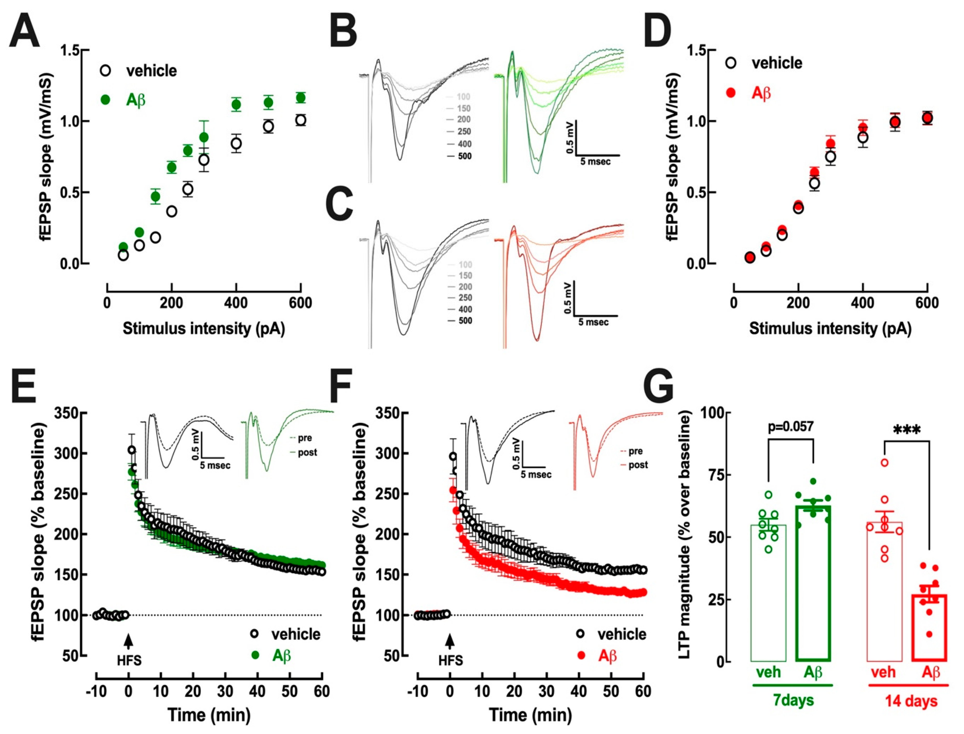
In spite of the absence of behavioral alteration at days 6–7 after Aβ-icv, there were alterations of synaptic excitability in hippocampal slices of these mice. Thus, the recording of synaptic transmission in Schaffer fibers-CA1 pyramid synapses, assessed as field excitatory synaptic potentials (fEPSP), revealed an increase in the input/output (I/O) curves in slices of mice at days 7–8 after Aβ-icv compared to vehicle-treated controls (Figure 2A,B). This increased excitability is likely to be a transient compensatory feature since it disappeared in slices of mice at days 15–16 after Aβ-icv, which displayed I/O superimposable to these of vehicle-treated mice (Figure 2C,D).
As shown in Figure 2E,G, synaptic plasticity, assessed as the magnitude of long-term potentiation (LTP), displayed a tendency for an increase (p = 0.057, Student’s t test) in slices collected at days 7–8 after Aβ-icv (62.7 ± 2.4% over baseline, n = 8) compared to vehicle-treated controls (55.0 ± 2.4% over baseline, n = 8). This tendency for an increased LTP magnitude observed before the onset of memory deficits reverted to a decreased LTP magnitude at the onset of memory deficits (Figure 2F,G), as previously reported [41]. In fact, in slices collected at days 15–16 after Aβ-icv, LTP magnitude (27.2 ± 3.2% over baseline, n = 8) was lower (p < 0.001, Student’s t test) than LTP magnitude in slices from vehicle-treated mice (56.1 ± 4.2% over baseline, n = 8).

3.3. Early Upregulation of Synaptic A2AR Is Responsible for Abnormal Hippocampal Function at 7–8 Days and at 15–16 Days after Aβ Administration

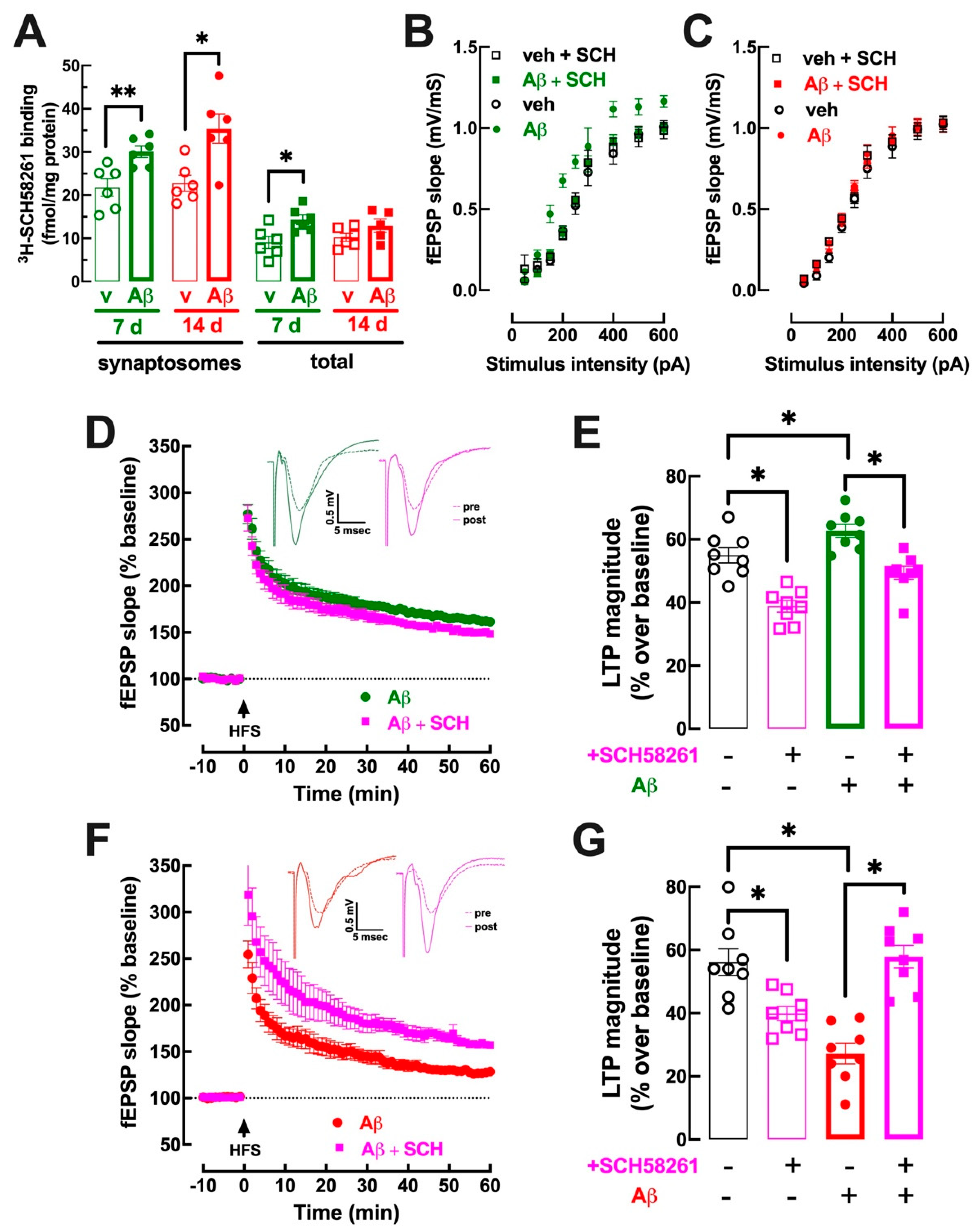
Since A2AR activation mimics [22,27,28] and A2AR blockade with SCH58261 prevents Aβ-icv-induced synaptic and memory deficits [25,41,52], and A2AR are up-regulated before the onset of some brain-induced dysfunction [26,45], we then tested if the density of A2AR in hippocampal synapses increased before the onset of synaptic and memory deficits triggered by Aβ-icv. As shown in Figure 3A, the binding density of a supramaximal but selective concentration (2 nM) of the A2AR antagonist 3H-SCH58261 [51] was larger (p < 0.008, Student’s t test) in hippocampal synaptosomal (i.e., synaptic) membranes from Aβ-icv-treated mice (30.1 ± 1.4 fmol/mg protein, n = 6) than from vehicle-treated mice at 7–8 days (21.8 ± 2.0 fmol/mg protein, n = 6), i.e., when memory performance was not yet modified (Figure 1C,D). In accordance with the previously described enrichment of A2AR in hippocampal synapses [17], the binding density of A2AR in synaptosomal membranes was higher (p = 0.006, Student’s t test) than in total membranes (9.1 ± 1.4 fmol/mg protein, n = 6) (Figure 3A, comparing first to fifth column from the left). However, still, in total hippocampal membranes, A2AR density was higher (p = 0.015, Student’s t test) 7–8 days after Aβ-icv administration 14.3 ± 1.1 fmol/mg protein, n = 6) compared to vehicle-treated mice (9.1 ± 1.4 fmol/mg protein, n = 6) (Figure 3A). When Aβ-induced memory deficits became evident, 15 days after Aβ-icv, A2AR density was still larger (p = 0.012, Student’s t test) in synaptosomal membranes from Aβ-icv treated (35.4 ± 3.4 fmol/mg protein, n = 6) compared to vehicle-treated mice (22.8 ± 1.8 fmol/mg protein, n = 6) (Figure 3A), but there was no significant difference (p = 0.175, Student’s t test) between the binding density of A2AR in total membranes from Aβ-icv-treated (13.0 ± 1.5 fmol/mg protein, n = 5) compared to vehicle-treated mice (10.2 ± 0.9 fmol/mg protein, n = 6) (Figure 3A).
We next investigated if A2AR blockade would be able to prevent the hyperexcitability observed in the hippocampus in the presymptomatic period, i.e., 7–8 days after Aβ-icv injection. The administration of a supramaximal concentration of SCH58261 (50 nM) [20] prevented the increased synaptic efficiency in the hippocampus 7–8 days after Aβ-icv administration, bringing the I/O curve to a profile nearly superimposable to that observed in vehicle-treated mice ((Figure 3B); note that the vehicle-icv and Aβ-icv groups in Figure 3B are the same as in Figure 2A). Moreover, the tendency for an increased LTP magnitude observed in hippocampal slices collected 7–8 days after Aβ-icv administration was eliminated by 50 nM SCH58261 ((Figure 3D); note that the vehicle-icv and Aβ-icv groups in Figure 3D,E are the same as in Figure 2C–E); thus, in slices collected 7–8 days after Aβ-icv administration, LTP magnitude was 62.7 ± 2.4% over baseline (n = 8) in the absence and was significantly lower (p < 0.001, Student’s t test) in the presence of SCH58261 (49.4 ± 2.1% over baseline, n = 8). A Tukey’s multiple comparison test after a two-way ANOVA (effect of Aβ: F1,28 = 18.82, p < 0.001; effect of SCH58261: F1,28 = 48.61, p < 0.001) revealed no significant difference between LTP magnitude in vehicle-treated and Aβ-icv-treated mice in the presence of SCH58261 (p = 0.291), although SCH58261 also decreased LTP magnitude in saline-treated mice (p < 0.001, Student’s t test), as previously reported (e.g., [26,35,41]).
The normalizing effect of SCH58261 in slices collected 15–16 days after Aβ-icv was similar to that previously described [41,53]. Thus, SCH58261 (50 nM) reverted (p < 0.001, Student’s t test) the Aβ-induced decrease in LTP magnitude (27.2.4 ± 3.2% over baseline, n = 8, in the absence and 57.9 ± 3.6% over baseline, n = 8, in the presence of SCH58261) ((Figure 3F,G); note that the vehicle-icv and Aβ-icv groups in Figure 3F,G are the same as in Figure 2D,E). A Tukey’s multiple comparison test after a two-way ANOVA (effect of Aβ: F1,28 = 6.61, p = 0.018; effect of SCH58261: F1,28 = 4.52, p = 0.043) revealed no significant differences of LTP magnitude between vehicle- and Aβ-icv-treated mice in the presence of SCH58261 (p = 0.983), although SCH58261 also decreased LTP magnitude in saline-treated mice (p = 0.011), as previously reported (e.g., [26,35,41]).

4. Discussion

The present study shows that an increased density and functional impact of A2AR in the hippocampus predates the Aβ-icv-induced deterioration of synaptic plasticity and of reference memory that are characteristic of this mouse model of AD. Thus, it was shown that the activity of these A2AR—with increased density in the presymptomatic period of AD that is maintained throughout the phenotypic expression of synaptic and memory deficits characteristic of early AD—is required both for the adaptive hyperexcitability before memory deficits as well as for the expression of synaptic and memory deficits present at the onset of symptomatic AD.
We first characterized experimental conditions indicative of a presymptomatic period of AD, where an increase in hippocampal excitability was observed at 7–8 days after Aβ-icv, without any evident alteration of memory-related behavioral performance. This is in agreement with several studies reporting an initial hyperexcitability in the hippocampus in early AD and pre-dating the onset of memory deficits (reviewed in [54,55,56]), as well as with the presence of subclinical epileptiform activity in the AD prodrome [57,58] and its association with the onset of AD [59]. This initial hippocampal hyperexcitability is linked to a deregulation of the inhibitory GABAergic network [60,61,62], involving a parallel alteration of the excitability of different interneurons [60,61,63] as well as an altered astrocytic support [64,65]. Importantly, this initial hippocampal hyperexcitability may contribute to the subsequent deterioration of information processing in hippocampal networks and the onset/aggravation of memory symptoms of AD [54,55,56], as heralded by the ability of drugs refraining from excessive excitation, such as levetiracetam, to improve performance on spatial memory tasks in early AD [66].
The exploitation of this tentative mouse model of presymptomatic AD then allowed us to conclude that A2AR undergo an early upregulation and seem to be paramount to controlling the increased excitability during this prodrome-like period in this Aβ-icv model of AD. In fact, we observed an increased density of A2AR in the synapses, where cortical A2AR are most abundant [17], but also in total membranes that mostly include non-synaptic and astrocytic membranes. Moreover, we showed that a selective A2AR antagonist, SCH58261 [51], abrogated the hippocampal hyperexcitability present before the onset of memory deficits in this Aβ-icv model of AD. Although the precise mechanism underlying hippocampal hyperexcitability in the pathogenesis of AD is still unclear, it has been proposed to involve a deregulation of GABAergic interneurons [60,61,63] and an altered astrocytic support [64,65]. Remarkably, hippocampal A2AR can control the excitability of GABAergic neurons [67], the release of GABA [68,69] and the stability of GABAergic synapses [70]. A2AR are also located in astrocytes in the hippocampus (e.g., [71,72,73]), controlling GABA uptake [74], and astrocytic A2AR control neuronal excitability and memory performance [73,75,76]. These mechanistic links are in agreement with the observed upregulation of A2AR in synapses as well as in total membranes of the hippocampus during the presymptomatic phase of AD; however, future work is required to unveil the relative contribution of these different mechanism(s) operated by A2AR to control hippocampal hyperexcitability present during the presymptomatic phase of AD.
The pattern of alteration of hippocampal synaptic plasticity in our tentative presymptomatic Aβ-icv model of AD is also in agreement with the currently accepted evolution of hippocampal activity during the evolution of AD, starting with a hyperexcitability in the presymptomatic phases that gradually evolves into a hypofunction with the onset and aggravation of memory deficits [55,77,78]. The density of synaptic A2AR is increased before and remains increased after the onset on synaptic plasticity and memory deficits in this Aβ-icv model of AD. Furthermore, A2AR blockade reverted the deficits of synaptic plasticity in a manner similar to that previously reported in the normalization of abnormal synaptic plasticity in the hippocampus in different animal models of AD [8,22,32,33,41] and those of other brain diseases [24,26,42,44,79,80,81,82,83]. The effects of A2AR in the control of abnormal synaptic plasticity likely reflect the key role of A2AR in the control of memory dysfunction, as heralded by the previous observations that A2AR activation is sufficient to trigger memory dysfunction [27,28,29], whereas A2AR blockade dampens memory dysfunction in AD models [8,22,25,32,33,34,41], specifically in the currently used Aβ-icv model of AD [25,41,52]. The mechanisms underlying this beneficial effect of A2AR antagonists are probably related to the impact of A2AR on the release of glutamate [18,19], on AMPA [84] and NMDA receptors [20,21,22,85,86], as well as the ability of A2AR to control astrocytic function [71,72,73,74,75,76] and microglia [87,88,89,90], impacting neuroinflammation (e.g., [91,92,93]). Future work is needed to clarify the contribution of these different pools of A2AR to controlling these different processes known to impact synaptic plasticity and memory performance in order to begin tackling the mechanisms associated with the ability of A2AR blockade to normalize synaptic plasticity. These will probably involve the engagement of different heteromers containing A2AR (reviewed in [94]) and the coupling of A2AR to different transducing mechanisms, which may change with the evolution of brain diseases and the upregulation of A2AR (reviewed in [14]). Thus, understanding the spatiotemporal dynamics of A2AR signaling in different compartments—which often have opposite effects on synaptic and behavioral outputs (see [95,96,97,98])—will be critical to understand this apparently paradoxical ability of A2AR blockade to prevent aberrant hyperexcitability and normalize synaptic plasticity both when it is increased as well as when it is decreased.

5. Conclusions

It is concluded that A2AR upregulation is an early event in the presymptomatic phase of AD that is maintained throughout the onset of memory deficits. This prompts consideration of the cortical A2AR upregulation as a putative novel biomarker of the risk of developing AD [46], and eventually as a general biomarker of neuropsychiatric diseases [99] in view of the association of A2AR overfunction with different neuropsychiatric diseases [99]. Moreover, the present observation that A2AR blockade prevents both the initial hyperexcitability during the presymptomatic phase of AD and the opposite decreased synaptic plasticity emerging with the onset of memory deficits confirms the prophylactic and therapeutic potential of A2AR antagonists and stresses the need to understand the different signaling mechanisms operated by A2AR in different cellular compartments.

Author Contributions

C.R.L., A.C.S., P.M.C. and J.P.L. carried out the surgeries and behavioral characterization, C.R.L., A.C.S., H.B.S. and J.P.L. carried out the electrophysiological recordings, A.C.S., P.A. and R.A.C. carried out the neurochemical assays, R.A.C. and P.A. coordinated the project and wrote the manuscript, which was revised and commented by all authors. All authors have read and agreed to the published version of the manuscript.

Funding

Supported by La Caixa Foundation (LCF/PR/HP17/52190001), Centro 2020 (CENTRO-01-0145-FEDER-000008:BrainHealth2020 and CENTRO-01-0246-FEDER-000010) and FCT (POCI-01-0145-FEDER-03127 and UIDB/04539/2020). Cátia R. Lopes is under receipt of a PhD grant from FCT (2021.06954.BD).

Institutional Review Board Statement

All animal procedures were approved by the Ethical Committee of the Center for Neuroscience and Cell Biology of Coimbra (ORBEA-128/2015).

Informed Consent Statement

Not applicable.

Data Availability Statement

The data that support the findings of this study are available from the corresponding author upon reasonable request.

Conflicts of Interest

R.A.C. is a scientific consultant for the Institute for Scientific Information on Coffee. All other authors declare no conflicts of interest.

Abbreviations

A2AR: adenosine A2A receptors; Aβ: amyloid β peptides; ACSF: artificial cerebrospinal fluid; AD: Alzheimer’s disease; EPSP: excitatory post-synaptic potential; icv: intracerebroventricular; LTP: long-term potentiation; SCH58261: 2-(2-furanyl)-7-(2-phenylethyl)-7H-pyrazolo [4,3-e][1,2,4]triazolo[1,5-c]pyrimidin-5-amine.

References

  1. Scheltens, P.; De Strooper, B.; Kivipelto, M.; Holstege, H.; Chételat, G.; Teunissen, C.E.; Cummings, J.; van der Flier, W.M. Alzheimer’s disease. Lancet 2021, 397, 1577–1590. [Google Scholar] [CrossRef] [PubMed]
  2. Selkoe, D.J. Alzheimer’s disease is a synaptic failure. Science 2002, 298, 789–791. [Google Scholar] [CrossRef] [PubMed] [Green Version]
  3. Pelucchi, S.; Gardoni, F.; Di Luca, M.; Marcello, E. Synaptic dysfunction in early phases of Alzheimer’s disease. Handb. Clin. Neurol. 2022, 184, 417–438. [Google Scholar] [CrossRef] [PubMed]
  4. Meftah, S.; Gan, J. Alzheimer’s disease as a synaptopathy: Evidence for dysfunction of synapses during disease progression. Front. Synaptic. Neurosci. 2023, 15, 1129036. [Google Scholar] [CrossRef]
  5. Kirvell, S.L.; Esiri, M.; Francis, P.T. Down-regulation of vesicular glutamate transporters precedes cell loss and pathology in Alzheimer’s disease. J. Neurochem. 2006, 98, 939–950. [Google Scholar] [CrossRef]
  6. Canas, P.M.; Simões, A.P.; Rodrigues, R.J.; Cunha, R.A. Predominant loss of glutamatergic terminal markers in a β-amyloid peptide model of Alzheimer’s disease. Neuropharmacology 2014, 76, 51–56. [Google Scholar] [CrossRef]
  7. Sri, S.; Pegasiou, C.-M.; Cave, C.A.; Hough, K.; Wood, N.; Gomez-Nicola, D.; Deinhardt, K.; Bannerman, D.; Perry, V.H.; Vargas-Caballero, M. Emergence of synaptic and cognitive impairment in a mature-onset APP mouse model of Alzheimer’s disease. Acta Neuropathol. Commun. 2019, 7, 25. [Google Scholar] [CrossRef] [Green Version]
  8. Viana da Silva, S.; Haberl, M.G.; Zhang, P.; Bethge, P.; Lemos, C.; Gonçalves, N.; Gorlewicz, A.; Malezieux, M.; Gonçalves, F.Q.; Grosjean, N.; et al. Early synaptic deficits in the APP/PS1 mouse model of Alzheimer’s disease involve neuronal adenosine A2A receptors. Nat. Commun. 2016, 7, 11915. [Google Scholar] [CrossRef] [Green Version]
  9. Scheff, S.W.; Price, D.A.; Schmitt, F.A.; Mufson, E.J. Hippocampal synaptic loss in early Alzheimer’s disease and mild cognitive impairment. Neurobiol. Aging 2006, 27, 1372–1384. [Google Scholar] [CrossRef]
  10. Galasko, D.; Xiao, M.; Xu, D.; Smirnov, D.; Salmon, D.P.; Dewit, N.; Vanbrabant, J.; Jacobs, D.; Vanderstichele, H.; Vanmechelen, E.; et al. Synaptic biomarkers in CSF aid in diagnosis, correlate with cognition and predict progression in MCI and Alzheimer’s disease. Alzheimer’s Dement. Transl. Res. Clin. Interv. 2019, 5, 871–882. [Google Scholar] [CrossRef]
  11. Zhang, J.; Wang, J.; Xu, X.; You, Z.; Huang, Q.; Huang, Y.; Guo, Q.; Guan, Y.; Zhao, J.; Liu, J.; et al. In vivo synaptic density loss correlates with impaired functional and related structural connectivity in Alzheimer’s disease. J. Cereb. Blood Flow Metab. 2023, 43, 977–988. [Google Scholar] [CrossRef]
  12. Wall, M.J.; Richardson, M.J.E. Localized adenosine signaling provides fine-tuned negative feedback over a wide dynamic range of neocortical network activities. J. Neurophysiol. 2015, 113, 871–882. [Google Scholar] [CrossRef] [Green Version]
  13. Lopes, C.R.; Gonçalves, F.Q.; Olaio, S.; Tomé, A.R.; Cunha, R.A.; Lopes, J.P. Adenosine A2A receptors shut down adenosine A1 receptor-mediated presynaptic inhibition to promote implementation of hippocampal long-term potentiation. Biomolecules 2023, 13, 715. [Google Scholar] [CrossRef]
  14. Cunha, R.A. How does adenosine control neuronal dysfunction and neurodegeneration? J. Neurochem. 2016, 139, 1019–1055. [Google Scholar] [CrossRef]
  15. Tetzlaff, W.; Schubert, P.; Kreutzberg, G.W. Synaptic and extrasynaptic localization of adenosine binding sites in the rat hippocampus. Neuroscience 1987, 21, 869–875. [Google Scholar] [CrossRef]
  16. Rebola, N.; Pinheiro, P.C.; Oliveira, C.R.; Malva, J.O.; Cunha, R.A. Subcellular localization of adenosine A1 receptors in nerve terminals and synapses of the rat hippocampus. Brain Res. 2003, 987, 49–58. [Google Scholar] [CrossRef] [Green Version]
  17. Rebola, N.; Canas, P.M.; Oliveira, C.R.; Cunha, R.A. Different synaptic and subsynaptic localization of adenosine A2A receptors in the hippocampus and striatum of the rat. Neuroscience 2005, 132, 893–903. [Google Scholar] [CrossRef] [PubMed]
  18. Lopes, L.V.; Cunha, R.A.; Kull, B.; Fredholm, B.B.; Ribeiro, J.A. Adenosine A2A receptor facilitation of hippocampal synaptic transmission is dependent on tonic A1 receptor inhibition. Neuroscience 2002, 112, 319–329. [Google Scholar] [CrossRef] [Green Version]
  19. Machado, N.J.; Simões, A.P.; Silva, H.B.; Ardais, A.P.; Kaster, M.P.; Garção, P.; Rodrigues, D.I.; Pochmann, D.; Santos, A.I.; Araújo, I.M.; et al. Caffeine reverts memory but not mood impairment in a depression-prone mouse strain with up-regulated adenosine A2A receptor in hippocampal glutamate synapses. Mol. Neurobiol. 2017, 54, 1552–1563. [Google Scholar] [CrossRef]
  20. Rebola, N.; Lujan, R.; Cunha, R.A.; Mulle, C. Adenosine A2A receptors are essential for long-term potentiation of NMDA-EPSCs at hippocampal mossy fiber synapses. Neuron 2008, 57, 121–134. [Google Scholar] [CrossRef] [Green Version]
  21. Mouro, F.M.; Rombo, D.M.; Dias, R.B.; Ribeiro, J.A.; Sebastião, A.M. Adenosine A2A receptors facilitate synaptic NMDA currents in CA1 pyramidal neurons. Br. J. Pharmacol. 2018, 175, 4386–4397. [Google Scholar] [CrossRef] [PubMed] [Green Version]
  22. Temido-Ferreira, M.; Ferreira, D.G.; Batalha, V.L.; Marques-Morgado, I.; Coelho, J.E.; Pereira, P.; Gomes, R.; Pinto, A.; Carvalho, S.; Canas, P.M.; et al. Age-related shift in LTD is dependent on neuronal adenosine A2A receptors interplay with mGluR5 and NMDA receptors. Mol. Psychiatry 2020, 25, 1876–1900. [Google Scholar] [CrossRef] [Green Version]
  23. Costenla, A.R.; Diógenes, M.J.; Canas, P.M.; Rodrigues, R.J.; Nogueira, C.; Maroco, J.; Agostinho, P.M.; Ribeiro, J.A.; Cunha, R.A.; de Mendonça, A. Enhanced role of adenosine A2A receptors in the modulation of LTP in the rat hippocampus upon ageing. Eur. J. Neurosci. 2011, 34, 12–21. [Google Scholar] [CrossRef] [PubMed]
  24. Kaster, M.P.; Machado, N.J.; Silva, H.B.; Nunes, A.; Ardais, A.P.; Santana, M.; Baqi, Y.; Müller, C.E.; Rodrigues, A.L.S.; Porciúncula, L.O.; et al. Caffeine acts through neuronal adenosine A2A receptors to prevent mood and memory dysfunction triggered by chronic stress. Proc. Natl. Acad. Sci. USA 2015, 112, 7833–7838. [Google Scholar] [CrossRef]
  25. Canas, P.M.; Porciúncula, L.O.; Cunha, G.M.A.; Silva, C.G.; Machado, N.J.; Oliveira, J.M.A.; Oliveira, C.R.; Cunha, R.A. Adenosine A2A receptor blockade prevents synaptotoxicity and memory dysfunction caused by β-amyloid peptides via p38 mitogen-activated protein kinase pathway. J. Neurosci. 2009, 29, 14741–14751. [Google Scholar] [CrossRef] [Green Version]
  26. Canas, P.M.; Porciúncula, L.O.; Simões, A.P.; Augusto, E.; Silva, H.B.; Machado, N.J.; Gonçalves, N.; Alfaro, T.M.; Gonçalves, F.Q.; Araújo, I.M.; et al. Neuronal adenosine A2A receptors are critical mediators of neurodegeneration triggered by convulsions. eNeuro 2018, 5, ENEURO.0385-18.2018. [Google Scholar] [CrossRef] [Green Version]
  27. Pagnussat, N.; Almeida, A.S.; Marques, D.M.; Nunes, F.; Chenet, G.C.; Botton, P.H.S.; Mioranzza, S.; Loss, C.M.; Cunha, R.A.; Porciúncula, L.O. Adenosine A2A receptors are necessary and sufficient to trigger memory impairment in adult mice. Br. J. Pharmacol. 2015, 172, 3831–3845. [Google Scholar] [CrossRef] [Green Version]
  28. Li, P.; Rial, D.; Canas, P.M.; Yoo, J.-H.; Li, W.; Zhou, X.; Wang, Y.; van Westen, G.J.P.; Payen, M.-P.; Augusto, E.; et al. Optogenetic activation of intracellular adenosine A2A receptor signaling in the hippocampus is sufficient to trigger CREB phosphorylation and impair memory. Mol. Psychiatry 2015, 20, 1339–1349. [Google Scholar] [CrossRef] [Green Version]
  29. Carvalho, K.; Faivre, E.; Pietrowski, M.J.; Marques, X.; Gomez-Murcia, V.; Deleau, A.; Huin, V.; Hansen, J.N.; Kozlov, S.; Danis, C.; et al. Exacerbation of C1q dysregulation, synaptic loss and memory deficits in tau pathology linked to neuronal adenosine A2A receptor. Brain 2019, 142, 3636–3654. [Google Scholar] [CrossRef]
  30. Chen, J.-F.; Sonsalla, P.K.; Pedata, F.; Melani, A.; Domenici, M.R.; Popoli, P.; Geiger, J.; Lopes, L.V.; de Mendonça, A. Adenosine A2A receptors and brain injury: Broad spectrum of neuroprotection, multifaceted actions and “fine tuning” modulation. Prog. Neurobiol. 2007, 83, 310–331. [Google Scholar] [CrossRef]
  31. Merighi, S.; Borea, P.A.; Varani, K.; Vincenzi, F.; Jacobson, K.A.; Gessi, S. A2A adenosine receptor antagonists in neurodegenerative diseases. Curr. Med. Chem. 2022, 29, 4138–4151. [Google Scholar] [CrossRef] [PubMed]
  32. Laurent, C.; Burnouf, S.; Ferry, B.; Batalha, V.L.; Coelho, J.E.; Baqi, Y.; Malik, E.; Mariciniak, E.; Parrot, S.; Van der Jeugd, A.; et al. A2A adenosine receptor deletion is protective in a mouse model of Tauopathy. Mol. Psychiatry 2016, 21, 97–107. [Google Scholar] [CrossRef] [PubMed] [Green Version]
  33. Silva, A.C.; Lemos, C.; Gonçalves, F.Q.; Pliássova, A.V.; Machado, N.J.; Silva, H.B.; Canas, P.M.; Cunha, R.A.; Lopes, J.P.; Agostinho, P. Blockade of adenosine A2A receptors recovers early deficits of memory and plasticity in the triple transgenic mouse model of Alzheimer’s disease. Neurobiol. Dis. 2018, 117, 72–81. [Google Scholar] [CrossRef] [PubMed]
  34. Orr, A.G.; Lo, I.; Schumacher, H.; Ho, K.; Gill, M.; Guo, W.; Kim, D.H.; Knox, A.; Saito, T.; Saido, T.C.; et al. Istradefylline reduces memory deficits in aging mice with amyloid pathology. Neurobiol. Dis. 2018, 110, 29–36. [Google Scholar] [CrossRef]
  35. Lopes, J.P.; Pliássova, A.; Cunha, R.A. The physiological effects of caffeine on synaptic transmission and plasticity in the mouse hippocampus selectively depend on adenosine A1 and A2A receptors. Biochem. Pharmacol. 2019, 166, 313–321. [Google Scholar] [CrossRef]
  36. Eskelinen, M.H.; Ngandu, T.; Tuomilehto, J.; Soininen, H.; Kivipelto, M. Midlife coffee and tea drinking and the risk of late-life dementia: A population-based CAIDE study. J. Alzheimer’s Dis. 2009, 16, 85–91. [Google Scholar] [CrossRef] [PubMed]
  37. Gelber, R.P.; Petrovitch, H.; Masaki, K.H.; Ross, G.W.; White, L.R. Coffee intake in midlife and risk of dementia and its neuropathologic correlates. J. Alzheimer’s Dis. 2011, 23, 607–615. [Google Scholar] [CrossRef] [PubMed]
  38. Dong, X.; Li, S.; Sun, J.; Li, Y.; Zhang, D. Association of coffee, decaffeinated coffee and caffeine intake from coffee with cognitive performance in older adults: National Health and Nutrition Examination Survey (NHANES) 2011–2014. Nutrients 2020, 12, 840. [Google Scholar] [CrossRef] [Green Version]
  39. Horgusluoglu-Moloch, E.; Nho, K.; Risacher, S.L.; Kim, S.; Foroud, T.; Shaw, L.M.; Trojanowski, J.Q.; Aisen, P.S.; Petersen, R.C.; Jack, C.R., Jr.; et al. Targeted neurogenesis pathway-based gene analysis identifies ADORA2A associated with hippocampal volume in mild cognitive impairment and Alzheimer’s disease. Neurobiol. Aging 2017, 60, 92–103. [Google Scholar] [CrossRef] [Green Version]
  40. Siokas, V.; Mouliou, D.S.; Liampas, I.; Aloizou, A.-M.; Folia, V.; Zoupa, E.; Papadimitriou, A.; Lavdas, E.; Bogdanos, D.P.; Dardiotis, E. Analysis of ADORA2A rs5760423 and CYP1A2 rs762551 genetic variants in patients with Alzheimer’s disease. Int. J. Mol. Sci. 2022, 23, 14400. [Google Scholar] [CrossRef]
  41. Gonçalves, F.Q.; Lopes, J.P.; Silva, H.B.; Lemos, C.; Silva, A.C.; Gonçalves, N.; Tomé, Â.R.; Ferreira, S.G.; Canas, P.M.; Rial, D.; et al. Synaptic and memory dysfunction in a β-amyloid model of early Alzheimer’s disease depends on increased formation of ATP-derived extracellular adenosine. Neurobiol. Dis. 2019, 132, 104570. [Google Scholar] [CrossRef] [PubMed]
  42. Augusto, E.; Gonçalves, F.Q.; Real, J.E.; Silva, H.B.; Pochmann, D.; Silva, T.S.; Matos, M.; Gonçalves, N.; Tomé, Â.R.; Chen, J.-F.; et al. Increased ATP release and CD73-mediated adenosine A2A receptor activation mediate convulsion-associated neuronal damage and hippocampal dysfunction. Neurobiol. Dis. 2021, 157, 105441. [Google Scholar] [CrossRef] [PubMed]
  43. Simões, A.P.; Gonçalves, F.Q.; Rial, D.; Ferreira, S.G.; Lopes, J.P.; Canas, P.M.; Cunha, R.A. CD73-mediated formation of extracellular adenosine is responsible for adenosine A2A receptor-mediated control of fear memory and amygdala plasticity. Int. J. Mol. Sci. 2022, 23, 12826. [Google Scholar] [CrossRef] [PubMed]
  44. Dias, L.; Pochmann, D.; Lemos, C.; Silva, H.B.; Real, J.I.; Gonçalves, F.Q.; Rial, D.; Gonçalves, N.; Simões, A.P.; Ferreira, S.G.; et al. Increased synaptic ATP release and CD73-mediated formation of extracellular adenosine in the control of behavioral and electrophysiological modifications caused by chronic stress. ACS Chem. Neurosci. 2023, 14, 1299–1309. [Google Scholar] [CrossRef] [PubMed]
  45. Gonçalves, F.Q.; Matheus, F.C.; Silva, H.B.; Real, J.I.; Rial, D.; Rodrigues, R.J.; Oses, J.-P.; Silva, A.C.; Gonçalves, N.; Prediger, R.D.; et al. Increased ATP release and higher impact of adenosine A2A receptors on corticostriatal plasticity in a rat model of presymptomatic Parkinson’s disease. Mol. Neurobiol. 2023, 60, 1659–1674. [Google Scholar] [CrossRef] [PubMed]
  46. Gessi, S.; Poloni, T.E.; Negro, G.; Varani, K.; Pasquini, S.; Vincenzi, F.; Borea, P.A.; Merighi, S. A2A adenosine receptor as a potential biomarker and a possible therapeutic target in Alzheimer’s disease. Cells 2021, 10, 2344. [Google Scholar] [CrossRef]
  47. Resende, R.; Ferreiro, E.; Pereira, C.; Resende de Oliveira, C. Neurotoxic effect of oligomeric and fibrillar species of amyloid-beta peptide 1–42: Involvement of endoplasmic reticulum calcium release in oligomer-induced cell death. Neuroscience 2008, 155, 725–737. [Google Scholar] [CrossRef]
  48. Hong, S.; Beja-Glasser, V.F.; Nfonoyim, B.M.; Frouin, A.; Li, S.; Ramakrishnan, S.; Merry, K.M.; Shi, Q.; Rosenthal, A.; Barres, B.A.; et al. Complement and microglia mediate early synapse loss in Alzheimer mouse models. Science 2016, 352, 712–716. [Google Scholar] [CrossRef] [Green Version]
  49. Kim, H.Y.; Lee, D.K.; Chung, B.-R.; Kim, H.V.; Kim, Y. Intracerebroventricular injection of amyloid-β peptides in normal mice to acutely induce Alzheimer-like cognitive deficits. J. Vis. Exp. 2016, 109, 53308. [Google Scholar] [CrossRef] [Green Version]
  50. Anderson, W.W.; Collingridge, G.L. Capabilities of the WinLTP data acquisition program extending beyond basic LTP experimental functions. J. Neurosci. Methods 2007, 162, 346–356. [Google Scholar] [CrossRef]
  51. Lopes, L.V.; Halldner, L.; Rebola, N.; Johansson, B.; Ledent, C.; Chen, J.F.; Fredholm, B.B.; Cunha, R.A. Binding of the prototypical adenosine A2A receptor agonist CGS 21680 to the cerebral cortex of adenosine A1 and A2A receptor knockout mice. Br. J. Pharmacol. 2004, 141, 1006–1014. [Google Scholar] [CrossRef] [PubMed] [Green Version]
  52. Cunha, G.M.; Canas, P.M.; Melo, C.S.; Hockemeyer, J.; Müller, C.E.; Oliveira, C.R.; Cunha, R.A. Adenosine A2A receptor blockade prevents memory dysfunction caused by β-amyloid peptides but not by scopolamine or MK-801. Exp. Neurol. 2008, 210, 776–781. [Google Scholar] [CrossRef]
  53. Lopes, C.R.; Amaral, I.M.; Pereira, M.F.; Lopes, J.P.; Madeira, D.; Canas, P.M.; Cunha, R.A.; Agostinho, P. Impact of blunting astrocyte activity on hippocampal synaptic plasticity in a mouse model of early Alzheimer’s disease based on amyloid-β peptide exposure. J. Neurochem. 2022, 160, 556–567. [Google Scholar] [CrossRef]
  54. Palop, J.J.; Mucke, L. Network abnormalities and interneuron dysfunction in Alzheimer disease. Nat. Rev. Neurosci. 2016, 17, 777–792. [Google Scholar] [CrossRef] [PubMed]
  55. Targa Dias Anastacio, H.; Matosin, N.; Ooi, L. Neuronal hyperexcitability in Alzheimer’s disease: What are the drivers behind this aberrant phenotype? Transl. Psychiatry 2022, 12, 257. [Google Scholar] [CrossRef] [PubMed]
  56. Tang, Y.; Yan, Y.; Mao, J.; Ni, J.; Qing, H. The hippocampus associated GABAergic neural network impairment in early-stage of Alzheimer’s disease. Ageing Res. Rev. 2023, 86, 101865. [Google Scholar] [CrossRef]
  57. Vossel, K.A.; Beagle, A.J.; Rabinovici, G.D.; Shu, H.; Lee, S.E.; Naasan, G.; Hegde, M.; Cornes, S.B.; Henry, M.L.; Nelson, A.B.; et al. Seizures and epileptiform activity in the early stages of Alzheimer disease. JAMA Neurol. 2013, 70, 1158–1166. [Google Scholar] [CrossRef] [PubMed] [Green Version]
  58. Ranasinghe, K.G.; Kudo, K.; Hinkley, L.; Beagle, A.; Lerner, H.; Mizuiri, D.; Findlay, A.; Miller, B.L.; Kramer, J.H.; Gorno-Tempini, M.L.; et al. Neuronal synchrony abnormalities associated with subclinical epileptiform activity in early-onset Alzheimer’s disease. Brain 2022, 145, 744–753. [Google Scholar] [CrossRef]
  59. Vossel, K.A.; Ranasinghe, K.G.; Beagle, A.J.; Mizuiri, D.; Honma, S.M.; Dowling, A.F.; Darwish, S.M.; Van Berlo, V.; Barnes, D.E.; Mantle, M.; et al. Incidence and impact of subclinical epileptiform activity in Alzheimer’s disease. Ann. Neurol. 2016, 80, 858–870. [Google Scholar] [CrossRef] [Green Version]
  60. Palop, J.J.; Chin, J.; Roberson, E.D.; Wang, J.; Thwin, M.T.; Bien-Ly, N.; Yoo, J.; Ho, K.O.; Yu, G.-Q.; Kreitzer, A.; et al. Aberrant excitatory neuronal activity and compensatory remodeling of inhibitory hippocampal circuits in mouse models of Alzheimer’s disease. Neuron 2007, 55, 697–711. [Google Scholar] [CrossRef] [Green Version]
  61. Ruiter, M.; Herstel, L.J.; Wierenga, C.J. Reduction of dendritic inhibition in CA1 pyramidal neurons in amyloidosis models of early Alzheimer’s disease. J. Alzheimer’s Dis. 2020, 78, 951–964. [Google Scholar] [CrossRef] [PubMed]
  62. Li, Y.; Zhu, K.; Li, N.; Wang, X.; Xiao, X.; Li, L.; Li, L.; He, Y.; Zhang, J.; Wo, J.; et al. Reversible GABAergic dysfunction involved in hippocampal hyperactivity predicts early-stage Alzheimer disease in a mouse model. Alzheimer’s Res. Ther. 2021, 13, 114. [Google Scholar] [CrossRef] [PubMed]
  63. Wander, C.M.; Li, Y.-D.; Bao, H.; Asrican, B.; Luo, Y.-J.; Sullivan, H.A.; Chao, T.-H.H.; Zhang, W.-T.; Chéry, S.L.; Tart, D.S.; et al. Compensatory remodeling of a septo-hippocampal GABAergic network in the triple transgenic Alzheimer’s mouse model. J. Transl. Med. 2023, 21, 258. [Google Scholar] [CrossRef] [PubMed]
  64. Ramos, B.; Baglietto-Vargas, D.; del Rio, J.C.; Moreno-Gonzalez, I.; Santa-Maria, C.; Jimenez, S.; Caballero, C.; Lopez-Tellez, J.F.; Khan, Z.U.; Ruano, D.; et al. Early neuropathology of somatostatin/NPY GABAergic cells in the hippocampus of a PS1xAPP transgenic model of Alzheimer’s disease. Neurobiol. Aging 2006, 27, 1658–1672. [Google Scholar] [CrossRef] [PubMed]
  65. Jo, S.; Yarishkin, O.; Hwang, Y.J.; Chun, Y.E.; Park, M.; Woo, D.H.; Bae, J.Y.; Kim, T.; Lee, J.; Chun, H.; et al. GABA from reactive astrocytes impairs memory in mouse models of Alzheimer’s disease. Nat. Med. 2014, 20, 886–896. [Google Scholar] [CrossRef] [PubMed]
  66. Vossel, K.; Ranasinghe, K.G.; Beagle, A.J.; La, A.; Ah Pook, K.; Castro, M.; Mizuiri, D.; Honma, S.M.; Venkateswaran, N.; Koestler, M.; et al. Effect of levetiracetam on cognition in patients with Alzheimer disease with and without epileptiform activity: A randomized clinical trial. JAMA Neurol. 2021, 78, 1345–1354. [Google Scholar] [CrossRef]
  67. Rombo, D.M.; Newton, K.; Nissen, W.; Badurek, S.; Horn, J.M.; Minichiello, L.; Jefferys, J.G.; Sebastiao, A.M.; Lamsa, K.P. Synaptic mechanisms of adenosine A2A receptor-mediated hyperexcitability in the hippocampus. Hippocampus 2015, 25, 566–580. [Google Scholar] [CrossRef]
  68. Cunha, R.A.; Ribeiro, J.A. Purinergic modulation of [3H]GABA release from rat hippocampal nerve terminals. Neuropharmacology 2000, 39, 1156–1167. [Google Scholar] [CrossRef] [PubMed]
  69. Saransaari, P.; Oja, S.S. GABA release modified by adenosine receptors in mouse hippocampal slices under normal and ischemic conditions. Neurochem. Res. 2005, 30, 467–473. [Google Scholar] [CrossRef]
  70. Gomez-Castro, F.; Zappettini, S.; Pressey, J.C.; Silva, C.G.; Russeau, M.; Gervasi, N.; Figueiredo, M.; Montmasson, C.; Renner, M.; Canas, P.M.; et al. Convergence of adenosine and GABA signaling for synapse stabilization during development. Science 2021, 374, eabk2055. [Google Scholar] [CrossRef]
  71. Matos, M.; Augusto, E.; Agostinho, P.; Cunha, R.A.; Chen, J.-F. Antagonistic interaction between adenosine A2A receptors and Na+/K+-ATPase-α2 controlling glutamate uptake in astrocytes. J. Neurosci. 2013, 33, 18492–18502. [Google Scholar] [CrossRef] [PubMed] [Green Version]
  72. Orr, A.G.; Hsiao, E.C.; Wang, M.M.; Ho, K.; Kim, D.H.; Wang, X.; Guo, W.; Kang, J.; Yu, G.-Q.; Adame, A.; et al. Astrocytic adenosine receptor A2A and Gs-coupled signaling regulate memory. Nat. Neurosci. 2015, 18, 423–434. [Google Scholar] [CrossRef] [PubMed] [Green Version]
  73. Barros-Barbosa, A.R.; Ferreirinha, F.; Oliveira, Â.; Mendes, M.; Lobo, M.G.; Santos, A.; Rangel, R.; Pelletier, J.; Sévigny, J.; Cordeiro, J.M.; et al. Adenosine A2A receptor and ecto-5’-nucleotidase/CD73 are upregulated in hippocampal astrocytes of human patients with mesial temporal lobe epilepsy (MTLE). Purinergic Signal. 2016, 12, 719–734. [Google Scholar] [CrossRef] [PubMed] [Green Version]
  74. Cristóvão-Ferreira, S.; Navarro, G.; Brugarolas, M.; Pérez-Capote, K.; Vaz, S.H.; Fattorini, G.; Conti, F.; Lluis, C.; Ribeiro, J.A.; McCormick, P.J.; et al. A1R-A2AR heteromers coupled to Gs and Gi/0 proteins modulate GABA transport into astrocytes. Purinergic Signal. 2013, 9, 433–449. [Google Scholar] [CrossRef] [Green Version]
  75. Matos, M.; Shen, H.-Y.; Augusto, E.; Wang, Y.; Wei, C.J.; Wang, Y.T.; Agostinho, P.; Boison, D.; Cunha, R.A.; Chen, J.-F. Deletion of adenosine A2A receptors from astrocytes disrupts glutamate homeostasis leading to psychomotor and cognitive impairment: Relevance to schizophrenia. Biol. Psychiatry 2015, 78, 763–774. [Google Scholar] [CrossRef] [Green Version]
  76. Madeira, D.; Lopes, C.R.; Simões, A.P.; Canas, P.M.; Cunha, R.A.; Agostinho, P. Astrocytic A2A receptors silencing negatively impacts hippocampal synaptic plasticity and memory of adult mice. Glia 2023, 71, 2137–2153. [Google Scholar] [CrossRef]
  77. Mufson, E.J.; Mahady, L.; Waters, D.; Counts, S.E.; Perez, S.E.; DeKosky, S.T.; Ginsberg, S.D.; Ikonomovic, M.D.; Scheff, S.W.; Binder, L.I. Hippocampal plasticity during the progression of Alzheimer’s disease. Neuroscience 2015, 309, 51–67. [Google Scholar] [CrossRef] [Green Version]
  78. Baazaoui, N.; Flory, M.; Iqbal, K. Synaptic compensation as a probable cause of prolonged mild cognitive impairment in Alzheimer’s disease: Implications from a transgenic mouse model of the disease. J. Alzheimer’s Dis. 2017, 56, 1385–1401. [Google Scholar] [CrossRef]
  79. Ferreira, D.G.; Batalha, V.L.; Vicente Miranda, H.; Coelho, J.E.; Gomes, R.; Gonçalves, F.Q.; Real, J.I.; Rino, J.; Albino-Teixeira, A.; Cunha, R.A.; et al. Adenosine A2A receptors modulate α-synuclein aggregation and toxicity. Cereb. Cortex 2017, 27, 718–730. [Google Scholar] [CrossRef] [PubMed] [Green Version]
  80. Rei, N.; Rombo, D.M.; Ferreira, M.F.; Baqi, Y.; Müller, C.E.; Ribeiro, J.A.; Sebastião, A.M.; Vaz, S.H. Hippocampal synaptic dysfunction in the SOD1G93A mouse model of Amyotrophic Lateral Sclerosis: Reversal by adenosine A2AR blockade. Neuropharmacology 2020, 171, 108106. [Google Scholar] [CrossRef]
  81. Moreira-de-Sá, A.; Gonçalves, F.Q.; Lopes, J.P.; Silva, H.B.; Tomé, Â.R.; Cunha, R.A.; Canas, P.M. Adenosine A2A receptors format long-term depression and memory strategies in a mouse model of Angelman syndrome. Neurobiol. Dis. 2020, 146, 105137. [Google Scholar] [CrossRef]
  82. Ferrante, A.; Boussadia, Z.; Borreca, A.; Mallozzi, C.; Pedini, G.; Pacini, L.; Pezzola, A.; Armida, M.; Vincenzi, F.; Varani, K.; et al. Adenosine A2A receptor inhibition reduces synaptic and cognitive hippocampal alterations in Fmr1 KO mice. Transl. Psychiatry 2021, 11, 112. [Google Scholar] [CrossRef] [PubMed]
  83. Li, X.-C.; Hong, F.-F.; Tu, Y.-J.; Li, Y.-A.; Ma, C.-Y.; Yu, C.-Y.; Fang, L.; Chen, J.-Y.; Li, Z.-L.; Bao, S.-J.; et al. Blockade of adenosine A2A receptor alleviates cognitive dysfunction after chronic exposure to intermittent hypoxia in mice. Exp. Neurol. 2022, 350, 113929. [Google Scholar] [CrossRef]
  84. Dias, R.B.; Ribeiro, J.A.; Sebastião, A.M. Enhancement of AMPA currents and GluR1 membrane expression through PKA-coupled adenosine A2A receptors. Hippocampus 2012, 22, 276–291. [Google Scholar] [CrossRef]
  85. Sarantis, K.; Tsiamaki, E.; Kouvaros, S.; Papatheodoropoulos, C.; Angelatou, F. Adenosine A₂A receptors permit mGluR5-evoked tyrosine phosphorylation of NR2B (Tyr1472) in rat hippocampus: A possible key mechanism in NMDA receptor modulation. J. Neurochem. 2015, 135, 714–726. [Google Scholar] [CrossRef] [PubMed] [Green Version]
  86. Franco, R.; Rivas-Santisteban, R.; Casanovas, M.; Lillo, A.; Saura, C.A.; Navarro, G. Adenosine A2A receptor antagonists affects NMDA glutamate receptor function. Potential to address neurodegeneration in Alzheimer’s disease. Cells 2020, 9, 1075. [Google Scholar] [CrossRef] [PubMed]
  87. Orr, A.G.; Orr, A.L.; Li, X.-J.; Gross, R.E.; Traynelis, S.F. Adenosine A2A receptor mediates microglial process retraction. Nat. Neurosci. 2009, 12, 872–878. [Google Scholar] [CrossRef] [Green Version]
  88. Gomes, C.; Ferreira, R.; George, J.; Sanches, R.; Rodrigues, D.I.; Gonçalves, N.; Cunha, R.A. Activation of microglial cells triggers a release of brain-derived neurotrophic factor (BDNF) inducing their proliferation in an adenosine A2A receptor-dependent manner: A2A receptor blockade prevents BDNF release and proliferation of microglia. J. Neuroinflammation 2013, 10, 16. [Google Scholar] [CrossRef] [PubMed] [Green Version]
  89. Chen, P.-Z.; He, W.-J.; Zhu, Z.-R.; E, G.-J.; Xu, G.; Chen, D.-W.; Gao, Y.-Q. Adenosine A2A receptor involves in neuroinflammation-mediated cognitive decline through activating microglia under acute hypobaric hypoxia. Behav. Brain Res. 2018, 347, 99–107. [Google Scholar] [CrossRef]
  90. Du, H.; Tan, Y.; Li, C.-H.; Zhao, Y.; Li, P.; Ning, Y.-L.; Gao, R.-B.; Wang, B.; Peng, Y.; Tan, S.-W.; et al. High glutamate concentration reverses the inhibitory effect of microglial adenosine 2A receptor on NLRP3 inflammasome assembly and activation. Neurosci. Lett. 2022, 769, 136431. [Google Scholar] [CrossRef] [PubMed]
  91. Rebola, N.; Simões, A.P.; Canas, P.M.; Tomé, A.R.; Andrade, G.M.; Barry, C.E.; Agostinho, P.M.; Lynch, M.A.; Cunha, R.A. Adenosine A2A receptors control neuroinflammation and consequent hippocampal neuronal dysfunction. J. Neurochem. 2011, 117, 100–111. [Google Scholar] [CrossRef] [PubMed]
  92. Meng, F.; Guo, Z.; Hu, Y.; Mai, W.; Zhang, Z.; Zhang, B.; Ge, Q.; Lou, H.; Guo, F.; Chen, J.; et al. CD73-derived adenosine controls inflammation and neurodegeneration by modulating dopamine signalling. Brain 2019, 142, 700–718. [Google Scholar] [CrossRef] [PubMed]
  93. Dias, L.; Lopes, C.R.; Gonçalves, F.Q.; Nunes, A.; Pochmann, D.; Machado, N.J.; Tomé, A.R.; Agostinho, P.; Cunha, R.A. Crosstalk between ATP-P2X7 and adenosine A2A receptors controlling neuroinflammation in rats subject to repeated restraint stress. Front. Cell. Neurosci. 2021, 15, 639322. [Google Scholar] [CrossRef] [PubMed]
  94. Franco, R.; Cordomí, A.; Llinas Del Torrent, C.; Lillo, A.; Serrano-Marín, J.; Navarro, G.; Pardo, L. Structure and function of adenosine receptor heteromers. Cell. Mol. Life Sci. 2021, 78, 3957–3968. [Google Scholar] [CrossRef]
  95. Shen, H.-Y.; Coelho, J.E.; Ohtsuka, N.; Canas, P.M.; Day, Y.-J.; Huang, Q.-Y.; Rebola, N.; Yu, L.; Boison, D.; Cunha, R.A.; et al. A critical role of the adenosine A2A receptor in extrastriatal neurons in modulating psychomotor activity as revealed by opposite phenotypes of striatum and forebrain A2A receptor knock-outs. J. Neurosci. 2008, 28, 2970–2975. [Google Scholar] [CrossRef] [PubMed] [Green Version]
  96. Shen, H.-Y.; Canas, P.M.; Garcia-Sanz, P.; Lan, J.-Q.; Boison, D.; Moratalla, R.; Cunha, R.A.; Chen, J.-F. Adenosine A2A receptors in striatal glutamatergic terminals and GABAergic neurons oppositely modulate psychostimulant action and DARPP-32 phosphorylation. PLoS ONE 2013, 8, e80902. [Google Scholar] [CrossRef]
  97. Wei, C.J.; Augusto, E.; Gomes, C.A.; Singer, P.; Wang, Y.; Boison, D.; Cunha, R.A.; Yee, B.K.; Chen, J.-F. Regulation of fear responses by striatal and extrastriatal adenosine A2A receptors in forebrain. Biol. Psychiatry 2014, 75, 855–863. [Google Scholar] [CrossRef] [Green Version]
  98. Chen, J.-F.; Choi, D.-S.; Cunha, R.A. Striatopallidal adenosine A2A receptor modulation of goal-directed behavior: Homeostatic control with cognitive flexibility. Neuropharmacology 2023, 226, 109421. [Google Scholar] [CrossRef]
  99. Moreira-de-Sá, A.; Lourenço, V.S.; Canas, P.M.; Cunha, R.A. Adenosine A2A receptors as biomarkers of brain diseases. Front. Neurosci. 2021, 15, 702581. [Google Scholar] [CrossRef]
Figure 1. Mice challenged with an intracerebroventricular (icv) injection of β-amyloid (1–42) (Aβ) displayed a selective alteration of hippocampal-dependent spatial memory after 14–15 days, without behavioral alterations after 6–7 days. Compared to vehicle-icv-injected mice (veh, thin bars and open symbols), Aβ-icv-injected mice (Aβ, thick bars and filled symbols) analyzed at 6–7 days (green bars and symbols) or 14–15 days (red bars and symbols) after icv injections displayed a preserved spontaneous locomotion (A) and a preserved pattern of anxiety (B) in an open field arena. Memory performance in the object displacement test (C) and in the modified Y-maze test (D) was preserved after 6–7 days and deteriorated at 14–15 days. Data are mean ± SEM; n = 8 mice per group; *** p < 0.001, Student’s t test with Welsh correction.
Biomolecules 13 01173 g001
Figure 2. Hippocampal slices collected from mice during the presymptomatic period 7–8 days after an intracerebroventricular (icv) injection of β-amyloid 1–42 (Aβ-icv) displayed an hyperexcitability that was converted to a decreased pattern of synaptic plasticity upon onset of memory deficits at 15–16 days after Aβ-icv. At 7–8 days after Aβ-icv, and when compared to vehicle-icv injected mice (black open symbols/lines), Aβ-icv mice (filled green symbols/lines) displayed an hyperexcitability assessed by the shift to left of the input/output curve of the field excitatory post-synaptic potentials (fEPSP) slope (A,B) and a tendency towards an increased magnitude of long-term potentiation (LTP) triggered by a high-frequency stimulation train (HFS, 100 Hz for 1 s) in Aβ-icv mice (E,G). In contrast, in the symptomatic phase, there was no difference in the input/output curve between control and Aβ-icv mice (filled red symbols/lines) (C,D); however, LTP magnitude was lower in Aβ-icv mice (F,G). Data are mean ± SEM of 8 mice per group, the same as in Figure 1; *** p < 0.001 between indicated groups using Student’s t test with Welsh correction.
Biomolecules 13 01173 g002
Figure 3. A2AR are upregulated from the presymptomatic phase of AD onward and control both hippocampal hyperexcitability in the presymptomatic phase and LTP deficits upon onset of memory deficits. (A) Binding of the A2AR antagonist 3H-SCH58261 (2 nM) in vehicle-icv mice (v, open symbols and thin bars) and in Aβ-icv mice (Aβ, filled symbols and thick bars) in hippocampal synaptosomal or total membranes at 7–8 days (green) or 15–16 days (red) after icv injections. Data are mean ± SEM of 6 mice per group; * p < 0.05 and ** p < 0.01 between indicated groups using Student’s t test with Welsh correction. (B) The addition of SCH58261 (50 nM) to hippocampal slices prevented the hyperexcitability in Aβ-icv mice and had no effect in vehicle-icv mice (veh) at 7–8 days, whereas SCH58261 was devoid of effects on hippocampal excitability in Aβ-icv and vehicle-icv mice at 15–16 days (C). SCH58261 decreased LTP magnitude triggered by a high-frequency stimulation train (HFS, 100 Hz for 1 s) in vehicle-icv-treated mice and normalized LTP magnitude in Aβ-icv both in the presymptomatic (D,E) and symptomatic phase (F,G). The vehicle-icv and Aβ-icv groups in Figure 3 are the same as in Figure 2. Data in (BG) are mean ± SEM of 8 mice per group; * p < 0.05 between indicated groups using Tukey’s test after a two-way ANOVA.
Biomolecules 13 01173 g003
Disclaimer/Publisher’s Note: The statements, opinions and data contained in all publications are solely those of the individual author(s) and contributor(s) and not of MDPI and/or the editor(s). MDPI and/or the editor(s) disclaim responsibility for any injury to people or property resulting from any ideas, methods, instructions or products referred to in the content.