Review

Fungal L-Methionine Biosynthesis Pathway Enzymes and Their Applications in Various Scientific and Commercial Fields

Department of Pharmaceutical Technology and Biochemistry, Gdansk University of Technology, 80-233 Gdansk, Poland
*
Author to whom correspondence should be addressed.
Biomolecules 2024, 14(10), 1315; https://doi.org/10.3390/biom14101315
Submission received: 31 August 2024 / Revised: 8 October 2024 / Accepted: 15 October 2024 / Published: 17 October 2024
(This article belongs to the Section Enzymology)

Abstract

:
L-methionine (L-Met) is one of the nine proteinogenic amino acids essential for humans since, in human cells, there are no complete pathways for its biosynthesis from simple precursors. L-Met plays a crucial role in cellular function as it is required for proper protein synthesis, acting as an initiator. Additionally, this amino acid participates in various metabolic processes and serves as a precursor for the synthesis of S-adenosylmethionine (AdoMet), which is involved in the methylation of DNA molecules and phospholipids, as well as in maintaining genome stability. Due to its importance, fungal L-methionine biosynthesis pathway enzymes are being intensively studied. This review presents the current state of the art in terms of their cellular function, usefulness as molecular markers, antifungal targets, or industrial approaches.

1. Introduction

L-methionine (L-Met) belongs to the aspartate family of amino acids, which are not synthesized by mammalian cells. It is a unique in structure amino acid, which is composed of a thioether hydrophobic side chain. L-Met is crucial for cell functioning as it plays an essential role in the initiation of translation of protein biosynthesis and takes part in various metabolic processes inside the cell [1]. This amino acid is biosynthesized by fungal, bacterial, and plant cells from L-homoserine (L-Hom), but the respective steps of the pathway leading to L-Met production differ across organisms. Due to the extensive biological interdependencies of the L-Met biosynthetic pathway, studying the mechanisms of the enzymes involved in this pathway can provide valuable insights into cellular functions and have applications in various scientific and commercial fields.
Because of the important role that L-Met plays in various organisms and the absence of an L-Met biosynthesis pathway in humans, enzymes involved in L-Met biosynthesis in fungi are considered by researchers as new possible targets for antimicrobial drugs [2]. Another positive aspect is that this amino acid level in human serum stands at only 20 μM [3], which is probably too low to rescue auxotrophic fungal strains incapable of methionine biosynthesis (as has been outlined in subsequent subsections). All of this led to deeper studies about the enzymes involved in the biosynthesis pathway of L-Met, their impact on the virulence of microorganisms, their phenotype, and their ability to grow.
Selected methionine biosynthesis enzymes are also considered as molecular markers. For years, strains of Saccharomyces cerevisiae with the deleted gene encoding the bi-functional O-acetyl-L-homoserine/O-acetyl-L-serine sulfhydrylase enzyme (Met17p), such as BY4741, have been commonly used in laboratories. The deletion of MET17 has been employed as an auxotrophic marker based on the assumption that this gene is essential for growth in media lacking organosulfur compounds [4]. Using an enzyme from the L-Met biosynthesis pathway in fungi is also an increasingly attractive approach for the commercial production of L-Met, an amino acid of significant industrial importance. Efforts are being made to develop a cost-effective method for producing L-Met from natural sources, without relying on genetically modified organisms [5]. Researchers also indicated that L-Met biosynthesis is connected with the production of intercellular H2S, an internal, physiological regulator in various cells, which exerts its effects on various targets and pathways by interacting with proteins, and DNA. Hydrogen sulfide, a volatile sulfur compound produced during yeast fermentation, plays an important role in the wine industry. The formation of H2S is correlated with an unpleasant odor in wine, similar to rotten eggs or sewage, and negatively impacts wine quality. The formation of H2S is closely related to the L-Met biosynthesis pathway, a route for the metabolism of organic sulfur compounds. The level of hydrogen sulfide in wine should be properly managed during the fermentation, as excess amounts can greatly overwhelm the aroma profile of the wine and reduce its quality and appeal [6].

2. L-Methionine Biosynthesis Pathway in Fungal Cells and Its Connections

L-Met is one of the five proteinogenic amino acids constituting the so-called aspartate family (includes L-Asp, L-Asn, L-Met, L-Thr and L-Ile). Biosynthesis of these amino acids starts from the common precursor, i.e., oxaloacetate, which is in the five initial steps converted into L-homoserine. Biosynthesis of methionine from the homoserine occurs in bacteria, fungi, and plants, but its course is not exactly the same in all these organisms [1]. In the main fungal version of methionine biosynthesis, as shown in Figure 1, starting from the branch at the L-Hom intermediate, it is first O-acetylated in the reaction catalyzed by L-homoserine transacetylase (Met2p, EC 2.3.1.31).
A sulfur atom is incorporated into O-acetyl-L-homoserine in two different ways, either by the direct sulfhydrylation pathway or the transsulfuration pathway. The direct sulfhydrylation pathway utilizes inorganic sulfide for the L-homocysteine production by a bifunctional O-acetyl-L-homoserine/O-acetyl-L-serine sulfhydrylase enzyme (Met17p, EC 2.5.1.49, EC 2.5.1.47). For S. cerevisiae this enzyme is also called Met15p [7]. L-homocysteine corresponds to a crossroads between direct sulfhydrylation and the transsulfuration pathway. The final step of the L-Met biosynthesis involves the methionine synthase (Met6p) enzyme, which, in contrast to the human equivalent, is cobalamin-independent, and utilizes L-homocysteine and 5-methyl-tetrahydrofolate [8]. The synthesis of L-homocysteine via the transsulfuration pathway involves the action of two different enzymes: cystathionine-γ-synthase (Str2p, EC 2.5.1.48) and cystathionine-β-lyase (Str3p, EC 4.4.1.8) which transfer sulfur atom between L-cysteine and L-homocysteine. The Str2p enzyme catalyzes the reaction of the formation of L-cystathionine from O-acetyl-L-homoserine and L-cysteine, which is then used by Str3p to yield L-homocysteine. In most fungal species, there also exists a reverse transsulfuration pathway that transforms L-homocysteine to L-cystathionine by cystathionine-β-synthase (Cys4p) followed by the formation of L-cysteine by cystathionine-γ-lyase (Cys3p) (Hébert et al. 2011). The reverse transsulfuration pathway is absent in Schizosaccharomyces pombe cells [9]. The L-Met biosynthesis pathway is linked to the pathway of L-cysteine biosynthesis. In contrast to bacterial cells, some fungal organisms, like Aspergillus nidulans and Neurospora crassa, synthesize L-cysteine in two distinct ways [10]: through the mentioned reverse transsulfuration pathway or the O-acetyl-L-serine (OAS) pathway, which begins with L-serine, followed by the reaction between sulfide and O-acetyl-L-serine. (Toh-e et al. 2018; de Melo et al. 2019). The OAS pathway is poorly characterized among fungal species. It is known that the OAS pathway is absent in Candida glabrata and S. cerevisiae species, which means that in these organisms the only functional route for L-Cys biosynthesis is the reverse transsulfuration pathway [11]. Interestingly, Schizosaccharomyces pombe, which lacks the reverse transsulfuration pathway, appears to possess two O-acetyl-L-homoserine-sulfhydrylases, linking features of filamentous fungi and yeasts [12].

3. Characterization of Fungal Enzymes Involved in the L-Methionine Biosynthesis Pathway

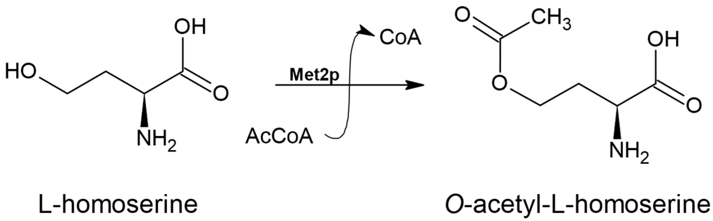
3.1. L-homoserine O-acetyltransferase (EC 2.3.1.31)

The first enzyme of the L-Met biosynthesis pathway in fungal cells is L-homoserine O-acetyltransferase (Met2p) encoded by the MET2 gene. Met2p requires L-homoserine (L-Hom) and acetyl coenzyme A (AcCoA) as substrates as it is responsible for the activation of the L-Hom hydroxyl group through its acetylation via a covalent acyl-enzyme intermediate [13] (Figure 2).
Met2p belongs to the α/β hydrolase fold enzyme superfamily, which is characterized by the eight-stranded β-sheet connected by α-helices. The structures of the Met2p enzyme from the Ascomycetes group exhibit a comparable overall folding pattern, characterized by the formation of two distinct domains: a main core domain and a lid domain, which plays a role in the enzyme’s dimerization [14]. The active site of these enzymes is found on a characteristic structural element known as the “nucleophilic elbow” involving a catalytic triad in which a nucleophilic residue may vary between serine, histidine, or aspartic acid [13]. L-homoserine O-acetyltransferase possesses a catalytic triad of conserved motif Ser-Asp-X-Leu-Phe, where serine acts as a nucleophilic residue whereas histidine and aspartic acid help to activate serine and take part in enzyme-acyl bond formation [13]. The same catalytic motif was observed for bacterial Mycobacterium smegmatis (PDBID: 6IOG), Haemophilus influenzae (PDBID: 2B61), and Leptospira interrogans (PDBID: 2PL5) as well as L-homoserine O-acetyltransferase from Candida albicans [15].
A series of studies has shown that homoserine O-acetyltransferase activity is essential for the prototrophy of C. albicans and S. cerevisiae fungi with respect to L-Met [16]. ΔMet2 S. cerevisiae mutants additionally exhibited resistance to the presence of methylmercury in the medium. Montoya et al. observed that Δmet2 Candida guilliermondii mutants, auxotrophic for L-Met, like the S. cerevisiae mutant, form brown colonies on a solid agar medium containing lead, probably due to the presence of a dark lead sulfide coating on the cell surface [17,18]. This team attempted to use this phenomenon as a selective marker in genetic modifications of the C. guilliermondii genome. It was demonstrated that the MET2 knockout strategy is not suitable for effectively disrupting multiple genes. However, the results indicate that the MET2 knockout cassette works well as a system for disrupting a single gene in C. guilliermondii [17].
Studies of Δmet2 Cryptococcus neoformans mutants showed avirulence in a mouse infection model via inhalation. As expected, the strain was auxotrophic for L-Met. A screening of a compound library of small molecules for Met2p inhibitors was also conducted, and the compound 6-carbamoyl-3a,4,5,9b-tetrahydro-3H-cyclo-penta[c]quinoline-4-carboxylic acid (CTCQC) was tested. Although CTCQC proved to be an effective in vitro inhibitor of the Met2p enzyme, it did not show antifungal activity against C. neoformans in a minimal medium at concentrations up to 128 µg mL−1 [19]. Comparable results were observed in studies of the bacterium Mycobacterium tuberculosis. The ΔmetA mutant, which has a deleted gene encoding bacterial L-homoserine O-acetyltransferase, is auxotrophic for O-acetylhomoserine, L-homocysteine, and L-Met. Despite their high levels in serum, these metabolites are not present in sufficient amounts in the host’s preferred niches, or the transport mechanisms for these metabolites in the bacteria may not be active in living organisms. Additionally, the ΔmetA mutant is incapable of proliferating in primary human macrophages and is highly susceptible to elimination in both immunocompetent and immunocompromised mice [20].
Similarly, the rice blast fungus, Magnaporthe oryzae, exhibited significantly reduced growth and pathogenicity following MET2 gene disruption compared to the wild type. Li et al. [21] additionally demonstrated that transgenic rice cultivars modified using the Host-Induced Gene Silencing (HIGS) technique to target the genes MET2 and CYS2 (homologs encoding proteins with L-homoserine O-acetyltransferase activity) exhibited strong resistance to the pathogen. They prepared a siRNA construct capable of silencing the expression of MET2 and CYS4 in M. oryzae and introduced it into transgenic plants, demonstrating both that L-homoserine O-acetyltransferase could be a potential target for novel fungicides and that HIGS could be an effective strategy for controlling rice blast [21].
These results indicate that L-homoserine O-acetyltransferase might be considered a great potential molecular target for antimicrobial chemotherapy [19]. In this regard, some structural analogs of L-homoserine were tested as Met2p inhibitors. Moderate inhibitory potential against Met2p from C. albicans was observed for L- and D-penicillamine [15] (Figure 3) This effect was related to antifungal activity. The minimal inhibitory concentrations (MIC50) at which growth is inhibited by 50% ranged from 32 to 1024 µg mL−1 for several fungal strains. For C. glabrata and S. cerevisiae, this value depends on the availability of L-Met in the medium, confirming that the molecular target is connected with the L-Met biosynthesis pathway.

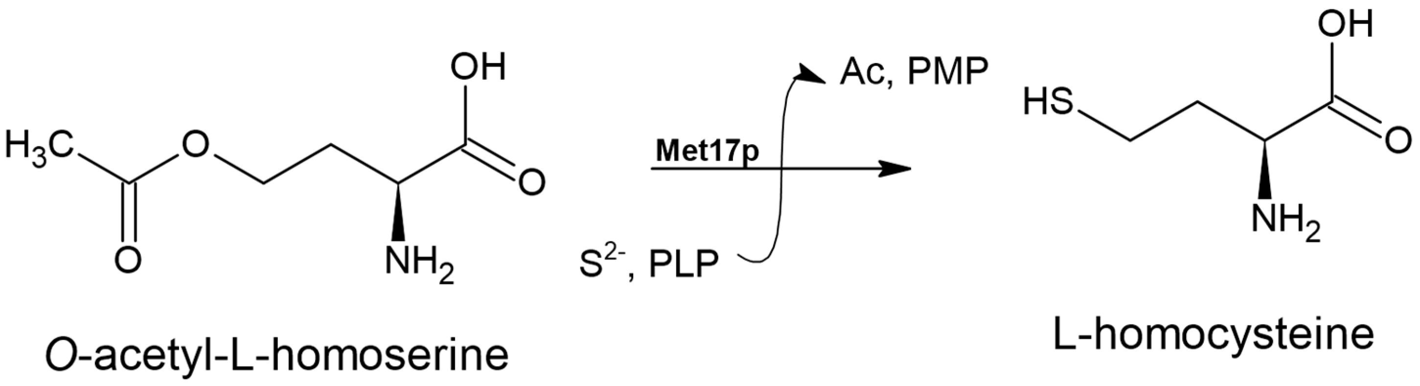
3.2. O-acetyl-L-homoserine Sulfhydrylase Enzyme (EC 2.5.1.49)

The O-acetyl-L-homoserine sulfhydrylase EC 2.5.1.49 is a PLP-dependent enzyme that uses O-acetyl-L-homoserine and a sulfide ion to catalyze the synthesis of L-homocysteine (L-HCT) (Figure 4), a reaction that plays a role in the direct sulfhydrylation pathway of the L-Met biosynthesis pathway [22,23].
In some fungi, an enzyme exists as a bi-functional O-acetyl-L-homoserine/O-acetyl-L-serine sulfhydrylase (EC 2.5.1.49, EC 2.5.1.47). One of the roles of this enzyme is to produce L-Cys from O-acetyl-L-serine (L-OAS) as a part of the transsulfuration pathway [22,24]. The bi-functional enzyme can be found in fungal cells such as S. cerevisiae, Kluyveromyces lactis, Yarrowia lipolytica, S. pombe, Trichosporon cutaneum and A. nidulans.
The bi-functional O-acetyl-L-homoserine/O-acetyl-L-serine sulfhydrylase enzyme from S. cerevisiae (Met17p) and bacterial O-acetyl-L-homoserine sulfhydrylase (Met15p) enzyme from Wolinella succinogenes have been analyzed in their crystal structures, showing that they are part of the PLP-dependent Cys/Met enzyme family [25,26]. The activity of this bi-functional enzyme (called Met17p in yeast) is essential for the proper functioning of S. cerevisiae cells. ΔMet17 mutants are auxotrophic for L-Met and sulfur, which can be mainly attributed to the toxic accumulation of hydrogen sulfide gas. In contrast, K. lactis yeast mutants lacking the gene encoding this enzyme grow more slowly compared to the wild-type strain and exhibit lower levels of sulfur-containing amino acids [24]. The content of sulfur-containing amino acids is changing also in T. cutaneum mutants but does not influence its growth rate. The enzyme is physiologically important, though not indispensable. The lack of activity of this enzyme in Y. lipolytica and S. pombe does not have a physiological impact, in contrast to the bacterial O-acetyl-L-homoserine sulfhydrylase enzyme, which is crucial for the virulence rate and survival of Streptococcus suis in mouse infection model [27]. The bi-functional O-acetyl-L-homoserine/O-acetyl-L-serine sulfhydrylase enzymes from Y. lipolytica, S. pombe, and T. cutaneum are probably mainly responsible for recycling the methylthio group from 5′-methylthioadenosine, a byproduct of polyamine synthesis from AdoMet [28]. This role was proved for enzymes from A. nidulans [28].
The S. cerevisiae Δmet17 mutant has been also used for years as a model organism equipped with an auxotrophic marker for a wide range of studies. However, it has been shown that the Met17p protein is not essential for the survival of S. cerevisiae in the absence of exogenous sulfur-containing organic compounds; the mutants are able to utilize an alternative homocysteine biosynthesis pathway involving the enzyme encoded by HSU1 (Yll058W). This enzyme can catalyze the reaction typically carried out by Met17p, albeit with lower efficiency [4,29,30].
Moreover, the bi-functional O-acetyl-L-homoserine/O-acetyl-L-serine sulfhydrylase enzyme from S. cerevisiae (Met17p) has attracted scientific interest due to its commercial potential. A method has been developed for L-Met production from O-acetyl-homoserine and 3-methylthiopropionaldehyde using recombinant Met17p purified from Escherichia coli BL21-Met17 [5]. L-Met is an amino acid whose commercial use is continually increasing. It is used as a flavoring agent in food additives, in therapies for liver diseases, as a nutritional component in infant formulas, parenteral nutrition, and in sports supplements [31]. The production of L-Met using recombinant Met17p addresses issues such as the formation of racemic D- and L-Met mixtures, chemical contamination of the final product associated with chemical synthesis, and the low yield and difficult purification processes observed in microbial fermentation methods [5]. Production of L-Met in a one-pot process using a combined method of fermentation and biocatalysis was reported by Zhu et al. in 2021. Mutant strains of E. coli and homoserine O-succinyltransferase enzyme were employed for efficient large-scale production. In contrast to the method described above, Zhu team used homoserine O-succinyltransferase which is a bacterial analog of Met2p, an enzyme involved in the L-methionine biosynthesis pathway in E. coli [32]. The Met17p enzyme from S. cerevisiae may also be used for the production of L-methionine analogs and organic thiols. Most of these compounds are not commercially available but are necessary for synthesizing S-adenosyl-L-methionine analogs [25]. The clinical applications of AdoMet include treatment of depression, osteoarthritis and alcoholic liver disease [33]. Moreover, AdoMet and its analogs are used in research investigating methyltransferases and the regulation of intracellular processes [34,35]. The discovery of novel AdoMet-dependent methyltransferases enables the industrial biosynthesis of various natural products in heterologous hosts [36].
In the wine industry, the aroma and bouquet of wine are of immense importance and directly impact economic gains. Excess H2S produced during the yeast fermentation process can lead to off-flavors reminiscent of rotten eggs, which significantly detracts from the quality of the wine. It was found that early-stage H2S production positively affects the aroma and bouquet of wine due to subsequent transformations of this compound into more desirable forms [37]. Enzymes Met2p and Met17p from S. cerevisiae were evaluated for their impact on the amount of H2S produced during fermentation. However, the S. cerevisiae Δmet17 and Δmet2 mutants produced higher levels of H2S in the later stages of fermentation [6].

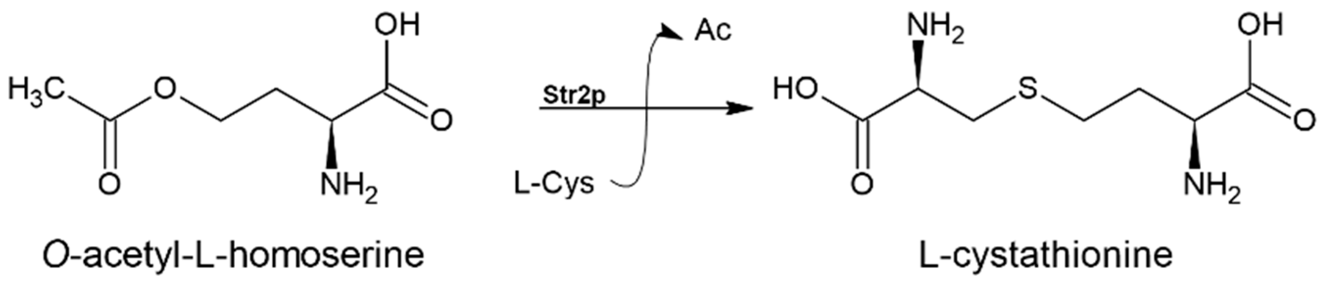
3.3. Cystathionine-γ-synthase (EC 2.5.1.48)

Incorporation of a sulfur atom into L-homocysteine can alternatively be carried out via the transsulfuration pathway in the presence of L-cysteine (Figure 1). The first step of the pathway during which L-cystathionine origins from L-cysteine and O-acetyl-L-homoserine (an activated form of L-Hom) is catalyzed by the cystathionine-γ-synthase (Figure 5).
Microorganisms activate L-Hom in different ways depending on the organism. Generally, in fungi by creating O-acetyl-L-homoserine, in bacteria O-succinyl-L-homoserine and in plants O-phospho-L-homoserine [38].
Many fungal species like N. crassa possess both Str2p and Met17p enzymes; however, Met17p was discovered not to be essential in L-Met biosynthesis in N. crassa cells [39]. Deletion of genes encoding Str2p resulted in auxotrophic growth towards L-Met, whereas deletion of Met17p encoding genes did not cause similar results.
Research on Botrytis cinerea (grey mold) has highlighted the significant role of the Str2p protein in fungal physiology, including growth, virulence, and response to environmental stresses. The Δstr2 mutant, which is auxotrophic for L-Met, displayed increased sensitivity to the fungicides fluazinam and fludioxonil. Additionally, disruption of the STR2 gene led to reduced conidia production and defects in sclerotia formation, which may impact its ability to survive. The Δstr2 mutant also showed heightened sensitivity to oxidative stress caused by H2O2, a reactive oxygen species produced by host plants that can result in substantial cellular damage, as well as to osmotic stress, which affects its growth in plant tissues [40].

3.4. Cystathionine-β-lyase (EC 4.4.1.8)

The second step of the transsulfuration pathway, during which L-cystathionine cleaves to L-homocysteine, is catalyzed by cystathionine β-lyase [41]. This reaction is shown in Figure 6.
Both cystathionine γ-synthase and cystathionine β-lyase are members of the PLP-dependent enzyme family and possess a homotetrameric structure. They share similarities in their overall architecture and the arrangement of main active-site residues, including an arginine that recognizes the carboxyl group of the substrate, a tyrosine associated with the PLP cofactor, and a lysine that is likely to collect a proton. Despite these similarities, their catalytic mechanisms differ significantly. Cystathionine β-lyase catalyzes a step in methionine biosynthesis and transfers the proton to L-homocysteine [38].
In A. nidulans or Fusarium graminearum, deletion of the gene responsible for encoding this enzyme causes an auxotrophy to L-Met [42]. A similar effect was observed for the mutant Salmonella gallinarum, a bacterium that is solely responsible for avian typhus [43]. The METC gene (encoding cystathionine β-lyase) deletion in S. gallinarum resulted in the inability to grow on a minimal medium without L-Met. The ΔmetC mutant was unable to kill chickens after oral inoculation, whereas only 10% of animals inoculated with the wild-type strain survived. The experiment showed that the mutant was not able to grow in the internal organs of the chickens, including the liver or spleen [43]. The virulence of Salmonella typhimurium mutant was also impaired in a mouse model of systemic infection in vivo [43].

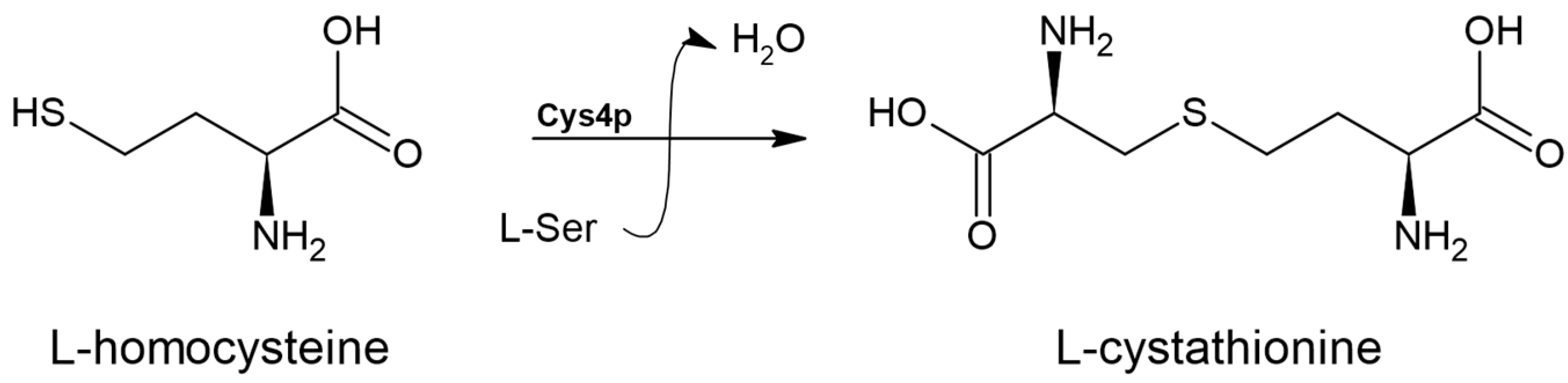
3.5. Cystathionine β-synthase (EC 4.2.1.22)

Many organisms can engage in a reverse transsulfuration pathway, where L-homocysteine is first transformed into L-cystathionine and subsequently into L-cysteine [44] (Figure 1). The initial step of this process is mediated by the enzyme cystathionine β-synthase (EC 4.2.1.22) (Figure 7).
Cystathionine β-synthase from C. albicans was identified as the key enzyme involved in the intracellular synthesis of H2S, an antioxidant molecule that plays a role in various host cell colonization strategies and is secreted to protect against the host’s immune response. Yeast cells with a deletion of the CYS4 gene exhibited growth defects closely linked to fungal pathogenicity. Additionally, the ability of Δcys4 mutants to form biofilms and hyphae was impaired. This modification also affected protein synthesis, folding, and mannosylation, and caused irregular respiration or mitochondrial dysfunction [45]. Chang et al. elucidated the crystal structure of the catalytic core of Cys4p from C. albicans (PDB: 7XRQ) and tested potent inhibitors of Cys4p from C. albicans: PA compound (isolated from an edible lichen, along with alternariol and usnic acid) and synthetic ethyl 2-(aminooxy)acetate (EAOA) (Figure 8). Both compounds inhibited fungal growth (C. albicans, C. glabrata, Candida parapsilosis, and Candida tropicalis) with MIC values of 32 and 64 µg mL−1 for EAOA and PA, respectively. EAOA and PA reduced the H2S generation of C. albicans, whereas PA more potently inhibited Cys4p than Cys3p and exhibited enzyme selectivity towards fungal Cys4p enzyme [45].
Aspergillus fumigatus MECA (gene encoding cystathionine β-synthase) deletion mutant was more sensitive to H2O2, the cell wall stressor SDS, and the antifungal agent fludioxonil. Furthermore, Sueiro-Olivares et al. demonstrated that the activity of cystathionine β-synthase and cystathionine γ-lyase is essential for survival [46].
It was reported that cystathionine β-synthases from the medicinal mushrooms Ganoderma lucidum and Lignosus rhinocerus possess angiotensin-converting enzyme (ACE) inhibitory activities. It is worth mentioning that ACE is responsible for blood pressure regulation and is associated with several diseases, such as Alzheimer’s, fibrosis, and lung injury [47]. Moreover, the activity of cystathionine β-synthase from the mushroom G. lucidum is linked to its cellulolytic ability and increases the adaptive capacity of this fungus [48]. The ability to break down cellulose is valuable in industries focused on biomass conversion. Additionally, enhancing the growth and adaptive capacity of G. lucidum could be beneficial due to its medicinal properties.

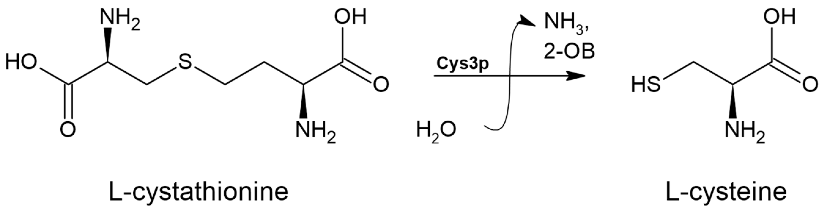
3.6. Cystathionine γ-lyase (EC 4.4.1.1)

The further conversion of L-cystathionine to L-cysteine through reverse transsulfuration is mediated by the enzyme cystathionine γ-lyase (EC 4.4.1.1). This enzyme is a member of the PLP-dependent enzyme family involved in cysteine and methionine metabolism, which also includes cystathionine γ-synthase, cystathionine β-lyase, and cystathionine β-synthase [49]. The cystathionine γ-lyase enzyme facilitates the transformation of L-cystathionine into L-cysteine, producing α-ketobutyrate and ammonia in the process, as illustrated in Figure 9.
Interestingly, the cystathionine γ-synthase from yeast or human sources has an active site that is nearly identical to that of the enzyme from E. coli [50]. The γ-synthase and γ-lyase activities of both enzymes depend on their position within the metabolic pathway and the availability of substrates [50].
It has been shown that S. cerevisiae cystathionine γ-lyase is able to convert S-propargyl-cysteine to H2S, a relevant gaseous signaling molecule that participated in numerous biological functions. Increasing the intracellular level of H2S led to enhanced expression of genes involved in sulfur amino acid biosynthesis and a significant increase in fungal growth [51]. Interestingly, it has been suggested that targeting the endogenous production of this gasotransmitter could be an effective approach to combat these pathogens. The production of H2S is also extensively studied in the wine industry to reduce the production of this gas during the fermentation process. The proteins Cys3p and Cys4p have been identified as participating in the H2S releasement. However, it has been shown that disruption of the genes encoding CYS3 and CYS4 from the S. cerevisiae genome did not reduce the levels of H2S in vivo [52]. Protein persulfidation (also called sulfhydration) of fungal and host proteins is closely related to the adaptation of A. fumigatus to its host and the host’s defense against this pathogen. Deleting the gene encoding cystathionine γ-lyase in fungal cells reduces the rate of persulfidation, a post-translational modification that modulates the activities of peroxiredoxin and alcohol dehydrogenase, both of which are associated with the pathogenic potential of A. fumigatus. Additionally, conidia of the mutant strain exhibited significantly higher susceptibility to killing by murine and human macrophage cell lines compared to the wild-type conidia and also displayed reduced virulence in a leukopenic mouse infection model. The A. fimugatus mutant lacking the cystathionine γ-lyase gene was also more sensitive to menadione, the thiol-oxidizing drug diamide, and H2O2. Furthermore, the proper activity of human cystathionine γ-lyase determines the level of persulfidation in host proteins, essential for maximizing the antifungal action of lung-resident alveolar macrophages and epithelial cells [46].
The protein Cys3p was also studied as a potential biological diagnostic target for invasive candidiasis; however, due to its high homology with cystathionine-β-lyase from Chlamydia, it cannot be considered a good biomarker [53].
This enzyme may also have commercial potential, as it has been shown that cystathionine γ-lyase from the Gymnopilus dilepis mushroom is positively correlated with the biosynthesis of psilocybin, a potential antidepressant alkaloid [54].

3.7. Methionine Synthase (EC 2.1.1.13)

In fungal microorganisms, the last step of the L-Met biosynthesis pathway is carried out by methionine synthase, which is encoded by the MET6 gene [44]. This enzyme transfers a methyl group from 5-methyltetrahydrofolate to L-homocysteine, resulting in the production of L-Met and tetrahydrofolate (THF) [55] (Figure 10).
It is important to note that there are two distinct classes of methionine synthases: cobalamin-dependent and cobalamin-independent. Mammalian methionine synthase, which is dependent on cobalamin, functions as an intermediate methyl carrier and plays a crucial role in L-Met biosynthesis [8,56]. In contrast, fungal enzymes, including those from C. albicans, do not require cobalamin and instead use 5-methyl-THF as a methyl donor. This difference in cofactor requirement leads to mechanistic variations between the two enzyme types. Cobalamin-independent methionine synthases are also found in some bacteria and higher plants, whereas organisms that cannot produce or obtain cobalamin rely on this type of enzyme [55].
Research revealed that in fungi like Pichia pastoris and C. albicans, Met6p is situated in the cell nucleus, whereas in S. cerevisiae, this enzyme is present in the cytosol [55]. The variations in structure and function between methionine synthases in humans and fungi highlight Met6p as a promising target for antifungal drug development [8]. The gene MET6 is essential for C. albicans viability as colonies of double deletion mutant did not grow even when provided with additional exogenous methionine [8]. Furthermore, evidence suggests that methionine synthase activity may play a crucial role in the virulence of bloodstream-isolated C. albicans. The levels of L-homocysteine and 5-methyltetrahydrofolate were elevated in invasive isolates or those that formed a high biofilm [57].
Pascon et al. [58] showed that mutant Δmet6 C. neoformans did not exhibit pathogenicity in the inhalation mouse model, displayed increased sensitivity to fluconazole and cyclosporin A and showed slowed growth, auxotrophy for L-Met and a defect in capsule formation (a virulence factor), whereas in A fumigatus, the gene responsible for encoding methionine synthase is considered essential for the invasion of host cells [44,59]. The lack of methionine synthase activity in this pathogen causes a metabolic disruption, resulting in decreased intracellular ATP levels, which inhibits fungal growth despite the presence of methionine [60]. Similarly, in the plant pathogen F. graminearum, deactivating the MSY1 gene encoding methionine synthase results in aerial hyphal growth defects and reduction in virulence followed by L-Met auxotrophy [42]. Another plant pathogen, M. oryzae, becomes avirulent after the deletion of the MET6 gene. ΔMet6 mutants were auxotrophic for L-Met; even when grown with abundant methionine, these mutants exhibited developmental issues, including decreased pigmentation of the mycelium, impaired formation of aerial hyphae, and reduced sporulation [61].
Studies aimed at improving the sensitivity of diagnostic tests for invasive candidiasis were also conducted using Met6p. Immunoenzymatic tests were performed to detect antibodies against various C. albicans proteins [62,63]. In immunocompromised patients, antibodies against Met6p proved to be the most effective biomarker [62].
Methionine synthase is widely present, but the versions found in humans and fungi differ significantly in their structure and function. Additionally, Met6p is exposed on the surface of C. albicans cells. These two facts were utilized to develop an effective vaccine protecting against disseminated C. albicans by targeting epitopes derived from fructose bisphosphate aldolase and methionine synthase [64]. It has also been shown that survival rates in mice infected with C. tropicalis, C. glabrata, and C. albicans were significantly improved when treated with dendritic cells pulsed with Met6p [65].
The interest in methionine synthase as a potential molecular target for antimicrobial drugs stems from the fact that inhibiting this enzyme causes the accumulation of toxic homocysteine. This accumulation leads to the formation of reactive homocysteine thiolactone, which interferes with the biosynthesis of ergosterol, an important component of fungal cell membranes [3,44]. Despite these promising results, no effective methionine synthase inhibitors with antifungal activity have yet been described [44].

4. Conclusions

Methionine and sulfur metabolism play critical roles in the growth and development of fungal cells. Studies concerning the methionine biosynthesis pathway and its connections with cysteine metabolism performed so far mainly focused on usefulness as molecular targets for antifungal agents (Table 1). The investigated pathway differs considerably between pathogens, and even the genes studied within one pathway are often not the same or, in some cases, even not present. The main strategy for using enzymes from this pathway appears to be that of targeting processes that are essential for fungal viability due to antifungal drug development. Although a few sources have reported that some methionine auxotrophs are non-pathogenic, others revealed that disruption of a homologous gene in other pathogenic fungi has no effect on virulence. Despite this, the strong relationship of this pathway with others has allowed the identification of effective enzyme inhibitors with antifungal activity. It is also worth mentioning that selected L-Met biosynthesis enzymes are important for pathogen–host interactions. Another approach related to the methionine biosynthesis pathway is the industrial potential of selected enzymes. Apart from the presence of selected genes’ usefulness as diagnostic markers and auxotrophic mutant strains’ utility as selection markers, a new biotechnological method for L-Met production using recombinant Met17p enzyme from the S. cerevisiae has been developed. This enzyme may also be used for the synthesis of commercially important L-methionine analogs and organic thiols. Moreover, the relevance of the yeast methionine biosynthesis pathway enzymes is investigated in relation to the organoleptic properties of food products produced by fermentation, such as wine. Studies performed so far aimed to reduce the production of intercellular H2S during the fermentation process.
Overall, the search for antifungal drug candidates among L-Met biosynthetic pathway enzyme inhibitors is undoubtedly worth considering. Moreover, with a better understanding of enzymes’ physiological functions and implications in sulfur metabolism, it is highly probable to discover new potential applications in industry.

Author Contributions

K.R. and I.G. contributed to the conception of the article. K.R., A.K. and I.G. find the relevant literature. A.K. and K.R. interpreted the literature and wrote the first draft of the manuscript. K.R. and I.G. revised it critically for important content. K.R. prepared the figures. K.R., A.K. and I.G. wrote sections of the manuscript. K.R. and I.G. contributed to the manuscript revision, read, and approved the submitted version. All authors have read and agreed to the published version of the manuscript.

Funding

This work has been funded by a grant from the Polish National Science Centre (NCN), OPUS 20 grant number UMO-2020/39/B/NZ7/01519.

Conflicts of Interest

The authors declare no conflicts of interest.

References

  1. Gophna, U.; Bapteste, E.; Doolittle, W.F.; Biran, D.; Ron, E.Z. Evolutionary Plasticity of Methionine Biosynthesis. Gene 2005, 355, 48–57. [Google Scholar] [CrossRef] [PubMed]
  2. Monserrat-Martinez, A.; Gambin, Y.; Sierecki, E. Thinking Outside the Bug: Molecular Targets and Strategies to Overcome Antibiotic Resistance. Int. J. Mol. Sci. 2019, 20, 1255. [Google Scholar] [CrossRef] [PubMed]
  3. Jastrzębowska, K.; Gabriel, I. Inhibitors of Amino Acids Biosynthesis as Antifungal Agents. Amino Acids 2015, 47, 227–249. [Google Scholar] [CrossRef] [PubMed]
  4. Van Oss, S.B.; Parikh, S.B.; Castilho Coelho, N.; Wacholder, A.; Belashov, I.; Zdancewicz, S.; Michaca, M.; Xu, J.; Kang, Y.P.; Ward, N.P.; et al. On the Illusion of Auxotrophy: Met15Δ Yeast Cells Can Grow on Inorganic Sulfur, Thanks to the Previously Uncharacterized Homocysteine Synthase Yll058w. J. Biol. Chem. 2022, 298, 102697. [Google Scholar] [CrossRef]
  5. Wang, H.; Li, Y.; Che, Y.; Yang, D.; Wang, Q.; Yang, H.; Boutet, J.; Huet, R.; Yin, S. Production of L -Methionine from 3-Methylthiopropionaldehyde and O-Acetylhomoserine by Catalysis of the Yeast O-Acetylhomoserine Sulfhydrylase. J. Agric. Food Chem. 2021, 69, 7932–7937. [Google Scholar] [CrossRef]
  6. Huang, C.W.; Walker, M.E.; Fedrizzi, B.; Roncoroni, M.; Gardner, R.C.; Jiranek, V. The Yeast TUM1 Affects Production of Hydrogen Sulfide from Cysteine Treatment during Fermentation. FEMS Yeast Res. 2016, 16, fow100. [Google Scholar] [CrossRef] [PubMed]
  7. Viaene, J.; Tiels, P.; Logghe, M.; Dewaele, S.; Martinet, W.; Contreras, R. MET15 as a Visual Selection Marker for Candida Albicans. Yeast 2000, 16, 1205–1215. [Google Scholar] [CrossRef]
  8. Suliman, H.S.; Appling, D.R.; Robertus, J.D. The Gene for Cobalamin-Independent Methionine Synthase Is Essential in Candida Albicans: A Potential Antifungal Target. Arch. Biochem. Biophys. 2007, 467, 218–226. [Google Scholar] [CrossRef]
  9. Bachhawat, A.K.; Yadav, A.K. Metabolic Pathways as Drug Targets: Targeting the Sulphur Assimilatory Pathways of Yeast and Fungi for Novel Drug Discovery. In Combating Fungal Infections: Problems and Remedy; Springer: Berlin/Heidelberg, Germany, 2010; pp. 327–346. [Google Scholar] [CrossRef]
  10. Takagi, H.; Yoshioka, K.; Awano, N.; Nakamori, S.; Ono, B.I. Role of Saccharomyces Cerevisiae Serine O-Acetyltransferase in Cysteine Biosynthesis. FEMS Microbiol. Lett. 2003, 218, 291–297. [Google Scholar] [CrossRef]
  11. Hébert, A.; Casaregola, S.; Beckerich, J.M. Biodiversity in Sulfur Metabolism in Hemiascomycetous Yeasts. FEMS Yeast Res. 2011, 11, 366–378. [Google Scholar] [CrossRef]
  12. Brzywczy, J.; Paszewski, A. Sulfur Amino Acid Metabolism in Schizosaccharomyces Pombe: Occurrence of Two O-Acetylhomoserine Sulfhydrylases and the Lack of the Reverse Transfulfuration Pathway. FEMS Microbiol. Lett. 1994, 121, 171–174. [Google Scholar] [CrossRef]
  13. Nazi, I.; Wright, G.D. Catalytic Mechanism of Fungal Homoserine Transacetylase. Biochemistry 2005, 44, 13560–13566. [Google Scholar] [CrossRef] [PubMed]
  14. Sagong, H.Y.; Hong, J.; Kim, K.J. Crystal Structure and Biochemical Characterization of O-Acetylhomoserine Acetyltransferase from Mycobacterium Smegmatis ATCC 19420. Biochem. Biophys. Res. Commun. 2019, 517, 399–406. [Google Scholar] [CrossRef] [PubMed]
  15. Kuplińska, A.; Rząd, K.; Wojciechowski, M.; Milewski, S.; Gabriel, I. Antifungal Effect of Penicillamine Due to the Selective Targeting of L-Homoserine O-Acetyltransferase. Int. J. Mol. Sci. 2022, 23, 7763. [Google Scholar] [CrossRef] [PubMed]
  16. Kingsbury, J.M.; McCusker, J.H. Cytocidal Amino Acid Starvation of Saccharomyces Cerevisiae and Candida Albicans Acetolactate Synthase (Ilv2Δ) Mutants Is Influenced by the Carbon Source and Rapamycin. Microbiology 2010, 156, 929–939. [Google Scholar] [CrossRef]
  17. Montoya, E.J.O.; Melin, C.; Blanc, N.; Lanoue, A.; Foureau, E.; Boudesocque, L.; Gildas, P.; Simkin, A.J.; Creche, J.; Atehortua, L.; et al. Disrupting the Methionine Biosynthetic Pathway in Candida guilliermondii: Characterization of the MET2 Gene as Counter-Selectable Marker. Yeast 2014, 31, 234–251. [Google Scholar] [CrossRef]
  18. Singh, A.; Sherman, F. Characteristics and Relationships of Mercury Resistant Mutants and Methionine Auxotrophs of Yeast. J. Bacteriol. 1974, 118, 911–918. [Google Scholar] [CrossRef]
  19. Nazi, I.; Scott, A.; Sham, A.; Rossi, L.; Williamson, P.R.; Kronstad, J.W.; Wright, G.D. Role of Homoserine Transacetylase as a New Target for Antifungal Agents. Antimicrob. Agents Chemother. 2007, 51, 1731–1736. [Google Scholar] [CrossRef]
  20. Berney, M.; Berney-Meyer, L.; Wong, K.W.; Chen, B.; Chen, M.; Kim, J.; Wang, J.; Harris, D.; Parkhill, J.; Chan, J.; et al. Essential Roles of Methionine and S-Adenosylmethionine in the Autarkic Lifestyle of Mycobacterium Tuberculosis. Proc. Natl. Acad. Sci. USA 2015, 112, 10008–10013. [Google Scholar] [CrossRef]
  21. Li, H.; Mo, P.; Zhang, J.; Xie, Z.; Liu, X.; Chen, H.; Yang, L.; Liu, M.; Zhang, H.; Wang, P.; et al. Methionine Biosynthesis Enzyme MoMet2 Is Required for Rice Blast Fungus Pathogenicity by Promoting Virulence Gene Expression via Reducing 5mC Modification. PLoS Genet. 2023, 19, e1010927. [Google Scholar] [CrossRef]
  22. Yamagata, S. Roles of O-Acetyl-l-Homoserine Sulfhydrylases in Microorganisms. Biochimie 1989, 71, 1125–1143. [Google Scholar] [CrossRef]
  23. Thomas, D.; Surdin-Kerjan, Y. Metabolism of Sulfur Amino Acids in Saccharomyces cerevisiae. Microbiol. Mol. Biol. Rev. 1997, 61, 503–532. [Google Scholar] [CrossRef] [PubMed]
  24. Brzywczy, J.; Paszewski, A. Role of O-acetylhomoserine Sulfhydrylase in Sulfur Amino Acid Synthesis in Various Yeasts. Yeast 1993, 9, 1335–1342. [Google Scholar] [CrossRef] [PubMed]
  25. Mohr, M.K.F.; Saleem-Batcha, R.; Cornelissen, N.V.; Andexer, J.N. Enzymatic Synthesis of L-Methionine Analogues and Application in a Methyltransferase Catalysed Alkylation Cascade. Chem.-A Eur. J. 2023, 29, e202301503. [Google Scholar] [CrossRef]
  26. Tran, T.H.; Krishnamoorthy, K.; Begley, T.P.; Ealick, S.E. A Novel Mechanism of Sulfur Transfer Catalyzed by O-Acetylhomoserine Sulfhydrylase in the Methionine-Biosynthetic Pathway of Wolinella Succinogenes. Acta Crystallogr. Sect. D Biol. Crystallogr. 2011, 67, 831–838. [Google Scholar] [CrossRef]
  27. Wu, T.; Jiang, H.; Li, F.; Jiang, X.; Wang, J.; Wei, S.; Sun, Y.; Tian, Y.; Chu, H.; Shi, Y.; et al. O-Acetyl-Homoserine Sulfhydrylase Deficient Streptococcus Suis Serotype 2 Strain SC19 Becomes an Avirulent Strain and Provides Immune Protection against Homotype Infection in Mice. Vet. Microbiol. 2024, 288, 109943. [Google Scholar] [CrossRef] [PubMed]
  28. Brzywczy, J.; Yamagata, S.; Paszewski, A. Comparative Studies on O-Acetylhomoserine Sulfhydrylase Physiological Role and Characterization of the A Nidulans Enzyme. Acta Biochim. Pol. 1993, 40, 421–428. [Google Scholar] [CrossRef]
  29. Sonal; Yuan, A.E.; Yang, X.; Shou, W. Collective Production of Hydrogen Sulfide Gas Enables Budding Yeast Lacking MET17 to Overcome Their Metabolic Defect. PLoS Biol. 2023, 21, e3002439. [Google Scholar] [CrossRef]
  30. Yu, J.S.L.; Heineike, B.M.; Hartl, J.; Aulakh, S.K.; Correia-Melo, C.; Lehmann, A.; Lemke, O.; Agostini, F.; Lee, C.T.; Demichev, V.; et al. Inorganic Sulfur Fixation via a New Homocysteine Synthase Allows Yeast Cells to Cooperatively Compensate for Methionine Auxotrophy. PLoS Biol. 2022, 20, e3001912. [Google Scholar] [CrossRef]
  31. Willke, T. Methionine Production—A Critical Review. Appl. Microbiol. Biotechnol. 2014, 98, 9893–9914. [Google Scholar] [CrossRef]
  32. Zhu, W.Y.; Niu, K.; Liu, P.; Cai, X.; Liu, Z.Q.; Zheng, Y.G. Combining Fermentation to Produce O-Succinyl-L-Homoserine and Enzyme Catalysis for the Synthesis of L-Methionine in One Pot. J. Biosci. Bioeng. 2021, 132, 451–459. [Google Scholar] [CrossRef] [PubMed]
  33. Niu, W.; Cao, S.; Yang, M.; Xu, L. Enzymatic Synthesis of S-Adenosylmethionine Using Immobilized Methionine Adenosyltransferase Variants on the 50-MM Scale. Catalysts 2017, 7, 238. [Google Scholar] [CrossRef]
  34. Rudenko, A.Y.; Mariasina, S.S.; Bolikhova, A.K.; Nikulin, M.V.; Ozhiganov, R.M.; Vasil’ev, V.G.; Ikhalaynen, Y.A.; Khandazhinskaya, A.L.; Khomutov, M.A.; Sergiev, P.V.; et al. Organophosphorus S-Adenosyl-L-Methionine Mimetics: Synthesis, Stability, and Substrate Properties. Front. Chem. 2024, 12, 1448747. [Google Scholar] [CrossRef] [PubMed]
  35. Yin, C.; Zheng, T.; Chang, X. Biosynthesis of S-Adenosylmethionine by Magnetically Immobilized Escherichia Coli Cells Highly Expressing a Methionine Adenosyltransferase Variant. Molecules 2017, 22, 1365. [Google Scholar] [CrossRef] [PubMed]
  36. Zhang, C.; Sultan, S.A.; Rehka, T.; Chen, X. Biotechnological Applications of S-Adenosyl-Methionine-Dependent Methyltransferases for Natural Products Biosynthesis and Diversification. Bioresour. Bioprocess. 2021, 8, 72. [Google Scholar] [CrossRef]
  37. Winter, G.; Henschke, P.A.; Higgins, V.J.; Ugliano, M.; Curtin, C.D. Effects of Rehydration Nutrients on H2S Metabolism and Formation of Volatile Sulfur Compounds by the Wine Yeast VL3. AMB Express 2011, 1, 36. [Google Scholar] [CrossRef]
  38. Steegborn, C.; Laber, B.; Messerschmidt, A.; Huber, R.; Clausen, T. Crystal Structures of Cystathionine γ-Synthase Inhibitor Complexes Rationalize the Increased Affinity of a Novel Inhibitor. J. Mol. Biol. 2001, 311, 789–801. [Google Scholar] [CrossRef]
  39. Kerr, D.S.; Flavin, M. The Regulation of Methionine Synthesis and the Nature of Cystathionine Gamma-Synthase in Neurospora. J. Biol. Chem. 1970, 245, 1842–1855. [Google Scholar] [CrossRef]
  40. Shao, W.; Yang, Y.; Zhang, Y.; Lv, C.; Ren, W.; Chen, C. Involvement of BcStr2 in Methionine Biosynthesis, Vegetative Differentiation, Multiple Stress Tolerance and Virulence in Botrytis Cinerea. Mol. Plant Pathol. 2016, 17, 438–447. [Google Scholar] [CrossRef]
  41. Sieńko, M.; Paszewski, A. The MetG Gene of Aspergillus Nidulans Encoding Cystathionine β-Lyase: Cloning and Analysis. Curr. Genet. 1999, 35, 638–646. [Google Scholar] [CrossRef]
  42. Seong, K.; Hou, Z.; Tracy, M.; Kistler, H.C.; Xu, J.R. Random Insertional Mutagenesis Identifies Genes Associated with Virulence in the Wheat Scab Fungus Fusarium Graminearum. Phytopathology 2005, 95, 744–750. [Google Scholar] [CrossRef] [PubMed]
  43. Shah, D.H.; Shringi, S.; Desai, A.R.; Heo, E.J.; Park, J.H.; Chae, J.S. Effect of MetC Mutation on Salmonella Gallinarum Virulence and Invasiveness in 1-Day-Old White Leghorn Chickens. Vet. Microbiol. 2007, 119, 352–357. [Google Scholar] [CrossRef] [PubMed]
  44. Kuplińska, A.; Rząd, K. Molecular Targets for Antifungals in Amino Acid and Protein Biosynthetic Pathways. Amino Acids 2021, 53, 961–991. [Google Scholar] [CrossRef] [PubMed]
  45. Chang, W.; Zhang, M.; Jin, X.; Zhang, H.; Zheng, H.; Zheng, S.; Qiao, Y.; Yu, H.; Sun, B.; Hou, X.; et al. Inhibition of Fungal Pathogenicity by Targeting the H2Ssynthesizing Enzyme Cystathionine β-Synthase. Sci. Adv. 2022, 8, eadd5366. [Google Scholar] [CrossRef] [PubMed]
  46. Sueiro-Olivares, M.; Scott, J.; Gago, S.; Petrovic, D.; Kouroussis, E.; Zivanovic, J.; Yu, Y.; Strobel, M.; Cunha, C.; Thomson, D.; et al. Fungal and Host Protein Persulfidation Are Functionally Correlated and Modulate Both Virulence and Antifungal Response. PLoS Biol. 2021, 19, e3001247. [Google Scholar] [CrossRef]
  47. Goh, N.Y.; Mohamad, R.; Muhammad, F.; Yap, Y.H.Y.; Ng, C.L.; Fung, S.Y. In Silico Analysis and Characterization of Medicinal Mushroom Cystathionine Beta-Synthase as an Angiotensin Converting Enzyme (ACE) Inhibitory Protein. Comput. Biol. Chem. 2022, 96, 107620. [Google Scholar] [CrossRef]
  48. Shangguan, J.; Qiao, J.; Liu, H.; Zhu, L.; Han, X.; Shi, L.; Zhu, J.; Liu, R.; Ren, A.; Zhao, M. The CBS/H2S Signalling Pathway Regulated by the Carbon Repressor CreA Promotes Cellulose Utilization in Ganoderma Lucidum. Commun. Biol. 2024, 7, 466. [Google Scholar] [CrossRef]
  49. El-Sayed, A.S.A.; Abdel-Azeim, S.; Ibrahim, H.M.; Yassin, M.A.; Abdel-Ghany, S.E.; Esener, S.; Ali, G.S. Biochemical Stability and Molecular Dynamic Characterization of Aspergillus Fumigatus Cystathionine γ-Lyase in Response to Various Reaction Effectors. Enzyme Microb. Technol. 2015, 81, 31–46. [Google Scholar] [CrossRef]
  50. Messerchmidt, A.; Worbs, M.; Steegborn, C.; Wahl, M.C.; Huber, R.; Laber, B.; Clausen, T. Determinants of Enzymatic Specificity in the Cys-Met-Metabolism PLP-Dependent Enzymes Family: Crystal Structure of Cystathionine γ-Lyase from Yeast and Intrafamiliar Structure Comparison. Biol. Chem. 2003, 384, 373–386. [Google Scholar] [CrossRef]
  51. Gu, Z.; Sun, Y.; Wu, F. Mechanism of Growth Regulation of Yeast Involving Hydrogen Sulfide From S-Propargyl-Cysteine Catalyzed by Cystathionine-γ-Lyase. Front. Microbiol. 2021, 12, 679563. [Google Scholar] [CrossRef]
  52. Huang, C.W.; Walker, M.E.; Fedrizzi, B.; Gardner, R.C.; Jiranek, V. Hydrogen Sulfide and Its Roles in Saccharomyces Cerevisiae in a Winemaking Context. FEMS Yeast Res. 2017, 17, fox058. [Google Scholar] [CrossRef]
  53. Bregón-Villahoz, M.; Menéndez-Manjón, P.; Carrano, G.; Díez-Villalba, A.; Arrieta-Aguirre, I.; Fernandez-de-Larrinoa, I.; Moragues, M.D. Candida Albicans CDNA Library Screening Reveals Novel Potential Diagnostic Targets for Invasive Candidiasis. Diagn. Microbiol. Infect. Dis. 2024, 109, 116311. [Google Scholar] [CrossRef]
  54. Yao, S.; Wei, C.; Lin, H.; Zhang, P.; Liu, Y.; Deng, Y.; Huang, Q.; Xie, B. Cystathionine Gamma-Lyase Regulate Psilocybin Biosynthesis in Gymnopilus Dilepis Mushroom via Amino Acid Metabolism Pathways. J. Fungi 2022, 8, 870. [Google Scholar] [CrossRef]
  55. Sahu, U.; Rajendra, V.K.H.; Kapnoor, S.S.; Bhagavat, R.; Chandra, N.; Rangarajan, P.N. Methionine Synthase Is Localized to the Nucleus in Pichia Pastoris and Candida Albicans and to the Cytoplasm in Saccharomyces Cerevisiae. J. Biol. Chem. 2017, 292, 14730–14746. [Google Scholar] [CrossRef]
  56. Ubhi, D.; Kavanagh, K.L.; Monzingo, A.F.; Robertus, J.D. Structure of Candida Albicans Methionine Synthase Determined by Employing Surface Residue Mutagenesis. Arch. Biochem. Biophys. 2011, 513, 19–26. [Google Scholar] [CrossRef]
  57. Kanchanapiboon, J.; Maiuthed, A.; Rukthong, P.; Thunyaharn, S.; Tuntoaw, S.; Poonsatha, S.; Santimaleeworagun, W. Metabolomics Profiling of Culture Medium Reveals Association of Methionine and Vitamin B Metabolisms with Virulent Phenotypes of Clinical Bloodstream-Isolated Candida Albicans. Res. Microbiol. 2023, 174, 104009. [Google Scholar] [CrossRef]
  58. Pascon, R.C.; Ganous, T.M.; Kingsbury, J.M.; Cox, G.M.; McCusker, J.H. Cryptococcus Neoformans Methionine Synthase: Expression Analysis and Requirement for Virulence. Microbiology 2004, 150, 3013–3023. [Google Scholar] [CrossRef]
  59. Amich, J.; Dümig, M.; O’Keeffe, G.; Binder, J.; Doyle, S.; Beilhack, A.; Krappmann, S. Exploration of Sulfur Assimilation of Aspergillus Fumigatus Reveals Biosynthesis of Sulfur-Containing Amino Acids as a Virulence Determinant. Infect. Immun. 2016, 84, 917–929. [Google Scholar] [CrossRef]
  60. Scott, J.; Sueiro-Olivares, M.; Thornton, B.P.; Owens, R.A.; Muhamadali, H.; Fortune-Grant, R.; Thomson, D.; Thomas, R.; Hollywood, K.; Doyle, S.; et al. Targeting Methionine Synthase in a Fungal Pathogen Causes a Metabolic Imbalance That Impacts Cell Energetics, Growth, and Virulence. MBio 2020, 11, e01985-20. [Google Scholar] [CrossRef]
  61. Saint-Macary, M.E.; Barbisan, C.; Gagey, M.J.; Frelin, O.; Beffa, R.; Lebrun, M.H.; Droux, M. Methionine Biosynthesis Is Essential for Infection in the Rice Blast Fungus Magnaporthe Oryzae. PLoS ONE 2015, 10, e0111108. [Google Scholar] [CrossRef]
  62. Díez, A.; Carrano, G.; Bregón-Villahoz, M.; Cuétara, M.S.; García-Ruiz, J.C.; Fernandez-de-Larrinoa, I.; Moragues, M.D. Biomarkers for the Diagnosis of Invasive Candidiasis in Immunocompetent and Immunocompromised Patients. Diagn. Microbiol. Infect. Dis. 2021, 101, 115509. [Google Scholar] [CrossRef] [PubMed]
  63. Vahedi, F.; Ghasemi, Y.; Atapour, A.; Zomorodian, K.; Ranjbar, M.; Monabati, A.; Nezafat, N.; Savardashtaki, A. B-Cell Epitope Mapping from Eight Antigens of Candida Albicans to Design a Novel Diagnostic Kit: An Immunoinformatics Approach. Int. J. Pept. Res. Ther. 2022, 28, 110. [Google Scholar] [CrossRef]
  64. Adams, A.L.; Eberle, K.; Colón, J.R.; Courville, E.; Xin, H. Synthetic Conjugate Peptide Fba-Met6 (MP12) Induces Complement-Mediated Resistance against Disseminated Candida Albicans. Vaccine 2021, 39, 4099–4107. [Google Scholar] [CrossRef]
  65. Xin, H. Effects of Immune Suppression in Murine Models of Disseminated Candida Glabrata and Candida Tropicalis Infection and Utility of a Synthetic Peptide Vaccine. Med. Mycol. 2019, 57, 745–756. [Google Scholar] [CrossRef]
Figure 1. The biosynthesis pathway of L-Met in S. cerevisiae cells. Met2p O-acetyl-L-homoserine O-acetyltransferase (EC 2.3.1.31); Met17p bifunctional O-acetyl-L-homoserine/O-acetyl-L-serine sulfhydrylase (EC 2.5.1.49, EC 2.5.1.47); Str2p cystathionine γ-synthase (EC 2.5.1.48); Str3p cystathionine β-lyase (EC 4.4.1.8); Cys4p cystathionine β-synthase (EC 4.2.1.22); Cys3p cystathionine γ-lyase (EC 4.4.1.1); Met6p methionine synthase (EC 2.1.1.13).
Biomolecules 14 01315 g001
Figure 2. The reaction catalyzed by L-homoserine O-acetyltransferase (EC 2.3.1.31).
Biomolecules 14 01315 g002
Figure 3. Structures of L-homoserine and its analog L-penicillamine, an inhibitor of C. albicans Met2p.
Biomolecules 14 01315 g003
Figure 4. The reaction catalyzed by the bi-functional O-acetyl-L-homoserine/O-acetyl-L-serine sulfhydrylase enzyme (EC 2.5.1.49, EC 2.5.1.47) from S. cerevisiae. ‘Ac’ denotes acetate.
Biomolecules 14 01315 g004
Figure 5. The reaction catalyzed by cystathionine-γ-synthase (EC 2.5.1.48). ‘Ac’ denotes acetate.
Biomolecules 14 01315 g005
Figure 6. The reaction catalyzed by cystathionine β-lyase (EC 4.4.1.8). ‘Pyr’ denotes pyruvate.
Biomolecules 14 01315 g006
Figure 7. The reaction catalyzed by cystathionine β-synthase (EC 4.2.1.22).
Biomolecules 14 01315 g007
Figure 8. Structures of EAOA and PA, inhibitors of Cy4p from C. albicans [45].
Biomolecules 14 01315 g008
Figure 9. The L-cystathionine conversion reaction catalyzed by cystathionine γ-lyase (EC4.4.1.1). ‘2-OB’ denotes 2-oxybutanoate.
Biomolecules 14 01315 g009
Figure 10. The reaction catalyzed by methionine synthase (EC 2.1.1.13). 5-MTHF and THF denote 5-methyltetrahydrofolate and tetrahydrofolate, respectively.
Biomolecules 14 01315 g010
Table 1. Summary of enzymes application.
EnzymeApplication
Met2pL-homoserine O acetyltransferase
  • The MET2 knockout cassette is a system for disrupting a single gene in C. guilliermondii [17]
  • Δmet2 C. neoformans mutants showed avirulence in a mouse infection model via inhalation [19]
  • Δmet2 M. oryzae mutants showed reduced growth and pathogenicity [21]
  • C. albicans Met2p inhibitor, L-penicillamine, showed antifungal activity [15]
Met15/Met17pbi-functional O-acetyl-L-homoserine/O-acetyl-L-serine sulfhydrylase
  • K. lactis deletion mutant grows slowly [24]
  • A.nidulans enzyme is responsible for recycling the methylthio group from 5′-methylthioadenosine, a byproduct of polyamine synthesis from S-adenosyl-L-methionine [28]
  • Recombinant S. cerevisiae Met17p purified from Escherichia coli BL21-Met17 has been developed for L-Met commercial production [5]
  • Met17p enzyme from S. cerevisiae may also be used for the production of L-methionine analogs and organic thiols [25]
Str2pcystathionine-γ-synthase
  • Δstr2 mutant of Botrytis cinerea displayed increased sensitivity to the fungicides, is less virulence, showed heightened sensitivity to oxidative stress as well as to osmotic stress [40]
Cys4pcystathionine β- synthase
  • Δcys4 mutant of C. albicans exhibited growth defects closely linked to fungal pathogenicity, defect in forming biofilms and hyphae, protein synthesis, folding, mannosylation, and irregular respiration or mitochondrial dysfunction [45]
  • C. albicans Cys4p inhibitors, PA and EAOA, showed antifungal activity [45]
  • Enzymes from the medicinal mushrooms Ganoderma lucidum and Lignosus rhinocerus are potent inhibitors of angiotensin-converting human enzyme [47]
  • Enzyme from G. lucidum is linked to its cellulolytic ability and increases the adaptive capacity of this fungus [48]
Cys3pcystathionine γ-lyase
  • Cys3p from S. cerevisiae is capable of converting S-propargyl-cysteine to H2S [51]
  • Deleting the gene encoding cystathionine γ-lyase in A. fumigatus reduces the fungus’s virulence [46]
  • Enzyme from the Gymnopilus dilepis mushroom is positively correlated with the biosynthesis of psilocybin, a potential antidepressant alkaloid [54]
Met6pmethionine synthase
  • Met6p is essential for C. albicans viability and may play a crucial role in the virulence of bloodstream-isolated C. albicans [8,57]
  • Mutant Δmet6 C. neoformans did not exhibit pathogenicity in the inhalation mouse model, displayed increased sensitivity to fluconazole and cyclosporin A, showed slowed growth, and a defect in capsule formation [58]
  • A fumigatus methionine synthase is considered essential for the invasion of host cells and growth [44,59]
  • F. graminearum with deactivating gene encoding methionine synthase has aerial hyphal growth defects and reduction in virulence [42]
  • Δmet6 mutant of M. oryzae is avirulent, has decreased pigmentation of the mycelium, impaired formation of aerial hyphae, and reduced sporulation [61]
  • Antibodies against Met6p proved to be the effective biomarker for C. albicans detection
  • Targeting epitopes derived from fructose bisphosphate aldolase and methionine synthase were utilized to develop an effective vaccine protecting against disseminated C. albicans [64]
  • Survival rates in mice infected with C. tropicalis, C. glabrata, and C. albicans were significantly improved when treated with dendritic cells pulsed with Met6p [65]
Disclaimer/Publisher’s Note: The statements, opinions and data contained in all publications are solely those of the individual author(s) and contributor(s) and not of MDPI and/or the editor(s). MDPI and/or the editor(s) disclaim responsibility for any injury to people or property resulting from any ideas, methods, instructions or products referred to in the content.

