Review

The Role of Inositols in Endocrine and Neuroendocrine Tumors

Oncological Endocrinology Unit, IRCCS Regina Elena National Cancer Institute, Via Elio Chianesi 53, 00144 Rome, Italy
*
Author to whom correspondence should be addressed.
These authors contributed equally to this work.
Biomolecules 2024, 14(8), 1004; https://doi.org/10.3390/biom14081004
Submission received: 5 July 2024 / Revised: 5 August 2024 / Accepted: 8 August 2024 / Published: 14 August 2024
(This article belongs to the Special Issue Inositol Phosphates in Health and Disease - Volume II)

Abstract

:
Inositols have demonstrated a role in cancer prevention and treatment in many kinds of neoplasms. Their molecular mechanisms vary from the regulation of survival and proliferative pathways to the modulation of immunity and oxidative stress. The dysregulation of many pathways and mechanisms regulated by inositols has been demonstrated in endocrine and neuroendocrine tumors but the role of inositol supplementation in this context has not been clarified. The aim of this review is to summarize the molecular basis of the possible role of inositols in endocrine and neuroendocrine tumors, proposing it as an adjuvant therapy.

1. Introduction

The antiproliferative effect of myo-inositol (myo-Ins) represents, to date, an interesting object of study for researchers and oncologists. Myo-Ins is a cyclic polyalcohol with six carbon atoms and represents one of the nine stereoisomers of the inositol family. Its most phosphorylated form, inositol hexakisphosphate (IP6), is a highly represented component in vegetables, cereals and dried fruit (Figure 1). It is also contained in many mammalian cells and is involved in many cellular functions ranging from signal transduction to cell proliferation and differentiation [1]. For this reason, in recent years, studies focused on the prevention of tumors and the mechanisms of cell growth and metastasis of tumors have paid considerable attention to the role of inositols and IP6 [2]. Several studies focusing on nutrients such as polyphenolic acids, flavonoids and inositols have demonstrated their valuable and interesting anti-tumor effect, also reinforcing the idea of the Mediterranean diet, rich in vegetables and cereals, as a source of microelements essential for human health [3]. For some tumor types, such as colon, breast and prostate, diet seems to play a role in modifying their incidence [4]. It has been demonstrated that the consumption of fibers with a high content of IP6, the main myo-Ins supplier from foods, is able to reduce the incidence of colon cancer [5,6].
Although strong evidence in the literature demonstrated a reduction in tumor growth (in vitro) in different tumor types, such as breast, prostate, colorectal cancer, melanoma and leukemia [7,8,9,10], the role of inositols in endocrine cancers has not been fully elucidated despite the very promising assumptions. Currently, endocrinologists and gynecologists show a huge clinical interest in inositol and its derivatives (especially D-chiro-inositol) since it can be used as an insulin sensitizer in the treatment of polycystic ovary syndrome (PCOS) and infertility.
The aim of this review is to collect the available evidence on the role of inositols in cancer prevention and in the treatment of endocrine and neuroendocrine neoplasms.

2. Materials and Methods

We performed a search of published English articles on the Pubmed database with the following keywords: “inositol”, “myo-inositol”, “IP6”, “endocrine cancer”, “neuroendocrine neoplasm”, “thyroid cancer”, “pituitary tumor” and “adrenal tumor”. Only articles reporting data on the role of inositols in endocrine and neuroendocrine neoplasms were considered for this review. This review was conducted according to the SANRA scale for the quality assessment of narrative review articles [11].

3. Results

3.1. Molecular Mechanisms of the Role of Inositols in Cancer

IP6 appears to be involved in cell cycle control with both cytostatic and cytotoxic effects [12,13]. Both IP6 and myo-Ins have been demonstrated to increase p53 oncosuppressive activity [14] and significantly reduce the expression of P13K and the activation of AKT, which are molecular pathways involved in cell growth [15,16]. Additionally, inositol modulates the Wnt/β-catenin pathway through cytoskeletal remodeling and β-catenin redistribution, thus contributing to substantive downregulation of different inflammatory markers [17]. IP6 has been shown to play a role in inducing cell apoptosis in several types of cancer in vitro and in vivo [18] and in inhibiting vascular endothelial growth factor (VEGF) secretion from tumor cells, which influences neoangiogenesis [19]. Furthermore, IP6 plays a crucial antioxidant role by reducing carcinogenesis mediated by active oxygen species and decreasing cellular damage through antioxidant function [20]. InsP6 and inositol also modulate the immune system and immunosuppression in the tumor microenvironment, increasing killer cell activity in vitro and neutralizing the carcinogen-induced depression of natural killer cell activity in vivo [21,22]. A synergistic anticancer action of IP6 combined with inositol was observed in animal models of colon and mammary tumor and metastatic fibrosarcoma [23,24,25].
In the context of endocrine and neuroendocrine tumors, it is important to consider the role of insulin resistance in cancer development. Myo-Ins is a component of the cell membrane, where it can be found as phosphatidyl-myoinositol, the precursor of inositol triphosphate (IP3), which acts as a second messenger in the transduction of several endocrine signals, including FSH, TSH and insulin [26]. Inositol plays a key role in insulin transduction signaling, and the deregulation of inositol metabolism has been demonstrated in different conditions of insulin resistance, which has been associated with several cancers [27,28]. Insulin resistance is involved in the pathogenesis of many cancers, and it is usually due to obesity, which can cause a chronic low-grade inflammation state that can favor neoplastic transformation [29]. Insulin resistance stimulates the hepatic synthesis of insulin-growth factor 1 (IGF-1) and increases its bioactivity by reducing insulin-growth factor binding protein 1 (IGFBP-1) and 2 (IGFBP-2) levels [30]. IGF proteins can likely promote mitogenic activation by binding not only their own receptors, but also the insulin receptor, which shares 84% of its identity with the IGF-1 receptor [31]. Functional activation of both the insulin receptor and the IGF-1 receptor results in the activation of mitogenic-activated protein (MAP) kinase and the PI3K/Akt pathway, frequently observed in malignant cells [27].
The PI3K/Akt pathways represent a key step in the activation of survival pathways, including Wnt and NF-kB activation [32], cell cycle progression and cell growth [33]. In this scenario, inositol may directly affect the carcinogenesis process by modulating several critical processes downstream of insulin stimulation, including endocrine modulation, antioxidant defenses and oxidative glucose metabolism [27]. Inositol and some of its phosphate derivatives (such as InsP6) have been demonstrated to reduce PI3K expression and Akt activation in neoplastic cells by inhibiting Akt phosphorylation [16]. The role of inositol in PI3K blocking could even go as far as reducing the aggressiveness of tumor cells, since PI3K/Akt pathway activity is required for triggering the epithelial–mesenchymal transition [15]. It has been demonstrated that inositol displays multi-targeted effects on several biochemical pathways involved in the epithelial–mesenchymal transition (EMT), a physiological process in which epithelial cells acquire the motility and the invasiveness of mesenchymal cells, leading to the promotion of the metastatic process. In breast cancer cells, inositol has been demonstrated to induce an EMT reversion through a significant downregulation of PI3K/Akt activity [15]. This evidence is supported by another in vitro study demonstrating that InsP6 blocks epidermal growth factor-induced PI3K activity in a dose-dependent manner in JB6 cells. Further strong evidence of the anti-tumoral effect of InsP6 in blocking the activation of PI3K derives from the impairment of epidermal growth factor or phorbol ester-induced JB6 cell transformation and extracellular signal-regulated protein kinase activation, as well as activator protein 1 activation [16].
Several epidemiological data have already shown an association between the use of metformin, an insulin-sensitizing drug, and a lower incidence of thyroid cancer and pancreatic neuroendocrine tumors (pNETS) in diabetic patients. Considering the proposed anticancer mechanism, an indirect modulation targeting insulin resistance should definitely be considered [34]. Figure 2 reports a summary of the main mechanisms proposed for inositol’s role as a blocker of carcinogenesis and tumor development.

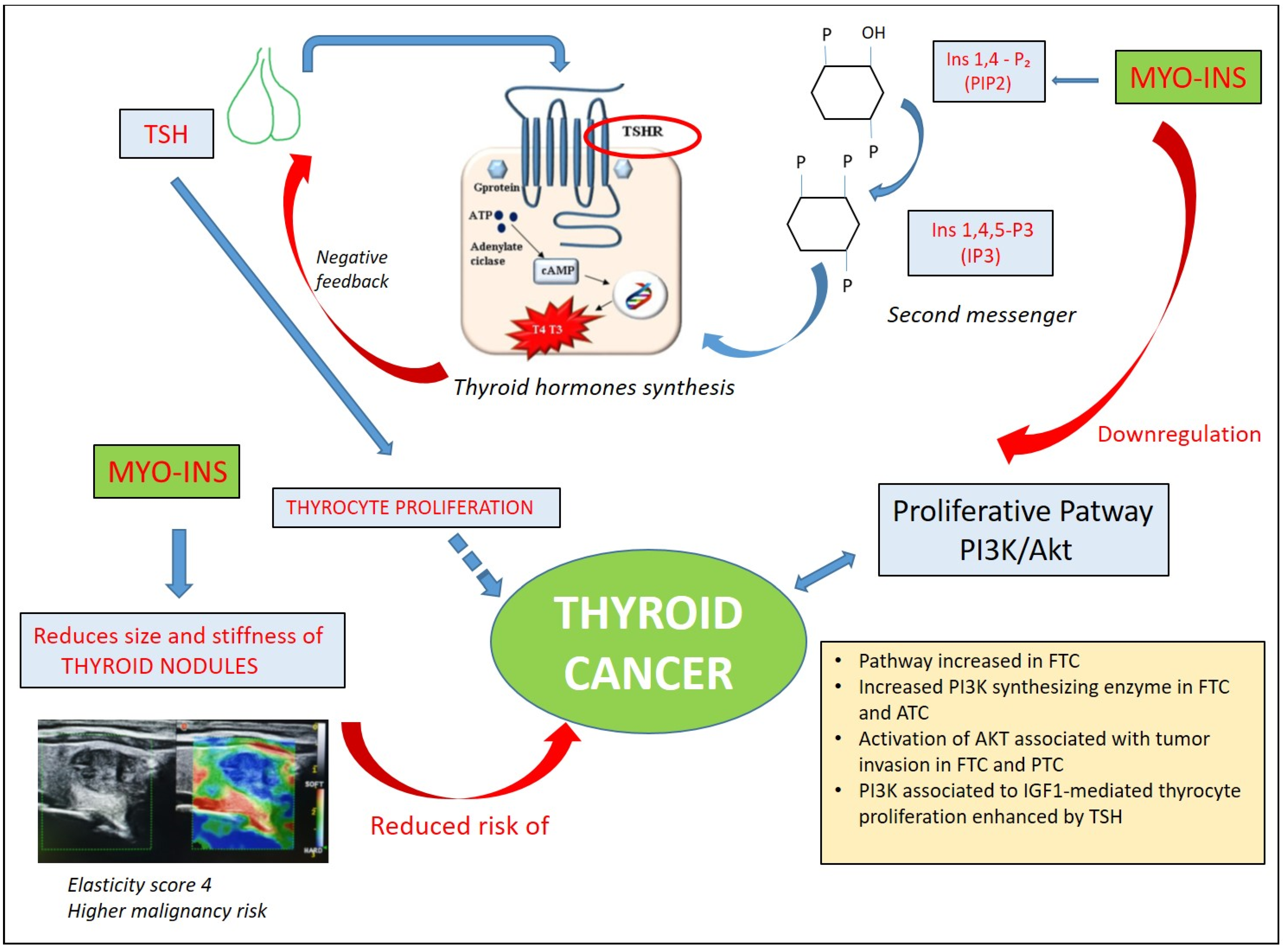
3.2. Inositols and Thyroid Cancer

Among endocrine tumors, thyroid cancer is undoubtedly the most common, with an increasing incidence in recent decades [35]. Palpable nodules constitute the smallest proportion of thyroid nodules (approximately 5%), but ultrasound is able to detect even the smallest thyroid nodules, with higher frequencies in women and the elderly. Most diagnosed nodules are benign and asymptomatic, requiring only observation and follow-up. Progression to thyroid cancer occurs in 7–15% of nodules and several risk factors, such as family history, age, sex [36,37] radiation exposure [38], can influence this process.
Different ultrasonographic features, including elastosonographic examination, allow us to estimate the risk of thyroid nodule malignancy. Poor elasticity due to alterations in the mechanical properties of the thyroid tissue is associated with an increased risk of malignancy [39]. In indeterminate lesions (TIR3A thyroid nodules), high elasticity has been shown to predict benignity, having been found in 102 out of 111 benign nodules and in only 1 out of 31 carcinomas (p < 0.0001) [40]. Nordio et al. demonstrated that supplementation with myo-Ins, with the addition of selenium, induces a morphological change in thyroid nodules classified as class I or II (at low and intermediate risk according to ultrasound classification of the AACE/ACE/AME 2016 guidelines [41]), reducing their size and stiffness, as evaluated by elastosonography [42]. The effect of myo-Ins supplementation on thyroid nodules can be traced back to the relevant role of phosphoinositides in thyroid function and their actions on pathways involved in cell proliferation.
Myo-Ins is a precursor of phosphoinositides, and among these, phosphatidylinositol 4,5-bisphosphate (PIP2) is the precursor of IP3, the second messenger of different hormones, including thyroid-stimulating hormone (TSH). As a second messenger in the phospholipase C (PLC)-dependent inositol phosphate Ca++/DAG pathway, myo-Ins contributes to thyroid hormone synthesis through H2O2 production.
Subsequently, it is natural to think that the depletion of inositol or the impairment of its signaling pathway linked to TSH may lead to a state of hypothyroidism. Several clinical studies have demonstrated that treatment with myo-Ins plus selenium is able to significantly reduce TSH levels in patients with subclinical hypothyroidism with or without autoimmune thyroiditis and that a reduction in TSH is also associated with a decline in anti-thyroid antibodies [43]. A systematic review including 28 studies with a total of 42,032 subjects and 5786 thyroid cancer cases demonstrated that higher serum TSH concentrations are associated with an increased risk of thyroid cancer, with a probable key role of anti-thyroid antibodies [44]. A meta-analysis including 53,538 participants demonstrated that each 1 mU/L increase in serum TSH was associated with a 14% greater risk of thyroid cancer for all histological thyroid cancer, 16% for differentiated thyroid carcinoma and 22% for papillary thyroid carcinoma (PTC), but, conversely, high serum TSH was associated with a reduced risk of follicular thyroid carcinoma (FTC) [45].
The relationship between thyroid autoantibodies and thyroid cancer is still a matter of debate [46,47,48]. Other studies have not found statistical significance; however, the risk of developing thyroid papillary thyroid carcinoma in Hashimoto thyroiditis patients might be considered [49]. Supplementation with myo-Ins and selenium has been shown to reduce both thyroid antibody titer and TSH levels [50,51]; consequently, a reduction in TSH levels, a risk factor for thyroid cancer, could be considered a protective factor against the development of malignant thyroid neoplasms.
Myo-Ins could be considered a thyroid cancer oncometabolite [52]. Proton nuclear magnetic resonance spectroscopy-based metabolomics demonstrates the ability to differentiate tumors from benign nodules. This is particularly interesting in the case of “indeterminate” nodules; metabolomics has demonstrated increased levels of phenylalanine, taurine and lactate, and reduced levels of choline/choline derivatives, myo- and scyllo-inositol, in malignant nodules compared to benign ones, suggesting a possible depletion of inositols in the case of malignant cells [53].
A crucial and interesting finding is the increased activity of the PI3K/Akt pathway observed in thyroid cancers. Ringel et al. demonstrated that human thyroid cancer tissues, especially FTC, are characterized by an increased expression of Akt1 and Akt2 and increased Akt activity compared with normal thyroid tissue [54]. Both in FTC and anaplastic thyroid cancers (ATCs), mutations or amplifications of the PIK3 synthesizing enzyme PI3KCA gene have been reported [55,56,57]. PIK3CA amplification was associated with an overexpression of the PIK3CA protein and phosphorylation of Akt, suggesting that this alteration represents a relevant oncogenic event in thyroid tumorigenesis via the PI3K/Akt pathway [57,58]. In particular, PIK3CA amplifications could exert a promoting role in the initial stages of thyroid tumorigenesis, since they are also found in benign thyroid adenoma (BTA), although with low prevalence [57]. In addition, other classical fusions associated with differentiated thyroid cancer, such as rearrangement involving the RET gene (RET/PTC), appear to activate the PI3K/AKT pathway [59,60]. Furthermore, RAS mutations, identified in many thyroid tumors, particularly in FTC and in the follicular variant of papillary thyroid carcinoma (FV-PTC) [57,61], may enhance thyroid tumorigenesis through their interaction with the PI3K/Akt pathway, which is a common and early event in FTC [62].
The role of PI3K signaling has also been detected in thyroid cancer progression. Studies in mouse models with metastatic FTC have demonstrated enhanced AKT activation both in primary and metastatic tumor tissue [63]. Vasko et al. reported that activation of AKT, particularly AKT 1, is associated with tumor invasion and metastasis both in FTC and PTC [64]. The investigation of molecular pathways in thyroid nodules caught the attention of some researchers who investigated the immunohistochemical expression of HGF, c-met, STAT3, phosphorylated-STAT3 (pSTAT3), PI3K, Akt and Rho in 83 benign thyroid nodules and 46 thyroid cancers (20 PTCs, 16 FTCs and 6 ATCs). The study showed that all seven proteins were expressed in 15% of follicular adenomas and all PTCs expressed the combination hepatocyte growth factor (HGF)/cmet/STAT3/pSTAT3/PI3. Among FTCs, the proportion of PI3K+ cells correlated with both the clinical phenotype and pathological stages [65].
In thyroid cells, the proliferative pathway PI3K can be activated by both TSH receptor (TSH-R) and tyrosine kinase receptors, such as insulin receptor (IR) and insulin-like growth factor 1 receptor (IGF-1R). In this context, TSH exerts its proliferative action on thyrocytes both through its own receptor and by inducing IGF-1R production and potentiating the IGF1-mediated proliferation pathways associated with PI3K and mitogen-activated protein kinase (MAPK) [66]. Thus, TSH and IGF1 are synergistic in promoting thyrocyte proliferation [67]. IGF1 is mainly produced locally in either a paracrine or autocrine manner and although it is present both in normal and in thyroid cancer tissues, IGF1 and its receptor IGF-1R are found to be overexpressed in thyroid cancer cell lines.
Considering the effects of insulin on promoting cell growth and the epidemiological evidence of the high prevalence of insulin resistance in patients with DTC [68], it may be possible to assimilate the effect of inositol to that of metformin, studied in goiter thyroid cells and in thyroid carcinoma cell lines [69]. Metformin has been demonstrated to reduce the cell viability of thyroid carcinoma cells, blocking cell cycle progression and inducing apoptosis, reducing the insulin-stimulated cell proliferation of thyroid carcinoma cells and their cancer stem cells.
The described effect of myo-Ins and IP6 in anticancer prevention, by inhibiting PI3K and the downregulation of Akt activity [15,16] and reducing insulin resistance, proposes myo-Ins as a protective factor for the malignant transformation of thyroid nodules and metastatic spread development (Figure 3). Some studies on thyroid nodules demonstrated the efficacy of myo-Ins and selenium in nodule proliferation, but the potential role of this supplementation should be explored further.

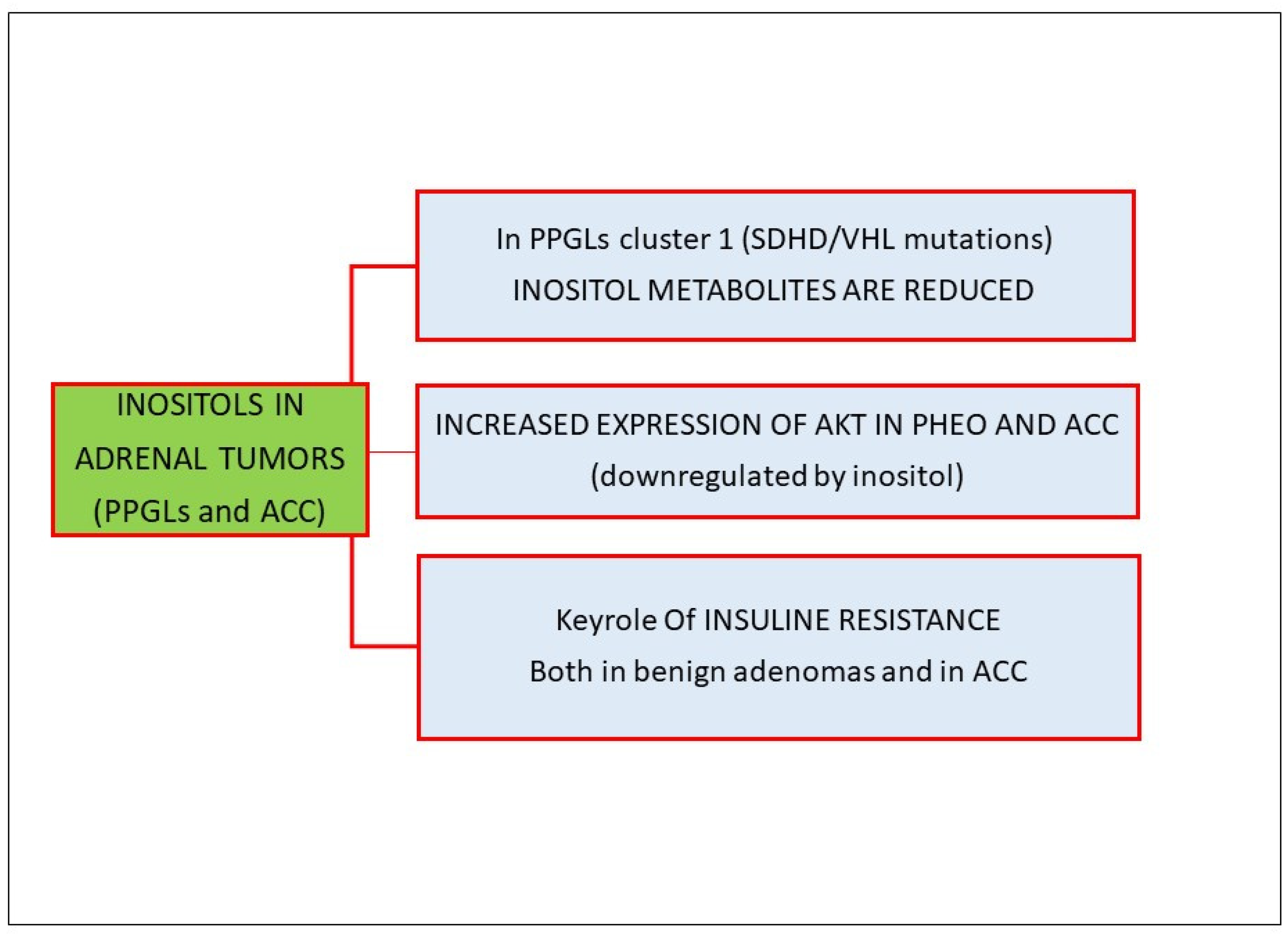
3.3. Inositols and Adrenal Tumors

Adrenal tumors include tumors arising from cortical (adrenocortical adenomas/carcinomas) and medullary tissue (pheochromocytomas), and both of them can be either benign or malignant. Pheochromocytomas and paragangliomas (PPGLs) derive from the chromaffin cell of the neural crest (from the adrenal medulla and sympathetic paraganglia, respectively); therefore, they should be considered neuroendocrine tumors. Although the majority of PPGLs are benign tumors, up to 20% of them can be malignant [70]. Based on genomic studies, PPGL tumors can be grouped into three clusters. Cluster 1 includes tumors derived by mutations in SHDx, VHL, EPAS1, FH and MDH2; cluster 2 comprises tumors with underlying RET, NF1, TMEM127, HRAS, KIF1B and MAX mutations; and the third cluster, called the Wnt-altered subtype, is derived from MAML3 fusions and CSDE1 somatic mutations [71,72,73].
Using mass spectrometry in 344 PPGLs, Murakami et al. showed that compounds related to inositol metabolism were decreased in cluster 1 and inositol cyclic phosphate, as an inositol-related metabolite, was also reduced in SHDH- and VHL- knockdown PC12 cells [74], thus suggesting a possible target therapy in patients with those specific mutations. Furthermore, several studies have suggested that insulin secretion is impaired in patients affected by PPGL, likely as a result of the inhibitory effect of catecholamines on pancreatic β cells; consequently, catecholamines antagonize insulin action in target organs, triggering an insulin resistance status [75,76]. Endogenous catecholamine excess in patients affected by pheochromocytoma can induce or aggravate insulin resistance both in patients with type 2 diabetes and in patients with normal glucose tolerance [77].
In the context of the PI3K/Akt pathway, in which inositol seems to play a key role, Fassnacht et al. observed an increased expression of Akt both in pheochromocytomas and adrenocortical carcinomas compared to normal adrenals or adenomas. Furthermore, the activated form, p-AKT, has also been reported to be overexpressed in pheochromocytomas [78]. Adler et al. demonstrated that pharmacologic inhibition of the PI3K-Akt pathway in pheochromocytoma cells downregulated the neuroendocrine phenotype, decreased hormonal secretion and promoted apoptosis [79].
Fewer data exist regarding the expression of p-AKT in adrenocortical carcinomas. The mammalian target of rapamycin (mTOR), a kinase of the PI3K-Akt signaling pathway, is a crucial intracellular mediator of the activity of growth factors receptors, comprising vascular endothelial growth factor (VEGF) and IGFs. Dysregulation of the mTOR pathway is a known feature of many tumors; therefore, the mTOR pathway is considered a target for antineoplastic therapy in different tumors. The role and functions of mTOR and its signaling pathway in the normal and tumoral adrenal gland have not been completely clarified but a dysregulation of AKT, leading to growth-simulating effects, has been described in adrenocortical carcinomas and pheochromocytomas [80].
It is worth highlighting the key role of insulin resistance in adrenocortical tumor growth [81], even if, at present, there is no unequivocal evidence of a clear causal relationship between hyperinsulinemia and adrenal tumors. Benign non-functioning adenomas are commonly associated with insulin resistance [76], although it is still a matter of debate whether this association is mediated by undetected cortisol secretion from adrenal tumor or if insulin resistance and the compensatory hyperinsulinemia stimulate adrenal tumor cell growth [82]. Additionally, in malignant neoplasms, hyperinsulinemia may potentiate the proliferative effect of IGF-2, which is overexpressed in 90% of adrenocortical carcinomas [83,84], through the binding of insulin receptor A (IR-A), IGF-1 receptor (IGF-1R) and hybrid receptor IR-A/IGF-1R. The binding of insulin or IGF-2 to these receptors leads to the activation of downstream PI3K/AKt pathways [85].
Figure 4 summarizes the role of inositol in adrenal tumors.

3.4. Inositols and Pituitary Neuroendocrine Tumors (Pit-NETs)

Low evidence is available regarding the role of inositol in Pit-NETs. Jones et al. demonstrated different expression levels of phosphoinositide in 29 pituitary adenoma tissues, which were divided into two groups, one with a high basal turnover and another with low basal phosphoinositide. The first group was associated with kinine response and produced interleukin-6, which is involved in promoting mitogenic growth and pituitary tumorigenesis, suggesting a possible role of inositols in the control of mechanisms involved in the cell growth of some, but not all, pituitary tumors [86].
A more recent systematic review investigated the metabolomic aspects of pituitary adenomas using mass spectrometry, suggesting an alteration of amino acid metabolism in these tumors. In detail, the authors demonstrated that phosphethanolamine, N-acetyl aspartate and myo-Ins are downregulated in prolactinomas, hypothesizing that a possible deficiency of these biomolecules in some tumors could determine their pathogenesis [87].
Currently, there are limited studies suggesting the growth-inhibitory effects of metformin, an anti-diabetic and insulin-sensitizing drug, on PitNETs. The antiproliferative effects of metformin have been observed on an ACTH-secreting mouse corticotroph tumor cell line and growth hormone-secreting PitNET cell lines, GH3 and GH1 [88]. In ACTH-secreting AtT20 cells, metformin inhibited cell proliferation by activating the AMPK signaling pathway and inhibiting the IGF-1R/AKT/mTOR pathway. In a recent study performed on primary cell cultures derived from PitNETs, metformin treatment inhibited cell viability in ACTH-secreting adenomas and non-functioning pituitary adenomas, but not prolactinomas, and in GH-secreting adenomas [89]. Inositol, which shares the same Akt/mTOR signaling pathway, could be hypothesized as a possible concomitant influencing factor in the cellular growth of Pit-NETs.

3.5. Inositols and Neuroendocrine Neoplasms

Neuroendocrine neoplasms (NENs) arise from neuroendocrine cells, located in all parts of the body, though the most common sites of origin are the lung and the gastro-entero-pancreatic (GEP) system. The incidence and prevalence of these tumors, while rare, is increasing [90]. The term NENs includes the well-differentiated forms, also known as neuroendocrine tumors (NETs), which have a low proliferation rate and are slow-growing, and the poorly differentiated neoplasms, also known as Neuroendocrine Carcinomas (NECs), characterized by a high proliferation rate and clinical aggressiveness [91]. NENs can be non-functioning or functioning, with the latter characterized by the secretion of hormones and peptides associated with a specific syndrome [92].
Many research articles demonstrated a pathogenetic role of the PI3K/Akt/mTOR pathways in the development of NENs. mTOR and PI3K signaling can regulate cell growth, proliferation, migration, survival and angiogenesis [93].
The PIK3CA gene, encoding for the catalytic subunit of class I PI3K, can be mutated in a sub-group of pNENs [94]. Preclinical studies using PI3K inhibitors (LY294002) in rat-derived GEP-NEN cell lines result in the inhibition of VEGF secretion by neoplastic endocrine cells [95]. Similarly, a study on BON cells, one of the few neuroendocrine cell lines available, has demonstrated that LY294002 blocks the constitutive activation of PI3K and ERKs [96]. PI3K signaling is also able to reduce neuroendocrine secretion in BON and QGP-1 cells [97].
mTOR hyperactivation has frequently been detected in neuroendocrine tumors [98,99] but also in poorly differentiated NECs [100]. Mutations of genes of the mTOR pathway, such as PTEN, TSC2 and PIK3CA, have been detected in about 16% of sporadic pancreatic NETs [101]. Additionally, low expression of TSC2 and PTEN was associated with reduced disease-free and overall survival in pancreatic NENs [102]. Everolimus, a mTOR inhibitor, is currently approved for the treatment of GEP and lung NETs [103]. However, NENs can develop resistance to everolimus, and one of the proposed pathways is the activation of PI3K, which could be potentially overcome by the use of inositols.
Finally, NEN cells are able to produce high amounts of IGF 1. In gastrinomas, high levels of IGF-1 and its receptor (IGF-1R) were associated with tumor development, progression and aggressiveness [104].
To date, there are no data on inositol use in patients affected by neuroendocrine cells, but studies on other cancer cells have suggested a role of IP6 in the induction of cell apoptosis by inhibiting the Akt/mTOR pathway and PI3k pathways, supporting the rationale for the use of inositols in combination with everolimus in NENs.

4. Clinical Use of Inositols and New Therapeutic Perspectives

Inositol supplementation (in the form of myo-Ins and D-chiro-inositol) is currently used as an effective alternative to the classic insulin-sensitizing molecule, metformin, in many gynecological and endocrinological diseases, such as PCOS, gestational diabetes mellitus (GDM) and male infertility [26].
In a recent systematic review and meta-analysis of 36 randomized controlled clinical trials with a total of 1691 PCOS patients randomized to inositol, metformin or placebo, it was demonstrated that inositol was superior to placebo and not inferior to metformin in normalizing menstrual cycles and superior to placebo in reducing BMI, blood levels of testosterone, androstenedione glucose and AUC insulin [105].
Regarding the use of inositol in the treatment of endocrine tumors, despite the promising evidence described above, we are still far from a possible clinical use. In many other tumor histotypes, there are already robust in vitro and in vivo studies that reinforce their potential use in human tumors. In a small pilot clinical trial involving 22 patients suffering from metastatic colon cancer, the combination of IP6 and inositol (InoCell) as an adjuvant to conventional chemotherapy caused a reduction in tumor growth, also lowering the side effects of chemotherapy (leukopenia, thrombocytopenia, nausea and vomiting) [106,107].

5. Conclusions

This review highlights the role of inositols in the pathogenesis and progression of the main types of endocrine and neuroendocrine cancers, proposing a solid rationale for the use of inositol supplementation as an adjuvant treatment in this field of oncology.
We believe that the current evidence on the molecular mechanisms of carcinogenesis, particularly in thyroid and adrenal tumors, can support researchers’ decisions to apply inositol in in vitro and in vivo studies in these settings to confirm these hypotheses.

Author Contributions

Conceptualization, M.A.; methodology, M.M., G.P. and M.A.; validation: R.L. and M.B.; resources, M.M. and G.P.; data curation, M.M. and G.P.; writing—original draft preparation, M.M. and G.P.; writing—review and editing, R.L., M.B. and M.A.; supervision, M.A. All authors have read and agreed to the published version of the manuscript.

Funding

This research received no external funding.

Conflicts of Interest

M.A. declares consultancy and research grants from Bayer, Eisai and Eli-Lilly. G.P. declares consultancy for Bayer. The other authors declare no conflicts of interest.

Abbreviations

myo-Insmyo-inositol
IP6inositol hexakisphosphate
IP3inositol triphosphate
IGF-1insulin-growth factor 1
IGFBP-1 insulin-growth factor binding protein 1
IGFBP-2insulin-growth factor binding protein 2
MAP mitogenic-activated protein
PI3K/Aktphosphoinositide-3 kinase/Akt
EMTepithelial–mesenchymal transition
pNETSpancreatic neuroendocrine tumors
PIP2phosphatidylinositol 4,5-bisphosphate
TSHthyroid-stimulating hormone
PLCphospholipase C
PTCpapillary thyroid carcinoma
FTCfollicular thyroid carcinoma
BTAbenign thyroid adenoma
FV-PTCfollicular variant of papillary thyroid carcinoma
pSTAT3phosphorylated-STAT3
HGFhepatocyte growth factor
MAPKmitogen-activated protein kinase
PPGLparagangliomas
mTORmammalian target of rapamycin
VEGFvascular endothelial growth factor
Pit-NET pituitary neuroendocrine tumors
NENneuroendocrine neoplasm
NETneuroendocrine tumor
NEC neuroendocrine carcinoma
GEPgastro-entero-pancreatic system
PHEOpheochromocytoma
ACCadrenocortical carcinoma
PCOS polycystic ovary syndrome
GDMgestational diabetes mellitus

References

  1. Shamsuddin, A.M. Metabolism and Cellular Functions of Ip6: A Review. Anticancer Res. 1999, 19, 3733–3736. [Google Scholar] [PubMed]
  2. Bizzarri, M.; Vucenik, I.; Appetecchia, M. Inositols as Adjuvant Treatments in Oncology. In A Clinical Guide to Inositols. Vittorio Unfer and Didier Dewailly Editors; Academic Press: New York, NY, USA, 2023. [Google Scholar] [CrossRef]
  3. Shanmugam, M.K.; Lee, J.H.; Chai, E.Z.; Kanchi, M.M.; Kar, S.; Arfuso, F.; Dharmarajan, A.; Kumar, A.P.; Ramar, P.S.; Looi, C.Y.; et al. Cancer Prevention and Therapy through the Modulation of Transcription Factors by Bioactive Natural Compounds. Semin. Cancer Biol. 2016, 40–41, 35–47. [Google Scholar] [CrossRef] [PubMed]
  4. Irving, D.; Drasar, B.S. Fibre and Cancer of the Colon. Br. J. Cancer 1973, 28, 462–463. [Google Scholar] [CrossRef] [PubMed]
  5. Graf, E.; Eaton, J.W. Dietary Suppression of Colonic Cancer. Fiber or Phytate? Cancer 1985, 56, 717–718. [Google Scholar] [CrossRef]
  6. Markiewicz, L.H.; Ogrodowczyk, A.M.; Wiczkowski, W.; Wroblewska, B. Phytate and Butyrate Differently Influence the Proliferation, Apoptosis and Survival Pathways in Human Cancer and Healthy Colonocytes. Nutrients 2021, 13, 1887. [Google Scholar] [CrossRef] [PubMed]
  7. Singh, R.P.; Agarwal, R. Prostate Cancer and Inositol Hexaphosphate: Efficacy and Mechanisms. Anticancer Res. 2005, 25, 2891–2903. [Google Scholar] [PubMed]
  8. Wawszczyk, J.; Kapral, M.; Lodowska, J.; Jesse, K.; Hollek, A.; Weglarz, L. Antiproliferative Effect of Inositol Hexaphosphate on Human Skin Melanoma Cells In Vitro. Acta Pol. Pharm. 2015, 72, 895–900. [Google Scholar]
  9. Liu, G.; Song, Y.; Cui, L.; Wen, Z.; Lu, X. Inositol Hexaphosphate Suppresses Growth and Induces Apoptosis in Ht-29 Colorectal Cancer Cells in Culture: Pi3k/Akt Pathway as a Potential Target. Int. J. Clin. Exp. Pathol. 2015, 8, 1402–1410. [Google Scholar] [PubMed]
  10. de Lima, E.M.; Kanunfre, C.C.; de Andrade, L.F.; Granato, D.; Rosso, N.D. Cytotoxic Effect of Inositol Hexaphosphate and Its Ni(Ii) Complex on Human Acute Leukemia Jurkat T Cells. Toxicol. In Vitro 2015, 29, 2081–2088. [Google Scholar] [CrossRef]
  11. Baethge, C.; Goldbeck-Wood, S.; Mertens, S. Sanra-a Scale for the Quality Assessment of Narrative Review Articles. Res. Integr. Peer Rev. 2019, 4, 5. [Google Scholar] [CrossRef]
  12. Agarwal, C.; Dhanalakshmi, S.; Singh, R.P.; Agarwal, R. Inositol Hexaphosphate Inhibits Growth and Induces G1 Arrest and Apoptotic Death of Androgen-Dependent Human Prostate Carcinoma Lncap Cells. Neoplasia 2004, 6, 646–659. [Google Scholar] [CrossRef] [PubMed]
  13. Deliliers, G.L.; Servida, F.; Fracchiolla, N.S.; Ricci, C.; Borsotti, C.; Colombo, G.; Soligo, D. Effect of Inositol Hexaphosphate (Ip(6)) on Human Normal and Leukaemic Haematopoietic Cells. Br. J. Haematol. 2002, 117, 577–587. [Google Scholar] [CrossRef] [PubMed]
  14. Weglarz, L.; Molin, I.; Orchel, A.; Parfiniewicz, B.; Dzierzewicz, Z. Quantitative Analysis of the Level of P53 and P21(Waf1) Mrna in Human Colon Cancer Ht-29 Cells Treated with Inositol Hexaphosphate. Acta Biochim. Pol. 2006, 53, 349–356. [Google Scholar] [CrossRef] [PubMed]
  15. Dinicola, S.; Fabrizi, G.; Masiello, M.G.; Proietti, S.; Palombo, A.; Minini, M.; Harrath, A.H.; Alwasel, S.H.; Ricci, G.; Catizone, A.; et al. Inositol Induces Mesenchymal-Epithelial Reversion in Breast Cancer Cells through Cytoskeleton Rearrangement. Exp. Cell Res. 2016, 345, 37–50. [Google Scholar] [CrossRef]
  16. Huang, C.; Ma, W.Y.; Hecht, S.S.; Dong, Z. Inositol Hexaphosphate Inhibits Cell Transformation and Activator Protein 1 Activation by Targeting Phosphatidylinositol-3’ Kinase. Cancer Res. 1997, 57, 2873–2878. [Google Scholar]
  17. Liao, J.; Seril, D.N.; Yang, A.L.; Lu, G.G.; Yang, G.Y. Inhibition of Chronic Ulcerative Colitis Associated Adenocarcinoma Development in Mice by Inositol Compounds. Carcinogenesis 2007, 28, 446–454. [Google Scholar] [CrossRef]
  18. Vucenik, I.; Shamsuddin, A.M. Protection against Cancer by Dietary Ip6 and Inositol. Nutr. Cancer 2006, 55, 109–125. [Google Scholar] [CrossRef]
  19. Vucenik, I.; Passaniti, A.; Vitolo, M.I.; Tantivejkul, K.; Eggleton, P.; Shamsuddin, A.M. Anti-Angiogenic Activity of Inositol Hexaphosphate (Ip6). Carcinogenesis 2004, 25, 2115–2123. [Google Scholar] [CrossRef]
  20. Graf, E.; Eaton, J.W. Antioxidant Functions of Phytic Acid. Free Radic. Biol. Med. 1990, 8, 61–69. [Google Scholar] [CrossRef]
  21. Baten, A.; Ullah, A.; Tomazic, V.J.; Shamsuddin, A.M. Inositol-Phosphate-Induced Enhancement of Natural Killer Cell Activity Correlates with Tumor Suppression. Carcinogenesis 1989, 10, 1595–1598. [Google Scholar] [CrossRef]
  22. Zhang, Z.; Song, Y.; Wang, X.L. Inositol Hexaphosphate-Induced Enhancement of Natural Killer Cell Activity Correlates with Suppression of Colon Carcinogenesis in Rats. World J. Gastroenterol. 2005, 11, 5044–5046. [Google Scholar] [CrossRef]
  23. Shamsuddin, A.M.; Ullah, A.; Chakravarthy, A.K. Inositol and Inositol Hexaphosphate Suppress Cell Proliferation and Tumor Formation in Cd-1 Mice. Carcinogenesis 1989, 10, 1461–1463. [Google Scholar] [CrossRef] [PubMed]
  24. Vucenik, I.; Sakamoto, K.; Bansal, M.; Shamsuddin, A.M. Inhibition of Rat Mammary Carcinogenesis by Inositol Hexaphosphate (Phytic Acid). A Pilot Study. Cancer Lett. 1993, 75, 95–102. [Google Scholar] [CrossRef] [PubMed]
  25. Vucenik, I.; Tomazic, V.J.; Fabian, D.; Shamsuddin, A.M. Antitumor Activity of Phytic Acid (Inositol Hexaphosphate) in Murine Transplanted and Metastatic Fibrosarcoma, a Pilot Study. Cancer Lett. 1992, 65, 9–13. [Google Scholar] [CrossRef] [PubMed]
  26. Dinicola, S.; Unfer, V.; Facchinetti, F.; Soulage, C.O.; Greene, N.D.; Bizzarri, M.; Lagana, A.S.; Chan, S.Y.; Bevilacqua, A.; Pkhaladze, L.; et al. Inositols: From Established Knowledge to Novel Approaches. Int. J. Mol. Sci. 2021, 22, 105753. [Google Scholar] [CrossRef] [PubMed]
  27. Bizzarri, M.; Dinicola, S.; Cucina, A. Modulation of Both Insulin Resistance and Cancer Growth by Inositol. Curr. Pharm. Des. 2017, 23, 5200–5210. [Google Scholar] [CrossRef] [PubMed]
  28. Tsugane, S.; Inoue, M. Insulin Resistance and Cancer: Epidemiological Evidence. Cancer Sci. 2010, 101, 1073–1079. [Google Scholar] [CrossRef] [PubMed]
  29. Calle, E.E.; Thun, M.J. Obesity and Cancer. Oncogene 2004, 23, 6365–6378. [Google Scholar] [CrossRef]
  30. Vigneri, P.; Frasca, F.; Sciacca, L.; Pandini, G.; Vigneri, R. Diabetes and Cancer. Endocr. Relat. Cancer 2009, 16, 1103–1123. [Google Scholar] [CrossRef]
  31. Denley, A.; Wallace, J.C.; Cosgrove, L.J.; Forbes, B.E. The Insulin Receptor Isoform Exon 11- (Ir-a) in Cancer and Other Diseases: A Review. Horm. Metab. Res. 2003, 35, 778–785. [Google Scholar] [CrossRef]
  32. Fresno Vara, J.A.; Casado, E.; de Castro, J.; Cejas, P.; Belda-Iniesta, C.; Gonzalez-Baron, M. Pi3k/Akt Signalling Pathway and Cancer. Cancer Treat. Rev. 2004, 30, 193–204. [Google Scholar] [CrossRef]
  33. Lim, J.; Kim, J.H.; Paeng, J.Y.; Kim, M.J.; Hong, S.D.; Lee, J.I.; Hong, S.P. Prognostic Value of Activated Akt Expression in Oral Squamous Cell Carcinoma. J. Clin. Pathol. 2005, 58, 1199–1205. [Google Scholar] [CrossRef] [PubMed]
  34. Thakur, S.; Daley, B.; Klubo-Gwiezdzinska, J. The Role of an Anti-Diabetic Drug Metformin in the Treatment of Endocrine Tumors. J. Mol. Endocrinol. 2019, 63, R17–R35. [Google Scholar] [CrossRef] [PubMed]
  35. Lim, H.; Devesa, S.S.; Sosa, J.A.; Check, D.; Kitahara, C.M. Trends in Thyroid Cancer Incidence and Mortality in the United States, 1974–2013. JAMA 2017, 317, 1338–1348. [Google Scholar] [CrossRef] [PubMed]
  36. Haugen, B.R.; Alexander, E.K.; Bible, K.C.; Doherty, G.M.; Mandel, S.J.; Nikiforov, Y.E.; Pacini, F.; Randolph, G.W.; Sawka, A.M.; Schlumberger, M.; et al. 2015 American Thyroid Association Management Guidelines for Adult Patients with Thyroid Nodules and Differentiated Thyroid Cancer: The American Thyroid Association Guidelines Task Force on Thyroid Nodules and Differentiated Thyroid Cancer. Thyroid 2016, 26, 1–133. [Google Scholar] [CrossRef]
  37. Hegedus, L. Clinical Practice. The Thyroid Nodule. N. Engl. J. Med. 2004, 351, 1764–1771. [Google Scholar] [CrossRef]
  38. Antonelli, A.; Silvano, G.; Bianchi, F.; Gambuzza, C.; Tana, L.; Salvioni, G.; Baldi, V.; Gasperini, L.; Baschieri, L. Risk of Thyroid Nodules in Subjects Occupationally Exposed to Radiation: A Cross Sectional Study. Occup. Environ. Med. 1995, 52, 500–504. [Google Scholar] [CrossRef]
  39. Asteria, C.; Giovanardi, A.; Pizzocaro, A.; Cozzaglio, L.; Morabito, A.; Somalvico, F.; Zoppo, A. Us-Elastography in the Differential Diagnosis of Benign and Malignant Thyroid Nodules. Thyroid 2008, 18, 523–531. [Google Scholar] [CrossRef]
  40. Rago, T.; Scutari, M.; Santini, F.; Loiacono, V.; Piaggi, P.; Di Coscio, G.; Basolo, F.; Berti, P.; Pinchera, A.; Vitti, P. Real-Time Elastosonography: Useful Tool for Refining the Presurgical Diagnosis in Thyroid Nodules with Indeterminate or Nondiagnostic Cytology. J. Clin. Endocrinol. Metab. 2010, 95, 5274–5280. [Google Scholar] [CrossRef]
  41. Gharib, H.; Papini, E.; Garber, J.R.; Duick, D.S.; Harrell, R.M.; Hegedus, L.; Paschke, R.; Valcavi, R.; Vitti, P.; Aace Ace Ame Task Force on Thyroid Nodules. American Association of Clinical Endocrinologists, American College of Endocrinology, and Associazione Medici Endocrinologi Medical Guidelines for Clinical Practice for the Diagnosis and Management of Thyroid Nodules--2016 Update. Endocr. Pract. 2016, 22, 622–639. [Google Scholar] [CrossRef]
  42. Nordio, M.; Basciani, S. Evaluation of Thyroid Nodule Characteristics in Subclinical Hypothyroid Patients under a Myo-Inositol Plus Selenium Treatment. Eur. Rev. Med. Pharmacol. Sci. 2018, 22, 2153–2159. [Google Scholar] [CrossRef] [PubMed]
  43. Benvenga, S.; Nordio, M.; Lagana, A.S.; Unfer, V. The Role of Inositol in Thyroid Physiology and in Subclinical Hypothyroidism Management. Front. Endocrinol. 2021, 12, 662582. [Google Scholar] [CrossRef] [PubMed]
  44. McLeod, D.S.; Watters, K.F.; Carpenter, A.D.; Ladenson, P.W.; Cooper, D.S.; Ding, E.L. Thyrotropin and Thyroid Cancer Diagnosis: A Systematic Review and Dose-Response Meta-Analysis. J. Clin. Endocrinol. Metab. 2012, 97, 2682–2692. [Google Scholar] [CrossRef] [PubMed]
  45. Hu, N.; Li, Z.M.; Liu, J.F.; Zhang, Z.Z.; Wang, L.S. An Overall and Dose-Response Meta-Analysis for Thyrotropin and Thyroid Cancer Risk by Histological Type. Oncotarget 2016, 7, 47750–47759. [Google Scholar] [CrossRef] [PubMed]
  46. de Alcantara-Jones, D.M.; de Alcantara-Nunes, T.F.; Rocha Bde, O.; de Oliveira, R.D.; Santana, A.C.; de Alcantara, F.T.; de Faria, T.M.; da Silva, I.C.; Araujo, L.M. Is There Any Association between Hashimoto’s Thyroiditis and Thyroid Cancer? A Retrospective Data Analysis. Radiol. Bras. 2015, 48, 148–153. [Google Scholar] [CrossRef] [PubMed]
  47. Lee, I.S.; Hsieh, A.T.; Lee, T.W.; Lee, T.I.; Chien, Y.M. The Association of Thyrotropin and Autoimmune Thyroid Disease in Developing Papillary Thyroid Cancer. Int. J. Endocrinol. 2017, 2017, 5940367. [Google Scholar] [CrossRef] [PubMed]
  48. Selek, A.; Cetinarslan, B.; Tarkun, I.; Canturk, Z.; Ustuner, B.; Akyay, Z. Thyroid Autoimmunity: Is Really Associated with Papillary Thyroid Carcinoma? Eur. Arch. Otorhinolaryngol. 2017, 274, 1677–1681. [Google Scholar] [CrossRef]
  49. Boi, F.; Pani, F.; Mariotti, S. Thyroid Autoimmunity and Thyroid Cancer: Review Focused on Cytological Studies. Eur. Thyroid J. 2017, 6, 178–186. [Google Scholar] [CrossRef]
  50. Nordio, M.; Basciani, S. Myo-Inositol Plus Selenium Supplementation Restores Euthyroid State in Hashimoto’s Patients with Subclinical Hypothyroidism. Eur. Rev. Med. Pharmacol. Sci. 2017, 21, 51–59. [Google Scholar]
  51. Payer, J.; Jackuliak, P.; Kuzma, M.; Dzupon, M.; Vanuga, P. Supplementation with Myo-Inositol and Selenium Improves the Clinical Conditions and Biochemical Features of Women with or at Risk for Subclinical Hypothyroidism. Front. Endocrinol. 2022, 13, 1067029. [Google Scholar] [CrossRef]
  52. Khatami, F.; Payab, M.; Sarvari, M.; Gilany, K.; Larijani, B.; Arjmand, B.; Tavangar, S.M. Oncometabolites as Biomarkers in Thyroid Cancer: A Systematic Review. Cancer Manag. Res. 2019, 11, 1829–1841. [Google Scholar] [CrossRef] [PubMed]
  53. Torregrossa, L.; Shintu, L.; Nambiath Chandran, J.; Tintaru, A.; Ugolini, C.; Magalhaes, A.; Basolo, F.; Miccoli, P.; Caldarelli, S. Toward the Reliable Diagnosis of Indeterminate Thyroid Lesions: A Hrmas Nmr-Based Metabolomics Case of Study. J. Proteome Res. 2012, 11, 3317–3325. [Google Scholar] [CrossRef] [PubMed]
  54. Ringel, M.D.; Hayre, N.; Saito, J.; Saunier, B.; Schuppert, F.; Burch, H.; Bernet, V.; Burman, K.D.; Kohn, L.D.; Saji, M. Overexpression and Overactivation of Akt in Thyroid Carcinoma. Cancer Res. 2001, 61, 6105–6111. [Google Scholar]
  55. Wu, G.; Mambo, E.; Guo, Z.; Hu, S.; Huang, X.; Gollin, S.M.; Trink, B.; Ladenson, P.W.; Sidransky, D.; Xing, M. Uncommon Mutation, but Common Amplifications, of the Pik3ca Gene in Thyroid Tumors. J. Clin. Endocrinol. Metab. 2005, 90, 4688–4693. [Google Scholar] [CrossRef]
  56. Garcia-Rostan, G.; Costa, A.M.; Pereira-Castro, I.; Salvatore, G.; Hernandez, R.; Hermsem, M.J.; Herrero, A.; Fusco, A.; Cameselle-Teijeiro, J.; Santoro, M. Mutation of the Pik3ca Gene in Anaplastic Thyroid Cancer. Cancer Res. 2005, 65, 10199–10207. [Google Scholar] [CrossRef] [PubMed]
  57. Hou, P.; Liu, D.; Shan, Y.; Hu, S.; Studeman, K.; Condouris, S.; Wang, Y.; Trink, A.; El-Naggar, A.K.; Tallini, G.; et al. Genetic Alterations and Their Relationship in the Phosphatidylinositol 3-Kinase/Akt Pathway in Thyroid Cancer. Clin. Cancer Res. 2007, 13, 1161–1170. [Google Scholar] [CrossRef] [PubMed]
  58. Liu, Z.; Hou, P.; Ji, M.; Guan, H.; Studeman, K.; Jensen, K.; Vasko, V.; El-Naggar, A.K.; Xing, M. Highly Prevalent Genetic Alterations in Receptor Tyrosine Kinases and Phosphatidylinositol 3-Kinase/Akt and Mitogen-Activated Protein Kinase Pathways in Anaplastic and Follicular Thyroid Cancers. J. Clin. Endocrinol. Metab. 2008, 93, 3106–3116. [Google Scholar] [CrossRef] [PubMed]
  59. Rodriguez-Viciana, P.; Warne, P.H.; Dhand, R.; Vanhaesebroeck, B.; Gout, I.; Fry, M.J.; Waterfield, M.D.; Downward, J. Phosphatidylinositol-3-Oh Kinase as a Direct Target of Ras. Nature 1994, 370, 527–532. [Google Scholar] [CrossRef] [PubMed]
  60. Segouffin-Cariou, C.; Billaud, M. Transforming Ability of Men2a-Ret Requires Activation of the Phosphatidylinositol 3-Kinase/Akt Signaling Pathway. J. Biol. Chem. 2000, 275, 3568–3576. [Google Scholar] [CrossRef]
  61. Zhu, Z.; Gandhi, M.; Nikiforova, M.N.; Fischer, A.H.; Nikiforov, Y.E. Molecular Profile and Clinical-Pathologic Features of the Follicular Variant of Papillary Thyroid Carcinoma. An Unusually High Prevalence of Ras Mutations. Am. J. Clin. Pathol. 2003, 120, 71–77. [Google Scholar] [CrossRef]
  62. Gupta, S.; Ramjaun, A.R.; Haiko, P.; Wang, Y.; Warne, P.H.; Nicke, B.; Nye, E.; Stamp, G.; Alitalo, K.; Downward, J. Binding of Ras to Phosphoinositide 3-Kinase P110alpha Is Required for Ras-Driven Tumorigenesis in Mice. An Unusually High Prevalence of Ras Mutations. Cell 2007, 129, 957–968. [Google Scholar] [CrossRef] [PubMed]
  63. Kim, C.S.; Vasko, V.V.; Kato, Y.; Kruhlak, M.; Saji, M.; Cheng, S.Y.; Ringel, M.D. Akt Activation Promotes Metastasis in a Mouse Model of Follicular Thyroid Carcinoma. Endocrinology 2005, 146, 4456–4463. [Google Scholar] [CrossRef] [PubMed]
  64. Vasko, V.; Saji, M.; Hardy, E.; Kruhlak, M.; Larin, A.; Savchenko, V.; Miyakawa, M.; Isozaki, O.; Murakami, H.; Tsushima, T.; et al. Akt Activation and Localisation Correlate with Tumour Invasion and Oncogene Expression in Thyroid Cancer. J. Med. Genet. 2004, 41, 161–170. [Google Scholar] [CrossRef] [PubMed]
  65. Ruggeri, R.M.; Vitarelli, E.; Barresi, G.; Trimarchi, F.; Benvenga, S.; Trovato, M. Hgf/C-Met System Pathways in Benign and Malignant Histotypes of Thyroid Nodules: An Immunohistochemical Characterization. Histol. Histopathol. 2012, 27, 113–121. [Google Scholar] [CrossRef] [PubMed]
  66. Ciampolillo, A.; De Tullio, C.; Perlino, E.; Maiorano, E. The Igf-I Axis in Thyroid Carcinoma. Curr. Pharm. Des. 2007, 13, 729–735. [Google Scholar] [CrossRef] [PubMed]
  67. Sarlis, N.J.; Benvenga, S. Molecular Signaling in Thyroid Cancer. Cancer Treat. Res. 2004, 122, 237–264. [Google Scholar] [CrossRef]
  68. Rezzonico, J.N.; Rezzonico, M.; Pusiol, E.; Pitoia, F.; Niepomniszcze, H. Increased Prevalence of Insulin Resistance in Patients with Differentiated Thyroid Carcinoma. Metab. Syndr. Relat. Disord. 2009, 7, 375–380. [Google Scholar] [CrossRef]
  69. Chen, G.; Xu, S.; Renko, K.; Derwahl, M. Metformin Inhibits Growth of Thyroid Carcinoma Cells, Suppresses Self-Renewal of Derived Cancer Stem Cells, and Potentiates the Effect of Chemotherapeutic Agents. J. Clin. Endocrinol. Metab. 2012, 97, E510–E520. [Google Scholar] [CrossRef]
  70. Lenders, J.W.; Duh, Q.Y.; Eisenhofer, G.; Gimenez-Roqueplo, A.P.; Grebe, S.K.; Murad, M.H.; Naruse, M.; Pacak, K.; Young, W.F., Jr.; Endocrine, S. Pheochromocytoma and Paraganglioma: An Endocrine Society Clinical Practice Guideline. J. Clin. Endocrinol. Metab. 2014, 99, 1915–1942. [Google Scholar] [CrossRef] [PubMed]
  71. Dahia, P.L.; Ross, K.N.; Wright, M.E.; Hayashida, C.Y.; Santagata, S.; Barontini, M.; Kung, A.L.; Sanso, G.; Powers, J.F.; Tischler, A.S.; et al. A Hif1alpha Regulatory Loop Links Hypoxia and Mitochondrial Signals in Pheochromocytomas. PLoS Genet. 2005, 1, e8. [Google Scholar] [CrossRef]
  72. Flynn, A.; Dwight, T.; Harris, J.; Benn, D.; Zhou, L.; Hogg, A.; Catchpoole, D.; James, P.; Duncan, E.L.; Trainer, A.; et al. Pheo-Type: A Diagnostic Gene-Expression Assay for the Classification of Pheochromocytoma and Paraganglioma. J. Clin. Endocrinol. Metab. 2016, 101, 1034–1043. [Google Scholar] [CrossRef] [PubMed]
  73. Castro-Vega, L.J.; Buffet, A.; De Cubas, A.A.; Cascon, A.; Menara, M.; Khalifa, E.; Amar, L.; Azriel, S.; Bourdeau, I.; Chabre, O.; et al. Germline Mutations in Fh Confer Predisposition to Malignant Pheochromocytomas and Paragangliomas. Hum. Mol. Genet. 2014, 23, 2440–2446. [Google Scholar] [CrossRef] [PubMed]
  74. Murakami, M.; Sun, N.; Greunke, C.; Feuchtinger, A.; Kircher, S.; Deutschbein, T.; Papathomas, T.; Bechmann, N.; William Wallace, P.; Peitzsch, M.; et al. Mass Spectrometry Imaging Identifies Metabolic Patterns Associated with Malignant Potential in Pheochromocytoma and Paraganglioma. Eur. J. Endocrinol. 2021, 185, 179–191. [Google Scholar] [CrossRef] [PubMed]
  75. Zeiger, M.A.; Thompson, G.B.; Duh, Q.Y.; Hamrahian, A.H.; Angelos, P.; Elaraj, D.; Fishman, E.; Kharlip, J.; American Association of Clinical, E.; American Association of Endocrine, S. American Association of Clinical Endocrinologists and American Association of Endocrine Surgeons Medical Guidelines for the Management of Adrenal Incidentalomas: Executive Summary of Recommendations. Endocr. Pract. 2009, 15, 450–453. [Google Scholar] [CrossRef] [PubMed]
  76. Terzolo, M.; Pia, A.; Ali, A.; Osella, G.; Reimondo, G.; Bovio, S.; Daffara, F.; Procopio, M.; Paccotti, P.; Borretta, G.; et al. Adrenal Incidentaloma: A New Cause of the Metabolic Syndrome? J. Clin. Endocrinol. Metab. 2002, 87, 998–1003. [Google Scholar] [CrossRef]
  77. Wiesner, T.D.; Bluher, M.; Windgassen, M.; Paschke, R. Improvement of Insulin Sensitivity after Adrenalectomy in Patients with Pheochromocytoma. J. Clin. Endocrinol. Metab. 2003, 88, 3632–3636. [Google Scholar] [CrossRef]
  78. Fassnacht, M.; Weismann, D.; Ebert, S.; Adam, P.; Zink, M.; Beuschlein, F.; Hahner, S.; Allolio, B. Akt Is Highly Phosphorylated in Pheochromocytomas but Not in Benign Adrenocortical Tumors. J. Clin. Endocrinol. Metab. 2005, 90, 4366–4370. [Google Scholar] [CrossRef]
  79. Adler, J.T.; Hottinger, D.G.; Kunnimalaiyaan, M.; Chen, H. Inhibition of the Pi3k Pathway Suppresses Hormonal Secretion and Limits Growth in Pheochromocytoma Cells. World J. Surg. 2009, 33, 2452–2457. [Google Scholar] [CrossRef]
  80. De Martino, M.C.; van Koetsveld, P.M.; Pivonello, R.; Hofland, L.J. Role of the Mtor Pathway in Normal and Tumoral Adrenal Cells. Neuroendocrinology 2010, 92 (Suppl. S1), 28–34. [Google Scholar] [CrossRef]
  81. Muscogiuri, G.; Sorice, G.P.; Prioletta, A.; Mezza, T.; Cipolla, C.; Salomone, E.; Giaccari, A.; Pontecorvi, A.; Della Casa, S. The Size of Adrenal Incidentalomas Correlates with Insulin Resistance. Is There a Cause-Effect Relationship? Clin. Endocrinol. 2011, 74, 300–305. [Google Scholar] [CrossRef]
  82. Altieri, B.; Tirabassi, G.; Della Casa, S.; Ronchi, C.L.; Balercia, G.; Orio, F.; Pontecorvi, A.; Colao, A.; Muscogiuri, G. Adrenocortical Tumors and Insulin Resistance: What Is the First Step? Int. J. Cancer 2016, 138, 2785–2794. [Google Scholar] [CrossRef] [PubMed]
  83. Ribeiro, T.C.; Latronico, A.C. Insulin-Like Growth Factor System on Adrenocortical Tumorigenesis. Mol. Cell Endocrinol. 2012, 351, 96–100. [Google Scholar] [CrossRef] [PubMed]
  84. Almeida, M.Q.; Fragoso, M.C.; Lotfi, C.F.; Santos, M.G.; Nishi, M.Y.; Costa, M.H.; Lerario, A.M.; Maciel, C.C.; Mattos, G.E.; Jorge, A.A.; et al. Expression of Insulin-Like Growth Factor-Ii and Its Receptor in Pediatric and Adult Adrenocortical Tumors. J. Clin. Endocrinol. Metab. 2008, 93, 3524–3531. [Google Scholar] [CrossRef] [PubMed]
  85. Belfiore, A.; Frasca, F.; Pandini, G.; Sciacca, L.; Vigneri, R. Insulin Receptor Isoforms and Insulin Receptor/Insulin-Like Growth Factor Receptor Hybrids in Physiology and Disease. Endocr. Rev. 2009, 30, 586–623. [Google Scholar] [CrossRef] [PubMed]
  86. Jones, T.H.; Kennedy, R.L.; Justice, S.K.; Price, A. Pituitary Adenomas with High and Low Basal Inositol Phospholipid Turnover; the Stimulatory Effect of Kinins and an Association with Interleukin-6 Secretion. Clin. Endocrinol. 1993, 39, 433–439. [Google Scholar] [CrossRef] [PubMed]
  87. Pinzariu, O.; Georgescu, B.; Georgescu, C.E. Metabolomics-a Promising Approach to Pituitary Adenomas. Front. Endocrinol. 2018, 9, 814. [Google Scholar] [CrossRef] [PubMed]
  88. Faggi, L.; Giustina, A.; Tulipano, G. Effects of Metformin on Cell Growth and Ampk Activity in Pituitary Adenoma Cell Cultures, Focusing on the Interaction with Adenylyl Cyclase Activating Signals. Mol. Cell Endocrinol. 2018, 470, 60–74. [Google Scholar] [CrossRef] [PubMed]
  89. Vazquez-Borrego, M.C.; Fuentes-Fayos, A.C.; Herrera-Martinez, A.D.; L-López, F.; Ibanez-Costa, A.; Moreno-Moreno, P.; Alhambra-Exposito, M.R.; Barrera-Martin, A.; Blanco-Acevedo, C.; Dios, E.; et al. Biguanides Exert Antitumoral Actions in Pituitary Tumor Cells through Ampk-Dependent and -Independent Mechanisms. J. Clin. Endocrinol. Metab. 2019, 104, 3501–3513. [Google Scholar] [CrossRef] [PubMed]
  90. Lee, M.R.; Harris, C.; Baeg, K.J.; Aronson, A.; Wisnivesky, J.P.; Kim, M.K. Incidence Trends of Gastroenteropancreatic Neuroendocrine Tumors in the United States. Clin. Gastroenterol. Hepatol. 2019, 17, 2212–2217.e1. [Google Scholar] [CrossRef]
  91. Rindi, G.; Mete, O.; Uccella, S.; Basturk, O.; La Rosa, S.; Brosens, L.A.A.; Ezzat, S.; de Herder, W.W.; Klimstra, D.S.; Papotti, M.; et al. Overview of the 2022 Who Classification of Neuroendocrine Neoplasms. Endocr. Pathol. 2022, 33, 115–154. [Google Scholar] [CrossRef]
  92. Vinik, A.I.; Chaya, C. Clinical Presentation and Diagnosis of Neuroendocrine Tumors. Hematol. Oncol. Clin. N. Am. 2016, 30, 21–48. [Google Scholar] [CrossRef] [PubMed]
  93. Briest, F.; Grabowski, P. Pi3k-Akt-Mtor-Signaling and Beyond: The Complex Network in Gastroenteropancreatic Neuroendocrine Neoplasms. Theranostics 2014, 4, 336–365. [Google Scholar] [CrossRef] [PubMed]
  94. Reidy-Lagunes, D.L.; Vakiani, E.; Segal, M.F.; Hollywood, E.M.; Tang, L.H.; Solit, D.B.; Pietanza, M.C.; Capanu, M.; Saltz, L.B. A Phase 2 Study of the Insulin-Like Growth Factor-1 Receptor Inhibitor Mk-0646 in Patients with Metastatic, Well-Differentiated Neuroendocrine Tumors. Cancer 2012, 118, 4795–4800. [Google Scholar] [CrossRef]
  95. Villaume, K.; Blanc, M.; Gouysse, G.; Walter, T.; Couderc, C.; Nejjari, M.; Vercherat, C.; Cordier-Bussat, M.; Roche, C.; Scoazec, J.Y. Vegf Secretion by Neuroendocrine Tumor Cells Is Inhibited by Octreotide and by Inhibitors of the Pi3k/Akt/Mtor Pathway. Neuroendocrinology 2010, 91, 268–278. [Google Scholar] [CrossRef] [PubMed]
  96. von Wichert, G.; Haeussler, U.; Greten, F.R.; Kliche, S.; Dralle, H.; Bohm, B.O.; Adler, G.; Seufferlein, T. Regulation of Cyclin D1 Expression by Autocrine Igf-I in Human Bon Neuroendocrine Tumour Cells. Oncogene 2005, 24, 1284–1289. [Google Scholar] [CrossRef]
  97. Li, J.; Song, J.; Cassidy, M.G.; Rychahou, P.; Starr, M.E.; Liu, J.; Li, X.; Epperly, G.; Weiss, H.L.; Townsend, C.M., Jr.; et al. Pi3k P110alpha/Akt Signaling Negatively Regulates Secretion of the Intestinal Peptide Neurotensin through Interference of Granule Transport. Mol. Endocrinol. 2012, 26, 1380–1393. [Google Scholar] [CrossRef] [PubMed]
  98. Shida, T.; Kishimoto, T.; Furuya, M.; Nikaido, T.; Koda, K.; Takano, S.; Kimura, F.; Shimizu, H.; Yoshidome, H.; Ohtsuka, M.; et al. Expression of an Activated Mammalian Target of Rapamycin (Mtor) in Gastroenteropancreatic Neuroendocrine Tumors. Cancer Chemother. Pharmacol. 2010, 65, 889–893. [Google Scholar] [CrossRef] [PubMed]
  99. Scarpa, A.; Chang, D.K.; Nones, K.; Corbo, V.; Patch, A.M.; Bailey, P.; Lawlor, R.T.; Johns, A.L.; Miller, D.K.; Mafficini, A.; et al. Whole-Genome Landscape of Pancreatic Neuroendocrine Tumours. Nature 2017, 543, 65–71. [Google Scholar] [CrossRef]
  100. Catena, L.; Bajetta, E.; Milione, M.; Ducceschi, M.; Valente, M.; Dominoni, F.; Colonna, V. Mammalian Target of Rapamycin Expression in Poorly Differentiated Endocrine Carcinoma: Clinical and Therapeutic Future Challenges. Target. Oncol. 2011, 6, 65–68. [Google Scholar] [CrossRef]
  101. De Wilde, R.F.; Edil, B.H.; Hruban, R.H.; Maitra, A. Well-Differentiated Pancreatic Neuroendocrine Tumors: From Genetics to Therapy. Nat. Rev. Gastroenterol. Hepatol. 2012, 9, 199–208. [Google Scholar] [CrossRef]
  102. Missiaglia, E.; Dalai, I.; Barbi, S.; Beghelli, S.; Falconi, M.; della Peruta, M.; Piemonti, L.; Capurso, G.; Di Florio, A.; delle Fave, G.; et al. Pancreatic Endocrine Tumors: Expression Profiling Evidences a Role for Akt-Mtor Pathway. J. Clin. Oncol. 2010, 28, 245–255. [Google Scholar] [CrossRef] [PubMed]
  103. Pusceddu, S.; Verzoni, E.; Prinzi, N.; Mennitto, A.; Femia, D.; Grassi, P.; Concas, L.; Vernieri, C.; Lo Russo, G.; Procopio, G. Everolimus Treatment for Neuroendocrine Tumors: Latest Results and Clinical Potential. Ther. Adv. Med. Oncol. 2017, 9, 183–188. [Google Scholar] [CrossRef] [PubMed]
  104. Furukawa, M.; Raffeld, M.; Mateo, C.; Sakamoto, A.; Moody, T.W.; Ito, T.; Venzon, D.J.; Serrano, J.; Jensen, R.T. Increased Expression of Insulin-Like Growth Factor I and/or Its Receptor in Gastrinomas Is Associated with Low Curability, Increased Growth, and Development of Metastases. Clin. Cancer Res. 2005, 11, 3233–3242. [Google Scholar] [CrossRef] [PubMed]
  105. Greff, D.; Juhasz, A.E.; Vancsa, S.; Varadi, A.; Sipos, Z.; Szinte, J.; Park, S.; Hegyi, P.; Nyirady, P.; Acs, N.; et al. Inositol Is an Effective and Safe Treatment in Polycystic Ovary Syndrome: A Systematic Review and Meta-Analysis of Randomized Controlled Trials. Reprod. Biol. Endocrinol. 2023, 21, 10. [Google Scholar] [CrossRef] [PubMed]
  106. Bacic, I.; Druzijanic, N.; Karlo, R.; Skific, I.; Jagic, S. Efficacy of Ip6 + Inositol in the Treatment of Breast Cancer Patients Receiving Chemotherapy: Prospective, Randomized, Pilot Clinical Study. J. Exp. Clin. Cancer Res. 2010, 29, 12. [Google Scholar] [CrossRef]
  107. Druzijanic, N.J.; Juricic, J.; Perko, Z.; Kraljevic, D. IP-6 & Inositol: Adjuvant to chemo-therapy of colon cancer. A pilot clinical trial. Rev. Oncol. 2002, 4, 480. [Google Scholar]
Figure 1. Myo-Inositol and its most phosphorylated form: inositol hexakisphosphate IP6.
Biomolecules 14 01004 g001
Figure 2. Effects of inositol on mechanisms of neoplastic development and progression. Inositol is able to inhibit the MAPK, PI3K/AKT and Wnt/NFKB pathways, both directly and through a reduction in insulin resistance. Pathways reported in red are blocked by inositol. In addition, inositol enhances immunity, reduces oxidative stress and the epithelial–mesenchymal transition. Abbreviations: IGF-1: insulin growth factor-1; IGFBP: IGF binding protein; MAPK: mitogen-activated kinase; PI3K: phosphoinositide-3 kinase; NFKB: nuclear factor kappa-light-chain-enhancer of activated B cells; NK: natural killer; Wnt: Wingless-related integration site.
Biomolecules 14 01004 g002
Figure 3. Effect of inositol on thyrocyte proliferation and thyroid cancer: myo-Ins, through its phosphoinositide derivatives (PIP2 and PI3) as the second messenger of TSH, contributes to the synthesis of thyroid hormones. This leads to a reduction in TSH levels, the hormone involved in the proliferation of thyrocytes and an increased risk of developing potentially malignant thyroid nodules. Myo-Ins regulates the PI3K/Akt pathway, which is increased in several types of thyroid cancer: FTC, ATC and PTC Abbreviations: FTC: follicular thyroid cancer; ATC: anaplastic thyroid cancer; PTC: papillary thyroid cancer; IGF1: insulin-like growth factor.
Biomolecules 14 01004 g003
Figure 4. The role of inositols in adrenal tumors: in medullary tissue-derived tumors, particularly in cluster 1 tumors, inositol-related metabolites are decreased. Both in PHEO and in ACC, the Akt pathway is overexpressed. Insulin resistance plays a key role both in benign adrenal neoplasms and in ACC, where hyperinsulinemia may enhance the proliferative effect of IGF-2, which is overexpressed in 90% of ACCs. Abbreviations: PPGLs: pheochromocytomas/paragangliomas; PHEO: pheochromocytoma; ACC: adrenocortical carcinoma.
Biomolecules 14 01004 g004
Disclaimer/Publisher’s Note: The statements, opinions and data contained in all publications are solely those of the individual author(s) and contributor(s) and not of MDPI and/or the editor(s). MDPI and/or the editor(s) disclaim responsibility for any injury to people or property resulting from any ideas, methods, instructions or products referred to in the content.

