Review

Complex Interplay between DNA Damage and Autophagy in Disease and Therapy

by
Aman Singh
1,
Naresh Ravendranathan
1,
Jefferson C. Frisbee
1 and
Krishna K. Singh
1,2,*
1
Department of Medical Biophysics, Schulich School of Medicine and Dentistry, University of Western Ontario, 1151 Richmond Street North, London, ON N6A 5C1, Canada
2
Anatomy and Cell Biology, Schulich School of Medicine and Dentistry, University of Western Ontario, London, ON N6A 5C1, Canada
*
Author to whom correspondence should be addressed.
Biomolecules 2024, 14(8), 922; https://doi.org/10.3390/biom14080922
Submission received: 25 June 2024 / Revised: 19 July 2024 / Accepted: 26 July 2024 / Published: 29 July 2024

Abstract

:
Cancer, a multifactorial disease characterized by uncontrolled cellular proliferation, remains a global health challenge with significant morbidity and mortality. Genomic and molecular aberrations, coupled with environmental factors, contribute to its heterogeneity and complexity. Chemotherapeutic agents like doxorubicin (Dox) have shown efficacy against various cancers but are hindered by dose-dependent cytotoxicity, particularly on vital organs like the heart and brain. Autophagy, a cellular process involved in self-degradation and recycling, emerges as a promising therapeutic target in cancer therapy and neurodegenerative diseases. Dysregulation of autophagy contributes to cancer progression and drug resistance, while its modulation holds the potential to enhance treatment outcomes and mitigate adverse effects. Additionally, emerging evidence suggests a potential link between autophagy, DNA damage, and caretaker breast cancer genes BRCA1/2, highlighting the interplay between DNA repair mechanisms and cellular homeostasis. This review explores the intricate relationship between cancer, Dox-induced cytotoxicity, autophagy modulation, and the potential implications of autophagy in DNA damage repair pathways, particularly in the context of BRCA1/2 mutations.

1. Introduction

Cancer, a complex and heterogeneous disease, remains a significant global health challenge characterized by uncontrolled cellular proliferation and growth due to genomic and molecular aberrations [1]. Cancer cells evade immune surveillance and exploit neighboring cells’ physiology to sustain their proliferation [2]. The metabolic mechanisms of cancer cells significantly overlap with those of host cells, posing a formidable challenge to cancer treatment [3]. In 2022 alone, the World Health Organization (WHO) reported a staggering 20 million new cancer cases worldwide, accompanied by 9.7 million cancer-related deaths, with projections indicating a rise to 35 million cases by 2050 [4]. Among the myriad cancer types, breast cancer ranks as the most prevalent [4]. These cancers arise from a combination of internal factors, such as inherited mutations and external factors acquired from the environment and infectious organisms [5] (Figure 1). Notably, DNA damage emerges as a critical player in cancer development, stemming mainly from mutations due to exposure to radiation or genotoxic agents or errors in DNA repair mechanisms triggering mutations or chromosomal instability, impacting crucial genes like oncogenes and tumor suppressor genes [6].
DNA damage can arise from various endogenous and exogenous sources, including oxidative stress, UV radiation, and genotoxic chemicals [7]. It represents a critical challenge to cellular integrity and genomic stability, necessitating intricate repair mechanisms to maintain genetic material. DNA damage encompasses a spectrum of lesions, ranging from single-strand breaks (SSBs) to double-strand breaks (DSBs) and base modifications. These lesions can disrupt normal DNA structure and function, potentially leading to mutations or genomic instability if left unrepaired. Cells have evolved a sophisticated network of DNA repair pathways, including base excision repair (BER), nucleotide excision repair (NER), mismatch repair (MMR), and homologous recombination (HR), among others, to mitigate the detrimental effects of DNA damage [8]. When DNA lesions persist without repair, prolonged DNA damage initiates specific cellular responses, including cell death and senescence. Breast cancer susceptibility gene 1 (BRCA1) and breast cancer susceptibility gene 2 (BRCA2) are critical components in maintaining genomic stability through their roles in the homologous recombination (HR) pathway. BRCA1 acts as a mediator and regulator of HR, facilitating the repair of DSBs [9]. Similarly, BRCA2 plays a key role in HR by aiding in the loading of RAD51 onto single-stranded DNA, crucial for strand invasion and repair of DSBs [10]. Mutations in BRCA1 or BRCA2 can lead to defective HR function, resulting in an accumulation of unrepaired DNA damage. This heightened genomic instability increases the risk of cancer development, particularly in breast and ovarian tissues where BRCA mutations are prevalent. A notable consequence of BRCA1/2 mutations is their impact on DNA repair efficiency and genome integrity. Cells with defective BRCA1 or BRCA2 are unable to effectively repair DNA damage, leading to the accumulation of lethal amounts of damaged DNA and spontaneous chromosomal abnormalities. This compromised DNA repair capacity predisposes cells to malignant transformation or apoptosis, highlighting the critical role of BRCA1 and BRCA2 in DNA damage repair in maintaining cellular health and preventing cancer [11,12,13,14]. A detailed review of BRCA1/2′s role in DNA damage repair is available elsewhere [15].
Although the world is still in search for an ideal therapy for cancer, the current therapies in use include surgery, radiation, hormonal and/or chemotherapy, and immunotherapy depending on the type, stage, and location of the cancer. Among these therapies, radiation and chemotherapy are discussed in detail here as these are the most common therapies and they exert their effect by inducing DNA damage in tumor cells. Details on hormonal and immunotherapies are provided elsewhere [16,17].
Radiation therapy stands as a cornerstone in the arsenal against cancer, harnessing the power of high-ionizing X-ray and gamma radiation to target and annihilate cancerous cells by inducing DNA damage and impeding their growth leading to cell death [18]. Radiation therapy plays a pivotal role in the treatment journey of over half of all cancer patients [19,20]. This therapeutic modality, however, confronts significant challenges as it unavoidably affects both malignant and healthy cells exposed to radiation. Radiation therapy comprises two primary modalities—(1) external beam radiation and (2) internal radiation or brachytherapy. External beam radiation therapy is the most prevalent form that employs a machine external to the body to deliver targeted radiation to the cancer site. Conversely, internal radiation therapy involves the placement of radioactive material directly into or near the tumor. This is particularly vital for patients with inoperable tumors or incomplete surgical resections, as well as those facing recurrent disease. Both are employed prophylactically to forestall recurrence and palliatively to alleviate symptoms of tumor progression and metastases, particularly in breast cancer treatment [21]. Furthermore, radiation therapy synergizes effectively with other modalities, such as chemotherapy and surgery [22,23,24]. However, despite its efficacy in combating cancer, concerns regarding its adverse effects on exposed healthy neighboring cells have been raised, and an increased risk of cardiovascular diseases has been documented [25,26]. Additionally, there exists a heightened risk of developing subsequent cancers following radiotherapy, including secondary malignancies [27]. These concerns underscore the ongoing imperative to refine and optimize radiation therapy protocols to minimize collateral damage while maximizing therapeutic efficacy in the ongoing battle against cancer.
Chemotherapy, which exerts its effect mainly by directly or indirectly targeting DNA, is widely used to treat different types of cancer, particularly advanced-stage breast cancer where the triple-negative subtype accounts for a notable 15–20% of all breast cancer cases [28]. Chemotherapeutic agents are categorized based on their mechanism of action and chemical structure, including alkylating agents and topoisomerase inhibitors [29]. Alkylating agents, for instance, disrupt DNA integrity by forming covalent adducts, hindering cell division, and inducing senescence. They interact with cellular DNA in their electrophilic forms, causing cross-linking of nucleic acids with proteins or peptides, leading to erroneous base pairing and DNA strand breakage [30]. This mechanism underlies their broader utility as frontline chemotherapeutic agents, particularly effective against slow-growing cancers. On the other hand, topoisomerase enzymes regulate DNA topology by facilitating the proper unwinding, separation, and rejoining of DNA strands during DNA replication and transcription process [31], and any dysregulation in their activity can lead to genomic instability and cancer. Thus, topoisomerase inhibitors act as anticancer agents and have emerged as a potential anticancer medication [31]. Inhibitors of DNA topoisomerase I and II exert their effects in cancer treatment. Type I inhibitors stabilize the cleavage complex, whereas type II inhibitors induce breaks in the complex along with DNA. These inhibitors demonstrate effectiveness in treating diverse cancers, including breast, lung, and ovarian cancers [31]. Chemotherapy can be administered intravenously, intramuscularly, intra-abdominally, topically, or through pressurized intraperitoneal aerosol chemotherapy, which is a new method for efficiently delivering intraperitoneal chemotherapy to patients with end-stage peritoneal metastases [32]. There are new therapeutic strategies on the rise that comprise small molecule inhibitors [33], amalgamations of anticancer agents coupled with immunotherapy [34], drugs utilizing nanotechnology and organo-seleno compounds [35,36]. Despite extensive research, targeted chemotherapy where the drug only reaches the tumor but not the healthy tissue is not established and, accordingly, poses significant challenges due to its adverse effects [37]. These include bone marrow injury [38], thrombocytopenia [39], substantial hepatotoxicity and nephrotoxicity [40,41], cardiotoxicity [42], and neurotoxicity [43]. Doxorubicin (Dox) exerts its effect mainly by inducing DNA damage and is renowned for its efficacy against various cancers, including breast, lung, leukemia, and carcinoma. However, its use is associated with significant drawbacks [44]. A detailed review comprising mechanistic details on Dox-induced DNA damage is available elsewhere [15]. Dox is derived from Streptomyces bacteria and typically administered intravenously at 21-day intervals with a dose ranging from 50 to 75 mg/m² [45]. One of the most concerning issues is its dose-dependent cytotoxicity on the heart and other organs, which has been the subject of an ongoing inquiry [10,42,46]. Administered in cancer therapy, Dox can precipitate cardiotoxic effects, potentially leading to heart failure [42,47]. Additionally, Dox’s neurotoxic effects can impair cognitive function, contributing to conditions called chemo-brain [43,46] and brain senescence [48]. Considering these adverse effects, optimizing therapeutic outcomes in cancer treatment requires vigilant monitoring and personalized approaches. The Dox’s cardiotoxic effect is such that in many of the cases, the treatment is halted to protect the heart regardless of the cancer status.

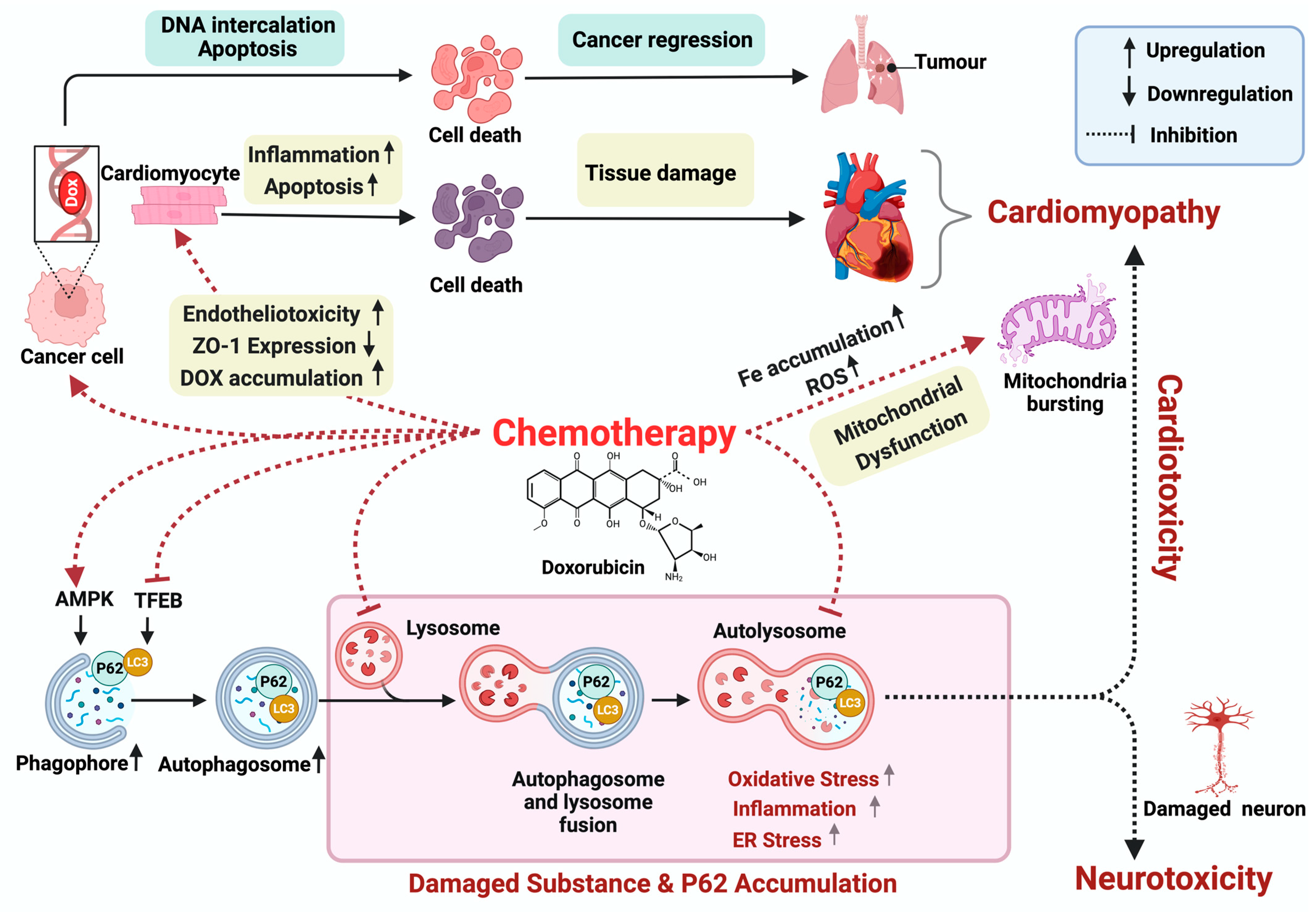
2. Doxorubicin-Induced Cardiotoxicity

Research on Dox-induced cardiotoxicity spans several decades. In the 1970s, Von Hoff and colleagues conducted early studies on 4018 patients confirming Dox-induced congestive heart failure (CHF) and its relationship with the cumulative dose of administered Dox, with a continuum of increasing risk with dose and/or age. Surprisingly, their research also revealed that a weekly dosing schedule of Dox was linked to a significantly lower incidence of CHF compared with the standard three-week schedule, challenging prevailing understandings of cumulative dose-related toxicity [45,49]. Throughout the 1980s and 1990s, investigations focused on dose dependency and risk factors in Dox-induced CHF [50]. By the late 1990s, Dox’s clinical use was limited by dose-related cardiomyopathy [51]. Further, histological evaluation in both human and animal models’ hearts revealed characteristic myocardial changes, such as myocardial fibrosis, myofibrillar disarray, and vacuolar degeneration of cardiomyocytes associated with Dox-induced cardiotoxicity [52]. Moreover, the incidence of Dox-induced cardiomyopathy significantly escalates beyond a cumulative dose of 550 mg/m^2 body surface area. Consequently, once this threshold is surpassed, the therapy is typically omitted from chemotherapy regimens [53]. This outcome underscores the potential deprivation of patients from an effective treatment option to mitigate Dox’s cardiotoxic side effects.
In the 2000s, research shifted toward understanding the mechanism of Dox-induced cytotoxicity and developing a personalized approach to mitigating this risk. The pathogenesis of Dox-induced cardiac dysfunction remains unclear; however, oxidative stress, apoptosis, and mitochondrial dysfunction are implicated [15]. This is characterized by an imbalance in reactive oxygen species (ROS) and reactive nitrogen species (RNS) causing dysregulated antioxidant mechanisms resulting in subcellular damage and apoptosis [15]. Additionally, studies on murine models have shown that Dox-induced cardiotoxicity primarily arises from oxidative stress and mitochondrial dysfunction, characterized by reduced oxygen consumption, decreased mitochondrial membrane potential, and elevated iron accumulation [54] (Figure 2). The investigation conducted by Pillai et al. reveals that the decline in SIRT3 levels, a mitochondrial enzyme that regulates cellular metabolism and oxidative stress response, which is crucial for maintaining mitochondrial function, correlates with the cardiotoxic repercussions of Dox [55]. Moreover, Jordan et al.’s findings in 2018 highlight the frequent occurrence of cardiac atrophy in patients subjected to Dox therapy [56]. Using an in vivo study, Ni et al. uncovered additional factors contributing to the adverse effects of Dox demonstrating its ability to induce inflammation in a more intricate manner. Notably, beyond stimulating CD11b+ macrophages to produce IFNγ, Dox was found to disrupt lipid metabolism, presenting a multifaceted mechanism underlying the development of cardiomyopathy [57]. In the presence of iron (Fe), Dox undergoes futile redox cycling, generating ROS and causing cellular damage. This process involves superoxide formation, conversion to hydrogen peroxide (H2O2), and production of highly toxic hydroxyl radicals through the Fenton reaction. Additionally, Dox directly interacts with Fe to form a Fe–Dox complex, further enhancing ROS production [58]. In the other study, Liu et al. (2018) observed that Dox also disrupts AMPK function and suppresses PGC1-α. This disruption alters downstream antioxidant signaling pathways involving NRF1, TFAM, and UCP2. Consequently, cardiomyocyte viability is compromised, which exacerbates cardiac injury [59]. Dox binding to eNOS reductase causes an imbalance between nitric oxide and superoxide, leading to cardiotoxicity via increased redox stimulation, and apoptosis [60,61].
Importantly, Dox disrupts the crucial endothelial cell–cardiomyocyte bond, exacerbating cardiac injury by enhancing ROS-driven damage, reducing zonula occludens-1 (ZO-1) expression, and increasing permeability, while also heightening Dox accumulation in the heart [15,62]. Furthermore, Dox-induced ROS and RNS production induce mitochondrial damage in endothelial cells, triggering apoptosis through cytochrome C release and caspase activation, disrupting their supportive role for cardiomyocytes [63]. Moreover, studies on cultured endothelial cells reveal ROS/RNS-independent direct DNA damage by Dox binding with CG-rich sequences, further disrupting endothelial–cardiomyocyte crosstalk and support [64]. Furthermore, exposure to Dox during the early stages of life in mice led to a failure in developing compensatory cardiac hypertrophy when later challenged with angiotensin-II (Ang-II)-induced hypertension [65]. This suggests that the cardiotoxic effects of Dox may interfere with the heart’s ability to adapt to increased blood pressure, potentially exacerbating the detrimental effects of hypertension on cardiac function. The extensive research on Dox-induced cardiotoxicity underscores its complex pathogenesis and multifaceted impact on cardiac function. From historical observations to contemporary insights, evidence highlights its detrimental effects, including dose-dependent cardiomyopathy, CHF, and disruption of cell interactions. Understanding these mechanisms and developing personalized approaches are crucial for mitigating risks and improving outcomes.

3. Doxorubicin-Induced Neurotoxicity
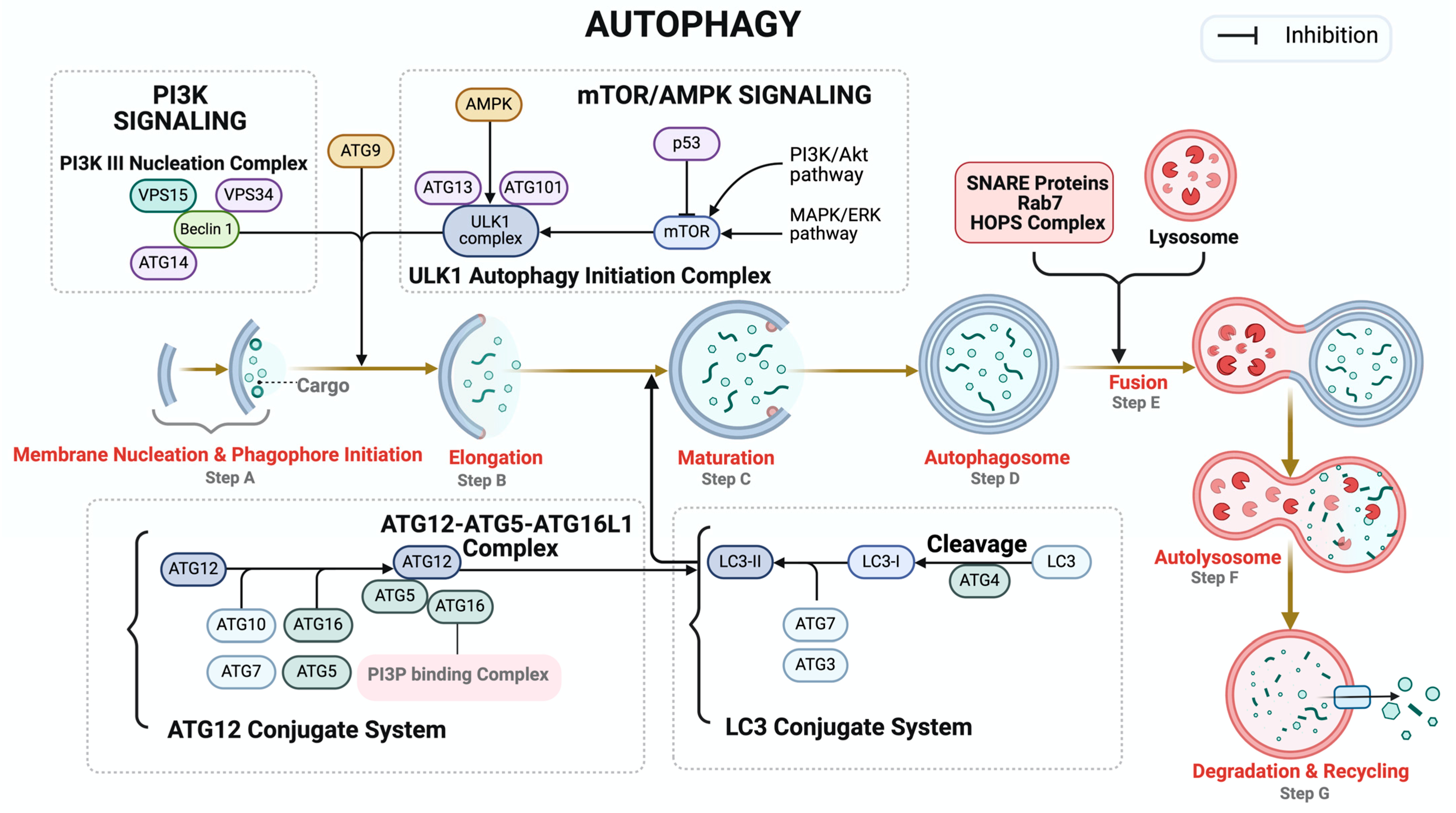
Chemotherapy-induced cognitive decline, often termed chemotherapy-related cognitive impairment (CRCI) or “chemo-brain”, is a recognized phenomenon observed in cancer patients undergoing Dox chemotherapy [46]. Manifesting as cognitive difficulties, this condition affects a significant proportion, approximately 17–70% of cancer patients, and is an outcome of a complex interplay of mechanisms [66]. These include the induction of DNA damage [67], augmentation of oxidative stress pathways [68,69], initiation of inflammatory cascades [70,71], disruption of apoptosis, modulation of neurotransmitter levels [72], perturbation of mitochondrial function [73], and inhibition of neurogenesis [74]. In the 1980s, there was a prevalent notion that Dox did not pose a risk of neurotoxicity due to its limited penetration of the blood–brain barrier (BBB) [75]. Nevertheless, doubts regarding the potential for Dox-induced neurotoxicity persisted. Thus, multiple studies conducted in the 2000s have suggested a potential indirect mechanism, which is Dox-induced elevated levels of peripheral TNF-α that may traverse the blood–brain barrier (BBB) and impede cellular antioxidant systems [76,77] . In a separate investigation conducted in mice, it was found that exposure to Dox induces significant mitochondrial damage and disrupts the levels of brain choline-containing metabolites and phospholipases, and the shifts in metabolic markers are closely associated with heightened oxidative stress mediated by TNF-α [73]. Indirect Dox-induced neurotoxicity by TNF-α, which has the potential to influence the volume of the hippocampus, can augment the inflammation by activating astrocytes and microglia in the brain [70,71]. Additionally, animal studies witnessed that Dox treatment significantly reduces neurogenesis, evidenced by a notable decrease in the number of cells labeled with the neuro-specific nuclear antigen bromodeoxyuridine (BrdUrd), which are crucial for spatial processing and memory formation [74]. In our current mice study, we observed increased DNA damage measured by immunoblotting for gH2AX—a marker for DNA damage—showing the potential role of DNA damage in Dox-induced neurotoxicity (unpublished data); however, it remains to be confirmed whether it is a direct or indirect effect of Dox. Among clinical observations spanning the past two decades, Freeman and Broshek (2002) elucidated the cognitive impairments associated with Dox-based chemotherapy, ranging from memory deficits to depressive symptoms in breast cancer patients [78]. Building upon these findings, studies in 2016 delineated a spectrum of neurological symptoms, including headaches, seizures, and encephalopathy, predominantly observed in patients treated with Dox [79,80]. In 2017, Cruz-Carreras et al. reported specific neurological symptoms, such as headaches and aphasia, in a young leukemia patient undergoing Dox chemotherapy. Additionally, these patients reported decreased strength in the right arm, which shows a potential connection with the central nervous system and the peripheral nervous system [81]. Most recently, a study illuminated the prevalence of posterior reversible encephalopathy syndrome (PRES) in children undergoing chemotherapy, with seizures emerging as a prominent clinical feature [82]. Indirect neurotoxicity resulting from Dox treatment is attributed to the induction of oxidative stress due to excessive production of ROS [69]. These ROS subsequently lead to the oxidative modification of proteins, lipids, and nucleic acids. Additionally, Hsieh et al. demonstrated that ROS could activate nuclear factor kappa B (NF-κB), further implicating oxidative stress in the neurotoxic effects of Dox [83]. The neurological impact of Dox is complex, necessitating further investigation crucial for optimizing therapies and minimizing adverse effects to improve patient outcomes. Current drug discovery focuses on novel derivatives with enhanced efficacy and reduced toxicity, including natural compounds from microbial, plant, and Ayurvedic sources [84,85,86].
Despite extensive investigation around the role and contribution of DNA damage to Dox-induced toxicity, there still exists a gap in knowledge about the role of different mechanisms. Recently, autophagy has emerged as a crucial pathway in both cancer and healthy cells that are exposed to Dox. Consequently, there has been a recent shift in research focus from DNA damage to autophagy in Dox-associated toxicity. While chemotherapy remains integral to cancer treatment, this review will concentrate on elucidating the adverse DNA damage effects induced by Dox and its direct and indirect intricate relationship with autophagy in disease and cancer. By delving into the complex pathways of autophagy and its multifaceted roles, this review also aims to highlight the potential of various factors in modulating autophagy to mitigate Dox-induced adverse effects, thereby offering promising avenues for therapeutic intervention.

4. Autophagy

Autophagy is a highly conserved cellular process involved in the degradation and recycling of damaged organelles and proteins, thus playing a crucial role in maintaining cellular homeostasis [87]. Dysregulation of autophagy has been implicated in various pathological conditions, including cardiovascular diseases [88,89,90], neurodegenerative diseases [91], cancer [92], metabolic disorders [93], and infectious diseases [94]. This tightly regulated process culminates in the formation of autophagosomes, which are double-membraned vesicles that ultimately fuse with lysosomes. This fusion allows for the degradation of protein aggregates and dysfunctional organelles, promoting cell survival. The maturation of autophagosomes involves several coordinated steps, including initiation, elongation, maturation, fusion, and degradation—all tightly governed by signaling pathways and protein complexes to ensure an efficient and selective breakdown of cellular components [95] (Figure 3). Initiation begins with the formation of the phagophore, facilitated by the activation of Unc-51-like kinases (ULKs). The activity of ULK is modulated by mTORC1, which is a nutrient sensor. Under conditions of nutrient abundance, mTORC1 activation suppresses autophagy initiation. Conversely, during nutrient deprivation, autophagy is activated by the inhibition of the mTORC1 [96]. AMPK, an energy sensor, activates autophagy by inhibiting mTORC1 [97,98,99]. Autophagy process requires two ubiquitin-like conjugation systems: the ATG12-ATG5-ATG16L1 complex and the microtubule-associated protein 1A/1B-light chain 3 (LC3) system [100]. The ATG12-ATG5-ATG16L1 complex is crucial for the lipidation of LC3 as it covalently attaches ATG3 to LC3 [101]. LC3 is processed by ATG4 and conjugated to phosphatidylethanolamine (PE) to form LC3-II, which is integrated into the autophagosome membrane by the E3-like complex to support its expansion and closure [102]. During the maturation step, the original phagophore develops into a fully matured autophagosome, supported by the recruitment of more LC3-II and the closing of the phagophore to produce a double-membraned autophagosome, which then fuses with lysosome for degradation (Figure 3) [103].
The Beneficial and Detrimental Role of Autophagy in Cancer: Prior to delving into the role of autophagy in Dox-induced toxicity and discussing the possibility of modulating autophagy to reduce unwanted Dox-induced toxicity, it is imperative to understand the role of autophagy in cancer itself. In cancer, autophagy exhibits diverse and, sometimes, conflicting roles that are influenced by factors such as tissue type, tumor stage, and the microenvironment, necessitating a comprehensive understanding for the development of effective therapeutic strategies [104,105,106]. Autophagy can act as a tumor suppressor mechanism that inhibits tumor growth and as a cytoprotective mechanism that promotes tumor survival [107,108]. Autophagy functions as a tumor suppressor through a variety of methods. It removes defective organelles and misfolded proteins to prevent cellular harm, which reduces inflammation, a known risk factor for cancer growth [109], and minimizes the accumulation of mutations that could lead to tumor initiation [110]. For example, the tumor suppressor gene, cation transport regulator homolog 2 (CHAC2), which is typically downregulated in gastric and colorectal cancer, is degraded by the ubiquitin–proteasome pathway. However, CHAC2 expression is essential to inhibiting tumor growth, proliferation, and migration, as CHAC2 induces mitochondrial apoptosis and autophagy [111]. Autophagy also reduces oxidative stress by digesting damaged mitochondria, a process known as mitophagy, which is a process of selectively targeting damaged mitochondria for degradation to regulate mitochondrial quality control [112]. Mitophagy is crucial to maintaining ROS levels, limiting DNA damage, and eventually preventing cancer [112]. By encouraging the elimination of damaged mitochondria and other cellular components, autophagy aids in preserving cellular energy balance and lowering oxidative stress [99,113].
Autophagy also serves as a pivotal mechanism in responding to cellular stress, capable of inducing cell cycle arrest and apoptosis. This response can potentially restrict the proliferation of cells carrying harmful mutations. Autophagy facilitates the degradation of key regulators of the cell cycle, such as cyclins and cyclin-dependent kinases, crucial for cell cycle progression. Notably, studies have demonstrated autophagic degradation of cyclin A and CDK2, effectively limiting the proliferation of mutation-bearing cells and impeding cancer progression [114,115]. Autophagy is also involved in the apoptotic pathway. Autophagy is used to degrade specific anti-apoptotic factors to promote the apoptotic pathway. Caspases are also activated by the degradation of caspase inhibitors or the release of pro-apoptotic factors from the mitochondria [116]. Several molecular pathways regulate the role of autophagy in cell cycle arrest and apoptosis. For example, the inhibition of mTOR induces autophagy. Research has demonstrated that substances such as apigetrin can cause cell cycle arrest and apoptosis through the PI3K/AKT/mTOR pathway, which, in turn, causes a decrease in cancer cell growth and an increase in cell death [117]. However, in established cancers, the observed detrimental role of autophagy promotes tumor survival, particularly under stressful conditions such as nutrient deprivation, hypoxia, and therapeutic stress [118]. In these contexts, autophagy recycles cellular components to provide a continuous source of energy and essential biomolecules to tumor cells, especially in nutrient-deprived conditions that support tumor growth and migration [106]. Additionally, autophagy helps cancerous cells adapt to different conditions, such as radiation and chemotherapy by degrading and recycling damaged cellular components, thereby enhancing cell survival under therapeutic stress and promoting therapeutic resistance [119]. Autophagy can influence the tumor microenvironment by regulating the availability of oxygen and degrading damaged organelles, which aids in the survival of cancer cells in hypoxic environments. In an unfavorable microenvironment, autophagic activity helps cancer cells survive and proliferate by recycling intracellular components to provide nutrition [105]. Autophagy also supports critical processes required for cancer spread, such as cell invasion and migration, by supplying energy and preserving cellular homeostasis that promotes metastasis.
Autophagy-based Clinical Trials in Cancer: Drugs that inhibit the late phases of autophagy, such as hydroxychloroquine (HCQ) and chloroquine (CQ), are being investigated to sensitize cancer cells to chemotherapy and other treatments [120]. Both CQ and HCQ inhibit autophagy by inhibiting autophagosome fusion with lysosomes, eventually dampening the growth of malignant cells. When the autophagy system is disrupted, defective autophagosomes accumulate resulting in cellular stress and, perhaps, leading to cancer cell death [121]. By increasing the susceptibility of cancer cells to treatment-induced cell death, this can improve the efficacy of other cancer therapies. These drugs are already approved for use in malaria and rheumatoid arthritis and are being repurposed for cancer therapy in various clinical trials [122]. However, the efficacy of treatment varies in different tissues due to different cancer lines relying on autophagy more than others. Clinical trials on CQ and HCQ have demonstrated their potential as useful adjuvant therapies in a variety of cancer treatments. In one trial, CQ and HCQ were used with antiestrogen treatments for estrogen receptor-positive (ER+) breast cancer, such as tamoxifen (TAM) and faslodex (ICI). The result demonstrated that CQ could reverse the resistance of breast cancer cells to estrogen. TAM and CQ demonstrated success collectively, indicating that CQ may improve the receptivity of ER+ breast cancer patients to antiestrogen treatments [123]. Another study investigated the treatment of glioblastoma using CQ in addition to radiation therapy, leading to an increase in radiation therapy’s efficacy, indicating that CQ may be a useful adjuvant therapy for treating glioblastoma [124]. Preclinical studies have also demonstrated HCQ’s efficacy in conjunction with temozolomide, a popular chemotherapeutic treatment for glioblastoma. The findings imply that the inhibition of autophagy by HCQ increases the susceptibility of glioblastoma cells to chemotherapy, which may improve tumor growth control and patient recovery [125]. In scenarios where enhancing autophagy may lead to cell death or prevent tumor initiation, activating autophagy could be beneficial. This approach could be particularly effective in early-stage cancers or as a preventive strategy in high-risk populations. Drugs like rapamycin and its analogs (temsirolimus, everolimus) inhibit the mTOR pathway, a negative regulator of autophagy [126]. These medications can cause death in cancer cells reliant on mTOR signaling by triggering autophagy. Treatments for certain diseases, including pancreatic neuroendocrine tumors, renal cell carcinoma, and breast cancer, involve the use of mTOR inhibitors, which may also improve the effectiveness of other cancer treatments [127]. Dox is the most common drug used in cancer chemotherapy and given the context-dependent complex, detrimental and beneficial, role of autophagy in different cancer types, it becomes imperative to characterize autophagy and its role for every cancer type before designing an autophagy-based therapy, such as Dox-based chemotherapy.
Doxorubicin Chemotherapy and Autophagy: Dox induces autophagy in various cancer cells. This autophagic reaction may function as a defense mechanism, enabling cancer cells to tolerate the damaging impacts of Dox. Conversely, Dox is also known to suppress autophagy, compromising DNA repair pathways and, potentially, inducing apoptosis in non-cancer cells, which contributes to dilated cardiomyopathy [128]. Several interrelated pathways, including mitochondrial damage and ROS production, forkhead box o3a (FOXO3a) and miR-223 regulation, PI3K/AKT/mTOR pathway, p53 pathway, and AMPK/mTOR signaling, play a role in the induction of autophagy by Dox [129,130,131]. These methods illustrate autophagy’s significance in cancer treatment by proving to be both a possible therapeutic target and a cell survival mechanism. Understanding these pathways can support the development of combination therapies that serve to manipulate autophagy and boost Dox’s efficacy while simultaneously minimizing its side effects. Dox-induced mitochondrial dysfunction can induce ROS production, which, in turn, promotes autophagy. Ozcan et al. highlighted this process in their discovery of higher levels of autophagic markers (LC3-II and Beclin-1) in cardiac tissues obtained from Dox-treated patients. This study demonstrated that the accumulation of damaged mitochondria and increased production of ROS were important driving factors of the autophagic response [130]. Dox treatment is also associated with an increased expression of ATGs such as ATG5 and Beclin-1, demonstrating elevated autophagic activity [129,132]. The conversion of LC3-I to LC3-II is an essential step in the development of autophagosomes [133]. Dox therapy raises LC3-II levels indicating increased autophagosome production, which is supported by the increase of autophagic vacuoles seen both in vitro and in vivo [129]. The transcription factor FOXO3a is a key regulator of autophagy. According to Zhou et al., hepatocellular carcinoma cells (HCCs) overexpress miR-223, leading to the downregulation of its target FOXO3a, which prevents Dox-induced autophagy and reverses chemoresistance in HCC cells [131]. One important regulator of cell proliferation, survival, and autophagy is the PI3K/AKT/mTOR pathway, and its inhibition activates autophagy. It has been shown that Dox interacts with this mechanism to induce autophagy. Moreover, natural substances such as magnoflorine have been shown to enhance Dox sensitivity by blocking the PI3K/AKT/mTOR pathway, thereby inducing autophagy and promoting cell death in breast cancer cells. This investigation demonstrated that the increased autophagic response displayed in Dox-treated cells is dependent on the inhibition of the PI3K/AKT/mTOR pathway [134]. Dox also activates the AMP-activated protein kinase (AMPK) pathway, which acts as an energy sensor in cells and suppresses the mTOR pathway and, thereby, activates autophagy in HCC cells [135]. Additionally, Ang-II, a key component of the renin-angiotensin system, is also reported to exacerbate Dox-induced cardiac injury through dysregulation of autophagy pathways [136].
Genomic integrity is a fundamental aspect of cellular health and disease prevention, and DNA damage poses significant threats to genomic stability and is primarily caused by factors like ROS, replication stalling, and genotoxic or radiation exposure [137]. Autophagy, a critical cellular process responsible for clearing damaged components, has become recognized for its role in maintaining genomic stability and its relevance to conditions like breast cancer [138,139]. Previous investigations utilizing genetic inhibition of autophagy (via ATG7 knockout) in fibroblasts have revealed autophagy’s impact on DNA repair mechanisms, primarily homologous recombination (HR), leading to HR deficiency characterized by genomic instability and an increased apoptosis [140]. Persistent HR deficiency leading to accumulation of DNA damage can lead to the activation of inflammatory pathways, and recent studies have linked DNA damage and inflammation in the context of Dox-induced cardiomyopathy. Dox-induced DNA damage triggers inflammatory responses characterized by the upregulation of pro-inflammatory cytokines, including tumor necrosis factor-alpha (TNF-α), which, in turn, can activate autophagy-related pathways (Figure 4) [141,142]. Inflammation is a key factor in Dox-induced cardiotoxicity where Dox-induced cytokines like interleukin-1 and TNF-α trigger NF-κB and p38-MAPK signaling pathways leading to inflammatory cascades [143,144]. NF-κB, a central regulator of inflammation, is activated by the IκB kinase complex involving transforming growth factor-β (TGF-β)-activated kinase 1 and its cofactors TAK1 and MAP3K7-binding proteins. Once activated, NF-κB translocates to the nucleus to induce an inflammatory response [143,145]. Inflammatory and autophagy pathways are interwoven as certain components associated with TAK1, like TGF-β-activated kinase 1-binding proteins 2/3, can engage Beclin-1 to promote autophagy [146]. Reports indicate that compromised autophagy leading to p62 accumulation triggers IKK activation, subsequently inducing NF-κB-mediated inflammation; conversely, enhanced autophagy prevents p62 accumulation, thereby impeding the NF-κB signaling pathway [147]. However, the precise molecular mechanisms mediating the communication between genomic instability, inflammation, and autophagy pathways remain to be fully elucidated.
Dox-induced dysregulation of autophagy leads to the accumulation of autophagosomes, disrupting cellular health and therapeutic efficacy [148]. This dysregulation underscores the intricate relationship between autophagy, DNA repair, and disease processes as evidenced by promising pre-treatment strategies that enhance autophagy to mitigate Dox-induced cardiotoxicity [129]. The essential role of autophagy in maintaining genomic integrity is highlighted by perturbations in checkpoint kinase-1 (CHK1) regulation and degradation mechanisms [138]. However, paradoxically, Dox suppresses autophagy, compromising DNA repair pathways and, potentially, inducing apoptosis, which contributes to dilated cardiomyopathy [128]. The interplay between DNA damage and autophagy is heavily influenced by DNA-damage repair (DDR) genes, such as breast cancer susceptibility genes 1 and 2 (BRCA1/BRCA2), and Fanconi anemia (FA) genes. Cells lacking DDR genes, particularly BRCA genes, are particularly susceptible to Dox-induced toxicity, showing a 70-fold increase in vulnerability compared with normal cells [149]. This susceptibility underscores the critical role of DNA double-strand break (DSB) repair mechanisms orchestrated by BRCA1/BRCA2, as well as the active involvement of the FA-BRCA pathway in DDR [150,151,152,153]. Of note, the chromatin complex, involving monoubiquitinated FANCD2 (FANCD2-L) and BRCA2, is implicated in homology-directed repair (HDR) although the precise nature of their interaction remains unclear. BRCA2 actively engages with RAD51 to facilitate DNA damage repair, while FANCD2-L likely assists in ensuring precise matching of genetic material for effective RAD51 unloading by BRCA2 at targeted repair loci [151]. Mutations in DDR molecules, particularly FA genes (e.g., PALB2) and BRCA genes (e.g., BRCA1) can lead to significant complications, including breast/ovarian cancer and severe forms of Fanconi anemia [154,155,156,157]. Studies have elucidated the relationship between BRCA genes and autophagy, revealing that BRCA1-deficient cells heavily rely on autophagy for survival, with disruption of autophagy leading to rapid cell death [158]. Conversely, studies observed that BRCA1 negatively regulates autophagy in MCF-7 breast cancer cells, with BRCA1-loss increasing autophagic vacuoles and ROS levels. This suggests a potential antioxidant role of BRCA1, influencing heightened autophagy due to mitochondrial dysfunction and increased ROS levels [159]. Additionally, BRCA1 levels have been found to decrease rapidly during nutrient deprivation-induced autophagy in MCF-7 cells [159]. Loss of BRCA2 in tumors already exhibiting BRCA1 allelic loss enhances autophagy and mitophagy, suggesting a potential link between BRCA2 and autophagy [160]. However, the clear role of BRCA2 in regulating autophagy remains unclear. Autophagy is known to be essential for enabling HR, and its absence leads to DNA damage accumulation and cell death (Figure 4) [138]. Exploring the role of BRCA2 in these processes could offer crucial insights into DNA repair, genome stability, and disease development. Furthermore, understanding how autophagy is influenced in different BRCA gene variants and HR-deficient tumors, as well as how Dox affects autophagy in BRCA2-mutated patients, is pivotal for refining breast cancer treatment strategies. Singh et al. found that cardiomyocyte-specific BRCA1-deficient mice exhibited significant metabolic disruptions, including decreased expression of glucose and fatty acid transporters, along with reduced levels of key enzymes involved in fatty acid oxidation. This led to diminished rates of glucose and fatty acid oxidation despite increased activation of AMPK and AKT, indicating an energy-starved heart and predisposing the heart to failure [161]. There is also a growing interest in understanding how BRCA genes responsible for DNA damage, known for their roles in cancer and immune response, may regulate autophagy, a process critical for cardiac homeostasis. However, the specific interaction between BRCA1 and autophagy pathways in the context of cardiac metabolism and heart failure remains unclear. Further investigation is essential to elucidating this potential interaction and its implications for therapeutic intervention [159,162].

5. Doxorubicin and Mitophagy

Mitochondria, often referred to as the powerhouse of the cell, are vital for maintaining genomic integrity alongside DDR genes [163]. Unlike nuclear DNA, mitochondrial DNA (mtDNA) is highly susceptible to damage, posing significant risks to cellular health and contributing to various diseases, such as neurodegenerative disorders and cancer [164]. Mitochondria dynamically adjust their content, undergo fusion and fission, and activate the unfolded protein response to ensure cellular homeostasis and steady energy flow, crucial for cellular signaling under stress [163,164,165]. However, there remains a gap in understanding how mitochondria respond to genomic DNA damage and their impact on DNA repair and cell fate despite knowing the intricate relationship between DNA damage and mitochondrial stress [166,167]. Mitophagy, selectively targeting damaged mitochondria for degradation, is pivotal for mitochondrial quality control, primarily mediated by the phosphatase and tensin homolog-induced putative kinase 1 (PINK1)-Parkin cascade, whereby impaired mitochondria are labeled with phosphorylated ubiquitin, and then targeted for degradation. Mitophagy plays a crucial role in the early stages of Dox-induced cardiomyopathy [112,168,169]. Yet, disruptions in mitophagy can lead to a cascade of deleterious effects, ranging from metabolic disorders to cancer, senescence, inflammation, genomic instability, and aging [170]. Moreover, excessive mitophagy can be detrimental as evidenced by elevated LC3 and Beclin 1 levels, decreased p62 expression during Dox treatment, and suppressed expression of PGC-1α, NRF1, and TFAM, along with reductions in mitochondrial protein levels. Furthermore, Dox induces concentration-dependent reductions in mitochondrial membrane potential (MMP) and mtDNA content [171]. Notably, cells treated with Dox exhibit significant MMP reductions to 55% of the control and mtDNA content declines up to 83% of the control at specific Dox doses [171]. Interestingly, recent insights suggest a potential involvement of DNA repair proteins such as FANCD2, BRCA1, and BRCA2 not only in repairing nuclear DNA but also in Parkin-mediated mitophagy [172]. Additionally, the presence of HR machinery within mitochondria, supported by the detection of recombination intermediates and the presence of RAD51 and its related proteins like XRCC3, suggest their involvement in mitochondrial HR processes [173,174]. While key DNA damage repair proteins like BRCA1 and 53BP1 are found in mitochondria, their direct role in mitochondrial repair remains unclear [175,176]. This intriguing intersection of DNA repair BRCA1/2 proteins and mitochondrial maintenance unveils a novel dimension of cellular quality control, offering insights into therapeutic potential in mitigating Dox-induced toxicity and preserving mitochondrial health.

6. Autophagy Modulation for Therapeutic Intervention

Autophagy modulation holds significant therapeutic potential across diseases like cancer [177]. In cancer, autophagy’s role can either promote or inhibit tumor growth depending on context, making its therapeutic modulation complex and context-dependent [178,179]. While autophagy inhibition can sensitize certain cancer cells to chemotherapy or radiation therapy, its activation may promote tumor cell death under specific conditions [179]. Chemotherapy, a common cancer treatment, often encounters drug resistance. Autophagy aids cancer cells in surviving chemotherapy by repairing DNA and eliminating toxins, thereby contributing to drug resistance [180]. Autophagy is implicated in resistance to drugs like Dox, with research showing elevated autophagic activity in drug-resistant cells and its inhibition leading to re-sensitization to Dox [135,181]. Conversely, inhibiting autophagy can restore drug sensitivity through agents like CQ or by downregulating autophagy-associated proteins such as ATG7 and ATG14 [182,183,184,185,186]. Pharmacological inhibition of autophagy by agents like CQ and bafilomycin affects Dox efficacy. CQ potentiates Dox-induced cell death by blocking autophagic flux and reducing drug resistance in various cancers [187,188]. Bafilomycin A1, which inhibits vacuolar-type H+-ATPase and blocks lysosomal acidification, impedes autophagosome–lysosome fusion, disrupting autophagic flux crucial for cellular degradation processes, which in turn promotes the efficacy of Dox against cancer [189]. In HepG2 liver cancer cells, treatment with bafilomycin A1 or chloroquine alone at lower doses enhanced inhibition of cell growth and increased apoptosis when combined with Dox. Additionally, these inhibitors promoted lysosomal membrane permeabilization and reduced mitochondrial membrane potential in response to Dox treatment, suggesting their potential to synergistically improve Dox’s anticancer effects in liver cancer therapy [190].
In a recent study, Montalvo et al. targeted autophagy with rAAV-dnATG5 in female rats exposed to Dox and revealed that while Dox had negative effects on left ventricular function, redox balance, and mitochondrial function, acute inhibition of autophagy mitigated the increase in mitochondrial ROS emission and improved cardiac health. Notably, this improvement was observed only at the acute stage, suggesting variations in ATG5–ATG12 conjugation kinetics [191]. Natural substances like epigallocatechin-3-gallate (EGCG) and magnoflorine have also been explored for enhancing Dox efficacy by regulating autophagy [134,192]. Another compound, metformin, a remarkable antidiabetic medication, not only regulates blood sugar levels but also holds tremendous promise in cancer therapy through its intricate modulation of cellular processes [193]. By activating AMPK, metformin elevates the AMPK/AMP ratio, setting off a cascade of events. AMPK activation, in turn, exerts its inhibitory influence on the mechanistic target of rapamycin complex 1 (mTORC1) pathway by phosphorylating key downstream targets like tuberous sclerosis complex protein 2 (TSC2) [194]. This inhibition of mTORC1 serves as a crucial checkpoint, curbing aberrant cell growth and proliferation, hallmarks of cancer [195]. Moreover, metformin’s impact extends to the modulation of autophagy, the cellular process of self-degradation and recycling. Through its influence on AMPK and mTORC1, metformin enhances autophagic activity, promoting the selective degradation of damaged proteins and organelles within cancer cells [196].
Studies also investigated whether altering autophagy could mitigate Dox-induced cardiotoxicity. Accordingly, rat cardiac myoblasts were treated with rapamycin, an mTOR inhibitor and autophagy inducer, prior to Dox exposure, which showed significantly improved cell viability, reduced apoptosis and ROS production, and enhanced mitochondrial function. To further validate findings, they used GFP-LC3 mice with EO771 tumors, where a single rapamycin injection followed by Dox injections preserved cardiac function, as evidenced by reduced caspase-3 expression, and maintained cardiomyocyte size [197]. Rapamycin’s autophagy induction potentially protects against Dox cardiotoxicity by promoting cellular survival mechanisms. Conversely, rapamycin complicates the therapeutic landscape by potentially promoting cancer cell survival against Dox-induced cytotoxicity. Recent research suggests that rapamycin-mediated autophagy induction might confer resistance to Dox in certain cancer contexts, highlighting the dual-edge sword of autophagy modulation in cancer therapy warranting future investigations [198]. Other agents, like thymoquinone (TQ), induced autophagy in Dox-treated cardiac myoblasts by activating LKB1/AMPK and inhibiting mTOR, and promoted cardiac myoblasts survival. Blocking autophagosome formation reversed TQ’s anti-apoptotic effects, suggesting its potential to prevent Dox-induced cardiotoxicity [199]. Another drug Spinacetin is proposed to protect cardiac myoblasts against Dox-induced cytotoxicity by enhancing autophagy and reducing apoptosis via AMPK/mTOR signaling that is mediated by increased SIRT3 expression [200]. Furthermore, curcumin induces autophagy in various tumor cells by suppressing the PI3K/Akt/mTOR pathway and enhancing LC3-II, Beclin1, and ATG protein expression. Studies in ovarian, gastric, and lung cancer cells highlight curcumin’s role in promoting autophagic vesicle formation and reducing p62 levels, potentially enhancing cancer treatment efficacy through autophagy modulation [201,202]. Separately, Zhang et al. and Wei Yu et al. independently demonstrated in their study that curcumin attenuates Dox-induced cardiotoxicity in mice [203,204]. These findings suggest curcumin’s potential as a protective agent against Dox-induced cardiotoxicity while illustrating its complex role in modulating autophagy in different contexts. Understanding the context-specific effects of these compounds on autophagy could promote precision medicine.
Precision medicine is mainly driven by omic technologies where, by analyzing genetic variants, protein expression patterns, and metabolic profiles, clinicians can stratify patients based on their individual molecular signatures. This personalized approach allows for tailored treatment strategies that maximize efficacy while minimizing adverse effects. Proteomic analysis enables the characterization of protein expression changes induced by autophagy modulation, identifying biomarkers associated with autophagy activity or response to specific drugs. For instance, biomarkers like the LC3-II/I ratio, reflecting autophagic flux, have been extensively studied in cancer to predict treatment efficacy. Elevated LC3-II levels indicate increased autophagy and may predict resistance to chemotherapy in certain cancer types [205]. Conversely, p62/SQSTM1 levels, which accumulate when autophagy is impaired, have been linked to poor prognosis in various cancers [206]. Proteomic techniques have pinpointed specific proteins like lysosomal-associated membrane protein 2 (LAMP2A) and serine/threonine kinase 11 interacting protein (STK11IP), crucial for regulating lysosomal targeting and mTORC1 signaling in diseases such as cancer [207,208]. These studies underscore how dynamic changes in autophagy-related protein expression influence disease pathogenesis, which can be monitored and used by clinicians to predict treatment responses [205]. By incorporating these biomarkers into clinical decision algorithms, physicians can tailor treatment regimens in real time, adjusting dosages or switching therapies based on individual patient responses. Genomic studies complement these insights by identifying mutations and epigenetic alterations in key autophagy genes (e.g., ATG5, ATG7), linking genetic variants to disease susceptibility and responses to treatment [209,210]. Genetic profiling has further identified polymorphisms in these genes that influence autophagy pathway dynamics and treatment responses [211]. Computational models integrate these genetic variants with clinical data to predict how patients will respond to autophagy modulation therapies. By understanding individual genetic predispositions, clinicians can adjust dosing regimens or select the most effective treatment options, thereby optimizing therapeutic outcomes. Computational models have successfully predicted treatment outcomes in cancer therapies by elucidating the complex interactions of autophagy modulation with chemotherapy drugs like Dox. For example, studies have shown that inhibiting autophagy using CQ enhances Dox’s cytotoxic effects in various cancers [187]. Computational simulations predicted the synergistic effects of combining CQ with Dox based on pharmacokinetic and pharmacodynamic data, guiding clinical decision-making [187]. In another study, computational models highlighted how rapamycin-induced autophagy protects against Dox-induced cardiotoxicity by promoting pro-survival mechanisms [197]. By simulating drug interactions within autophagy pathways, these models identify optimal dosing strategies to mitigate cardiotoxic effects while maintaining therapeutic efficacy in cancer treatment. These genetic variants can impact sensitivity to autophagy inhibitors or activators, highlighting the importance of personalized treatment approaches based on patient-specific genetic profiles. Furthermore, metabolomic profiling reveals alterations in cellular metabolism induced by autophagy modulation, providing deeper insights into its physiological consequences [212].
Moreover, integrating omics methods with advanced bioinformatics tools enhances our understanding of autophagy modulation and facilitates precision medicine approaches in treating autophagy-related disorders. Bioinformatics and computational modeling tools play a pivotal role in leveraging omics data to predict patient responses to autophagy modulation therapies. Computational models have been instrumental in studying how autophagy modulation affects treatment outcomes, particularly in cancer therapies [213,214]. Similar computational approaches have successfully elucidated complex interactions between autophagy activity and disease progression in Alzheimer’s disease and degenerative aging [215,216]. These models explore how failures in regulatory mechanisms can lead to elevated autophagy levels, potentially contributing to neuronal dysfunction through amyloid-β (Aβ) accumulation. Computational modeling approaches, exemplified by studies on Dox treatment in triple-negative breast cancer patients, provide insights into optimizing drug interactions and dosing strategies tailored to individual patient profiles, thereby advancing precision medicine in cancer therapy [217]. Future directions in this field involve refining computational models to integrate multi-omics data, including biomarkers. By combining genomics, proteomics, and metabolomics with clinical outcomes, researchers aim to develop predictive models for patient-specific responses to autophagy modulation therapies. These models will enable tailored treatment strategies based on individual genetic profiles, molecular signatures, and metabolic profiles, optimizing therapeutic efficacy while minimizing adverse effects.

7. Challenges and Future Directions

Translating preclinical data on autophagy-based therapies into clinical practice encounters several challenges [218]. These include potential off-target effects of pharmacological agents, limited specificity of autophagy modulators, and the necessity for biomarkers to monitor autophagic activity in patients [219]. Furthermore, the complexity of autophagy regulation and its context-dependent effects necessitate further research to optimize therapeutic strategies and identify patient populations most likely to benefit from autophagy modulation [179]. It is noteworthy that a transient surge in autophagy signaling post-Dox exposure cautions against prolonged autophagy inhibition due to potential pathological ramifications [191]. Thus, future research avenues are warranted to fine-tune autophagy modulation in therapeutic interventions. While both augmentation and suppression of autophagy have been proposed as potential approaches, the current clinical landscape predominantly leans toward inhibiting autophagy [182,220]. This inclination underscores a prevailing skepticism regarding the efficacy of autophagy enhancement in cancer therapy. However, as clinical trials explore this intricate interplay, the decision to predominantly target autophagy inhibition signals a cautious yet determined stride toward unraveling the complexities of cancer treatment. The relationship between autophagy and Dox in cancer treatment is complex and multifaceted [221]. Depending on numerous variables, including cancer type, the patient’s genetic background, and treatment circumstances, autophagy may either shield cancer cells from Dox-induced cytotoxic effects or promote cell death. Understanding these mechanisms is pivotal for developing effective therapeutic approaches that optimize autophagy regulation to enhance Dox’s efficacy in cancer therapy. Additionally, recent studies suggest that BRCA1/BRCA2, well-known tumor suppressor genes frequently mutated in hereditary breast and ovarian cancers, may play a role in autophagy regulation. BRCA1 has been implicated in promoting autophagy under cellular stress conditions, while BRCA2 deficiency has been associated with impaired autophagy flux, potentially contributing to cancer development and treatment resistance [138,159]. Investigating the interplay and molecular mechanism between BRCA1/BRCA2 and autophagy could provide further insights into cancer biology and therapeutic strategies promoting precision medicine. Through an in-depth investigation of the complex interaction between autophagy and Dox, researchers and medical professionals can potentially devise targeted and efficient cancer treatments, overcoming drug resistance challenges and improving patient responses to treatments.

8. Conclusions

The intricate interplay among autophagy, inflammation, and genomic stability underscores their collective impact on cellular health and disease processes. Autophagy, essential for removing damaged components, plays a crucial role in maintaining genomic stability. In contrast, inflammation, often triggered by DNA damage, can worsen cellular dysfunction. This complex relationship is evident in conditions like Dox-induced cardiomyopathy where DNA damage-induced inflammation activates autophagy, further influencing disease progression. Moreover, the disruption of autophagy and DNA repair mechanisms, as observed in Dox-induced cardiotoxicity, highlights their importance in determining therapeutic outcomes. The involvement of DNA damage repair genes, particularly BRCA1/2 emphasizes their critical role in mediating DNA repair, autophagy, and disease susceptibility. However, the exact mechanisms through which these genes regulate autophagy remain unclear, presenting a crucial area for further investigation. Additionally, mitophagy emerges as a pivotal mechanism in maintaining mitochondrial quality control and cellular homeostasis, with implications for disease development and therapeutic interventions. From a therapeutic standpoint, modulating autophagy shows promise for various diseases, including cancer. However, translating autophagy-based therapies into clinical practice faces challenges, such as off-target effects and the limited specificity of pharmacological agents. Further research is necessary to refine therapeutic strategies and identify patient populations who are most likely to benefit from autophagy modulation. Ultimately, unraveling the complex interplay among autophagy, inflammation, and genomic stability will lead to the development of novel therapeutic interventions and personalized medicine approaches.

Author Contributions

Conceptualization, K.K.S., J.C.F. and A.S.; resources, K.K.S.; writing—original draft preparation, A.S., N.R. and K.K.S.; writing—review and editing, A.S., N.R. and K.K.S.; visualization, A.S. and J.C.F.; supervision, K.K.S.; and funding acquisition, K.K.S. All authors have read and agreed to the published version of the manuscript.

Funding

This research was funded by the Heart and Stroke Foundation, grant number G-22-0032104 (KS). KS is also the recipient of the 2018/19 National New Investigator Award—Salary Support from the Heart and Stroke Foundation of Canada, Canada.

Conflicts of Interest

The authors declare that they have no conflicts of interest with the contents of this article.

References

  1. Brown, J.S.; Amend, S.R.; Austin, R.H.; Gatenby, R.A.; Hammarlund, E.U.; Pienta, K.J. Updating the Definition of Cancer. Mol. Cancer Res. 2023, 21, 1142–1147. [Google Scholar] [CrossRef]
  2. Janssen, L.M.E.; Ramsay, E.E.; Logsdon, C.D.; Overwijk, W.W. The immune system in cancer metastasis: Friend or foe? J. Immunother. Cancer 2017, 5, 79. [Google Scholar] [CrossRef]
  3. Kalyanaraman, B. Teaching the basics of cancer metabolism: Developing antitumor strategies by exploiting the differences between normal and cancer cell metabolism. Redox Biol. 2017, 12, 833–842. [Google Scholar] [CrossRef] [PubMed]
  4. Siegel, R.L.; Giaquinto, A.N.; Jemal, A. Cancer statistics, 2024. CA Cancer J. Clin. 2024, 74, 12–49. [Google Scholar] [CrossRef]
  5. Anand, P.; Kunnumakara, A.B.; Sundaram, C.; Harikumar, K.B.; Tharakan, S.T.; Lai, O.S.; Sung, B.; Aggarwal, B.B. Cancer is a Preventable Disease that Requires Major Lifestyle Changes. Pharm. Res. 2008, 25, 2097–2116. [Google Scholar] [CrossRef] [PubMed]
  6. Torgovnick, A.; Schumacher, B. DNA repair mechanisms in cancer development and therapy. Front. Genet. 2015, 6, 157. [Google Scholar] [CrossRef]
  7. Rodriguez-Rocha, H.; Garcia-Garcia, A.; Panayiotidis, M.I.; Franco, R. DNA damage and autophagy. Mutat. Res. 2011, 711, 158–166. [Google Scholar] [CrossRef] [PubMed]
  8. Jackson, S.P.; Bartek, J. The DNA-damage response in human biology and disease. Nature 2009, 461, 1071–1078. [Google Scholar] [CrossRef]
  9. Shukla, P.C.; Singh, K.K.; Quan, A.; Al-Omran, M.; Teoh, H.; Lovren, F.; Cao, L.; Rovira, I.I.; Pan, Y.; Brezden-Masley, C.; et al. BRCA1 is an essential regulator of heart function and survival following myocardial infarction. Nat. Commun. 2011, 2, 593. [Google Scholar] [CrossRef]
  10. Singh, K.K.; Shukla, P.C.; Quan, A.; Desjardins, J.F.; Lovren, F.; Pan, Y.; Garg, V.; Gosal, S.; Garg, A.; Szmitko, P.E.; et al. BRCA2 protein deficiency exaggerates doxorubicin-induced cardiomyocyte apoptosis and cardiac failure. J. Biol. Chem. 2012, 287, 6604–6614. [Google Scholar] [CrossRef]
  11. Singh, S.; Nguyen, H.; Michels, D.; Bazinet, H.; Matkar, P.N.; Liu, Z.; Esene, L.; Adam, M.; Bugyei-Twum, A.; Mebrahtu, E.; et al. BReast CAncer susceptibility gene 2 deficiency exacerbates oxidized LDL-induced DNA damage and endothelial apoptosis. Physiol. Rep. 2020, 8, e14481. [Google Scholar] [CrossRef] [PubMed]
  12. Rahman, N.; Stratton, M.R. The genetics of breast cancer susceptibility. Annu. Rev. Genet. 1998, 32, 95–121. [Google Scholar] [CrossRef] [PubMed]
  13. Connor, F.; Bertwistle, D.; Mee, P.J.; Ross, G.M.; Swift, S.; Grigorieva, E.; Tybulewicz, V.L.; Ashworth, A. Tumorigenesis and a DNA repair defect in mice with a truncating Brca2 mutation. Nat. Genet. 1997, 17, 423–430. [Google Scholar] [CrossRef] [PubMed]
  14. Nikfarjam, S.; Singh, K.K. DNA damage response signaling: A common link between cancer and cardiovascular diseases. Cancer Med. 2023, 12, 4380–4404. [Google Scholar] [CrossRef] [PubMed]
  15. Nguyen, H.C.; Frisbee, J.C.; Singh, K.K. Different Mechanisms in Doxorubicin-Induced Cardiomyopathy: Impact of BRCA1 and BRCA2 Mutations. Hearts 2024, 5, 54–74. [Google Scholar] [CrossRef]
  16. Abraham, J.; Staffurth, J. Hormonal therapy for cancer. Medicine 2016, 44, 30–33. [Google Scholar] [CrossRef]
  17. Ling, S.P.; Ming, L.C.; Dhaliwal, J.S.; Gupta, M.; Ardianto, C.; Goh, K.W.; Hussain, Z.; Shafqat, N. Role of Immunotherapy in the Treatment of Cancer: A Systematic Review. Cancers 2022, 14, 5205. [Google Scholar] [CrossRef] [PubMed]
  18. Masuda, Y.; Kamiya, K. Molecular nature of radiation injury and DNA repair disorders associated with radiosensitivity. Int. J. Hematol. 2012, 95, 239–245. [Google Scholar] [CrossRef] [PubMed]
  19. Durante, M.; Loeffler, J.S. Charged particles in radiation oncology. Nat. Rev. Clin. Oncol. 2010, 7, 37–43. [Google Scholar] [CrossRef]
  20. Baskar, R.; Dai, J.; Wenlong, N.; Yeo, R.; Yeoh, K.W. Biological response of cancer cells to radiation treatment. Front. Mol. Biosci. 2014, 1, 24. [Google Scholar] [CrossRef]
  21. Delaney, G.; Jacob, S.; Featherstone, C.; Barton, M. The role of radiotherapy in cancer treatment: Estimating optimal utilization from a review of evidence-based clinical guidelines. Cancer 2005, 104, 1129–1137. [Google Scholar] [CrossRef] [PubMed]
  22. Rallis, K.S.; Lai Yau, T.H.; Sideris, M. Chemoradiotherapy in Cancer Treatment: Rationale and Clinical Applications. Anticancer. Res. 2021, 41, 1–7. [Google Scholar] [CrossRef] [PubMed]
  23. Morrow, C.P.; Bundy, B.N.; Homesley, H.D.; Creasman, W.T.; Hornback, N.B.; Kurman, R.; Thigpen, J.T. Doxorubicin as an adjuvant following surgery and radiation therapy in patients with high-risk endometrial carcinoma, stage I and occult stage II: A Gynecologic Oncology Group Study. Gynecol. Oncol. 1990, 36, 166–171. [Google Scholar] [CrossRef] [PubMed]
  24. Rotman, M.Z. Chemoirradiation: A new initiative in cancer treatment. 1991 RSNA annual oration in radiation oncology. Radiology 1992, 184, 319–327. [Google Scholar] [CrossRef] [PubMed]
  25. Cuzick, J.; Stewart, H.; Rutqvist, L.; Houghton, J.; Edwards, R.; Redmond, C.; Peto, R.; Baum, M.; Fisher, B.; Host, H. Cause-specific mortality in long-term survivors of breast cancer who participated in trials of radiotherapy. J. Clin. Oncol. 1994, 12, 447–453. [Google Scholar] [CrossRef] [PubMed]
  26. Early Breast Cancer Trialists’ Collaborative Group. Favourable and unfavourable effects on long-term survival of radiotherapy for early breast cancer: An overview of the randomised trials. Lancet 2000, 355, 1757–1770. [Google Scholar] [CrossRef]
  27. Dracham, C.B.; Shankar, A.; Madan, R. Radiation induced secondary malignancies: A review article. Radiat. Oncol. J. 2018, 36, 85–94. [Google Scholar] [CrossRef] [PubMed]
  28. Li, X.; Yang, J.; Peng, L.; Sahin, A.A.; Huo, L.; Ward, K.C.; O’Regan, R.; Torres, M.A.; Meisel, J.L. Triple-negative breast cancer has worse overall survival and cause-specific survival than non-triple-negative breast cancer. Breast Cancer Res. Treat. 2017, 161, 279–287. [Google Scholar] [CrossRef] [PubMed]
  29. Bukowski, K.; Kciuk, M.; Kontek, R. Mechanisms of Multidrug Resistance in Cancer Chemotherapy. Int. J. Mol. Sci. 2020, 21, 3233. [Google Scholar] [CrossRef] [PubMed]
  30. Qiu, H.; Wang, Y. Exploring DNA-Binding Proteins with In Vivo Chemical Cross-Linking and Mass Spectrometry. J. Proteome Res. 2009, 8, 1983–1991. [Google Scholar] [CrossRef]
  31. Yakkala, P.A.; Penumallu, N.R.; Shafi, S.; Kamal, A. Prospects of Topoisomerase Inhibitors as Promising Anti-Cancer Agents. Pharmaceuticals 2023, 16, 1456. [Google Scholar] [CrossRef]
  32. Giger-Pabst, U.; Bucur, P.; Roger, S.; Falkenstein, T.A.; Tabchouri, N.; Le Pape, A.; Lerondel, S.; Demtröder, C.; Salamé, E.; Ouaissi, M. Comparison of Tissue and Blood Concentrations of Oxaliplatin Administrated by Different Modalities of Intraperitoneal Chemotherapy. Ann. Surg. Oncol. 2019, 26, 4445–4451. [Google Scholar] [CrossRef]
  33. Sun, G.; Rong, D.; Li, Z.; Sun, G.; Wu, F.; Li, X.; Cao, H.; Cheng, Y.; Tang, W.; Sun, Y. Role of Small Molecule Targeted Compounds in Cancer: Progress, Opportunities, and Challenges. Front. Cell Dev. Biol. 2021, 9, 694363. [Google Scholar] [CrossRef] [PubMed]
  34. Apetoh, L.; Ladoire, S.; Coukos, G.; Ghiringhelli, F. Combining immunotherapy and anticancer agents: The right path to achieve cancer cure? Ann. Oncol. 2015, 26, 1813–1823. [Google Scholar] [CrossRef] [PubMed]
  35. Vivek, R.; Thangam, R.; NipunBabu, V.; Rejeeth, C.; Sivasubramanian, S.; Gunasekaran, P.; Muthuchelian, K.; Kannan, S. Multifunctional HER2-antibody conjugated polymeric nanocarrier-based drug delivery system for multi-drug-resistant breast cancer therapy. ACS Appl. Mater. Interfaces 2014, 6, 6469–6480. [Google Scholar] [CrossRef] [PubMed]
  36. Piktel, E.; Niemirowicz, K.; Watek, M.; Wollny, T.; Deptula, P.; Bucki, R. Recent insights in nanotechnology-based drugs and formulations designed for effective anti-cancer therapy. J. Nanobiotechnol. 2016, 14, 39. [Google Scholar] [CrossRef]
  37. Shankar, S.M.; Marina, N.; Hudson, M.M.; Hodgson, D.C.; Adams, M.J.; Landier, W.; Bhatia, S.; Meeske, K.; Chen, M.H.; Kinahan, K.E.; et al. Monitoring for cardiovascular disease in survivors of childhood cancer: Report from the Cardiovascular Disease Task Force of the Children’s Oncology Group. Pediatrics 2008, 121, e387–e396. [Google Scholar] [CrossRef]
  38. Wang, Y.; Probin, V.; Zhou, D. Cancer therapy-induced residual bone marrow injury-Mechanisms of induction and implication for therapy. Curr. Cancer Ther. Rev. 2006, 2, 271–279. [Google Scholar] [CrossRef]
  39. Kuter, D.J. Managing thrombocytopenia associated with cancer chemotherapy. Oncology 2015, 29, 282–294. [Google Scholar]
  40. Horie, S.; Oya, M.; Nangaku, M.; Yasuda, Y.; Komatsu, Y.; Yanagita, M.; Kitagawa, Y.; Kuwano, H.; Nishiyama, H.; Ishioka, C.; et al. Guidelines for treatment of renal injury during cancer chemotherapy 2016. Clin. Exp. Nephrol. 2018, 22, 210–244. [Google Scholar] [CrossRef]
  41. Maor, Y.; Malnick, S. Liver injury induced by anticancer chemotherapy and radiation therapy. Int. J. Hepatol. 2013, 2013, 815105. [Google Scholar] [CrossRef] [PubMed]
  42. Chatterjee, K.; Zhang, J.; Honbo, N.; Karliner, J.S. Doxorubicin cardiomyopathy. Cardiology 2010, 115, 155–162. [Google Scholar] [CrossRef] [PubMed]
  43. Kaminska, K.; Cudnoch-Jedrzejewska, A. A Review on the Neurotoxic Effects of Doxorubicin. Neurotox. Res. 2023, 41, 383–397. [Google Scholar] [CrossRef] [PubMed]
  44. Johnson-Arbor, K.; Dubey, R. Doxorubicin. In StatPearls; StatPearls Publishing: Treasure Island, FL, USA, 2024. [Google Scholar]
  45. Sritharan, S.; Sivalingam, N. A comprehensive review on time-tested anticancer drug doxorubicin. Life Sci. 2021, 278, 119527. [Google Scholar] [CrossRef] [PubMed]
  46. Eide, S.; Feng, Z.P. Doxorubicin chemotherapy-induced “chemo-brain”: Meta-analysis. Eur. J. Pharmacol. 2020, 881, 173078. [Google Scholar] [CrossRef]
  47. Xu, F.; Zang, T.; Chen, H.; Zhou, C.; Wang, R.; Yu, Y.; Shen, L.; Qian, J.; Ge, J. Deubiquitinase OTUB1 regulates doxorubicin-induced cardiotoxicity via deubiquitinating c-MYC. Cell Signal 2024, 113, 110937. [Google Scholar] [CrossRef] [PubMed]
  48. Demby, T.; Gross, P.S.; Mandelblatt, J.; Huang, J.K.; Rebeck, G.W. The chemotherapeutic agent doxorubicin induces brain senescence, with modulation by APOE genotype. Exp. Neurol. 2024, 371, 114609. [Google Scholar] [CrossRef]
  49. Von Hoff, D.D.; Layard, M.W.; Basa, P.; Davis, H.L., Jr.; Von Hoff, A.L.; Rozencweig, M.; Muggia, F.M. Risk factors for doxorubicin-induced congestive heart failure. Ann. Intern. Med. 1979, 91, 710–717. [Google Scholar] [CrossRef]
  50. Swain, S.M.; Whaley, F.S.; Ewer, M.S. Congestive heart failure in patients treated with doxorubicin: A retrospective analysis of three trials. Cancer 2003, 97, 2869–2879. [Google Scholar] [CrossRef]
  51. Doroshow, J.H. Doxorubicin-induced cardiac toxicity. N. Engl. J. Med. 1991, 324, 843–845. [Google Scholar] [CrossRef]
  52. Bristow, M.R.; Mason, J.W.; Billingham, M.E.; Daniels, J.R. Doxorubicin cardiomyopathy: Evaluation by phonocardiography, endomyocardial biopsy, and cardiac catheterization. Ann. Intern. Med. 1978, 88, 168–175. [Google Scholar] [CrossRef]
  53. De Beer, E.L.; Bottone, A.E.; Voest, E.E. Doxorubicin and mechanical performance of cardiac trabeculae after acute and chronic treatment: A review. Eur. J. Pharmacol. 2001, 415, 1–11. [Google Scholar] [CrossRef] [PubMed]
  54. Ichikawa, Y.; Ghanefar, M.; Bayeva, M.; Wu, R.; Khechaduri, A.; Naga Prasad, S.V.; Mutharasan, R.K.; Naik, T.J.; Ardehali, H. Cardiotoxicity of doxorubicin is mediated through mitochondrial iron accumulation. J. Clin. Investig. 2014, 124, 617–630. [Google Scholar] [CrossRef] [PubMed]
  55. Pillai, V.B.; Kanwal, A.; Fang, Y.H.; Sharp, W.W.; Samant, S.; Arbiser, J.; Gupta, M.P. Honokiol, an activator of Sirtuin-3 (SIRT3) preserves mitochondria and protects the heart from doxorubicin-induced cardiomyopathy in mice. Oncotarget 2017, 8, 34082–34098. [Google Scholar] [CrossRef] [PubMed]
  56. Jordan, J.H.; Castellino, S.M.; Melendez, G.C.; Klepin, H.D.; Ellis, L.R.; Lamar, Z.; Vasu, S.; Kitzman, D.W.; Ntim, W.O.; Brubaker, P.H.; et al. Left Ventricular Mass Change After Anthracycline Chemotherapy. Circ. Heart Fail. 2018, 11, e004560. [Google Scholar] [CrossRef] [PubMed]
  57. Ni, C.; Ma, P.; Wang, R.; Lou, X.; Liu, X.; Qin, Y.; Xue, R.; Blasig, I.; Erben, U.; Qin, Z. Doxorubicin-induced cardiotoxicity involves IFNgamma-mediated metabolic reprogramming in cardiomyocytes. J. Pathol. 2019, 247, 320–332. [Google Scholar] [CrossRef]
  58. Keizer, H.G.; Pinedo, H.M.; Schuurhuis, G.J.; Joenje, H. Doxorubicin (adriamycin): A critical review of free radical-dependent mechanisms of cytotoxicity. Pharmacol. Ther. 1990, 47, 219–231. [Google Scholar] [CrossRef] [PubMed]
  59. Liu, D.; Ma, Z.; Di, S.; Yang, Y.; Yang, J.; Xu, L.; Reiter, R.J.; Qiao, S.; Yuan, J. AMPK/PGC1alpha activation by melatonin attenuates acute doxorubicin cardiotoxicity via alleviating mitochondrial oxidative damage and apoptosis. Free Radic. Biol. Med. 2018, 129, 59–72. [Google Scholar] [CrossRef] [PubMed]
  60. Drummond, G.R.; Cai, H.; Davis, M.E.; Ramasamy, S.; Harrison, D.G. Transcriptional and posttranscriptional regulation of endothelial nitric oxide synthase expression by hydrogen peroxide. Circ. Res. 2000, 86, 347–354. [Google Scholar] [CrossRef]
  61. Bu, S.; Nguyen, H.C.; Nikfarjam, S.; Michels, D.C.R.; Rasheed, B.; Maheshkumar, S.; Singh, S.; Singh, K.K. Endothelial cell-specific loss of eNOS differentially affects endothelial function. PLoS ONE 2022, 17, e0274487. [Google Scholar] [CrossRef]
  62. Wilkinson, E.L.; Sidaway, J.E.; Cross, M.J. Cardiotoxic drugs Herceptin and doxorubicin inhibit cardiac microvascular endothelial cell barrier formation resulting in increased drug permeability. Biol. Open 2016, 5, 1362–1370. [Google Scholar] [CrossRef] [PubMed]
  63. Luu, A.Z.; Chowdhury, B.; Al-Omran, M.; Teoh, H.; Hess, D.A.; Verma, S. Role of Endothelium in Doxorubicin-Induced Cardiomyopathy. JACC Basic. Transl. Sci. 2018, 3, 861–870. [Google Scholar] [CrossRef] [PubMed]
  64. Wojcik, T.; Buczek, E.; Majzner, K.; Kolodziejczyk, A.; Miszczyk, J.; Kaczara, P.; Kwiatek, W.; Baranska, M.; Szymonski, M.; Chlopicki, S. Comparative endothelial profiling of doxorubicin and daunorubicin in cultured endothelial cells. Toxicol. In Vitro 2015, 29, 512–521. [Google Scholar] [CrossRef] [PubMed]
  65. Matsumura, N.; Zordoky, B.N.; Robertson, I.M.; Hamza, S.M.; Parajuli, N.; Soltys, C.M.; Beker, D.L.; Grant, M.K.; Razzoli, M.; Bartolomucci, A.; et al. Co-administration of resveratrol with doxorubicin in young mice attenuates detrimental late-occurring cardiovascular changes. Cardiovasc. Res. 2018, 114, 1350–1359. [Google Scholar] [CrossRef] [PubMed]
  66. Xu, Z.; Luo, F.; Wang, Y.; Zou, B.S.; Man, Y.; Liu, J.S.; Li, H.; Arshad, B.; Li, H.; Li, S.; et al. Cognitive impairments in breast cancer survivors treated with chemotherapy: A study based on event-related potentials. Cancer Chemother. Pharmacol. 2020, 85, 61–67. [Google Scholar] [CrossRef] [PubMed]
  67. Manchon, J.F.; Dabaghian, Y.; Uzor, N.E.; Kesler, S.R.; Wefel, J.S.; Tsvetkov, A.S. Levetiracetam mitigates doxorubicin-induced DNA and synaptic damage in neurons. Sci. Rep. 2016, 6, 25705. [Google Scholar] [CrossRef] [PubMed]
  68. Shokoohinia, Y.; Hosseinzadeh, L.; Moieni-Arya, M.; Mostafaie, A.; Mohammadi-Motlagh, H.R. Osthole attenuates doxorubicin-induced apoptosis in PC12 cells through inhibition of mitochondrial dysfunction and ROS production. BioMed Res. Int. 2014, 2014, 156848. [Google Scholar] [CrossRef] [PubMed]
  69. Zhu, H.; Sarkar, S.; Scott, L.; Danelisen, I.; Trush, M.A.; Jia, Z.; Li, Y.R. Doxorubicin Redox Biology: Redox Cycling, Topoisomerase Inhibition, and Oxidative Stress. React. Oxyg. Species 2016, 1, 189–198. [Google Scholar] [CrossRef] [PubMed]
  70. Haroon, E.; Miller, A.H.; Sanacora, G. Inflammation, Glutamate, and Glia: A Trio of Trouble in Mood Disorders. Neuropsychopharmacology 2017, 42, 193–215. [Google Scholar] [CrossRef]
  71. Wang, L.W.; Chang, Y.C.; Chen, S.J.; Tseng, C.H.; Tu, Y.F.; Liao, N.S.; Huang, C.C.; Ho, C.J. TNFR1-JNK signaling is the shared pathway of neuroinflammation and neurovascular damage after LPS-sensitized hypoxic-ischemic injury in the immature brain. J. Neuroinflamm. 2014, 11, 215. [Google Scholar] [CrossRef]
  72. Lim, I.; Joung, H.Y.; Yu, A.R.; Shim, I.; Kim, J.S. PET Evidence of the Effect of Donepezil on Cognitive Performance in an Animal Model of Chemobrain. BioMed Res. Int. 2016, 2016, 6945415. [Google Scholar] [CrossRef] [PubMed]
  73. Ren, X.; Keeney, J.T.R.; Miriyala, S.; Noel, T.; Powell, D.K.; Chaiswing, L.; Bondada, S.; St Clair, D.K.; Butterfield, D.A. The triangle of death of neurons: Oxidative damage, mitochondrial dysfunction, and loss of choline-containing biomolecules in brains of mice treated with doxorubicin. Advanced insights into mechanisms of chemotherapy induced cognitive impairment (“chemobrain”) involving TNF-alpha. Free Radic. Biol. Med. 2019, 134, 1–8. [Google Scholar] [CrossRef] [PubMed]
  74. Christie, L.A.; Acharya, M.M.; Parihar, V.K.; Nguyen, A.; Martirosian, V.; Limoli, C.L. Impaired cognitive function and hippocampal neurogenesis following cancer chemotherapy. Clin. Cancer Res. 2012, 18, 1954–1965. [Google Scholar] [CrossRef] [PubMed]
  75. Bigotte, L.; Arvidson, B.; Olsson, Y. Cytofluorescence localization of adriamycin in the nervous system. I. Distribution of the drug in the central nervous system of normal adult mice after intravenous injection. Acta Neuropathol. 1982, 57, 121–129. [Google Scholar] [CrossRef] [PubMed]
  76. Tangpong, J.; Sompol, P.; Vore, M.; St Clair, W.; Butterfield, D.A.; St Clair, D.K. Tumor necrosis factor alpha-mediated nitric oxide production enhances manganese superoxide dismutase nitration and mitochondrial dysfunction in primary neurons: An insight into the role of glial cells. Neuroscience 2008, 151, 622–629. [Google Scholar] [CrossRef] [PubMed]
  77. Tangpong, J.; Cole, M.P.; Sultana, R.; Estus, S.; Vore, M.; St. Clair, W.; Ratanachaiyavong, S.; St. Clair, D.K.; Butterfield, D.A. Adriamycin-mediated nitration of manganese superoxide dismutase in the central nervous system: Insight into the mechanism of chemobrain. J. Neurochem. 2007, 100, 191–201. [Google Scholar] [CrossRef] [PubMed]
  78. Freeman, J.R.; Broshek, D.K. Assessing cognitive dysfunction in breast cancer: What are the tools? Clin. Breast Cancer 2002, 3 (Suppl. 3), S91–S99. [Google Scholar] [CrossRef] [PubMed]
  79. Khan, R.B.; Sadighi, Z.S.; Zabrowski, J.; Gajjar, A.; Jeha, S. Imaging Patterns and Outcome of Posterior Reversible Encephalopathy Syndrome During Childhood Cancer Treatment. Pediatr. Blood Cancer 2016, 63, 523–526. [Google Scholar] [CrossRef]
  80. Kamiya-Matsuoka, C.; Paker, A.M.; Chi, L.; Youssef, A.; Tummala, S.; Loghin, M.E. Posterior reversible encephalopathy syndrome in cancer patients: A single institution retrospective study. J. Neurooncol 2016, 128, 75–84. [Google Scholar] [CrossRef]
  81. Cruz-Carreras, M.T.; Chaftari, P.; Shamsnia, A.; Guha-Thakurta, N.; Gonzalez, C. Methotrexate-induced leukoencephalopathy presenting as stroke in the emergency department. Clin. Case Rep. 2017, 5, 1644–1648. [Google Scholar] [CrossRef]
  82. Abdou, M.m.A.A.; El Kiki, H.A.; Madney, Y.; Youssef, A.A. Chemotherapy-related neurotoxicity in pediatric cancer patients: Magnetic resonance imaging and clinical correlation. Egypt. J. Radiol. Nucl. Med. 2021, 52, 230. [Google Scholar] [CrossRef]
  83. Hsieh, H.L.; Yang, C.M. Role of redox signaling in neuroinflammation and neurodegenerative diseases. BioMed Res. Int. 2013, 2013, 484613. [Google Scholar] [CrossRef] [PubMed]
  84. Anand, U.; Tudu, C.K.; Nandy, S.; Sunita, K.; Tripathi, V.; Loake, G.J.; Dey, A.; Prockow, J. Ethnodermatological use of medicinal plants in India: From ayurvedic formulations to clinical perspectives—A review. J. Ethnopharmacol. 2022, 284, 114744. [Google Scholar] [CrossRef]
  85. Paul, S.; Chakraborty, S.; Anand, U.; Dey, S.; Nandy, S.; Ghorai, M.; Saha, S.C.; Patil, M.T.; Kandimalla, R.; Prockow, J.; et al. Withania somnifera (L.) Dunal (Ashwagandha): A comprehensive review on ethnopharmacology, pharmacotherapeutics, biomedicinal and toxicological aspects. Biomed. Pharmacother. 2021, 143, 112175. [Google Scholar] [CrossRef] [PubMed]
  86. Lin, S.R.; Chang, C.H.; Hsu, C.F.; Tsai, M.J.; Cheng, H.; Leong, M.K.; Sung, P.J.; Chen, J.C.; Weng, C.F. Natural compounds as potential adjuvants to cancer therapy: Preclinical evidence. Br. J. Pharmacol. 2020, 177, 1409–1423. [Google Scholar] [CrossRef]
  87. Mizushima, N.; Komatsu, M. Autophagy: Renovation of cells and tissues. Cell 2011, 147, 728–741. [Google Scholar] [CrossRef] [PubMed]
  88. De Meyer, G.R.; De Keulenaer, G.W.; Martinet, W. Role of autophagy in heart failure associated with aging. Heart Fail. Rev. 2010, 15, 423–430. [Google Scholar] [CrossRef] [PubMed]
  89. Zech, A.T.L.; Singh, S.R.; Schlossarek, S.; Carrier, L. Autophagy in cardiomyopathies. Biochim. Biophys. Acta Mol. Cell Res. 2020, 1867, 118432. [Google Scholar] [CrossRef] [PubMed]
  90. Bu, S.; Singh, K.K. Epigenetic Regulation of Autophagy in Cardiovascular Pathobiology. Int. J. Mol. Sci. 2021, 22, 6544. [Google Scholar] [CrossRef]
  91. Rubinsztein, D.C. The roles of intracellular protein-degradation pathways in neurodegeneration. Nature 2006, 443, 780–786. [Google Scholar] [CrossRef]
  92. Liang, X.H.; Jackson, S.; Seaman, M.; Brown, K.; Kempkes, B.; Hibshoosh, H.; Levine, B. Induction of autophagy and inhibition of tumorigenesis by beclin 1. Nature 1999, 402, 672–676. [Google Scholar] [CrossRef] [PubMed]
  93. Yang, L.; Li, P.; Fu, S.; Calay, E.S.; Hotamisligil, G.S. Defective hepatic autophagy in obesity promotes ER stress and causes insulin resistance. Cell Metab. 2010, 11, 467–478. [Google Scholar] [CrossRef] [PubMed]
  94. Zhang, C.; Li, Y.; Li, J. Dysregulated autophagy contributes to the pathogenesis of enterovirus A71 infection. Cell Biosci. 2020, 10, 142. [Google Scholar] [CrossRef] [PubMed]
  95. Chun, Y.; Kim, J. Autophagy: An Essential Degradation Program for Cellular Homeostasis and Life. Cells 2018, 7, 278. [Google Scholar] [CrossRef] [PubMed]
  96. Lampada, A.; O’Prey, J.; Szabadkai, G.; Ryan, K.M.; Hochhauser, D.; Salomoni, P. mTORC1-independent autophagy regulates receptor tyrosine kinase phosphorylation in colorectal cancer cells via an mTORC2-mediated mechanism. Cell Death Differ. 2017, 24, 1045–1062. [Google Scholar] [CrossRef] [PubMed]
  97. Gwinn, D.M.; Shackelford, D.B.; Egan, D.F.; Mihaylova, M.M.; Mery, A.; Vasquez, D.S.; Turk, B.E.; Shaw, R.J. AMPK phosphorylation of raptor mediates a metabolic checkpoint. Mol. Cell 2008, 30, 214–226. [Google Scholar] [CrossRef] [PubMed]
  98. Park, J.M.; Lee, D.H.; Kim, D.H. Redefining the role of AMPK in autophagy and the energy stress response. Nat. Commun. 2023, 14, 2994. [Google Scholar] [CrossRef] [PubMed]
  99. Wang, S.; Li, H.; Yuan, M.; Fan, H.; Cai, Z. Role of AMPK in autophagy. Front. Physiol. 2022, 13, 1015500. [Google Scholar] [CrossRef]
  100. Iriondo, M.N.; Etxaniz, A.; Varela, Y.R.; Ballesteros, U.; Lazaro, M.; Valle, M.; Fracchiolla, D.; Martens, S.; Montes, L.R.; Goni, F.M.; et al. Effect of ATG12-ATG5-ATG16L1 autophagy E3-like complex on the ability of LC3/GABARAP proteins to induce vesicle tethering and fusion. Cell Mol. Life Sci. 2023, 80, 56. [Google Scholar] [CrossRef]
  101. Lystad, A.H.; Carlsson, S.R.; Simonsen, A. Toward the function of mammalian ATG12-ATG5-ATG16L1 complex in autophagy and related processes. Autophagy 2019, 15, 1485–1486. [Google Scholar] [CrossRef]
  102. Kauffman, K.J.; Yu, S.; Jin, J.; Mugo, B.; Nguyen, N.; O’Brien, A.; Nag, S.; Lystad, A.H.; Melia, T.J. Delipidation of mammalian Atg8-family proteins by each of the four ATG4 proteases. Autophagy 2018, 14, 992–1010. [Google Scholar] [CrossRef] [PubMed]
  103. Lai, S.C.; Devenish, R.J. LC3-Associated Phagocytosis (LAP): Connections with Host Autophagy. Cells 2012, 1, 396–408. [Google Scholar] [CrossRef] [PubMed]
  104. Amaral, C.; Borges, M.; Melo, S.; da Silva, E.T.; Correia-da-Silva, G.; Teixeira, N. Apoptosis and autophagy in breast cancer cells following exemestane treatment. PLoS ONE 2012, 7, e42398. [Google Scholar] [CrossRef] [PubMed]
  105. Ma, J.; Xue, H.; He, L.H.; Wang, L.Y.; Wang, X.J.; Li, X.; Zhang, L. The Role and Mechanism of Autophagy in Pancreatic Cancer: An Update Review. Cancer Manag. Res. 2021, 13, 8231–8240. [Google Scholar] [CrossRef] [PubMed]
  106. Patergnani, S.; Missiroli, S.; Morciano, G.; Perrone, M.; Mantovani, C.M.; Anania, G.; Fiorica, F.; Pinton, P.; Giorgi, C. Understanding the Role of Autophagy in Cancer Formation and Progression Is a Real Opportunity to Treat and Cure Human Cancers. Cancers 2021, 13, 5622. [Google Scholar] [CrossRef] [PubMed]
  107. White, E.; DiPaola, R.S. The double-edged sword of autophagy modulation in cancer. Clin. Cancer Res. 2009, 15, 5308–5316. [Google Scholar] [CrossRef] [PubMed]
  108. Cristofani, R.; Montagnani Marelli, M.; Cicardi, M.E.; Fontana, F.; Marzagalli, M.; Limonta, P.; Poletti, A.; Moretti, R.M. Dual role of autophagy on docetaxel-sensitivity in prostate cancer cells. Cell Death Dis. 2018, 9, 889. [Google Scholar] [CrossRef] [PubMed]
  109. Singh, N.; Baby, D.; Rajguru, J.P.; Patil, P.B.; Thakkannavar, S.S.; Pujari, V.B. Inflammation and cancer. Ann. Afr. Med. 2019, 18, 121–126. [Google Scholar] [CrossRef] [PubMed]
  110. Van Drie, J.H. Protein folding, protein homeostasis, and cancer. Chin. J. Cancer 2011, 30, 124–137. [Google Scholar] [CrossRef]
  111. Liu, S.; Fei, W.; Shi, Q.; Li, Q.; Kuang, Y.; Wang, C.; He, C.; Hu, X. CHAC2, downregulated in gastric and colorectal cancers, acted as a tumor suppressor inducing apoptosis and autophagy through unfolded protein response. Cell Death Dis. 2017, 8, e3009. [Google Scholar] [CrossRef]
  112. Bernardini, J.P.; Lazarou, M.; Dewson, G. Parkin and mitophagy in cancer. Oncogene 2017, 36, 1315–1327. [Google Scholar] [CrossRef] [PubMed]
  113. Lee, J.W.; Park, S.; Takahashi, Y.; Wang, H.G. The association of AMPK with ULK1 regulates autophagy. PLoS ONE 2010, 5, e15394. [Google Scholar] [CrossRef] [PubMed]
  114. Mathiassen, S.G.; De Zio, D.; Cecconi, F. Autophagy and the Cell Cycle: A Complex Landscape. Front. Oncol. 2017, 7, 51. [Google Scholar] [CrossRef] [PubMed]
  115. Zhang, J.; Gan, Y.; Li, H.; Yin, J.; He, X.; Lin, L.; Xu, S.; Fang, Z.; Kim, B.W.; Gao, L.; et al. Inhibition of the CDK2 and Cyclin A complex leads to autophagic degradation of CDK2 in cancer cells. Nat. Commun. 2022, 13, 2835. [Google Scholar] [CrossRef] [PubMed]
  116. Xi, H.; Wang, S.; Wang, B.; Hong, X.; Liu, X.; Li, M.; Shen, R.; Dong, Q. The role of interaction between autophagy and apoptosis in tumorigenesis (Review). Oncol. Rep. 2022, 48, 208. [Google Scholar] [CrossRef] [PubMed]
  117. Kim, S.M.; Vetrivel, P.; Ha, S.E.; Kim, H.H.; Kim, J.A.; Kim, G.S. Apigetrin induces extrinsic apoptosis, autophagy and G2/M phase cell cycle arrest through PI3K/AKT/mTOR pathway in AGS human gastric cancer cell. J. Nutr. Biochem. 2020, 83, 108427. [Google Scholar] [CrossRef] [PubMed]
  118. Yun, C.W.; Lee, S.H. The Roles of Autophagy in Cancer. Int. J. Mol. Sci. 2018, 19, 3466. [Google Scholar] [CrossRef] [PubMed]
  119. Sui, X.; Chen, R.; Wang, Z.; Huang, Z.; Kong, N.; Zhang, M.; Han, W.; Lou, F.; Yang, J.; Zhang, Q.; et al. Autophagy and chemotherapy resistance: A promising therapeutic target for cancer treatment. Cell Death Dis. 2013, 4, e838. [Google Scholar] [CrossRef] [PubMed]
  120. Agalakova, N.I. Chloroquine and Chemotherapeutic Compounds in Experimental Cancer Treatment. Int. J. Mol. Sci. 2024, 25, 945. [Google Scholar] [CrossRef]
  121. Wear, D.; Bhagirath, E.; Balachandar, A.; Vegh, C.; Pandey, S. Autophagy Inhibition via Hydroxychloroquine or 3-Methyladenine Enhances Chemotherapy-Induced Apoptosis in Neuro-Blastoma and Glioblastoma. Int. J. Mol. Sci. 2023, 24, 12052. [Google Scholar] [CrossRef] [PubMed]
  122. Zhou, W.; Wang, H.; Yang, Y.; Chen, Z.S.; Zou, C.; Zhang, J. Chloroquine against malaria, cancers and viral diseases. Drug Discov. Today 2020, 25, 2012–2022. [Google Scholar] [CrossRef]
  123. Cook, K.L.; Warri, A.; Soto-Pantoja, D.R.; Clarke, P.A.; Cruz, M.I.; Zwart, A.; Clarke, R. Hydroxychloroquine inhibits autophagy to potentiate antiestrogen responsiveness in ER+ breast cancer. Clin. Cancer Res. 2014, 20, 3222–3232. [Google Scholar] [CrossRef]
  124. Kimura, T.; Takabatake, Y.; Takahashi, A.; Isaka, Y. Chloroquine in cancer therapy: A double-edged sword of autophagy. Cancer Res. 2013, 73, 3–7. [Google Scholar] [CrossRef] [PubMed]
  125. Abdel-Aziz, A.K.; Saadeldin, M.K.; Salem, A.H.; Ibrahim, S.A.; Shouman, S.; Abdel-Naim, A.B.; Orecchia, R. A Critical Review of Chloroquine and Hydroxychloroquine as Potential Adjuvant Agents for Treating People with Cancer. Future Pharmacol. 2022, 2, 431–443. [Google Scholar] [CrossRef]
  126. Qi, W.X.; Huang, Y.J.; Yao, Y.; Shen, Z.; Min, D.L. Incidence and risk of treatment-related mortality with mTOR inhibitors everolimus and temsirolimus in cancer patients: A meta-analysis. PLoS ONE 2013, 8, e65166. [Google Scholar] [CrossRef]
  127. Ballou, L.M.; Lin, R.Z. Rapamycin and mTOR kinase inhibitors. J. Chem. Biol. 2008, 1, 27–36. [Google Scholar] [CrossRef] [PubMed]
  128. Zhang, S.; Wei, X.; Zhang, H.; Wu, Y.; Jing, J.; Huang, R.; Zhou, T.; Hu, J.; Wu, Y.; Li, Y.; et al. Doxorubicin downregulates autophagy to promote apoptosis-induced dilated cardiomyopathy via regulating the AMPK/mTOR pathway. Biomed. Pharmacother. 2023, 162, 114691. [Google Scholar] [CrossRef]
  129. Koleini, N.; Kardami, E. Autophagy and mitophagy in the context of doxorubicin-induced cardiotoxicity. Oncotarget 2017, 8, 46663–46680. [Google Scholar] [CrossRef]
  130. Ozcan, M.; Guo, Z.; Valenzuela Ripoll, C.; Diab, A.; Picataggi, A.; Rawnsley, D.; Lotfinaghsh, A.; Bergom, C.; Szymanski, J.; Hwang, D.; et al. Sustained alternate-day fasting potentiates doxorubicin cardiotoxicity. Cell Metab. 2023, 35, 928–942.e4. [Google Scholar] [CrossRef]
  131. Zhou, Y.; Chen, E.; Tang, Y.; Mao, J.; Shen, J.; Zheng, X.; Xie, S.; Zhang, S.; Wu, Y.; Liu, H.; et al. miR-223 overexpression inhibits doxorubicin-induced autophagy by targeting FOXO3a and reverses chemoresistance in hepatocellular carcinoma cells. Cell Death Dis. 2019, 10, 843. [Google Scholar] [CrossRef]
  132. Hiensch, A.E.; Bolam, K.A.; Mijwel, S.; Jeneson, J.A.L.; Huitema, A.D.R.; Kranenburg, O.; van der Wall, E.; Rundqvist, H.; Wengstrom, Y.; May, A.M. Doxorubicin-induced skeletal muscle atrophy: Elucidating the underlying molecular pathways. Acta Physiol. 2020, 229, e13400. [Google Scholar] [CrossRef]
  133. Bu, S.; Singh, A.; Nguyen, H.C.; Peddi, B.; Bhatt, K.; Ravendranathan, N.; Frisbee, J.C.; Singh, K.K. Protein Disulfide Isomerase 4 Is an Essential Regulator of Endothelial Function and Survival. Int. J. Mol. Sci. 2024, 25, 3913. [Google Scholar] [CrossRef]
  134. Wei, T.; Xiaojun, X.; Peilong, C. Magnoflorine improves sensitivity to doxorubicin (DOX) of breast cancer cells via inducing apoptosis and autophagy through AKT/mTOR and p38 signaling pathways. Biomed. Pharmacother. 2020, 121, 109139. [Google Scholar] [CrossRef] [PubMed]
  135. Li, J.; Zhou, W.; Mao, Q.; Gao, D.; Xiong, L.; Hu, X.; Zheng, Y.; Xu, X. HMGB1 Promotes Resistance to Doxorubicin in Human Hepatocellular Carcinoma Cells by Inducing Autophagy via the AMPK/mTOR Signaling Pathway. Front. Oncol. 2021, 11, 739145. [Google Scholar] [CrossRef]
  136. Toko, H.; Oka, T.; Zou, Y.; Sakamoto, M.; Mizukami, M.; Sano, M.; Yamamoto, R.; Sugaya, T.; Komuro, I. Angiotensin II type 1a receptor mediates doxorubicin-induced cardiomyopathy. Hypertens. Res. 2002, 25, 597–603. [Google Scholar] [CrossRef]
  137. Stein, A.; Sia, E.A. Mitochondrial DNA repair and damage tolerance. Front. Biosci. 2017, 22, 920–943. [Google Scholar] [CrossRef]
  138. Gillespie, D.A.; Ryan, K.M. Autophagy is critically required for DNA repair by homologous recombination. Mol. Cell Oncol. 2016, 3, e1030538. [Google Scholar] [CrossRef] [PubMed]
  139. Karantza-Wadsworth, V.; Patel, S.; Kravchuk, O.; Chen, G.; Mathew, R.; Jin, S.; White, E. Autophagy mitigates metabolic stress and genome damage in mammary tumorigenesis. Genes. Dev. 2007, 21, 1621–1635. [Google Scholar] [CrossRef]
  140. Liu, E.Y.; Xu, N.; O’Prey, J.; Lao, L.Y.; Joshi, S.; Long, J.S.; O’Prey, M.; Croft, D.R.; Beaumatin, F.; Baudot, A.D.; et al. Loss of autophagy causes a synthetic lethal deficiency in DNA repair. Proc. Natl. Acad. Sci. USA 2015, 112, 773–778. [Google Scholar] [CrossRef] [PubMed]
  141. Hoesel, B.; Schmid, J.A. The complexity of NF-kappaB signaling in inflammation and cancer. Mol. Cancer 2013, 12, 86. [Google Scholar] [CrossRef]
  142. Clayton, Z.S.; Brunt, V.E.; Hutton, D.A.; Casso, A.G.; Ziemba, B.P.; Melov, S.; Campisi, J.; Seals, D.R. Tumor Necrosis Factor Alpha-Mediated Inflammation and Remodeling of the Extracellular Matrix Underlies Aortic Stiffening Induced by the Common Chemotherapeutic Agent Doxorubicin. Hypertension 2021, 77, 1581–1590. [Google Scholar] [CrossRef] [PubMed]
  143. Qing, G.; Yan, P.; Xiao, G. Hsp90 inhibition results in autophagy-mediated proteasome-independent degradation of IkappaB kinase (IKK). Cell Res. 2006, 16, 895–901. [Google Scholar] [CrossRef] [PubMed]
  144. Salminen, A.; Hyttinen, J.M.; Kauppinen, A.; Kaarniranta, K. Context-Dependent Regulation of Autophagy by IKK-NF-kappaB Signaling: Impact on the Aging Process. Int. J. Cell Biol. 2012, 2012, 849541. [Google Scholar] [CrossRef] [PubMed]
  145. Niso-Santano, M.; Criollo, A.; Malik, S.A.; Michaud, M.; Morselli, E.; Marino, G.; Lachkar, S.; Galluzzi, L.; Maiuri, M.C.; Kroemer, G. Direct molecular interactions between Beclin 1 and the canonical NFkappaB activation pathway. Autophagy 2012, 8, 268–270. [Google Scholar] [CrossRef] [PubMed]
  146. Lee, H.M.; Shin, D.M.; Yuk, J.M.; Shi, G.; Choi, D.K.; Lee, S.H.; Huang, S.M.; Kim, J.M.; Kim, C.D.; Lee, J.H.; et al. Autophagy negatively regulates keratinocyte inflammatory responses via scaffolding protein p62/SQSTM1. J. Immunol. 2011, 186, 1248–1258. [Google Scholar] [CrossRef] [PubMed]
  147. Zhou, H.F.; Yan, H.; Hu, Y.; Springer, L.E.; Yang, X.; Wickline, S.A.; Pan, D.; Lanza, G.M.; Pham, C.T. Fumagillin prodrug nanotherapy suppresses macrophage inflammatory response via endothelial nitric oxide. ACS Nano 2014, 8, 7305–7317. [Google Scholar] [CrossRef] [PubMed]
  148. Abdullah, C.S.; Alam, S.; Aishwarya, R.; Miriyala, S.; Bhuiyan, M.A.N.; Panchatcharam, M.; Pattillo, C.B.; Orr, A.W.; Sadoshima, J.; Hill, J.A.; et al. Doxorubicin-induced cardiomyopathy associated with inhibition of autophagic degradation process and defects in mitochondrial respiration. Sci. Rep. 2019, 9, 2002. [Google Scholar] [CrossRef] [PubMed]
  149. Nakamura, J. Potential Doxorubicin-Mediated Dual-Targeting Chemotherapy in FANC/BRCA-Deficient Tumors via Modulation of Cellular Formaldehyde Concentration. Chem. Res. Toxicol. 2020, 33, 2659–2667. [Google Scholar] [CrossRef] [PubMed]
  150. Marmorstein, L.Y.; Ouchi, T.; Aaronson, S.A. The BRCA2 gene product functionally interacts with p53 and RAD51. Proc. Natl. Acad. Sci. USA 1998, 95, 13869–13874. [Google Scholar] [CrossRef]
  151. Yang, H.; Jeffrey, P.D.; Miller, J.; Kinnucan, E.; Sun, Y.; Thoma, N.H.; Zheng, N.; Chen, P.L.; Lee, W.H.; Pavletich, N.P. BRCA2 function in DNA binding and recombination from a BRCA2-DSS1-ssDNA structure. Science 2002, 297, 1837–1848. [Google Scholar] [CrossRef]
  152. Wu, J.; Lu, L.Y.; Yu, X. The role of BRCA1 in DNA damage response. Protein Cell 2010, 1, 117–123. [Google Scholar] [CrossRef] [PubMed]
  153. Su, X.; Huang, J. The Fanconi anemia pathway and DNA interstrand cross-link repair. Protein Cell 2011, 2, 704–711. [Google Scholar] [CrossRef]
  154. Chen, H.; Zhang, S.; Wu, Z. Fanconi anemia pathway defects in inherited and sporadic cancers. Transl. Pediatr. 2014, 3, 300–304. [Google Scholar] [CrossRef] [PubMed]
  155. Svojgr, K.; Sumerauer, D.; Puchmajerova, A.; Vicha, A.; Hrusak, O.; Michalova, K.; Malis, J.; Smisek, P.; Kyncl, M.; Novotna, D.; et al. Fanconi anemia with biallelic FANCD1/BRCA2 mutations—Case report of a family with three affected children. Eur. J. Med. Genet. 2016, 59, 152–157. [Google Scholar] [CrossRef]
  156. King, M.C.; Marks, J.H.; Mandell, J.B.; New York Breast Cancer Study, G. Breast and ovarian cancer risks due to inherited mutations in BRCA1 and BRCA2. Science 2003, 302, 643–646. [Google Scholar] [CrossRef] [PubMed]
  157. Rahman, N.; Seal, S.; Thompson, D.; Kelly, P.; Renwick, A.; Elliott, A.; Reid, S.; Spanova, K.; Barfoot, R.; Chagtai, T.; et al. PALB2, which encodes a BRCA2-interacting protein, is a breast cancer susceptibility gene. Nat. Genet. 2007, 39, 165–167. [Google Scholar] [CrossRef] [PubMed]
  158. Tang, M.K.; Kwong, A.; Tam, K.F.; Cheung, A.N.; Ngan, H.Y.; Xia, W.; Wong, A.S. BRCA1 deficiency induces protective autophagy to mitigate stress and provides a mechanism for BRCA1 haploinsufficiency in tumorigenesis. Cancer Lett. 2014, 346, 139–147. [Google Scholar] [CrossRef] [PubMed]
  159. Esteve, J.M.; Armengod, M.E.; Knecht, E. BRCA1 negatively regulates formation of autophagic vacuoles in MCF-7 breast cancer cells. Exp. Cell Res. 2010, 316, 2618–2629. [Google Scholar] [CrossRef] [PubMed]
  160. Arun, B.; Akar, U.; Gutierrez-Barrera, A.M.; Hortobagyi, G.N.; Ozpolat, B. The PARP inhibitor AZD2281 (Olaparib) induces autophagy/mitophagy in BRCA1 and BRCA2 mutant breast cancer cells. Int. J. Oncol. 2015, 47, 262–268. [Google Scholar] [CrossRef]
  161. Singh, K.K.; Shukla, P.C.; Yanagawa, B.; Quan, A.; Lovren, F.; Pan, Y.; Wagg, C.S.; Teoh, H.; Lopaschuk, G.D.; Verma, S. Regulating cardiac energy metabolism and bioenergetics by targeting the DNA damage repair protein BRCA1. J. Thorac. Cardiovasc. Surg. 2013, 146, 702–709. [Google Scholar] [CrossRef]
  162. Morand, S.; Stanbery, L.; Walter, A.; Rocconi, R.P.; Nemunaitis, J. BRCA1/2 Mutation Status Impact on Autophagy and Immune Response: Unheralded Target. JNCI Cancer Spectr. 2020, 4, pkaa077. [Google Scholar] [CrossRef]
  163. Osellame, L.D.; Blacker, T.S.; Duchen, M.R. Cellular and molecular mechanisms of mitochondrial function. Best. Pract. Res. Clin. Endocrinol. Metab. 2012, 26, 711–723. [Google Scholar] [CrossRef]
  164. Wallace, D.C. A mitochondrial paradigm of metabolic and degenerative diseases, aging, and cancer: A dawn for evolutionary medicine. Annu. Rev. Genet. 2005, 39, 359–407. [Google Scholar] [CrossRef] [PubMed]
  165. Garza-Lombo, C.; Pappa, A.; Panayiotidis, M.I.; Franco, R. Redox homeostasis, oxidative stress and mitophagy. Mitochondrion 2020, 51, 105–117. [Google Scholar] [CrossRef] [PubMed]
  166. Yamamori, T.; Yasui, H.; Yamazumi, M.; Wada, Y.; Nakamura, Y.; Nakamura, H.; Inanami, O. Ionizing radiation induces mitochondrial reactive oxygen species production accompanied by upregulation of mitochondrial electron transport chain function and mitochondrial content under control of the cell cycle checkpoint. Free Radic. Biol. Med. 2012, 53, 260–270. [Google Scholar] [CrossRef]
  167. Poyton, R.O.; McEwen, J.E. Crosstalk between nuclear and mitochondrial genomes. Annu. Rev. Biochem. 1996, 65, 563–607. [Google Scholar] [CrossRef]
  168. Lazarou, M.; Sliter, D.A.; Kane, L.A.; Sarraf, S.A.; Wang, C.; Burman, J.L.; Sideris, D.P.; Fogel, A.I.; Youle, R.J. The ubiquitin kinase PINK1 recruits autophagy receptors to induce mitophagy. Nature 2015, 524, 309–314. [Google Scholar] [CrossRef] [PubMed]
  169. Kobayashi, S.; Volden, P.; Timm, D.; Mao, K.; Xu, X.; Liang, Q. Transcription factor GATA4 inhibits doxorubicin-induced autophagy and cardiomyocyte death. J. Biol. Chem. 2010, 285, 793–804. [Google Scholar] [CrossRef] [PubMed]
  170. Fang, E.F.; Scheibye-Knudsen, M.; Chua, K.F.; Mattson, M.P.; Croteau, D.L.; Bohr, V.A. Nuclear DNA damage signalling to mitochondria in ageing. Nat. Rev. Mol. Cell Biol. 2016, 17, 308–321. [Google Scholar] [CrossRef]
  171. Yin, J.; Guo, J.; Zhang, Q.; Cui, L.; Zhang, L.; Zhang, T.; Zhao, J.; Li, J.; Middleton, A.; Carmichael, P.L.; et al. Doxorubicin-induced mitophagy and mitochondrial damage is associated with dysregulation of the PINK1/parkin pathway. Toxicol In Vitro 2018, 51, 1–10. [Google Scholar] [CrossRef]
  172. Sumpter, R., Jr.; Sirasanagandla, S.; Fernandez, A.F.; Wei, Y.; Dong, X.; Franco, L.; Zou, Z.; Marchal, C.; Lee, M.Y.; Clapp, D.W.; et al. Fanconi Anemia Proteins Function in Mitophagy and Immunity. Cell 2016, 165, 867–881. [Google Scholar] [CrossRef] [PubMed]
  173. Pohjoismaki, J.L.; Goffart, S.; Tyynismaa, H.; Willcox, S.; Ide, T.; Kang, D.; Suomalainen, A.; Karhunen, P.J.; Griffith, J.D.; Holt, I.J.; et al. Human heart mitochondrial DNA is organized in complex catenated networks containing abundant four-way junctions and replication forks. J. Biol. Chem. 2009, 284, 21446–21457. [Google Scholar] [CrossRef]
  174. Mishra, A.; Saxena, S.; Kaushal, A.; Nagaraju, G. RAD51C/XRCC3 Facilitates Mitochondrial DNA Replication and Maintains Integrity of the Mitochondrial Genome. Mol. Cell Biol. 2018, 38, e00489-17. [Google Scholar] [CrossRef] [PubMed]
  175. Coene, E.D.; Hollinshead, M.S.; Waeytens, A.A.; Schelfhout, V.R.; Eechaute, W.P.; Shaw, M.K.; Van Oostveldt, P.M.; Vaux, D.J. Phosphorylated BRCA1 is predominantly located in the nucleus and mitochondria. Mol. Biol. Cell 2005, 16, 997–1010. [Google Scholar] [CrossRef] [PubMed]
  176. Youn, C.K.; Kim, H.B.; Wu, T.T.; Park, S.; Cho, S.I.; Lee, J.-H. 53BP1 contributes to regulation of autophagic clearance of mitochondria. Sci. Rep. 2017, 7, 45290. [Google Scholar] [CrossRef]
  177. White, E.; Karp, C.; Strohecker, A.M.; Guo, Y.; Mathew, R. Role of autophagy in suppression of inflammation and cancer. Curr. Opin. Cell Biol. 2010, 22, 212–217. [Google Scholar] [CrossRef]
  178. Chen, J.L.; Wu, X.; Yin, D.; Jia, X.H.; Chen, X.; Gu, Z.Y.; Zhu, X.M. Autophagy inhibitors for cancer therapy: Small molecules and nanomedicines. Pharmacol. Ther. 2023, 249, 108485. [Google Scholar] [CrossRef]
  179. Amaravadi, R.K.; Kimmelman, A.C.; Debnath, J. Targeting Autophagy in Cancer: Recent Advances and Future Directions. Cancer Discov. 2019, 9, 1167–1181. [Google Scholar] [CrossRef]
  180. Chandra, A.; Rick, J.; Yagnik, G.; Aghi, M.K. Autophagy as a mechanism for anti-angiogenic therapy resistance. Semin. Cancer Biol. 2020, 66, 75–88. [Google Scholar] [CrossRef]
  181. Zhang, S.; Liu, X.; Abdulmomen Ali Mohammed, S.; Li, H.; Cai, W.; Guan, W.; Liu, D.; Wei, Y.; Rong, D.; Fang, Y.; et al. Adaptor SH3BGRL drives autophagy-mediated chemoresistance through promoting PIK3C3 translation and ATG12 stability in breast cancers. Autophagy 2022, 18, 1822–1840. [Google Scholar] [CrossRef]
  182. Liu, L.Q.; Wang, S.B.; Shao, Y.F.; Shi, J.N.; Wang, W.; Chen, W.Y.; Ye, Z.Q.; Jiang, J.Y.; Fang, Q.X.; Zhang, G.B.; et al. Hydroxychloroquine potentiates the anti-cancer effect of bevacizumab on glioblastoma via the inhibition of autophagy. Biomed. Pharmacother. 2019, 118, 109339. [Google Scholar] [CrossRef] [PubMed]
  183. Amaravadi, R.K.; Yu, D.; Lum, J.J.; Bui, T.; Christophorou, M.A.; Evan, G.I.; Thomas-Tikhonenko, A.; Thompson, C.B. Autophagy inhibition enhances therapy-induced apoptosis in a Myc-induced model of lymphoma. J. Clin. Investig. 2007, 117, 326–336. [Google Scholar] [CrossRef]
  184. Lin, J.Z.; Wang, W.W.; Hu, T.T.; Zhu, G.Y.; Li, L.N.; Zhang, C.Y.; Xu, Z.; Yu, H.B.; Wu, H.F.; Zhu, J.G. FOXM1 contributes to docetaxel resistance in castration-resistant prostate cancer by inducing AMPK/mTOR-mediated autophagy. Cancer Lett. 2020, 469, 481–489. [Google Scholar] [CrossRef] [PubMed]
  185. Torisu, K.; Singh, K.K.; Torisu, T.; Lovren, F.; Liu, J.; Pan, Y.; Quan, A.; Ramadan, A.; Al-Omran, M.; Pankova, N.; et al. Intact endothelial autophagy is required to maintain vascular lipid homeostasis. Aging Cell 2016, 15, 187–191. [Google Scholar] [CrossRef]
  186. Singh, K.K.; Lovren, F.; Pan, Y.; Quan, A.; Ramadan, A.; Matkar, P.N.; Ehsan, M.; Sandhu, P.; Mantella, L.E.; Gupta, N.; et al. The essential autophagy gene ATG7 modulates organ fibrosis via regulation of endothelial-to-mesenchymal transition. J. Biol. Chem. 2015, 290, 2547–2559. [Google Scholar] [CrossRef]
  187. Maes, H.; Kuchnio, A.; Peric, A.; Moens, S.; Nys, K.; De Bock, K.; Quaegebeur, A.; Schoors, S.; Georgiadou, M.; Wouters, J.; et al. Tumor vessel normalization by chloroquine independent of autophagy. Cancer Cell 2014, 26, 190–206. [Google Scholar] [CrossRef] [PubMed]
  188. Bano, N.; Ansari, M.I.; Kainat, K.M.; Singh, V.K.; Sharma, P.K. Chloroquine synergizes doxorubicin efficacy in cervical cancer cells through flux impairment and down regulation of proteins involved in the fusion of autophagosomes to lysosomes. Biochem. Biophys. Res. Commun. 2023, 656, 131–138. [Google Scholar] [CrossRef]
  189. Yoshimori, T.; Yamamoto, A.; Moriyama, Y.; Futai, M.; Tashiro, Y. Bafilomycin A1, a specific inhibitor of vacuolar-type H(+)-ATPase, inhibits acidification and protein degradation in lysosomes of cultured cells. J. Biol. Chem. 1991, 266, 17707–17712. [Google Scholar] [CrossRef] [PubMed]
  190. Li, Y.; Sun, Y.; Jing, L.; Wang, J.; Yan, Y.; Feng, Y.; Zhang, Y.; Liu, Z.; Ma, L.; Diao, A. Lysosome Inhibitors Enhance the Chemotherapeutic Activity of Doxorubicin in HepG2 Cells. Chemotherapy 2017, 62, 85–93. [Google Scholar] [CrossRef]
  191. Montalvo, R.N.; Doerr, V.; Kwon, O.S.; Talbert, E.E.; Yoo, J.-K.; Hwang, M.-H.; Nguyen, B.L.; Christou, D.D.; Kavazis, A.N.; Smuder, A.J. Protection against Doxorubicin-Induced Cardiac Dysfunction Is Not Maintained Following Prolonged Autophagy Inhibition. Int. J. Mol. Sci. 2020, 21, 8105. [Google Scholar] [CrossRef]
  192. Chen, L.; Ye, H.L.; Zhang, G.; Yao, W.M.; Chen, X.Z.; Zhang, F.C.; Liang, G. Autophagy inhibition contributes to the synergistic interaction between EGCG and doxorubicin to kill the hepatoma Hep3B cells. PLoS ONE 2014, 9, e85771. [Google Scholar] [CrossRef] [PubMed]
  193. Dowling, R.J.; Zakikhani, M.; Fantus, I.G.; Pollak, M.; Sonenberg, N. Metformin inhibits mammalian target of rapamycin-dependent translation initiation in breast cancer cells. Cancer Res. 2007, 67, 10804–10812. [Google Scholar] [CrossRef] [PubMed]
  194. Yoshida, S.; Hong, S.; Suzuki, T.; Nada, S.; Mannan, A.M.; Wang, J.; Okada, M.; Guan, K.L.; Inoki, K. Redox regulates mammalian target of rapamycin complex 1 (mTORC1) activity by modulating the TSC1/TSC2-Rheb GTPase pathway. J. Biol. Chem. 2011, 286, 32651–32660. [Google Scholar] [CrossRef] [PubMed]
  195. Meric-Bernstam, F.; Gonzalez-Angulo, A.M. Targeting the mTOR signaling network for cancer therapy. J. Clin. Oncol. 2009, 27, 2278–2287. [Google Scholar] [CrossRef] [PubMed]
  196. Kasznicki, J.; Sliwinska, A.; Drzewoski, J. Metformin in cancer prevention and therapy. Ann. Transl. Med. 2014, 2, 57. [Google Scholar] [CrossRef] [PubMed]
  197. Sishi, B.J.; Loos, B.; van Rooyen, J.; Engelbrecht, A.M. Autophagy upregulation promotes survival and attenuates doxorubicin-induced cardiotoxicity. Biochem. Pharmacol. 2013, 85, 124–134. [Google Scholar] [CrossRef] [PubMed]
  198. Panwar, V.; Singh, A.; Bhatt, M.; Tonk, R.K.; Azizov, S.; Raza, A.S.; Sengupta, S.; Kumar, D.; Garg, M. Multifaceted role of mTOR (mammalian target of rapamycin) signaling pathway in human health and disease. Signal Transduct. Target. Ther. 2023, 8, 375. [Google Scholar] [CrossRef] [PubMed]
  199. Liu, D.; Zhao, L. Thymoquinone-induced autophagy mitigates doxorubicin-induced H9c2 cell apoptosis. Exp. Ther. Med. 2022, 24, 694. [Google Scholar] [CrossRef] [PubMed]
  200. Liu, D.; Zhao, L. Spinacetin alleviates doxorubicin-induced cardiotoxicity by initiating protective autophagy through SIRT3/AMPK/mTOR pathways. Phytomedicine 2022, 101, 154098. [Google Scholar] [CrossRef]
  201. Liu, L.D.; Pang, Y.X.; Zhao, X.R.; Li, R.; Jin, C.J.; Xue, J.; Dong, R.Y.; Liu, P.S. Curcumin induces apoptotic cell death and protective autophagy by inhibiting AKT/mTOR/p70S6K pathway in human ovarian cancer cells. Arch. Gynecol. Obs. 2019, 299, 1627–1639. [Google Scholar] [CrossRef]
  202. Fu, H.; Wang, C.; Yang, D.; Wei, Z.; Xu, J.; Hu, Z.; Zhang, Y.; Wang, W.; Yan, R.; Cai, Q. Curcumin regulates proliferation, autophagy, and apoptosis in gastric cancer cells by affecting PI3K and P53 signaling. J. Cell Physiol. 2018, 233, 4634–4642. [Google Scholar] [CrossRef] [PubMed]
  203. He, H.; Luo, Y.; Qiao, Y.; Zhang, Z.; Yin, D.; Yao, J.; You, J.; He, M. Curcumin attenuates doxorubicin-induced cardiotoxicity via suppressing oxidative stress and preventing mitochondrial dysfunction mediated by 14-3-3gamma. Food Funct. 2018, 9, 4404–4418. [Google Scholar] [CrossRef] [PubMed]
  204. Yu, W.; Qin, X.; Zhang, Y.; Qiu, P.; Wang, L.; Zha, W.; Ren, J. Curcumin suppresses doxorubicin-induced cardiomyocyte pyroptosis via a PI3K/Akt/mTOR-dependent manner. Cardiovasc. Diagn. Ther. 2020, 10, 752–769. [Google Scholar] [CrossRef] [PubMed]
  205. Mizushima, N.; Yoshimori, T.; Levine, B. Methods in mammalian autophagy research. Cell 2010, 140, 313–326. [Google Scholar] [CrossRef]
  206. Jain, A.; Lamark, T.; Sjottem, E.; Larsen, K.B.; Awuh, J.A.; Overvatn, A.; McMahon, M.; Hayes, J.D.; Johansen, T. p62/SQSTM1 is a target gene for transcription factor NRF2 and creates a positive feedback loop by inducing antioxidant response element-driven gene transcription. J. Biol. Chem. 2010, 285, 22576–22591. [Google Scholar] [CrossRef] [PubMed]
  207. Zi, Z.; Zhang, Z.; Feng, Q.; Kim, C.; Wang, X.D.; Scherer, P.E.; Gao, J.; Levine, B.; Yu, Y. Quantitative phosphoproteomic analyses identify STK11IP as a lysosome-specific substrate of mTORC1 that regulates lysosomal acidification. Nat. Commun. 2022, 13, 1760. [Google Scholar] [CrossRef] [PubMed]
  208. Kacal, M.; Zhang, B.; Hao, Y.; Norberg, E.; Vakifahmetoglu-Norberg, H. Quantitative proteomic analysis of temporal lysosomal proteome and the impact of the KFERQ-like motif and LAMP2A in lysosomal targeting. Autophagy 2021, 17, 3865–3874. [Google Scholar] [CrossRef] [PubMed]
  209. Liu, T.; Lin, Y.; Wu, C. Resolving mutational signatures in cancer development. Cancer Cell 2022, 40, 711–713. [Google Scholar] [CrossRef]
  210. Mani, D.R.; Krug, K.; Zhang, B.; Satpathy, S.; Clauser, K.R.; Ding, L.; Ellis, M.; Gillette, M.A.; Carr, S.A. Cancer proteogenomics: Current impact and future prospects. Nat. Rev. Cancer 2022, 22, 298–313. [Google Scholar] [CrossRef]
  211. Levine, B.; Kroemer, G. Autophagy in the pathogenesis of disease. Cell 2008, 132, 27–42. [Google Scholar] [CrossRef]
  212. Hong, L.; Zhu, Y.C.; Liu, S.; Wu, T.; Li, Y.; Ye, L.; Diao, L.; Zeng, Y. Multi-omics reveals a relationship between endometrial amino acid metabolism and autophagy in women with recurrent miscarriagedagger. Biol. Reprod. 2021, 105, 393–402. [Google Scholar] [CrossRef]
  213. Shirin, A.; Klickstein, I.S.; Feng, S.; Lin, Y.T.; Hlavacek, W.S.; Sorrentino, F. Prediction of Optimal Drug Schedules for Controlling Autophagy. Sci. Rep. 2019, 9, 1428. [Google Scholar] [CrossRef]
  214. Kapuy, O.; Papp, D.; Vellai, T.; Banhegyi, G.; Korcsmaros, T. Systems-Level Feedbacks of NRF2 Controlling Autophagy upon Oxidative Stress Response. Antioxidants 2018, 7, 39. [Google Scholar] [CrossRef] [PubMed]
  215. Hoffman, T.E.; Barnett, K.J.; Wallis, L.; Hanneman, W.H. A multimethod computational simulation approach for investigating mitochondrial dynamics and dysfunction in degenerative aging. Aging Cell 2017, 16, 1244–1255. [Google Scholar] [CrossRef] [PubMed]
  216. Han, K.; Kim, S.H.; Choi, M. Computational modeling of the effects of autophagy on amyloid-beta peptide levels. Theor. Biol. Med. Model. 2020, 17, 2. [Google Scholar] [CrossRef]
  217. McKenna, M.T.; Weis, J.A.; Barnes, S.L.; Tyson, D.R.; Miga, M.I.; Quaranta, V.; Yankeelov, T.E. A Predictive Mathematical Modeling Approach for the Study of Doxorubicin Treatment in Triple Negative Breast Cancer. Sci. Rep. 2017, 7, 5725. [Google Scholar] [CrossRef] [PubMed]
  218. Dikic, I.; Elazar, Z. Mechanism and medical implications of mammalian autophagy. Nat. Rev. Mol. Cell Biol. 2018, 19, 349–364. [Google Scholar] [CrossRef] [PubMed]
  219. Rubinsztein, D.C.; Codogno, P.; Levine, B. Autophagy modulation as a potential therapeutic target for diverse diseases. Nat. Rev. Drug Discov. 2012, 11, 709–730. [Google Scholar] [CrossRef]
  220. Amaravadi, R.; Kimmelman, A.C.; White, E. Recent insights into the function of autophagy in cancer. Genes. Dev. 2016, 30, 1913–1930. [Google Scholar] [CrossRef]
  221. Gewirtz, D.A. The four faces of autophagy: Implications for cancer therapy. Cancer Res. 2014, 74, 647–651. [Google Scholar] [CrossRef]
Figure 1. Cancer progression and treatment options. The figure illustrates the progression from normal to cancerous cells, detailing the factors contributing to DNA damage and subsequent cancer development. Inherited mutations, immune disorders, genotoxic agents, environment such as radiation, and infectious agents are shown as sources of DNA damage, initiating the formation of precancerous cells and promoting their proliferation. Key drivers like genomic instability and the inactivation of tumor suppressor genes, such as BRCA1 and BRCA2 (BRCA1/2), are depicted as crucial in cancer initiation. The figure also outlines cancer treatment options, including hormonal therapy, chemotherapy, and radiotherapy. BRCA1/2: Breast Cancer Susceptibility Gene-1 and -2. Created with Biorender.com.
Biomolecules 14 00922 g001
Figure 2. Chemotherapy-induced toxicity. This figure elucidates the repercussions of chemotherapeutic drugs on neuronal and cardiac systems. Specifically, doxorubicin (Dox) is portrayed as an agent that inhibits TFEB activity while stimulating AMPK, thereby fostering the upregulation of phagophore and autophagosome formation. However, Dox concurrently hampers lysosomal function and autophagosome maturation, leading to the accumulation of damaged substrates and p62. This accumulation exacerbates oxidative stress, inflammation, ER stress, and mitochondrial dysfunction, culminating in neuronal damage, thereby inciting neurotoxicity and contributing to cardiotoxicity. Additionally, Dox intercalates with DNA in cancer cells, leading to apoptosis and regression of cancer. Dox-induced cardiotoxicity primarily arises from mitochondrial dysfunction, characterized by elevated iron accumulation and increased reactive oxygen species generation. Dox promotes tissue permeability by inducing endothelial toxicity and inhibiting ZO-1 expression, which intensifies drug retention within cardiac tissues. Consequently, this provokes inflammation, apoptosis, tissue degeneration, and, ultimately, cardiomyopathy. The figure illustrates how the inhibition of autophagosome and lysosome maturation by Dox exacerbates cellular damage and dysfunction, amplifying the pathogenesis of neurotoxicity and cardiotoxicity. The accumulation of damaged substances and p62 further highlights the dysregulation of autophagy and its contribution to cellular pathology, including mitochondrial dysfunction, which further exacerbates cardiotoxicity. Dox: Doxorubicin, ROS: Reactive Oxygen Species, p62: Sequestosome 1, TFEB: Transcription Factor EB, AMPK: AMP-activated Protein Kinase, ZO-1: Zonula Occludens-1. Created with Biorender.com.
Biomolecules 14 00922 g002
Figure 3. Autophagy process and protein complexes. Autophagy, a complex cellular degradation process, progresses through initiation, elongation, maturation, fusion, and degradation phases. Its regulation involves mTOR inhibition and AMPK activation, while autophagy-related proteins (ATGs) such as VPS15 and VPS34 and BECN1 are essential for its execution. In cancer, p53 inhibits mTOR, which is a negative regulator of autophagy, thus exacerbating autophagy for cancer cells’ survival under stress. When autophagy is initiated, unwanted cytoplasmic materials (autophagic cargo) are engulfed by a double membrane, signaling the beginning of the cleaning process (Step A). Next, the cell forms a structure called the phagophore, akin to preparing a basket to collect items for cleaning (Step A). After the phagophore expands to form the autophagosome, it increases its capacity to hold more cellular components slated for recycling (Step B, Step C, Step D). Subsequently, the autophagosome fuses with acidic lysosomes (Step E) to form autolysosomes, wherein SNARE proteins, the HOPS complex, and Rab7 play crucial roles in mediating the fusion between autophagosomes and lysosomes, facilitating cargo degradation within autolysosomes (Step F). These cellular organelles are responsible for degradation and recycling, facilitating the breakdown of the collected material into reusable components (Step G). Central to the orchestration of autophagy are various protein complexes: the ULK1, an autophagy initiation complex, kickstarts the process; the nucleation complex and PI3k-binding complex aid in the formation and guidance of the phagophore akin to architectural blueprints and construction crews assembling a structure. ATG9-containing vesicles, along with proteins like VPS15 and VPS34, support phagophore expansion by generating PI3P on membranes, recruiting necessary components. In the ATG12 conjugation system, ATG12 is conjugated with ATG5 to form a complex with ATG16L1. This complex then interact with the PI3P-binding complex, which is involved in phagophore elongation. The ATG12–ATG5–ATG16L1 complex also promotes conjugation of LC3, whereby LC3 is cleaved by the protease ATG4 to form LC3-I, which is then conjugated to form LC3-II. Once LC3 conjugation is complete, it assists in forming a mature phagophore by extending the phagophore. Then the autophagosome fuses with a lysosome to form the autolysosome and degrade the contents. ATG9 vesicles supply membrane material, ensuring efficient cellular maintenance. AMPK: AMP-activated protein kinase, ATG: autophagy-related proteins, mTOR: mechanistic target of rapamycin, BECN1: Beclin1, VPS34/15: vesicular protein sorting 34/15, PI3P: phosphatidylinositol 3-phosphate, PE: phosphatidylethanolamine, SNARE: soluble N-ethylmaleimide-sensitive factor attachment protein receptor, HOPS: homotypic fusion and protein sorting, Rab7: ras-related protein Rab-7, WIPIs: WD repeat domain phosphoinositide-interacting proteins, DFCP1: zinc-finger FYVE domain-containing protein 1, LC3: microtubule-associated protein light chain 3, and ULK: unc-51-like autophagy activating kinase 1. Created with Biorender.com.
Biomolecules 14 00922 g003
Figure 4. The interplay between DNA damage, inflammation, and autophagy in Doxorubicin-induced cellular responses: role of BRCA1/2. The figure presents a comprehensive overview of the cellular response to Dox-induced DNA damage, highlighting the intricate interplay between DNA damage repair, inflammation, BRCA1/2 genes, and autophagy-mediated cellular homeostasis. Dox-induced DNA damage triggers a cascade of events leading to the release of proinflammatory cytokines, such as TNF-α, which activate signaling pathways involving TAK1 and BECLIN, ultimately promoting autophagy. This autophagic response is crucial for mitigating inflammation and facilitating DNA damage repair; however, impaired/inhibited autophagy exacerbates cellular stress, promotes inflammation via NF-kB activation, and contributes to DNA damage accumulation. The complex crosstalk and the underlying mechanism between DNA damage and inflammation induced by autophagy are not clear, as shown by question mark sign. Additionally, Dox is also known to inhibit autophagy, which causes HR deficiency and excessive accumulation of DNA damage compromising genomic integrity. Notably, the figure also illustrates the involvement of key DNA damage repair proteins, BRCA1 and BRCA2, in the HR pathway. While BRCA negatively regulates autophagy, exhibiting an antioxidant role under starved conditions. Despite this understanding, the precise molecular mechanisms underlying BRCA modulation of autophagy remain elusive, as shown by question mark sign. The figure thus provides a comprehensive portrayal of the interconnected pathways governing cellular response to DNA damage; however, it also underscores the need for deeper mechanistic insights to fully elucidate the crosstalk between autophagy, DNA damage repair, and inflammation regulation, which could hold significant implications for therapeutic strategies targeting DNA damage-associated pathologies. Dox: Doxorubicin, BRCA1/2: breast cancer type 1/2 susceptibility protein, TAK1: transforming growth factor beta-activated kinase 1, TNF-α: tumor necrosis factor-alpha, HR: homologous recombination. Created with Biorender.com.
Biomolecules 14 00922 g004
Disclaimer/Publisher’s Note: The statements, opinions and data contained in all publications are solely those of the individual author(s) and contributor(s) and not of MDPI and/or the editor(s). MDPI and/or the editor(s) disclaim responsibility for any injury to people or property resulting from any ideas, methods, instructions or products referred to in the content.

