Review

Genetics of Female Pelvic Organ Prolapse: Up to Date

1
National Clinical Research Center for Obstetrical and Gynecological Diseases, Department of Gynecology, Tongji Hospital, Tongji Medical College, Huazhong University of Science and Technology, Wuhan 430030, China
2
Key Laboratory of Cancer Invasion and Metastasis, Ministry of Education, Wuhan 430030, China
*
Author to whom correspondence should be addressed.
Biomolecules 2024, 14(9), 1097; https://doi.org/10.3390/biom14091097
Submission received: 1 August 2024 / Revised: 16 August 2024 / Accepted: 30 August 2024 / Published: 1 September 2024

Abstract

:
Pelvic organ prolapse (POP) is a benign disease characterized by the descent of pelvic organs due to weakened pelvic floor muscles and fascial tissues. Primarily affecting elderly women, POP can lead to various urinary and gastrointestinal tract symptoms, significantly impacting their quality of life. The pathogenesis of POP predominantly involves nerve–muscle damage and disorders in the extracellular matrix metabolism within the pelvic floor. Recent studies have indicated that genetic factors may play a crucial role in this condition. Focusing on linkage analyses, single-nucleotide polymorphisms, genome-wide association studies, and whole exome sequencing studies, this review consolidates current research on the genetic predisposition to POP. Advances in epigenetics are also summarized and highlighted, aiming to provide theoretical recommendations for risk assessments, diagnoses, and the personalized treatment for patients with POP.

1. Introduction

Pelvic organ prolapse (POP) is a benign disease characterized by the descent of pelvic organs due to weakened pelvic floor muscles and fascial tissues, predominantly affecting elderly women. The organs involved include the anterior and posterior vaginal walls, the vaginal vault, and the uterus. This displacement often leads to the protrusion of neighboring organs into the vagina, resulting in cystocele, rectocele, and enterocele [1]. The primary symptoms are a sensation of a vaginal bulge or protrusion, vaginal heaviness, urinary and bowel symptoms, and sexual dysfunction, all of which can significantly impair a patient’s quality of life [2]. With an aging population, the medical burden associated with this disease is expected to rise. The prevalence of POP varies based on the population studied and the diagnostic criteria employed. A National Health and Nutrition Examination Survey conducted in America from 2005 to 2010 involving 7924 women over the age of 20 years reported a prevalence of symptomatic POP at 2.9%, with most patients being asymptomatic prior to diagnosis [3]. Conversely, a study from 2014 to 2016 involving 53,178 adult Chinese females identified a symptomatic POP prevalence of 9.6%, noting an increase with age [4]. Despite several identified risk factors, the pathogenesis of POP remains poorly understood. Recent research has increasingly focused on the genetic contributions to its pathogenesis. Therefore, this review summarizes recent advancements in understanding the pathogenesis and genetic predisposition to POP.

2. Materials and Methods

A comprehensive search of the published English language literature was conducted on PubMed. The search was performed using the MeSH terms combined with the following keywords: “pelvic organ prolapse”, “prolapse”, “genetics”, “polymorphism”, “SNP”, “genome wide association study”, “GWAS”, “exome sequencing”, “pathogenesis”, “molecular mechanism”, “muscle”, “nerve”, “extracellular matrix”, “cytokine”, “inflammation”, “senescence”, “aging”, and “epigenetic”. After the initial evaluation of titles and abstracts, we included original articles and reviews related to the genetic susceptibility and molecular mechanisms of POP pathogenesis. The references in the retrieved publications were hand-searched for relevant studies. Then, two authors independently extracted and made quality judgements on the data, and a third author was consulted in case of disagreement. The quality evaluation system mainly included relevance to genetic studies and molecular mechanisms of pathogenesis of POP, as well as methodological issues.

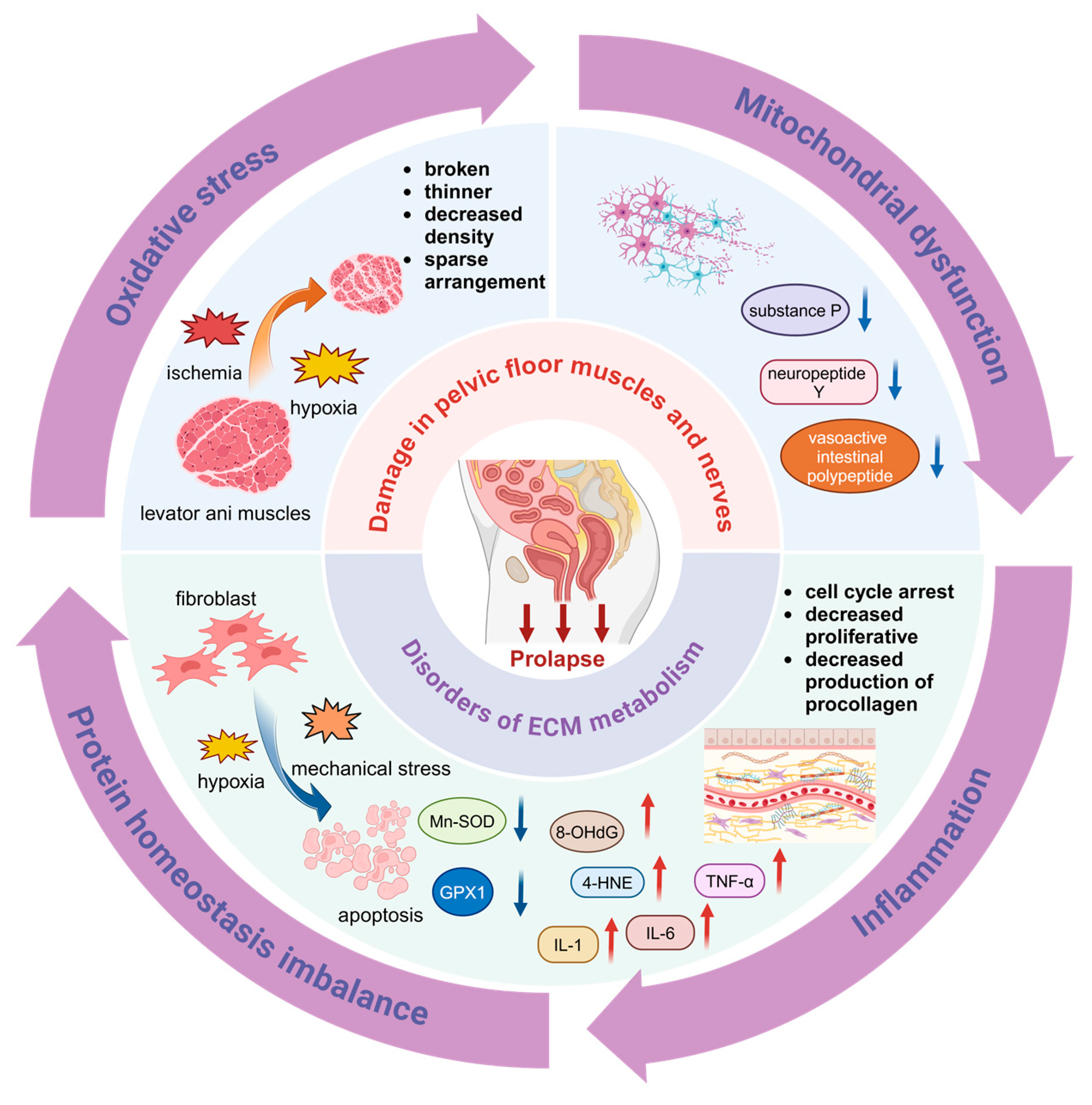
3. Pathogenesis of POP

Current research supports the notion that POP is a polygenic disorder influenced by a combination of genetic and environmental factors. Aging and vaginal delivery are deemed the most significant risk factors for the development of POP [5]. Consequently, POP may be regarded as a degenerative condition closely associated with tissue injury and impaired repair mechanisms [6]. Disruptions in pelvic floor support structures, such as tears in the levator ani muscles and the loss of apical support ligaments, along with alterations in the quantities and ratios of the extracellular matrix (ECM) can significantly affect the biomechanical properties of the tissues [7,8]. The maintenance of pelvic floor organs in their physiological positions is contingent not only upon the integrity of these support structures, but also on their functional normalcy. Pathophysiological states, such as oxidative stress (OS), mitochondrial dysfunction, protein homeostasis imbalance, and persistent inflammation, are apparent in the pelvic floor tissues and may contribute to compromised pelvic floor support [9,10,11]. This review discusses the potential pathogenesis of POP by focusing on both the pelvic floor muscles and ligamentous fascia, with an emphasis on the pathophysiological alterations occurring in both aging and injury states (Figure 1).

3.1. Damage in Pelvic Floor Muscles and Nerves

The predominant muscle of the pelvic floor, the levator ani, is a striated muscle that includes the pubococcygeus, iliococcygeus, and coccygeus muscles [12]. The continuous contraction of these slow-twitch fibers helps maintain the pelvic organs in their normal anatomical positions. In patients with POP, however, the levator ani muscle fibers are often fragmented, display a reduced diameter, decreased density, and are sparsely arranged [13]. Compared to controls, women with POP frequently exhibited defects in the levator ani muscles and produce less vaginal closure force during maximal contraction [14].
During vaginal delivery, pelvic floor tissues may suffer from ischemia and hypoxia, leading to immediate muscle damage. While this damage can gradually repair post-delivery, a significant portion remains improperly healed. Furthermore, chronic increased abdominal pressure may also contribute to persistent stress, exacerbating pelvic floor muscle damage. In vitro experiments have demonstrated that mechanical stress on myofibroblasts and levator ani muscles results in notable OS [15], triggering cellular mitochondrial dysfunction and impeding tissue repair. This eventually contributes to pelvic floor weakness [16]. These findings suggest that the inadequate repair of post-delivery levator ani muscle damage, coupled with chronic injury, are likely initial factors for POP.
The physiological function of muscle is contingent upon nerve innervation. In the absence of innervation, the differentiation and formation of mature muscle fibers are hindered, potentially leading to muscle atrophy due to the lack of neurotrophic support. Consequently, both the physiological function and structure of the muscle are compromised. Notably, denervation changes, including significant reductions or the absence of neuropeptide Y, vasoactive intestinal polypeptide, and substance P immunoreactive nerves, have been observed in the levator ani muscles of patients with POP [17]. However, previous studies have not identified any neuropathy in the levator ani muscles of POP patients [12], leaving the nerve injury hypothesis for the levator ani muscles open to question.
The degradation of muscle function inevitably occurs with aging. The disruption of protein homeostasis and mitochondrial dysfunction are key manifestations of muscle aging. This includes the decline of the heat shock protein family, autophagy-lysosome pathways, ubiquitin–proteasome systems, and mitochondrial quality control [18,19]. The role of mitochondrial disease and cumulative OS in the pathophysiology of pelvic floor disorders has been documented [20]. However, no studies have specifically addressed the aging of pelvic floor skeletal muscles. Further exploration into the mechanisms of aging in these muscles could enhance our understanding of the onset of POP.

3.2. Disorders of ECM Metabolism in the Pelvic Floor

The ECM comprises structural proteins such as collagen and elastin, along with matrix adhesion molecules including fibronectin, laminin, and proteoglycans. Primarily, collagen and elastic fibers are synthesized by fibroblasts. Factors inducing fibroblast apoptosis and dysfunction may weaken pelvic floor connective tissue by impacting ECM metabolism. On the other hand, an imbalance between proteolytic enzymes matrix metalloproteinase (MMPs) and the tissue inhibitor of matrix metalloproteinases (TIMPs) can also lead to excessive ECM degradation, contributing to the weakening of pelvic floor tissues observed in POP [21]. Recent studies highlight the influence of OS, mitochondrial dysfunction, hormone metabolism, and inflammation on fibroblast behavior.

3.2.1. Oxidative Stress (OS)

The cytoskeleton in fibroblasts can sense mechanical stress and convert it into biochemical signals that are transmitted to the nucleus to regulate gene expression and protein translation [22]. In vitro studies have demonstrated that mechanical stress can lead to an accumulation of reactive oxygen species (ROS) in fibroblasts. Elevated levels of intracellular OS can induce cellular senescence and apoptosis and reduce the expression of type I collagen. During this process, the phosphorylation activation of the PI3K-Akt signaling pathway in prolapsed tissues increases the phosphorylation level of FOXO1 and inhibits its transcriptional regulatory activity, which in turn downregulates the expression of antioxidant proteins, such as glutathione peroxidase 1 (GPX1) and Mn-superoxide dismutase (Mn-SOD). This results in an imbalance between the reduced antioxidative capacity and the accumulation of intracellular ROS [11]. Markers of oxidative damage, such as 8-hydroxyguanosine (8-OHdG) and 4-hydroxynonenal (4-HNE), were significantly higher in POP patients compared to the controls. A further in vitro challenge of human USL fibroblasts using hydrogen peroxide (H2O2) revealed that H2O2 can regulate the cellular collagen metabolism in a dose-dependent manner, with small doses promoting collagen synthesis and higher doses enhancing collagen breakdown. Concurrently, levels of MMP2 and TIMP2 varied with the degree of OS [10]. Notably, in patients with POP, the overexpression of mitochondrial fusion 2 (MFN2) and advanced glycation end products mediate a reduction in the fibroblasts’ proliferative capacity, causing cell cycle arrest in the G0/G1 phase and the diminished production of type I and III procollagen via the Ras-Raf-ERK axis, RAGE and/or p-p38 MAPK, and NF-κB-p-p65 pathways, as documented in references [23,24,25]. This further suggests that OS may influence the ECM metabolism. Chronic long-term stress induces significant OS in pelvic floor tissues, potentially affecting their ECM metabolism.

3.2.2. Mitochondrial Dysfunction

Pelvic floor tissues become hypoxic during labor or chronic increased abdominal pressure. Hypoxia induces the elevated expression of hypoxia-inducible factor 1α (HIF-1α) in the fibroblasts of the uterosacral ligament, resulting in mitochondrial dysfunction and the subsequent apoptosis of fibroblasts [26]. Mitochondrial DNA in the uterosacral ligament was increased in premenopausal patients with POP and significantly decreased in postmenopausal POP patients compared to the non-POP group. The elevated copy number of mitochondrial DNA may provide energy for tissue repair; however, this compensatory process was absent in postmenopausal patients. Furthermore, the expression of the transcriptional coactivator PGC-1α, a principal regulator of mitochondrial biogenesis, was upregulated in POP patients, potentially compensating for mitochondrial dysfunction by enhancing mitochondrial biogenesis in prolapsed tissues [27]. This evidence suggests that mitochondrial dysfunction plays a role in the development of POP.

3.2.3. Hormonal Changes

The role of estrogen in the development of POP remains controversial. Predominantly, patients with POP are postmenopausal, suggesting that reduced estrogen levels may facilitate POP development. Estrogen regulates the expression of genes associated with the ECM metabolism [28,29], including lysyl oxidase-like-1 (LOXL1), an enzyme that catalyzes the deamination of lysine residues in protoelastin. These residues form cross-links to create stable elastin and promote covalent linkages between elastin and collagen [30]. Recent studies indicate that a stiff ECM can enhance the transformation of fibroblasts into myofibroblasts, leading to excessive collagen deposition, tissue contracture, and dysfunction. Conversely, estrogen impedes fibroblast differentiation by upregulating DNA methyltransferase1 (DNMT1) expression, thus inhibiting POP progression [31]. However, it has been observed that estrogen markedly suppresses fibroblast proliferation, and estrogen supplementation has not proven to be an effective treatment [32]. Clinical trial results also revealed that perioperative vaginal estrogen did not increase surgical success rates, suggesting that estrogen supplementation primarily mitigates symptoms related to vaginal atrophy with a limited therapeutic impact on POP [33]. In addition to an estrogen deficiency, other hormonal shifts including a variation in progesterone levels can influence ECM metabolism. Although findings on progesterone receptor (PR) expression levels are mixed, an increasing number of studies indicate elevated PR in patients with POP [34,35,36]. The research demonstrates that both estrogen and progesterone contribute to the integrity of pelvic floor support tissues by inhibiting MMP13 and collagenase production, thus reducing ECM degradation [37,38]. It is hypothesized that postmenopausal women experience a loss of this protective effect due to a hormone deficiency. Nonetheless, the exact role of progesterone remains unclear.

3.2.4. Inflammation

Inflammatory cytokines such as IL-1, IL-6, and TNF-α have been implicated in the pathogenesis of POP through the promotion of ECM component breakdown. It has been observed that in patients with POP, the levels of pro-inflammatory cytokines IL-6 and TNF-α positively correlate with MMP1 and MMP2, while inversely correlate with TIMP1 [21,39]. Furthermore, IL-1β plays a role in the regulation of elastin expression [40]. Repeated mechanical stress, resulting from factors such as childbirth, heavy lifting, or chronic coughing, can upregulate genes associated with inflammation, subsequently leading to ECM degradation and pelvic floor dysfunction [38,41].
In conclusion, damage to the supporting tissues of the pelvic floor, including muscles, nerves, and ECM, is intricately involved in the pathogenesis of POP. Factors such as aging, continuous stress, and fluctuations in estrogen and progesterone contribute to the inevitable onset and progression of POP by promoting OS overproduction, mitochondrial dysfunction, the chronic inflammatory environment, and protein destabilization. Notably, current research has largely overlooked the role of pelvic floor muscles, whereas the impact of the ECM in pelvic floor tissues is increasingly recognized as more significant.

4. Genetic Studies of POP

Different populations and individuals exhibit varying predispositions to polygenic diseases due to distinct genetic structures and the influence of external environments. Notably, the prevalence of POP varies significantly among women of different races, suggesting that diverse genetic backgrounds may influence the etiology of POP [42]. Moreover, genetic variations in the number, structure, or function of pelvic floor connective tissue components across races may affect the incidence of POP.
Epidemiologic studies highlight the genetic predisposition to POP. The research indicates that sisters of patients with severe POP are five times more likely to develop the condition compared to women without a familial history [43]. A family history of POP is associated with a 2.7-fold increased risk (odds ratio (OR): 2.64; 95% confidence interval (CI): 2.07, 3.35) and a 1.4-fold increased risk of recurrence (OR: 1.44; 95% CI: 1.00, 2.08) [44]. Importantly, the aggregation of POP within families is not solely attributable to genetic factors; environmental influences, such as similar lifestyle habits and their socioeconomic status, also play a significant role. A comparative study of 3376 identical twins and 5067 fraternal twins found that identical twins had a higher concordance for developing POP. Quantitative genetic analyses suggest that genetic factors account for 43% of POP cases, while environmental factors contribute to 57%. Thus, genetic predisposition significantly influences the development of POP [45].
In recent years, the genetic susceptibility of POP has been partially elucidated through genomic studies, including candidate gene association studies, genome-wide association studies (GWAS), linkage analyses, and whole-exome sequencing (WES). The majority of these studies have concentrated on genes involved in the ECM metabolism in pelvic floor connective tissue. Numerous case–control studies have demonstrated that the abnormal composition and metabolism of connective tissue are linked to POP. Additionally, genetic knockout mouse models related to the elastic fiber metabolism, such as LOXL1 [46,47] and fibronectin-5 (FIBN5), have exhibited severe POP symptoms, vaginal wall weakness, paraurethral lesions, and lower urinary tract dysfunction following pregnancy and vaginal delivery [5,48,49].

4.1. Linkage Analyses

Linkage analysis is a technique used to map the location of genes associated with specific traits or diseases by studying inheritance patterns of genetic markers. If family members affected by the same disease share a specific chromosome region, it is likely that the susceptibility gene for this disease is located in or near that region. The principles of genetic linkage are employed to genotype a reference locus within the family line and then mathematically determine whether the locus co-segregates with the disease, thereby exploring the relationship between causative genes and the reference locus [50]. Nikolova et al. first conducted a linkage analysis on POP family, where three generations of females exhibited symptoms of prolapse at a very young age. They identified ten linkage regions with potentially strong candidate genes involved in ECM metabolism, such as LAMC1, LAMC2, and COL8A1. Furthermore, they discovered that POP was inherited in an autosomal dominant manner with significant epistasis in this family [51]. Subsequently, Allen-Brady et al. performed a linkage analysis on 70 members from 32 families, each with at least two members suffering from moderate-to-severe POP. Their study identified a significant linkage of pelvic floor dysfunction susceptibility genes in the p21 region of chromosome 9 [52]. In a further linkage analysis involving 299 POP patients from 83 families, chromosomes 10q and 17q were found to be closely associated with the development of POP, indicating potential regions for susceptibility genes [53]. Future studies are needed to further narrow down these regions to pinpoint the gene loci associated with POP.

4.2. Candidate Gene Association Studies

Single-nucleotide polymorphisms (SNPs) are sites in DNA where individual nucleotides vary from person to person, representing the most abundant type of DNA sequence variation in the human genome. Only a small fraction of these SNPs are biologically significant and contribute to human genome diversity. SNPs can influence the promoter activity of DNA and pre-mRNA conformation, leading to alterations in amino acid sequences and protein function, thereby playing a direct or indirect role in phenotypic expression. Numerous studies have investigated POP-related SNPs in patients by DNA sequencing, targeting genes related to ECM synthesis and catabolism, including COL1A1, COL3A1, LAMC1, LOXL1, FBLN5, and MMP. Additionally, polymorphisms of ER may also be associated with POP (Table 1). However, these studies often face limitations such as small sample sizes and varying criteria for identifying cases and controls. Overall, no SNPs have yet been identified that strongly correlate with the development of POP.

4.2.1. COL1A1

The most abundant ECM of pelvic floor connective tissue is composed of collagen types I and III [80]. Type I collagen is a heterotrimeric protein consisting of two α1 chains and one α2 chain, with the α1 protein chain encoded by COL1A1 located on chromosome 17, q21.31–q22. The rs1800012 of COL1A1 was found to be associated with an increased risk of stress urinary incontinence (SUI) [81]. The substitution of guanine with thymine (G/T) at the Sp1 binding site in COL1A1′s transcription factor leads to the abnormal production of the α1 chain, increasing the formation of homotrimeric proteins and decreasing connective tissue strength. Both POP and SUI are believed to share an etiological basis, prompting studies on the association between this polymorphism and POP. Feiner et al. reported an OR of 1.75 for the GT genotype prevalence of rs1800012 between POP cases and controls [57]. Subsequent studies in diverse populations such as Brazil, Korea, Italy, China, and Japan found no significant correlations [53,54,55,57,58,59]. However, Cartwright et al.’s meta-analysis identified an association between COL1A1 rs1800012 (G→T) and POP (T vs. G, OR: 1.33, 95% CI: 1.02–1.73) [82]. Similarly, Allen-Brady et al. conducted a meta-analysis from 2015 to 2020, which also suggested an association between rs1800012 and POP [83]. Although individual studies may not have found significant associations, the data integration from global studies indicates a potential link between the mutant T allele and POP, though further validation in larger cohorts is necessary.

4.2.2. COL3A1

COL3A1, located at chromosome 2, q24.3–q31, encodes the α1 protein chain of type III collagen. This fibrillar protein comprises three identical α1 chains, forming fibers that are finer, more sparsely arranged, and more elastic than type I collagen fibers, significantly contributing to tissue repair. In patients with POP, an increased presence of type III collagen at non-prolapse sites possibly indicates tissue repair after pelvic floor injury [84]. The polymorphism of COL3A1 may alter the α1 protein chain, impacting the mechanical properties of type III collagen and affecting the structures supporting the pelvic floor. Chen et al. assessed the genetic association using a polymerase chain reaction and restriction fragment length polymorphism (PCR-RFLP) analysis in a study of 84 POP patients and 147 controls from Taiwan, discovering a significant association between the AA genotype of COL3A1 (rs1800255) and POP. The genetic mutation in exon 30, from a guanine to an adenine (G→A), transforms the encoded alanine to threonine, potentially disrupting the triple-helical structure of type III collagen due to increased hydrophilicity [64]. Similarly, Kluivers et al. found an association between rs1800255 and POP in a study involving 202 patients and 102 controls [63]. Moreover, a meta-analysis up to December 2012 indicated a significant association of rs1800255 with POP in Asian and Dutch females, with an OR of 4.79 (95% CI: 1.91–11.98) [85].
All the aforementioned studies have utilized PCR-RFLP to draw their final conclusions. Additionally, Lince et al. confirmed the polymorphism of gene rs1800255 by employing the high-resolution melting curve (HRMC) technique. They observed differential expression between the case and control groups in Dutch women; however, it was not possible to demonstrate that this polymorphism was associated with POP. Consequently, they recommended that the conclusions of other association studies using PCR-RFLP should be approached with caution [62]. Subsequent studies involving Brazilian, Japanese, and Chinese females have all indicated that COL3A1 rs1800255 is not linked to POP [54,55,56,61]. Nevertheless, a meta-analysis of data up to March 2021 suggests that rs1800255 may be a risk factor for POP in Caucasian populations. Moreover, Caucasian individuals carrying the A allele or AA genotype are at an increased risk of developing POP [86].
Jeon et al. conducted PCR-RFLP analyses on 36 POP cases and 36 normal controls, discovering that the G/A mutation at position +2092 of COL3A1 exon 31 was associated with POP in Korean women, with an OR of 3.2 for POP in women carrying the G allele [66]. In contrast, a study in a Brazilian population found no association between this polymorphism in exon 31 of COL3A1 and POP [65]. Thus, the research on COL3A1 polymorphisms and POP has yielded inconsistent results. The connection between COL3A1 polymorphism and POP remains inconclusive, influenced by factors such as the testing technology, racial differences, and the sample size.

4.2.3. LAMC1

Laminin, a crucial non-collagenous component of the ECM glycoproteins, constitutes the basement membrane and plays a vital role in mediating cell adhesion, as well as regulating cell growth and differentiation. It is a heterotrimer composed of α, β, and γ chains and is present in at least 15 distinct forms. The gene LAMC1, which encodes the γ-chain, is located on chromosome 1 at q25.3 [67]. The altered laminin expression in tissues from patients with SUI suggests that changes in laminin may compromise the integrity of the basement membrane [87]. Previously, Nikolova et al. identified a candidate gene in vaginal tissues of POP-afflicted family through linkage and co-segregation analyses, discovering a mutation (C→T) in the LAMC1 promoter region, rs10911193, which may heighten the risk of an early-onset POP [51]. Further research involving Caucasian and African–American females with a stage II-IV POP revealed racial differences in LAMC1 polymorphisms, though no significant correlation was found between the POP stage and LAMC1 variants [68]. Another study across diverse ethnic groups also found no association between LAMC1 polymorphisms and POP [67]. Thus, the relationship between this gene polymorphism and POP requires additional validation across different racial groups.

4.2.4. LOXL

Lysyl oxidase-like gene 1 (LOXL1), situated on chromosome 15 at q24.1, encodes a copper-dependent monoamine oxidase. Within the ECM, lysyl oxidase catalyzes the cross-linking of lysine residues in elastin and hydroxylysine residues in collagen, thereby stabilizing and forming an elastic meshwork [30]. Liu et al. knocked out Loxl1 in a mouse model and observed that, in contrast to wild-type mice, Loxl1−/− mice exhibited abnormal elastic fiber deposition in the uterus post-delivery and developed symptoms related to POP. This indicates that LOXL1 is crucial in maintaining the structure and function of elastic fibers, and its genetic deficiency may foster the progression of POP [47]. Moreover, molecular analyses of anterior vaginal wall tissues from POP patients revealed the significantly reduced expression of LOXL1 and LOXL3, suggesting that LOXL defects could lead to abnormal elastic fiber remodeling in pelvic connective tissues, thereby contributing to the onset and exacerbation of POP. Nonetheless, case–control studies conducted with Japanese and Brazilian women have not demonstrated an association, and no SNPs in this gene have been linked to POP [54,69].
Furthermore, a linkage analysis revealed that chromosome 10 q24–26 is associated with POP. LOXL4, located on chromosome 10 q24.2, is identified as the most likely candidate gene in this region [53]. It plays a crucial role in connective tissue biogenesis by catalyzing the cross-linking between collagen and elastin. A study involving Brazilian women found no correlation between the LOXL4 gene (rs2862296) and POP [70]. In contrast, this polymorphism was linked to the development of POP in Japanese women [54]. Given the limited research on the association between LOXL4 polymorphisms and POP and the varying conclusions of different studies, further validation through more extensive research is necessary.

4.2.5. FBLN5

Fibulin is a class of secreted glycoproteins distributed in the ECM. Fibulin5 (FBLN5) acts as an adaptor protein linking cross-linking enzymes with structural components of elastic fibers, such as elastin, fibrillin-1, and LOXL1. It organizes the assembly of elastic fibers on microfibrillar scaffolds and stabilizes the structure of the basement membrane [48]. Studies on Fbln5−/− mice have shown progressive symptoms of genital prolapse, indicating that FBLN5 may play a crucial role in maintaining the stability of the pelvic floor support structure. Furthermore, FBLN5 can suppress the expression of MMP9, which regulates the homeostasis of the pelvic floor’s ECM and prevents the occurrence of POP [48]. Khadzhieva et al. conducted the first study on FBLN5 polymorphisms in patients with stage III-IV POP and 292 normal controls, finding significant associations with POP, especially in patients with a history of pelvic floor injury, at rs2018736 and rs12589592 [71]. Additionally, a meta-analysis from 2015 to 2020 indicated that the polymorphism rs12589592 in FBLN5 was connected to POP risk with an OR of 1.46 and a 95% CI of 1.11–1.82 [83]. However, Paula et al. found no association between FBLN5 polymorphisms and POP in Brazilian women [72].

4.2.6. MMPs and TIMPs

MMPs, a class of proteases capable of degrading the ECM and basement membrane components, have their increased expression levels directly associated with the onset and progression of POP [88,89]. MMP1, also known as mesenchymal collagenase, targets type I, II, III, and IV collagens and is primarily located in genital tissues, the colon, and blood vessels. In Italian women, an insertion or deletion of a guanine at −1607/1608 upstream of the MMP1 transcriptional start site has been linked with POP development [58]. Two polymorphic loci in POP patients were examined, including the MMP1 promoter region −1607/−1608 G insertion or deletion and the MMP3 promoter region −1612/1617 A insertion or deletion. Although no individual SNP was associated with POP, a combined analysis of these two SNPs indicated the higher expression of the gene polymorphism in patients with POP compared to controls [73].
MMP9, also known as type IV collagenase or gelatinase B, primarily expressed in blood vessels and pelvic tissues, is responsible for degrading collagen, proteoglycans, laminin, and fibronectin. Chen et al. reported significant associations of the AG and GG genotypes of MMP9 rs17576 with POP [76]. Additionally, two other polymorphic sites in MMP9, rs3918253 and rs3918256, have been linked to POP in non-Hispanic white women [75]. Furthermore, it has been observed that polymorphisms in MMP10, specifically rs17435959, may contribute to POP onset. This polymorphism involves a substitution of leucine with valine at amino acid 4 in the first exon of the gene. A case–control study indicated a significant difference in the frequencies of the rs17435959 G/C between the cases and controls [77].
TIMP serves as an inhibitor of endogenous MMPs and is crucial for maintaining the metabolic balance between ECM synthesis and degradation. Reduced TIMP expression levels in pelvic floor tissues have been directly linked with the onset and progression of POP [88]. Furthermore, a linkage analysis has identified chromosome 17q25 as potentially related to POP, with TIMP2 being the most likely candidate gene in this region [53]. However, no studies currently explore the association between this gene polymorphism and the pathogenesis of POP.
In summary, research examining the relationship between polymorphisms in genes associated with ECM degradation and POP has produced inconsistent results. Further studies with larger sample sizes across diverse ethnic groups are necessary for additional clarification.

4.2.7. ER

Estrogen receptors (ER) are categorized into two primary subtypes: ERα and ERβ. ERα is predominantly expressed in the uterus of adult women, whereas ERβ is mainly expressed in other estrogen-targeted tissues [90]. Both ERα and ERβ can influence the strength of pelvic floor supportive tissues by regulating the synthesis and degradation of the ECM. In patients with POP, the expression levels of ER in the uterine and USL are significantly reduced prior to menopause [91]. Chen et al. observed that the frequency of the ERβ haplotype CGCGC was elevated in women with POP (16.7%) [92]. Research conducted in Chinese and Israeli populations has demonstrated a significant association between the rs2228480 polymorphism in ERα and the development of POP, identifying both heterozygous and homozygous subtypes as risk factors [79]. Moreover, a meta-analysis has indicated that polymorphisms in ERα, specifically rs2228480, are linked to POP [83]. Additionally, polymorphisms rs2234693 and rs17847075 have also been significantly associated with POP in the Chinese population [78].
Overall, the evidence for the association of polymorphisms in ER with POP is still insufficient. Although numerous SNP loci associated with POP have been identified across various ethnic groups, the reliability of these findings necessitates further verification. This underscores the multiplicity of factors influencing POP as a polygenic disease. Furthermore, case–control studies that evaluate SNPs in multiple candidate genes are often limited by factors such as small sample sizes and low accuracy of testing methods.

4.3. Whole Exome Sequencing (WES)

WES refers to the sequencing of the entire coding region of a gene with high chromosomal coverage and accuracy, which is extensively applied to detect mutations causing diseases. Rao et al. initially conducted WES on peripheral blood DNA samples from eight patients with POP, identifying two missense variants: c.2668 G>A in WNK1 (p.G890R) and c.6761 C>T in another gene (p.P2254L). These variants were not present in 231 healthy controls. Additionally, functional experiments revealed that fibroblasts in the uterosacral ligament from POP patients with WNK1 mutations displayed a looser and more irregular arrangement than those from healthy controls [93]. However, due to the small sample size, the conclusions of this study were limited.

4.4. Genome-Wide Association Studies (GWAS)

GWAS employs a case–control association design where SNP loci are identified in both the case group and normal controls. The SNPs are analyzed on a genome-wide scale, allowing for the calculation of variant allele frequencies [94]. Allen-Brady et al. performed the first GWAS analysis of 115 patients with a family history of POP by using Illumina 550K. They identified significant associations with POP at six loci, 4q21, 8q24, 9q22, 15q11, 20p13, and 21q22, in the Dutch population, confirming the role of genetic factors in POP development [95]. Subsequently, Giri et al. performed a GWAS on African– and Hispanic–American women, involving 1274 normal controls and 1427 patients with varying stages of POP. They identified several potential susceptibility loci, including CPE, AL132709.5, DPP6, PGBD5, and SHC3 [96].
Recently, several teams have conducted GWAS analysis for POP with larger sample sizes. Olafsdottir et al. analyzed data from the United Kingdom, comprising 15,010 cases and 340,734 controls. They identified eight sequence variants at seven loci associated with POP (p < 5 × 10⁻⁸), further suggesting a role for connective tissue metabolism and estrogen in POP pathogenesis [97]. The advancement of gene chip technology has facilitated genetic screenings on large scales. Utilizing Illumina Human CoreExome microarrays, Cox et al. detected associations between 20 million SNPs and POP from 1329 patients and 16,383 normal controls. Additionally, four previously reported suspect SNPs were replicated, reinforcing the genetic contribution to POP [98]. A meta-analysis synthesizing GWAS results from 28,086 POP patients and 546,291 controls identified 26 genome-wide significant loci (p < 5 × 10⁻⁸), which included 19 novel loci and 7 previously reported ones. This study demonstrated correlations between several candidate genes and POP, including LOXL1, WNT4, EFEMP1, FAT4, IMPDH1, TBX5, and SALL1, alongside new candidate genes such as CHRDL2, ACADVL, and PLA2G6. It further revealed that molecular alterations in connective tissues and abnormalities in urogenital development are crucial in the pathogenesis of POP [99].
The above GWAS results suggest that connective tissue abnormalities are associated with POP. Future research involving larger sample sizes and diverse racial groups will elucidate the genetic predispositions to POP further.

5. Epigenetic Studies of POP

In addition to genetic predisposition, environmental influences are undoubtedly crucial in the development of POP. Under environmental effects, the coding pattern of DNA undergoes hereditary alterations, known as epigenetic inheritance. Such mechanisms induce inheritable cellular changes without altering the DNA sequence, including DNA methylation, histone modification, microRNA (miRNA) expression, and DNA replication timing, leading to selective gene expression or repression. Recent studies have indicated that epigenetic mechanisms may partially contribute to the pathogenesis of POP [100].

5.1. DNA Methylation

DNA methylation, the most prevalent epigenetic mechanism, is influenced by environmental risk factors. It has been reported that the methylation of promoter regions can potentially inhibit LOX gene expression in women with POP. Researchers observed an 8.2-fold decrease in the LOX expression and increased methylation of CpG sites in tissue from women with grade III prolapse compared to controls [101]. Another study identified differences in the genome-wide methylation in the USL between POP patients and controls through a genome-wide DNA methylation analysis [100]. This study reported 3723 differentially methylated CpG sites, with the gene ontology analysis suggesting an association with the ECM. It was demonstrated that specific key members involved in the ECM metabolism were DNA-methylated. However, due to the limitations imposed by small sample sizes in these studies, these findings require further investigation in future studies.

5.2. MicroRNA

MicroRNAs (miRNAs) are small non-coding RNAs that bind to mRNA regulatory sites in target genes, modifying their expression via translational repression or mRNA degradation. One study indicated that miR-221 and miR-222 are upregulated in the USL of women with POP, potentially leading to a reduced ERα expression [102]. Similarly, an increased miR-92 expression and decreased ERβ1 level was observed in POP patients, with the ERβ1 expression being inversely correlated with the miR-92 levels [36]. Additionally, studies have demonstrated that overexpressed miR-30d and miR-181a suppress the expression of HOXA11 in the USLs of POP patients. As a crucial transcription factor regulating the collagen metabolism and homeostasis in the USLs, deficient HOXA11 signaling may contribute to the development of POP [103]. Overall, the current research focuses on the correlation between the expression levels of various miRNAs and the development of POP, primarily affecting the expression of ECM-related metabolic genes. The validity of these findings and the specific mechanisms involved require further investigation.

6. Conclusions

POP is a benign gynecological disease that seriously affects women’s quality of life. It has a complex etiology and its pathogenesis remains unclear. Numerous studies have indicated the involvement of genetic factors, identifying several candidate genes and SNPs in the metabolism of pelvic floor connective tissue potentially associated with POP. However, the existing genetic research on POP predominantly comprises isolated case–control studies focusing on single populations or small sample sizes, and has yet to pinpoint definitive causative genes and loci. A multicenter GWAS is anticipated to yield more accurate conclusions.
Injury to the levator ani muscles and the dysregulation of the connective tissue component of the pelvic floor may initiate this disorder, or the progression of this disorder itself may lead to molecular changes in the pelvic floor tissue. Patients with POP may possess a genetic predisposition that increases their likelihood of developing pelvic floor dysfunction spontaneously, or as a result of factors such as vaginal delivery, aging, or chronic increased intra-abdominal pressure. Conversely, POP may directly affect gene expression and regulation through tissue stretching. Functional in vitro experiments are essential to further explore the impact of mechanical stress and other factors on the homeostasis of the ECM. Investigating genetic susceptibility and the pathogenesis of POP will aid in providing theoretical recommendations for an individualized risk assessment and diagnosis, as well as the development of targeted therapeutic strategies.

Author Contributions

Conceptualization: M.W.; writing—original draft preparation: Y.L. (Yuting Li); writing—review and editing: Z.L., Y.L. (Yinuo Li) and T.W.; charting-revised draft: X.G. and Y.H. All authors have read and agreed to the published version of the manuscript.

Funding

This research was funded by the National Key Research and Development Program of China, 2023YFC2706003 and 2023YFC2706001.

Acknowledgments

The authors thank all the reviewers for their constructive evaluations which have improved this manuscript.

Conflicts of Interest

The authors declare no conflicts of interest.

References

  1. Jelovsek, J.E.; Maher, C.; Barber, M.D. Pelvic organ prolapse. Lancet 2007, 369, 1027–1038. [Google Scholar] [CrossRef] [PubMed]
  2. Barber, M.D. Pelvic organ prolapse. BMJ 2016, 354, i3853. [Google Scholar] [CrossRef] [PubMed]
  3. Wu, J.M.; Vaughan, C.P.; Goode, P.S.; Redden, D.T.; Burgio, K.L.; Richter, H.E.; Markland, A.D. Prevalence and Trends of Symptomatic Pelvic Floor Disorders in U.S. Women. Obstet. Gynecol. 2014, 123, 141–148. [Google Scholar] [CrossRef] [PubMed]
  4. Li, Z.Y.; Zhu, L.; Xu, T.; Liu, Q.; Li, Z.A.; Gong, J.; Wang, Y.L.; Wang, J.T.; Lai, T.; Wu, L.; et al. An epidemiologic study of pelvic organ prolapse in urban Chinese women: A population-based sample in China. Zhonghua Yi Xue Za Zhi 2019, 99, 857–861. [Google Scholar] [CrossRef]
  5. Drewes, P.G.; Yanagisawa, H.; Starcher, B.; Hornstra, I.; Csiszar, K.; Marinis, S.I.; Keller, P.; Word, R.A. Pelvic Organ Prolapse in Fibulin-5 Knockout Mice. Am. J. Pathol. 2007, 170, 578–589. [Google Scholar] [CrossRef]
  6. Tinelli, A.; Malvasi, A.; Rahimi, S.; Negro, R.; Vergara, D.; Martignago, R.; Pellegrino, M.; Cavallotti, C. Age-related pelvic floor modifications and prolapse risk factors in postmenopausal women. Menopause 2010, 17, 204–212. [Google Scholar] [CrossRef]
  7. Kieserman-Shmokler, C.; Swenson, C.W.; Chen, L.; Desmond, L.M.; Ashton-Miller, J.A.; DeLancey, J.O. From molecular to macro: The key role of the apical ligaments in uterovaginal support. Am. J. Obstet. Gynecol. 2020, 222, 427–436. [Google Scholar] [CrossRef]
  8. Chi, N.; Lozo, S.; Rathnayake, R.A.C.; Botros-Brey, S.; Ma, Y.; Damaser, M.; Wang, R.R. Distinctive structure, composition and biomechanics of collagen fibrils in vaginal wall connective tissues associated with pelvic organ prolapse. Acta Biomater. 2022, 152, 335–344. [Google Scholar] [CrossRef]
  9. Orlicky, D.J.; Guess, M.K.; Bales, E.S.; Rascoff, L.G.; Arruda, J.S.; Hutchinson-Colas, J.A.; Johnson, J.; Connell, K.A. Using the novel pelvic organ prolapse histologic quantification system to identify phenotypes in uterosacral ligaments in women with pelvic organ prolapse. Am. J. Obstet. Gynecol. 2021, 224, 67.e1–67.e18. [Google Scholar] [CrossRef]
  10. Liu, C.; Yang, Q.; Fang, G.; Li, B.-S.; Wu, D.-B.; Guo, W.-J.; Hong, S.-S.; Hong, L. Collagen metabolic disorder induced by oxidative stress in human uterosacral ligament-derived fibroblasts: A possible pathophysiological mechanism in pelvic organ prolapse. Mol. Med. Rep. 2016, 13, 2999–3008. [Google Scholar] [CrossRef]
  11. Li, B.-S.; Guo, W.-J.; Hong, L.; Liu, Y.-D.; Liu, C.; Hong, S.-S.; Wu, D.-B.; Min, J. Role of mechanical strain-activated PI3K/Akt signaling pathway in pelvic organ prolapse. Mol. Med. Rep. 2016, 14, 243–253. [Google Scholar] [CrossRef]
  12. Jundt, K.; Kiening, M.; Fischer, P.; Bergauer, F.; Rauch, E.; Janni, W.; Peschers, U.; Dimpfl, T. Is the histomorphological concept of the female pelvic floor and its changes due to age and vaginal delivery correct? Neurourol. Urodyn. 2005, 24, 44–50. [Google Scholar] [CrossRef]
  13. Zhu, L.; Lang, J.H.; Chen, J.; Chen, J. Morphologic study on levator ani muscle in patients with pelvic organ prolapse and stress urinary incontinence. Int. Urogynecol. J. 2005, 16, 401–404. [Google Scholar] [CrossRef]
  14. DeLancey, J.O.L.; Morgan, D.M.; Fenner, D.E.; Kearney, R.; Guire, K.; Miller, J.M.; Hussain, H.; Umek, W.; Hsu, Y.; Ashton-Miller, J.A. Comparison of Levator Ani Muscle Defects and Function in Women with and without Pelvic Organ Prolapse. Obstet. Gynecol. 2007, 109, 295–302. [Google Scholar] [CrossRef]
  15. Huang, G.; He, Y.; Hong, L.; Zhou, M.; Zuo, X.; Zhao, Z. Restoration of NAD+ homeostasis protects C2C12 myoblasts and mouse levator ani muscle from mechanical stress-induced damage. Anim. Cells Syst. 2022, 26, 192–202. [Google Scholar] [CrossRef] [PubMed]
  16. Yi, Y.; Wang, L.; Li, S.; Li, B.; Liu, C.; Hong, L. Effects of mechanical trauma on the differentiation and ArfGAP3 expression of C2C12 myoblast and mouse levator ani muscle. Int. Urogynecol. J. 2020, 31, 1913–1924. [Google Scholar] [CrossRef]
  17. Busacchi, P.; Perri, T.; Paradisi, R.; Oliverio, C.; Santini, D.; Guerrini, S.; Barbara, G.; Stanghellini, V.; Corinaldesi, R.; De Giorgio, R. Abnormalities of somatic peptide-containing nerves supplying the pelvic floor of women with genitourinary prolapse and stress urinary incontinence. Urology 2004, 63, 591–595. [Google Scholar] [CrossRef] [PubMed]
  18. McArdle, A.; Pollock, N.; Staunton, C.A.; Jackson, M.J. Aberrant redox signalling and stress response in age-related muscle decline: Role in inter- and intra-cellular signalling. Free Radic. Biol. Med. 2019, 132, 50–57. [Google Scholar] [CrossRef] [PubMed]
  19. Picca, A.; Lozanoska-Ochser, B.; Calvani, R.; Coelho-Júnior, H.J.; Leewenburgh, C.; Marzetti, E. Inflammatory, mitochondrial, and senescence-related markers: Underlying biological pathways of muscle aging and new therapeutic targets. Exp. Gerontol. 2023, 178, 112204. [Google Scholar] [CrossRef]
  20. Yiou, R.; Authier, F.-J.; Gherardi, R.; Abbou, C. Evidence of Mitochondrial Damage in the Levator Ani Muscle of Women with Pelvic Organ Prolapse. Eur. Urol. 2009, 55, 1241–1243. [Google Scholar] [CrossRef]
  21. Cao, L.L.; Yu, J.; Yang, Z.L.; Qiao, X.; Ye, H.; Xi, C.L.; Zhou, Q.C.; Hu, C.C.; Zhao, C.J.; Gong, Z.L. MMP-1/TIMP-1 expressions in rectal submucosa of females with obstructed defecation syndrome associated with internal rectal prolapse. Histol. Histopathol. 2019, 34, 265–274. [Google Scholar] [CrossRef] [PubMed]
  22. Guler, Z.; Roovers, J.P. Role of Fibroblasts and Myofibroblasts on the Pathogenesis and Treatment of Pelvic Organ Prolapse. Biomolecules 2022, 12, 94. [Google Scholar] [CrossRef] [PubMed]
  23. Wang, X.; Wang, X.; Zhou, Y.; Peng, C.; Chen, H.; Lu, Y. Mitofusin2 regulates the proliferation and function of fibroblasts: The possible mechanisms underlying pelvic organ prolapse development. Mol. Med. Rep. 2019, 20, 2859–2866. [Google Scholar] [CrossRef] [PubMed]
  24. Chen, H.; Lu, Y.; Qi, Y.; Bai, W.; Liao, Q. Relationship between the expressions of mitofusin-2 and procollagen in uterosacral ligament fibroblasts of postmenopausal patients with pelvic organ prolapse. Eur. J. Obstet. Gynecol. Reprod. Biol. 2014, 174, 141–145. [Google Scholar] [CrossRef]
  25. Chen, Y.-S.; Wang, X.-J.; Feng, W.; Hua, K.-Q. Advanced glycation end products decrease collagen I levels in fibroblasts from the vaginal wall of patients with POP via the RAGE, MAPK and NF-κB pathways. Int. J. Mol. Med. 2017, 40, 987–998. [Google Scholar] [CrossRef]
  26. Zhao, X.; Liu, L.; Li, R.; Wei, X.; Luan, W.; Liu, P.; Zhao, J. Hypoxia-Inducible Factor 1-α (HIF-1α) Induces Apoptosis of Human Uterosacral Ligament Fibroblasts through the Death Receptor and Mitochondrial Pathways. Med. Sci. Monit. 2018, 24, 8722–8733. [Google Scholar] [CrossRef]
  27. Sun, M.-J.; Cheng, Y.-S.; Liu, C.-S.; Sun, R. Changes in the PGC-1α and mtDNA copy number may play a role in the development of pelvic organ prolapse in pre-menopausal patients. Taiwan. J. Obstet. Gynecol. 2019, 58, 526–530. [Google Scholar] [CrossRef]
  28. ZONG, W.; JIANG, Y.; ZHAO, J.; ZHANG, J.; GAO, J. Estradiol plays a role in regulating the expression of lysyl oxidase family genes in mouse urogenital tissues and human Ishikawa cells. J. Zhejiang Univ. Sci. B 2015, 16, 857–864. [Google Scholar] [CrossRef]
  29. Sflomos, G.; Battista, L.; Aouad, P.; De Martino, F.; Scabia, V.; Stravodimou, A.; Ayyanan, A.; Ifticene-Treboux, A.; Bucher, P.; Fiche, M.; et al. Intraductal xenografts show lobular carcinoma cells rely on their own extracellular matrix and LOXL1. EMBO Mol. Med. 2021, 13, e13180. [Google Scholar] [CrossRef]
  30. Trackman, P.C. Lysyl Oxidase Isoforms and Potential Therapeutic Opportunities for Fibrosis and Cancer. Expert. Opin. Ther. Targets 2016, 20, 935–945. [Google Scholar] [CrossRef]
  31. Zhao, Z.; Huang, G.; He, Y.; Zuo, X.; Han, W.; Li, H. Estrogen inhibits the differentiation of fibroblasts induced by high stiffness matrix by enhancing DNMT1 expression. Tissue Cell 2023, 85, 102207. [Google Scholar] [CrossRef] [PubMed]
  32. Liu, Y.M.; Choy, K.W.; Lui, W.T.; Pang, M.W.; Wong, Y.F.; Yip, S.K. 17β-Estradiol suppresses proliferation of fibroblasts derived from cardinal ligaments in patients with or without pelvic organ prolapse. Hum. Reprod. 2006, 21, 303–308. [Google Scholar] [CrossRef] [PubMed]
  33. Rahn, D.D.; Richter, H.E.; Sung, V.W.; Pruszynski, J.E.; Hynan, L.S. Perioperative Vaginal Estrogen as Adjunct to Native Tissue Vaginal Apical Prolapse Repair: A Randomized Clinical Trial. JAMA 2023, 330, 615. [Google Scholar] [CrossRef]
  34. Reddy, R.A.; Cortessis, V.; Dancz, C.; Klutke, J.; Stanczyk, F.Z. Role of sex steroid hormones in pelvic organ prolapse. Menopause 2020, 27, 941–951. [Google Scholar] [CrossRef] [PubMed]
  35. Fuermetz, A. Change of steroid receptor expression in the posterior vaginal wall after local estrogen therapy. Eur. J. Obstet. Gynecol. Reprod. Biol. 2015, 187, 45–50. [Google Scholar] [CrossRef]
  36. He, K.; Niu, G.; Gao, J.; Liu, J.-X.; Qu, H. MicroRNA-92 expression may be associated with reduced estrogen receptor β1 mRNA levels in cervical portion of uterosacral ligaments in women with pelvic organ prolapse. Eur. J. Obstet. Gynecol. Reprod. Biol. 2016, 198, 94–99. [Google Scholar] [CrossRef] [PubMed]
  37. Zong, W.; Meyn, L.A.; Moalli, P.A. The Amount and Activity of Active Matrix Metalloproteinase 13 Is Suppressed by Estradiol and Progesterone in Human Pelvic Floor Fibroblasts. Biol. Reprod. 2009, 80, 367–374. [Google Scholar] [CrossRef]
  38. Zong, W.; Jallah, Z.C.; Moalli, P.A. Repetitive Mechanical Stretch Increases Extracellular Collagenase Activity in Vaginal Fibroblasts. Female Pelvic Med. Reconstr. Surg. 2010, 16, 257–262. [Google Scholar] [CrossRef]
  39. Zhou, C.; Wu, Y.; Wan, S.; Lou, L.; Gu, S.; Peng, J.; Zhao, S.; Hua, X. Exosomes isolated from TNF -α-treated bone marrow mesenchymal stem cells ameliorate pelvic floor dysfunction in rats. J. Cell. Mol. Med. 2024, 28, e18451. [Google Scholar] [CrossRef]
  40. Zhao, B.; Sun, Q.; Fan, Y.; Hu, X.; Li, L.; Wang, J.; Cui, S. Transplantation of bone marrow-derived mesenchymal stem cells with silencing of microRNA-138 relieves pelvic organ prolapse through the FBLN5/IL-1β/elastin pathway. Aging 2021, 13, 3045–3059. [Google Scholar] [CrossRef]
  41. Vashaghian, M. Gentle cyclic straining of human fibroblasts on electrospun scaffolds enhances their regenerative potential. Acta Biomater. 2019, 84, 159–168. [Google Scholar] [CrossRef] [PubMed]
  42. Whitcomb, E.L.; Rortveit, G.; Brown, J.S.; Creasman, J.M.; Thom, D.H.; Van Den Eeden, S.K.; Subak, L.L. Racial Differences in Pelvic Organ Prolapse. Obstet. Gynecol. 2009, 114, 1271–1277. [Google Scholar] [CrossRef] [PubMed]
  43. Jack, G.S.; Nikolova, G.; Vilain, E.; Raz, S.; Rodríguez, L.V. Familial tranmission of genitovaginal prolapse. Int. Urogynecol. J. 2006, 17, 498–501. [Google Scholar] [CrossRef] [PubMed]
  44. Samimi, P.; Jones, S.H.; Giri, A. Family history and pelvic organ prolapse: A systematic review and meta-analysis. Int. Urogynecol. J. 2021, 32, 759–774. [Google Scholar] [CrossRef]
  45. Altman, D.; Forsman, M.; Falconer, C.; Lichtenstein, P. Genetic Influence on Stress Urinary Incontinence and Pelvic Organ Prolapse. Eur. Urol. 2008, 54, 918–923. [Google Scholar] [CrossRef]
  46. Alperin, M.; Debes, K.; Abramowitch, S.; Meyn, L.; Moalli, P.A. LOXL1 deficiency negatively impacts the biomechanical properties of the mouse vagina and supportive tissues. Int. Urogynecol. J. 2008, 19, 977–986. [Google Scholar] [CrossRef]
  47. Liu, X.; Zhao, Y.; Gao, J.; Pawlyk, B.; Starcher, B.; Spencer, J.A.; Yanagisawa, H.; Zuo, J.; Li, T. Elastic fiber homeostasis requires lysyl oxidase–like 1 protein. Nat. Genet. 2004, 36, 178–182. [Google Scholar] [CrossRef]
  48. Budatha, M.; Roshanravan, S.; Zheng, Q.; Weislander, C.; Chapman, S.L.; Davis, E.C.; Starcher, B.; Word, R.A.; Yanagisawa, H. Extracellular matrix proteases contribute to progression of pelvic organ prolapse in mice and humans. J. Clin. Investig. 2011, 121, 2048–2059. [Google Scholar] [CrossRef]
  49. Clark-Patterson, G.L.; Roy, S.; Desrosiers, L.; Knoepp, L.R.; Sen, A.; Miller, K.S. Role of fibulin-5 insufficiency and prolapse progression on murine vaginal biomechanical function. Sci. Rep. 2021, 11, 20956. [Google Scholar] [CrossRef]
  50. Ott, J.; Wang, J.; Leal, S.M. Genetic linkage analysis in the age of whole-genome sequencing. Nat. Rev. Genet. 2015, 16, 275–284. [Google Scholar] [CrossRef]
  51. Nikolova, G.; Lee, H.; Berkovitz, S.; Nelson, S.; Sinsheimer, J.; Vilain, E.; Rodríguez, L.V. Sequence variant in the laminin γ1 (LAMC1) gene associated with familial pelvic organ prolapse. Hum. Genet. 2007, 120, 847–856. [Google Scholar] [CrossRef] [PubMed]
  52. Allen-Brady, K.; Norton, P.A.; Farnham, J.M.; Teerlink, C.; Cannon-Albright, L.A. Significant Linkage Evidence for a Predisposition Gene for Pelvic Floor Disorders on Chromosome 9q21. Am. J. Hum. Genet. 2009, 84, 678–682. [Google Scholar] [CrossRef] [PubMed]
  53. Allen-Brady, K.; Cannon-Albright, L.A.; Farnham, J.M.; Norton, P.A. Evidence for pelvic organ prolapse predisposition genes on chromosomes 10 and 17. Am. J. Obstet. Gynecol. 2015, 212, 771.e1–771.e7. [Google Scholar] [CrossRef] [PubMed]
  54. Ashikari, A.; Suda, T.; Miyazato, M. Collagen type 1A1, type 3A1, and LOXL1/4 polymorphisms as risk factors of pelvic organ prolapse. BMC Res. Notes 2021, 14, 15. [Google Scholar] [CrossRef]
  55. Batista, N.C.; Bortolini, M.A.T.; Silva, R.S.P.; Teixeira, J.B.; Melo, N.C.; Santos, R.G.M.; Pepicelli, F.A.A.; Castro, R.A. Collagen I and collagen III polymorphisms in women with pelvic organ prolapse. Neurourol. Urodyn. 2020, 39, 1977–1984. [Google Scholar] [CrossRef]
  56. Li, L.; Sun, Z.; Chen, J.; Zhang, Y.; Shi, H.; Zhu, L. Genetic polymorphisms in collagen-related genes are associated with pelvic organ prolapse. Menopause 2020, 27, 223–229. [Google Scholar] [CrossRef]
  57. Feiner, B.; Fares, F.; Azam, N.; Auslender, R.; David, M.; Abramov, Y. Does COLIA1 SP1-binding site polymorphism predispose women to pelvic organ prolapse? Int. Urogynecol. J. 2009, 20, 1061–1065. [Google Scholar] [CrossRef]
  58. Ferrari, M.M.; Rossi, G.; Biondi, M.L.; Viganò, P.; Dell’Utri, C.; Meschia, M. Type I collagen and matrix metalloproteinase 1, 3 and 9 gene polymorphisms in the predisposition to pelvic organ prolapse. Arch. Gynecol. Obstet. 2012, 285, 1581–1586. [Google Scholar] [CrossRef]
  59. Cho, H.J.; Jung, H.J.; Kim, S.K.; Choi, J.R.; Cho, N.H.; Bai, S.W. Polymorphism of a COLIA1 Gene Sp1 Binding Site in Korean Women with Pelvic Organ Prolapse. Yonsei Med. J. 2009, 50, 564–568. [Google Scholar] [CrossRef]
  60. Rodrigues, A.M.; Girão, M.J.B.C.; da Silva, I.D.C.G.; Sartori, M.G.F.; Martins, K.d.F.; Castro, R.d.A. COL1A1 Sp1-binding site polymorphism as a risk factor for genital prolapse. Int. Urogynecol. J. 2008, 19, 1471–1475. [Google Scholar] [CrossRef]
  61. Teixeira, F.H.; Fernandes, C.E.; do Souto, R.P.; de Oliveira, E. Polymorphism rs1800255 from COL3A1 gene and the risk for pelvic organ prolapse. Int. Urogynecol. J. 2020, 31, 73–78. [Google Scholar] [CrossRef] [PubMed]
  62. Lince, S.L.; van Kempen, L.C.; Dijkstra, J.R.; IntHout, J.; Vierhout, M.E.; Kluivers, K.B. Collagen type III alpha 1 polymorphism (rs1800255, COL3A1 2209 G>A) assessed with high-resolution melting analysis is not associated with pelvic organ prolapse in the Dutch population. Int. Urogynecol. J. 2014, 25, 1237–1242. [Google Scholar] [CrossRef] [PubMed]
  63. Kluivers, K.B.; Dijkstra, J.R.; Hendriks, J.C.M.; Lince, S.L.; Vierhout, M.E.; van Kempen, L.C.L. COL3A1 2209G>A is a predictor of pelvic organ prolapse. Int. Urogynecol. J. 2009, 20, 1113–1118. [Google Scholar] [CrossRef]
  64. Chen, H.-Y.; Chung, Y.-W.; Lin, W.-Y.; Wang, J.-C.; Tsai, F.-J.; Tsai, C.-H. Collagen type 3 alpha 1 polymorphism and risk of pelvic organ prolapse. Int. J. Gynecol. Obstet. 2008, 103, 55–58. [Google Scholar] [CrossRef]
  65. Martins, K.d.F.; de Jármy-DiBella, Z.I.K.; da Fonseca, A.M.R.M.; Castro, R.A.; da Silva, I.D.C.G.; Girão, M.J.B.C.; Sartori, M.G.F. Evaluation of demographic, clinical characteristics, and genetic polymorphism as risk factors for pelvic organ prolapse in Brazilian women. Neurourol. Urodyn. 2011, 30, 1325–1328. [Google Scholar] [CrossRef] [PubMed]
  66. Jeon, M.J.; Chung, S.M.; Choi, J.R.; Jung, H.J.; Kim, S.K.; Bai, S.W. The relationship between COL3A1 exon 31 polymorphism and pelvic organ prolapse. J. Urol. 2009, 181, 1213–1216. [Google Scholar] [CrossRef]
  67. Wu, J.M.; Visco, A.G.; Grass, E.A.; Craig, D.M.; Fulton, R.G.; Haynes, C.; Amundsen, C.L.; Shah, S.H. Comprehensive analysis of LAMC1 genetic variants in advanced pelvic organ prolapse. Am. J. Obstet. Gynecol. 2012, 206, 447.e1–447.e6. [Google Scholar] [CrossRef]
  68. Chen, C.; Hill, L.D.; Schubert, C.M.; Strauss, J.F.; Matthews, C.A. Is laminin gamma-1 a candidate gene for advanced pelvic organ prolapse? Am. J. Obstet. Gynecol. 2010, 202, e505.e1–e505.e5. [Google Scholar] [CrossRef]
  69. Costa E Silva, C.L.; Bortolini, M.a.T.; Batista, N.C.; Silva, R.S.P.; Teixeira, J.B.; Oliveira, É.; Souto, R.P.; Castro, R.A. The rs2165241 polymorphism of the Loxl1 gene in postmenopausal women with pelvic organ prolapse. Climacteric 2022, 25, 407–412. [Google Scholar] [CrossRef]
  70. Dos Santos, R.G.M.; Pepicelli, F.C.A.; Batista, N.C.; de Carvalho, C.V.; Bortolini, M.A.T.; Castro, R.A. Collagen XVIII and LOXL-4 polymorphisms in women with and without advanced pelvic organ prolapse. Int. Urogynecol. J. 2018, 29, 893–898. [Google Scholar] [CrossRef]
  71. Khadzhieva, M.B.; Kamoeva, S.V.; Chumachenko, A.G.; Ivanova, A.V.; Volodin, I.V.; Vladimirov, I.S.; Abilev, S.K.; Salnikova, L.E. Fibulin-5 (FBLN5) gene polymorphism is associated with pelvic organ prolapse. Maturitas 2014, 78, 287–292. [Google Scholar] [CrossRef]
  72. Paula, M.V.B.d.; Lira Júnior, M.A.d.F.; Monteiro, V.C.e.S.C.; Souto, R.P.; Fernandes, C.E.; Oliveira, E.d.e. Evaluation of the fibulin 5 gene polymorphism as a factor related to the occurrence of pelvic organ prolapse. Rev. Assoc. Med. Bras. 2020, 66, 680–686. [Google Scholar] [CrossRef] [PubMed]
  73. Skorupski, P.; Jankiewicz, K.; Miotła, P.; Marczak, M.; Kulik-Rechberger, B.; Rechberger, T. The polymorphisms of the MMP-1 and the MMP-3 genes and the risk of pelvic organ prolapse. Int. Urogynecol. J. 2013, 24, 1033–1038. [Google Scholar] [CrossRef]
  74. Maeda, P.M.; Bicudo, A.P.S.L.; Watanabe, R.T.M.; Fonseca, T.S.M.; do Souto, R.P.; Fernandes, C.E.; de Oliveira, E. Study of the polymorphism rs3025058 of the MMP-3 gene and risk of pelvic organ prolapse in Brazilian women. Eur. J. Obstet. Gynecol. Reprod. Biol. X 2019, 3, 100031. [Google Scholar] [CrossRef] [PubMed]
  75. Wu, J.M.; Visco, A.G.; Grass, E.A.; Craig, D.M.; Fulton, R.G.; Haynes, C.; Weidner, A.C.; Shah, S.H. Matrix Metalloproteinase-9 Genetic Polymorphisms and the Risk for Advanced Pelvic Organ Prolapse. Obstet. Gynecol. 2012, 120, 587–593. [Google Scholar] [CrossRef] [PubMed]
  76. Chen, H.-Y.; Lin, W.-Y.; Chen, Y.-H.; Chen, W.-C.; Tsai, F.-J.; Tsai, C.-H. Matrix metalloproteinase-9 polymorphism and risk of pelvic organ prolapse in Taiwanese women. Eur. J. Obstet. Gynecol. Reprod. Biol. 2010, 149, 222–224. [Google Scholar] [CrossRef] [PubMed]
  77. Wang, H.; Zhang, Z.-Q.; Wang, S.-Z.; Lu, J.-L.; Wang, X.-L.; Zhang, Z.-Y. Association of matrix metalloproteinase-10 polymorphisms with susceptibility to pelvic organ prolapse: Genetic mutations of MMP-10 in POP. J. Obstet. Gynaecol. Res. 2015, 41, 1972–1981. [Google Scholar] [CrossRef]
  78. Abulaizi, A.; Abula, A.; Ababaikeli, G.; Wan, X.; Du, R.; Zhakeer, A. Identification of pelvic organ prolapse risk susceptibility gene SNP locus in Xinjiang women. Int. Urogynecol. J. 2020, 31, 123–130. [Google Scholar] [CrossRef]
  79. Nakad, B.; Fares, F.; Azzam, N.; Feiner, B.; Zilberlicht, A.; Abramov, Y. Estrogen receptor and laminin genetic polymorphism among women with pelvic organ prolapse. Taiwan. J. Obstet. Gynecol. 2017, 56, 750–754. [Google Scholar] [CrossRef]
  80. Li, Y.; Nie, N.; Gong, L.; Bao, F.; An, C.; Cai, H.; Yao, X.; Liu, Y.; Yang, C.; Wu, B.; et al. Structural, functional and molecular pathogenesis of pelvic organ prolapse in patient and Loxl1 deficient mice. Aging 2021, 13, 25886–25902. [Google Scholar] [CrossRef]
  81. Skorupski, P.; Król, J.; Staręga, J.; Adamiak, A.; Jankiewicz, K.; Rechberger, T. An α-1 chain of type I collagen Sp1-binding site polymorphism in women suffering from stress urinary incontinence. Am. J. Obstet. Gynecol. 2006, 194, 346–350. [Google Scholar] [CrossRef] [PubMed]
  82. Cartwright, R.; Kirby, A.C.; Tikkinen, K.A.O.; Mangera, A.; Thiagamoorthy, G.; Rajan, P.; Pesonen, J.; Ambrose, C.; Gonzalez-Maffe, J.; Bennett, P.; et al. Systematic review and metaanalysis of genetic association studies of urinary symptoms and prolapse in women. Am. J. Obstet. Gynecol. 2015, 212, 199.e1–199.e24. [Google Scholar] [CrossRef] [PubMed]
  83. Allen-Brady, K.; Chua, J.W.F.; Cuffolo, R.; Koch, M.; Sorrentino, F.; Cartwright, R. Systematic review and meta-analysis of genetic association studies of pelvic organ prolapse. Int. Urogynecol. J. 2021, 33, 67–82. [Google Scholar] [CrossRef] [PubMed]
  84. Kerkhof, M.H.; Ruiz-Zapata, A.M.; Bril, H.; Bleeker, M.C.G.; Belien, J.A.M.; Stoop, R.; Helder, M.N. Changes in tissue composition of the vaginal wall of premenopausal women with prolapse. Am. J. Obstet. Gynecol. 2014, 210, 168.e1–168.e9. [Google Scholar] [CrossRef] [PubMed]
  85. Ward, R.M.; Velez Edwards, D.R.; Edwards, T.; Giri, A.; Jerome, R.N.; Wu, J.M. Genetic epidemiology of pelvic organ prolapse: A systematic review. Am. J. Obstet. Gynecol. 2014, 211, 326–335. [Google Scholar] [CrossRef]
  86. Niu, K.; Chen, X.; Lu, Y. COL3A1 rs1800255 polymorphism is associated with pelvic organ prolapse susceptibility in Caucasian individuals: Evidence from a meta-analysis. PLoS ONE 2021, 16, e0250943. [Google Scholar] [CrossRef]
  87. Chen, B.; Wen, Y.; Zhang, Z.; Wang, H.; Warrington, J.A.; Polan, M.L. Menstrual phase-dependent gene expression differences in periurethral vaginal tissue from women with stress incontinence. Am. J. Obstet. Gynecol. 2003, 189, 89–97. [Google Scholar] [CrossRef]
  88. Wang, X.; Li, Y.; Chen, J.; Guo, X.; Guan, H.; Li, C. Differential expression profiling of matrix metalloproteinases and tissue inhibitors of metalloproteinases in females with or without pelvic organ prolapse. Mol. Med. Rep. 2014, 10, 2004–2008. [Google Scholar] [CrossRef]
  89. Dviri, M.; Leron, E.; Dreiher, J.; Mazor, M.; Shaco-Levy, R. Increased matrix metalloproteinases-1,-9 in the uterosacral ligaments and vaginal tissue from women with pelvic organ prolapse. Eur. J. Obstet. Gynecol. Reprod. Biol. 2011, 156, 113–117. [Google Scholar] [CrossRef]
  90. Jia, M.; Dahlman-Wright, K.; Gustafsson, J.-Å. Estrogen receptor alpha and beta in health and disease. Best. Pract. Res. Clin. Endocrinol. Metab. 2015, 29, 557–568. [Google Scholar] [CrossRef]
  91. Lang, J.H.; Zhu, L.; Sun, Z.J.; Chen, J. Estrogen levels and estrogen receptors in patients with stress urinary incontinence and pelvic organ prolapse. Int. J. Gynecol. Obstet. 2003, 80, 35–39. [Google Scholar] [CrossRef] [PubMed]
  92. Chen, H.-Y.; Wan, L.; Chung, Y.-W.; Chen, W.-C.; Tsai, F.-J.; Tsai, C.-H. Estrogen receptor beta gene haplotype is associated with pelvic organ prolapse. Eur. J. Obstet. Gynecol. Reprod. Biol. 2008, 138, 105–109. [Google Scholar] [CrossRef] [PubMed]
  93. Rao, S.; Lang, J.; Zhu, L.; Chen, J. Exome Sequencing Identifies a Novel Gene, WNK1, for Susceptibility to Pelvic Organ Prolapse (POP). PLoS ONE 2015, 10, e0119482. [Google Scholar] [CrossRef] [PubMed]
  94. Tam, V.; Patel, N.; Turcotte, M.; Bossé, Y.; Paré, G.; Meyre, D. Benefits and limitations of genome-wide association studies. Nat. Rev. Genet. 2019, 20, 467–484. [Google Scholar] [CrossRef]
  95. Allen-Brady, K.; Cannon-Albright, L.; Farnham, J.M.; Teerlink, C.; Vierhout, M.E.; van Kempen, L.C.L.; Kluivers, K.B.; Norton, P.A. Identification of six loci associated with pelvic organ prolapse using genome-wide association analysis. Obstet. Gynecol. 2011, 118, 1345–1353. [Google Scholar] [CrossRef]
  96. Giri, A.; Wu, J.M.; Ward, R.M.; Hartmann, K.E.; Park, A.J.; North, K.E.; Graff, M.; Wallace, R.B.; Bareh, G.; Qi, L.; et al. Genetic Determinants of Pelvic Organ Prolapse among African American and Hispanic Women in the Women’s Health Initiative. PLoS ONE 2015, 10, e0141647. [Google Scholar] [CrossRef]
  97. Olafsdottir, T.; Thorleifsson, G.; Sulem, P.; Stefansson, O.A.; Medek, H.; Olafsson, K.; Ingthorsson, O.; Gudmundsson, V.; Jonsdottir, I.; Halldorsson, G.H.; et al. Genome-wide association identifies seven loci for pelvic organ prolapse in Iceland and the UK Biobank. Commun. Biol. 2020, 3, 129. [Google Scholar] [CrossRef]
  98. Cox, C.K.; Pandit, A.; Zawistowski, M.; Dutta, D.; Narla, G.; Swenson, C.W. Genome-Wide Association Study of Pelvic Organ Prolapse Using the Michigan Genomics Initiative. Female Pelvic Med. Reconstr. Surg. 2021, 27, 502–506. [Google Scholar] [CrossRef]
  99. Pujol-Gualdo, N.; Läll, K.; Lepamets, M.; Estonian Biobank Research Team; Rossi, H.-R.; Arffman, R.K.; Piltonen, T.T.; Mägi, R.; Laisk, T. Advancing our understanding of genetic risk factors and potential personalized strategies for pelvic organ prolapse. Nat. Commun. 2022, 13, 3584. [Google Scholar] [CrossRef]
  100. Zhang, L.; Zheng, P.; Duan, A.; Hao, Y.; Lu, C.; Lu, D. Genome-wide DNA methylation analysis of uterosacral ligaments in women with pelvic organ prolapse. Mol. Med. Rep. 2019, 19, 391–399. [Google Scholar] [CrossRef]
  101. Klutke, J.; Stanczyk, F.Z.; Ji, Q.; Campeau, J.D.; Klutke, C.G. Suppression of lysyl oxidase gene expression by methylation in pelvic organ prolapse. Int. Urogynecol. J. 2010, 21, 869–872. [Google Scholar] [CrossRef] [PubMed]
  102. Shi, Z.; Zhang, T.; Zhang, L.; Zhao, J.; Gong, J.; Zhao, C. Increased microRNA-221/222 and decreased estrogen receptor α in the cervical portion of the uterosacral ligaments from women with pelvic organ prolapse. Int. Urogynecol. J. 2012, 23, 929–934. [Google Scholar] [CrossRef] [PubMed]
  103. Jeon, M.J.; Kim, E.J.; Lee, M.; Kim, H.; Choi, J.R.; Chae, H.D.; Moon, Y.J.; Kim, S.K.; Bai, S.W. MicroRNA-30d and microRNA-181a regulate HOXA11 expression in the uterosacral ligaments and are overexpressed in pelvic organ prolapse. J. Cell. Mol. Med. 2015, 19, 501–509. [Google Scholar] [CrossRef] [PubMed]
Figure 1. The possible pathogenesis of POP. HIF 1α: hypoxia-inducible factor 1α; GPX1: glutathione peroxidase 1; Mn-SOD: Mn-superoxide dismutase; 8-OHdG: 8-hydroxyguanosine; 4-HNE: 4-hydroxynonenal. IL-1: interleukin 1; IL-6: interleukin 6; TNF-α: tumor necrosis factor α. The straight blue arrows indicate decreased expression levels, and the straight red arrows indicate increased expression levels. Created with BioRender.com.
Biomolecules 14 01097 g001
Table 1. Analysis of SNPs for POP in ethnically diverse population.
GeneSNPsSample Size (Case/Control)PopulationCorrelation to POP YearReference
COL1A1rs180001252/28JapaneseNo2021[54]
348/286BrazilianNo2020[55]
48/48ChineseNo2019[56]
36/36Caucasian, Ashkenazi–Jewish Israeli No2009[57]
Sp1 G>T137/96ItalianNo2012[58]
15/15KoreanNo2009[59]
107/209BrazilianNo2008[60]
COL3A1rs1800255 52/28JapaneseNo2021[54]
348/286BrazilianNo2020[55]
48/48ChineseNo2019[54]
112/180BrazilianNo2019[61]
272/82DutchNo2014[62]
202/102NetherlandPositive2009[63]
84/147TaiwanNegative2008[64]
Exon 31, 2092G>A107/209BrazilianNo2011[65]
36/36KoreanPositive2008[66]
rs7642556948/48ChinesePositive2019[56]
rs38822248/48ChinesePositive2019[56]
rs228196848/48ChinesePositive2019[56]
LAMC1rs10911193(C/T)239/197Non-Hispanic white womenNo2012[67]
165/246Caucasian/African–AmericanNo2010[68]
family pedigreeAmericanPositive2007[51]
rs20563 (A/G)165/246
239/197
Caucasian/African–American non-Hispanic white womenNo2010[68]
rs20558 (T/C)165/246Caucasian/African–AmericanNo2010[68]
LOXL1rs2165241426/410BrazilianNo2022[69]
52/28JapaneseNo2021[54]
LOXL4rs286229652/28JapanesePositive2021[54]
285/247BrazilianNo2018[70]
FBLN5rs2018736210/292RussianPositive2014[71]
rs12589592210/292RussianPositive2014[71]
rs12586948112/180BrazilianNo2020[72]
MMP1−1607/−1608,1G/2G133/132PolandNo2013[73]
137/96ItalianPositive2012[58]
MMP3−1612/−1617,5A/6A133/132PolandNo2013[73]
−1171 5A/6A137/96ItalianNo2012[58]
−1171 5A/6A rs3025058112/180BrazilianNo2019[74]
MMP9−1562 C/T137/96ItalianNo2012[58]
rs17576239/197non-Hispanic white womenNo2012[75]
92/152TaiwanesePositive2010[76]
MMP10rs1743595991/172ChinesePositive2015[77]
ESR1rs223469388/108ChinesePositive2019[78]
rs1784707588/108ChinesePositive2019[78]
rs2228480 G/A26/26IsraelPositive2017[79]
Disclaimer/Publisher’s Note: The statements, opinions and data contained in all publications are solely those of the individual author(s) and contributor(s) and not of MDPI and/or the editor(s). MDPI and/or the editor(s) disclaim responsibility for any injury to people or property resulting from any ideas, methods, instructions or products referred to in the content.

