Article

L-Cysteine Upregulates Testosterone Biosynthesis and Blood–Testis Barrier Genes in Cultured Human Leydig Cells and THP-1 Monocytes and Increases Testosterone Secretion in Human Leydig Cells

by
Jeffrey Justin Margret
and
Sushil K. Jain
*
Department of Pediatrics, Louisiana State University Health Sciences Center, Shreveport, LA 71103, USA
*
Author to whom correspondence should be addressed.
Biomolecules 2024, 14(9), 1171; https://doi.org/10.3390/biom14091171
Submission received: 2 August 2024 / Revised: 28 August 2024 / Accepted: 16 September 2024 / Published: 18 September 2024
(This article belongs to the Section Molecular Reproduction)

Abstract

:
Leydig cells are the primary source of testosterone or androgen production in male mammals. The blood–testis barrier (BTB) maintains structural integrity and safeguards germ cells from harmful substances by blocking their entry into the seminiferous tubules. L-cysteine is essential to the production of glutathione, a powerful antioxidant crucial to protecting against oxidative stress-induced damage. Animal studies have demonstrated the protective effect of L-cysteine in preventing testicular damage caused by chemicals or radiation. This study examines whether L-cysteine enhances the expression of testosterone biosynthesis and the BTB genes in human Leydig cells and THP-1 monocytes. The Leydig cells and THP-1 monocytes were treated with L-cysteine for 24 h. RNA was extracted following treatment, and the gene expression was analyzed using quantitative RT-PCR. Testosterone levels in the cell supernatant were measured using an ELISA kit. L-cysteine treatment in Leydig cells significantly upregulated the expression of CYP11A1 (p = 0.03) and the BTB genes CLDN1 (p = 0.03), CLDN11 (p = 0.02), and TJP1 (p = 0.02). Similarly, L-cysteine significantly upregulated the expression of CYP11A1 (p = 0.03) and CYP19A1 (p < 0.01), and the BTB genes CLDN1 (p = 0.04), CLDN2 (p < 0.01), CLDN4 (p < 0.01), CLDN11 (p < 0.01), and TJP1 (p = 0.03) in THP-1 monocytes. Further, L-cysteine supplementation increased the testosterone secretion levels in human Leydig cells. The findings suggest that L-cysteine supplementation could be used as an adjuvant therapy to promote the integrity of the BTB genes, testosterone biosynthesis and secretion, and the maintenance of testicular functions, which in turn mitigates the risk of male infertility.

1. Introduction

Leydig cells are found in the interstitial compartment of the testis and serve as the main site for the biosynthesis and secretion of testosterone or androgens in male mammals [1,2]. Their importance lies in their role in sperm production, the regulation of sexual development, and the maintenance of secondary sexual characteristics and behaviors. Testosterone levels are significantly elevated in the testes compared to the bloodstream [3]. A reduction in intratesticular testosterone levels has been linked to impaired spermatogenesis, which is correlated with diminished expression of crucial proteins responsible for germ cell regulation [4]. Leydig cells express genes related to steroidogenesis [5]. The cholesterol side chain cleavage enzyme (CYP11A1) is a key enzyme in the testosterone synthesis pathway. In Leydig cells, cholesterol is hydroxylated by CYP11A1, which undergoes a series of reactions that eventually produce testosterone [6]. Decreased expression of essential proteins, such as CYP11A1, has been linked to decreased intratesticular testosterone levels and associated defects in spermatogenesis [4]. Defects in the CYP11A1 gene cause pseudohermaphroditism and lipoid congenital adrenal hyperplasia [7,8]. Serum testosterone levels decline progressively with aging in rodents and humans [9].
The spermatogenic cells are interconnected and this dynamic relationship is regulated by tight junctions (TJs) and gap junctions (GJs). The TJ is important for the formation and function of the blood–testis barrier (BTB) [10]. During the active phase of spermatogenesis, germ cells differentiate and travel across the BTB. This process is unequivocally dynamic, with cells projecting to contact others, and adhesion molecules being rearranged precisely to admit the passage of germ cells without affecting barrier permeability [11]. Claudins (CLDN1, CLDN2, CLDN4, CLDN11, and CLDN15), zonula occludens-1 (ZO-1 or TJP1), and occludin (OCLN), a group of cell junctional proteins, serve as the backbone of the TJ. Claudin family members perform dual roles; some have barrier activities, while others mediate the permeability of small molecules and ions [12]. Defects in these proteins can cause BTB dysfunction, which may elicit an immune response against meiotic and postmeiotic cells, leading ultimately to spermatogenic failure and male infertility. Increased apoptosis and irregular tight junction (TJ) development are linked with claudin-11, occludin, and ZO-1 in cases of nonobstructive azoospermia [13]. In addition, the function of the BTB may also be compromised due to the defects in the genes that regulate the formation and function of cell junctions [14].
The amino acid L-cysteine is a critical building block required for the synthesis of proteins. L-cysteine acts as a precursor for glutathione biosynthesis, an important antioxidant. The reduced form of glutathione (GSH) plays a fundamental role in the organism’s defense against damage caused by oxidative stress. GSH is abundantly present in Leydig cells, and it safeguards cellular macromolecules from reactive oxygen and nitrogen species while directly neutralizing free radicals. With the aging of Leydig cells, there is a decline in GSH levels, resulting in heightened oxidative stress and diminished testosterone synthesis [15]. In animal studies, the antioxidant effect of N-acetyl-L-cysteine (NAC) reduces oxidative stress and protects against chromium-induced oxidative damage in mouse testis [16], treatment with sodium fluoride in rat testis [17], and reduces the damage caused by various chemicals and radiation to testicular cells [18,19,20,21]. In humans, oral supplementation with NAC improves sperm parameters and reduces oxidative stress in infertile males [22].
Oxidative stress is one of the primary factors that cause damage to Leydig cells by initiating lipid peroxidation and apoptosis, impairing mitochondrial function, and decreasing testosterone synthesis [23]. Dysregulation in Leydig cell function and BTB integrity ultimately leads to male infertility. Elevating gene expression could improve testosterone production, strengthen the integrity of the BTB, and alleviate the impacts of male infertility. No previous study has examined whether the protective effects of L-cysteine are mediated at the level of upregulation of BTB genes or/and testosterone biosynthesis genes and secretion. This study has examined the hypothesis that the expression of testosterone-regulatory and BTB genes and testosterone secretion are upregulated by L-cysteine. We treated both the human Leydig cells and THP-1 monocytes with L-cysteine to examine the expression of testosterone regulatory genes (CYP11A1 and CYP19A1) as well as BTB genes (CLDN1, CLDN2, CLDN4, CLDN11, CLDN15, TJP1, and OCLN).

2. Materials and Methods

2.1. Cell Culture Treatment

The human THP-1 monocyte cell line was obtained from the American Type Culture Collection (ATCC, Manassas, VA, USA) and maintained as described previously [24,25]. Cells (1 × 10⁶/mL) were pretreated with different concentrations of L-cysteine (500–1000 µM) for 24 h. Control cells were maintained with a normal glucose concentration (7 mM). Viability of cells was determined using the Alamar Blue reduction bioassay (Alamar Biosciences, Sacramento, CA, USA).
The human Leydig cells (HLCs) were obtained from ScienCell Research Laboratories (Carlsbad, CA, USA). They were cultured in poly-L-lysine (PLL)-coated flasks with Leydig cell medium (ScienCell) and maintained according to the manufacturer’s protocol. Leydig cells have a limited expansion capacity, which makes them unsuitable for long-term culture, so they were treated with 500 µM L-cysteine for 24 h. Both the cell lines were used as per the institutional biosafety guidelines (#B 95-230).

2.2. Testosterone Assay

After treatment, the cell supernatant was collected in an Eppendorf tube and preserved at −80 °C. The testosterone levels in the human Leydig cells and THP-1 monocytes were assessed using a commercially available ELISA kit (R&D Systems, MN, USA). First, 100 µL of cell culture supernatant was used for the assay. All necessary controls and standards as outlined by the manufacturer’s instructions were implemented. The absorbance was read at 450 nm. Each sample was analyzed in duplicate simultaneously, and the concentration was determined using a standard curve along with suitable blanks.

2.3. RNA Extraction and qPCR

Following treatment, the cells were harvested, and total RNA was extracted using an RNeasy Mini Kit (Qiagen, Hilden, Germany). The human Leydig cells were trypsinized and the lysate was transferred to the column for RNA isolation. The concentration and quality of the isolated RNA were measured on a NanoDrop spectrophotometer (Thermo Scientific, Waltham, MA, USA). cDNA was synthesized from 1 µg RNA using a High-Capacity RNA-to-cDNA Kit (Applied Biosystems, Waltham, MA, USA) in a 20 µL reaction volume and then incubated at 37 °C for 60 min followed by 95 °C for 5 min. Quantitative real-time PCR was performed to determine the expression of mRNA using RT2 SYBR Green (Qiagen) in an Opus 384-2 Real-Time System (BioRad, Hercules, CA, USA). Gene-specific primers were designed using PrimerBlast software (v2.5.0) and synthesized by Invitrogen (Table 1). Melting curve analysis was performed each time to check the specificity of primers. Each sample was run in triplicate and the relative expression (ΔΔt) of the mRNA was calculated and normalized to the housekeeping gene GAPDH.

2.4. Statistical Analysis
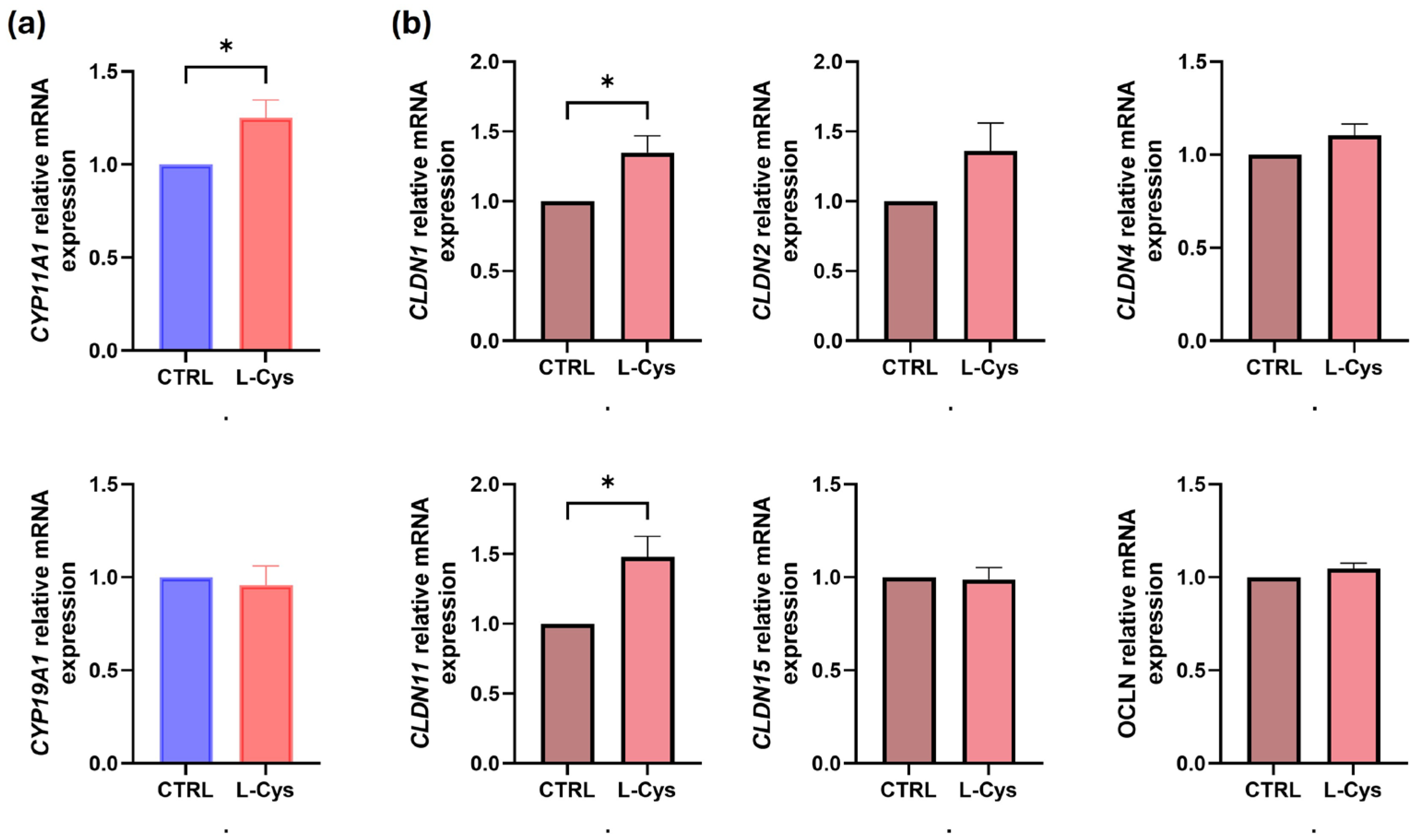
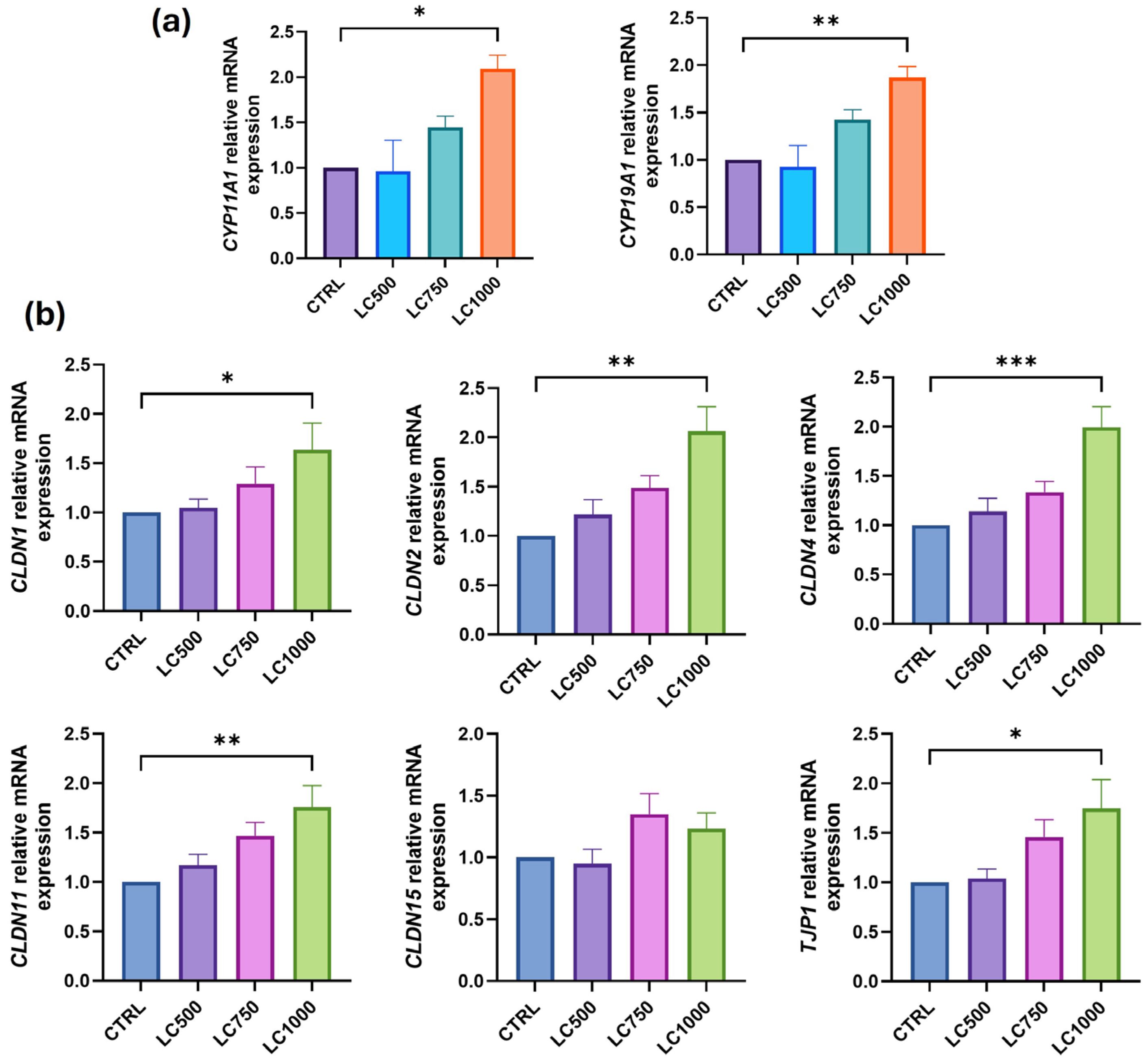
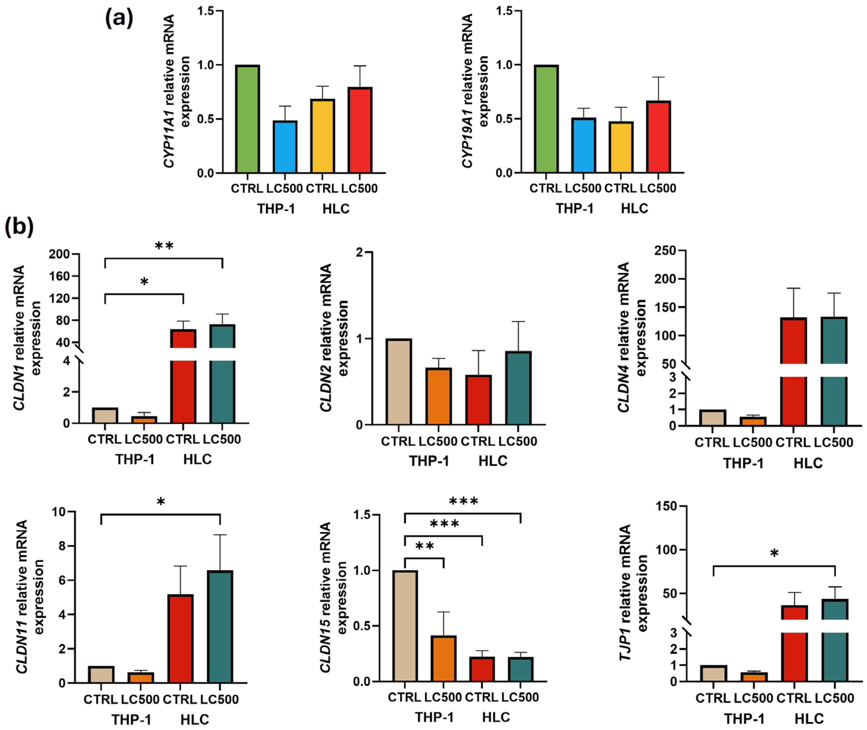
Unless otherwise stated, data are presented as mean ± standard error of the mean (SEM). Data from four independent experiments were cumulated for the analysis. To determine the significant differences between the groups, normally distributed data were compared using the unpaired Student’s t-test. A two-sided p-value ≤ 0.05 was considered statistically significant. Analysis was performed using GraphPad Prism (v10).

3. Results

The human Leydig cells and THP-1 monocytes were treated with L-cysteine for 24 h, followed by measurement of the mRNA expression of testosterone regulatory genes and BTB genes. The L-cysteine treatment did not impact cell viability in either Leydig cells or THP-1 monocytes. L-cysteine (500 µM) significantly upregulated (p = 0.03) the mRNA expression of the CYP11A1 gene in human Leydig cells. Conversely, there was no observed impact on the expression of the CYP19A1 gene (Figure 1a). Additionally, the expression of the BTB genes CLDN1 (p = 0.03), CLDN11 (p = 0.02) and TJP1 (p = 0.02) was upregulated, while no significant changes were noted in the expression of CLDN2, CLDN4, and CLDN15 genes (Figure 1b).
THP-1 monocytes were exposed to varying concentrations of L-cysteine, with the highest concentration (1000 µM) resulting in a significant increase in the expression of CYP11A1 (p = 0.03) and CYP19A1 (p < 0.01) (Figure 2a). Additionally, the expression of BTB genes, CLDN1 (p = 0.04), CLDN2 (p < 0.01), CLDN4 (p < 0.01), CLDN11 (p < 0.01), and TJP1 (p = 0.03) was significantly upregulated after treatment with L-cysteine (1000 µM). However, there was no notable change in the gene expression of CLDN15 (Figure 2b) and lower concentrations of L-cysteine (500 µM and 750 µM) did not impact the gene expression profile.
Gene expression levels were compared between Leydig cells and THP-1 monocytes, revealing higher expression of BTB genes CLDN1 (p < 0.01), CLDN11 (p = 0.03), and TJP1 (p = 0.04) in Leydig cells compared to THP-1 monocytes (Figure 3b). No significant difference was observed in the expression of testosterone-regulatory genes between Leydig cells and THP-1 monocytes (Figure 3a).
The testosterone levels in the human Leydig cells and THP-1 supernatants were measured after treatment with L-cysteine. Figure 4 illustrates that L-cysteine (500 µM) significantly increased (p < 0.01) the testosterone levels in the Leydig cells. However, L-cysteine treatment did not show any effect on the testosterone secretion in the THP-1 monocytes. The untreated Leydig cells comparatively (p < 0.01) secreted more testosterone (2-fold) than the untreated as well as treated THP-1 monocytes.

4. Discussion

Approximately 5 million men in the United States, including 20–50% of men aged 60 and older, experience a significant decrease in their serum testosterone levels [26]. Among couples seeking medical assistance for infertility issues, around 15% of men are affected [27]. Hypogonadism, characterized by low testosterone levels, can affect men of various ages. However, primary testicular deficiency, also known as primary hypogonadism, typically does not lead to a change in serum LH levels. The stimulation of testosterone production in response to LH administration is generally lower in older men than in young men, indicating a decline in the responsiveness of Leydig cells with age [28]. LH administration is often unsuccessful in boosting testosterone levels in men due to its inability to stimulate Leydig cell testosterone production. On the other hand, the administration of exogenous testosterone replacement therapy has proven effective in alleviating symptoms associated with low testosterone; nevertheless, it is important to note that men who undergo this therapy face an elevated risk of stroke, heart attack, and prostate cancer [29].
The present study is the first to investigate the effect of L-cysteine treatment on the relative mRNA expression of the testosterone regulatory genes and testosterone secretion in human Leydig cells and THP-1 monocytes. Leydig cells are specialized endocrine cells essential for male fertility due to their involvement in testosterone production and support of spermatogenesis [19]. These cells are susceptible to oxidative stress from reactive oxygen species (ROS) produced by cytochrome 450 enzymes and have a significant detrimental effect on Leydig cell functions on the steroidogenic pathway. Further, due to their proximity to testicular macrophages, potentially leading to damage and dysfunction, they ultimately play a role in male infertility [30]. Furthermore, Leydig cells play a crucial role in the local immune response by releasing cytokines and chemokines, aiding in the defense against pathogens, and maintaining testicular health [31].
Research indicates that there is an increase in superoxide levels, while the antioxidant defense molecules such as glutathione peroxidase and GSH exhibit a decline in aged Leydig cells [32,33]. Furthermore, various studies have documented age-related declines in GSH levels across multiple biological systems, including human serum and the livers and brains of rats [34,35,36]. A decrease in GSH levels within Leydig cells could lead to a decline in steroid hormone production [15].
L-cysteine is a powerful antioxidant and a potential treatment option to reduce the risk of several disease conditions [37,38,39,40]. L-cysteine supplementation helps to restore glutathione synthesis, safeguards the body against diseases caused by imbalanced redox reactions, and counteracts oxidative stress by activating genes [41,42,43]. L-cysteine is widely used in the development of numerous drugs. However, the number of clinical trials testing the effects of L-cysteine on human health and wellness is small. N-acetyl-L-cysteine (NAC) is commonly used to combat inflammation and oxidative stress both in vivo and in vitro [44]. It has the potential to enhance animal reproduction and reproductive performance as a nutritional supplement by improving placental function and regulating hormone production [45]. Furthermore, NAC provides significant protective effects against busulfan-induced male reproductive impairment, possibly through modification of the Nrf2/HO-1 signaling pathway [46]. Co-supplementation with alpha-lipoic acid and NAC prevented intensive swimming-induced testicular spermatogenic and steroidogenic disorders induced by ROS generation [47].
Our study reports that L-cysteine upregulates the steroidogenic gene CYP11A1 in human Leydig cells and THP-1 monocytes. The overexpression of the CYP11A1 gene resulted in increased testosterone secretion by L-cysteine-treated Leydig cells in comparison to the untreated cells. CYP11A1 plays a vital role in converting cholesterol into other intermediates, which subsequently serve as precursors for the synthesis of steroid hormones [6,48]. Previous studies have documented the decline in levels of CYP11A1 gene expression and testosterone production in aging Leydig cells [49,50]. Despite the decline in steroidogenic enzyme levels in aged cells, the availability of sufficient cholesterol to the inner mitochondrial membrane steroidogenic enzyme CYP11A1 can still result in high testosterone levels [51]. In addition to sexual function, testosterone increases skeletal muscle mass and strength as well as bone mineral density [52]. The supplementation of L-cystine may promote the expression of the CYP11A1 gene, potentially leading to an increase in testosterone synthesis. CYP19A1 catalyzes the final stages of estrogen production and has been identified in the expression of mammalian trophoblasts [53]. Previous studies in male rats supplemented with NAC showed no changes in the expression of the testicular CYP11A1 gene [38]. However, when NAC was administered to rat granulosa cells, it led to an increase in the gene expression of both CYP11A1 and CYP19A1 [54]. A low dose of NAC (1 µM) in porcine placental trophoblast cells upregulated the CYP19A1 gene expression, whereas a high dose of NAC (10 mM) downregulated CYP11A1 mRNA [45]. In this study, treatment with 500 µM and 1000 µM L-cysteine significantly upregulated the expression of the CYP11A1 gene in human Leydig cells and THP-1 cells, respectively.
L-cysteine supplementation resulted in an increase in BTB gene expression in both Leydig cells and THP-1 monocytes. Unexplained male infertility can often be linked to endocrine disturbances in testicular development during the neonatal period, which can be influenced by a combination of environmental pollutants, genetic factors, and epigenetic changes. These factors are linked to the BTB, as they are the main target of different environmental toxins [55]. Endocrine-disrupting compounds are believed to cause testicular damage and affect the integrity of the BTB and the endocrine function of Leydig cells [56,57]. The permeability of the BTB is increased by the presence of nanoparticles and ionizing radiation, which induce structural damage. Consequently, this leads to impaired reproductive cell function and a decline in sperm quality [58,59,60]. In addition to maintaining structural integrity, cell junction proteins facilitate several events in spermatogenesis. The function of the BTB in protecting germ cells is compromised with age due to a decrease in BTB proteins, leading to an inability to block the passage of harmful substances into the seminiferous tubules [61]. NAC attenuates the damage caused to the BTB by SR X-rays [62] and may be used as a preventive measure against iron overload-induced testicular damage [41].
The BTB is a complex structure present in the seminiferous tubules of the mammalian testis [62]. It differs from other tissue barriers in that it is composed of four different cell junctions. The BTB is created by tight junctions (TJs), ectoplasmic specializations, desmosomes, and gap junctions (GJs) [63]. Testosterone secreted by Leydig cells in the presence of LH is required for the maintenance of the BTB, spermatogenesis, and fertility. It also promotes both Sertoli-germ cell junction assembly and disassembly [63]. TJ, which has gate and fence functions, is the most important component of the BTB. The gate function prevents the passage of water, solutes, and other large molecules in the paracellular space (by creating a barrier), whereas the fence function restricts the movement of proteins and lipids between apical and basolateral domains (by generating cell polarity) [63]. Factors such as cytokines and tissue damage affect junctional integrity and could enhance the permeability of endothelial and epithelial barriers [64,65,66]. TJs contain many proteins including claudins [67].
We compared the expression levels of these genes in human Leydig cells and THP-1 cells. Interestingly, the CLDN1, CLDN11, and TJP1 genes showed significantly higher expression levels in HLC than in THP-1 monocytes. This suggests a potential role for these genes in the specific functions and characteristics of Leydig cells, highlighting the importance of further investigation into their regulatory mechanisms and implications in cellular function. The expansion capacity of human Leydig cells is limited, which means that they cannot be grown for a long duration, so we used THP-1 monocytes as an additional model to study the effect of L-cysteine in the regulation of testosterone synthesis. Due to their homogenous genetic background, the THP-1 monocytes facilitate the reproducibility of the findings by minimizing the effect of variability in the cell phenotype [68]. The concentration of L-cysteine used was similar to those found in the bloodstream [69].
The strength of our study is the novel finding that L-cysteine supplementation significantly upregulated the expression of CLDN1, CLDN11, and TJP1 genes in the human Leydig cells, while CLDN1, CLDN2, CLDN4, CLDN11, and TJP1 genes were significantly upregulated in the THP-1 monocytes. Two different cell lines were used to test the hypothesis and compare the expression profile between them. L-cysteine possesses antioxidant properties that can counteract reactive particles, preventing potential damage to cells and tissues [70]. These results indicate that L-cysteine has the potential to serve as a beneficial supplement for controlling the expression of BTB genes, which can mitigate the harmful effects of oxidative stress and hinder the entry of cytotoxic drugs into the seminiferous tubules through the BTB. This has the potential to improve spermatogenesis and the development of germ cells. Clinical studies are required to determine whether L-cysteine co-supplementation can benefit aging men by increasing the integrity of the BTB and boosting testosterone production. If so, co-supplementation with L-cysteine could provide a safer alternative to treatment with testosterone alone, which suppresses LH levels. Being able to explore the development of new therapies to increase intratesticular bioactive androgen levels without any side effects can help reduce infertility. The strength of our study is that we have used Human Leydig cells; however, these cells present significant challenges in terms of growth and culture. Their slow proliferation and restricted expansion capacity resulted in a limited number of cells available for experimentation, leading us to employ a single concentration of L-cysteine (500 µM) for treatment, with untreated Leydig cells serving as a control. Different concentrations of L-cysteine could have provided more comprehensive data. In contrast, THP-1 cells exhibited a more rapid growth rate, allowing for the examination of multiple L-cysteine concentrations. Additionally, a limitation of this study is the absence of concurrent analysis of protein levels in the treated cells.

5. Conclusions

Leydig cells, also known as the interstitial cells of the testes, are found adjacent to the seminiferous tubules in the testicle and are vital for testosterone biosynthesis. Previous studies have documented an association between the decline in levels of CYP11A1 gene expression and testosterone production in aging Leydig cells and infertility. Disruption of any of these interactions can lead to impaired spermatogenesis and ultimately affect male fertility. This study demonstrates that the supplementation of L-cysteine leads to augmented expression of the genes associated with testosterone and BTB in both human Leydig cells and THP-1 monocytes. Supplementation with L-cysteine resulted in upregulation of the testosterone biosynthesis gene and testosterone secretion, which can potentially enhance spermatogenesis (Figure 5). Clinical studies are necessary to investigate the potential advantages of L-cysteine co-supplementation for elderly males, focusing on strengthening the integrity of the blood–testis barrier and elevating testosterone levels to reduce the likelihood of male infertility.

Author Contributions

Conceptualization, S.K.J.; methodology, validation, formal analysis, investigation, J.J.M.; writing—original draft preparation, J.J.M.; writing—review and editing, J.J.M. and S.K.J.; supervision, S.K.J.; funding acquisition, S.K.J. All authors have read and agreed to the published version of the manuscript.

Funding

This research was funded by the National Institute of Health/The National Center for Complementary and Integrative Health (NIH/NCCIH), under grant numbers 5R33AT010637-01A1 and 3R33AT010637-02S1, and the Malcolm Feist Endowed Chair in Diabetes.

Institutional Review Board Statement

Not applicable.

Informed Consent Statement

Not applicable.

Data Availability Statement

The original contributions presented in the study are included in the article; further inquiries can be directed to the corresponding author.

Acknowledgments

The authors thank Georgia Morgan for the excellent editing of this manuscript.

Conflicts of Interest

The authors declare no conflicts of interest.

References

  1. Al-Agha, O.M.; Axiotis, C.A. An in-depth look at Leydig cell tumor of the testis. Arch. Pathol. Lab. Med. 2007, 131, 311–317. [Google Scholar] [CrossRef] [PubMed]
  2. Kumar, S.; Kim, H.J.; Lee, C.H.; Choi, H.S.; Lee, K. Leydig Cell-Specific DAX1-Deleted Mice Has Higher Testosterone Level in the Testis during Pubertal Development. Reprod. Sci. 2022, 29, 955–962. [Google Scholar] [CrossRef] [PubMed]
  3. Aladamat, N.; Tadi, P. Histology, Leydig Cells; StatPearls Publishing: Treasure Island, FL, USA, 2024. [Google Scholar]
  4. Griswold, S.L.; Behringer, R.R. Fetal Leydig cell origin and development. Sex. Dev. 2009, 3, 1–15. [Google Scholar] [CrossRef] [PubMed]
  5. Marinkovic, D.Z.; Medar, M.L.J.; Becin, A.P.; Andric, S.A.; Kostic, T.S. Growing up under Constant Light: A Challenge to the Endocrine Function of the Leydig Cells. Front. Endocrinol. 2021, 12, 653602. [Google Scholar] [CrossRef]
  6. Hu, Y.; Wang, L.; Yang, G.; Wang, S.; Guo, M.; Lu, H.; Zhang, T. VDR promotes testosterone synthesis in mouse Leydig cells via regulation of cholesterol side chain cleavage cytochrome P450 (Cyp11a1) expression. Genes. Genom. 2023, 45, 1377–1387. [Google Scholar] [CrossRef]
  7. Katsumata, N.; Ohtake, M.; Hojo, T.; Ogawa, E.; Hara, T.; Sato, N.; Tanaka, T. Compound heterozygous mutations in the cholesterol side-chain cleavage enzyme gene (CYP11A) cause congenital adrenal insufficiency in humans. J. Clin. Endocrinol. Metab. 2002, 87, 3808–3813. [Google Scholar] [CrossRef]
  8. Richmond, E.J.; Flickinger, C.J.; McDonald, J.A.; Lovell, M.A.; Rogol, A.D. Lipoid congenital adrenal hyperplasia (CAH): Patient report and a mini-review. Clin. Pediatr. 2001, 40, 403–407. [Google Scholar] [CrossRef]
  9. Chen, H.; Ge, R.S.; Zirkin, B.R. Leydig cells: From stem cells to aging. Mol. Cell Endocrinol. 2009, 306, 9–16. [Google Scholar] [CrossRef]
  10. Acikel-Elmas, M.; Algilani, S.A.; Sahin, B.; Bingol Ozakpinar, O.; Gecim, M.; Koroglu, K.; Arbak, S. Apocynin Ameliorates Monosodium Glutamate Induced Testis Damage by Impaired Blood-Testis Barrier and Oxidative Stress Parameters. Life 2023, 13, 822. [Google Scholar] [CrossRef]
  11. Luaces, J.P.; Toro-Urrego, N.; Otero-Losada, M.; Capani, F. What do we know about blood-testis barrier? current understanding of its structure and physiology. Front. Cell Dev. Biol. 2023, 11, 1114769. [Google Scholar] [CrossRef]
  12. Bhat, A.A.; Syed, N.; Therachiyil, L.; Nisar, S.; Hashem, S.; Macha, M.A.; Yadav, S.K.; Krishnankutty, R.; Muralitharan, S.; Al-Naemi, H.; et al. Claudin-1, A Double-Edged Sword in Cancer. Int. J. Mol. Sci. 2020, 21, 569. [Google Scholar] [CrossRef] [PubMed]
  13. Aydin, S.; Billur, D.; Kizil, S.; Ozkavukcu, S.; Topal Celikkan, F.; Aydos, K.; Erdemli, E. Evaluation of blood-testis barrier integrity in terms of adhesion molecules in nonobstructive azoospermia. Andrologia 2020, 52, e13636. [Google Scholar] [CrossRef] [PubMed]
  14. Jiang, X.H.; Bukhari, I.; Zheng, W.; Yin, S.; Wang, Z.; Cooke, H.J.; Shi, Q.H. Blood-testis barrier and spermatogenesis: Lessons from genetically-modified mice. Asian J. Androl. 2014, 16, 572–580. [Google Scholar] [CrossRef] [PubMed]
  15. Chen, H.; Pechenino, A.S.; Liu, J.; Beattie, M.C.; Brown, T.R.; Zirkin, B.R. Effect of glutathione depletion on Leydig cell steroidogenesis in young and old brown Norway rats. Endocrinology 2008, 149, 2612–2619. [Google Scholar] [CrossRef]
  16. Bosgelmez, I.I.; Guvendik, G. Beneficial Effects of N-Acetyl-L-cysteine or Taurine Pre- or Post-treatments in the Heart, Spleen, Lung, and Testis of Hexavalent Chromium-Exposed Mice. Biol. Trace Elem. Res. 2019, 190, 437–445. [Google Scholar] [CrossRef]
  17. Feng, D.; Huang, H.; Yang, Y.; Yan, T.; Jin, Y.; Cheng, X.; Cui, L. Ameliorative effects of N-acetylcysteine on fluoride-induced oxidative stress and DNA damage in male rats’ testis. Mutat. Res. Genet. Toxicol. Environ. Mutagen. 2015, 792, 35–45. [Google Scholar] [CrossRef]
  18. Kemahli, E.; Uyeturk, U.; Cetinkaya, A.; Erimsah, S.; Uyeturk, U.; Gucuk, A. Protective Effects of N-Acetyl Cysteine on Undescended Testis after Orchiopexy: A Rat-model Study. J. Coll. Physicians Surg. Pak. 2023, 33, 319–324. [Google Scholar] [CrossRef]
  19. Abedi, B.; Tayefi-Nasrabadi, H.; Kianifard, D.; Basaki, M.; Shahbazfar, A.A.; Piri, A.; Dolatyarieslami, M. The effect of co-administration of artemisinin and N-acetyl cysteine on antioxidant status, spermatological parameters and histopathology of testis in adult male mice. Horm. Mol. Biol. Clin. Investig. 2023, 44, 207–214. [Google Scholar] [CrossRef]
  20. Acer-Demir, T.; Mammadov, M.; Ocbe, P.; Coruhlu, A.; Coskun, D.; Nazik, Y.; Tufekci, I.; Guney, L.H.; Hicsonmez, A. The long term effects of intrascrotal low dose and high dose N-acetylcysteine on testis damage in rat model of testicular torsion. J. Pediatr. Surg. 2020, 55, 672–680. [Google Scholar] [CrossRef]
  21. Bodur, A.; Alver, A.; Kahraman, C.; Altay, D.U.; Ince, I. Investigation of N-acetylcysteine on contralateral testis tissue injury by experimental testicular torsion: Long-term effect. Am. J. Emerg. Med. 2016, 34, 1069–1074. [Google Scholar] [CrossRef]
  22. Jannatifar, R.; Parivar, K.; Roodbari, N.H.; Nasr-Esfahani, M.H. Effects of N-acetyl-cysteine supplementation on sperm quality, chromatin integrity and level of oxidative stress in infertile men. Reprod. Biol. Endocrinol. 2019, 17, 24. [Google Scholar] [CrossRef] [PubMed]
  23. Monageng, E.; Offor, U.; Takalani, N.B.; Mohlala, K.; Opuwari, C.S. A Review on the Impact of Oxidative Stress and Medicinal Plants on Leydig Cells. Antioxidants 2023, 12, 1559. [Google Scholar] [CrossRef]
  24. Manna, P.; Jain, S.K. L-cysteine and hydrogen sulfide increase PIP3 and AMPK/PPARgamma expression and decrease ROS and vascular inflammation markers in high glucose treated human U937 monocytes. J. Cell Biochem. 2013, 114, 2334–2345. [Google Scholar] [CrossRef] [PubMed]
  25. Kanikarla-Marie, P.; Jain, S.K. L-Cysteine supplementation reduces high-glucose and ketone-induced adhesion of monocytes to endothelial cells by inhibiting ROS. Mol. Cell Biochem. 2014, 391, 251–256. [Google Scholar] [CrossRef] [PubMed]
  26. Surampudi, P.N.; Wang, C.; Swerdloff, R. Hypogonadism in the aging male diagnosis, potential benefits, and risks of testosterone replacement therapy. Int. J. Endocrinol. 2012, 2012, 625434. [Google Scholar] [CrossRef]
  27. Hwang, K.; Walters, R.C.; Lipshultz, L.I. Contemporary concepts in the evaluation and management of male infertility. Nat. Rev. Urol. 2011, 8, 86–94. [Google Scholar] [CrossRef]
  28. Zirkin, B.R.; Papadopoulos, V. Leydig cells: Formation, function, and regulation. Biol. Reprod. 2018, 99, 101–111. [Google Scholar] [CrossRef]
  29. Bosland, M.C. Testosterone treatment is a potent tumor promoter for the rat prostate. Endocrinology 2014, 155, 4629–4633. [Google Scholar] [CrossRef]
  30. Diemer, T.; Allen, J.A.; Hales, K.H.; Hales, D.B. Reactive oxygen disrupts mitochondria in MA-10 tumor Leydig cells and inhibits steroidogenic acute regulatory (StAR) protein and steroidogenesis. Endocrinology 2003, 144, 2882–2891. [Google Scholar] [CrossRef]
  31. Zhang, Y.F.; Su, P.K.; Wang, L.J.; Zheng, H.Q.; Bai, X.F.; Li, P.; Meng, X.P.; Yang, J.Y. T-2 toxin induces apoptosis via the Bax-dependent caspase-3 activation in mouse primary Leydig cells. Toxicol. Mech. Methods 2018, 28, 23–28. [Google Scholar] [CrossRef]
  32. Luo, L.; Chen, H.; Trush, M.A.; Show, M.D.; Anway, M.D.; Zirkin, B.R. Aging and the brown Norway rat leydig cell antioxidant defense system. J. Androl. 2006, 27, 240–247. [Google Scholar] [CrossRef] [PubMed]
  33. Chen, H.; Cangello, D.; Benson, S.; Folmer, J.; Zhu, H.; Trush, M.A.; Zirkin, B.R. Age-related increase in mitochondrial superoxide generation in the testosterone-producing cells of Brown Norway rat testes: Relationship to reduced steroidogenic function? Exp. Gerontol. 2001, 36, 1361–1373. [Google Scholar] [CrossRef] [PubMed]
  34. Jones, D.P.; Mody, V.C., Jr.; Carlson, J.L.; Lynn, M.J.; Sternberg, P., Jr. Redox analysis of human plasma allows separation of pro-oxidant events of aging from decline in antioxidant defenses. Free Radic. Biol. Med. 2002, 33, 1290–1300. [Google Scholar] [CrossRef] [PubMed]
  35. Sandhu, S.K.; Kaur, G. Alterations in oxidative stress scavenger system in aging rat brain and lymphocytes. Biogerontology 2002, 3, 161–173. [Google Scholar] [CrossRef]
  36. Liu, R.M. Down-regulation of gamma-glutamylcysteine synthetase regulatory subunit gene expression in rat brain tissue during aging. J. Neurosci. Res. 2002, 68, 344–351. [Google Scholar] [CrossRef]
  37. Larsson, S.C.; Hakansson, N.; Wolk, A. Dietary cysteine and other amino acids and stroke incidence in women. Stroke 2015, 46, 922–926. [Google Scholar] [CrossRef]
  38. Doosti, A.; Lotfi, Y.; Moossavi, A.; Bakhshi, E.; Talasaz, A.H.; Hoorzad, A. Comparison of the effects of N-acetyl-cysteine and ginseng in prevention of noise induced hearing loss in male textile workers. Noise Health 2014, 16, 223–227. [Google Scholar] [CrossRef]
  39. Xu, Y.J.; Tappia, P.S.; Neki, N.S.; Dhalla, N.S. Prevention of diabetes-induced cardiovascular complications upon treatment with antioxidants. Heart Fail. Rev. 2014, 19, 113–121. [Google Scholar] [CrossRef]
  40. Salaspuro, V.; Hietala, J.; Kaihovaara, P.; Pihlajarinne, L.; Marvola, M.; Salaspuro, M. Removal of acetaldehyde from saliva by a slow-release buccal tablet of L-cysteine. Int. J. Cancer 2002, 97, 361–364. [Google Scholar] [CrossRef]
  41. Ezzat, G.M.; Nassar, A.Y.; Bakr, M.H.; Mohamed, S.; Nassar, G.A.; Kamel, A.A. Acetylated Oligopeptide and N-acetyl cysteine Protected Against Oxidative Stress, Inflammation, Testicular-Blood Barrier Damage, and Testicular Cell Death in Iron-Overload Rat Model. Appl. Biochem. Biotechnol. 2023, 195, 5053–5071. [Google Scholar] [CrossRef]
  42. Achari, A.E.; Jain, S.K. l-Cysteine supplementation increases insulin sensitivity mediated by upregulation of GSH and adiponectin in high glucose treated 3T3-L1 adipocytes. Arch. Biochem. Biophys. 2017, 630, 54–65. [Google Scholar] [CrossRef] [PubMed]
  43. Jain, S.K.; Micinski, D.; Parsanathan, R. l-Cysteine Stimulates the Effect of Vitamin D on Inhibition of Oxidative Stress, IL-8, and MCP-1 Secretion in High Glucose Treated Monocytes. J. Am. Coll. Nutr. 2021, 40, 327–332. [Google Scholar] [CrossRef] [PubMed]
  44. Williams, L.; Burgos, E.S.; Vuguin, P.M.; Manuel, C.R.; Pekson, R.; Munnangi, S.; Reznik, S.E.; Charron, M.J. N-Acetylcysteine Resolves Placental Inflammatory-Vasculopathic Changes in Mice Consuming a High-Fat Diet. Am. J. Pathol. 2019, 189, 2246–2257. [Google Scholar] [CrossRef] [PubMed]
  45. Ding, H.; Yang, Y.; Wei, S.; Spicer, L.J.; Kenez, A.; Xu, W.; Liu, Y.; Feng, T. Influence of N-acetylcysteine on steroidogenesis and gene expression in porcine placental trophoblast cells. Theriogenology 2021, 161, 49–56. [Google Scholar] [CrossRef] [PubMed]
  46. Kim, K.H.; Park, M.J.; Park, N.C.; Park, H.J. Effect of N-acetyl-L-cysteine on Testicular Tissue in Busulfan-Induced Dysfunction in the Male Reproductive System. World J. Mens. Health 2023, 41, 882–891. [Google Scholar] [CrossRef]
  47. Jana, K.; Dutta, A.; Chakraborty, P.; Manna, I.; Firdaus, S.B.; Bandyopadhyay, D.; Chattopadhyay, R.; Chakravarty, B. Alpha-lipoic acid and N-acetylcysteine protects intensive swimming exercise-mediated germ-cell depletion, pro-oxidant generation, and alteration of steroidogenesis in rat testis. Mol. Reprod. Dev. 2014, 81, 833–850. [Google Scholar] [CrossRef]
  48. Redouane, S.; Harmak, H.; Elkarhat, Z.; Charoute, H.; Malki, A.; Barakat, A.; Rouba, H. Exploring the impact of CYP11A1’s missense SNPs on the interaction between CYP11A1 and cholesterol: A comprehensive structural analysis and MD simulation study. Comput. Biol. Chem. 2023, 106, 107937. [Google Scholar] [CrossRef]
  49. Chen, H.; Hardy, M.P.; Huhtaniemi, I.; Zirkin, B.R. Age-related decreased Leydig cell testosterone production in the brown Norway rat. J. Androl. 1994, 15, 551–557. [Google Scholar] [CrossRef]
  50. Wang, C.; Leung, A.; Sinha-Hikim, A.P. Reproductive aging in the male brown-Norway rat: A model for the human. Endocrinology 1993, 133, 2773–2781. [Google Scholar] [CrossRef]
  51. Culty, M.; Luo, L.; Yao, Z.X.; Chen, H.; Papadopoulos, V.; Zirkin, B.R. Cholesterol transport, peripheral benzodiazepine receptor, and steroidogenesis in aging Leydig cells. J. Androl. 2002, 23, 439–447. [Google Scholar] [CrossRef]
  52. Corona, G.; Rastrelli, G.; Di Pasquale, G.; Sforza, A.; Mannucci, E.; Maggi, M. Testosterone and Cardiovascular Risk: Meta-Analysis of Interventional Studies. J. Sex. Med. 2018, 15, 820–838. [Google Scholar] [CrossRef] [PubMed]
  53. Noyola-Martinez, N.; Halhali, A.; Zaga-Clavellina, V.; Olmos-Ortiz, A.; Larrea, F.; Barrera, D. A time-course regulatory and kinetic expression study of steroid metabolizing enzymes by calcitriol in primary cultured human placental cells. J. Steroid Biochem. Mol. Biol. 2017, 167, 98–105. [Google Scholar] [CrossRef] [PubMed]
  54. Tripathi, A.; Pandey, V.; Sahu, A.N.; Singh, A.; Dubey, P.K. Di-(2-ethylhexyl) phthalate (DEHP) inhibits steroidogenesis and induces mitochondria-ROS mediated apoptosis in rat ovarian granulosa cells. Toxicol. Res. 2019, 8, 381–394. [Google Scholar] [CrossRef] [PubMed]
  55. Dankers, A.C.; Roelofs, M.J.; Piersma, A.H.; Sweep, F.C.; Russel, F.G.; van den Berg, M.; van Duursen, M.B.; Masereeuw, R. Endocrine disruptors differentially target ATP-binding cassette transporters in the blood-testis barrier and affect Leydig cell testosterone secretion in vitro. Toxicol. Sci. 2013, 136, 382–391. [Google Scholar] [CrossRef]
  56. Marettova, E.; Maretta, M.; Legath, J. Toxic effects of cadmium on testis of birds and mammals: A review. Anim. Reprod. Sci. 2015, 155, 1–10. [Google Scholar] [CrossRef]
  57. Shen, Y.; You, Y.; Zhu, K.; Fang, C.; Yu, X.; Chang, D. Bibliometric and visual analysis of blood-testis barrier research. Front. Pharmacol. 2022, 13, 969257. [Google Scholar] [CrossRef]
  58. Son, Y.; Heo, K.; Bae, M.J.; Lee, C.G.; Cho, W.S.; Kim, S.D.; Yang, K.; Shin, I.S.; Lee, M.Y.; Kim, J.S. Injury to the blood-testis barrier after low-dose-rate chronic radiation exposure in mice. Radiat. Prot. Dosim. 2015, 167, 316–320. [Google Scholar] [CrossRef]
  59. Lu, Y.; Liu, M.; Tursi, N.J.; Yan, B.; Cao, X.; Che, Q.; Yang, N.; Dong, X. Uropathogenic Escherichia coli Infection Compromises the Blood-Testis Barrier by Disturbing mTORC1-mTORC2 Balance. Front. Immunol. 2021, 12, 582858. [Google Scholar] [CrossRef]
  60. Wang, R.; Song, B.; Wu, J.; Zhang, Y.; Chen, A.; Shao, L. Potential adverse effects of nanoparticles on the reproductive system. Int. J. Nanomed. 2018, 13, 8487–8506. [Google Scholar] [CrossRef]
  61. Paul, C.; Robaire, B. Impaired function of the blood-testis barrier during aging is preceded by a decline in cell adhesion proteins and GTPases. PLoS ONE 2013, 8, e84354. [Google Scholar] [CrossRef]
  62. Zhang, T.; Liu, T.; Shao, J.; Sheng, C.; Hong, Y.; Ying, W.; Xia, W. Antioxidant protects blood-testis barrier against synchrotron radiation X-ray-induced disruption. Spermatogenesis 2015, 5, e1009313. [Google Scholar] [CrossRef] [PubMed]
  63. Mruk, D.D.; Cheng, C.Y. The Mammalian Blood-Testis Barrier: Its Biology and Regulation. Endocr. Rev. 2015, 36, 564–591. [Google Scholar] [CrossRef] [PubMed]
  64. Liu, T.; Zhang, T.; Yu, H.; Shen, H.; Xia, W. Adjudin protects against cerebral ischemia reperfusion injury by inhibition of neuroinflammation and blood-brain barrier disruption. J. Neuroinflamm. 2014, 11, 107. [Google Scholar] [CrossRef] [PubMed]
  65. Bruewer, M.; Luegering, A.; Kucharzik, T.; Parkos, C.A.; Madara, J.L.; Hopkins, A.M.; Nusrat, A. Proinflammatory cytokines disrupt epithelial barrier function by apoptosis-independent mechanisms. J. Immunol. 2003, 171, 6164–6172. [Google Scholar] [CrossRef] [PubMed]
  66. Capaldo, C.T.; Nusrat, A. Cytokine regulation of tight junctions. Biochim. Biophys. Acta 2009, 1788, 864–871. [Google Scholar] [CrossRef]
  67. Liman, N. The abundance and localization of claudin-1 and -5 in the adult tomcats (Felis catus) testis, tubules rectus, rete testis, efferent ductules, and epididymis. Anat. Rec. 2023, 306, 2153–2169. [Google Scholar] [CrossRef]
  68. Chanput, W.; Mes, J.J.; Wichers, H.J. THP-1 cell line: An in vitro cell model for immune modulation approach. Int. Immunopharmacol. 2014, 23, 37–45. [Google Scholar] [CrossRef]
  69. Wang, N.; Chen, M.; Gao, J.; Ji, X.; He, J.; Zhang, J.; Zhao, W. A series of BODIPY-based probes for the detection of cysteine and homocysteine in living cells. Talanta 2019, 195, 281–289. [Google Scholar] [CrossRef]
  70. Clemente Plaza, N.; Reig Garcia-Galbis, M.; Martinez-Espinosa, R.M. Effects of the Usage of l-Cysteine (l-Cys) on Human Health. Molecules 2018, 23, 575. [Google Scholar] [CrossRef]
Figure 1. (a) Testosterone and (b) BTB relative gene expression after human Leydig cells were treated with 500 μM of L-cysteine. * p < 0.05.
Biomolecules 14 01171 g001
Figure 2. (a) Testosterone and (b) BTB relative gene expression after THP-1 monocytes were treated with different concentrations of L-cysteine (500–1000 μM). * p < 0.05; ** p < 0.01; *** p < 0.001.
Biomolecules 14 01171 g002
Figure 3. Comparison of (a) testosterone regulatory and (b) BTB relative gene expression among human Leydig cells and THP-1 after treatment with L-cysteine (500 μM). * p < 0.05; ** p < 0.01; *** p < 0.001.
Biomolecules 14 01171 g003
Figure 4. Testosterone concentration in the cell culture supernatant after L-cysteine treatment in human Leydig cells and THP-1 monocytes. ** p < 0.01; *** p < 0.001.
Biomolecules 14 01171 g004
Figure 5. Summary diagram illustrating that L-cysteine supplementation increased the gene expression of testosterone biosynthesis and BTB genes and enhanced testosterone secretion.
Biomolecules 14 01171 g005
Table 1. List of primers used for qPCR analysis.
GeneOrientationPrimer Sequence (5′–3′)
CYP11A1ForwardCGTCAGATCCATCGGGTTAATG
ReverseCATTCCAACCATCCAGGTATCG
CYP19A1ForwardGAGAACCAGGCTACAAGAGAAA
ReverseTGGTGGAATCGGGTCTTTATG
CLDN1ForwardCCAGTTAGAAGAGGTAGTGTGAAT
ReverseCAGCCAGCTGAGCAAATAAAG
CLDN2ForwardGGTGACATCCAGTGCAATCT
ReverseCTACCGCCACTCTGTCTTTG
CLDN4ForwardCAACTGCCTGGAGGATGAAA
ReverseCACCGGCACTATCACCATAAG
CLDN11ForwardGTGTGATCTCGGCTCATGTA
ReverseGTAGTAGTGAACGCCTGTAGTC
CLDN15ForwardTGACTCTGCCAAACAGCTAC
ReverseGTCGGTGGCACAGCTAAA
TJP1ForwardCCTTAGTGTCCAAACCAGACC
ReverseAAAGCTGCCTGAGCAGTATC
OCLNForwardCATTGCCATCTTTGCCTGTG
ReverseCCAAAGCCACTTCCTCCATAA
GAPDHForwardAGTATGACAACAGCCTCAAGAT
ReverseGTCCTTCCACGATACCAAA
Disclaimer/Publisher’s Note: The statements, opinions and data contained in all publications are solely those of the individual author(s) and contributor(s) and not of MDPI and/or the editor(s). MDPI and/or the editor(s) disclaim responsibility for any injury to people or property resulting from any ideas, methods, instructions or products referred to in the content.

