

Biosynthesized Chitosan-Coated Silver Nanoparticles: Insecticide Activity and Sublethal Effects Against Drosophila suzukii (Diptera: Drosophilidae)
 ,
,  , ,
, ,  ,
,
Abstract
1. Introduction
2. Materials and Methods
2.1. Plant Collection
2.2. Biosynthesis of Silver Nanoparticles
2.3. Biosynthesis of Chitosan-Coated Silver Nanoparticles
2.4. Characterization of Biosynthesized AgChNPs
2.5. Rearing of SWD
2.6. Insecticidal Effect of Biosynthesized AgChNPs
2.6.1. Chronic Toxicity
Larvicidal Bioassay
Pupicidal Activity
2.6.2. Acute Toxicity
Adulticidal Bioassay
2.7. Statistical Analysis
3. Results and Discussion
3.1. Characterization of AgChNPs
3.1.1. UV-Vis of AgChNPs
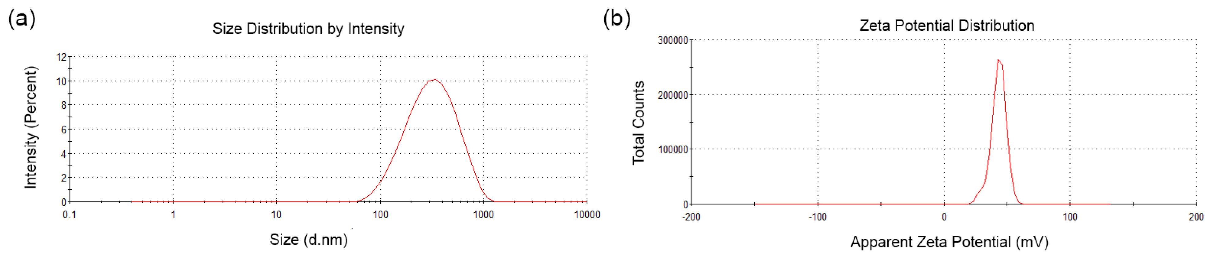
3.1.2. Hydrodynamic Diameter (nm), PDI, and Z Potential
3.1.3. X-Ray Diffraction (XRD) of AgChNPs
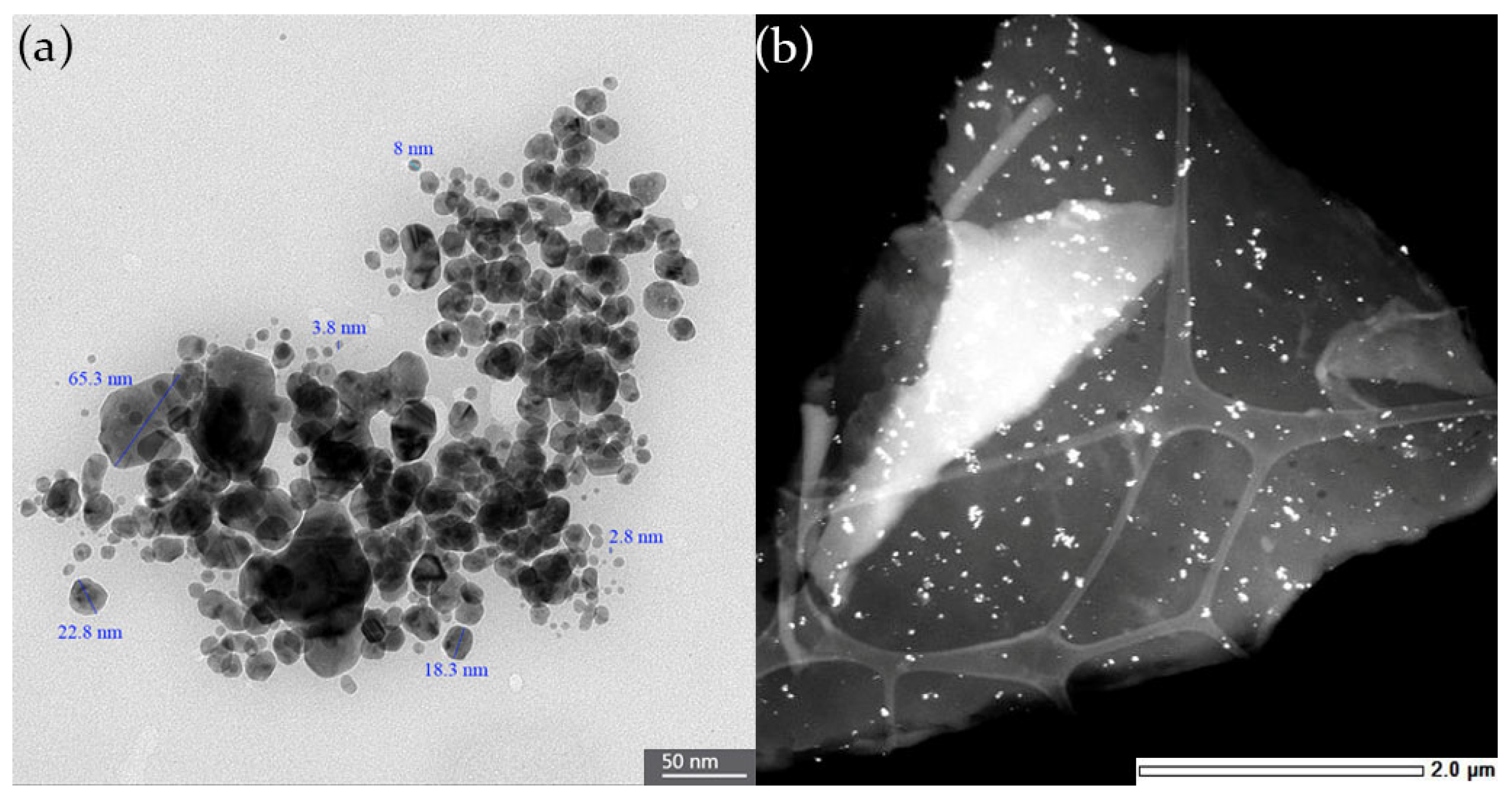
3.1.4. TEM Analysis of AgChNPs
3.2. Chronic Toxicity
3.2.1. Larvicidal Bioassay
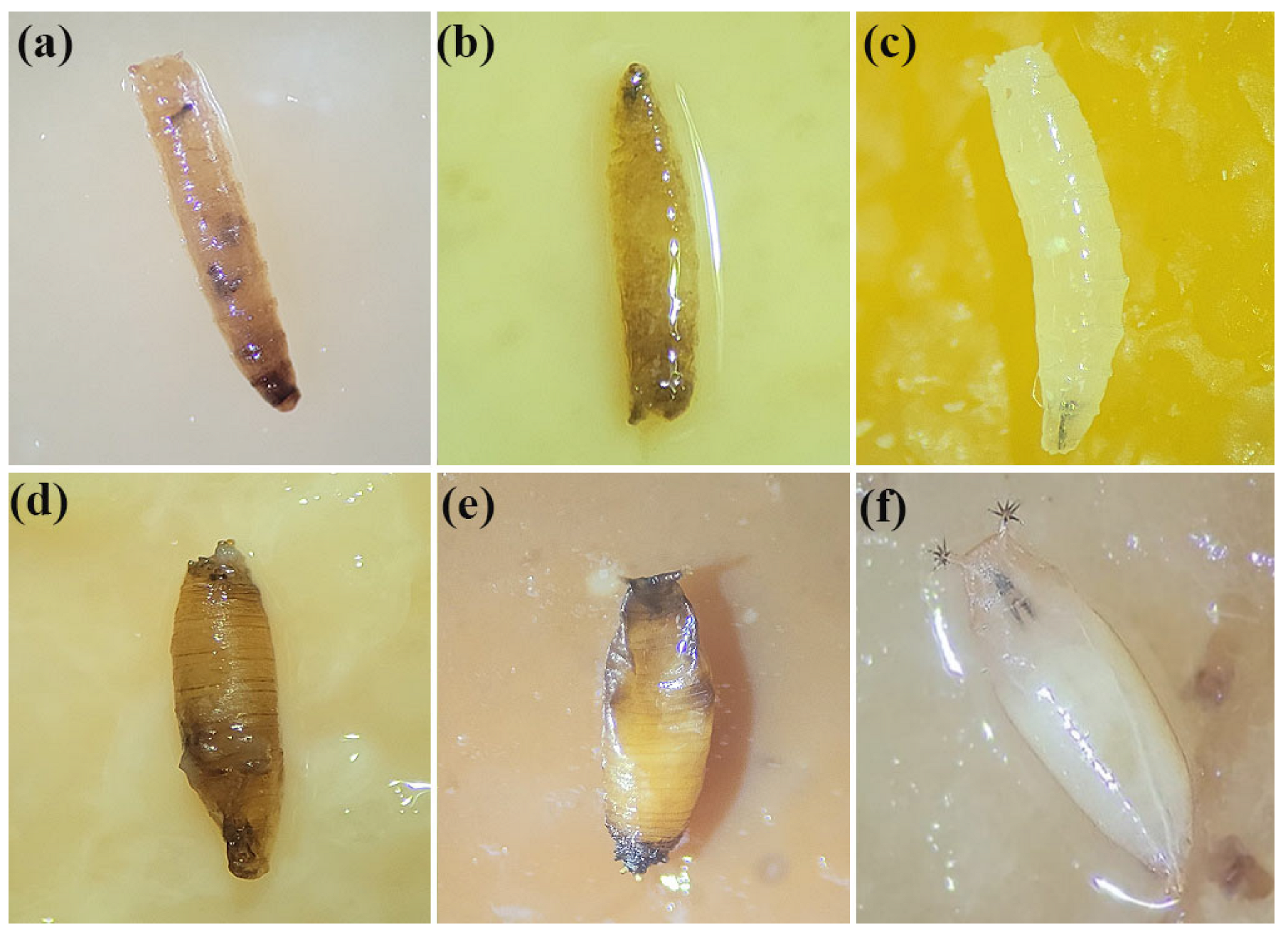
3.2.2. Pupicidal Activity
3.2.3. Adult Flies Emerged
3.3. Acute Toxicity
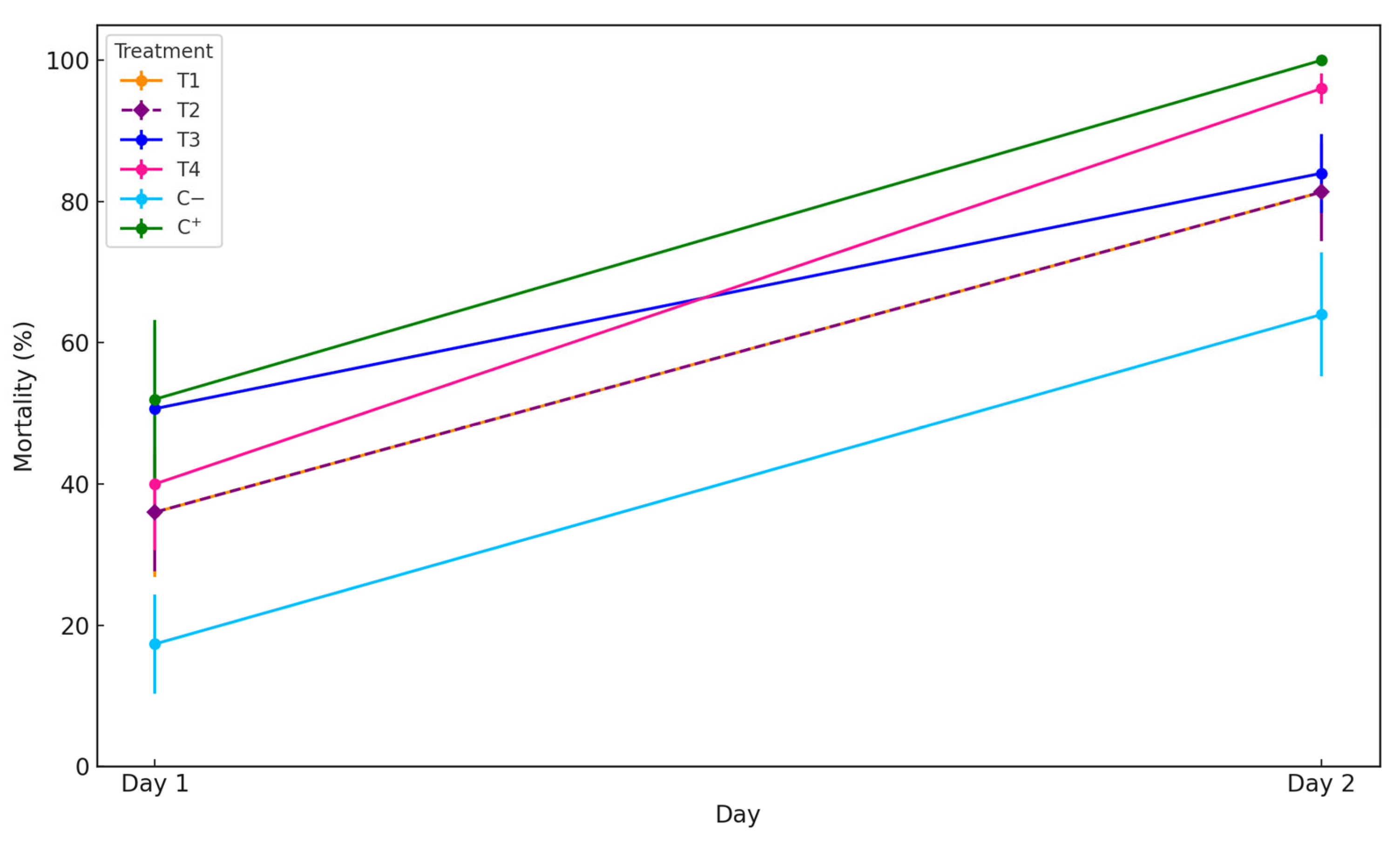
Adulticidal Bioassay
4. Conclusions
Author Contributions
Funding
Institutional Review Board Statement
Informed Consent Statement
Data Availability Statement
Conflicts of Interest
References
- Xiao, Z.; Yin, K.; Geng, L.; Wu, J.; Zhang, F.; Liu, Y. Pest identification via hyperspectral image and deep learning. Signal Image Video Process 2022, 16, 873–880. [Google Scholar] [CrossRef]
- Chagas, M.; Rakes, M.; Pasini, R.A.; Grützmacher, A.D.; Nava, D.E.; Bernardi, D. Toxicity and Transgeneracional effects of insecticides on Trichopria anastrephae (Hymenoptera: Diapriidae). Neotropical Èntomol. 2021, 51, 143–150. [Google Scholar] [CrossRef]
- McIntosh, H.; Atucha, A.; Townsend, P.A.; Hills, W.B.; Guédot, C. Plastic mulches reduce adult and larval populations of Drosophila suzukii in fall-bearing raspberry. J. Insect Sci. 2022, 95, 525–536. [Google Scholar] [CrossRef]
- Kamiyama, M.; Guédot, C. Varietal and Developmental Susceptibility of Tart Cherry (Rosales: Rosaceae) to Drosophila suzukii (Diptera: Drosophilidae). J. Econ. Entomol. 2019, 112, 1789–1797. [Google Scholar] [CrossRef] [PubMed]
- SENASICA. Mosca del Vinagre de alas Manchadas Drosophila suzukii Matsumura; Ficha técnica No. 7; SADER: Mexico City, Mexico, 2015; ISBN 978-607-715-117-3. [Google Scholar]
- Lizama, M.; Alves-Santos, F.; Navas-Gracia, L.; Martínez-Cisterna, D.; Medina, C.; Rebolledo, R.; Chacón-Fuentes, M.; Bardehle, L. The use of novel alginate capsules in a monitoring system for Drosophila suzukii in a cherry orchard in the Region of La Araucanía, Chile. Insects 2025, 16, 13. [Google Scholar] [CrossRef]
- SAG. Resolución Exenta N°: 1943. Medidas Fitosanitarias De Emergencia Provisionales Para La Plaga Drosófila De Alas Manchadas- Drosophila Suzukii (Matsumura). Diptera: Drosophila. 2019. Available online: https://bcn.cl/pO6fjY (accessed on 23 March 2025).
- Bizama, G. Invasión de Drosophila suzukii (Matsumura) en Chile: Utilizando los modelos de distribución de especies como herramienta de bioseguridad. Rev. Chil. Entomol. 2020, 46, 61–71. [Google Scholar] [CrossRef]
- Buzzetti, K. The spotted wing Drosophila in the South of the World: Chilean case and its first productive impacts. In Drosophila suzukii Management; Van Timmeren, S., Ed.; IntechOpen: London, UK, 2020; pp. 1–11. [Google Scholar] [CrossRef]
- Guillickson, M.; Rogers, M.; Burkness, E.C.; Hutchison, W.D. Efficacy of organic and conventional insecticides for Drosophila suzukii when combined with erythritol, a non-nutritive feeding stimulant. Crop. Prot. 2019, 125, 104878. [Google Scholar] [CrossRef]
- Saleem, S.; Solanki, B.; Khan, M. Beyond agrochemicals: Potential of nanoparticles as nanofertilizer and nanopesticide in legumes. Theor. Exp. Plant. Physiol. 2025, 37, 7. [Google Scholar] [CrossRef]
- Vurro, M.; Miguel-Rojas, C.; Pérez, A. Safe nanotechnologies for increasing the effectiveness of environmentally friendly natural agrochemicals. Pest. Manag. Sci. 2019, 75, 2403–2412. [Google Scholar] [CrossRef]
- Armstrong, N.; Ramamoorthy, M.; Lyon, D.; Jones, K.; Duttaroy, A. Mechanism of Silver Nanoparticles Action on Insect Pigmentation Reveals Intervention of Copper Homeostasis. PLoS ONE 2013, 8, e53186. [Google Scholar] [CrossRef]
- Thamilarasan, V.; Sethuraman, V.; Gopinath, K.; Balalakshmi, C.; Govindarajan, M.; Mothana, R.A.; Siddiqui, N.A.; Khaled, J.M.; Benelli, G. Single Step Fabrication of Chitosan Nanocrystals Using Penaeus semisulcatus: Potential as New Insecticides, Antimicrobials and Plant Growth Promoters. J. Clust. Sci. 2018, 29, 375–384. [Google Scholar] [CrossRef]
- Abdoon, F.M.; Hasan, M.; Salman, S.; Ameen, S.; Birhan, M. Exploiting of green synthesized silver nanoparticles using Capparis spinosa L. fruit for spectrophotometric determination of diphendydramine HCL in pure forms and commercial products. J. Exp. Nano 2023, 18, 2161525. [Google Scholar] [CrossRef]
- Akyüz, G.; Kaymazlar, E.; Ay, H.; Andaç, M.; Andaç, Ö. Use of silver nanoparticles loaded locust bean gum coatings to extend the shelf-life of fruits. Biointerface Res. Appl. Chem. 2023, 13, 289. [Google Scholar] [CrossRef]
- Manosalva, N.; Tortella, G.; Diez, M.C.; Schalchli, H.; Seabra, A.B.; Durán, N.; Rubilar, O. Green synthesis of silver nanoparticles: Efect of synthesis reaction parameters on antimicrobial activity. World J. Microbiol. Biotechnol. 2019, 35, 88. [Google Scholar] [CrossRef] [PubMed]
- Mishra, V.K.; Husen, A.; Rahman, Q. Plant-based fabrication of silver nanoparticles and their application. Nanomater. Plant. Potential. 2019, 5, 135–175. [Google Scholar] [CrossRef]
- Rastogi, K.; Vashishtha, R.; Shaloo; Dan, S. Scientific advances and Pharmacological Applications of Marine Derived-Collagen and Chitosan. Biointerface Res. Appl. Chem. 2022, 12, 3540–3558. [Google Scholar] [CrossRef]
- Asghar, M.A.; Yousuf, R.; Shoaib, M.; Asghar, M.A. Synergistic nanocomposites of different antibiotics coupled with green synthesized chitosan-based silver nanoparticles: Characterization, Antibacterial, in vivo Toxicological and Biodistribution Studies. Int. J. Nanomed. 2020, 15, 7841–7859. [Google Scholar] [CrossRef]
- Martínez-Cisterna, D.; Rubilar, O.; Tortella, G.; Chen, L.; Chacón-Fuentes, M.; Lizama, M.; Parra, P.; Bardehle, L. Silver nanoparticles as a potent nanopesticide: Toxic effects and mechanisms of action—A review. Molecules 2024, 29, 5520. [Google Scholar] [CrossRef]
- Kamaraj, C.; Rajakumar, G.; Rahuman, A.A.; Velayutham, K.; Bagavan, A.; Zahir, A.A.; Elango, G. Feeding deterrent activity of synthesized silver nanoparticles using manilkara zapota leaf extract against the house fly, Musca domestica (Diptera: Muscidae). Parasitol. Res. 2012, 111, 2439–2448. [Google Scholar] [CrossRef]
- Panacek, A.; Prucek, R.; Safavora, D.; Dittrich, M.; Richtrova, J.; Benickova, K.; Zboril, R.; Kvitek, L. Acute and Chronic Toxicity Effects of Silver Nanoparticles (Nps) on Drosophila melanogaster. Environ. Sci. Technol. 2011, 45, 4974–4979. [Google Scholar] [CrossRef]
- Posgai, R.; Ahamed, M.; Hussain, S.M.; Rowe, J.; Nielsen, M. Inhalation method for delivery of nanoparticles to the Drosophila respiratory system for toxicity testing. Sci. Total Environ. 2009, 408, 439–443. [Google Scholar] [CrossRef] [PubMed]
- Demir, E.; Vales, G.; Kaya, B.; Creus, A.; Marcos, R. Genotoxic analisis of silver nanoparticles in Drosophila. Nanotoxicology 2011, 5, 417–424. [Google Scholar] [CrossRef] [PubMed]
- Wulandari, I.; Pebriatin, B.; Valiana, V.; Hadisaputra, S.; Ananto, A.D.; Sabarudin, A. Green Synthesis of Silver Nanoparticles Coated by Water Soluble Chitosan and Its Potency as Non-Alcoholic Hand Sanitizer Formulation. Materials 2022, 15, 4641. [Google Scholar] [CrossRef]
- Kalajdzic, P.; Schetelig, M.F. CRISPR/Cas-mediated gene editing using purified protein in Drosophila suzukii. Entomol. Exp. Appl. 2017, 164, 350–362. [Google Scholar] [CrossRef]
- Dalton, D.T.; Walton, V.M.; Shearer, P.W.; Walsh, D.B.; Caprile, J.; Isaacs, R. Laboratory survival of Drosophila suzukii under simulated winter conditions of the pacific northwest and seasonal field trapping in five primary regions of small and stone fruit production in the United States. Pest. Manag. Sci. 2011, 67, 1368–1374. [Google Scholar] [CrossRef]
- Abbot, W. A method of computing the effectiveness of an insecticide. J. Econ. Entomol 1925, 18, 265–267. [Google Scholar] [CrossRef]
- Zare-Bidake, M.; Mohammadparast-Tabas, P.; Khorashadizade, M.; Mohammadparast-Tabas, P.; Alemzadeh, E.; Saberi, A.; Kabiri-Rad, H.; Eghbali, S. Bio-synthesized AGS@AgNPs for wound healing, antioxidant support, antibacterial defense, and anticancer intervention. Biocatal. Agric. Biotechnol. 2024, 61, 103402. [Google Scholar] [CrossRef]
- Cortés, Y.Z.; Valenzuela, L.M.; Peña, E.A.; Sánchez, B.L. Antibacterial Activity of Electrospun Nanocomposites Fabricated by In situ Chitosan/Silver Nanoparticles. IEEE Trans. Nanobiosci. 2022, 21, 89–96. [Google Scholar] [CrossRef]
- Subramaniam, B.; Siddik, Z.; Nagoor, N. Optimization of nanostructured lipid carriers: Understanding the types, designs, and parameters in the process of formulations. J. Nanopart. Res. 2020, 22, 141. [Google Scholar] [CrossRef]
- Karuppaiah, A.; Babu, D.; Servaraj, D.; Natrajan, T.; Rajan, R.; Gautam, M.; Ranganathan, H.; Siram, K.; Nesamony, J.; Sankar, V. Building and behavior of a pH-stimuli responsive chitosan nanoparticles loaded with folic acid conjugated gemcitabine silver colloids in MDA-MB-453 metastatic breast cancer cell line and pharmacokinetics in rats. Eur. J. Pharm. Sci. 2021, 165, 105938. [Google Scholar] [CrossRef]
- Bilal, M.; Rasheed, T.; Iqbal, H.; Lib, C.; Hu, H.; Zhang, X. Development of silver nanoparticles loaded chitosan-alginate constructs with biomedical potentialities. Int. J. Biol. Macromol. 2017, 105, 393–400. [Google Scholar] [CrossRef] [PubMed]
- Priya, K.; Vijayakumar, M.; Janani, B. Chitosan-mediated synthesis of biogenic silver nanoparticles (AgNPs), nanoparticle characterisation and in vitro assessment of anticancer activity in human hepatocellular carcinoma HepG2 cells. Int. J. Biol. Macromol. 2020, 149, 844–852. [Google Scholar] [CrossRef] [PubMed]
- Seekonda, S.; Rani, R. Eco-friendly synthesis, characterization, catalytic, antibacterial, antidiabetic, and antioxidant activities of Embelia robusta seeds extract stabilized AgNPs. J. Sci-Adv. Mater. Dev. 2022, 7, 100480. [Google Scholar] [CrossRef]
- Murugan, K.; Dinesh, D.; Paulpandi, M.; Meqbel, A.A.; Subramaniam, K.; Madhiyazhagan, P.; Wang, L.; Suresh, U.; Kumar, P.M.; Mohan, J.; et al. Nanoparticles in the fight against mosquito-borne diseases: Bioactivity of Bruguiera cylindrica-synthesized nanoparticles against dengue virus DEN-2 (in vitro) and its mosquito vector Aedes aegypti (Diptera: Culicidae). Parasitol. Res. 2015, 114, 4349–4361. [Google Scholar] [CrossRef]
- Murugan, K.; Benelli, G.; Ayyapan, S.; Dinesh, D.; Panneerselvam, C.; Nicoletti, M.; Hwang, J.; Kumar, P.M.; Subramaniam, J.; Suresh, U. Toxicity of seaweed-synthesized silver nanoparticles against the filariasis vector Culex quinquefasciatus and its impact on predation efficiency of the cyclopoid crustacean Mesocyclops longisetus. Parasitol. Res. 2015, 114, 2243–2253. [Google Scholar] [CrossRef]
- Thirunavoukkarasu, M.; Balaji, U.; Behera, S.; Panda, P.K.; Mishra, B.K. Biosynthesis of silver nanoparticle from leaf extract of Desmodium gangeticum (L.) DC. and its biomedical potential. Spectrochim. Acta. A. Mol. Biomol. Spectrosc. 2013, 116, 424–427. [Google Scholar] [CrossRef]
- De Matteis, V.; Cascione, M.; Costa, D.; Martano, S.; Manno, D.; Cannavale, A.; Mazzota, S.; Paladini, F.; Martino, M.; Rinaldi, R. Aloe vera silver nanoparticles addition in chitosan films: Improvement of physicochemical properties for eco-friendly food packaging material. J. Mater. Res. Technol. 2023, 24, 1015–1033. [Google Scholar] [CrossRef]
- Santos, T.; Varize, C.; Sanchez-Lopez, E.; Jain, S.A.; Souto, E.B.; Severino, P.; Mendonça, M. Entomopathogenic Fungi-Mediated AgNPs: Synthesis and Insecticidal Effect against Plutella xylostella (Lepidoptera: Plutellidae). Materials 2022, 15, 7596. [Google Scholar] [CrossRef]
- Arvind, R.S.; Raja, S.K. Biogenic silver nanoparticles mediated stress on developmental period and gut physiology of major lepidopteran pest Spodoptera litura (Fab.) (Lepidoptera: Noctuidae)-An eco-friendly approach of insect pest control. J. Environ. Chem. Eng. 2017, 5, 453–467. [Google Scholar] [CrossRef]
- Sayed, A.M.; Kim, S.; Behle, R.W. Characterization of silver nanoparticles synthesized by Bacillus thuringiensis as a nanobiopesticide for insect pest control. Biocontrol. Sci. Technol. 2017, 27, 1308. [Google Scholar] [CrossRef]
- Meng, X.; Abdlli, N.; Wang, N.; Lü, P.; Nie, Z.; Dong, X.; Lu, S.; Chen, K. Effects of Ag Nanoparticles on Growth and Fat Body Proteins in Silkworms (Bombyx mori). Biol. Trace. Elem. Res. 2017, 180, 327–337. [Google Scholar] [CrossRef] [PubMed]
- Pavunraj, M.; Baskar, K.; Duraipandiyan, V.; Al-Dhabi, N.A.; Rajendran, V.; Benelli, G. Toxicity of Ag Nanoparticles Synthesized Using Stearic Acid from Catharanthus roseus Leaf Extract Against Earias vittela and Mosquito Vectors (Culex quinquefasciatus and Aedes Aegypti). J. Clust. Sci. 2017, 28, 2477–2492. [Google Scholar] [CrossRef]
- Suresh, U.; Murugan, K.; Panneerselvam, C.; Rajaganesh, R.; Roni, M.; Aziz, A.; Naji, H.A.; Trivedi, S.; Rehman, H.; Kumar, S.; et al. Suaeda maritima-based herbal coils and green nanoparticles as potential biopesticides against the dengue vector Aedes aegypti and the tobacco cutworm Spodoptera litura. Physiol. Mol. Plant Pathol. 2017, 101, 225–235. [Google Scholar] [CrossRef]
- Rehman, H.; Majeed, B.; Farooqi, M.; Rasul, A.; Sagheer, M.; Ali, Q.; Akhtar, Z. Green synthesis of silver nitrate nanoparticles from Camelina sativa (L.) and its effect to control insect pests of stored grains. Int. J. Trop. Insect. Sci. 2021, 41, 3031–3039. [Google Scholar] [CrossRef]
- Manimegalai, T.; Raguvaran, K.; Kalpana, M.; Maheswaran, R. Green synthesis of silver nanoparticle using Leonitis nepetifolia and their toxicity against vector mosquitoes of Aedes aegypti and Culex quinquefasciatus and agricultural pests of Spodoptera litura and Helicoverpa armigera. Environ. Sci. Pollut. Res. 2020, 27, 43103–43116. [Google Scholar] [CrossRef]
- Mohammed, A.; Al Thabiani, A.; Subrata, T.; Chellasamy, P. Efficacy of chitosan silver nanoparticles from shrimp-shell wastes against major mosquito vectors of public health importance. Green. Process. Synth. 2020, 9, 675–684. [Google Scholar] [CrossRef]
- Achari, G.; Kowshik, M. Recent Developments on Nanotechnology in Agriculture: Plant Mineral Nutrition, Health, and Interactions with Soil Microflora. J. Agric. Food Chem. 2018, 66, 8647–8661. [Google Scholar] [CrossRef]
- Salah-Eddin, A.; Salem, S.A.; Ghabeish, I.H.; Awwad, A.M. Toxicity of Nanoparticles against Drosophila melanogaster (Diptera: Drosophilidae). J. Nanomater. 2015, 2015, 758132. [Google Scholar] [CrossRef]
- Almadiy, A.A.; Nenaah, G.E.; Shawer, D.M. Facile synthesis of silver nanoparticles using harmala alkaloids and their insecticidal and growth inhibitory activities against the khapra beetle. J. Pest. Sci. 2017, 91, 727–737. [Google Scholar] [CrossRef]
- Yasur, J.; Pathipati, U.R. Lepidopteran insect susceptibility to silver nanoparticles and measurement of changes in their growth, development and physiology. Chemosphere 2015, 124, 92–102. [Google Scholar] [CrossRef]
- Abenaim, L.; Conti, B. Chitosan as a control tool for insect pest management: A review. Insects 2023, 14, 949. [Google Scholar] [CrossRef] [PubMed]


| Test Sample | Concentration (ppm) | Repellency (%) | Larvicidal Activity (%) | Pupicidal Activity (%) | Total Mortality (%) |
|---|---|---|---|---|---|
| AgChNPs | 100 | 8.3 ± 3.1 a | 10 ± 4.9 a | 8.3 ± 3.3 a | 18.3 ± 6.6 a |
| 250 | 43.3 ± 8.8 c | 3.3 ± 3.3 a | 48.3 ± 7.0 d | 51.7 ± 7.4 b | |
| 500 | 24.9 ± 5.9 b | 3.3 ± 2.3 a | 44.9 ± 7.4 cd | 48.3 ± 7.4 b | |
| 1000 | 81.6 ± 7.4 d | 43.3 ± 4.9 b | 29.9 ± 3.3 bc | 73.3 ± 3.9 c | |
| Control + | AgNO3 | 0 ± 0 | 80 ± 0 c | 20 ± 7.0 ab | 100 ± 0 d |
| Control − | H2Od | 3.3 ± 2.3 a | 3.3 ± 2.3 a | 19.9 ± 5.6 ab | 23.3 ± 5.5 a |
| Test Sample | Concentration (ppm) | Hatched Flies (%) | Malformations (%) | Demelanization (%) |
|---|---|---|---|---|
| AgChNPs | 100 | 81.7 ± 6.6 d | 30 ± 8.7 b | 18.3 ± 6.6 b |
| 250 | 46.7 ± 7.8 c | 19.9 ± 7.4 b | 10 ± 4.8 ab | |
| 500 | 51.7 ± 7.4 c | 23.3 ± 6.4 b | 6.6 ± 3.0 ab | |
| 1000 | 26.6 ± 3.9 b | 16.6 ± 5.1 ab | 14.9 ± 5.1 b | |
| Control + | AgNO3 | 0 ± 0 a | 0 ± 0 a | 0 ± 0 a |
| Control − | H2Od | 76.7 ± 5.5 d | 0 ± 0 a | 0 ± 0 a |
| Total affected flies | All concentrations | 71.7 | 62.8 a | 37.2 b |
Disclaimer/Publisher’s Note: The statements, opinions and data contained in all publications are solely those of the individual author(s) and contributor(s) and not of MDPI and/or the editor(s). MDPI and/or the editor(s) disclaim responsibility for any injury to people or property resulting from any ideas, methods, instructions or products referred to in the content. |
© 2025 by the authors. Licensee MDPI, Basel, Switzerland. This article is an open access article distributed under the terms and conditions of the Creative Commons Attribution (CC BY) license (https://creativecommons.org/licenses/by/4.0/).
Share and Cite
Martínez-Cisterna, D.; Rubilar, O.; Chen, L.; Lizama, M.; Chacón-Fuentes, M.; Quiroz, A.; Parra, P.; Rebolledo, R.; Bardehle, L. Biosynthesized Chitosan-Coated Silver Nanoparticles: Insecticide Activity and Sublethal Effects Against Drosophila suzukii (Diptera: Drosophilidae). Biomolecules 2025, 15, 490. https://doi.org/10.3390/biom15040490
Martínez-Cisterna D, Rubilar O, Chen L, Lizama M, Chacón-Fuentes M, Quiroz A, Parra P, Rebolledo R, Bardehle L. Biosynthesized Chitosan-Coated Silver Nanoparticles: Insecticide Activity and Sublethal Effects Against Drosophila suzukii (Diptera: Drosophilidae). Biomolecules. 2025; 15(4):490. https://doi.org/10.3390/biom15040490
Chicago/Turabian StyleMartínez-Cisterna, Daniel, Olga Rubilar, Lingyun Chen, Marcelo Lizama, Manuel Chacón-Fuentes, Andrés Quiroz, Pablo Parra, Ramón Rebolledo, and Leonardo Bardehle. 2025. "Biosynthesized Chitosan-Coated Silver Nanoparticles: Insecticide Activity and Sublethal Effects Against Drosophila suzukii (Diptera: Drosophilidae)" Biomolecules 15, no. 4: 490. https://doi.org/10.3390/biom15040490
APA StyleMartínez-Cisterna, D., Rubilar, O., Chen, L., Lizama, M., Chacón-Fuentes, M., Quiroz, A., Parra, P., Rebolledo, R., & Bardehle, L. (2025). Biosynthesized Chitosan-Coated Silver Nanoparticles: Insecticide Activity and Sublethal Effects Against Drosophila suzukii (Diptera: Drosophilidae). Biomolecules, 15(4), 490. https://doi.org/10.3390/biom15040490