Article

Identification of a Chondrocyte-Specific Enhancer in the Hoxc8 Gene

by
Stephania A. Cormier
1 and
Claudia Kappen
2,*
1
Department of Respiratory Immunology and Toxicology, Pennington Biomedical Research Center, Louisiana State University System, 6400 Perkins Road, Baton Rouge, LA 70808, USA
2
Department of Developmental Biology, Pennington Biomedical Research Center, Louisiana State University System, 6400 Perkins Road, Baton Rouge, LA 70808, USA
*
Author to whom correspondence should be addressed.
J. Dev. Biol. 2024, 12(1), 5; https://doi.org/10.3390/jdb12010005
Submission received: 4 December 2023 / Revised: 16 January 2024 / Accepted: 19 January 2024 / Published: 24 January 2024
(This article belongs to the Special Issue The 10th Anniversary of JDB: Feature Papers)

Abstract

:
Hox genes encode transcription factors whose roles in patterning animal body plans during embryonic development are well-documented. Multiple studies demonstrate that Hox genes continue to act in adult cells, in normal differentiation, in regenerative processes, and, with abnormal expression, in diverse types of cancers. However, surprisingly little is known about the regulatory mechanisms that govern Hox gene expression in specific cell types, as they differentiate during late embryonic development, and in the adult organism. The murine Hoxc8 gene determines the identity of multiple skeletal elements in the lower thoracic and lumbar region and continues to play a role in the proliferation and differentiation of cells in cartilage as the skeleton matures. This study was undertaken to identify regulatory elements in the Hoxc8 gene that control transcriptional activity, specifically in cartilage-producing chondrocytes. We report that an enhancer comprising two 416 and 224 bps long interacting DNA elements produces reporter gene activity when assayed on a heterologous transcriptional promoter in transgenic mice. This enhancer is distinct in spatial, temporal, and molecular regulation from previously identified regulatory sequences in the Hoxc8 gene that control its expression in early development. The identification of a tissue-specific Hox gene regulatory element now allows mechanistic investigations into Hox transcription factor expression and function in differentiating cell types and adult tissues and to specifically target these cells during repair processes and regeneration.

1. Background

The homeobox gene Hoxc8 is expressed in the developing mouse embryo in the neural tube and mesoderm, as well as their derivatives [1,2,3,4,5]. Disruption of the Hoxc8 gene by gene targeting produced mutants with homeotic transformations in the thoracic and lumbar skeleton [6], demonstrating that Hoxc8 is required during the patterning phase of skeletal development. Roles for Hoxc8 have also been described in motorneuron innervation and survival [7] and in the adult hematopoietic system, as evidenced by the reduced propensity for erythrogenic and granulocyte–macrophage differentiation in Hoxc8 knockout mutant mice [8]. We have previously shown that Hoxc8 exerts a tissue-specific function in the developing skeleton by regulating chondrocyte differentiation and maturation [9]. When the Hoxc8 gene was overexpressed in transgenic mice under the control of its own upstream region, profound cartilage defects were observed that were incompatible with survival. The rib cartilage of these mice exhibited increased proliferation of immature chondrocytes at the expense of cell maturation [9]. Consistent with a cell-type-specific function in chondrocytes, a reduction in Hoxc8 expression in cultured primary rib chondrocytes by an antisense–oligonucleotide approach was associated with decreased cell proliferation due to prolonged transit through and delayed exit from the M phase [10]. These approaches provided evidence that Hoxc8, in addition to its function in skeletal patterning, plays a role in chondrocyte proliferation.
However, the molecular mechanisms controlling Hoxc8 expression in tissue- and cell-type-specific fashion have not been identified to date. We and others showed previously that the 5′ upstream region flanking the Hoxc8 promoter is sufficient to direct chondrocyte-specific expression of a reporter transgene [9,11], consistent with the earlier finding that a LacZ gene inserted into the Hoxc8 locus by homologous recombination is expressed in the developing skeleton at birth [6]. In this study, we sought to identify the particular regulatory element(s) in the Hoxc8 upstream region that control chondrocyte-specific gene expression. Previously, Shashikant et al. [12] characterized a 335 basepair long regulatory element that is active as an enhancer in the developing nervous system in a pattern that reflects the “early” domain of Hoxc8 expression in the embryo. However, the activity of this “early” enhancer is downregulated in mesodermal derivatives prior to skeleton formation [12]. Thus, it appears that any mesoderm-specific, and particularly cartilage-specific, enhancer activity is controlled by different areas in the 5 kb Hoxc8 upstream region. We, therefore, set out to identify the cartilage-specific regulatory element(s) using the LacZ reporter gene approach in transgenic mice. Analyses of reporter gene activity were performed at day 15.5 of gestation when skeletogenesis was well underway and chondrogenic cells were differentiating.

2. Methods and Materials

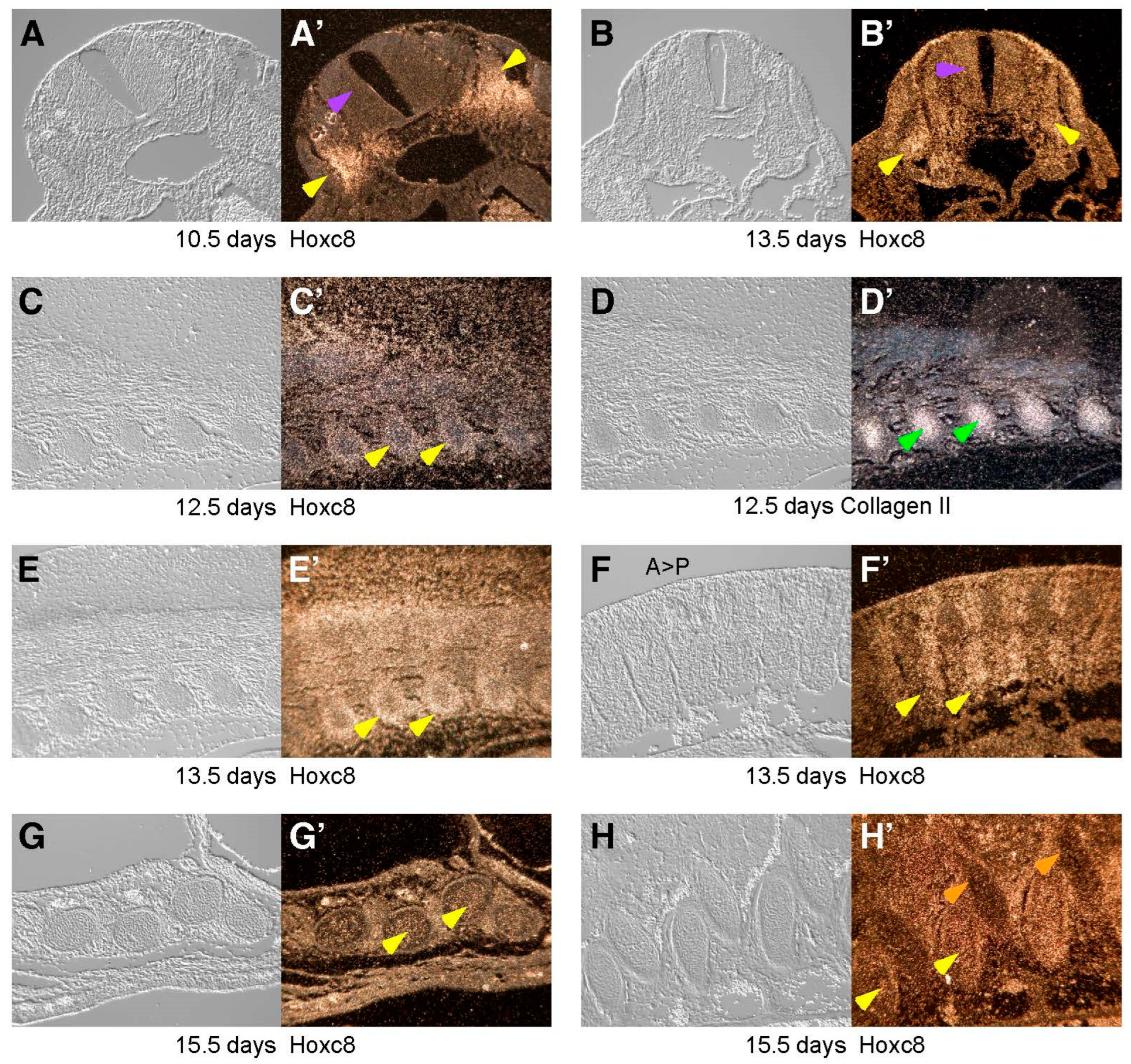
2.1. In Situ Hybridization

In situ hybridization was performed as described in Utset et al. [3]. Sections were cut to 10 μm thickness from paraffin-embedded mouse embryos isolated at 12.5 through 18.5 days of gestation. Sections were hybridized with 35 S-labeled riboprobes for Collagen II [13] and Hoxc8 [3]. The Hoxc8 probe detects the 5′ region of the RNA, excluding the homeobox.

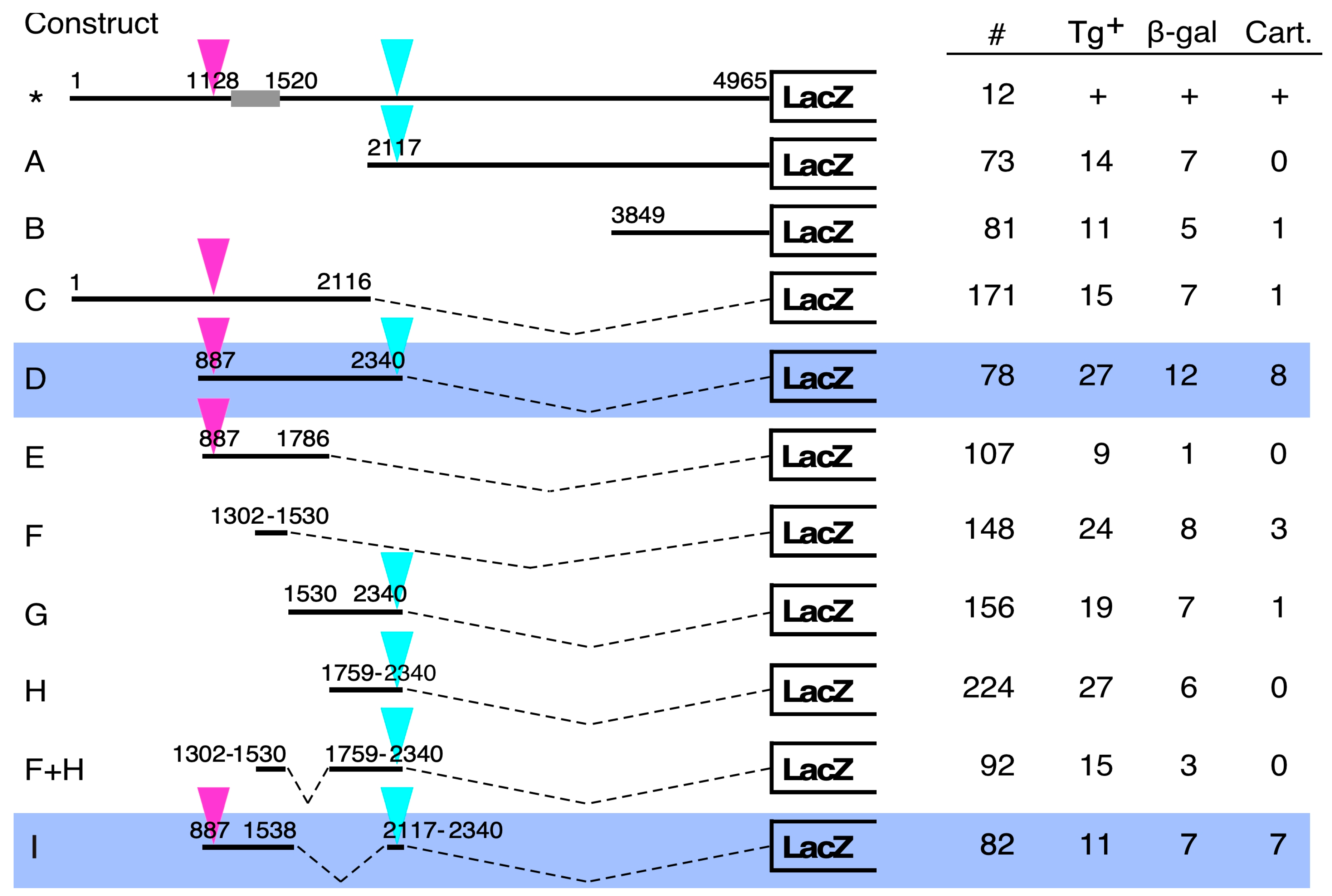
2.2. LacZ Transgene Constructs

The Hoxc8 upstream region fragments were cloned into the pIE-LacZ vector, in which the LacZ gene is linked to the minimal promoter from the Herpes Simplex Virus ICP4 gene [14]. We have previously demonstrated that this reporter is inactive in transgenic mice in the absence of an enhancer element in the construct [15]. The subcloning of fragments was performed using the XbaI site as the acceptor for blunt-ended inserts and XbaI and PstI or XhoI for directional cloning of inserts.
Constructs A and C were generated using the HindIII site at position 2116, and construct B was created using the NcoI site at position 3849. For constructs D through H, specific 5′ and 3′ primer pairs were designed that carried XhoI and XbaI sites, respectively, allowing directional cloning of an amplified and appropriately digested PCR product. Construct F + H was generated from construct D by combining PCR-generated fragments, Construct I was created by first designing complementary primers for each strand that span the deletion, followed by independent PCR in the respective 3′ direction using the respective end primers. In a third step, both PCR products were mixed and amplified using only the end primers, which also incorporated XbaI and PstI sites for directional cloning into the pIE-LacZ vector. All constructs were verified by DNA sequencing.

2.3. Generation and Analysis of Transient Transgenic Embryos

Microinjections were performed as described before [16], and FVB mice were used as embryo donors throughout the project. Embryos were isolated at day 15.5 of gestation, and yolk sacs or placentas were used for genotyping using primers specific for the LacZ transgene [15]. All procedures for analysis of β-gal expression were performed exactly as published before [15,16,17], with the exception that embryos were eviscerated, and fixation prior to staining was performed in 1% paraformaldehyde overnight; staining itself was also allowed to proceed overnight.
Reporter gene activity and staining patterns were deemed consistent and cartilage-specific when a given transgene construct produced the same staining pattern in at least three LacZ transgene-positive samples of progeny, each from independent pregnancies.

2.4. Histology

After microscopic inspection, β-galactosidase-positive embryos were embedded in paraffin for serial sectioning in order to enable inspection of the staining patterns at cellular resolution. Cross-sections of 10 μm thickness were produced, deparaffinized, and scored under DIC microscopy for the presence of β-galactosidase staining in skeletal structures and any other tissues.

2.5. Sequence Analysis

The sequence upstream of the mouse Hoxc8 gene has previously been published [18] and is listed in the UCSC genome browser at coordinates 102,893,993–102,898,962 on chromosome 15 (mm31). The human and rat Hoxc8 sequences were retrieved by searching with the Hox3.2–3.1 intergenic region [18] from Genbank accession #M35603. Conserved regions were visualized using VISTA (http://genome.lbl.gov/vista/index.shtml accessed on 1 January 2023). Putative transcription factor binding sites were retrieved from the Jaspar database of curated information (http://jaspar.genereg.net,accessed on 1 January 2023), as visualized through the UCSC genome browser (https://www.genome.ucsc.edu accessed on 1 January 2023) using genome assembly mm10. Only motifs that were predicted by both the 2018 and the 2020 versions of Jaspar were considered in our analyses.

3. Results

The initial indication that the 5 kb fragment upstream from the Hoxc8 promoter confers cartilage-specific enhancer activity came from experiments performed using the VP16-based binary transgenic system [9,15]. In this system, the LacZ transgene is activated by the viral transcriptional transactivator VP16. Driven by the Hoxc8 5 kb upstream fragment, β-galactosidase expression was observed specifically in the growth zones of the developing skeleton and cartilaginous structures. In order to further characterize the expression of the Hoxc8 gene in developing cartilage, we performed in situ hybridization using a probe that is specific for the 5′ region of the Hoxc8 mRNA. Figure 1 shows that during mouse development, Hoxc8 expression is detected in the somitic mesoderm and later in skeletogenic cells that condense to form cartilage anlagen. By 15.5 days of gestation (E15.5), Hoxc8 expression is detected in chondrocytes of the ribs and prevertebrae.
We then asked which specific regulatory elements control Hoxc8 expression in cartilage. Our first objective was to ascertain cartilage specificity with a conventional transgene in which the Hoxc8 upstream DNA is directly linked to the LacZ gene. Transgenic mice for such a construct had previously been generated [11] by Dr. C. Bieberich, who graciously provided them to us. We analyzed LacZ expression in embryos from several independent litters at 15.5 days of development. All animals that were transgenic for the construct (* in Figure 2) exhibited β-galactosidase activity. Reproducibly, staining was localized to the skeleton in a pattern congruent with the staining observed with the VP16-dependent binary transgenic system ([9], see also Figure 3A,B). These results confirmed with an independent transgene that the Hoxc8 upstream region contains a genuine enhancer with specificity in chondrogenic cells of the developing skeleton.
To map this enhancer within the 5 kb Hoxc8 upstream region, we performed deletion analyses using the LacZ reporter linked to a generic viral minimal promoter [14,15], applying a stringent criterion for enhancer function: the activation of transcription in vivo from a heterologous promoter. Deletion constructs were produced by restriction digest or cloning (see Methods for details) and assayed in transient transgenic embryos (generated as the F0 generation). Embryos containing constructs A or B exhibited no specific activity of β-galactosidase in developing cartilage. A few animals exhibited β-galactosidase activity in multiple independent patterns, which lacked evidence of consistent enhancer activity. We then assayed the 5′ half of the Hoxc8 upstream fragment (construct C), but again, found no specific activity in cartilage or skeletal structures. On the basis of these data, we considered the possibility that the restriction digest at nucleotide 2116 might have destroyed a critical site. Alternatively, elements on both the 5′ and 3′ halves (constructs A and C) may cooperate to form a cartilage-specific enhancer.
To distinguish between these possibilities, we created construct D, which extends beyond the restriction cut site at 2116 bp. This reporter gene produced activity in cartilage in eight out of twelve transgene-expressing embryos (66.7%). The distribution of β-galactosidase staining was consistent between all eight embryos, indicating that the 1.4 kb insert of construct D contains genuine cartilage-specific enhancer activity. This was confirmed in histological sections, where β-galactosidase staining was found localized to skeletogenic cells (Figure 3H). No staining was observed in other somite-derived tissues, such as muscle. These results provide evidence that sequences between nucleotides 887 and 2340 are crucial for cartilage-specific enhancer function.
In an attempt to further reduce the DNA region containing cartilage active regulatory elements, we generated additional deletion constructs E, F, and G, which subdivided the 1.4 kb fragment of construct C. None of these smaller DNA constructs produced cartilage-specific β-galactosidase activity in transgenic embryos. Construct E exhibited staining in only one out of nine transgenic embryos. Construct F also did not confer consistent cartilage-specificity; the three transgenic embryos with some β-galactosidase activity in cartilage exhibited seemingly random patterns of activity in different skeletogenic structures, arguing against the presence of a strong enhancer between 1302 and 1530. A lack of activity of constructs G and H, encompassing basepairs 1530–2340 and 1759–2340, respectively, provided additional evidence against an active site around nucleotide 2116. From these results combined, we concluded that cooperation of (a) site(s) present between nucleotides 887 and 1786 and of (a) site(s) between nucleotides 1759 and 2340 are required for cartilage enhancer activity.
The latter fragment was then combined with the upstream fragment encompassing nucleotides 1302–1530. This fragment had previously been shown to contain transcription factor binding sites involved in “early” enhancer activity in transgenic embryos at E9.5, producing staining that was weaker than in the neural tube but still appreciable in the mesoderm [12]. Our rationale in combining this element with the 3′ part was that a mesoderm-specific element with activity in early embryos might cooperate with other DNA sequences to maintain gene expression in mesodermal derivatives at later stages of development. However, we found no cartilage-specific reporter gene expression from the combined construct (F + H). Therefore, the postulated interaction with an element located between 2117 and 2340 must involve sequences 5′ of bp 1302.
This proposition was tested with construct I, which combined sequences between nucleotides 887 and 1538 and 2117 and 2340. This construct also addressed the concern that a critical site could be located right at 1530 by extending the fragment 8 bps further downstream. We deleted region 1539–2116, which we concluded was not required on the basis of results from constructs F, G, H, and F + H. Construct I produced cartilage-specific activity in 5/7 (71%) of the transgenic embryos generated with this DNA fragment. Because the fragment comprising basepairs 2117–2340 is also contained in construct F + H, which is negative for enhancer activity, a functional interaction with the sequence between nucleotides 1302 and 1530 can be excluded. From the comparison of construct I to constructs G and H, the region spanning basepairs 1759–2116 can also be excluded as potentially relevant. Instead, the minimal sequences defined by our deletion analysis comprise the 416 basepairs of the 887–1302 upstream element and the 224 basepairs of the 2117–2340 downstream element.
By assaying LacZ reporter activity by β-galactosidase staining, we observed a signal generated by construct I in the ribs (Figure 3E) and the developing vertebra (Figure 3F). It is interesting to note that in addition to the cartilaginous portions of the ribs, β-galactosidase staining was also found in the perichondrium of the ossifying dorsal parts of the ribs. This is consistent with the in situ hybridization results at E15.5 (see Figure 1H,H’) and indicates that enhancer activity is maintained in skeletogenic precursor cells, in addition to differentiating chondrocytes. A second intriguing feature of the cartilage enhancer is that even the minimal elements present in construct I exhibit region-specific activity in the developing skeleton (as did construct D), in that activity is prominent only below the level corresponding to thoracic vertebra 3 (see Figure 3D,E). Thus, in all constructs with enhancer activity, the regulatory element also conferred regional restriction along the anterior–posterior axis.
To gain insight into the transcriptional mechanisms involved in the regulation of the cartilage-specific enhancer, we performed sequence analyses of the Hoxc8 upstream region. Cross-species comparisons (Figure 4) revealed evolutionary conservation of non-coding sequences with particularly high similarities in the areas between bps 1100 and 1650 and 1800 and 2500. The previously described “early enhancer” ([12], the gray box in Figure 4) is known to be highly conserved among vertebrates [20]. Our analysis reveals additional areas of sequence conservation, which cover the interacting elements of our cartilage-specific enhancer (pink and turquoise bars in Figure 4). Analysis of the mouse sequence, annotated by the Jaspar transcription factor binding site collection, predicts the presence of potential binding sites for transcription factors of diverse classes (Figure 5): both elements of the cartilage-specific enhancer contain binding motifs for POU-domain/homeodomain transcription factors, Tbox transcription factors, and bHLH transcription factors. Unique to the upstream element are motifs for winged helix-domain transcription factors, double homeoboxes, BarH-like homeoboxes, and homeobox/OAR-domain transcription factors, a motif for nuclear receptor binding (NR2C2), and most notably, an element for binding of high-mobility group transcription factors, which are known to be required for initiation and maintenance of cartilage-specific gene transcription. Unique to the downstream element are motifs for the binding of GATA factors and nuclear receptors of the GATA class. Several of the motifs overlap, precluding unequivocal attribution of particular transcription factor binding activities or combinations thereof.

4. Discussion

This study aimed to identify the regulatory element(s) in the Hoxc8 gene that are necessary and sufficient for controlling gene transcription in skeletogenic cells at later stages of embryonic development. Using a transient transgenic reporter approach in mice, we show that cartilage-specific enhancer activity requires the cooperation of two sequence elements in the 5 Kb upstream region of the Hoxc8 gene: a 416 basepair element (encompassing bps 887–1302) and a 224 basepair element (comprising bps 2117–2340). The localization of these elements relative to each other does not seem to affect their enhancer activity, as they are 579 bp farther apart from each other in the native configuration and in construct D compared to construct I. The latter two constructs also indicate that the cartilage-specific enhancer maintains activity, even at variable distances to the transcription start site, at least with the heterologous promoter tested. Thus, by employing the transient transgenic in vivo assay, we have identified a bona fide chondrocyte-specific enhancer upstream of the Hoxc8 gene.
The cartilage-specific activity of the enhancer is observed in the actively growing ventral sections of the developing ribs at E15.5, with reporter staining maintained in perichondrial cells in the rib sections that have started hypertrophy and ossification (see the open arrows in Figure 3A,D). Activity is also evident in the developing sternum and the vertebrae (Figure 3D,F), indicating a role for the enhancer in all skeletal elements at a given segmental level. Of note, the more intense reporter staining in the binary transgenic system (panels A, B) is likely due to the amplification of transcriptional activation by the VP16 transactivator [9,19]. Within cartilaginous elements, not all cells are uniformly stained to the same extent (see Figure 3B,C), indicating tissue-specific “mosaicism”, which was also observed when the LacZ reporter gene was knocked into the endogenous Hoxc8 locus [6]. Thus, the activity of our enhancer elements recapitulates cellular patterns of expression activity that are also observed in the native Hoxc8 locus. However, which DNA sequences in particular control the activity remains to be tested by targeted mutagenesis.
Combinatorial action of multiple elements in transcriptional regulation has been described for developmental enhancers in multiple Hox genes, such as, for example, Hoxa7 [21], Hoxb4 [22] Hoxb7 [23], Hoxb8 [24], Hoxd11 [25], and in the Hoxc8 gene itself. Shashikant et al. [12] defined a 335 bp enhancer (nucleotides 1128–1462) with activity in the neural tube that initially was described as encompassing four interacting elements and was later extended by 64 bp to include a fifth element that confers activity early in mesodermal cells (1128–1526; [26]). Binding motifs were identified for Cdx transcription factors, forkhead transcription factor Foxa2/HNF3, Hox transcription factors, and HMG domain-containing proteins. These sites are closely spaced within a promoter–proximal 200 bp fragment (nucleotides 1328–1527), the so-called “early enhancer” [12], with activity in developing neural tube posterior to the 10th somite pair and in mesoderm posterior to the 14th some pair. The deletion of this 200 bp sequence from the endogenous mouse Hoxc8 locus in vivo [27] revealed that it is not required for the transcription of the endogenous Hoxc8 gene or regional specificity, but the “early enhancer” sequence plays a role for proper temporal regulation.
Indeed, the cartilage-specific activity of our enhancer does not require these “early enhancer” sequences, as evidenced by a lack of activity in the developing skeleton with construct F, which comprises the “early enhancer”. The addition of this DNA segment to the 224 bp downstream element we defined (in construct F + H) also failed to stimulate reporter transcription in chondrogenic cells. Thus, we have delineated a tissue-specific enhancer with activity at later stages of development that is distinct from early enhancers, comprising elements spaced both more distal from and more proximal to the transcription start site. Our sequence analysis reveals potential binding sites for many types of transcription factor molecules, among them HMG domain proteins, and Pbx/Hox transcription factor complexes. The HMG domain proteins Sox9, Sox5, and Sox6 are well-known transcriptional regulators in chondrocytes [28]. The implication of particular transcription factors in the activation and maintenance of activity of the Hoxc8 cartilage-specific enhancer will have to await further investigation by mutational analysis.
Intriguingly, even though the enhancer displayed strong specificity for cartilage, it maintained a regional restriction to the thoracic and lumbar/sacral regions of the developing skeleton, with weaker staining in ribs in the T3 through T5 segments and more prominent staining in T6 and T7 ribs, continuing more posteriorly. This is consistent with increasingly stronger staining in the lower sternal ribs observed in mice where the LacZ gene is inserted into the Hoxc8 locus [6]. These and other authors already noted that the rostral boundary of reporter gene expression at later developmental stages (E12.5) was located between T3 and T4 skeletal elements [5,6] and at the newborn stage extended to T1, including the first pair of ribs [6]. This is consistent with our findings for Hoxc8 upstream region activity with the binary transgenic system in newborn mice [9]. The mechanisms underlying this anterior shift compared to earlier developmental stages are unknown but could involve a lack of activity of the “early enhancer” at later stages, either due to the absence of relevant transcription factors in chondrogenic cells or epigenetic modifications that make it inaccessible. Alternatively, the earlier posterior restriction in skeletogenic precursor structures (i.e., the somites) could be mediated by a silencing element, preventing the action of the “early enhancer” in anterior regions. If such a repressive element were to exist, it is obviously no longer active by the time chondrogenic cells differentiate in the thoracic and lumbosacral regions of the developing skeleton.
It is noteworthy that the cartilage-specific Hoxc8 enhancer that we have identified is also active in perichondrial cells (see the arrows in Figure 3C,E) before ossification is completed. The perichondrium is known to play a critical role in catch-up growth [29] and the regeneration of long-bone cartilage, such as rib cartilage [30]. Our enhancer will now allow studies that follow Hoxc8 expression in these cells during the regeneration process. It is also intriguing that despite widely documented functional roles of Hox genes in adult tissues and cancer [31,32,33,34,35,36,37,38,39,40,41,42,43,44], few attempts have been reported to identify regulatory elements controlling Hox gene expression in specific adult cell types in vivo. For example, Tabaries et al. [45] identified regulatory sequences in the Hoxa5 gene with strong activity in thoracic prevertebrae, but whether the activity is maintained through late development or postnatally has not been investigated. Similarly, Pruett et al. [34] described regionally distinct reporter activity specifically in vascular smooth muscle cells driven by sequences from the Hoxa3 and Hoxc11 genes, but the responsible regulatory elements have not been identified. Thus, our study is one of only a few reports that characterize regulatory enhancer elements for cell-type-specific Hox gene expression through an in vivo approach. This now makes it possible to investigate the activity and regulation of this enhancer and, by implication, its transcription regulatory mechanisms, in postnatal skeletogenic processes, such as chondrocyte proliferation and differentiation during the growth of the skeleton, hormonal regulation of bone growth completion during puberty, cartilage and bone regeneration after injury and bone fracture repair, and transplantation and bioengineering approaches to cartilage tissue regeneration.

Author Contributions

Conceptualization, S.A.C. and C.K.; validation, S.A.C. and C.K.; investigation, S.A.C. and C.K.; resources, S.A.C. and C.K.; formal analysis, S.A.C. and C.K.; all writing and editing, S.A.C. and C.K.; visualization, S.A.C. and C.K.; finding acquisition, S.A.C. and C.K.; project administration, C.K. All authors have read and agreed to the published version of the manuscript.

Funding

This research was funded in parts by the Mayo Foundation for Medical Education and Research, NIH-R03-AR44945 (to C.K.), and NRSA F32-AR008545 (to S.A.C.). More recent work on this project and the completion of this manuscript was supported by the Peggy M. Pennington Cole Endowed Chair for Developmental Biology at Pennington Biomedical Research Center.

Institutional Review Board Statement

All animal experimentation protocols in this study were reviewed and approved by the respective Institutional Animal Care and Use Committees of Mayo Clinic Scottsdale (IACUC protocol number A87-97, approved in 1997), University of Nebraska Medical Center (IACUC protocol number 00-002-02, approved in 2002), and Pennington Biomedical Research Center, Louisiana State University System (IACUC protocol number 06-442, approved in 2006). All procedures involving mice were compliant with the “Guide for Care and Use of Animals in Laboratory Research” (https://pubmed.ncbi.nlm.nih.gov/21595115/; DOI: 10.17226/12910 accessed on 1 January 2023) by the US National Institutes of Health.

Informed Consent Statement

Not applicable.

Data Availability Statement

The datasets used and analyzed during the current study are available from the corresponding author upon reasonable request, in compliance with institutional policies of Pennington Biomedical Research Center/Louisiana State University System. DNA constructs will be made available upon request.

Acknowledgments

Major portions of this study were conducted while the authors were affiliated with the Samuel C. Johnson Medical Research Center at Mayo Clinic Scottsdale, 13400 E. Shea Boulevard, Scottsdale, AZ 85259, and the experimental work was completed when C.K. was affiliated with the Center for Human Molecular Genetics, Munroe-Meyer Institute, and the Department of Genetics, Cell Biology, and Anatomy at the University of Nebraska Medical Center, Omaha, NE. We thank T. Tinder, G. Basu, D.L. Costanzo, T. Wiggins, and M. Northam for their technical assistance and C.H. Rundle for contributions to the early phase of the project. M.A. Jennings provided expert histology, and the Mayo Transgenic Facility and the UNMC Mouse Genome Engineering Facility delivered superb microinjection services. We are also grateful to C.A. Bieberich for providing Hoxc8 upstream region-LacZ mice and C.S. Shashikant and J.M. Salbaum for critical discussions. S. Fisher provided secretarial assistance with early drafts of the manuscript.

Conflicts of Interest

The authors declare no conflicts of interest.

References

  1. LeMouellic, H.; Condamine, H.; Brulet, P. Pattern of transcription of the homeo gene Hox-3.1 in the mouse embryo. Genes. Dev. 1988, 2, 125–135. [Google Scholar] [CrossRef] [PubMed]
  2. Breier, G.; Dressler, G.R.; Gruss, P. Primary structure and developmental expression pattern of Hox 3.1, a member of the murine Hox 3 homeobox gene cluster. EMBO J. 1988, 7, 1329–1336. [Google Scholar] [CrossRef] [PubMed]
  3. Utset, M.F.; Awgulewitsch, A.; Ruddle, F.H.; McGinnis, W. Region-specific expression of two mouse homeo box genes. Science 1987, 235, 1379–1382. [Google Scholar] [CrossRef] [PubMed]
  4. Belting, H.G.; Shashikant, C.S.; Ruddle, F.H. Multiple phases of expression and regulation of mouse Hoxc-8 during early embryogenesis. J. Exp. Zool. 1998, 282, 196–222. [Google Scholar] [CrossRef]
  5. Kwon, Y.; Shin, J.; Park, H.W.; Kim, M.H. Dynamic expression pattern of Hoxc8 during mouse early embryogenesis. Anat. Rec. A Discov. Mol. Cell Evol. Biol. 2005, 283, 187–192. [Google Scholar] [CrossRef] [PubMed]
  6. LeMouellic, H.; Lallemand, Y.; Brulet, P. Homeosis in the mouse induced by a null mutation in the Hox-3.1 gene. Cell 1992, 69, 251–264. [Google Scholar] [CrossRef] [PubMed]
  7. Tiret, L.; Le Mouellic, H.; Maury, M.; Brulet, P. Increased apoptosis of motoneurons and altered somatotopic maps in the brachial spinal cord of Hoxc-8-deficient mice. Development 1998, 125, 279–291. [Google Scholar] [CrossRef] [PubMed]
  8. Shimamoto, T.; Tang, Y.; Naot, Y.; Nardi, M.; Brulet, P.; Bieberich, C.J.; Takeshita, K. Hematopoietic progenitor cell abnormalities in Hoxc-8 null mutant mice. J. Exp. Zool. 1999, 283, 186–193. [Google Scholar] [CrossRef]
  9. Yueh, Y.G.; Gardner, D.P.; Kappen, C. Evidence for regulation of cartilage differentiation by the homeobox gene Hoxc-8. Proc. Natl. Acad. Sci. USA 1998, 95, 9956–9961. [Google Scholar] [CrossRef]
  10. Kamel, S.; Kruger, C.; Salbaum, J.M.; Kappen, C. Morpholino-mediated knockdown in primary chondrocytes implicates Hoxc8 in regulation of cell cycle progression. Bone 2009, 44, 708–716. [Google Scholar] [CrossRef]
  11. Bieberich, C.J.; Utset, M.F.; Awgulewitsch, A.; Ruddle, F.H. Evidence for positive and negative regulation of the Hox-3.1 gene. Proc. Natl. Acad. Sci. USA 1990, 87, 8462–8466. [Google Scholar] [CrossRef] [PubMed]
  12. Shashikant, C.S.; Bieberich, C.J.; Belting, H.-G.; Wang, J.C.H.; Borbely, M.A.; Ruddle, F.H. Regulation of Hoxc-8 during mouse embryonic development: Identification and characterization of critical elements involved in early neural tube expression. Development 1995, 121, 4339–4347. [Google Scholar] [CrossRef] [PubMed]
  13. Sandell, L.J. In situ expression of collagen and proteoglycan genes in notochord and during skeletal development and growth. Microsc. Res. Technol. 1994, 28, 470–482. [Google Scholar] [CrossRef] [PubMed]
  14. Byrne, G.W.; Ruddle, F.H. Multiplex gene regulation: A two-tiered approach to transgene regulation in transgenic mice. Proc. Natl. Acad. Sci. USA 1989, 86, 5473–5477. [Google Scholar] [CrossRef] [PubMed]
  15. Gardner, D.P.; Byrne, G.W.; Ruddle, F.H.; Kappen, C. Spatial and temporal regulation of a LacZ reporter transgene in a binary transgenic mouse system. Transgenic Res. 1996, 5, 37–48. [Google Scholar] [CrossRef]
  16. Yaworsky, P.J.; Kappen, C. Heterogeneity of neural progenitor cells revealed by enhancers in the nestin gene. Dev. Biol. 1999, 205, 309–321. [Google Scholar] [CrossRef]
  17. Yaworsky, P.J.; Gardner, D.P.; Kappen, C. Transgenic analyses reveal neuron and muscle specific elements in the murine neurofilament light chain gene promoter. J. Biol. Chem. 1997, 272, 25112–25120. [Google Scholar] [CrossRef]
  18. Awgulewitsch, A.; Bieberich, C.; Bogarad, L.; Shashikant, C.; Ruddle, F.H. Structural analysis of the Hox-3.1 transcription unit and the Hox-3.2--Hox-3.1 intergenic region. Proc. Natl. Acad. Sci. USA 1990, 87, 6428–6432. [Google Scholar] [CrossRef]
  19. Rundle, C.H.; Macias, M.P.; Gardner, D.P.; Yueh, Y.G.; Kappen, C. Transactivation of Hox Gene Expression in a VP16-Dependent Binary Transgenic Mouse System. Biochim. Biophys. Acta 1998, 1398, 164–178. [Google Scholar] [CrossRef]
  20. Shashikant, C.S.; Bolanowsky, S.A.; Anand, S.; Anderson, S.M. Comparison of diverged Hoxc8 early enhancer activities reveals modification of regulatory interactions at conserved cis-acting elements. J. Exp. Zool. B Mol. Dev. Evol. 2007, 308, 242–249. [Google Scholar] [CrossRef]
  21. Puschel, A.W.; Balling, R.; Gruss, P. Separate elements cause lineage restriction and specify boundaries of Hox-1.1 expression. Development 1991, 112, 279–287. [Google Scholar] [CrossRef] [PubMed]
  22. Whiting, J.; Marshall, H.; Cook, M.; Krumlauf, R.; Rigby, P.W.; Stott, D.; Allemann, R.K. Multiple spatially specific enhancers are required to reconstruct the pattern of Hox-2.6 gene expression. Genes Dev. 1991, 5, 2048–2059. [Google Scholar] [PubMed]
  23. Vogels, R.; Charite, J.; Degraaff, W.; Deschamps, J. Proximal cis-acting elements cooperate to set hoxb-7 (hox-2.3) expression boundaries in transgenic mice. Development 1993, 118, 71–82. [Google Scholar] [CrossRef] [PubMed]
  24. Charite, J.; de Graaff, W.; Vogels, R.; Meijlink, F.; Deschamps, J. Regulation of the Hoxb-8 gene: Synergism between multimerized cis-acting elements increases responsiveness to positional information. Dev. Biol. 1995, 171, 294–305. [Google Scholar] [CrossRef] [PubMed]
  25. Gerard, M.; Duboule, D.; Zakany, J. Structure and activity of regulatory elements involved in the activation of the hoxd-11 gene during late gastrulation. Embo J. 1993, 12, 3539–3550. [Google Scholar] [CrossRef]
  26. Shashikant, C.S.; Ruddle, F.H. Combinations of closely situated cis-acting elements determine tissue-specific patterns and anterior extent of early Hoxc8 expression. Proc. Natl. Acad. Sci. USA 1996, 93, 12364–12369. [Google Scholar] [CrossRef] [PubMed]
  27. Juan, A.H.; Ruddle, F.H. Enhancer timing of Hox gene expression: Deletion of the endogenous Hoxc8 early enhancer. Development 2003, 130, 4823–4834. [Google Scholar] [CrossRef] [PubMed]
  28. Liu, C.F.; Samsa, W.E.; Zhou, G.; Lefebvre, V. Transcriptional control of chondrocyte specification and differentiation. Semin. Cell Dev. Biol. 2017, 62, 34–49. [Google Scholar] [CrossRef]
  29. Rosello-Diez, A.; Joyner, A.L. Regulation of Long Bone Growth in Vertebrates; It Is Time to Catch Up. Endocr. Rev. 2015, 36, 646–680. [Google Scholar] [CrossRef]
  30. Srour, M.K.; Fogel, J.L.; Yamaguchi, K.T.; Montgomery, A.P.; Izuhara, A.K.; Misakian, A.L.; Lam, S.; Lakeland, D.L.; Urata, M.M.; Lee, J.S.; et al. Natural large-scale regeneration of rib cartilage in a mouse model. J. Bone Miner. Res. 2015, 30, 297–308. [Google Scholar] [CrossRef]
  31. Bieberich, C.J.; Ruddle, F.H.; Stenn, K.S. Differential expression of the Hox 3.1 gene in adult mouse skin. Ann. N. Y. Acad. Sci. 1991, 642, 346–353, discussion in 353–344. [Google Scholar] [CrossRef] [PubMed]
  32. Brancaz, M.V.; Iratni, R.; Morrison, A.; Mancini, S.J.; Marche, P.; Sundberg, J.; Nonchev, S. A new allele of the mouse hairless gene interferes with Hox/LacZ transgene regulation in hair follicle primordia. Exp. Mol. Pathol. 2004, 76, 173–181. [Google Scholar] [CrossRef] [PubMed]
  33. Du, H.; Taylor, H.S. The Role of Hox Genes in Female Reproductive Tract Development, Adult Function, and Fertility. Cold Spring Harb. Perspect. Med. 2015, 6, a023002. [Google Scholar] [CrossRef]
  34. Pruett, N.D.; Visconti, R.P.; Jacobs, D.F.; Scholz, D.; McQuinn, T.; Sundberg, J.P.; Awgulewitsch, A. Evidence for Hox-specified positional identities in adult vasculature. BMC Dev. Biol. 2008, 8, 93. [Google Scholar] [CrossRef] [PubMed]
  35. Rux, D.R.; Song, J.Y.; Swinehart, I.T.; Pineault, K.M.; Schlientz, A.J.; Trulik, K.G.; Goldstein, S.A.; Kozloff, K.M.; Lucas, D.; Wellik, D.M. Regionally Restricted Hox Function in Adult Bone Marrow Multipotent Mesenchymal Stem/Stromal Cells. Dev. Cell 2016, 39, 653–666. [Google Scholar] [CrossRef]
  36. Song, J.Y.; Pineault, K.M.; Dones, J.M.; Raines, R.T.; Wellik, D.M. Hox genes maintain critical roles in the adult skeleton. Proc. Natl. Acad. Sci. USA 2020, 117, 7296–7304. [Google Scholar] [CrossRef]
  37. Taylor, H.S.; Vanden Heuvel, G.B.; Igarashi, P. A conserved Hox axis in the mouse and human female reproductive system: Late establishment and persistent adult expression of the Hoxa cluster genes. Biol. Reprod. 1997, 57, 1338–1345. [Google Scholar] [CrossRef]
  38. Abramovich, C.; Pineault, N.; Ohta, H.; Humphries, R.K. Hox genes: From leukemia to hematopoietic stem cell expansion. Ann. N. Y. Acad. Sci. 2005, 1044, 109–116. [Google Scholar] [CrossRef]
  39. Bhatlekar, S.; Fields, J.Z.; Boman, B.M. Role of HOX Genes in Stem Cell Differentiation and Cancer. Stem Cells Int. 2018, 2018, 3569493. [Google Scholar] [CrossRef]
  40. Chen, H.; Sukumar, S. HOX genes: Emerging stars in cancer. Cancer Biol. Ther. 2003, 2, 524–525. [Google Scholar] [CrossRef]
  41. Grier, D.G.; Thompson, A.; Kwasniewska, A.; McGonigle, G.J.; Halliday, H.L.; Lappin, T.R. The pathophysiology of HOX genes and their role in cancer. J. Pathol. 2005, 205, 154–171. [Google Scholar] [CrossRef] [PubMed]
  42. Javed, S.; Langley, S.E. Importance of HOX genes in normal prostate gland formation, prostate cancer development and its early detection. BJU Int. 2014, 113, 535–540. [Google Scholar] [CrossRef] [PubMed]
  43. Smith, J.; Zyoud, A.; Allegrucci, C. A Case of Identity: HOX Genes in Normal and Cancer Stem Cells. Cancers 2019, 11, 512. [Google Scholar] [CrossRef] [PubMed]
  44. Awgulewitsch, A. Hox in hair growth and development. Naturwissenschaften 2003, 90, 193–211. [Google Scholar] [CrossRef]
  45. Tabaries, S.; Lapointe, J.; Besch, T.; Carter, M.; Woollard, J.; Tuggle, C.K.; Jeannotte, L. Cdx protein interaction with Hoxa5 regulatory sequences contributes to Hoxa5 regional expression along the axial skeleton. Mol. Cell Biol. 2005, 25, 1389–1401. [Google Scholar] [CrossRef]
Figure 1. Hoxc8 expression in the developing skeleton at 10.5–15.5 days post-coitum. In situ hybridization with probes specific for Hoxc8 and Collagen II to sections of normal mouse embryos isolated at various developmental stages. The left panels (AH) in each pair depict paraffin sections in brightfield under differential interference contrast, with the in situ hybridization on that section imaged under darkfield conditions and depicted in the corresponding right panels (A’H’). (A,A’) At E10.5, strong Hoxc8 expression is apparent in prevertebral cells (yellow arrowheads), while at this axial level, Hoxc8 expression in the neural tube is low (purple arrowhead). (B,B’) Section through an embryo at E13.5 that displays Hoxc8 expression both in the neural tube (purple arrowhead) and mesoderm (yellow arrowheads). (C,C’) At E12.5, Hoxc8 expression is strongest at the periphery of condensing precartilaginous rib structures (yellow arrowheads) surrounding committed chondrocytes, as evidenced by Collagen II expression within the structures (green arrowheads) in the panels (D,D’,E,E’). At E13.5, Hoxc8 expression becomes stronger, as prevertebral structures condense and undergo cartilage differentiation. (F,F’) Within each segment in the vertebral column on this parasagittal section, Hoxc8 is more strongly expressed in the caudal part (yellow arrowheads, A>P indicates anterior–posterior direction). (G,G’,H,H’) At E15.5, Hoxc8 expression is evident in ventral (G,G’) and dorsal (H,H’) portions of the ribs (yellow arrowheads) and in the periphery of the vertebral bodies (orange arrowheads).
Jdb 12 00005 g001
Figure 2. Deletion constructs and assays for reporter gene activity in transgenic mice. The first construct, labeled by an asterisk, depicts the structure of the reporter construct containing the 5 Kb Hox8 upstream region that was previously published [11] but had not been analyzed at later developmental stages. For constructs produced in this study, all genomic DNA fragments from the Hoxc8 locus were ligated to a LacZ reporter gene that is linked to the minimal promoter from the herpes simplex virus ICP4 gene [14]. This reporter gene is silent in the absence of a specific enhancer [14,15,19]. The addition of enhancer-containing DNA leads to reporter gene expression in transgenic mice, as analyzed at 15.5 days of development. The numbers of all embryos recovered from microinjections of a given construct are given, and the number of transgenic embryos (Tg+) was determined by a PCR assay for the LacZ transgene. β-galactosidase staining was detected macroscopically, and microscopic inspection of embryos and histological section was used to ascertain activity at specific sites. Construct F contains the previously identified so-called “early enhancer” [12], which produced staining in some skeletal elements that differed between three (out of eight transgenic) individuals and was deemed not active in the skeleton at the later stages we investigated. The minimal DNA sequences required for cartilage-specific enhancer activity consist of two elements (pink and turquoise triangles) located between nucleotides 887 and 1301 (pink) and 2117 and 2340 (turquoise). See the manuscript text for the rationale supporting this conclusion.
Jdb 12 00005 g002
Figure 3. β-galactosidase activity in the cartilage of transgenic embryos. Transcriptional activation of LacZ reporter gene activity was assayed by staining for the reporter gene product β-galactosidase. (A) Transgenic embryo at E17.5 generated in the binary system [9] with tissue-specific β-galactosidase expression in the skeletogenic cells of the ribs, including the ventral rib cartilage. (B) The histological section of a rib reveals β-galactosidase staining in zones containing proliferative (p), prehypertrophic (ph), and hypertrophic chondrocytes (black arrows). (C) The section at an oblique angle from an E15.5 paraffin-embedded transgenic embryo carrying deletion construct D. β-galactosidase staining is apparent in the ribs (corresponding to proliferative zones of the ribs at T3, T4, and T5) and in perichondrial cells (arrows) but not in adjacent cells of mesodermal origin, such as, e.g., muscles. (D) A whole-mount partially eviscerated and β-galactosidase-stained transgenic embryo at E15.5 carrying construct I displays staining in the lower sternum (purple arrow) and the ribs (numbered in rostrocaudal direction by black arrows), with weaker intensity in segments T3 through T5 and a stronger signal in the lower ventral ribs. (E) Side view of another embryo transgenic for construct I; in addition to the black arrows for rostrocaudal numbering of ribs, white arrows point to areas of staining localized around the ossifying dorsal portions of ribs. (F) View of the lower body cavity of another construct I transgenic embryo from which the caudal region was removed; blue staining reveals reporter gene activity in the developing vertebrae at lower thoracic levels and in the corresponding rib heads of the lower floating ribs (visible in red are segmental blood vessels).
Jdb 12 00005 g003
Figure 4. Evolutionary comparison of the Hoxc8 upstream flanking region, including the developmentally early enhancer and the chondrocyte-specific enhancer. VISTA plot for pairwise comparisons of rat–mouse (top row), mouse–human (middle row), and rat–human (lower row) Hoxc8 upstream sequences. Several regions of conservation (a window size of 100 bp) across species above 75% (red shading) are evident, one of which contains the previously described Hoxc8 “early enhancer” (gray bar below the comparison plot). Pink- and turquoise-colored bars below the comparison plot depict the location of the cartilage-specific enhancer elements defined in our studies, as depicted in Figure 2.
Jdb 12 00005 g004
Figure 5. Sequence analysis of the Hoxc8 cartilage-specific enhancer and transcription factor binding sites. Sequences of the minimal elements defined from the results depicted in Figure 2. Motifs for potential transcription factor binding were collected, identified by both JASPAR2018 and JASPAR2020 versions, and visualized in the UCSC genome browser.
Jdb 12 00005 g005
Disclaimer/Publisher’s Note: The statements, opinions and data contained in all publications are solely those of the individual author(s) and contributor(s) and not of MDPI and/or the editor(s). MDPI and/or the editor(s) disclaim responsibility for any injury to people or property resulting from any ideas, methods, instructions or products referred to in the content.

