Cataract, uncorrected refractive error, Glaucoma, Age-related macular degeneration, Diabetic Retinopathy, corneal opacity, trachoma, and others are responsible for vision impairment. Among the listed medical conditions, uncorrected refractive error, cataract, and glaucoma are the major cause of blindness. Statistics from the World Health Organization (WHO) states that 81% of the people who are blind are above 50 years of age. It is estimated that the number of people who are blind will increase from 38.5 million in 2020 to 115 million in 2050 [
1]. Diabetic retinopathy is a major cause evolving for blindness. WHO report on diabetes states that diabetes will be the seventh major cause of death in 2030. 2.6% of blindness reported is attributed to diabetic retinopathy. The percentage reported may be minuscule, but a report from WHO states that the number of people with diabetes has increased by a factor of 3.5 times i.e., from 118 million in 1980 to 424 million in 2014 [
2]. These statistics show that diabetic retinopathy cases will drastically increase in the near future. Because the availability of expert ophthalmologists with specialization in Retinal disorders is not on par with the forecast of the disease, an automated system is required. The availability of sophisticated imaging and computing systems helps in developing a Computer Aided Diagnosis (CAD) system for the faster diagnosis of Diabetic retinopathy. The CAD system assists the experts in diagnosing diabetic retinopathy by extracting different parts of the eye.
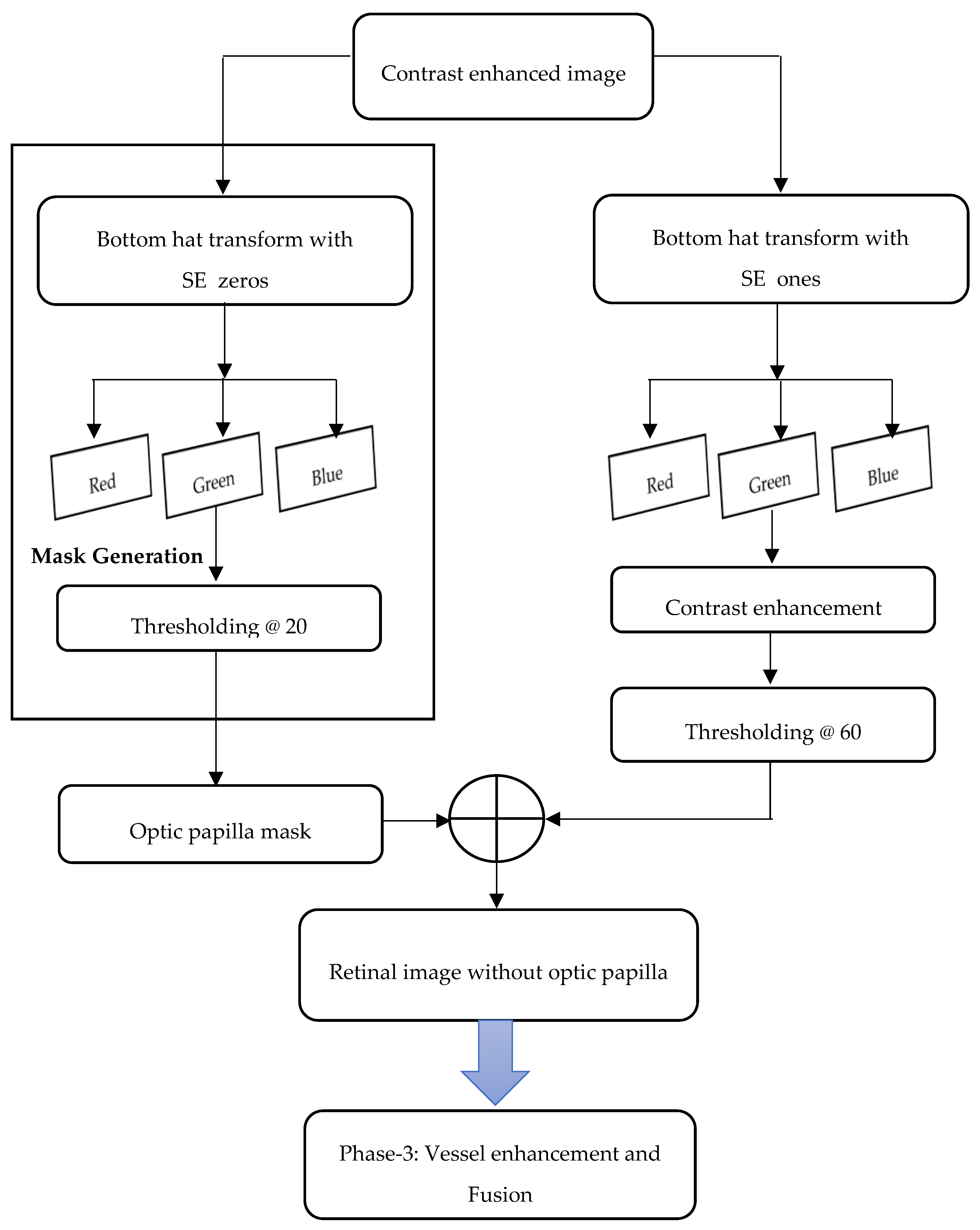
Fundus of the eye refers to the posterior surface of the eye, which consists of optic disc, retina, macula, fovea, and posterior pole. The Fundus Camera is used to capture the posterior surface of the eye. It uses monocular indirect ophthalmoscopy principle for acquiring the fundus images. Most pathological changes in the eye happen in the blood vessels, both in the arteries and veins. Many retinal diseases are characterized by the changes in blood vessels. Automatic detection of the optic disc, blood vessel, fovea, and macula are required for diagnosing diabetic retinopathy. Retinal maps are automatically generated through temporal and multimodal image registration. Temporal image registration helps in identifying the spread of the disease. Multimodal image registration aids in the better identification of some lesions. The structure of blood vessels is unique for individuals and it can be used for biometric-based authentication. Manual segmentation of blood vessels is a time-consuming process that involves extensive training and skill sets. The initial step in a CAD system for ophthalmic disorders involves the automatic segmentation of blood vessels and the identification of optic disk. Many algorithms have been proposed by researchers for segmenting the blood vessels, which are discussed in the forthcoming section. This paper proposes a hybrid segmentation approach to extract the blood vessels from the fundus image. Workflow of the proposed methodology is given in
Figure 1. The proposed methodology has three phases and these phases are depicted in
Figure 2,
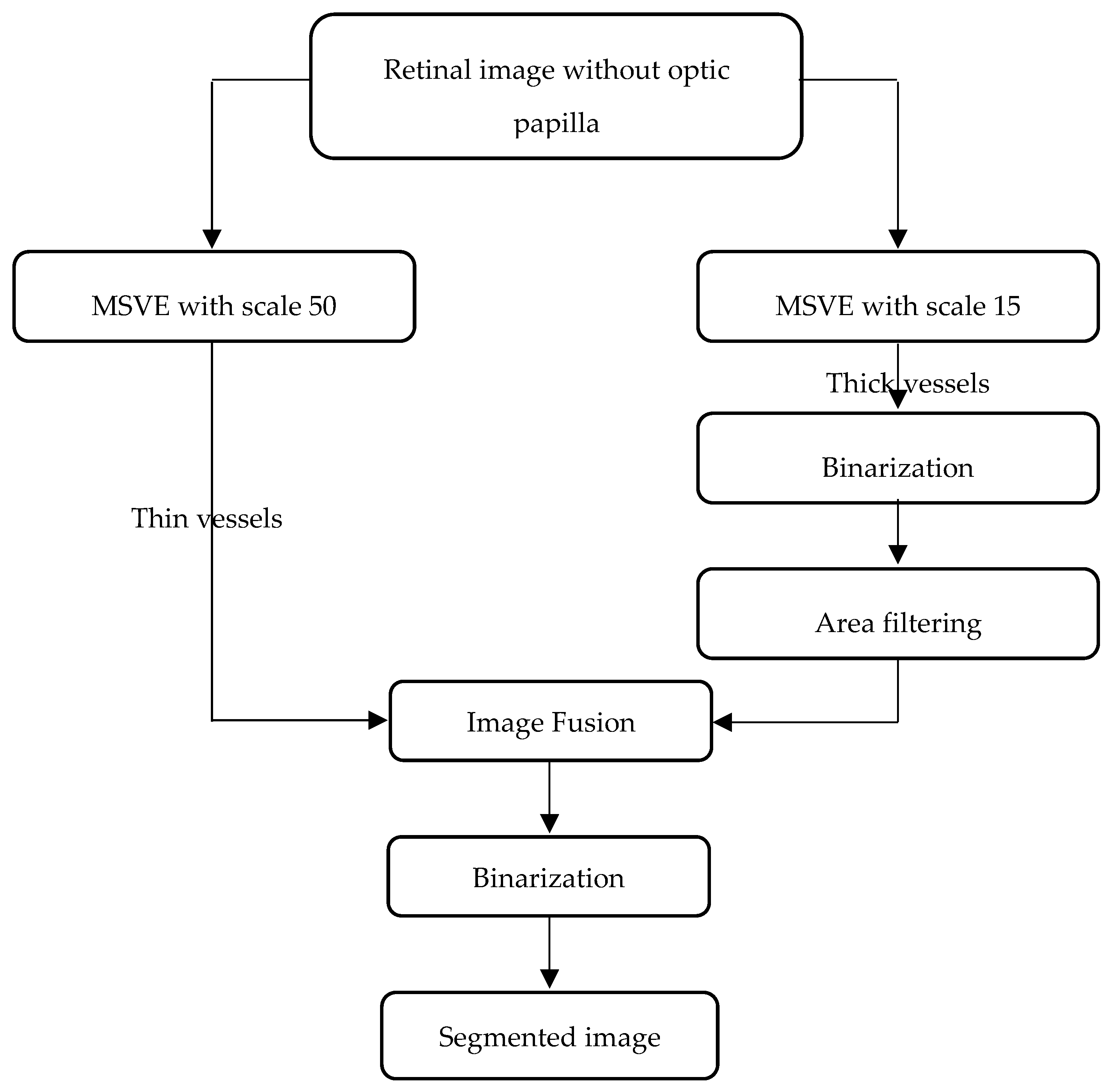
Figure 3, and
Figure 4.
1.1. Related Works
The segmentation and analysis of blood vessels through image processing is required in the diversified fields of medicine. Many researchers have contributed efficient algorithms for extraction and analysis. Some significant contributions in the field of Ophthalmology are given in this section.
Segmentation of blood vessels can be done by two different methods:
Pixel-based methods and tracing/tracking-based methods. In pixel-based methods, every pixel is processed to reveal whether it is a vessel or background pixel. Pixel-based methods use thresholding, morphological operations, and kernels for filtering and pattern recognition.
Pattern recognition-based methods use classifiers and clustering algorithms for segmenting blood vessels from the fundus image.
Soares et al. have proposed a supervised classification using two-dimensional (2D) Gabor wavelet [
3]. Ricci et al. have used line operators and Support Vector Machine (SVM) for segmenting blood vessels [
4]. Moment invariants-based feature and 7D feature based on gray-level were used to train the neural network by Marin et al. [
5]. Tolias et al. have proposed a method based on Fuzzy C-means clustering for segmenting the blood vessels [
6]. Niemeijer et al. proposed a
k-Nearest Neighborhood (kNN) based classifier for segmenting blood vessels [
7]. Salem et al. have used a novel algorithm (RACAL), which is a partially supervised algorithm for segmenting the blood vessels [
8].
Kernels are filters that are used in images for identifying the pixels of interest. The most common kernels are the edge filters that are used for finding the edges in the images. Prominent kernels used for edge detection are Robert, Sobel, Prewitt and Canny. Apart from this, kernels of specific type can be customized for an application to identify the edges. Chaudhuri et al. have proposed a kernel based matched filtering mechanism for blood vessel segmentation [
9]. It uses 12 different templates that were generated by rotating the actual template by 15 degrees. Al-Rawi et al. proposed an improved matched filtering mechanism based on Chaudhuri et al’s matched filtering mechanism [
10]. Cinsdikici et al. have proposed an algorithm that uses matched filtering with ant colony optimization [
11]. Zhang et al. proposed a modified matched filter, called Matched Filter-First order Derivative of Gaussian (MF-FDOG) [
12]. Odstrcilik et al. proposed a novel matched filtering kernel for segmenting blood vessel in fundus image [
13].
Thresholding of fundus image is another method that is used for segmenting blood vessels. Thresholding can be global, local, or adaptive. Adaptive thresholding is mostly used for segmentation and it gives better results. Hoover et al. proposed a piecewise threshold probing of the matched filter response for segmenting the blood vessels [
14]. Jiang et al. proposed an adaptive thresholding based on multi-threshold probing scheme [
15]. Reza et al. proposed automatic tracing algorithm for detecting optic disc and exudates using fixed and variable thresholds [
16]. They have also proposed a quadtree based blood vessel detection algorithm using RGB (Red-Blue-Green) color components of fundus images [
17].
Morphological operators are quite handy in segmenting the object of interest using mathematical operations. There are many morphological operators that are defined for image processing. The most commonly used morphological operations are dilation, erosion, closing, and opening [
18]. These operators are applied mainly to binary images. However, they can also be applied for grayscale images. Zana et al. employed a morphology-based method with cross curvature evaluation for segmenting vasculature from the medical image [
19]. Heneghan et al. combined morphological operations with the second order derivative operator to locate both the primary and secondary vessels [
20]. Yang et al. employed a combination of fuzzy clustering algorithm and morphological operator [
21]. Mehrotra et al. employed a morphological operator for highlighting the blood vessels and then later applied the Kohonen Clustering Network to segment the blood vessels [
22]. Miri et al. used Forward Discrete Cosine Transform (FDCT) for image contrast enhancement followed by morphological operations for extracting the blood vessels [
23]. Bharkad used top hat, a morphological operator with three different structuring elements [
24]. Yavuz et al. enhanced the retinal image using Gabor, Frangi, and Gaussian filters, followed by the use of top hat transform and clustering mechanism for segmenting the blood vessels [
25].
Employing the tracking or tracing based method, retinal vasculature can be segmented. Most tracking algorithms need a seed point to trace the vasculature. The success of the algorithm depends on the seed point. Gao et al. modelled the gray level distribution using the Gaussian function [
26]. Using this, the vessels are tracked to segment the blood vessels. Liu et al. employed an adaptive tracking algorithm in a three-stage recursive procedure [
27]. Delibasis et al. proposed a tracking algorithm that uses the geometric model and automatically seeks vessel bifurcation without user intervention [
28]. Vlachos et al. employed a procedure that starts with a small group of pixels that are based on the brightness rule and stops when the cross-sectional profile becomes invalid [
29]. Sheng et al. have proposed the Minimum Spanning Superpixel Tree (MSST) detector for segmenting retinal blood vessels [
30]. MSST uses geometrical structures, texture, and space information in superpixel graph.
Deformable models are also used for segmenting vasculature. Espona et al. have used an active contour that incorporates blood vessel topological properties [
31]. Al-Diri et al. proposed a contour-based model that uses two pairs of active contour model for segmenting blood vessels [
32]. In this method, the generalized morphological order operator is used to identify approximate center lines of the vessel. Palomera-Pérez et al. proposed a parallel implementation based on multiscale feature extraction and the region growing algorithm [
33]. Zhao et al. proposed a segmentation process based on level set and region growing method [
34]. Initially, adaptive histogram equalization and Gabor wavelet transform are used for enhancing the blood vessels. After preprocessing, the level set and region growing methods are applied independently and post-processing is done to obtain the final result. Instead of active contour, the graph cut technique with Markov Random field was used by Salazar et al. for segmenting blood vessels and optic disk [
35]. Zhao et al. have proposed an infinite active contour model that uses the Lebesgue measure of the γ-neighbourhood for infinite perimeter regularization [
36]. This method also adopts the advantage of region information, such as the combination of intensity information and local phase-based enhancement map. Gao et al. proposed an automated segmentation approach for extracting the retinal vessels using U-shaped fully convolutional neural network, called the U-net. The authors have used Gaussian matched filter for preprocessing the retinal fundus images [
37]. Li et al. framed a supervised vascular segmentation approach for retinal fundus images using multi-scale convolutional neural networks. They have also used the label processing approach to achieve better segmentation accuracy [
38]. Dasgupta et al. formulated the retinal vessels segmentation task as a multi-label inference task, which includes the convolutional neural network and structured prediction [
39].
From the literature survey, it is found that pattern- and morphology-based methods are predominantly used for segmenting blood vessels. Pattern-based methods consume more time for classifying the blood vessels. Morphology-based methods are easier to compute, but they require other filters to achieve high accuracy while segmenting blood vessels. These filters are dependent on the type of morphological operator used. Hence, an attempt is made to develop a hybrid segmentation approach using morphological operators, MSVE, and image fusion.


