
The Diagnostic Relevance and Interfaces Covered by Mach Band Effect in Dentistry: An Analysis of the Literature


Abstract
1. Introduction
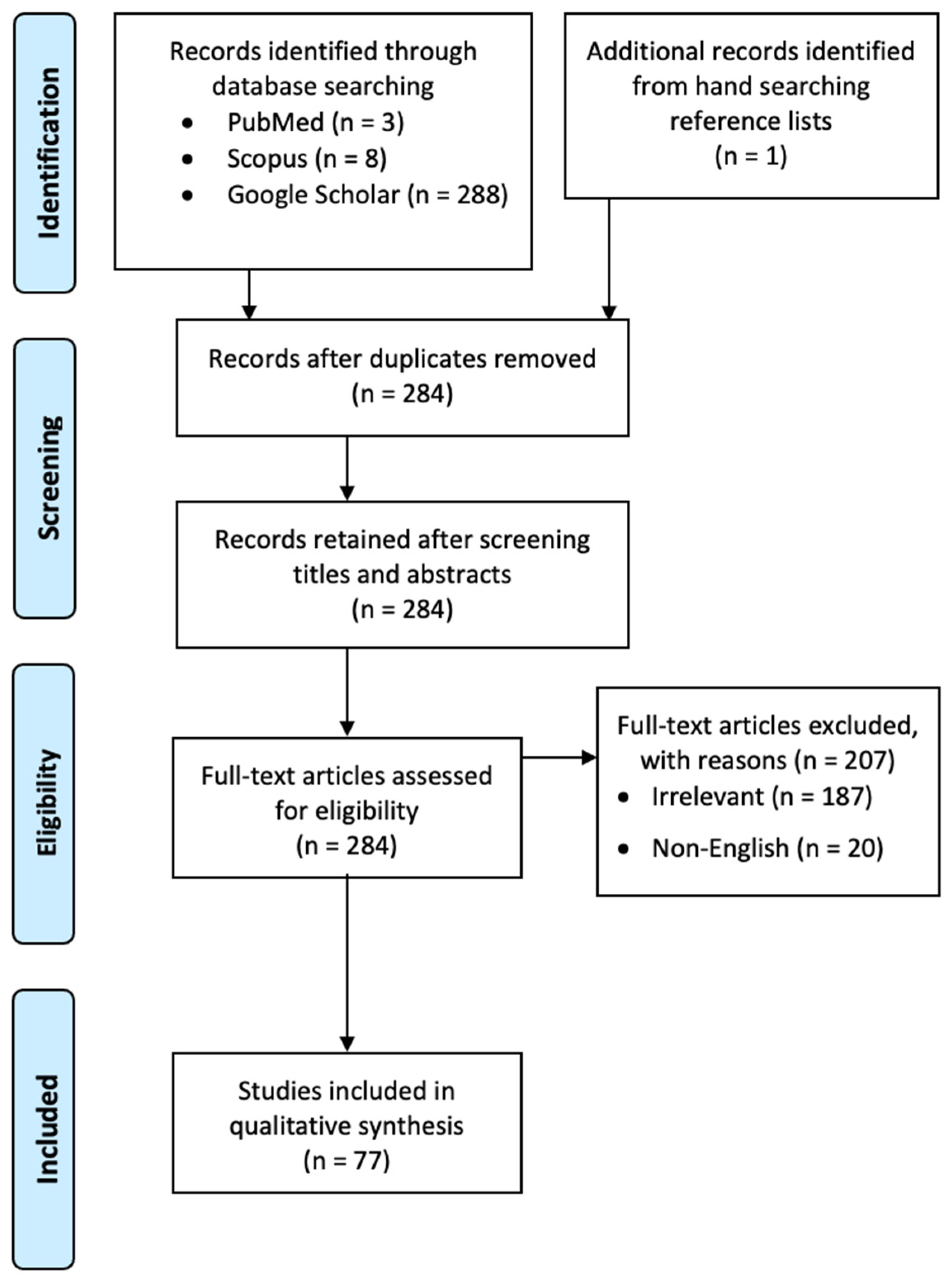
2. Materials and Methods
3. Results
4. Discussion
- “The radiographs were interpreted with caution, bearing in mind the possibility of false-positive diagnoses that might arise from the Mach-band effect…” [22]
- “A tendency to make false-positive scores is common, and this could be due to the Mach-band effect…” [23]
- “A high radio-opacity near a less radioopaque area can cause the Mach Band effect… This effect might be misinterpreted as caries…” [24]
- “Observers had to score voids within the root filling and not around it. The artefactual burnout phenomena (Mach bands) seen on CBCT images around root fillings were explained and the observers were told not to score them as a void…” [14]
- “To avoid false positive diagnoses caused by the Mach band effect, only obvious radiolucencies in the dentin were scored as dentin caries. This applied in particular to small radiolucencies adjacent to the enamel-dentin border of occlusal surfaces; surfaces with such radiolucencies were scored as sound…” [27]
- “Since the contrast between enamel and dentin may result in the Mach-band effect, the suspected area was carefully scrutinized and compared to the DEJs [enamel-dentinal junction] in the other teeth on the patient’s bitewings…” [16]
5. Conclusions
Supplementary Materials
Funding
Institutional Review Board Statement
Informed Consent Statement
Data Availability Statement
Conflicts of Interest
References
- Mach, E. Uber die wirkung der raumlichen Vertheilung des Lichterizes auf die netzhaut, I. Sitzungsberichte der mathematischnaturwissenschaftlichen Classe der kaiserlichen. Akad. Wiss. 1865, 52, 303–322. [Google Scholar]
- Lane, E.J.; Proto, A.V.; Phillips, T.W. Mach bands and density perception. Radiology 1976, 121, 9–17. [Google Scholar] [CrossRef]
- Gordenne, W.; Malchair, F. Mach bands in mammography. Radiology 1988, 169, 55–58. [Google Scholar] [CrossRef]
- Daffner, R. Pseudofracture of the dens: Mach bands. AJR Am. J. Roentgenol. 1977, 128, 607–612. [Google Scholar] [CrossRef]
- Friedman, A.; Lautin, E.; Rothenberg, L. Mach bands and pneumomediastinum. J. Can. Assoc. Radiol. 1981, 32, 232–235. [Google Scholar] [PubMed]
- Berry, H.M., Jr. Cervical burnout and Mach band: Two shadows of doubt in radiologic interpretation of carious lesions. J. Am. Dent. Assoc. 1983, 106, 622–625. [Google Scholar] [CrossRef] [PubMed]
- Manson, J. The lamina dura. Oral Surg. Oral Med. Oral Pathol. 1963, 16, 432–438. [Google Scholar]
- Kilpinen, E.; Hakala, P. Reproduction of the lamina dura in dental radiographs. Dentomaxillofac. Radiol. 1978, 7, 51–54. [Google Scholar] [CrossRef]
- Nielsen, C.J. Effect of scenario and experience on interpretation of mach bands. J. Endod. 2001, 27, 687–691. [Google Scholar] [CrossRef] [PubMed]
- Gunderman, R.B.; Patel, P. Perception’s crucial role in radiology education. Acad. Radiol. 2019, 26, 141–143. [Google Scholar] [CrossRef] [PubMed]
- Kelly, B.S.; Rainford, L.A.; Darcy, S.P.; Kavanagh, E.C.; Toomey, R.J. The development of expertise in radiology: In chest radiograph interpretation,“expert” search pattern may predate “expert” levels of diagnostic accuracy for pneumothorax identification. Radiology 2016, 280, 252–260. [Google Scholar] [CrossRef]
- Waite, S.; Grigorian, A.; Alexander, R.G.; Macknik, S.L.; Carrasco, M.; Heeger, D.J.; Martinez-Conde, S. Analysis of perceptual expertise in radiology—Current knowledge and a new perspective. Front. Hum. Neurosci. 2019, 13, 213. [Google Scholar] [CrossRef] [PubMed]
- Briley, J.; Dove, S.; Mertz-Fairhurst, E.; Hermesch, C. Computer-assisted densitometric image analysis (CADIA) of previously sealed carious teeth: A pilot study. Oper. Dent. 1997, 22, 105–114. [Google Scholar] [PubMed]
- Huybrechts, B.; Bud, M.; Bergmans, L.; Lambrechts, P.; Jacobs, R. Void detection in root fillings using intraoral analogue, intraoral digital and cone beam CT images. Int. Endod. J. 2009, 42, 675–685. [Google Scholar] [CrossRef]
- MacDonald, D. Viewing conditions and physiological phenomena and radiological interpretation. In Oral and Maxillofacial Radiology: A Diagnostic Approach; Wiley: Hoboken, NJ, USA, 2019; pp. 47–61. [Google Scholar]
- Mertz-Fairhurst, E.J.; Williams, J.E.; Schuster, G.S.; Smith, C.D.; Pierce, K.L.; Mackert Jr, J.R.; Sherrer, J.D.; Wenner, K.K.; Davis, Q.B.; Garman, T.A. Ultraconservative sealed restorations: Three-year results. J. Public Health Dent. 1991, 51, 239–250. [Google Scholar] [CrossRef]
- Qaramaleki, A.S.; Hassanpour, H. A New Method to Secondary Caries Detection in Restored Teeth. Int. J. Sci. Eng. Res. 2011, 2, 1–5. [Google Scholar]
- Secgin, C.K.; Gulsahi, A.; Arhun, N. Diagnostic challenge: Instances mimicking a proximal carious lesion detected by bitewing radiography. Oral Health Dent. Manag. 2016, 15, 1–5. [Google Scholar]
- Wilson, N.; Lynch, C.; Brunton, P.; Hickel, R.; Meyer-Lueckel, H.; Gurgan, S.; Pallesen, U.; Shearer, A.; Tarle, Z.; Cotti, E. Criteria for the replacement of restorations: Academy of Operative Dentistry European Section. Oper. Dent. 2016, 41, S48–S57. [Google Scholar] [CrossRef] [PubMed]
- Movahhedian, N.; Adibi, S.; Tavakoli, H.S.; Baseri, H. How does triangular-shaped radiolucency affect caries diagnosis? Oral Radiol. 2017, 33, 32–37. [Google Scholar] [CrossRef]
- Kuhnisch, J.; Pasler, F.; Bucher, K.; Hickel, R.; Heinrich-Weltzien, R. Frequency of non-carious triangular-shaped radiolucencies on bitewing radiographs. Dentomaxillofac. Radiol. 2008, 37, 23–27. [Google Scholar] [CrossRef]
- Chu, C.; Lo, E.; You, D. Clinical diagnosis of fissure caries with conventional and laser-induced fluorescence techniques. Lasers Med. Sci. 2010, 25, 355–362. [Google Scholar] [CrossRef] [PubMed][Green Version]
- Diniz, M.B.; Rodrigues, J.A.; Neuhaus, K.W.; Cordeiro, R.C.; Lussi, A. Influence of examiner’s clinical experience on the reproducibility and accuracy of radiographic examination in detecting occlusal caries. Clin. Oral Investig. 2010, 14, 515–523. [Google Scholar] [CrossRef] [PubMed]
- Lachowski, K.M.; Botta, S.; Lascala, C.; Matos, A.; Sobral, M. Study of the radio-opacity of base and liner dental materials using a digital radiography system. Dentomaxillofac. Radiol. 2013, 42, 20120153. [Google Scholar] [CrossRef]
- Nicholson, J.M.; Mordaunt, M.; Lopez, P.; Uppala, A.; Rosati, D.; Rodrigues, N.P.; Grabitz, P.; Rife, S.C. Scite: A smart citation index that displays the context of citations and classifies their intent using deep learning. Quant. Sci. Stud. 2021, 2, 882–898. [Google Scholar] [CrossRef]
- Yeung, A.W.K.; Cushing, C.A.; Lee, A.L. A bibliometric evaluation of the impact of theories of consciousness in academia and on social media. Conscious. Cogn. 2022, 100, 103296. [Google Scholar] [CrossRef]
- Ridell, K.; Olsson, H.; Mejàre, I. Unrestored dentin caries and deep dentin restorations in Swedish adolescents. Caries Res. 2008, 42, 164–170. [Google Scholar] [CrossRef] [PubMed]
- Buckle, C.E.; Udawatta, V.; Straus, C.M. Now you see it, now you don’t: Visual illusions in radiology. Radiographics 2013, 33, 2087–2102. [Google Scholar] [CrossRef] [PubMed]
- Roberts, B.; Harris, M.G.; Yates, T.A. The roles of inducer size and distance in the Ebbinghaus illusion (Titchener circles). Perception 2005, 34, 847–856. [Google Scholar] [CrossRef] [PubMed]
- Hung, K.; Yeung, A.W.K.; Tanaka, R.; Bornstein, M.M. Current applications, opportunities, and limitations of AI for 3D imaging in dental research and practice. Int. J. Environ. Res. Public Health 2020, 17, 4424. [Google Scholar] [CrossRef] [PubMed]


| No. of Publications | % (of 77) | |
|---|---|---|
| (A) Structural interface | ||
| Implant/bone | 10 | 13.0 |
| Restoration/tooth | 28 | 36.4 |
| Root filling/root canal | 3 | 3.9 |
| Enamel-dentinal junction | 21 | 27.3 |
| (B) Diagnostic relevance | ||
| Relevance to edge detection | 1 | 1.3 |
| Mimics caries | 47 | 61.0 |
| Mimics marginal defect | 5 | 6.5 |
| Mimics void in root filling | 3 | 3.9 |
| Mimics triangular-shaped radiolucency | 2 | 2.6 |
| Mimics root fracture | 2 | 2.6 |
| Mimics fracture at mandibular angle | 1 | 1.3 |
| Mimics a capsule around a lesion | 2 | 2.6 |
| No. of Publications | % (of 53) | |
|---|---|---|
| Introduction | 13 | 24.5 |
| Methods | 7 | 13.2 |
| Results | 2 | 3.8 |
| Discussion | 39 | 73.6 |
| Conclusion | 1 | 1.9 |
Publisher’s Note: MDPI stays neutral with regard to jurisdictional claims in published maps and institutional affiliations. |
© 2022 by the author. Licensee MDPI, Basel, Switzerland. This article is an open access article distributed under the terms and conditions of the Creative Commons Attribution (CC BY) license (https://creativecommons.org/licenses/by/4.0/).
Share and Cite
Yeung, A.W.K. The Diagnostic Relevance and Interfaces Covered by Mach Band Effect in Dentistry: An Analysis of the Literature. Healthcare 2022, 10, 632. https://doi.org/10.3390/healthcare10040632
Yeung AWK. The Diagnostic Relevance and Interfaces Covered by Mach Band Effect in Dentistry: An Analysis of the Literature. Healthcare. 2022; 10(4):632. https://doi.org/10.3390/healthcare10040632
Chicago/Turabian StyleYeung, Andy Wai Kan. 2022. "The Diagnostic Relevance and Interfaces Covered by Mach Band Effect in Dentistry: An Analysis of the Literature" Healthcare 10, no. 4: 632. https://doi.org/10.3390/healthcare10040632
APA StyleYeung, A. W. K. (2022). The Diagnostic Relevance and Interfaces Covered by Mach Band Effect in Dentistry: An Analysis of the Literature. Healthcare, 10(4), 632. https://doi.org/10.3390/healthcare10040632