Article

Diagnostic Utility of Hysteroscopic Biopsy in Cases of Suspected Lobular Endocervical Glandular Hyperplasia and Comparison with Cervical Conization

Department of Obstetrics and Gynecology, Kindai University Faculty of Medicine, Osakasayama 589-8511, Japan
*
Author to whom correspondence should be addressed.
Healthcare 2023, 11(11), 1619; https://doi.org/10.3390/healthcare11111619
Submission received: 12 April 2023 / Revised: 24 May 2023 / Accepted: 29 May 2023 / Published: 1 June 2023

Abstract

:
Background: Cervical cystic lesions encompass a range of benign and malignant pathologies. Magnetic resonance imaging or cytology alone cannot provide a definitive diagnosis, and conventional practice involves performing a cervical biopsy by conization to confirm the histology in cases exhibiting potential signs of lobular endocervical glandular hyperplasia (LEGH) or malignancy. However, as postoperative complications resulting from conization can impact future fertility and pregnancy, alternative diagnostic methods are needed for reproductive-age patients. This study aimed to establish the efficacy of a hysteroscopic biopsy for diagnosing cervical cystic lesions and compare it with conization. Methods: Thirteen patients with cervical cystic lesions suspected of LEGH or malignancy underwent a hysteroscopic biopsy, while 23 underwent conization. Patient background information, preoperative evaluation, histology, and postoperative outcomes were collected and compared retrospectively. Results: No significant differences were found between the hysteroscopy and conization groups in terms of mean patient age (45 vs. 48 years), operating time (23 vs. 35 min), blood loss (small amount vs. 43 mL), and postoperative hospitalization (1.1 vs. 1.6 days). Conclusion: A hysteroscopic biopsy allows for targeted resection of the cervix while maintaining diagnostic accuracy. It may serve as an efficient method for diagnosing cervical cystic lesions.

1. Introduction

Cystic lesions of the uterine cervix encompass a range of benign and malignant conditions, including nabothian cysts, lobular endocervical glandular hyperplasia (LEGH), and gastric-type mucinous carcinoma, including minimal deviation adenocarcinoma (MDA) [1]. LEGH, first described by Nucci et al., is a lesion that needs to be differentiated from MDA and is considered a potential precursor to cervical cancer [2,3,4]. The incidence of LEGH is rare, estimated at 0.7% [5]. It commonly affects women in their 40 s and 50 s presenting with typical symptoms such as increased vaginal discharge. However, some cases are asymptomatic, posing a challenge for early detection [6]. Ultrasonography, a standard tool in obstetric and gynecological examinations, is insufficient for a definitive diagnosis. In cases where cervical polycysts are observed on an ultrasound, magnetic resonance imaging (MRI) is often performed.
Characteristic imaging findings, such as the “cosmos pattern”, have been reported for LEGH and other malignant lesions [7,8]. Although a MRI is expected to aid in the diagnosis, a definitive diagnosis cannot rely solely on imaging [9,10]. Another suggested diagnostic method involves the detection of gastric mucin by collecting cervical mucus and using a HIK1083 latex agglutination kit [11]. However, this method often fails to achieve successful detection, rendering it an adjunct to the diagnosis [12]. While cervical cytology is commonly performed in gynecological practices and can identify atypical glandular cells in some cases of LEGH and malignancy, other cases may yield a diagnosis negative for intraepithelial lesions or malignancy. Therefore, when MRI findings of patients with cervical cystic lesions suggest the possibility of LEGH or malignancy, a cervical biopsy of the cystic lesions and histopathological assessment, including immunostaining, are required to confirm the current conditions [2,5].
Traditionally, cervical biopsy by conization has been the standard approach for cervical cystic lesions [7]. However, in such cases, cervical conization often results in a deeper resection of the cervix compared to the conventional conical resection performed for conditions such as cervical intraepithelial neoplasia (CIN). This is due to the frequent proximity of cystic lesions to the internal os of the uterus. Deeper cervical resection increases the risk of miscarriage and preterm birth due to cervical shortening during postoperative pregnancy [13,14]. Moreover, it raises the risk of postoperative cervical stenosis and closure, potentially reducing fertility in women of childbearing age [15]. Although cervical conization is considered a fertility-preserving procedure for reproductive-age females, it can inadvertently complicate future fertility and pregnancies. Deep conization for a biopsy site can negatively impact the quality of life in older patients, as cervical stenosis and closure can lead to menstrual abnormalities and hematometra [16]. Therefore, concerns arise regarding the long-term complications associated with deep conization for biopsy purposes.
To date, no literature has been published regarding the postoperative complications of cervical conization or the risk of miscarriage and preterm birth in cases of cystic lesions of the uterine cervix requiring a cervical biopsy for diagnosis. However, it is widely recognized that a history of deeper resection through cervical conization for CIN cases increases the risk of miscarriage and preterm delivery [17,18], suggesting a similar outcome for cervical cystic lesions. Even if a deeper resection of the cervix is performed in cases suspected of LEGH or malignancy, the biopsy may not yield reliable histological findings to determine whether uterine preservation is a viable option for the patient. Consequently, some patients may opt for a total hysterectomy due to concerns about overlooking malignancy. This decision presents a significant challenge for individuals desiring pregnancy, as they may face permanent infertility if no abnormalities are found, leading to an irreversible loss of fertility.
Alternative methods for diagnosing cervical cystic lesions that are less invasive yet maintain diagnostic accuracy are crucial, particularly for patients of reproductive age. One such approach involves the use of a hysteroscope for the biopsy. A hysteroscope is an endoscope that is inserted through the cervix into the uterine corpus [19]. Originally designed for submucosal fibroids and endometrial polyps protruding into the uterine cavity, hysteroscopic surgery allows for resectioning the lesion using various types of electrodes located at the tip of the hysteroscope. The excised tissue is then removed from the uterus through the cervical canal [20,21,22]. This technique provides surgeons with direct visualization of the cystic lesions from within the uterus, allowing for targeted biopsy and minimizing the extent of resection while ensuring an accurate diagnosis.
While a cervical biopsy of cystic lesions using transcervical resection with a hysteroscope has been reported previously [8], the limited number of cases hinders the ability to establish the effectiveness of a hysteroscopic biopsy. In our study, we performed hysteroscopic biopsies on 13 cases, with MRI findings indicating the potential presence of LEGH or malignancy. This report compares these cases and previous cases that underwent conventional conization for biopsy purposes.

2. Materials and Methods

Patients referred to Kindai University Hospital for multiple cystic lesions in the cervix underwent pelvic MRI scans. Those whose T2-weighted images demonstrated the cosmos pattern (diffuse or microcystic parts surrounded by medium-to-large cysts) [8], suggesting LEGH or malignancy, were offered a transcervical biopsy by hysteroscopic resection. The patients who agreed to a hysteroscopic biopsy received cervical dilation through the transcervical insertion of a laminaria tent the day before the biopsy. Transcervical resection using monopolar was performed using the OES Pro resectoscope (OLYMPUS Co., Tokyo, Japan), equipped with the HF surgery system UES-40S (OLYMPUS Co., Tokyo, Japan), a high-frequency power generator with an output power of 50 W for excision and 50 W for coagulation. The scope was fitted with a loop electrode for excision and a ball electrode for coagulation. During the hysteroscopic procedure, 3% D-sorbitol was perfused. The hysteroscopic observation revealed elevated cysts around the cervical canal (Figure 1A). As the surfaces of the cysts were resected, the inner cavities were exposed, and mucinous contents leaked out (Figure 1B,C). The cyst surface was removed around the entire circumference of the cervical canal. Bleeding areas were coagulated, and the hysteroscope was removed from the uterus once hemostasis was confirmed (Figure 1D). Patients were discharged from the hospital on the first postoperative day and scheduled for a follow-up appointment at the outpatient clinic in 2–4 weeks.
Second, we reviewed similar cases of cervical cystic lesions over the past decade at our hospital, which required cervical conization for a biopsy. Cervical resection was performed in a conical shape using HARMONIC SYNERGY® (Johnson & Johnson, Tokyo, Japan), an ultrasonic scalpel, for the conization procedure. A hooked blade was attached to the device for resection. Since the lesions were typically located near the internal os of the uterus, resection was performed with a depth of 2 cm or more to ensure complete removal of the lesions. Patients were discharged on the first postoperative day.
In this study, we assessed 13 patients who underwent a hysteroscopic biopsy and 23 patients who underwent conventional conization when a preoperative diagnosis of LEGH or higher lesions was suspected but not confirmed. Conization was the chosen technique for both procedures between 2011 and 2019. Conversely, a hysteroscopic biopsy was performed on the cases between 2020 and 2022. Therefore, it is important to note that the two groups in this study are not contemporaneous but rather represent a retrospective study with different study periods.
Patient information such as backgrounds, preoperative assessment, perioperative complications, and postoperative outcomes was collected for each group from their medical charts. The diagnostic accuracy of the pathology results was also evaluated in patients who underwent a hysteroscopic biopsy or conization followed by total hysterectomy. Additional details regarding the histological diagnosis were examined for cases involving a hysterectomy. A comparison was made between the two groups in terms of the mean age of the patients, operation time, blood loss, and postoperative hospital stay. Statistical analysis was performed using a Student’s t-test, with a significance level set at p < 0.05. This study received approval from the institutional review board of the Kindai University Faculty of Medicine (RR03-25).

3. Results

Thirteen patients underwent a hysteroscopic biopsy for cervical cystic lesions between April 2020 and September 2022. Table 1 presents the patients’ backgrounds, preoperative assessments, and postoperative outcomes. Of the 13 patients who had a hysteroscopic biopsy, five had nabothian cysts, and three had no significant histological findings. Two cases were diagnosed with LEGH, while one case revealed gastric-type mucinous carcinoma. All patients diagnosed with LEGH and mucinous carcinoma underwent a total hysterectomy. In the case of a LEGH diagnosis, two patients were given the options of a hysterectomy or observation with close monitoring, and both chose the former. One patient opted for a hysterectomy to mitigate the potential risk of developing cervical cancer, while the other desired treatment to alleviate abundant mucinous discharge.
From January 2011 to March 2020, a total of 23 patients with cervical cystic lesions underwent cervical conization for a biopsy. The patients’ backgrounds, preoperative assessments, and postoperative outcomes are detailed in Table 2. Among the cases, 12 were diagnosed with nabothian cysts or showed no significant histological findings. LEGH was identified in two cases, while malignancy was detected in four cases (three cases of MDA and one case of endocervical mucinous carcinoma). Among the cases that underwent a total hysterectomy after conization, two cases initially diagnosed with LEGH had additional findings of AIS and mucinous carcinoma. Despite two cases being diagnosed with no findings and glandular hyperplasia, a recommendation was made to proceed with a hysterectomy following conization due to concerns that the procedure might have failed to reach the lesions, resulting in a potential underdiagnosis. A flowchart illustrating the postoperative pathology results for each technique is presented in Figure 2.
In principle, a hysteroscopic biopsy was performed in cases of suspected LEGH or higher, and the diagnostic accuracy is shown in Table 3 for cases in which a total hysterectomy was finally performed. The sensitivity was 100%, the specificity was 67%, the positive predictive value was 80%, and the negative predictive value was 100%. Table 4 shows the results of the same study for conization. The sensitivity was 86%, specificity 50%, positive predictive value 86%, and negative predictive value 50%.
A comparison between the hysteroscopy and conization groups revealed no significant difference in the mean age of the patients (45 vs. 48 years). The hysteroscopy group had a relatively shorter mean operative time (23 vs. 35 min), although the difference was not statistically significant. Minimal blood loss was observed in all hysteroscopy cases, with an average of 43 mL in the conization procedures. The postoperative hospital stays were 1.1 and 1.6 days, respectively, with no significant difference observed (Table 5).

4. Discussion

We have used both hysteroscopic resection and conization as biopsy methods for cervical cystic lesions, with the MRI findings indicating the potential for LEGH or malignancy. The MRI findings in this study revealed a range of lesion sizes or cysts within the lesions, which were consistently identified as elevated cysts during hysteroscopy (Figure 3). More than half of the patients were diagnosed with nabothian cysts or no lesions. In cases of LEGH, which carry an inherent risk of developing into malignancy [6], a total hysterectomy could be considered for patients not seeking pregnancy, while close observation with cytology and an imaging assessment should be recommended for those desiring pregnancy [23,24,25]. However, two cases in the conization group were diagnosed with cervical malignancy post-hysterectomy, despite the preoperative biopsy indicating LEGH. Therefore, the possibility of underdiagnosing becomes a concern when conization is used for a biopsy with a result of LEGH. Conversely, two cases in the hysteroscopy group initially diagnosed with gastric-type lesions were ultimately found to have nabothian cysts and LEGH, respectively, after a hysterectomy. Consequently, determining the need for additional interventions based on this diagnosis has become a matter for further consideration.
Conization is a commonly employed biopsy method for cervical cystic lesions. However, compared to conization performed for CIN, it necessitates a deeper resection, which can result in increased blood loss and a longer operative time. Furthermore, it is associated with obstetric complications, such as a 1.5- to 3-times higher risk of preterm delivery during pregnancy compared to the overall rate of 8 to 15% [13]. Recent reports have also demonstrated a correlation between deep conical resection and an elevated risk of preterm delivery [26]. Complications arising from cervical conization include cervical stenosis and cervical atresia [15], which may require additional intervention [27,28]. In severe cases, a total hysterectomy may be the only viable option [28]. Therefore, attempts are being made to develop devices that can be inserted into the cervical canal [27,28]. Nonetheless, it is crucial to avoid complications such as cervical canal stenosis or closure.
Hysteroscopic procedures are associated with specific complications, including uterine perforation and fluid overload [29,30,31]. However, since the operative time was typically under half an hour in most cases, the risk of fluid overload due to a prolonged operative time was unlikely to be a concern. In our previous cases, we utilized a monopolar technique without an electrolyte solution. Nevertheless, in recent years, the mainstream approach has shifted towards a bipolar biopsy with an electrolyte solution. As a result, we now perform bipolar hysteroscopic biopsies in our department. Our protocol recommends excising only the surfaces of the lesions that can be visualized as elevated cysts around the cervical canal using a hysteroscope. There is no need for deep cervical resection to access the lesions, thereby minimizing the risk of uterine perforation. Although there may be concerns about the adequacy of tissue collection, the fact that the hysteroscopic biopsy in this study allowed for a pathological assessment without compromising the diagnostic yield supports the effectiveness of our protocol. Therefore, the excision technique used in hysteroscopic biopsies minimizes the loss of normal cervical tissue. While there were no significant differences in the perioperative complications between hysteroscopic resection and conization, the hysteroscopy group exhibited shorter operative times and minimal blood loss. Conversely, conization was associated with an increased operative time and blood loss in a few cases, likely due to the necessity for deep conization, which carries a higher risk of bleeding. Even when deep resection was performed during conization, two cases in the conization group eventually required a hysterectomy due to concerns about an underdiagnosis. Hysteroscopic resection allows surgeons to visualize cystic lesions and confirm the biopsy site, ensuring the quality of the biopsy. It is considered an alternative method for efficiently and minimally invasively conducting biopsies of cervical cystic lesions.
Regarding the costs of both procedures, in Japan, a conization surgery costs the patient approximately 33,300 JPY (about 247 USD) in terms of surgical expenses (additional fees for anesthesia and other services apply). The disposable ultrasonic scalpel HARMONIC SYNERGY® costs 47,000 JPY (about 348 USD) at the listed price (the actual delivered price is slightly lower). On the other hand, the cost of a monopolar hysteroscopic procedure is 47,300 JPY (about 350 USD), while the bipolar procedure using saline solution costs the patient approximately 66,300 JPY (about 491 USD). Regarding the equipment, both loop and roller electrodes have a listed price of 15,000 JPY (about 111 USD). However, there is a potential for cost reduction for these electrodes. For saline bipolar, both loop and roller electrodes have a listed price of 19,000 JPY (about 141 USD). These electrodes are disposable and must be discarded after use. Various techniques can be used for both conization and hysteroscopic surgery, each with its associated costs. However, a cost comparison conducted at our hospital revealed no significant difference between the two procedures (note that hysteroscopic surgery requires an initial investment in equipment such as monitors).
One limitation of this study is the small number of cases and the short duration of follow-up. While the long-term complications resulting from cervical conization have been extensively discussed, the impact of partial resection of the cervix through a hysteroscopic approach on the cervical physiology has not been well documented. In this series of cases, the postoperative effects of hysteroscopic resection on fertility and future pregnancies remain unclear, as no patients have attempted pregnancy following the hysteroscopic biopsy. Since hysteroscopic resection involves minimal cervical resection compared to deep conization, it is anticipated to carry a lower risk for fertility and future pregnancies. Further investigation and data accumulation are necessary to assess the long-term complications associated with this biopsy method. Additionally, this study focuses on the examination of the technique itself. The most crucial aspect of a new technique is diagnostic accuracy. We believe that the accuracy of this new method should be reevaluated as the number of cases increases in the future. Nevertheless, the current data are comparable to those obtained through conventional methods.

5. Conclusions

A hysteroscopic biopsy holds promise as a valuable method for diagnosing cystic lesions of the cervix. The key advantage of this technique lies in direct visualization of the lesion through an endoscope, enabling targeted biopsy of the identified area. However, further studies are required to expand the number of cases, extend the observation period, and investigate additional aspects such as diagnostic accuracy.

Author Contributions

R.S.: Concept and design, data collection, article writing, and approved the final version. Y.K. (Yasushi Kotani): Data collection, data analysis, article writing, and approved the final version. M.O.: Data collection, article writing, and approved the final version. H.S.: Data collection, article writing, and approved the final version. Y.K. (Yoko Kashima): Data collection, article writing, and approved the final version. K.M.: Data collection, data analysis, article writing, and approved the final version. K.K.: Data collection, article writing, and approved the final version. H.N.: Supervision, article writing, and approved the final version. N.M.: Data analysis, article writing, and approved the final version. All authors contributed to the article and approved the submitted version. All authors have read and agreed to the published version of the manuscript.

Funding

This research received no external funding.

Institutional Review Board Statement

The study was conducted in accordance with the Declaration of Helsinki and approved by the Institutional Review Board of Kindai University (protocol code RR03-25, 8 December 2022).

Informed Consent Statement

All study subjects provided informed consent in principle, but those who could not be contacted were given the opportunity to refuse to participate in the study by opting out. Those who refused to give consent were excluded from the study.

Data Availability Statement

Not applicable.

Conflicts of Interest

The authors declare no conflict of interest.

References

  1. Mikami, Y.; McCluggage, W.G. Endocervical glandular lesions exhibiting gastric differentiation: An emerging spectrum of benign, premalignant, and malignant lesions. Adv. Anat. Pathol. 2013, 20, 227–237. [Google Scholar] [CrossRef] [PubMed]
  2. Nucci, M.R.; Clement, P.B.; Young, R.H. Lobular endocervical glandular hyperplasia, not otherwise specified: A clinicopathologic analysis of thirteen cases of a distinctive pseudoneoplastic lesion and comparison with fourteen cases of adenoma malignum. Am. J. Surg. Pathol. 1999, 23, 886–891. [Google Scholar] [CrossRef] [PubMed]
  3. Mikami, Y.; Kiyokawa, T.; Hata, S.; Fujiwara, K.; Moriya, T.; Sasano, H.; Manabe, T.; Akahira, J.; Ito, K.; Tase, T.; et al. Gastrointestinal immunophenotype in adenocarcinomas of the uterine cervix and related glandular lesions: A possible link between lobular endocervical glandular hyperplasia/pyloric gland metaplasia and ‘adenoma malignum’. Mod. Pathol. 2004, 17, 962–972. [Google Scholar] [CrossRef] [PubMed]
  4. Takatsu, A.; Miyamoto, T.; Fuseya, C.; Suzuki, A.; Kashima, H.; Horiuchi, A.; Ishii, K.; Shiozawa, T. Clonality analysis suggests that STK11 gene mutations are involved in progression of lobular endocervical glandular hyperplasia (LEGH) to minimal deviation adenocarcinoma (MDA). Virchows Arch. 2013, 462, 645–651. [Google Scholar] [CrossRef]
  5. Mikami, Y.; Hata, S.; Fujiwara, K.; Imajo, Y.; Kohno, I.; Manabe, T. Florid endocervical glandular hyperplasia with intestinal and pyloric gland metaplasia: Worrisome benign mimic of “adenoma malignum”. Gynecol. Oncol. 1999, 74, 504–511. [Google Scholar] [CrossRef]
  6. Mikami, Y.; Hata, S.; Melamed, J.; Fujiwara, K.; Manabe, T. Lobular endocervical glandular hyperplasia is a metaplastic process with a pyloric gland phenotype. Histopathology 2001, 39, 364–372. [Google Scholar] [CrossRef]
  7. Miyamoto, T.; Kobara, H.; Shiozawa, T. Biology and management of lobular endocervical glandular hyperplasia. J. Obstet. Gynaecol. Res. 2022, 48, 3056–3067. [Google Scholar] [CrossRef]
  8. Yamada, Y.; Kobara, H.; Shinagawa, M.; Ono, M.; Takeuchi, H.; Ida, K.; Miyamoto, T.; Shiozawa, T. Effectiveness of hysteroscopic transcervical resection in the diagnosis of lobular endocervical glandular hyperplasia: A case report. Jap. J. Gynecoloc. Obstet. Endosc. 2021, 37, 211–215. [Google Scholar] [CrossRef]
  9. Takatsu, A.; Shiozawa, T.; Miyamoto, T.; Kurosawa, K.; Kashima, H.; Yamada, T.; Kaku, T.; Mikami, Y.; Kiyokawa, T.; Tsuda, H.; et al. Preoperative differential diagnosis of minimal deviation adenocarcinoma and lobular endocervical glandular hyperplasia of the uterine cervix: A multicenter study of clinicopathology and magnetic resonance imaging findings. Int. J. Gynecol. Cancer 2011, 21, 1287–1296. [Google Scholar] [CrossRef]
  10. Stoehr, A.; Nann, D.; Staebler, A.; Oberlechner, E.; Brucker, S.Y.; Bachmann, C. Difficulties in diagnosis of a minimal deviation adenocarcinoma of uterine cervix diagnosed postoperatively: Brief communication and literature review. Arch. Gynecol. Obstet. 2019, 300, 1029–1043. [Google Scholar] [CrossRef]
  11. Ishii, K.; Kumagai, T.; Tozuka, M.; Ota, H.; Katsuyama, T.; Kurihara, M.; Shiozawa, T.; Noguchi, H. A new diagnostic method for adenoma malignum and related lesions: Latex agglutination test with a new monoclonal antibody, HIK1083. Clin. Chim. Acta 2001, 312, 231–233. [Google Scholar] [CrossRef]
  12. Ida, K.; Yamanoi, K.; Asaka, S.; Takeuchi, H.; Miyamoto, T.; Shiozawa, T.; Nakayama, J. alphaGlcNAc and its catalyst alpha4GnT are diagnostic and prognostic markers in uterine cervical tumor, gastric type. Sci. Rep. 2019, 9, 13043. [Google Scholar] [CrossRef]
  13. Kyrgiou, M.; Athanasiou, A.; Paraskevaidi, M.; Mitra, A.; Kalliala, I.; Martin-Hirsch, P.; Arbyn, M.; Bennett, P.; Paraskevaidis, E. Adverse obstetric outcomes after local treatment for cervical preinvasive and early invasive disease according to cone depth: Systematic review and meta-analysis. BMJ 2016, 354, i3633. [Google Scholar] [CrossRef]
  14. Bevis, K.S.; Biggio, J.R. Cervical conization and the risk of preterm delivery. Am. J. Obstet. Gynecol. 2011, 205, 19–27. [Google Scholar] [CrossRef]
  15. Baldauf, J.J.; Dreyfus, M.; Ritter, J.; Meyer, P.; Philippe, E. Risk of cervical stenosis after large loop excision or laser conization. Obstet. Gynecol. 1996, 88, 933–938. [Google Scholar] [CrossRef]
  16. Tanaka, Y.; Ueda, Y.; Kakuda, M.; Kubota, S.; Matsuzaki, S.; Iwamiya, T.; Okazawa, A.; Matsuzaki, S.; Hashimoto, K.; Kobayashi, E.; et al. Predictors for recurrent/persistent high-grade intraepithelial lesions and cervical stenosis after therapeutic conization: A retrospective analysis of 522 cases. Int. J. Clin. Oncol. 2017, 22, 921–926. [Google Scholar] [CrossRef]
  17. Berghella, V.; Pereira, L.; Gariepy, A.; Simonazzi, G. Prior cone biopsy: Prediction of preterm birth by cervical ultrasound. Am. J. Obstet. Gynecol. 2004, 191, 1393–1397. [Google Scholar] [CrossRef]
  18. Acharya, G.; Kjeldberg, I.; Hansen, S.M.; Sorheim, N.; Jacobsen, B.K.; Maltau, J.M. Pregnancy outcome after loop electrosurgical excision procedure for the management of cervical intraepithelial neoplasia. Arch. Gynecol. Obstet. 2005, 272, 109–112. [Google Scholar] [CrossRef]
  19. Centini, G.; Troia, L.; Lazzeri, L.; Petraglia, F.; Luisi, S. Modern operative hysteroscopy. Minerva Ginecol. 2016, 68, 126–132. [Google Scholar]
  20. Keizer, A.L.; Jacobs, B.L.; Thurkow, A.L.; de Lange, M.E.; Radder, C.M.; van Kesteren, P.J.M.; Hanstede, M.M.F.; Huirne, J.A.F.; Hehenkamp, W.J.K. The effect of transcervical resection of submucous fibroids on menstrual blood loss: A prospective cohort study. Eur. J. Obstet. Gynecol. Reprod. Biol. 2022, 274, 128–135. [Google Scholar] [CrossRef]
  21. Preutthipan, S.; Herabutya, Y. Hysteroscopic polypectomy in 240 premenopausal and postmenopausal women. Fertil. Steril. 2005, 83, 705–709. [Google Scholar] [CrossRef] [PubMed]
  22. Moore, J.F.; Carugno, J. Hysteroscopy; StatPearls Publishing: Treasure Island, FL, USA, 2022. [Google Scholar]
  23. Kobara, H.; Miyamoto, T.; Otsuki, T.; Ohya, A.; Shiozawa, T. Worsening cytology and lesion enlargement are useful indicators for malignant transformation of lobular endocervical glandular hyperplasia during follow-up: A case report. Gynecol. Oncol. Rep. 2020, 32, 100571. [Google Scholar] [CrossRef] [PubMed]
  24. Okuyama, R.; Hashimoto, H.; Miura, T.; Sugita, M.; Arai, M.; Tsunoda, H.; Sasajima, Y.; Horiuchi, H. Two cases of adenocarcinoma in situ arising in lobular endocervical glandular hyperplasia indicating localization of mucin on the cluster surface as an early cytological finding of malignant transformation. Diagn. Cytopathol. 2017, 45, 842–847. [Google Scholar] [CrossRef] [PubMed]
  25. Ando, H.; Miyamoto, T.; Kashima, H.; Takatsu, A.; Ishii, K.; Fujinaga, Y.; Shiozawa, T. Usefulness of a management protocol for patients with cervical multicystic lesions: A retrospective analysis of 94 cases and the significance of GNAS mutation. J. Obstet. Gynaecol. Res. 2016, 42, 1588–1598. [Google Scholar] [CrossRef]
  26. Wiik, J.; Kärrberg, C.; Nilsson, S.; Strander, B.; Jacobsson, B.; Sengpiel, V. Associations between cervical intraepithelial neoplasia during pregnancy, previous excisional treatment, cone-length and preterm delivery: A register-based study from western Sweden. BMC Med. 2022, 20, 61. [Google Scholar] [CrossRef]
  27. Vieira, M.A.; de Araujo, R.L.C.; da Cunha Andrade, C.E.M.; Schmidt, R.L.; Filho, A.L.; Reis, R.D. A randomized clinical trial of a new anti-cervical stenosis device after conization by loop electrosurgical excision. PLoS ONE 2021, 16, e0242067. [Google Scholar] [CrossRef]
  28. Grund, D.; Kohler, C.; Krauel, H.; Schneider, A. A new approach to preserve fertility by using a coated nitinol stent in a patient with recurrent cervical stenosis. Fertil. Steril. 2007, 87, 1212.e13–1212.e16. [Google Scholar] [CrossRef]
  29. Jansen, F.W.; Vredevoogd, C.B.; van Ulzen, K.; Hermans, J.; Trimbos, J.B.; Trimbos-Kemper, T.C. Complications of hysteroscopy: A prospective, multicenter study. Obstet. Gynecol. 2000, 96, 266–270. [Google Scholar] [CrossRef]
  30. Elahmedawy, H.; Snook, N.J. Complications of operative hysteroscopy: An anaesthetist’s perspective. BJA Educ. 2021, 21, 240–242. [Google Scholar] [CrossRef]
  31. Aas-Eng, M.K.; Langebrekke, A.; Hudelist, G. Complications in operative hysteroscopy—Is prevention possible? Acta Obstet. Gynecol. Scand. 2017, 96, 1399–1403. [Google Scholar] [CrossRef]
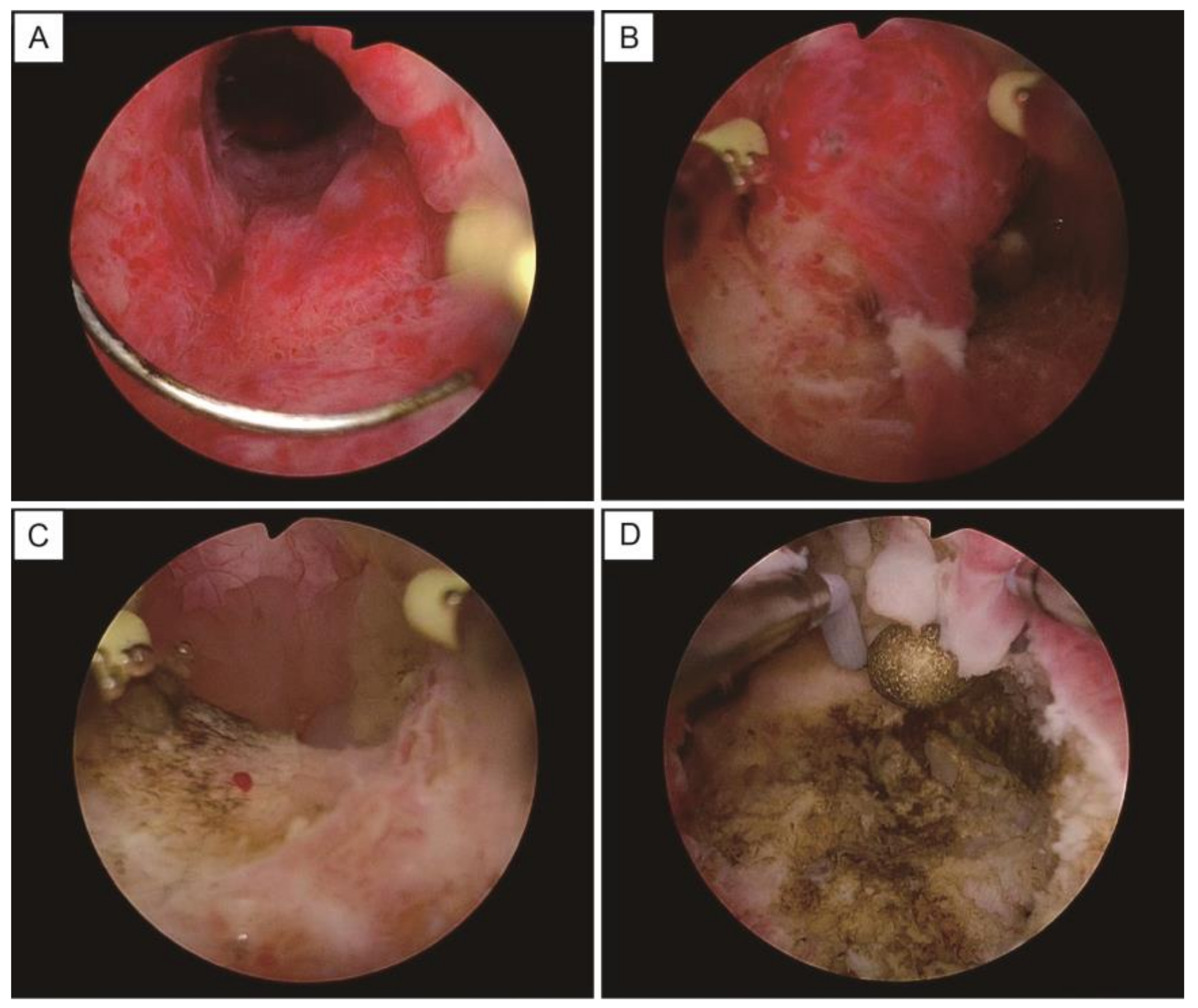
Figure 1. The images of a hysteroscopic biopsy. (A) The lesions are detected as elevated cysts around the cervical canal. (B) The surfaces of cysts are excised with a loop electrode. (C) The inner cavities of the cysts are exposed, with the mucinous contents leaking out of the cysts. (D) The bleeding sites are coagulated with a ball electrode.
Healthcare 11 01619 g001
Figure 2. Flowchart of the postoperative pathology results for hysteroscopy surgery and conization. Abbreviations: LEGH: lobular endocervical glandular hyperplasia and AIS: adenocarcinoma in situ.
Healthcare 11 01619 g002
Figure 3. MRI of the uterine cervix, and images of the lesions by hysteroscopic observation. (A) T2-weighted image, sagittal section. Cystic lesions in the cervix are stretching to the internal os of the uterus (arrows). (B) T2-weighted image, horizontal section. The sizes of the lesions and cysts differ among cases. (C) Cervical cystic lesions observed from inside the cervix by a hysteroscope.
Healthcare 11 01619 g003
Table 1. Patients’ background, preoperative evaluation, postoperative diagnosis, and postoperative course of cases in the hysteroscopy group.
CaseAgeGPGastric MucinCervical CytologyPathology after BiopsyHysterectomy after BiopySurgeryPathology after
Hysterectomy
144G0N/ALSILgastric-type lesionYTLHnabothian cyst
245G4P4positiveNILMLEGHYTLHLEGH
333G0N/AAGC-FNgastric-type mucinous carcinomaYLRHgastric-type mucinous carcinoma, pT2a2
431G0negativeNILMnabothian cystN
550G3P3negativeNILMnabothian cystN
637G1P0positiveNILMgastric-type lesionYTLHLEGH
748G1P1N/AAGC-FNnabothian cystN
848G2P2negativeNILMno lesionsY
(request by the patient)
TLHnabothian cyst
959G4P3negativeNILMnabothian cystN
1049G1P1N/ANILMnabothian cystY
(due to hypermenorrhea)
TLHnabothian cyst
1144G2P2negativeNILMno lesionsN
1248G4P1N/AAGC-NOSLEGHYTLHLEGH
1349G0N/AAGC-NOSno lesionsN
Abbreviations: GP: gravida/para, N/A: not applicable, LSIL: low-grade squamous intraepithelial lesion, NILM: negative for intraepithelial lesion or malignancy, AGC-FN: atypical glandular cells—favor neoplastic, AGC-NOS: atypical glandular cells—not otherwise specified, LEGH: lobular endocervical glandular hyperplasia, Y: Yes, N: No, TLH: total laparoscopic hysterectomy, and LRH: laparoscopic radical hysterectomy.
Table 2. Patients’ background, preoperative evaluation, postoperative diagnosis, and postoperative course of cases in the conization group.
CaseAgeGPGastric MucinCervical CytologyPathology after BiopsyHysterectomy after BiopsySurgeryPathology after
Hysterectomy
137G0N/ANILMno lesionN
240G0N/ANILMno lesionN
355G6P3N/ANILMnabothian cystN
441G2P2N/ANILMnabothian cystN
559G1P1N/AAGCnabothian cystN
635G1P1N/ANILMno lesionN
733G0N/ANILMnabothian cystY
(due to hypermenorrhea)
TAHnabothian cyst
869G1P0N/AAGCno lesionN
931G0N/AAGCMDAYmRHMDA
1048G1P0N/AAGCMDAYRHLEGH
1146G1P1N/AAGCLEGHYTLHLEGH, AIS
1246G2P1N/AAGCmicroglandular hyperplasiaN
1381G4P3N/Aadenocarcinoma nabothian cystN
1444G0N/AAGC-NOSno lesionYTLHLEGH
1555G4P3N/ANILMLEGHYTAHmucinous carcinoma
1645G0N/ANILMmicroglandular hyperplasiaN
1742G1P1N/AAGC-NOSnabothian cyst,
microglandular hyperplasia
N
1844G2P1N/ANILMmicroglandular hyperplasiaYTLHLEGH
1946G0N/AAGCMDAYRHLEGH
2037G0N/ANILMatypical glandsN
2156G4P3N/AAGCendocervical adenocarcinoma, usual typeYTLHno lesion
2252G3P2N/AASC-USCIN 1, nabothian cystN
2351G2P2N/ANILMno lesionN
Abbreviations: GP: gravida/para, N/A: not applicable, NILM: negative for intraepithelial lesion or malignancy, AGC: atypical glandular cells, AGC-NOS: atypical glandular cells—not otherwise specified, ASC-US: atypical squamous cells of undetermined significance, MDA: minimal deviation adenocarcinoma, LEGH: lobular endocervical glandular hyperplasia, CIN: cervical intraepithelial neoplasia, Y: Yes, N: No, TAH: total abdominal hysterectomy, mRH: modified radical hysterectomy, TLH: total laparoscopic hysterectomy, RH: radical hysterectomy, and AIS: adenocarcinoma in situ.
Table 3. Diagnostic accuracy in the hysteroscopic biopsy and total hysterectomy.
Total Hysterectomy
LEGH
or Greater Lesion
Nabothian Cyst
No Lesion
Total
Hysteroscopic
Biopsy
LEGH
or greater lesion
415
nabothian cyst
no lesion
022
Total437
Table 4. Diagnostic accuracy in the conization and total hysterectomy.
Total Hysterectomy
LEGH
or Greater Lesion
Nabothian Cyst
No Lesion
Total
ConizationLEGH
or greater lesion
617
nabothian cyst
no lesion
112
Total729
Table 5. Comparison of patients’ backgrounds and surgical outcomes between a hysteroscopic biopsy and a cervical conization.
Hysteroscpopy (n = 13)Conization (n = 23)p Value
mean age
(year)
45 ± 848 ± 120.49
operative time
(minute)
23 ± 1235 ± 200.06
total blood loss
(mL)
small amounts
(not countable)
43 ± 70
postoperative hospital stay
(day)
1.1 ± 0.21.6 ± 1.40.22
Disclaimer/Publisher’s Note: The statements, opinions and data contained in all publications are solely those of the individual author(s) and contributor(s) and not of MDPI and/or the editor(s). MDPI and/or the editor(s) disclaim responsibility for any injury to people or property resulting from any ideas, methods, instructions or products referred to in the content.