The Characteristics of Adjacent Anatomy of Mandibular Third Molar Germs: A CBCT Pilot Study in Patients with Osteogenesis Imperfecta

 , , ,
, , ,  and
and
Abstract
:1. Introduction
Development of the Mandibular Third Molar
2. Important Factors Which Contribute to Failure of Tooth Eruption
3. Materials and Methods
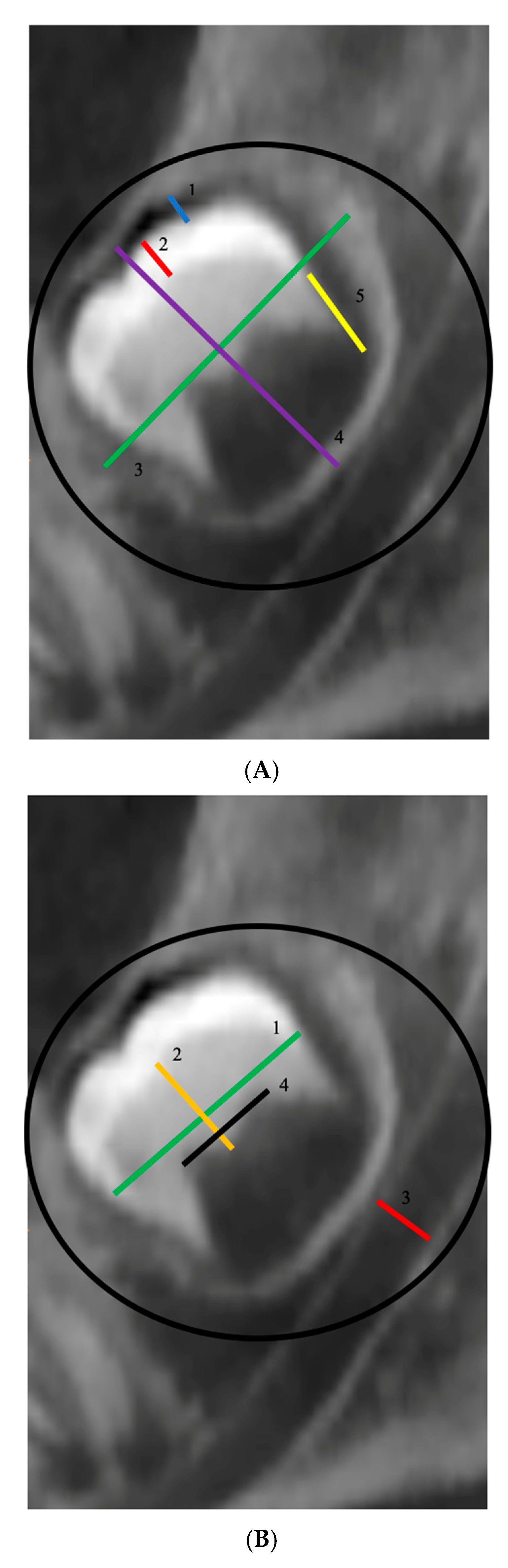
Instruments and Procedures
4. Data Analysis
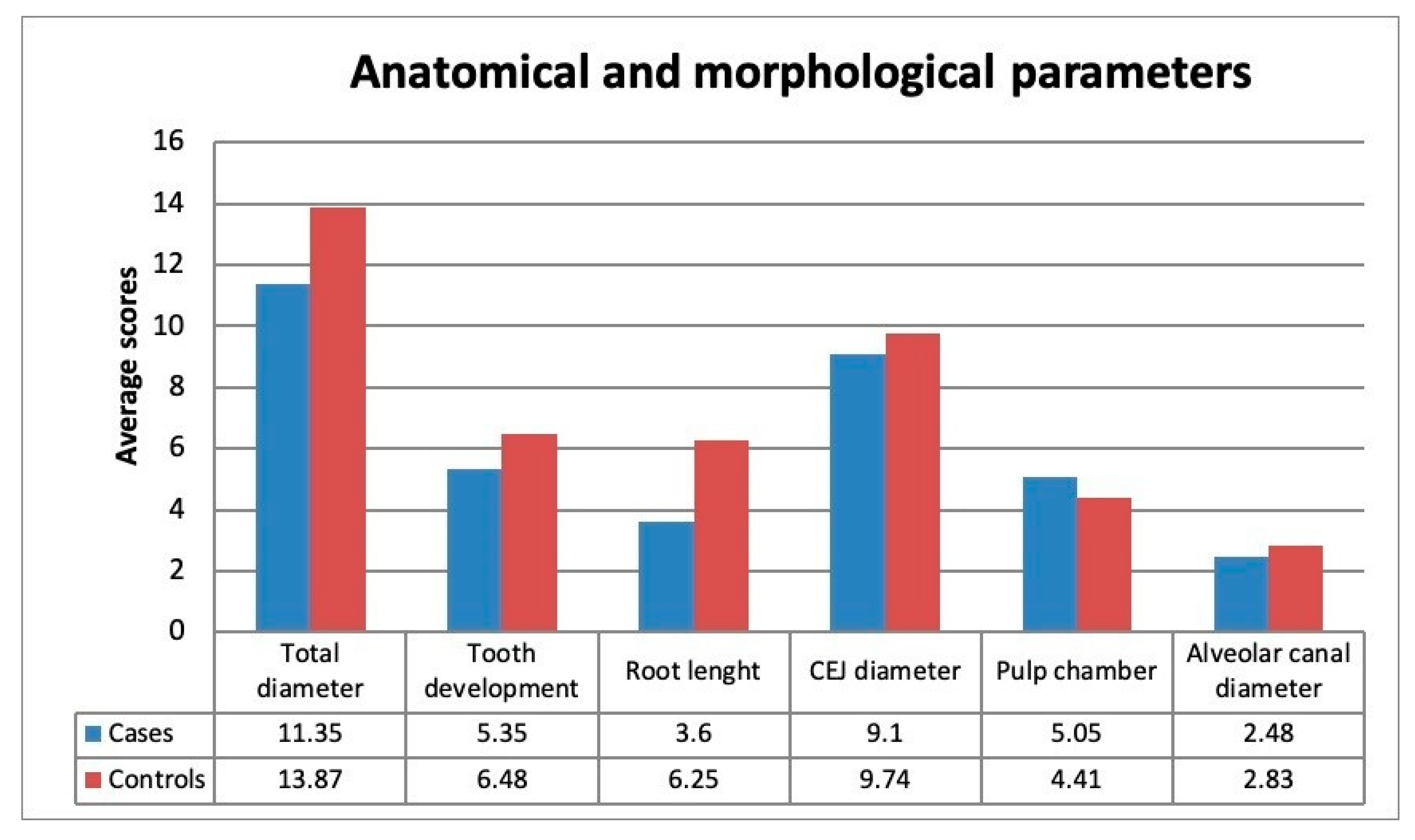
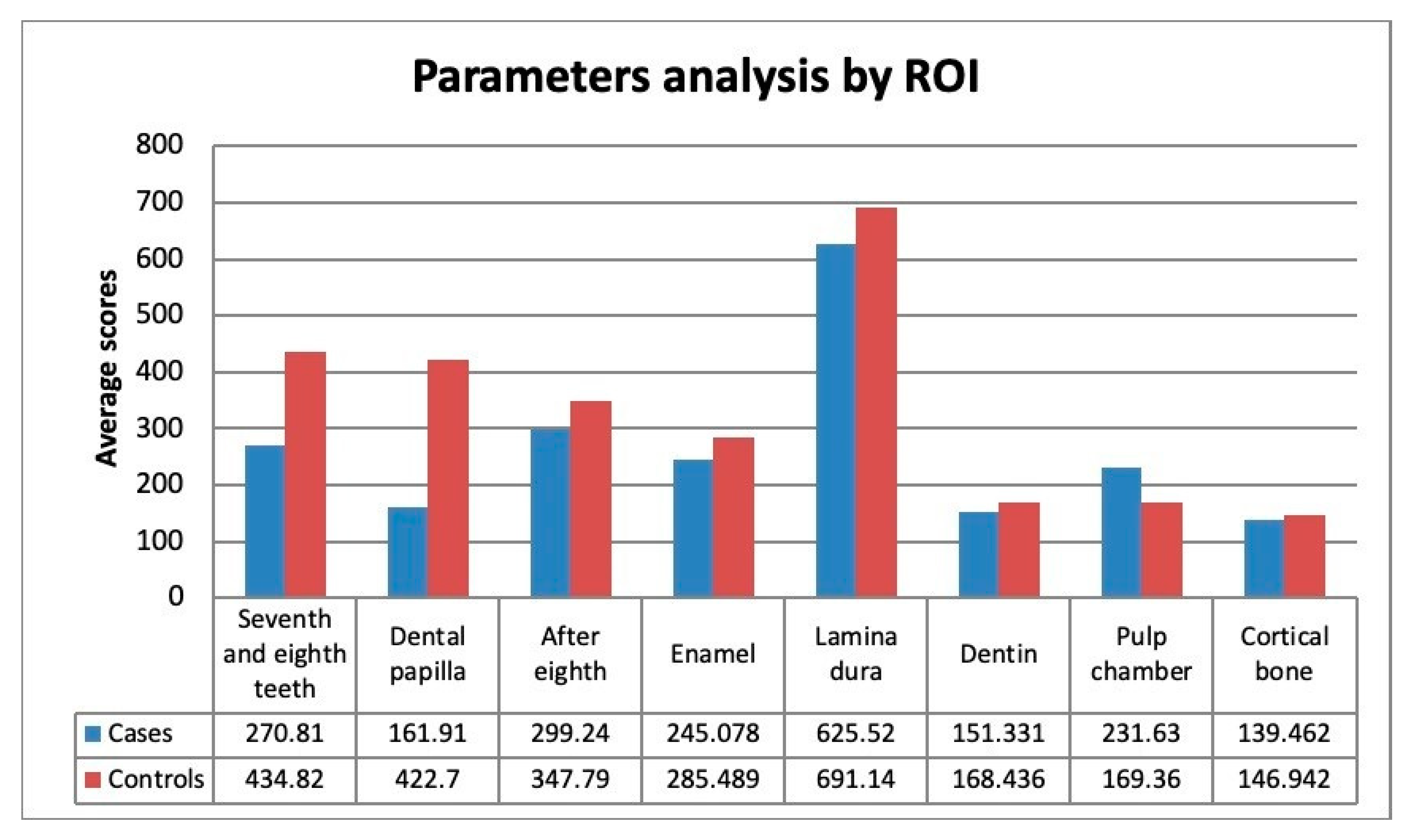
5. Results
5.1. Comparing Groups
5.2. Association
6. Discussion
7. Conclusions
Author Contributions
Funding
Conflicts of Interest
References
- Ben, A.M.; Rauch, F.; Monti, E. Osteogenesis imperfecta. Pediatr. Endocrinol. Rev. 2013, 10, 397–405. [Google Scholar]
- Sillence, D. Osteogenesis imperfecta: An expanding panorama of variants. Clin. Orthop. Relat. Res. 1981, 159, 11–25. [Google Scholar] [CrossRef]
- Greggio, N.A.; Rigon, F.; Zacchello, F. Osteogenesi Imperfetta. Osteopat. Pediatriche Emergenti 2003, 5, 139–152. [Google Scholar]
- Monti, E.; Mottes, M.; Fraschini, P. Current and emerging treatments for the management of osteogenesis imperfecta. Ther. Clin. Risk Manag. 2010, 6, 367–381. [Google Scholar] [PubMed] [Green Version]
- Russell, R.G.; Watts, N.B.; Ebetino, F.H. Mechanisms of action of bisphosphonates: Similarities and differences and their potential influence on clinical efficacy. Osteoporos. Int. 2008, 19, 733–759. [Google Scholar] [CrossRef] [PubMed]
- Bedogni, A.; Campisi, G.; Fusco, V.; Agrillo, A. Raccomandazioni clinico-terapeutiche sull’osteonecrosi delle ossa mascellari associata a bisfosfonati e sua prevenzione. In Proceedings of the SICMF-SIPMO, Palermo, Italia, 2013. [Google Scholar]
- Ierardo, G.; Bossù, M.; D’Angeli, G.; Celli, M.; Sfasciotti, G. Bisphosphonates therapy in children with Osteogenesis imperfecta: Clinical experience in oral surgery. Oral Implant. 2017, 10, 311–316. [Google Scholar] [CrossRef] [PubMed]
- Rantanen, A.V. The age of eruption of the third molar teeth. Acta Odontol. Scand. 1967, 25, 1–48. [Google Scholar]
- Engström, C.; Engström, H.; Sagne, S. Lower third molar development in relation to skeletal maturity and chronological age. Angle Orthod. 1983, 53, 97–106. [Google Scholar] [PubMed]
- Richardson, E.R.; Malhotra, S.K.; Semenya, K. Longitudinal study of three views of mandibular third molar eruption in males. Am. J. Orthod. 1984, 86, 119–129. [Google Scholar] [CrossRef]
- Richardson, E.M. The effect of mandibular first premolar extraction on third molar space. Angle Orthod. 1989, 59, 291–294. [Google Scholar] [PubMed]
- Richardson, M. Pre-eruptive movements of the mandibular third molar. Angle Orthod. 1978, 48, 187–193. [Google Scholar] [PubMed]
- Ng, F.; Burns, M.; Kerr, W.J.S. The impacted lower third molar and its relationship to tooth size and arch form. Eur. J. Orthod. 1986, 8, 254–258. [Google Scholar] [CrossRef] [PubMed]
- Forsberg, C.-M. Tooth size, spacing, and crowding in relation to eruption or impaction of third molars. Am. J. Orthod. Dentofac. Orthop. 1988, 94, 57–62. [Google Scholar] [CrossRef]
- Miloro, M.; Ghali, G.E.; Larsen, P.E.; Waite, P.D. (Eds.) Peterson’s Principles of Oral and Maxillofacial Surgery, 2nd ed.; BC Decker Inc.: Hamilton, London, UK, 2004. [Google Scholar]
- Frazier-Bowers, S.A.; Koehler, K.E.; Ackerman, J.L.; Proffit, W.R. Primary failure of eruption: Further characterization of a rare eruption disorder. Am. J. Orthod. Dentofac. Orthop. 2007, 131, 578.e1–578.e11. [Google Scholar] [CrossRef]
- Sodek, J.; McKee, M.D. Molecular and cellular biology of alveolar bone. Periodontology 2000, 24, 99–126. [Google Scholar] [CrossRef] [PubMed]
- Van Wesenbeeck, L.; Odgren, P.R.; Mackay, C.A.; D’Angelo, M.; Safadi, F.F.; Popoff, S.N.; Van Hul, W.; Marks, J.S.C. The osteopetrotic mutation toothless (tl) is a loss-of-function frameshift mutation in the rat Csf1 gene: Evidence of a crucial role for CSF-1 in osteoclastogenesis and endochondral ossification. Proc. Natl. Acad. Sci. USA 2002, 99, 14303–14308. [Google Scholar] [CrossRef] [Green Version]
- Dobbins, D.E.; Sood, R.; Hashiramoto, A.; Hansen, C.T.; Wilder, R.L.; Remmers, E. Mutation of macrophage colony stimulating factor (Csf1) causes osteopetrosis in the tl rat. Biochem. Biophys. Res. Commun. 2002, 294, 1114–1120. [Google Scholar] [CrossRef]
- Harokopakis-Hajishengallis, E. Physiologic root resorption in primary teeth: Molecular and histological events. J. Oral Sci. 2007, 49, 1–12. [Google Scholar] [CrossRef] [Green Version]
- Sahara, N.; Okafuji, N.; Toyoki, A. Odontoclastic resorption of the superficial nonmineralized layer of predentine in the shedding of human deciduous teeth. Cell Tissue Res. 1994, 277, 19–26. [Google Scholar] [CrossRef]
- Fukushima, H.; Kajiya, H.; Takada, K.; Okamoto, F.; Okabe, K. Expression and role of RANKL in periodontal ligament cells during physiological root-resorption in human deciduous teeth. Eur. J. Oral Sci. 2003, 111, 346–352. [Google Scholar] [CrossRef]
- Oshiro, T.; Shibasaki, Y.; Martin, T.J.; Sasaki, T. Immunolocalization of vacuolar-type H+-ATPase, cathepsin K, matrix metalloproteinase-9, and receptor activator of NFkB ligand in odontoclasts during physiological root resorption of human deciduous teeth. Anat. Rec. Adv. Integr. Anat. Evol. Boil. 2001, 264, 305–311. [Google Scholar] [CrossRef] [PubMed]
- Yoda, S.; Suda, N.; Kitahara, Y.; Komori, T.; Onyama, K. Delayed tooth eruption and suppressed osteoclast number in the eruption pathway of heterozygous Runx2/Cbfa1 knockout mice. Arch. Oral Biol. 2004, 49, 435–442. [Google Scholar] [CrossRef] [PubMed]
- Iizuka, T.; Cielinski, M.; Aukerman, S.; Marks, S.C. The effects of colony-stimulating factor-1 on tooth eruption in the toothless (osteopetrotic) rat in relation to the critical periods for bone resorption during tooth eruption. Arch. Oral Boil. 1992, 37, 629–636. [Google Scholar] [CrossRef]
- O’Connell, A.; Marini, J. Evaluation of oral problems in an osteogenesis imperfecta population. Oral Surg. Oral Med. Oral Pathol. Oral Radiol. Endodontol. 1999, 87, 189–196. [Google Scholar] [CrossRef]
- Mossey, P.A. The Heritability of Malocclusion: Part 2. The Influence of Genetics in Malocclusion. J. Orthod. 1999, 26, 195. [Google Scholar] [CrossRef] [PubMed]
- Barroso, M.; Arriola-Guillén, L.E.; Rodríguez-Cárdenas, Y.A.; Ruíz-Mora, G.A.; Guerrero, M.E.; Flores-Mir, C. Tridimensional assessment of the dental follicle dimensions of impacted mandibular third molars using cone-beam CTh. J. Clin. Exp. Dent. 2018, 10, 726–731. [Google Scholar]
- Nolla, C.M. The development of permanent teeth. J. Dent. Child. 1960, 27, 254–266. [Google Scholar]
- Nandlal, B.; Patil, K.; Ravi, S. Estimation of dental age by Nolla’s method using orthopantomographs among rural free residential school children. Int. J. Med. Res. Health Sci. 2014, 3, 273–277. [Google Scholar] [CrossRef] [Green Version]
- Proffit, W.R.; Fields, H.W.; Sarver, D.M. Ortodonzia Moderna; Edra Masson Libri: Milano, Italia, 2013. [Google Scholar]
- Ghaeminia, H.; Meijer, G.; Soehardi, A.; Borstlap, W.; Mulder, J.; Bergé, S. Position of the impacted third molar in relation to the mandibular canal. Diagnostic accuracy of cone beam computed tomography compared with panoramic radiography. Int. J. Oral Maxillofac. Surg. 2009, 38, 964–971. [Google Scholar] [CrossRef]
- Sun, R.; Cai, Y.; Yuan, Y.; Zhao, J.-H. The characteristics of adjacent anatomy of mandibular third molar germs: A CBCT study to assess the risk of extraction. Sci. Rep. 2017, 7, 14154. [Google Scholar] [CrossRef] [Green Version]
- Suprijanto, E.L.; Hajarini, M.S.; Juliastuti, E.; Susanti, H. Image analysis for dental bone quality assessment using CBCT imaging. J. Phys. Conf. Ser. 2016, 694, 12065. [Google Scholar] [CrossRef]
- Aksoy, U.; Kermeoğlu, F.; Kalender, A.; Eren, H.; Kolsuz, M.E.; Orhan, K. Cone-beam computed tomography evaluation of palatogingival grooves: A retrospective study with literature review. Oral Radiol. 2017, 33, 193. [Google Scholar] [CrossRef]
- Kröpil, P.; Hakimi, A.R.; Jungbluth, P. Cone Beam CT in Assessment of Tibial Bone Defect Healing: An Animal Study. Acad. Radiol. 2012, 19, 320–325. [Google Scholar] [CrossRef] [PubMed]
- Agbaje, J.O.; Jacobs, R.; Michiels, K.; Abu-Ta’A, M.; Van Steenberghe, D. Bone healing after dental extractions in irradiated patients: A pilot study on a novel technique for volume assessment of healing tooth sockets. Clin. Oral Investig. 2009, 13, 257–261. [Google Scholar] [CrossRef] [PubMed]
- Shokri, A.; Jamalpour, M.R.; Eskandarloo, A.; Godiny, M.; Amini, P.; Khavid, A. Performance of Cone Beam Computed Tomography Systems in Visualizing the Cortical Plate in 3D Image Reconstruction: An In Vitro Study. Open Dent. J. 2018, 12, 586–595. [Google Scholar] [CrossRef] [PubMed]
- Levin, L.S. The Dentition in the Osteogenesis Imperfecta Syndromes. Clin. Orthop. Relat. Res. 1981, 159, 64–74. [Google Scholar] [CrossRef]
- Schwartz, S.; Tsipouras, P. Oral findings in osteogenesis imperfecta. Oral Surg. Oral Med. Oral Pathol. 1984, 57, 161–167. [Google Scholar] [CrossRef]
- Petersen, K.E.; Wetzel, W. Recent findings in classification of osteogenesis imperfecta by means of existing dental symptoms. ASDC J. Dent. Child. 1998, 65, 305–354. [Google Scholar]
- Johnson, D.; Tinanoff, N. Malocclusion. In Nelson Textbook of Pediatrics, 16th ed.; Behrman, R.E., Kliegman, R.M., Eds.; Saunders W.B: Philadelphia, PA, USA, 2000; pp. 1110–1111. [Google Scholar]
- Karjodkar, F.R. Textbook of Dental and Maxillofacial Radiology, 2nd ed.; Jaypee Brothers: New Delhi, India, 2009. [Google Scholar]
- Enrico, P. Trattato di Chirurgia Odontostomatologica e Maxillo-Facciale: Chirurgia Della Lingua; Masson: Milano, Italy, 1990; ISBN 8821417379. [Google Scholar]
- Ferrini, F.; Ugo Covani, D. Chirurgia Orale; Edizioni Martina: Bologna, Italy, 2003. [Google Scholar]
- Lukinmaa, P.L.; Ranta, H.; Ranta, K.; Kaitila, I. Dental findings in osteogenesis imperfecta: I. Occurrence and expression of type I dentinogenesis imperfecta. J. Cranio-Fac. Genet. Dev. Boil. 1987, 7, 115–125. [Google Scholar]
- Lindau, B.; Dietz, W.; Hoyer, I.; Lundgren, T.; Storhaug, K.; Norén, J. Morphology of dental enamel and dentine–enamel junction in osteogenesis imperfecta. Int. J. Paediatr. Dent. 1999, 9, 13–21. [Google Scholar] [CrossRef]
- Parfitt, A.; Travers, R.; Rauch, F.; Glorieux, F. Structural and cellular changes during bone growth in healthy children. Bone 2000, 27, 487–494. [Google Scholar] [CrossRef]
- Nagai, N.; Hosokawa, M.; Itohara, S.; Adachi, E.; Matsushita, T.; Hosokawa, N.; Nagata, K. Embryonic Lethality of Molecular Chaperone Hsp47 Knockout Mice Is Associated with Defects in Collagen Biosynthesis. J. Cell Boil. 2000, 150, 1499–1506. [Google Scholar] [CrossRef] [PubMed] [Green Version]
- Thomson, C.A.; Ananthanarayanan, V.S. Structure–function studies on Hsp47: pH-dependent inhibition of collagen fibril formation in vitro. Biochem. J. 2000, 349, 877–883. [Google Scholar] [CrossRef] [PubMed] [Green Version]
- Fedarko, N.S.; D’Avis, P.; Frazier, C.; Burrill, M.; Fergusson, V.; Tayback, M.; Sponseller, P.; Shapiro, J. Cell proliferation of human fibroblasts and osteoblasts in osteogenesis imperfecta: Influence of age. J. Bone Miner. Res. 2009, 10, 1705–1712. [Google Scholar] [CrossRef] [PubMed]
- Wenstrup, R.J.; Witte, D.P.; Florer, J.B. Abnormal Differentiation in MC3T3-E1 Preosteoblasts Expressing a Dominant-Negative Type I Collagen Mutation. Connect. Tissue Res. 1996, 35, 249–257. [Google Scholar] [CrossRef]
- Wenstrup, R.J.; Fowlkes, J.L.; Witte, D.P.; Florer, J.B. Discordant expression of osteoblast markers in MC3T3eE1 eels that synthesize a high turnover matrix. J. Biol. Chem. 1996, 271, 10271–10276. [Google Scholar] [CrossRef] [Green Version]
- Alanay, Y.; Avaygan, H.; Camacho, N. Mutations in gene encoding the RER protein FKBP65 cause autosomal-recessive osteogenesis imperfecta. Am. J. Hum. Genet. 2010, 86, 551–559. [Google Scholar] [CrossRef] [Green Version]
- Rauch, F.; Travers, R.; Parfitt, A.M.; Glorieux, F.H. Static and dynamic bone histomorphometry in children with osteogenesis imperfecta. Bone 2000, 26, 581–589. [Google Scholar] [CrossRef]

| Axial | ||||
|---|---|---|---|---|
| I | IIA | IIB | IIC | |
| Cases | 22 (95.6%) | 1 (4.4%) | 0 | 0 |
| Controls | 20 (87%) | 0 | 3 (13%) | 0 |
| Parasagittal | ||||
| I | IIA | IIB | IIC | |
| Cases | 19 (82.6%) | 3 (13%) | 1 (4.4%) | 0 |
| Controls | 19 (82.6%) | 0 | 4 (17.4%) | 0 |
| Coronal | ||||
| I | IIA | IIB | IIC | |
| Cases | 22 (95.6%) | 1 (4.4%) | 0 | 0 |
| Controls | 14 (60.9%) | 1 (4.4%) | 3 (13%) | 5 (21.7%) |
| State of Growth | ||||
| A | B | |||
| Cases | 2 (8.7%) | 21 (91.3%) | ||
| Controls | 0 | 23 (100%) | ||
| Altered State of Dentin | ||||
| No | Yes | |||
| Cases | 16 (84.2%) | 3 (15.8%) | ||
| Controls | 22 (100%) | 0 | ||
| Interruption of the Lingual Cortical Plate on the Axial Axis Cone-Beam | ||||
| No | Yes | |||
| Cases | 14 (60.8%) | 9 (39.2%) | ||
| Controls | 23 (100%) | 0 | ||
© 2020 by the authors. Licensee MDPI, Basel, Switzerland. This article is an open access article distributed under the terms and conditions of the Creative Commons Attribution (CC BY) license (http://creativecommons.org/licenses/by/4.0/).
Share and Cite
D’Angeli, G.; Messineo, D.; Riminucci, M.; Corsi, A.; Celli, M.; Vozza, I.; Sfasciotti, G.L. The Characteristics of Adjacent Anatomy of Mandibular Third Molar Germs: A CBCT Pilot Study in Patients with Osteogenesis Imperfecta. Healthcare 2020, 8, 372. https://doi.org/10.3390/healthcare8040372
D’Angeli G, Messineo D, Riminucci M, Corsi A, Celli M, Vozza I, Sfasciotti GL. The Characteristics of Adjacent Anatomy of Mandibular Third Molar Germs: A CBCT Pilot Study in Patients with Osteogenesis Imperfecta. Healthcare. 2020; 8(4):372. https://doi.org/10.3390/healthcare8040372
Chicago/Turabian StyleD’Angeli, Giacomo, Daniela Messineo, Mara Riminucci, Alessandro Corsi, Mauro Celli, Iole Vozza, and Gian Luca Sfasciotti. 2020. "The Characteristics of Adjacent Anatomy of Mandibular Third Molar Germs: A CBCT Pilot Study in Patients with Osteogenesis Imperfecta" Healthcare 8, no. 4: 372. https://doi.org/10.3390/healthcare8040372