
The Advances of Hydrosol–Gel Transition-Based Sensors

1
School of Pharmaceutical Sciences, Qilu University of Technology (Shandong Science Academy), Jinan 250014, China
2
School of Mechanical and Material Engineering, Washington State University, Pullman, WA 99164, USA
*
Authors to whom correspondence should be addressed.
Chemosensors 2022, 10(10), 415; https://doi.org/10.3390/chemosensors10100415
Submission received: 26 August 2022
/
Revised: 2 October 2022
/
Accepted: 10 October 2022
/
Published: 12 October 2022
(This article belongs to the Special Issue Feature Papers on Luminescent Sensing)
Abstract
:Hydrogels, as a type of three-dimensional porous material, have attracted a lot of attention in the fields of drug delivery, artificial tissue engineering, and sensing. Due to their excellent biocompatibility and high sensitivity to external stimuli, they are widely used in the development of various sensors. Among them, the sensors constructed based on the sol–gel transition of target-responsive hydrogels are particularly welcome. Herein, the status of the sensors on the basis of sol–gel transition has been presented. The types of hydrogel sensors and the analytical methods in various application scenarios are illustrated. In addition, the future trends of the sensing systems based on sol–gel transition are briefly discussed.
1. Introduction
Hydrogels are crosslinked hydrophilic polymer porous network materials. Like other widely used porous materials such as MOFs [1], hydrogels also possess attractive physical and chemical properties. A large number of hydrogels have high molecular permeability, high mechanical properties, remarkable ionic conductivity, controllable microstructures, excellent biocompatibility, and impressive stability, which show great potential in the spheres of drug delivery [2,3,4,5,6,7,8,9,10], artificial tissue engineering [11,12,13,14], sensing [15,16,17,18,19,20,21], etc. Generally, hydrogels contain a large amount of water, whose weight is even a few hundred times more than that of dry hydrogel scaffolds [22]. The gel materials with various properties have different water storage capacities, leading to various applications. Hydrogels used in force change sensing often need strong mechanical properties and are often prepared by the method of physical adsorption and ultrasonic treatment [23,24]. Hydrogels used in biosensing are often prepared by the method of chemical covalent and require the integration of hydrogel scaffolds and recognition units, causing a highly sensitive response to various environmental stimuli. The recognition units of the hydrogels can specifically respond to external stimuli, which may be easily detected and quantified without complex operations.
As shown in this review, in response to small changes in temperature, pH, light, electricity, pressure, solvent composition, and radiation, hydrogels can generate appropriate signals due to changes in physical structures or chemical properties. For most analytical methods, it is easier to directly obtain signals caused by the change in physical properties of the materials. In past years, the sensors employing hydrogels have been widely applied in point-of-care testing (POCT), and the targets included organic and inorganic molecules, especially metal ions, organic toxins, and biomarkers. The sensors are usually developed based on the phase transition, volume change [25], and analyte interception [26,27] of hydrogels. Among them, the sol–gel transition is a popular sensing strategy that applies external stimuli to trigger the destruction or construction of the hydrogel scaffold structures. The structure changes affect the signal molecules embedded inside the hydrogel scaffolds, thereby inducing the generation of readable signals. The previous review of hydrogels was often classified based on their materials or properties, such as conductivity [28] and stretchable properties [29]. In this paper, sol–gel transition sensing strategy is selected as the basis to review the research progress of hydrogel sensors.
There are mainly two sensing methods that apply the sol–gel transition mechanism. One is to embed the recognition units and signal molecules into the hydrogels, which are dissolved with the presence of the target substances to release the signal molecules [30]. According to the porous three-dimensional structures of hydrogels, the signal molecules can be encapsulated to avoid their rapid interaction with the external environment. For example, considering the amount of water trapped in hydrogels as a type of signal, the destruction degree of hydrogel scaffolds can be determined [31]. The other method hinders the initial signals via the target-induced formation of hydrogels. For instance, the formation of Ca2+-triggered hydrogels on the surface of glass carbon electrodes greatly increases the impedance signals [32]. Compared to other advanced detection methods such as gold nanoparticles colorimetric assay [33], detection based on sol–gel transition takes advantage of the simple and repetitive structure of hydrogels to allow the signal molecules to embed in but not modify the materials, which simplifies the preparing operations. It is also more convenient as a detection method that the signal amplification is achieved according to the number of embedded signal molecules. Thus, the hydrogel sensing based on sol–gel transition is particularly important in the detection of biological enzyme activity, small molecules such as organic pesticides and toxins, heavy metal ions and so on.
This paper summarizes the different scaffold materials normally used to fabricate stimuli-responsive hydrogels. Detection technologies based on sol–gel transition are introduced in detail, including signal output modes, their application scenarios, as well as quantitative capabilities. Finally, future trends for sensing technologies based on sol–gel transition are briefly discussed. Discussions on the development and progress of the sensors will help to broaden the design methodologies to construct functional hydrogel sensors and provide new perspectives for the rapid and quantitative recognition of specific analytes in practical applications.
2. Types of Hydrogels in the Development of Sol–Gel Transition-Based Sensors
Hydrogel scaffolds with different compositions endow hydrogels with different responsive and destructive methods. According to various materials, three major kinds of hydrogels have been reported, involving DNA hydrogels, polypeptide hydrogels, and polysaccharide hydrogels. DNA hydrogels usually contain functional DNA fragments to recognize targets. Polypeptide hydrogels are usually used to detect enzymes due to their responses to different hydrolases. For polysaccharide hydrogels, besides the scaffold formed by polymers, the recognized units are usually acting as crosslinking agents. This section summarizes the various types of hydrogels (Table 1), including their output signals, targets, and recognition units. In addition, hydrogels with diverse supporting scaffolds and various detection principles are also discussed.
2.1. DNA Hydrogels
For DNA hydrogels, DNA acts as a crosslinking agent or scaffold, and recognition units are often DNA-related substances, such as aptamer, DNA probe, and complementary strand. Based on the complementary characteristics of DNA double strands, the combination and separation of recognition units from scaffolds is the core of sol–gel sensing. According to different scaffold components, two major types of DNA hydrogels have been extensively studied, involving pure and hybrid DNA hydrogels.
For pure DNA hydrogels, their scaffolds are composed of DNA strands with complementary bases [46]. They always have good biocompatibility and biodegradability [47]. Zhang et al. fabricated ctDNA-triggered and DNAzyme-functionalized hydrogels for visible detection of circulating tumor DNA (ctDNA) (Figure 1) [37]. Target ctDNA triggered the rolling circle amplification (RCA), thus resulting in DNA hydrogels with G-quadruplex structures on a macroscopical scale, which was utilized as the direct detection of ctDNA. Chu selected two partially complementary strands, Pb2+ dependent DNAzyme strand and substrate strand, to manufacture the DNA hydrogels [48]. Pb2+ presenting in the sample activated the DNAzyme strands, which led to the cleavage of the substrate strands and signal output.
Hybrid DNA hydrogels are generally formed by the hybridization of modified polymers and deoxyribonucleotide chains. They always possess high mechanical strength and stability [47]. For the preparation of hybrid DNA hydrogels, two common methods were developed. The one method involves DNA modification on organic polymer chains. Acrylamide [20,35,49] and acrydite [50] are always used as monomers for polymerization. Lin developed the hybrid DNA hydrogels which were made by three different kinds of ssDNA strands combining with non-DNA polymers [50]. Their scaffolds consisted of the acrydite-modified adenosine aptamer and the acrydite-modified ssDNA partially complementary to the adenosine aptamer with the heme aptamer acting as the crosslinker. To improve the sensitivity of polymer-DNA hydrogels, Liao’s group wrapped the fluorescent quantum dots in DNA-based acrylamide hydrogel microcapsules [19]. All fluorescent quantum dots in the microcapsules can be obtained without completely collapsing the hydrogel. Another method is to modify DNA on microbeads, such as gold nanoparticles and SiO2 nanoparticles. Jie’s group developed a versatile fluorescence strategy through DNA-SiO2 hybrid hydrogels, which were used to detect miRNA-141 with ultra-sensitivity [34]. The target miRNA-141 firstly induced the production of large amounts of DNA S3 by walking amplification. Then, S3 linked with DNA hairpin H1 which was attached to SiO2 microspheres. After the hybridization chain reaction (HCR) of H1 and other hairpin DNA modifying on SiO2, DNA crosslinked hydrogels can be formed on SiO2. Additionally, Liu used DNA-modified gold nanoparticle to fabricate hybrid hydrogels film for the biosensing system [51]. To enhance the mechanical strength of hydrogels, Ji et al. further integrated the two methods [52,53]. They firstly linked DNA with SiO2 nanoparticles, then modified poly methacrylic acid on the terminal of DNA complementary strand. Finally, polymer hydrogel scaffolds were constructed outside the DNA-SiO2 nanoparticles via DNA complementary binding.
DNA can continuously synthesize long DNA strands and form pure DNA hydrogels under the catalysis of polymerase. The operations of synthesis and signal amplification are relatively simple. Hybrid DNA hydrogels not only enrich the diversity of DNA hydrogels, provide more sensing strategies, but have better stability to adapt to complex detection environments.
2.2. Polypeptide Hydrogels
Polypeptide-based hydrogels benefit from their low immunogenicity, high bio-compatibility and biodegradability, and lower cost than DNA hydrogels, drawing great attention in the sphere of biological applications [54,55,56,57]. On the one hand, the types of amino acids are much more than the types of nucleobases in nucleic acids, which greatly enrich the diversity of peptide chains; On the other hand, unlike nucleic acids that rely on complementary base pairing to form scaffolds, peptide chains can be linked to each other by modifying reactive groups or photosensitive groups at the end of the chain, or directly through hydrogen bonds or hydrophobic interactions.
Polypeptide hydrogels can be used for the detection of polypeptide enzymes without introducing extra recognition groups. For example, it is feasible to quantitatively detect trypsin [31] and thrombin [39] by measuring the water molecules released from gelatin and fibrinogen hydrogels due to the enzymatic hydrolysis. Ahmad and colleagues crosslinked 4-arm poly (ethylene glycol) norbornene with bis cysteine peptide to form polypeptide hydrogels, which were used to detect trace collagenase with the assistance of quartz crystal microbalance [58]. In addition, combining with DNA probes, aptamers, and other recognition units with polypeptides can enrich the types of polypeptide-based hydrogels and expand the functionality. For example, to detect cancer DNA, Jandl used the method of terminal modification to combine DNA with peptide chain, and the fluorophores entered the polypeptide hydrogels via hydrogen bonds [56].
For the good specificity of proteolytic enzymes, polypeptide hydrogels can be directly recognized by related enzymes, and the abundant enzyme-binding sites in the hydrogel scaffold facilitate the signal amplification.
2.3. Polysaccharide Hydrogels
As a kind of natural polymer material, polysaccharide hydrogels have attracted extensive attention. Alginate hydrogels as polymer backbones, have been widely used in biological analysis. Yang and co-workers noticed that it was easy to utilize alginate hydrogels with “egg-box” structures to encapsulate enzymes [59]. This method facilitated the analysis of different enzymes using the immobilized enzyme reactor (IMER) with same hydrogel structures. The species of detected enzymes depended on the substrate injected in capillary electrophoresis. Later, they provided an approach for on-line enzyme assays for penicillinase utilizing capillary electrophoresis-integrated immobilized enzyme reactors (CE-IMERs) [60]. To fabricate the CE-IMERs (Figure 2a), the hydrosol stock suspension was prepared and injected into the pretreated capillary. The suspension contained penicillinase, sodium alginate, and calcium carbonate. Then, the penicillin G sodium salt (PG) solution was introduced into the inlet of the capillary with electrokinetics. The hydrolysis of PG by penicillinase-produced penicilloic acid (PA), which made the solution weakly acidic, thus promoting the hydrolysis of CaCO3 to produce Ca2+. With the help of Ca2+, the sodium alginate hydrosol transited into hydrogels.
Based on Ca2+-triggered pH-response sodium alginate hydrogels precipitation, Ma’s group developed a cascaded signal-amplification strategy for a sandwich-type impedimetric immunosensor, which was particularly useful for the detection of prostate specific antigen (PSA) [32]. As illustrated in Figure 2b, the secondary antibody was firstly connected to gold nanoparticle-CaCO3 microspheres (AuNP-CaCO3). When AuNP-CaCO3 was deposited on the surface of the glass carbon electrode (GCE) by binding the target molecules to the primary and secondary antibodies, the dissolution of CaCO3 and release of Ca2+ in a weakly acidic solution were induced. Consequently, Ca2+ was crosslinked with alginate to form insoluble alginate salt hydrogels’ precipitation on the sensing interface, which significantly increased the impedance signal. Benefiting from the cascaded signal amplification, the impedimetric immunosensor exhibited ultrahigh sensitivity.
At present, although there are many types of polysaccharide hydrogels, sodium alginate hydrogels are more researched for sensing because their unique “egg-box” structure and Ca2+-induced gelation are suitable for sensing strategy.
3. The Methods Used for the Development of Sol–Gel Transition-Based Sensors
Various sol–gel transition-based sensors are introduced according to different sensing methods. The brief information of these sensors is put in Table 2.
3.1. Colorimetric Assay
The detection of solution absorbance only needs the simple operation of a portable spectrometer, from which reliable quantitative information can be obtained, and even qualitative signals can be carried out through naked eye observation. Thus, colorimetric detection is one of the most suitable methods for rapid detection [51,75,76,77,78]. Hydrogels with color nanoparticles swell or shrink in response to external stimulations, which will change the lattice spacing. According to Bragg′s Law [25,75], lattice spacing changes result in a blue shift or red shift in the spectrum of hydrogels. Alternatively, the hydrogels are collapsed into hydrosol due to their combination with the target and release nanoparticles into the supernatant, and then color change can be seen.
Using the colorimetric method, Liang’s group fabricated an aptamer-based hydrogel containing trapped horseradish peroxidase (HRP), which was utilized for T-2 mycotoxin detection [61]. The target toxin triggered the collapse of hydrogels because it was preferentially bound to the T-2 toxin aptamer, and released HRP to react with H2O2 and KI. Then, I2 etched Au-nanorods, causing different degrees of colorimetric blue shift, which were related to toxin concentration. The detection range was 0.01–104 ng/mL with a low limit of detection (LOD) of 0.87 pg/mL.
As mentioned above, alginate hydrogels have been widely utilized for bio-analysis. Tan and co-workers developed the ascorbic acid (AA)-responsive alginate hydrogels (RhB@Alg/Fe3+) for the visible detection of alkaline phosphatase (ALP) (Figure 3a) [62]. And rhodamine B (RhB) was embedded in the hydrogels as an indicating reagent to assist visual detection. In the presence of ALP, the hydrolysis of ascorbate 2-phosphate to AA resulted in the reduction of Fe3+ to Fe2+, which showed a weak affinity to alginate hydrogel scaffolds. Consequently, Fe2+ triggered the dissolution of RhB@Alg/Fe3+, giving the sol solution an observable red color, and thus a detectable absorbance signal was read out for the amount of ALP. This visual approach for ALP detection had a low detection limit of 0.37 mU/mL and an excellent selectivity over other proteins.
Xu et al. reported a DNA hydrogel sensor with a novel detection mode for ochratoxin A (OTA) (Figure 3b) [63]. They pre-fabricated double-stranded DNA duplex, which was formed by primer and the complementary binding of OTA aptamer. With the addition of OTA, the primers were released, and combined with the padlock probe. Under the catalysis of T4 ligase and Phi29 DNA polymerase, the rolling cycle amplification (RCA) was started. The AuNPs in the solution were directly assembled inside the hydrogels in the process of RCA. The higher concentration of OTA, the more DNA hydrogels were yielded, and the fewer AuNPs were in the supernatant. Thus, the absorbance of the supernatant was negatively correlated with the concentration of OTA. The linear range was from 0.05 to 10 ng/mL with the detection of 0.005 ng/mL.
Gold nanoparticles (AuNPs) have long been known for their colorimetric detection. However, the color changes are not obvious when the gold nanoparticles in the supernatant are lacking. To overcome this deficiency, Li’s group combined the aptamer crosslinked zearalenone (ZEN)-responsive hydrogel with tetramethylbenzidine (TMB)-H2O2-AuNPs, and realized its low-dose visual detection for ZEN [79]. When exposed to ZEN, the binding of ZEN and its aptamer led to the destruction of hydrogels and the release of AuNPs. To ensure that ZEN can be detected even at low concentrations, Li’s group utilized the color reaction of TMB and H2O2 as a quantitative readout strategy for this hydrogel system, which generated an obvious change. This established method displayed excellent selectivity and sensitivity over other mycotoxins.
3.2. Fluorescence Assay
For the colorimetric method, the color change becomes slight with the decrease in target concentration, resulting in a low sensitivity [79]. Fluorescence analysis has higher sensitivity and a lower detection limit than the colorimetric method. Through chemical modification or physical insertion, fluorescent groups can be fixed on the hydrogels crosslinking chains [46,80,81]. Moreover, the three-dimensional porous structures of hydrogels are naturally suitable for encapsulating fluorescent groups. Therefore, fluorescence analysis based on sol–gel transition is also a common sensing method.
Employing a fluorescence-based detection strategy, Tan and colleagues fabricated pyrophosphate ion (PPi)-responsive alginate (Alg) hydrogels for alkaline phosphatase (ALP) detection [64]. The generated CDs@Cu/Alg hydrogels used Cu2+ as crosslinkers and immobilized the green fluorescent carbon nanodots (CDs) in the matrix. Because of the ultrahigh affinity between PPi and Cu2+, the presence of PPi could collapse the network structures of alginate hydrogels through the competitive coordination with Cu2+, causing the release of CDs into the solution. The gel–sol transition process could be observed by naked eyes, and the fluorescent intensity of the solution increased with the increase in PPi concentration. Therefore, when there was target ALP in the sample, ALP hydrolyzed PPi, whereas the hydrolysate phosphate ion (Pi) had no affinity to Cu2+, and CDs in the gel were not released. This fluorescent sensing platform showed the linear relationship between ALP and fluorescent intensity in the range of 0.05–1 mM with a LOD of 21.3 μM.
Based on the self-assembly of rolling circle amplification (RCA) products, Wu et al. developed a sensitive fluorescent DNA hydrogel sensor for ochratoxin A (OTA) detection in beer (Figure 4) [65]. The addition of OTA destroyed the complex of OTA aptamer and complementary primer, resulting in the release of the primer, which subsequently hybridized with the padlock probe to form a circular template. The RCA reaction was then initiated by adding T4 ligase, DNA polymerase, and fluorescent Cy3-modified deoxy-ribonucleoside triphosphates. A linear range between fluorescence intensity and OTA concentration ranging from 0.05 ng/mL to 100 ng/mL was obtained with a detection limit of 0.01 ng/mL. The sol–gel transition hydrogel-based fluorescent assay is easy to operate with a low detection limit and high reliability, but the problem of background noise restricts its applications.
3.3. Surface-Enhanced Raman Spectroscopy (SERS) Assay
SERS has been widely investigated in sensing, benefiting from its non-destruction, high sensitivity, good reproducibility, and specificity. Compared to other methods, the advantage of SERS is that it can be used to detect trace molecules even down to a single molecular level [82]. Wang et al. fabricated a flexible DNAzyme-based hydrogel SERS active biofilm for UO22+ detection in aquatic products [66]. As shown in Figure 5, polyacrylonitrile (PAN) nanorods arrays were arranged on the surface of the film and then decorated with Ag-NPs in high density. Acrydite-modified DNA was used as the scaffolds, and the crosslinker strand and enzyme strand were used as the crosslinking agents to form hydrogels, with rhodamine B (RhB) wrapped in it. The prepared hydrogels were then modified on PAN arrays. When UO22+ triggered the enzyme strand and caused the cleavage of the crosslinker strand, Raman reporters RhB were released from the hydrogels and finally captured by PAN arrays. After Raman signals were amplified twice, the limit of detection of the hydrogel biosensors reached 8.38 × 10⁻¹³ M for UO22+ with a wide linear range from 1.0 pM to 0.1 μM.
Zhuang’s group proposed a method that the target aptamer was used as a DNA hydrogel crosslinker and embedded immunoglobulin G (IgG) in it to detect α-fetoprotein (AFP), the specific marker of hepatocellular carcinoma, by SERS [67]. In the presence of AFP, the aptamer specifically recognized AFP and controlled the release of IgG accurately. The released IgG was captured by the antigen-modified magnetic beads, and combined with the SERS probe with the help of antigen-antibody interaction. After separation, the signal of Raman tags was positively correlated with the AFP concentration. The method above had a wide detection linear range from 50 pg/mL to 0.5 μg/mL and a low detection limit down to 50 pg/mL. As a sensitive method, it showed a lower detection limit than many traditional methods, but the sulfurization and oxidation of silver atoms easily occurred in the ambient atmosphere [83].
3.4. Magnetic Relaxation Switching (MRS) Assay
Under the applied magnetic field, the magnetic field between paramagnetic or superparamagnetic nanoparticles is not uniform. When aqueous protons get through this non-uniform magnetic field, the vibration frequency will inevitably change, resulting in the alteration of transverse relaxation time (T2), which can be quantified by nuclear magnetic resonance technology [84]. The response of MRS depends on magnetic signal rather than optical signal. Therefore, the complex sample matrix has little effect on the background value of this method, which greatly simplifies the sample pretreatment steps. In addition, MRS sensing is a homogeneous reaction system, which reduces the operation steps such as plate washing or color reaction, and greatly improves the detection efficiency. It is therefore especially suitable for detecting targets in turbid and complex systems such as biochemical samples [85,86,87].
Chen developed a magnetic transverse relaxation time-based biosensing strategy (Figure 6) and employed it to detect foodborne pathogens such as S. enteritidis [45]. The conversion of the biological signal to magnetic signal relied on the alkaline phosphatase (ALP)-mediated sol–gel transition. ALP catalyzed the hydrolysis of 2-phospho-L-ascorbic acid (AAP), which generated an acidic environment and transformed the alginate solution into hydrogels. This gelation process can directly affect the relaxation behavior of water molecules, resulting in the change of magnetic transverse relaxation time. The primary antibody-modified magnetic beads were used to capture the S. enteritidis, and then the streptavidin (SA)-modifying secondary antibody and ALP were bound to the S. enteritidis. The concentration of obtained ALP is related to the concentration of S. enteritidis, which is basic to the next gelation reaction. This method displayed high sensitivity for detecting 50 CFU/mL S. enteritidis within 2 h. Currently, combining methods with satisfactory reliability and detection limit with responsive hydrogel materials is a trend, and the researches for sol–gel transition hydrogel-based magnetic relaxation switching assay are gradually increasing.
3.5. pH-Based Assay
Among the various pH-sensitive hydrogels [88,89,90,91,92], target-responsive hydrogels are the most common materials that can be adopted to construct hydrogel sensors. However, the signal readout equipment made in the laboratory often faces the problem of insufficient sensitivity. In order to solve this problem, combining commercially portable equipment with pH-responsive hydrogels provides the advantages of high sensitivity and multi-target detection. With the popularity of handheld pH meters, quantitative information on target molecules can be obtained through the pH of the solution when sol–gel transitions are generated.
Chen’s group realized the specific detection of aflatoxin B1 (AFB1) by using the AFB1-responsive aptamer-crosslinked hydrogels and a handheld pH meter (Figure 7) [38]. Upon the addition of AFB1, the high-affinity binding of AFB1 and aptamer led to the collapse of DNA-hybrid hydrogels, causing the release of urease into the solution. As urease catalyzed the hydrolysis of added urea, uric acids were produced, then induced the changes in pH value, which was directly related to the concentration of AFB1. Finally, quantitative detection of AFB1 in food samples was successfully achieved by the portable pH meter. The linear range was 0.2–20 μM with a LOD of 0.1 μM.
3.6. Weight Assay by Electronic Balance
In recent years, novel biosensors that couple simple devices with different recognition strategies, are utilized to detect diverse targets by measuring their physical parameters, such as length, weight, etc. The electronic balance is the most common device in laboratories, and has been used as a readout device in constructing biosensors. These weight-based biosensors are attracting growing interest because of their portable, low-cost, and easily operated properties.
Lin and co-workers demonstrated a novel method to realize timely detection of hyaluronidase (HAase) by electronic balance [40]. They found that encapsulating catalysts in the hydrogels could amplify the sol–gel transition signal into another reaction with an obvious phenomenon, such as the reaction of generating gas. As shown in Figure 8, Pt@SiO2 nanoparticles were embedded in the hydrogels, which were constructed by polyethyleneimine (PEI) and hyaluronic acid (HA). In the presence of HAase, the hydrogels were broken down and released the Pt@SiO2, which disintegrated H2O2 into H2O and O2. Collected O2 enhanced the pressure in the drainage device and then a certain amount of water overflowed from the device. The collected H2O was finally quantified by electronic balance. Its weight had a linear relationship with the HAase concentration in the range of 1–60 U/mL.
In addition, they introduced aptamers to construct DNA hydrogels, and amplified the signal through enzyme cycles. The aflatoxin B1 (AFB1) target-responsive double crosslinked hydrogels were constructed using hyaluronic acid-grafted single-strand DNA complex as the backbone, and AFB1 aptamer and polyethyleneimine as crosslinkers [68]. Platinum nanoparticles had been embedded in hydrogels previously. Once AFB1 bound with aptamer, it can be released from hydrogels and cleaved by exonuclease I. Free AFB1 then returned to hydrogels to bind with aptamer again, which finally caused the collapse of hydrogels and the release of Pt nanoparticles (PtNPs). Adding PtNPs into a drainage device containing H2O2 resulted in the production of O2 and the release of water from the device. The AFB1 concentration can be measured by weighting the water. By this means, the linear range was 31.2 μg/kg–6.2 mg/kg with the detection limit of 9.4 μg/kg.
Recently, the improved hydrogels biosensor formed a membrane for sensitive detection of HAase [69]. The hyaluronic acid (HA)-polyethyleneimine (PEI) hydrogels were used to fabricate a thin film, which was covered on a mixed cellulose microporous membrane (MCEM) to form a HA-PEI-MCEM, which was utilized in a filtration system. After the addition of HAase, HA in the hydrogels were hydrolyzed, which increased the permeability of the membrane. The volume of liquid flowing through the filter membrane was linear with the concentration of HAase in a range of 1.0–36 U mL−1 with a detection limit of 0.35 U mL−1. The electronic balance-based method with the ingenious signal transition strategy was easy to operate with satisfying reliability.
3.7. Glucose Assay by Personal Glucose Meter
Personal glucose meter (PGM), a commercial rapid detection device, can obtain results according to the concentration of glucose in the sample in a few seconds. Due to its advantages of portability, cheapness, and user-friendliness, it has been widely utilized for the clinical diagnosis of hyperglycemia. Recently, the combination of PGM with enzymes that can catalyze the production of glucose offered a new strategy for more diverse target detection [93,94].
Li et al. designed a simple and easy handling POCT tool to test the activity of DNA adenine methyltransferase (Dam) (Figure 9) [70]. The tool consisted of a personal glucose meter (PGM) and DNA tetrahedra-based hydrogels. The DNA hydrogels modified on paper discs were used to trap the amyloglucosidase for target recognition and signal transduction. The edges of DNA tetrahedrons were methylated by Dam and subsequently cleaved by the methylation restriction endonuclease DpnI. As a result, the hydrogel structures were destroyed, causing the release of loaded amyloglucosidase, which catalyzed the generation of glucose for a final quantitative readout using PGM. The reading of PGM was linearly correlated with the activity of Dam, with a direct detection limit down to 0.001 U/mL.
3.8. Distance-Based Lateral Flow Assay
Paper-based analysis through measuring the changes of samples on the paper has a long history in the field of analytical chemistry. The combination of dry chemistry test method and paper-folding can be easily achieved for integrated analysis [95,96,97,98,99,100] and multi-channel analysis [101,102,103,104]. Due to their essential virtues of portability, simplicity, rapidity, low cost, and perceptual intuition, paper-based sensors have attracted tremendous research interest in the development of sensing devices.
Lateral flow assay (LFA) is the key to constructing a hydrogel-assisted distance-based sensor on paper. LFA provides a quantifiable linear range and establishes the relationship between the wetting area of the paper and the concentration of target molecules. In this way, there is no need to consider the transmission and amplification of biological signals, because the water can be directly captured by the paper and transformed into a distance. Thus, hydrogel-assisted paper-based lateral flow sensors, which rely on the water volume released from hydrogels, have drawn extensive attention due to their simplicity, high portability, and inexpensive production.
Yang’s group used mixed cellulose ester (MCE) filter paper based on acetylcholinesterase (AChE)-mediated alginate hydrogels for quantitative sensing of organophosphorus pesticides (OPs) (Figure 10) [71]. The target molecules, OPs (inhibitors of AChE), were added to the reaction mixture. The production of hydrogen ions from the enzymatic reaction of AChE was inhibited, and the gelation of alginate was inhibited, accordingly. Therefore, the viscosity of the solution and the diameter of its diffusion on the filter paper were linearly related to the concentration of the target molecules. The dynamic range of the method reached 66.7 ng/mL with a LOD of 3.3 ng/mL.
Based on the above strategy, Yang et al. developed a dual-readout paper-based sensor for quantitative determination of penicillinase via smartphone (Figure 11) [72]. They utilized the catalysis of penicillinase to generate penicilloic acid and to decrease the pH of the solution. When penicillin G sodium salt was enzymatically hydrolyzed, the Ca2+ that was produced by the decomposition of CaCO3 can promote the formation of alginate hydrogels. The existence of the target made the pH and viscosity of the solution change, which induced the changes in color and diffusion diameter of the spot, so two linear relationships could be established. The advantage of the method was that dual signal of the targets was easily achieved simultaneously. The pH was indicated by the color change of acid-base indicator, and viscosity change was indicated by the diameter of diffusion on the filter paper. The limits of detection are 2.67 × 10⁻³ mU/μL for distance-readout and 2.67 × 10⁻² mU/μL for color-readout.
Using the visual distance-readout method, Zhang et al. fabricated a paper-based sensor to determine the concentration of glucose based on the glucose oxidase (GOx)-mediated sodium alginate gelation [73]. They introduced a mechanism that GOx catalyzed the oxidation of glucose to generate gluconic acid and H2O2, which gradually changed the pH value of the solution to acidic. Similarly, an acidic environment led to the production of Ca2+ and the formation of alginate hydrogels. As the change in viscosity is related to the change in the diffusion diameter, the glucose concentration could be read out by vernier caliper. Under optimal conditions, the increase in diffusion diameter showed a good linear relationship with glucose concentration between 1.4–7.0 mM, and the limit of quantification was 1.4 mM.
Capillary sensing quantifies the target through the rising height of liquid level, which is a signal amplification method completely separated from the assistance of electronic analysis equipment. According to the conclusion of surface physics, the surface tension (γ) of liquid in the capillary is determined only by the temperature and the properties of compounds at the composition interface. Therefore, the requirements for the detection environment are very low, which shows great advantages in rapid on-site detection [105,106,107].
On the basis of the capillary action, Wen et al. reported a DNA-based hydrogels capillary sensor (DHCS) for the detection of Pb2+ (Figure 12) [74]. The substrate, crosslinking agents, and the response units, Pb2+-dependent GR-5 DNAzymes, were fixed on the scaffolds of the DNA hydrogels, which changed the capillary forces. In the presence of Pb2+, the GR-5 DNAzyme was activated and cleaved the crosslinker substrate strand, resulting in the fracture of hydrogels. Accordingly, the mesh size of the hydrogel film plugging the end of the capillary tube changed. With the increasing concentration of Pb2+, the solution could flow into the capillary tube in a shorter time. Therefore, quantitative detection of Pb2+ was achieved by reading the distance and duration time. Benefiting from the high sensitivity and selectivity of capillary sensor, this sensor was capable of detecting as low as 10 nM Pb2+ with the linear range of 0.01–50 μM.
Recently, Hu’s group demonstrated a new type of paper-based sensing strategy for disease diagnosis and drug screening, which was based on sol–gel transition caused by enzymatic hydrolysis. By measuring the distance of water flowing along the pH indicator strips, they constructed various paper-based biosensors for the detection of biological enzymes and their inhibitors [31]. The detection of trypsin and its inhibitor by the hydrogel-assisted lateral flow assay was demonstrated. The gelatin was employed to form hydrogels and the pH paper strips were chosen to catch the released water molecules. In the presence of trypsin, the gelatin hydrogels were hydrolyzed, which caused the release of the water to flow along the pH paper strips. However, in the absence of trypsin, water could not be released because it was trapped in the gelatin hydrogels. Therefore, the quantitative detection of trypsin could be realized by measuring the length of water moving distance. The detection limit of the paper-based sensor reached as low as 1.0 × 10⁻⁶ mg/mL.
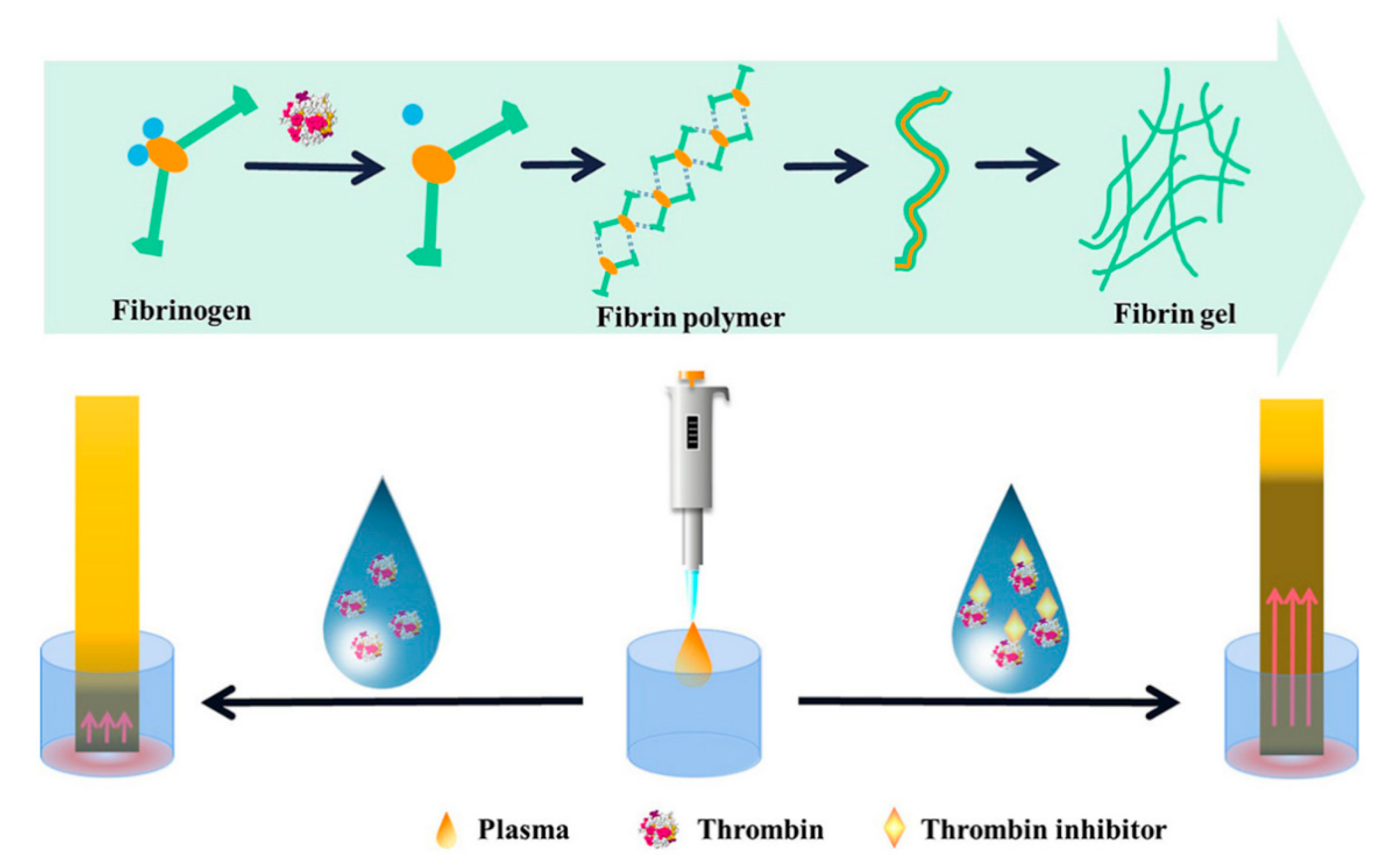
Later, Wu et al. developed a lateral flow sensor for thrombin detection and selection of inhibitors (Figure 13) [39]. During the blood clotting process, the water was captured efficiently, as the fibrinogen crosslinked and formed a fibrin porous network with the assistance of thrombin. In the presence of thrombin, the water in plasma could not diffuse along with the pH paper strips. Upon the inactivation of thrombin by the inhibitors, water could diffuse along the test paper. In this way, the detection limit of thrombin reached about 16.1 mU/mL.
4. Conclusions
In summary, this review introduces hydrogel-related sensing strategies based on the sol–gel transition, mostly referring to the papers of the past five years. Various polymers used in sol–gel transition-based sensing are summarized, and the obtained sensing is classified according to the output modes of the generated signals. The sensing principle, application scenario, and quantitative capability of various sensors are introduced in detail. From the perspective of analytes, the sol–gel based hydrogels are particularly important in the detection of biological enzyme activity, small molecules such as organic pesticides and toxins, heavy metal ions and so on. Most of the analytes are small enough to allow the diffusion inside the porous hydrogels and reaction with their scaffolds. Because the hydrogels possess loose network structures in the microcosm, each scaffold can be modified with recognition units, so this sol–gel transition easily occurs. For the wrapping capacity of hydrogels, the collapse of the scaffold can release a tremendous amount of embedded signal molecules. In a word, sol–gel based hydrogels are helpful signal amplification tools. Additionally, the biocompatible materials endow the hydrogel with good potential in the field of biomedicine and biomarker detection.
In terms of the sensitive detection, SERS technology and MRS sensors achieve a lower detection limit. It is worthy of note that the paper-based assay and capillary assay have great potential to achieve simple, convenient, and rapid detection without the assistance of large devices. Considerable efforts have been invested in constructing hydrogel sensing systems assisted by various analysis software or equipment. Nevertheless, their applications in POCT are limited by the compromised sensitivity depending on the signal readout strategy. A promising approach is the combination of portable devices to address this issue. Personal glucose meter, pH meter, electronic balance, and more commercially available apparatus tend to be integrated with sol–gel transition-based hydrogels to detect various targets. It can be predicted that mobile phones, as intelligent devices, will be more and more used for on-site detection. Overall, sol–gel transition-based sensors are promising for rapid, sensitive, and robust detection in various applications.
Author Contributions
Writing—original draft preparation, H.S.; writing—review and editing, M.Z.; writing—review and editing, S.D.; project administration and funding acquisition, supervision, writing—review and editing, Q.H. All authors have read and agreed to the published version of the manuscript.
Funding
This work was funded by [Shandong Scientific and Technical Small and Medium-sized Enterprises Innovation Capacity Improvement Project], grant number [2021TSGC1193], [the Taishan Scholars Program], grant number [tsqn201812088] and Science, Education and Industry Integration Innovation Pilot Project from Qilu University of Technology (Shandong Academy of Sciences), grant number [2022JBZ02-04].
Institutional Review Board Statement
Not applicable.
Informed Consent Statement
Not applicable.
Data Availability Statement
Not applicable.
Conflicts of Interest
The authors declare no conflict of interest.
References
- Karimi, M.; Mehrabadi, Z.; Farsadrooh, M.; Bafkary, R.; Derikvandi, H.; Hayati, P.; Mohammadi, K. Chapter 4—Metal–organic framework. In Interface Science and Technology; Ghaedi, M., Ed.; Elsevier: Amsterdam, The Netherlands, 2021; Volume 33, pp. 279–387. [Google Scholar]
- Dera, R.; Diliën, H.; Billen, B.; Gagliardi, M.; Rahimi, N.; Den Akker, N.M.S.; Molin, D.G.M.; Grandfils, C.; Adriaensens, P.; Guedens, W.; et al. Phosphodiester hydrogels for cell scaffolding and drug release applications. Macromol. Biosci. 2019, 19, 1900090. [Google Scholar] [CrossRef] [PubMed]
- Pivato, R.V.; Rossi, F.; Ferro, M.; Castiglione, F.; Trotta, F.; Mele, A. β-cyclodextrin nanosponge hydrogels as drug delivery nanoarchitectonics for multistep drug release kinetics. ACS Appl. Polym. Mater. 2021, 3, 6562–6571. [Google Scholar] [CrossRef]
- Tang, J.; Qiao, Y.; Chu, Y.; Tong, Z.; Zhou, Y.; Zhang, W.; Xie, S.; Hu, J.; Wang, T. Magnetic double-network hydrogels for tissue hyperthermia and drug release. J. Mater. Chem. B 2019, 7, 1311–1321. [Google Scholar] [CrossRef] [PubMed]
- Xie, Z.; Shen, J.; Sun, H.; Li, J.; Wang, X. Polymer-based hydrogels with local drug release for cancer immunotherapy. Biomed. Pharmacother. 2021, 137, 111333. [Google Scholar] [CrossRef]
- Weng, L.; Rostambeigi, N.; Zantek, N.D.; Rostamzadeh, P.; Bravo, M.; Carey, J.; Golzarian, J. An in situ forming biodegradable hydrogel-based embolic agent for interventional therapies. Acta Biomater. 2013, 9, 8182–8191. [Google Scholar] [CrossRef]
- Wang, W.; Song, H.; Zhang, J.; Li, P.; Li, C.; Wang, C.; Kong, D.; Zhao, Q. An injectable, thermosensitive and multicompartment hydrogel for simultaneous encapsulation and independent release of a drug cocktail as an effective combination therapy platform. J. Control Release 2015, 203, 57–66. [Google Scholar] [CrossRef]
- Guilbaud-Chéreau, C.; Dinesh, B.; Wagner, L.; Chaloin, O.; Ménard-Moyon, C.; Bianco, A. Aromatic dipeptide homologue-based hydrogels for photocontrolled drug release. Nanomaterials 2022, 12, 1643. [Google Scholar] [CrossRef]
- Hu, J.; Chen, Y.; Li, Y.; Zhou, Z.; Cheng, Y. A thermo-degradable hydrogel with light-tunable degradation and drug release. Biomaterials 2016, 112, 133–140. [Google Scholar] [CrossRef]
- Lou, C.; Tian, X.; Deng, H.; Wang, Y.; Jiang, X. Dialdehyde-β-cyclodextrin-crosslinked carboxymethyl chitosan hydrogel for drug release. Carbohydr. Polym. 2019, 231, 115678. [Google Scholar] [CrossRef]
- Gomez-Florit, M.; Pardo, A.; Domingues, R.M.A.; Graça, A.L.; Babo, P.S.; Reis, R.L.; Gomes, M.E. Natural-based hydrogels for tissue engineering applications. Molecules 2020, 25, 5858. [Google Scholar] [CrossRef]
- Khuu, N.; Kheiri, S.; Kumacheva, E. Structurally anisotropic hydrogels for tissue engineering. Trends Chem. 2021, 3, 1002–1026. [Google Scholar] [CrossRef]
- Yi, Y.; Xie, C.; Liu, J.; Zheng, Y.; Wang, J.; Lu, X. Self-adhesive hydrogels for tissue engineering. J. Mater. Chem. B 2021, 9, 8739–8767. [Google Scholar] [CrossRef]
- Zhao, H.; Liu, M.; Zhang, Y.; Yin, J.; Pei, R. Nanocomposite hydrogels for tissue engineering applications. Nanoscale 2020, 12, 14976–14995. [Google Scholar] [CrossRef]
- Fu, L.; Wang, A.; Lyu, F.; Lai, G.; Yu, J.; Lin, C.-T.; Liu, Z.; Yu, A.; Su, W. A solid-state electrochemical sensing platform based on a supramolecular hydrogel. Sens. Actuators B Chem. 2018, 262, 326–333. [Google Scholar] [CrossRef]
- Sun, X.; He, S.; Qin, Z.; Li, J.; Yao, F. Fast self-healing zwitterion nanocomposite hydrogel for underwater sensing. Compos. Commun. 2021, 26, 100784. [Google Scholar] [CrossRef]
- Zhang, J.; Jin, J.; Wan, J.; Jiang, S.; Wu, Y.; Wang, W.; Gong, X.; Wang, H. Quantum dots-based hydrogels for sensing applications. Chem. Eng. J. 2020, 408, 127351. [Google Scholar] [CrossRef]
- Bhattacharya, S.; Sarkar, R.; Nandi, S.; Porgador, A.; Jelinek, R. Detection of reactive oxygen species by a carbon-dot–ascorbic acid hydrogel. Anal. Chem. 2016, 89, 830–836. [Google Scholar] [CrossRef]
- Chang, W.-H.; Lee, Y.-F.; Liu, Y.-W.; Willner, I.; Liao, W.-C. Stimuli-responsive hydrogel microcapsules for the amplified detection of microRNAs. Nanoscale 2021, 13, 16799–16808. [Google Scholar] [CrossRef]
- Ma, Y.; Mao, Y.; An, Y.; Tian, T.; Zhang, H.; Yan, J.; Zhu, Z.; Yang, C.J. Target-responsive DNA hydrogel for non-enzymatic and visual detection of glucose. Analyst 2018, 143, 1679–1684. [Google Scholar] [CrossRef]
- Vassalini, I.; Ribaudo, G.; Gianoncelli, A.; Casula, M.F.; Alessandri, I. Plasmonic hydrogels for capture, detection and removal of organic pollutants. Environ. Sci. Nano 2020, 7, 3888–3900. [Google Scholar] [CrossRef]
- Xu, X.; Jerca, V.V.; Hoogenboom, R. Bioinspired double network hydrogels: From covalent double network hydrogels via hybrid double network hydrogels to physical double network hydrogels. Mater. Horiz. 2020, 8, 1173–1188. [Google Scholar] [CrossRef]
- Gao, Y.; Gu, S.; Jia, F.; Wang, Q.; Gao, G. “All-in-one” hydrolyzed keratin protein-modified polyacrylamide composite hydrogel transducer. Chem. Eng. J. 2020, 398, 125555. [Google Scholar] [CrossRef]
- Xie, H.; Yu, Q.; Mao, J.; Wang, S.; Hu, Y.; Guo, Z. A conductive polyacrylamide/double bond chitosan/polyaniline hydrogel for flexible sensing. J. Mater. Sci. Mater. Electron. 2020, 31, 10381–10389. [Google Scholar] [CrossRef]
- Hayashi, T.; Takinoue, M.; Onoe, H. DNA aptamer-linked structural-color hydrogel for repeatable biochemical sensing. In Proceedings of 20th International Conference on Solid-State Sensors, Actuators and Microsystems and Eurosensors XXXIII (TRANSDUCERS and EUROSENSORS), Berlin, Germany, 23–27 June 2019; pp. 582–585. [Google Scholar]
- Dai, J.; Zhang, H.; Huang, C.; Chen, Z.; Han, A. A Gel-Based Separation-Free Point-of-Care Device for Whole Blood Glucose Detection. Anal. Chem. 2020, 92, 16122–16129. [Google Scholar] [CrossRef]
- Erfkamp, J.; Guenther, M.; Gerlach, G. Enzyme-Functionalized Piezoresistive Hydrogel Biosensors for the Detection of Urea. Sensors 2019, 19, 2858. [Google Scholar] [CrossRef] [Green Version]
- Qin, T.; Liao, W.; Yu, L.; Zhu, J.; Wu, M.; Peng, Q.; Han, L.; Zeng, H. Recent progress in conductive self-healing hydrogels for flexible sensors. J. Polym. Sci. 2022, 60, 2607–2634. [Google Scholar] [CrossRef]
- Liu, Y.; Wang, L.; Mi, Y.; Zhao, S.; Qi, S.; Sun, M.; Peng, B.; Xu, Q.; Niu, Y.; Zhou, Y. Transparent stretchable hydrogel sensors: Materials, design and applications. J. Mater. Chem. C 2022, 10, 13351–13371. [Google Scholar] [CrossRef]
- Ata, S.; Banerjee, S.L.; Singha, N.K. Polymer nano-hybrid material based on graphene oxide/POSS via surface initiated atom transfer radical polymerization (SI-ATRP): Its application in specialty hydrogel system. Polymer 2016, 103, 46–56. [Google Scholar] [CrossRef]
- Ping, J.; Wu, W.; Qi, L.; Liu, J.; Liu, J.; Zhao, B.; Wang, Q.; Yu, L.; Lin, J.M.; Hu, Q. Hydrogel-assisted paper-based lateral flow sensor for the detection of trypsin in human serum. Biosens. Bioelectron. 2021, 192, 113548. [Google Scholar] [CrossRef]
- Zhao, L.; Yin, S.; Ma, Z. Ca(2+)-triggered pH-response sodium alginate hydrogel precipitation for amplified sandwich-type impedimetric immunosensor of tumor marker. ACS Sens. 2019, 4, 450–455. [Google Scholar] [CrossRef]
- Priyadarshini, E.; Pradhan, N. Gold nanoparticles as efficient sensors in colorimetric detection of toxic metal ions: A review. Sens. Actuators B Chem. 2017, 238, 888–902. [Google Scholar] [CrossRef]
- Li, C.; Li, H.; Ge, J.; Jie, G. Versatile fluorescence detection of microRNA based on novel DNA hydrogel-amplified signal probes coupled with DNA walker amplification. Chem. Commun. 2019, 55, 3919–3922. [Google Scholar] [CrossRef] [PubMed]
- Cai, W.; Xie, S.; Zhang, J.; Tang, D.; Tang, Y. An electrochemical impedance biosensor for Hg2+ detection based on DNA hydrogel by coupling with DNAzyme-assisted target recycling and hybridization chain reaction. Biosens. Bioelectron. 2017, 98, 466–472. [Google Scholar] [CrossRef] [PubMed]
- Hong, C.A.; Park, J.C.; Na, H.; Jeon, H.; Nam, Y.S. Short DNA-catalyzed formation of quantum dot-DNA hydrogel for enzyme-free femtomolar specific DNA assay. Biosens. Bioelectron. 2021, 182, 113110. [Google Scholar] [CrossRef]
- Mao, X.; Pan, S.; Zhou, D.; He, X.; Zhang, Y. Fabrication of DNAzyme-functionalized hydrogel and its application for visible detection of circulating tumor DNA. Sens. Actuators B Chem. 2019, 285, 385–390. [Google Scholar] [CrossRef]
- Zhao, M.; Wang, P.; Guo, Y.; Wang, L.; Luo, F.; Qiu, B.; Guo, L.; Su, X.; Lin, Z.; Chen, G. Detection of aflatoxin B1 in food samples based on target-responsive aptamer-cross-linked hydrogel using a handheld pH meter as readout. Talanta 2018, 176, 34–39. [Google Scholar] [CrossRef]
- Han, C.; Yuan, X.; Shen, Z.; Xiao, Y.; Wang, X.; Khan, M.; Liu, S.; Li, W.; Hu, Q.; Wu, W. A paper-based lateral flow sensor for the detection of thrombin and its inhibitors. Anal. Chim. Acta 2022, 1205, 339756. [Google Scholar] [CrossRef]
- Li, Z.; Tang, C.; Huang, D.; Qin, W.; Luo, F.; Wang, J.; Guo, L.; Qiu, B.; Lin, Z. Sensitive hyaluronidase biosensor based on target-responsive hydrogel using electronic balance as readout. Anal. Chem. 2019, 91, 11821–11826. [Google Scholar] [CrossRef]
- Danish, E.Y.; Bakhsh, E.M.; Akhtar, K. Design of chitosan nanocomposite hydrogel for sensitive detection and removal of organic pollutants. Int. J. Biol. Macromol. 2020, 159, 276–286. [Google Scholar] [CrossRef]
- Li, C.; Duan, L.; Cheng, X. Facile method to synthesize fluorescent chitosan hydrogels for selective detection and adsorption of Hg2+/Hg+. Carbohydr. Polym. 2022, 288, 119417. [Google Scholar] [CrossRef]
- Adnan, P.K.M.; Sreejith, L. Compact poly-electrolyte complex hydrogels of gelatin and sodium alginate for sensing wound status. Mater. Lett. 2022, 313, 131705. [Google Scholar] [CrossRef]
- Lengert, E.; Saveleva, M.; Abalymov, A.; Atkin, V.; Wuytens, P.C.; Kamyshinsky, R.; Vasiliev, A.L.; Gorin, D.A.; Sukhorukov, G.B.; Skirtach, A.G.; et al. Silver alginate hydrogel micro- and nanocontainers for theranostics: Synthesis, encapsulation, remote release, and detection. ACS Appl. Mater. Interfaces 2017, 9, 21949–21958. [Google Scholar] [CrossRef]
- Wei, L.; Wang, Z.; Feng, C.; Xianyu, Y.; Chen, Y. Direct transverse relaxation time biosensing strategy for detecting foodborne pathogens through enzyme-mediated sol−gel transition of hydrogels. Anal. Chem. 2021, 93, 6613–6619. [Google Scholar] [CrossRef]
- Yao, S.; Xiang, L.; Wang, L.; Gong, H.; Chen, F.; Cai, C. pH-responsive DNA hydrogels with ratiometric fluorescence for accurate detection of miRNA-21. Anal. Chim. Acta 2022, 1207, 339795. [Google Scholar] [CrossRef]
- Chen, M.; Wang, Y.; Zhang, J.; Peng, Y.; Li, S.; Han, D.; Ren, S.; Qin, K.; Li, S.; Gao, Z. Stimuli-responsive DNA-based hydrogels for biosensing applications. J. Nanobiotechnol. 2022, 20, 40. [Google Scholar] [CrossRef]
- Chu, J.; Chen, C.; Li, X.; Yu, L.; Li, W.; Cheng, M.; Tang, W.; Xiong, Z. A responsive pure DNA hydrogel for label-free detection of lead ion. Anal. Chim. Acta 2021, 1157, 338400. [Google Scholar] [CrossRef]
- Wang, Z.; Chen, R.; Hou, Y.; Qin, Y.; Li, S.; Yang, S.; Gao, Z. DNA hydrogels combined with microfluidic chips for melamine detection. Anal. Chim. Acta 2022, 1228, 340312. [Google Scholar] [CrossRef]
- Lin, Y.; Wang, X.; Sun, Y.; Dai, Y.; Sun, W.; Zhu, X.; Liu, H.; Han, R.; Gao, D.; Luo, C. A chemiluminescent biosensor for ultrasensitive detection of adenosine based on target-responsive DNA hydrogel with Au@HKUST-1 encapsulation. Sens. Actuators B Chem. 2019, 289, 56–64. [Google Scholar] [CrossRef]
- Liu, C.; Gou, S.; Bi, Y.; Gao, Q.; Sun, J.; Hu, S.; Guo, W. Smart DNA-gold nanoparticle hybrid hydrogel film based portable, cost-effective and storable biosensing system for the colorimetric detection of lead (II) and uranyl ions. Biosens. Bioelectron. 2022, 210, 114290. [Google Scholar] [CrossRef]
- Ji, X.; Lv, H.; Sun, X.; Ding, C. Green-emitting carbon dot loaded silica nanoparticles coated with DNA-cross-linked hydrogels for sensitive carcinoembryonic antigen detection and effective targeted cancer therapy. Chem. Commun. 2019, 55, 15101–15104. [Google Scholar] [CrossRef]
- Ji, X.; Wang, J.; Niu, S.; Ding, C. Size-controlled DNA-cross-linked hydrogel coated silica nanoparticles served as a ratiometric fluorescent probe for the detection of adenosine triphosphate in living cells. Chem. Commun. 2019, 55, 5243–5246. [Google Scholar] [CrossRef]
- Qiao, Y.; Liu, X.; Zhou, X.; Zhang, H.; Zhang, W.; Xiao, W.; Pan, G.; Cui, W.; Santos, H.A.; Shi, Q. Gelatin templated polypeptide co-cross-linked hydrogel for bone tegeneration. Adv. Healthc. Mater. 2020, 9, 1901239. [Google Scholar] [CrossRef]
- Cheng, L.; Cai, Z.; Ye, T.; Yu, X.; Chen, Z.; Yan, Y.; Qi, J.; Wang, L.; Liu, Z.; Cui, W.; et al. Injectable polypeptide-protein hydrogels for promoting infected wound healing. Adv. Funct. Mater. 2020, 30, 2001196. [Google Scholar] [CrossRef]
- Jandl, B.; Sedghiniya, S.; Carstens, A.; Astakhova, K. Peptide–fluorophore hydrogel as a signal boosting approach in rapid detection of cancer DNA. ACS Omega 2019, 4, 13889–13895. [Google Scholar] [CrossRef] [Green Version]
- Liu, Y.X.; Xie, T.J.; Li, C.H.; Ye, Q.C.; Tian, L.L.; Li, Y.F.; Huang, C.Z.; Zhen, S.J. A crosslinked submicro-hydrogel formed by DNA circuit-driven protein aggregation amplified fluorescence anisotropy for biomolecules detection. Anal. Chim. Acta 2021, 1154, 338319. [Google Scholar] [CrossRef]
- Ahmad, N.; Colak, B.; Zhang, D.-W.; Gibbs, M.J.; Watkinson, M.; Becer, C.R.; Gautrot, J.E.; Krause, S. Peptide cross-linked poly (ethylene glycol) hydrogel films as biosensor coatings for the detection of collagenase. Sensors 2019, 19, 1677. [Google Scholar] [CrossRef] [Green Version]
- Yang, J.; Hu, X.; Xu, J.; Liu, X.; Yang, L. Single-step In situ acetylcholinesterase-mediated alginate hydrogelation for enzyme encapsulation in CE. Anal. Chem. 2018, 90, 4071–4078. [Google Scholar] [CrossRef]
- Hu, X.; Yang, J.; Chen, C.; Khan, H.; Guo, Y.; Yang, L. Capillary electrophoresis-integrated immobilized enzyme microreactor utilizing single-step in-situ penicillinase-mediated alginate hydrogelation: Application for enzyme assays of penicillinase. Talanta 2018, 189, 377–382. [Google Scholar] [CrossRef]
- Sun, Y.; Li, S.; Chen, R.; Wu, P.; Liang, J. Ultrasensitive and rapid detection of T-2 toxin using a target-responsive DNA hydrogel. Sens. Actuators B Chem. 2020, 311, 127912. [Google Scholar] [CrossRef]
- Gao, L.; Li, Y.; Huang, Z.Z.; Tan, H. Visual detection of alkaline phosphatase based on ascorbic acid-triggered gel-sol transition of alginate hydrogel. Anal. Chim. Acta 2021, 1148, 238193. [Google Scholar] [CrossRef]
- Hao, L.; Liu, X.; Xu, S.; An, F.; Gu, H.; Xu, F. A novel aptasensor based on DNA hydrogel for sensitive visual detection of ochratoxin A. Mikrochim. Acta 2021, 188, 395. [Google Scholar] [CrossRef] [PubMed]
- Li, Y.; Huang, Z.Z.; Weng, Y.; Tan, H. Pyrophosphate ion-responsive alginate hydrogel as an effective fluorescent sensing platform for alkaline phosphatase detection. Chem. Commun. 2019, 55, 11450–11453. [Google Scholar] [CrossRef] [PubMed]
- Hao, L.; Wang, W.; Shen, X.; Wang, S.; Li, Q.; An, F.; Wu, S. A fluorescent DNA hydrogel aptasensor based on the self-assembly of rolling circle amplification products for sensitive detection of ochratoxin A. J. Agric. Food Chem. 2020, 68, 369–375. [Google Scholar] [CrossRef] [PubMed]
- He, X.; Zhou, X.; Liu, W.; Liu, Y.; Wang, X. Flexible DNA hydrogel SERS active biofilms for conformal ultrasensitive detection of uranyl Ions from aquatic products. Langmuir 2020, 36, 2930–2936. [Google Scholar] [CrossRef]
- Wang, Q.; Hu, Y.; Jiang, N.; Wang, J.; Yu, M.; Zhuang, X. Preparation of aptamer responsive DNA functionalized hydrogels for the sensitive detection of alpha-fetoprotein using SERS method. Bioconjug. Chem. 2020, 31, 813–820. [Google Scholar] [CrossRef]
- Tang, L.; Huang, Y.; Lin, C.; Qiu, B.; Guo, L.; Luo, F.; Lin, Z. Highly sensitive and selective aflatoxin B1 biosensor based on Exonuclease I-catalyzed target recycling amplification and targeted response aptamer-crosslinked hydrogel using electronic balances as a readout. Talanta 2020, 214, 120862. [Google Scholar] [CrossRef]
- Dong, N.; Cai, Q.; Li, Z.; Xu, L.; Wu, H.; Lin, Z.; Qiu, B.; Li, C.; Lin, Z. Convenient hyaluronidase biosensors based on the target-trigger enhancing of the permeability of a membrane using an electronic balance as a readout. Analyst 2021, 146, 3299–3304. [Google Scholar] [CrossRef]
- Gao, X.; Li, X.; Sun, X.; Zhang, J.; Zhao, Y.; Liu, X.; Li, F. DNA tetrahedra-cross-linked hydrogel functionalized paper for onsite analysis of DNA methyltransferase activity using a personal glucose meter. Anal. Chem. 2020, 92, 4592–4599. [Google Scholar] [CrossRef]
- Xu, J.; Hu, X.; Khan, H.; Tian, M.; Yang, L. Converting solution viscosity to distance-readout on paper substrates based on enzyme-mediated alginate hydrogelation: Quantitative determination of organophosphorus pesticides. Anal. Chim. Acta 2019, 1071, 1–7. [Google Scholar] [CrossRef]
- Xu, J.; Chen, X.; Khan, H.; Yang, L. A dual-readout paper-based sensor for on-site detection of penicillinase with a smartphone. Sens. Actuators B Chem. 2021, 335, 129707. [Google Scholar] [CrossRef]
- Zhang, H.; Li, X.; Qian, Z.M.; Wang, S.; Yang, F.Q. Glucose oxidase-mediated sodium alginate gelation: Equipment-Free detection of glucose in fruit samples. Enzym. Microb. Technol. 2021, 148, 109805. [Google Scholar] [CrossRef]
- Jiang, C.; Li, Y.; Wang, H.; Chen, D.; Wen, Y. A portable visual capillary sensor based on functional DNA crosslinked hydrogel for point-of-care detection of lead ion. Sens. Actuators B Chem. 2019, 307, 127625. [Google Scholar] [CrossRef]
- Gao, Y.; Chen, Y.; Li, M.; Jia, L.; Zhang, L.; Zhu, J. Gelatin-based photonic hydrogels for visual detection of pathogenic Pseudomonas aeruginosa. Sens. Actuators B Chem. 2020, 329, 129137. [Google Scholar] [CrossRef]
- Mao, Y.; Li, J.; Yan, J.; Ma, Y.; Song, Y.; Tian, T.; Liu, X.; Zhu, Z.; Zhou, L.; Yang, C. A portable visual detection method based on a target-responsive DNA hydrogel and the color change of gold nanorods. Chem. Commun. 2017, 53, 6375–6378. [Google Scholar] [CrossRef] [Green Version]
- Wu, M.; Zhang, Y.; Liu, Q.; Huang, H.; Wang, X.; Shi, Z.; Li, Y.; Liu, S.; Xue, L.; Lei, Y. A smart hydrogel system for visual detection of glucose. Biosens. Bioelectron. 2019, 142, 111547. [Google Scholar] [CrossRef]
- Wu, W.-Z.; Huang, M.-X.; Huang, Q.-D.; Lyu, C.-H.; Lai, J.-P.; Sun, H. Molecularly imprinted photonic hydrogels for visual detection of methylanthranilate in wine. Chin. J. Anal. Chem. 2019, 47, 1330–1336. [Google Scholar] [CrossRef]
- Liu, M.; Zhang, J.; Liu, S.; Li, B. A label-free visual aptasensor for zearalenone detection based on target-responsive aptamer-cross-linked hydrogel and color change of gold nanoparticles. Food Chem. 2022, 389, 133078. [Google Scholar] [CrossRef]
- Lu, S.; Wang, S.; Zhao, J.; Sun, J.; Yang, X. A pH-controlled bidirectionally pure DNA hydrogel: Reversible self-assembly and fluorescence monitoring. Chem. Commun. 2018, 54, 4621–4624. [Google Scholar] [CrossRef]
- Wu, L.; Wang, Y.; He, R.; Zhang, Y.; He, Y.; Wang, C.; Lu, Z.; Liu, Y.; Ju, H. Fluorescence hydrogel array based on interfacial cation exchange amplification for highly sensitive microRNA detection. Anal. Chim. Acta 2019, 1080, 206–214. [Google Scholar] [CrossRef]
- Huang, Z.; Zhang, A.; Zhang, Q.; Cui, D. Nanomaterial-based SERS sensing technology for biomedical application. J. Mater. Chem. B 2019, 7, 3755–3774. [Google Scholar] [CrossRef]
- Wang, S.; Jiang, J.; He, X.; Yang, S.; Wu, H.; Qin, Z.; Chu, M.; Zhang, Z.; Liao, J.; Wang, X. Research progress of SERS on uranyl ions and uranyl compounds: A review. J. Mater. Chem. C 2022, 10, 4006–4018. [Google Scholar] [CrossRef]
- Alcantara, D.; Lopez, S.; García-Martin, M.L.; Pozo, D. Iron oxide nanoparticles as magnetic relaxation switching (MRSw) sensors: Current applications in nanomedicine. Nanomed. Nanotechnol. Biol. Med. 2016, 12, 1253–1262. [Google Scholar] [CrossRef]
- Wang, S.; Zhang, Y.; An, W.; Wei, Y.; Liu, N.; Chen, Y.; Shuang, S. Magnetic relaxation switch immunosensor for the rapid detection of the foodborne pathogen Salmonella enterica in milk samples. Food Control 2015, 55, 43–48. [Google Scholar] [CrossRef] [Green Version]
- Chen, Y.; Xie, M. A magnetic relaxation switching immunosensor for one-step detection of salbutamol based on gold nanoparticle–streptavidin conjugate. RSC Adv. 2015, 5, 95401–95404. [Google Scholar] [CrossRef]
- Jia, F.; Xu, L.; Yan, W.; Wu, W.; Yu, Q.; Tian, X.; Dai, R.; Li, X. A magnetic relaxation switch aptasensor for the rapid detection of Pseudomonas aeruginosa using superparamagnetic nanoparticles. Microchim. Acta 2017, 184, 1539–1545. [Google Scholar] [CrossRef]
- Alqurashi, Y.; Elsherif, M.; Hendi, A.; Essa, K.; Butt, H. Optical Hydrogel Detector for pH Measurements. Biosensors 2022, 12, 40. [Google Scholar] [CrossRef]
- Han, B.; Yan, Q.; Liu, Q.; Li, D.; Chen, Y.; He, G. Bright green emission non-conjugated polymer dots: pH trigged hydrogel for specific adsorption of anionic dyes and visual detection of tert-butylhydroquinone. Sep. Purif. Technol. 2022, 292, 121023. [Google Scholar] [CrossRef]
- Lei, M.; Zhang, Y.-N.; Han, B.; Zhao, Q.; Zhang, A.; Fu, D. In-line Mach-Zehnder interferometer and FBG with smart hydrogel for simultaneous pH and temperature detection. IEEE Sens. J. 2018, 18, 7499–7504. [Google Scholar] [CrossRef]
- Scarpa, E.; Mastronardi, V.M.; Guido, F.; Algieri, L.; Qualtieri, A.; Fiammengo, R.; Rizzi, F.; De Vittorio, M. Wearable piezoelectric mass sensor based on pH sensitive hydrogels for sweat pH monitoring. Sci. Rep. 2020, 10, 10854. [Google Scholar] [CrossRef]
- Zhao, L.; Li, G.; Gan, J.; Yang, Z. Hydrogel optical fiber based ratiometric fluorescence sensor for highly sensitive pH detection. J. Light. Technol. 2021, 39, 6653–6659. [Google Scholar] [CrossRef]
- Yan, L.; Zhu, Z.; Zou, Y.; Huang, Y.; Liu, D.; Jia, S.; Xu, D.; Wu, M.; Zhou, Y.; Zhou, S.; et al. Target-responsive “sweet” hydrogel with glucometer readout for portable and quantitative detection of non-glucose targets. J. Am. Chem. Soc. 2013, 135, 3748–3751. [Google Scholar] [CrossRef] [PubMed]
- Liu, D.; Jia, S.; Zhang, H.; Ma, Y.; Guan, Z.; Li, J.; Zhu, Z.; Ji, T.; Yang, C.J. Integrating target-responsive hydrogel with pressuremeter readout enables simple, sensitive, user-friendly, quantitative point-of-care testing. ACS Appl. Mater. Interfaces 2017, 9, 22252–22258. [Google Scholar] [CrossRef] [PubMed]
- Arduini, F.; Cinti, S.; Caratelli, V.; Amendola, L.; Palleschi, G.; Moscone, D. Origami multiple paper-based electrochemical biosensors for pesticide detection. Biosens. Bioelectron. 2018, 126, 346–354. [Google Scholar] [CrossRef] [PubMed]
- Ding, J.; Li, B.; Chen, L.; Qin, W. A three-dimensional origami paper-based device for potentiometric biosensing. Angew. Chem. Int. Ed. 2016, 55, 13033–13037. [Google Scholar] [CrossRef]
- Liang, B.; Zhu, Q.; Fang, L.; Cao, Q.; Liang, X.; Ye, X. An origami paper device for complete elimination of interferents in enzymatic electrochemical biosensors. Electrochem. Commun. 2017, 82, 43–46. [Google Scholar] [CrossRef]
- Xue, W.; Dan Zhao, N.; Zhang, Q.; Chang, Y.; Liu, M. An origami paper-based analytical device for DNA damage analysis. Chem. Commun. 2021, 57, 11465–11468. [Google Scholar] [CrossRef]
- Chauhan, A.; Toley, B.J. Barrier-free microfluidic paper analytical devices for multiplex colorimetric detection of analytes. Anal. Chem. 2021, 93, 8954–8961. [Google Scholar] [CrossRef]
- Huang, C.-H.; Chong, K.-Y.; Lei, K.F. Analysis of the internal hypoxic environment in solid tumor tissue using a folding paper system. ACS Appl. Mater. Interfaces 2021, 13, 33885–33893. [Google Scholar] [CrossRef]
- Fan, Y.; Zhang, L.; Jia, J.; Chen, H.; Fu, H.; She, Y. Development of a triple channel colorimetric paper sensor array based on quantum dots: A robust tool for process monitoring and quality control of basic liquors of Baijiu. Sens. Actuators B Chem. 2020, 319, 128260. [Google Scholar] [CrossRef]
- Pomili, T.; Donati, P.; Pompa, P.P. Paper-based multiplexed colorimetric device for the simultaneous detection of salivary biomarkers. Biosensors 2021, 11, 443. [Google Scholar] [CrossRef]
- Xiong, X.; Zhang, J.; Wang, Z.; Liu, C.; Xiao, W.; Han, J.; Shi, Q. Simultaneous multiplexed detection of protein and metal ions by a colorimetric microfluidic paper-based analytical device. BioChip J. 2020, 14, 429–437. [Google Scholar] [CrossRef]
- Vaquer, A.; Barón, E.; de la Rica, R. Dissolvable polymer valves for sweat chrono-sampling in wearable paper-based analytical devices. ACS Sens. 2022, 7, 488–494. [Google Scholar] [CrossRef]
- Jeong, J.Y.; Do, J.Y.; Hong, C.A. Target DNA- and pH-responsive DNA hydrogel–based capillary assay for the optical detection of short SARS-CoV-2 cDNA. Microchim. Acta 2021, 189, 34. [Google Scholar] [CrossRef]
- Jiang, C.; Wang, F.; Zhang, K.; Min, T.; Chen, D.; Wen, Y. Distance-based biosensor for ultrasensitive detection of uracil-DNA glycosylase using membrane filtration of DNA hydrogel. ACS Sens. 2021, 6, 2395–2402. [Google Scholar] [CrossRef]
- Li, Y.; Ma, Y.; Jiao, X.; Li, T.; Lv, Z.; Yang, C.J.; Zhang, X.; Wen, Y. Control of capillary behavior through target-responsive hydrogel permeability alteration for sensitive visual quantitative detection. Nat. Commun. 2019, 10, 1036. [Google Scholar] [CrossRef] [Green Version]
- Zhao, M.; Luo, L.; Guo, Y.; Zhao, B.; Chen, X.; Shi, X.; Khan, M.; Lin, J.M.; Hu, Q. Viscosity-based flow sensor on paper for quantitative and label-free detection of alpha-amylase and Its inhibitor. ACS Sens. 2022, 7, 593–600. [Google Scholar] [CrossRef]
- Zhao, B.; Qi, L.; Tai, W.; Zhao, M.; Chen, X.; Yu, L.; Shi, J.; Wang, X.; Lin, J.M.; Hu, Q. Paper-based flow sensor for the detection of hyaluronidase via an enzyme hydrolysis-induced viscosity change in a polymer solution. Anal. Chem. 2022, 94, 4643–4649. [Google Scholar] [CrossRef]
Figure 1.
Schematic illustration of pure DNA hydrogels formed by RCA reaction for ctDNA visible detection. Reproduced with permission from Xiaoxia Mao, Sensors and Actuators B: Chemical; published by Elsevier, 2019.
Figure 1.
Schematic illustration of pure DNA hydrogels formed by RCA reaction for ctDNA visible detection. Reproduced with permission from Xiaoxia Mao, Sensors and Actuators B: Chemical; published by Elsevier, 2019.
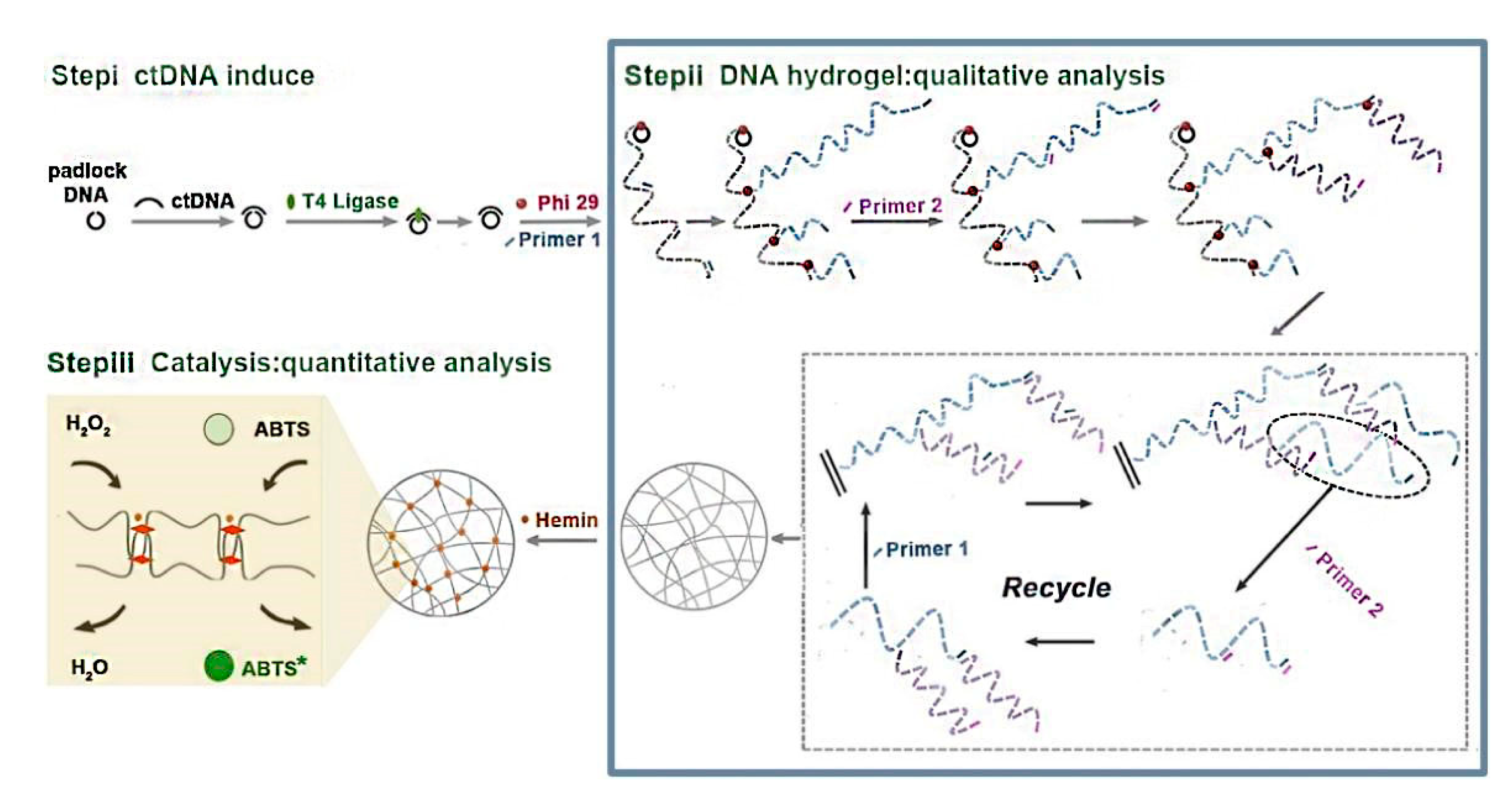
Figure 2.
Schematic illustrations of the principle of polysaccharide hydrogel sensors. (a) The on-line enzyme assays for penicillinase utilizing CE-IMERs; (b) The cascaded signal amplification sandwich-type impedimetric immunosensor based on sodium alginate hydrogels’ precipitation for prostate specific antigen (PSA) detection. Reproduced with permission from Xiaotong Hu (a) and Lihua Zhao (b), Talanta (a) and ACS Sensors (b); published by Elsevier (a) and American Chemical Society (b), 2018 (a) and 2019 (b).
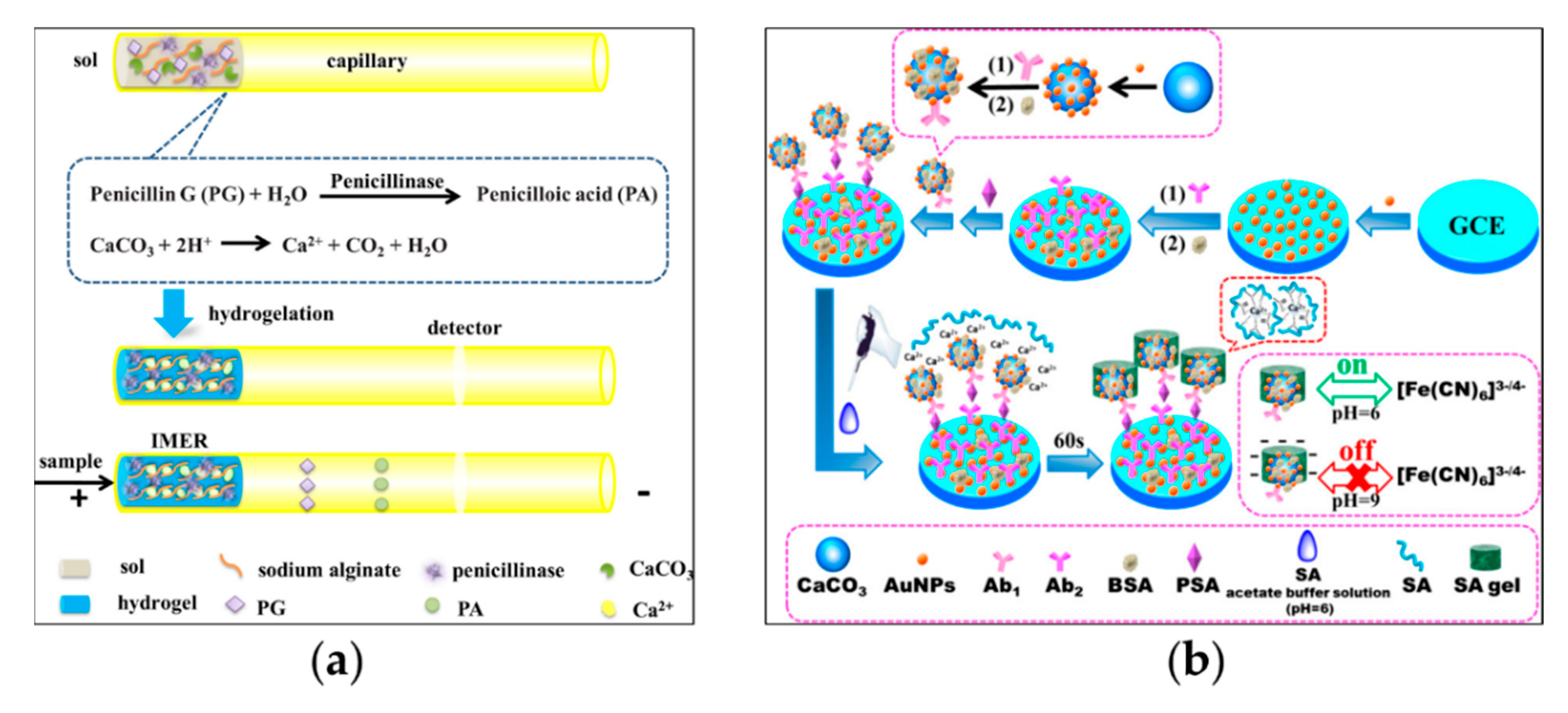
Figure 3.
Schematic illustration of hydrogels’ transition signal conversed into colorimetric signal. (a) The AA-responsive alginate hydrogels for the colorimetric detection of ALP; (b) The self-assembled DNA hydrogels based on RCA products for the visual OTA detection. Reproduced with permission from Liping Gao (a) and Liling Hao (b), Analytica Chimica Acta (a) and Microchimica Acta (b); published by Elsevier (a) and Springer (b), 2021.

Figure 4.
Schematic illustration of fluorescent DNA hydrogels biosensor based on self-assembly of RCA productions for the detection of OTA. Reproduced with permission from Liling Hao, Journal of Agricultural and Food Chemistry; published by American Chemical Society, 2020.
Figure 4.
Schematic illustration of fluorescent DNA hydrogels biosensor based on self-assembly of RCA productions for the detection of OTA. Reproduced with permission from Liling Hao, Journal of Agricultural and Food Chemistry; published by American Chemical Society, 2020.
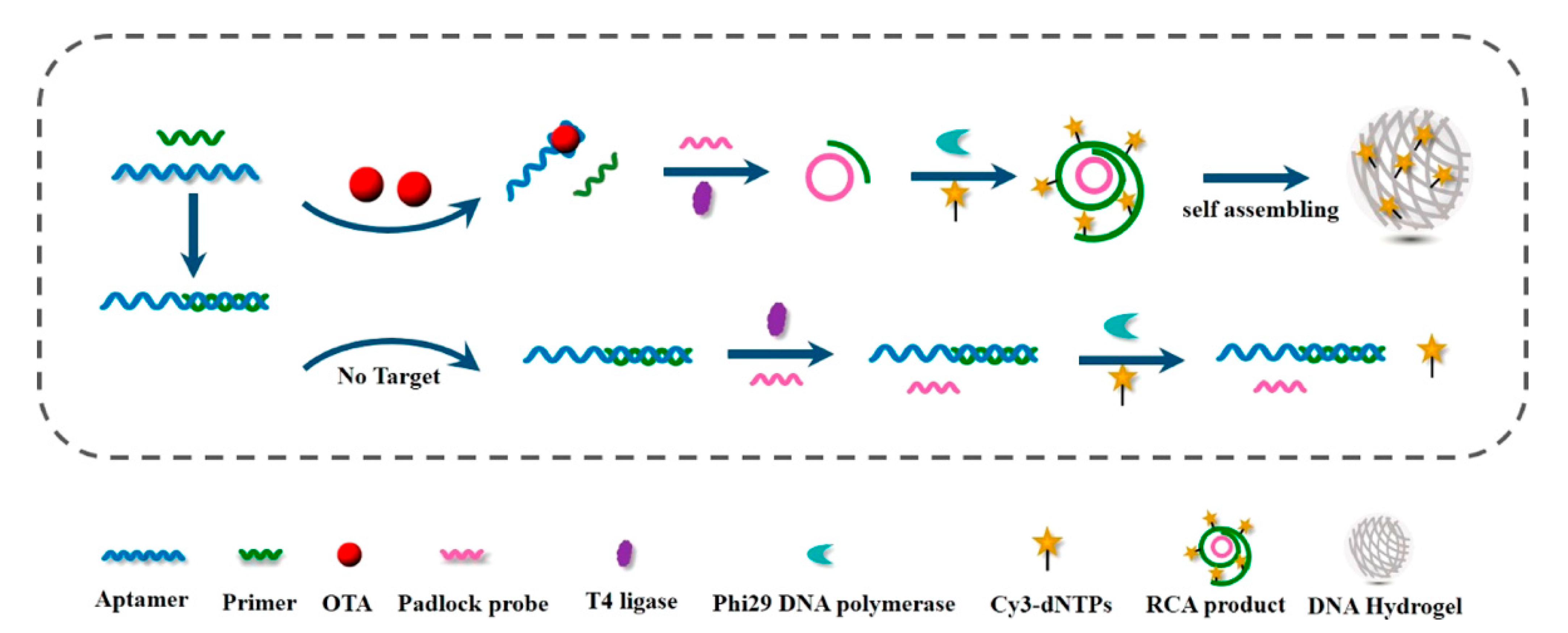
Figure 5.
Schematic illustration of the DNA hydrogels coating to SERS substrate as a flexible biosensor for UO22+ detection. Reproduced with permission from Xuan He, Langmuir; published by American Chemical Society, 2020.
Figure 5.
Schematic illustration of the DNA hydrogels coating to SERS substrate as a flexible biosensor for UO22+ detection. Reproduced with permission from Xuan He, Langmuir; published by American Chemical Society, 2020.

Figure 6.
Schematic illustration of the hydrogelation of alginate hydrogels and T2 biosensing strategy with the assistance of enzyme for the immunoassay of S. enteritidis. Reproduced with permission from Luyu Wei, Analytical Chemistry; published by the American Chemical Society, 2021.
Figure 6.
Schematic illustration of the hydrogelation of alginate hydrogels and T2 biosensing strategy with the assistance of enzyme for the immunoassay of S. enteritidis. Reproduced with permission from Luyu Wei, Analytical Chemistry; published by the American Chemical Society, 2021.
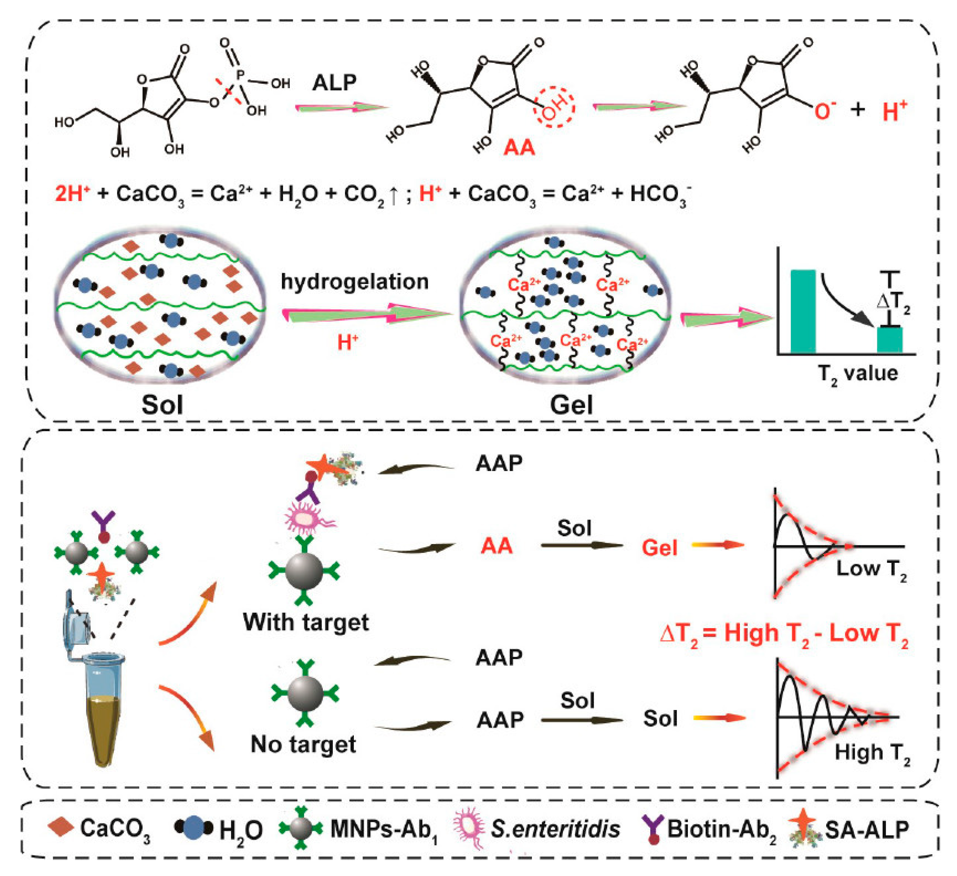
Figure 7.
Schematic illustration of the portable device for the detection of AFB1 based on the DNA hydrogels and pH meter. Reproduced with permission from Mengmeng Zhao, Talanta; published by Elsevier, 2017.
Figure 7.
Schematic illustration of the portable device for the detection of AFB1 based on the DNA hydrogels and pH meter. Reproduced with permission from Mengmeng Zhao, Talanta; published by Elsevier, 2017.

Figure 8.
Schematic illustration of HA hydrogels containing Pt@SiO2NPs for HAase detection using electronic balance as readout device. Reproduced with permission from Zhixin Li, Analytical Chemistry; published by American Chemical Society, 2019.
Figure 8.
Schematic illustration of HA hydrogels containing Pt@SiO2NPs for HAase detection using electronic balance as readout device. Reproduced with permission from Zhixin Li, Analytical Chemistry; published by American Chemical Society, 2019.
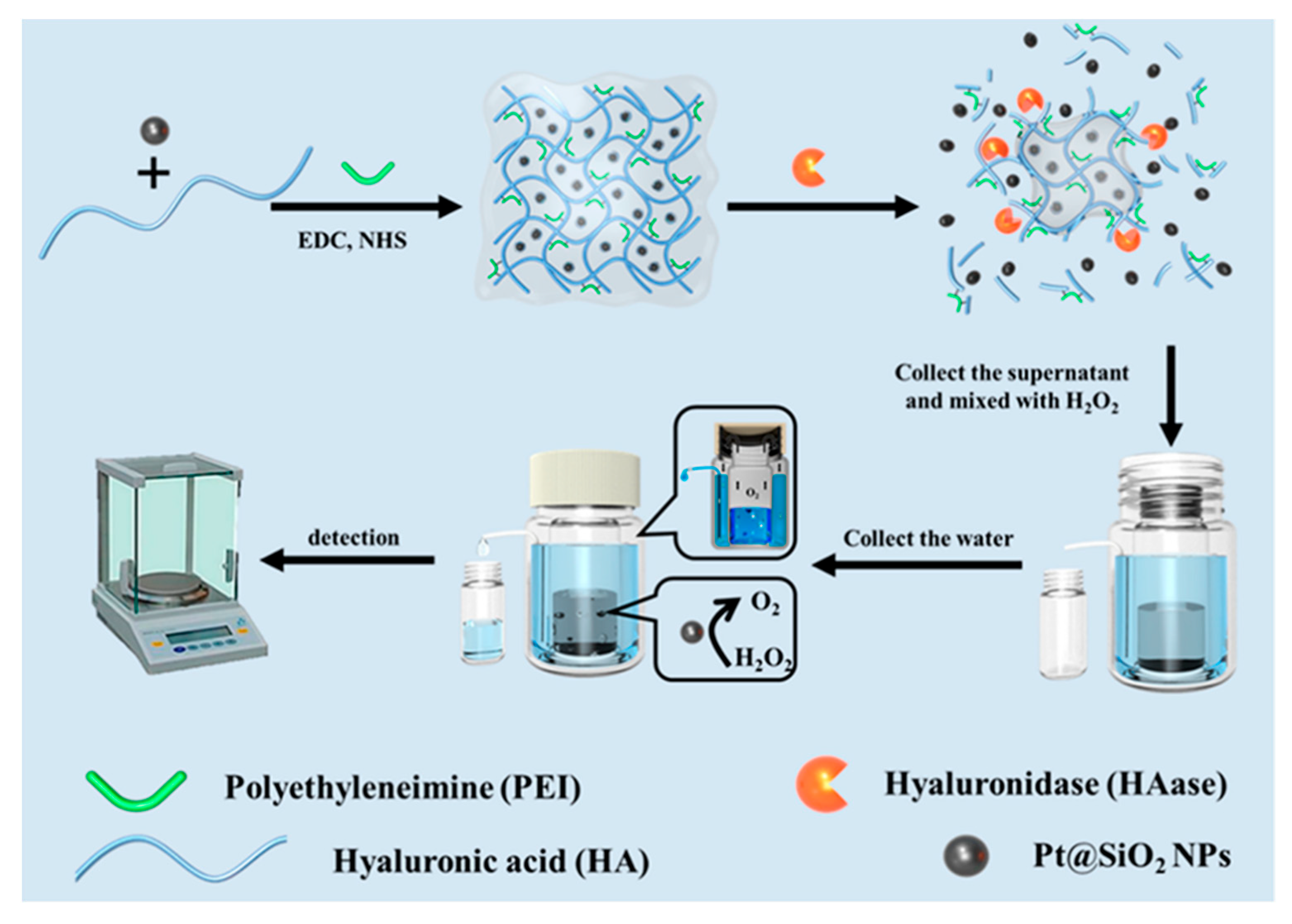
Figure 9.
Schematic illustration of POCT tool integrating Dam-responsive DNA tetrahedra-crosslinked hydrogels and its evaluation strategy of Dam activity and quantitative. Reproduced with permission from Xin Gao, Analytical Chemistry; published by American Chemical Society, 2020.
Figure 9.
Schematic illustration of POCT tool integrating Dam-responsive DNA tetrahedra-crosslinked hydrogels and its evaluation strategy of Dam activity and quantitative. Reproduced with permission from Xin Gao, Analytical Chemistry; published by American Chemical Society, 2020.
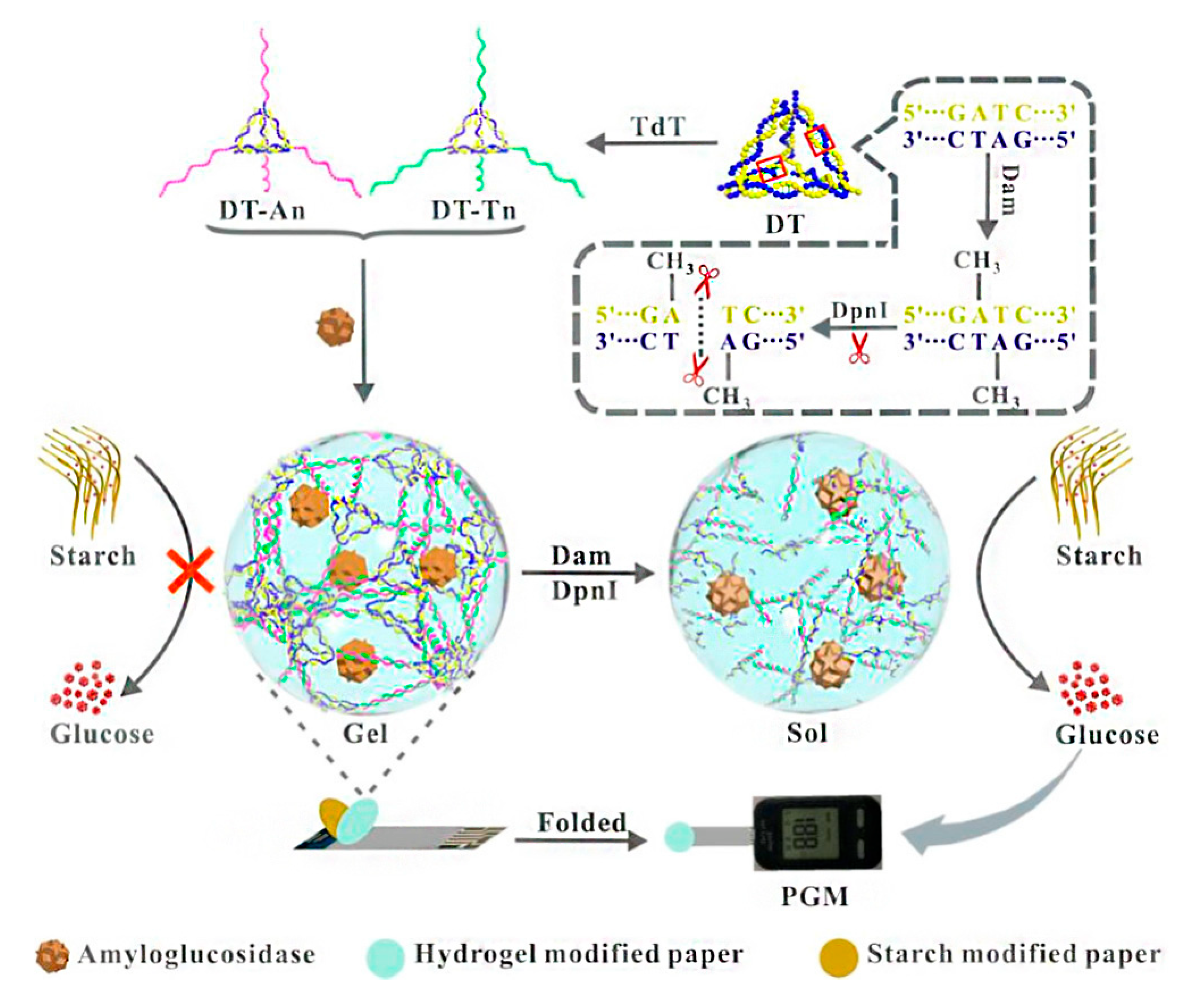
Figure 10.
Schematic illustration of paper-based sensors with pH-responsive alginate hydrogels and the analytical strategy for the detection of OPs. Reproduced with permission from Jia Xu, Analytica Chimica Acta; published by Elsevier, 2019.
Figure 10.
Schematic illustration of paper-based sensors with pH-responsive alginate hydrogels and the analytical strategy for the detection of OPs. Reproduced with permission from Jia Xu, Analytica Chimica Acta; published by Elsevier, 2019.

Figure 11.
Schematic illustration of the dual-readout paper-based sensor using pH-responsive hydrogels for the detection of penicillinase and its discoloration principle. (a) The formation principle of the sensing materials, detection phenomena and the profile-view of the detection equipment are given; (b) The discoloration principle of the color sensing spots for the detection of pH. Reproduced with permission from JiaXu, Sensors and Actuators B: Chemical; published by Elsevier, 2021.
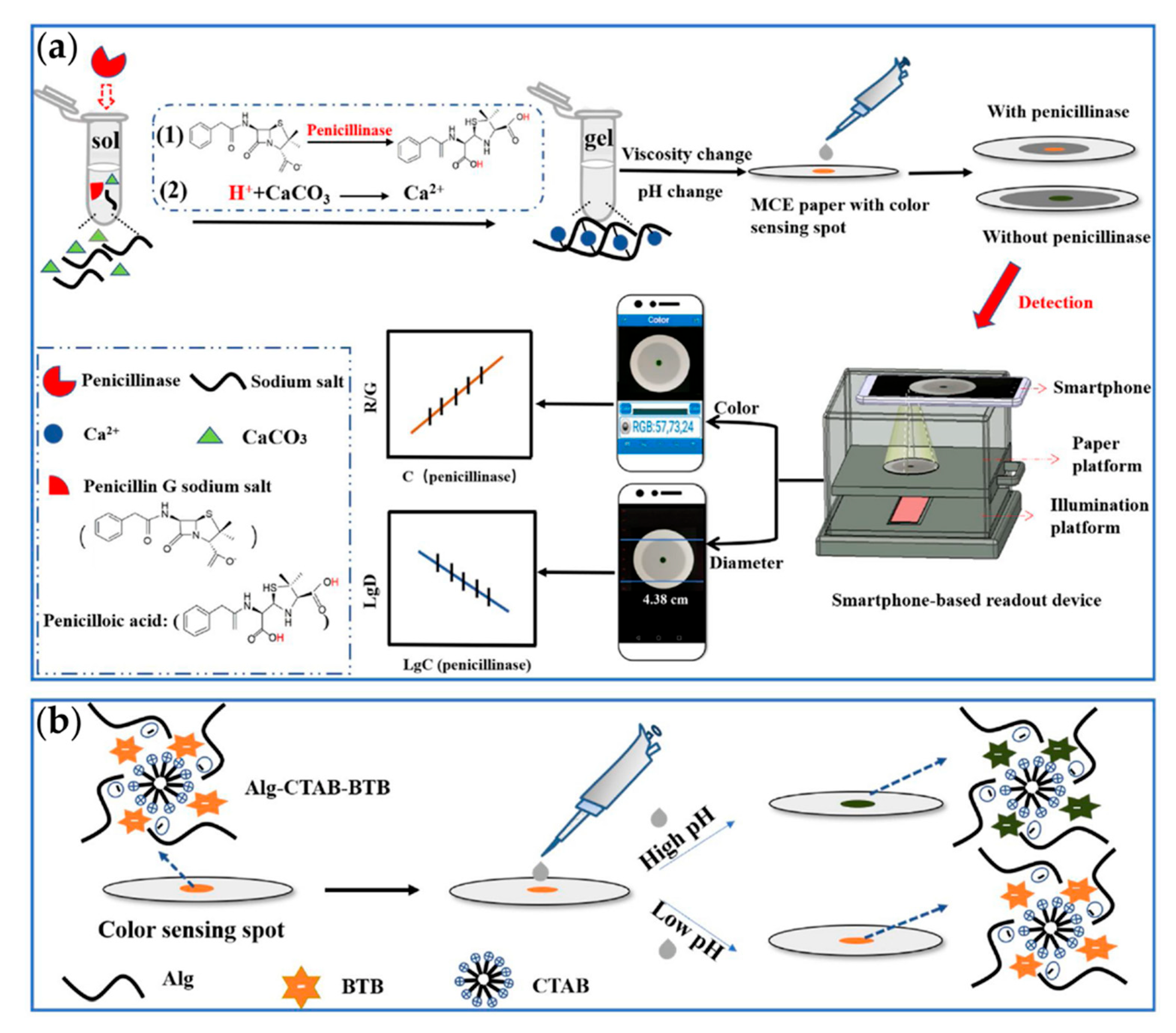
Figure 12.
Schematic illustration of DNA hydrogels in capillary for the detection of Pb2+. Reproduced with permission from Chao Jiang, Sensors and Actuators B: Chemical; published by Elsevier, 2020.
Figure 12.
Schematic illustration of DNA hydrogels in capillary for the detection of Pb2+. Reproduced with permission from Chao Jiang, Sensors and Actuators B: Chemical; published by Elsevier, 2020.
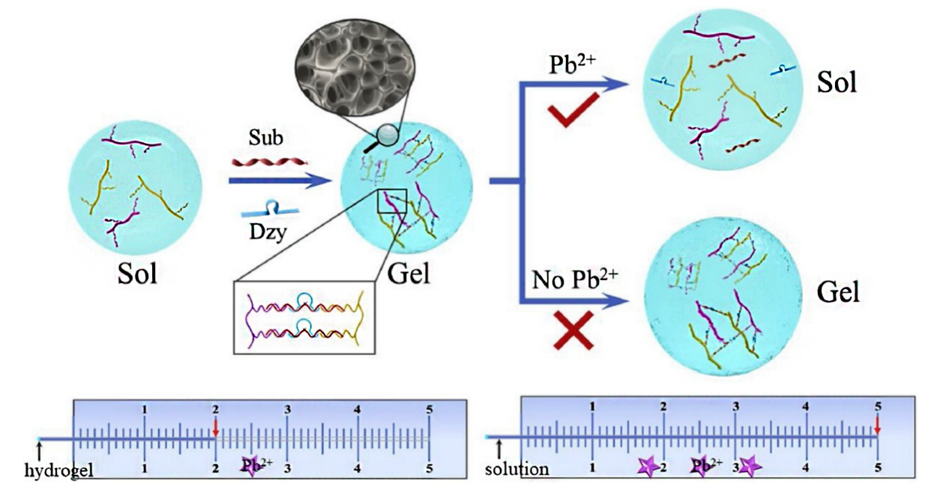
Figure 13.
Schematic illustration of paper-based sensing for the detection of thrombin and its inhibitor via monitoring the viscosity change of the amylopectin solution. Reproduced with permission from Chengliang Han, Analytica Chimica Acta; published by Elsevier, 2022.
Figure 13.
Schematic illustration of paper-based sensing for the detection of thrombin and its inhibitor via monitoring the viscosity change of the amylopectin solution. Reproduced with permission from Chengliang Han, Analytica Chimica Acta; published by Elsevier, 2022.

Table 1.
Examples of hydrogels with different recognition units.
| Type of Hydrogels | Output Signal | Target | Recognition Unit | Reference |
|---|---|---|---|---|
| Hybrid/pure DNA hydrogels | Fluorescence | MicroRNA-141 | DNA probe | [34] |
| Color | Glucose | Target aptamer strand | [20] | |
| Electricity | Hg2+ | DNAzyme/Hairpin DNA | [35] | |
| Fluorescence | Drug-resistant bacteria gene | Hairpin DNA | [36] | |
| Color | Circulating tumor DNA | G-quadruplex | [37] | |
| pH | Aflatoxin B1 | Target aptamer strand | [38] | |
| Polypeptide hydrogels | Distance | Thrombin/inhibitors | Thrombin | [39] |
| Weight | Hyaluronidase | Pt@SiO2NPs | [40] | |
| Polysaccharide hydrogels | Electrochemistry | 4-Nitrophenol | Glutaraldehyde | [41] |
| Fluorescence | Hg2+/Hg+ | Glutaraldehyde | [42] | |
| Color | pH | Ca2+ crosslinker | [43] | |
| Surface-Enhanced Raman Scattering | BSA-tetramethylrhodamine | Ag-nanoparticle crosslinker | [44] | |
| Magnetic Resonance Spectroscopy | Foodborne pathogens | ALP-modified antibody | [43,45] |
Table 2.
The application and limit of detection of sensors with different using methods.
| Method | Detection Application | Limit of Detection | Reference |
|---|---|---|---|
| Colorimetric assay | T-2 mycotoxin | 0.87 pg/mL | [61] |
| Alkaline phosphatase | 0.37 mU/mL | [62] | |
| Ochratoxin A | 0.005 ng/mL | [63] | |
| Fluorescence assay | Alkaline phosphatase | 21.3 μM | [64] |
| Ochratoxin A | 0.01 ng/mL | [65] | |
| Surface-enhanced Raman spectroscopy (SERS) assay | UO22+ | 8.38 × 10⁻¹³ M | [66] |
| α-fetoprotein | 50 pg/mL | [67] | |
| Magnetic relaxation switching (MRS) assay | Foodborne pathogens | 50 CFU/mL | [45] |
| pH-based assay | Aflatoxin B1 | 0.1 μM | [38] |
| Weight assay by electronic balance | Hyaluronidase | 0.2 U/mL | [40] |
| AFB1 | 9.4 μg/kg | [68] | |
| Hyaluronidase | 0.35 U mL−1 | [69] | |
| Glucose assay by personal glucose meter | DNA adenine methyltransferase | 0.001 U/mL | [70] |
| Distance-based lateral flow assay | Organophosphorus pesticides | 3.3 ng/mL | [71] |
| Penicillinase | 2.67 × 10⁻³ mU/μL 2.67 × 10⁻² mU/μL | [72] | |
| Glucose | 1.4 mM | [73] | |
| Pb2+ | 10 nM | [74] | |
| Thrombin and inhibitor | 16.1 mU/mL | [39] |
Publisher’s Note: MDPI stays neutral with regard to jurisdictional claims in published maps and institutional affiliations. |
© 2022 by the authors. Licensee MDPI, Basel, Switzerland. This article is an open access article distributed under the terms and conditions of the Creative Commons Attribution (CC BY) license (https://creativecommons.org/licenses/by/4.0/).
Share and Cite
MDPI and ACS Style
Song, H.; Ding, S.; Zhao, M.; Hu, Q. The Advances of Hydrosol–Gel Transition-Based Sensors. Chemosensors 2022, 10, 415. https://doi.org/10.3390/chemosensors10100415
AMA Style
Song H, Ding S, Zhao M, Hu Q. The Advances of Hydrosol–Gel Transition-Based Sensors. Chemosensors. 2022; 10(10):415. https://doi.org/10.3390/chemosensors10100415
Chicago/Turabian StyleSong, Haoyang, Shichao Ding, Mei Zhao, and Qiongzheng Hu. 2022. "The Advances of Hydrosol–Gel Transition-Based Sensors" Chemosensors 10, no. 10: 415. https://doi.org/10.3390/chemosensors10100415
Note that from the first issue of 2016, this journal uses article numbers instead of page numbers. See further details here.