Review

A Review on the Advances in Nanomaterials for Electrochemical Non-Enzymatic Glucose Sensors Working in Physiological Conditions

by
Velia Osuna
1,
Eider Pedro Aparicio Martínez
2,
Rocio B. Dominguez
1,* and
Alejandro Vega Rios
2,*
1
CONAHCYT-Centro de Investigación en Materiales Avanzados, S.C. (CIMAV), Miguel de Cervantes #120, Complejo Industrial Chihuahua, Chihuahua 31136, Mexico
2
Centro de Investigación en Materiales Avanzados, S.C. (CIMAV), Miguel de Cervantes #120, Complejo Industrial Chihuahua, Chihuahua 31136, Mexico
*
Authors to whom correspondence should be addressed.
Chemosensors 2024, 12(8), 159; https://doi.org/10.3390/chemosensors12080159
Submission received: 29 May 2024 / Revised: 18 July 2024 / Accepted: 29 July 2024 / Published: 8 August 2024

Abstract

:
Although an enzymatic electrochemical biosensor is a major keystone in Diabetes Mellitus management, its replacement with a low-cost and stable non-enzymatic glucose sensor (NEGS) is of high interest to scientific and industrial fields. However, most NEGS for direct glucose electrooxidation (DGE) must be performed under extreme alkaline conditions, implying additional pretreatments before detection and a limited application for on-body, real-time monitoring. Thus, research on DGE in physiological conditions is fundamental to successfully translating the current NEGS into clinical applications. In physiological conditions, drawbacks such as low current, low selectivity, and poisoning appear due to the reduction of OH ions in neutral electrolytes and the presence of chloride ions in biofluids. Therefore, an increasing number of nanomaterials based on Pt, Au, and their nanocomposites have been proposed to improve the electrochemical performance. Additionally, transition metals such as Cu, Pd, Ni, or Co combined with high surface area supports have shown promising results in increasing catalytic sites for DGE. The molecular interaction of phenylboronic acid with glucose has also been demonstrated in neutral conditions. Overall, the present review summarizes the current strategies for DGE in physiological conditions and highlights the challenges still faced for further development of functional glucose NEGS.

Graphical Abstract

1. Introduction

Diabetes Mellitus (DM) is a chronic metabolic, degenerative disease characterized by elevated blood glucose levels [1]. According to recent statistics from the International Diabetes Federation (IDF), a total of 10.5% of the worldwide adult population between 20 and 79 suffers from DM; see Figure 1a [2]. These alarming numbers also estimate a gradual increase to 783 million cases by 2045; see Figure 1b [2]. Compared to IDF data from 1990, the number of patients with DM increased by about 450% in 31 years [3].
Diabetes patients must regularly monitor their blood glucose levels to determine therapeutic decisions, which may involve changes in diet, drug intake, or insulin doses [4]. The classification of diabetes is crucial for determining the treatment that should be followed and the frequency of monitoring. Diabetes can be classified as type 1 diabetes, type 2 diabetes, gestational diabetes, and specific types of diabetes due to other causes. Type 1 diabetes is related to the autoimmune destruction of pancreatic cells, leading to absolute insulin deficiency. Type 2 diabetes is caused by progressive pancreatic β-cell function loss, decreasing insulin secretion. Gestational diabetes is defined as any degree of carbohydrate intolerance with onset for the first time during the second or third trimester of pregnancy [2].
The guidance for monitoring indicates that type 1 diabetes patients need to perform blood glucose monitoring (BGM) five to six times per day, while, in the case of type 2 diabetes individuals with an insulin dosage, this number is three to four times per day. When not adequately controlled, DM possesses a high risk of comorbidity and can lead to significant complications, e.g., cardiovascular disease, neuropathy, retinopathy, nephropathy, and skin conditions. Furthermore, during the COVID-19 pandemic, the prevalence of diabetes was observed to increase the severity of COVID-19, with DM accounting for 9.5% of severe cases and 16.8% of deaths [1]. Thus, it is vital to have timely diagnoses and adequate control of the disease, in which BGM plays a prominent role.
The current standards for DM diagnosis include fasting plasma glucose values above 126 mg/dL, according to the American Diabetes Association. Although a laboratory test is usually the first analytical method, it is common that, once diagnosed, the patient performs self-monitoring blood glucose using a point of care (PoC) detector. The technology for DM monitoring has experienced significant advantages and innovation since the first dextrostix strips, following the handheld PoC glucose meter and the current continuous glucose monitor (CGM). Nevertheless, enzymatic detection has been at the center of such advances [5].
Enzymatic glucose sensors (EGS) employ biomolecules such as glucose oxidase or glucose dehydrogenase to selectively catalyze glucose oxidation, generating an electrical signal that correlates with the glucose concentration. The measurement can be directly performed either from capillary blood in the case of a glucose meter or from interstitial fluid for CGM, achieving a good correlation with plasma glucose in both cases. Although EGS has revolutionized glucose monitoring for individuals with diabetes, users should remain aware of their limitations and ensure proper maintenance and calibration for reliable results. EGS are also susceptible to interference from substances other than glucose, which may be present in biological fluids, and have a limited operational lifespan, often ranging from several days to a couple of weeks [6,7]. In addition, sensor performance can be impacted by external factors such as temperature, humidity, oxygen levels, or extreme conditions [7].
Non-enzymatic glucose sensors (NEGS) have been developed as an alternative to address the limitations and drawbacks associated with EGS. In the last decade, this field has experienced substantial growth driven by the clinical importance of BGM and the expansion of the glucose monitoring market. The basis of the operation of NEGS is modifying the working electrode surface with novel materials to promote direct glucose electrooxidation (DGE). The vast literature regarding NEGS has been systematically reviewed, highlighting the role of materials such as carbon nanomaterials [8,9], conducting polymers [10,11], metallic nanomaterials [12,13], metallic oxides [14], or metallic organic frameworks [10] in achieving sensitive glucose detection. In these processes, glucose molecules are oxidized at the NEGS-modified surface, releasing electrons that produce a current proportional to glucose concentration.
Most NEGS studies typically employ alkaline electrolytes (e.g., NaOH and KOH) to facilitate the sensing mechanism, therefore promoting DGE [10]. Additionally, the electrochemical activity of many carbon-based electrode materials commonly used in NEGS is increased under these alkaline conditions. Glucose selectivity is also improved, because alkaline electrolytes can reduce interference from electroactive species in the sample. Nevertheless, glucose under strongly alkaline conditions can be subjected to extensive isomerization and even chain degradation. This effect is caused by the establishment of an equilibrium between the monosaccharide and an enediol structure through a slow process hydride ion transfer. The enediol from glucose can subsequently be oxidized at higher potentials [15,16].
Therefore, NEGS working at physiological pH conditions are preferred to be as functional as their EGS counterparts for several reasons. First, unlike EGS, most NEGS need a previous pH adjustment of the sample under analyses to achieve the fullest analytical capabilities of the sensor. Compared to commercial disposable strips, this is noncompetitive but has further relevance for implantable devices where the highly alkaline environment is incompatible with the body environment [17,18]. Second, using an electrolyte at the physiological pH minimizes the risk of tissue damage or irritation when the sensor is intended for implanted applications. For wearable or implantable glucose sensors, comfort and user acceptance is crucial. In addition, operating at the physiological pH helps reduce discomfort, irritation, or inflammation at the sensor site, enhancing the user’s overall experience. Finally, sensors operating at the physiological pH are more likely to gain regulatory approval and clinical acceptance due to their compatibility with the human body and adherence to safety standards [17].
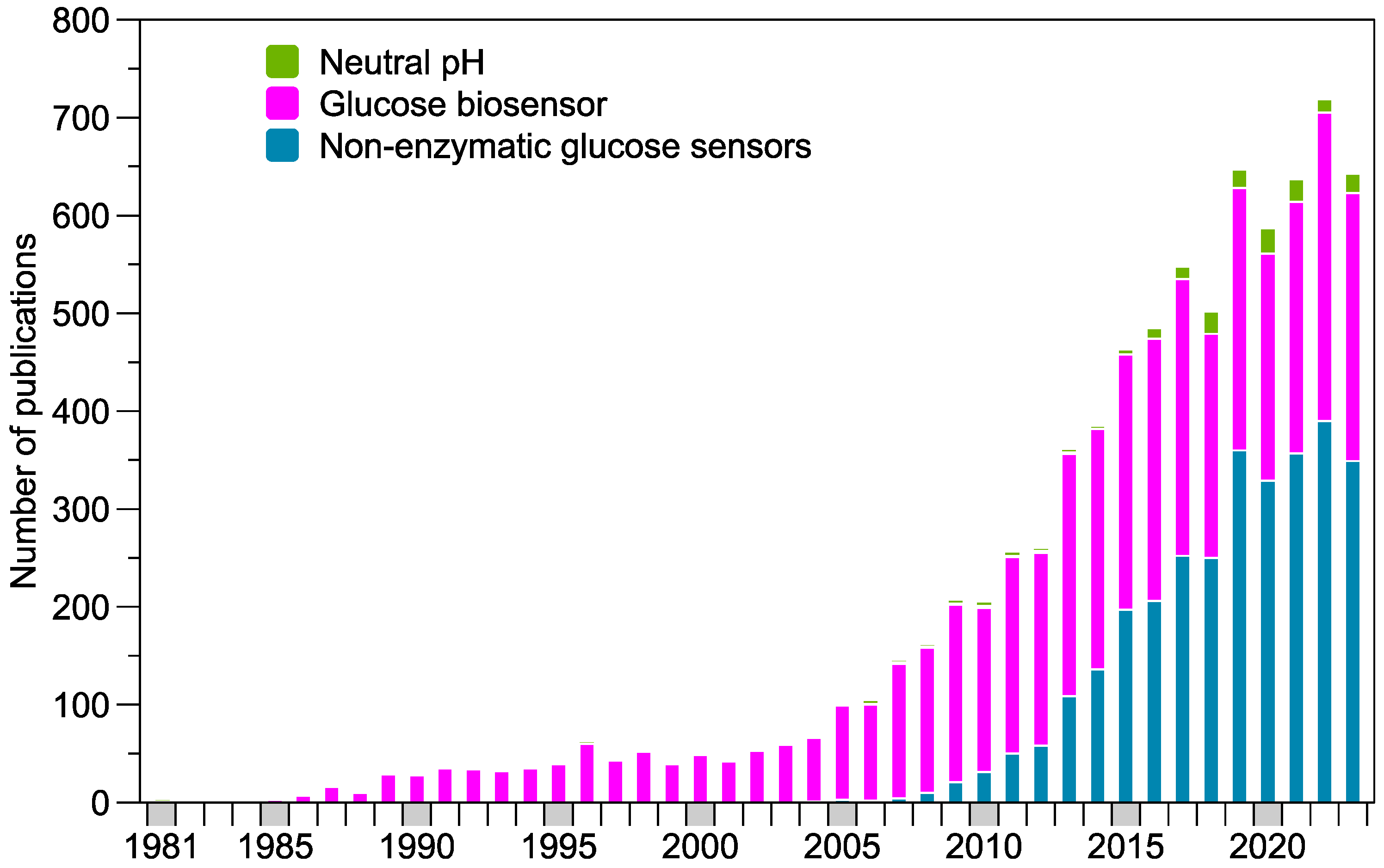
As observed in Figure 2, the literature trend was dominated by EGS until a recent shift towards NEGS, mainly in the last 5 years. However, few works within this group were devoted to non-enzymatic detection at physiological conditions (NEDPC), especially pH 7.4. Some early attempts during the 1980s and 1990s involved noble metal electrodes to catalyze glucose oxidation, with platinum (Pt) and gold (Au) as the leading candidates [19,20,21,22]. Nevertheless, these electrodes made of bulk materials often had limitations, including sensitivity issues and susceptibility to interference. Furthermore, additional challenges appeared as researchers focused on changing the typical NaOH alkaline electrolyte to a solution that resembles physiological conditions, e.g., phosphate-buffered saline (PBS). The lower presence of OH anions makes it challenging to record a significant current density for DGE, resulting in novel strategies for the local pH increase, such as water splitting or surface functionalization. In addition, as nanostructured materials, such as carbon nanotubes, graphene, or metal oxide nanoparticles, began to be incorporated onto modified electrode surfaces, improved sensor sensitivity and selectivity were obtained due to an enhanced surface area and catalytic properties [18].
In this work, we reviewed the existing literature regarding electrochemical NEDPC. In addition to the introductory section, we included a brief background on BGM covering the current commercial PoC. In the third section, we described the reported mechanism for glucose sensing under physiological conditions, including the well-known incipient hydrous oxide adatom mediator (IHOAM) model and the strategies for adding OH anions. The following section presented the current sensors for NEDPC, where Pt- and Au-based nanomaterials were the most studied materials. Recent trends based on additional transition metals (Ag, Pd, Cu, Ni, and Co) and their composites were also discussed, along with boronic acid-based copolymers. The recent developments in wearable glucose sensors for physiological conditions are summarized in Section 5. Finally, perspectives and conclusions of DGE at physiological conditions for the further development of functional NEGS are included at the end of the document.

2. Electrochemical Glucose Detection Background

2.1. Current Formats for Commercial Glucose Detection

Currently, the commercially available choices for glucose self-monitoring involve either blood glucose meters (BGMT) or continuous glucose meters (CGM). Despite working under EGS, their inherent functioning implies fundamental differences such as the analyzed sample, the required accuracy, biocompatibility aspects, frequency of monitoring, or long-term stability [5]. All these characteristics need to be considered in the design of alternative NEGS. BGMT are handheld devices comprising an enzymatic disposable test strip and a portable meter. The monitoring involves pricking the finger to obtain a small capillary blood sample to measure the glucose concentration directly. Although its performance can be affected by environmental, physiological, and medication factors, BGMT provides a good correlation with plasma glucose and remains a valuable element of diabetes management [5]. Most of the reviewed literature on NEDPC falls in this category, as the proposed materials are intended for single use and are mainly evaluated in blood samples or related fluids, e.g., plasma or serum. Usually, these NEDPC are manufactured using rigid transducers such as screen-printed carbon electrodes (SPCEs), glassy carbon electrodes (GCEs), or fluorine-doped tin oxide that are subsequently modified with catalytic materials for DGE. However, in order to be considered as a competitive alternative, the proposed NEDPC devices need to consider the working range of commercial enzymatic BGMT, the null pretreatment of the sample, and the low cost of enzymatic strips [17].
One main disadvantage of BGMT is the limited monitoring due to incommodities associated with skin pricking, which can prevent the identification of glucose peak episodes during the day or night [6]. Thus, it is unsurprising that the CGM reports a rapid user increase in recent years. CGMs provide real-time glucose measurements and do not require regular fingerstick tests. In this device, a small sensor with microneedles is inserted under the skin (typically on the abdomen or arm) and continuously measures glucose levels in the interstitial fluid; then, the recorded data are sent to a receiver or a smartphone app [6]. Currently, the number of CGM based on NEGS at a neutral pH is quite limited, possibly due to the complexity of the operation and biocompatibility in an implantable operation. However, other wearable devices for CGM at laboratory stages aim to relate blood glucose levels with the glucose content in non-invasive samples, such as sweat, saliva, urine, or tears [23]. In this format, the detector is closely attached to the body but the collection of samples involves only excreted fluids rather than inner fluids, simplifying the design process. Even so, the NEDPC sensors need to be incorporated in flexible substrates that allow on-body detection, such as textiles, polymers, carbon nanomaterials, or metal foils. Additionally, wearable glucose sensors need to exhibit high sensitivity due to the low concentration found in these body fluids compared to that found in blood. Table 1 illustrates the glucose concentration in body fluids according to their working operation principle.

2.2. Accuracy and Precision

The reliability of glucose sensors is a crucial aspect of their development and clinical use. These sensors have the advantage of requiring small blood samples and delivering results quickly. However, factors such as glycolysis and the instability of blood glucose levels can impact their accuracy [5,26]. To assess the accuracy of a BGMT, one can distinguish between clinical and analytical accuracy. The former refers to the significance of the device’s results in clinical decision-making and is gauged based on the Parkes error grid [27]. Conversely, analytical accuracy compares the sensor’s readings to a gold-standard method.
Several organizations have established criteria to measure the precision of a BGMT. The key ones are detailed in Table 2. For instance, a comprehensive evaluation of the performance of a BGMT, the ISO 15197 standard from 2015, includes additional parameters to characterize [28]. One of them is bias, which estimates the systematic error in a measurement and can be calculated by subtracting a reference value from the average value obtained from a set of measurements. Precision, which measures the consistency of a series of replicated measurements under specific conditions, must also be considered and can be quantified using standard deviation, variance, or the coefficient of variation.
Moreover, the Food and Drug Administration (FDA) recommends assessing imprecision through repeated measurements of the same sample, termed “within-run precision evaluation”, and advises evaluating imprecision under simulated conditions of regular use, testing a sample for at least 10 days, known as an “Intermediate precision evaluation”. The data obtained from these studies after carrying out a complete statistical study must include the mean, standard deviation, and the coefficient of variation (%CV) of the different glucose concentrations used in each case [29,30].

2.3. Selectivity, Interferences, and Poisoning

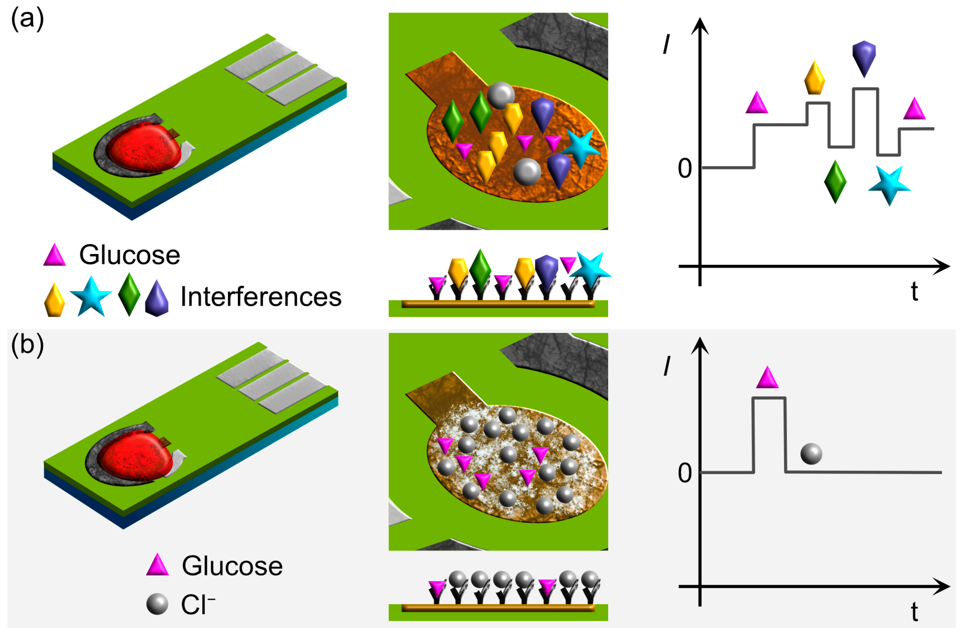
Selectivity in a glucose sensor refers to its ability to detect only glucose despite the presence of other substances. Interference is a parameter that describes the degree of influence these substances have on the magnitude of the sensor’s response; see Figure 3. These interfering compounds can be endogenous, exogenous, or resulting from the effects of a disease [27]. Table 3 displays different interfering substances and their recommended evaluation concentrations by the FDA for self-monitoring blood glucose devices.
Additionally, for these devices, it is recommended that tests be conducted to assess the interference caused by varying levels of hematocrit and oxygen in the blood on the accuracy of the glucometer [27,28]. In the case of NEGS, evaluating the poisoning of the sensing materials is recommended. The poisoning pertains to the partial or total loss of sensitivity experienced by the sensing material caused by the chemisorption of substances on its surface [32]. This poisoning can be caused by the chemisorption of substances on its surface, such as chlorine ions (Cl) [33], carbon monoxide [34], and gluconolactone [35]. When performing this evaluation, it is essential to use concentrations in ranges that are within those expected in the body.
Moreover, Cl are known to bind to metal species, inhibiting the formation of metal oxides crucial for the glucose detection mechanism. In particular, the presence of Cl can completely inhibit the oxidation reaction, especially at the high concentration of 0.1 M typically found in blood. Noble metals such as Pt, Ag, and Au, among others, are more susceptible to this type of problem. The most useful strategy reported is to place a thin film of NafionTM that prevents the interaction of Cl with the sensing material because of electrostatic repulsion [36]. Another popular approach is to incorporate bimetallic composites, e.g., Ni-Cu [36], Ni-Co-S [31], Pd-Au [37], and Co-Zn [38]. The regeneration of the surface of the sensor material has also been tested by applying appropriate potentials to reduce the metal oxides to their metallic form and then applying the potential to regenerate the metal oxide species prior to the sensing process. Finally, to evaluate the sensor’s response more accurately in the presence of interfering substances, it is recommended to use a biological matrix—that is, a real sample of blood or interstitial fluid, depending on the potential application of the sensor.

3. Sensing Mechanisms for Materials Applied for Non-Enzymatic Detection at Neutral pH

The DGE is the heart of the NEGS occurring on the working electrode. The theories of activated chemisorption and the IHOAM are the most recognized ones that can explain DGE on a metal transition electrode, i.e., its sensing mechanism. The activated chemisorption model was proposed by Pletcher et al. [39], who employed Pt, Ir, and Ni electrodes. The unpaired d-electrons and unfilled d-orbitals of transition metal are responsible for forming a bond with the analyte, i.e., glucose. In addition, the chemical composition of the transition metal—in particular, its oxidation state and its geometric arrangement of the center—are vital factors to consider in the design of the electrodes.
In contrast, Burke L. [40] proposed the IHOAM model, where OH plays a fundamental role on the Au electrode. The first step is the activated chemisorption of OH (through oxygen) species on the electrode, generating pre-monolayer oxidation on metal. Secondly, the glucose interacts with OH, causing glucose oxidation.
Moreover, in both models, an electric current is generated owing to DGE, which the device transforms into a response. Although both models were based on bulk (macro) electrodes with a polished surface, these theoretical principles are still utilized today to explain the sensing mechanism of the novel electrodes (e.g., bimetallic, carbon-based, and nanocomposites, among others).

3.1. Platinum

The kinetics and mechanism of DGE for Pt have mainly been studied in the 1970s and 1980s—in particular, studies focused on neutral pH using Pt in combination with materials or elements are of interest. Ernst and Heitbaum investigated the DGE on smooth Pt in PBS, allowing them to elucidate a mechanism with bases to an interaction between an adsorbed hydrogen and a free electron pair of the hemiacetal group; see Figure 4a [41,42]. In addition, the adsorbed radical formed in the oxidation process can be stabilized either by desorption or by a chemisorptive bond. Above 400 mV (double-layer region), glucose is absorbed on the Pt surface, then dehydrogenation and, finally, oxidation; see Figure 4b [42]. A similar approach was made by Pletcher [39] but from the point of view of the molecular theory of the metallic electrode.
Additionally, the process of DGE on a smooth Pt electrode has been investigated in a wide range of pH values. Vassilyev et al. [19] studied the kinetics and mechanism of DGE on Pt under steady-state and potentiodynamic conditions. Their findings show that the Pt electrode is not a very suitable catalyst, because it can adsorb various substances, including Cl and blood proteins, present in physiological solutions. It also lacks selectivity; that is, it is capable of oxidizing different organic substances. Furthermore, the oxidation current does not decrease with the addition of Cl at a concentration of 10−3 M. Nonetheless, at physiological chloride concentrations, the current decreases by a factor of 3–5, depending on the potential value. Finally, it was concluded that the overall kinetics of DGE at bare Pt is too sluggish to produce significant faradaic currents.
Recently, Singh et al. reported a PtAu/C NEGS, where they utilized the Ernst and Heitbaum principles to comprehend the sensing mechanism [43]. Furthermore, Wu et al. [44] studied the mechanisms of DGE on concave Pd core/island Pt shell nanoparticles by using the electrochemical quartz crystal balance. They established that the hydrogen region and double-layer region for the glucose-free medium occur between −500 mV and −300 mV (vs. Ag/AgCl) and from −300 mV to 200 mV (vs. Ag/AgCl), respectively, due to the chemisorption, dehydrogenation, and oxidation of D-glucose and the formation of gluconolactone on the electrode surface.

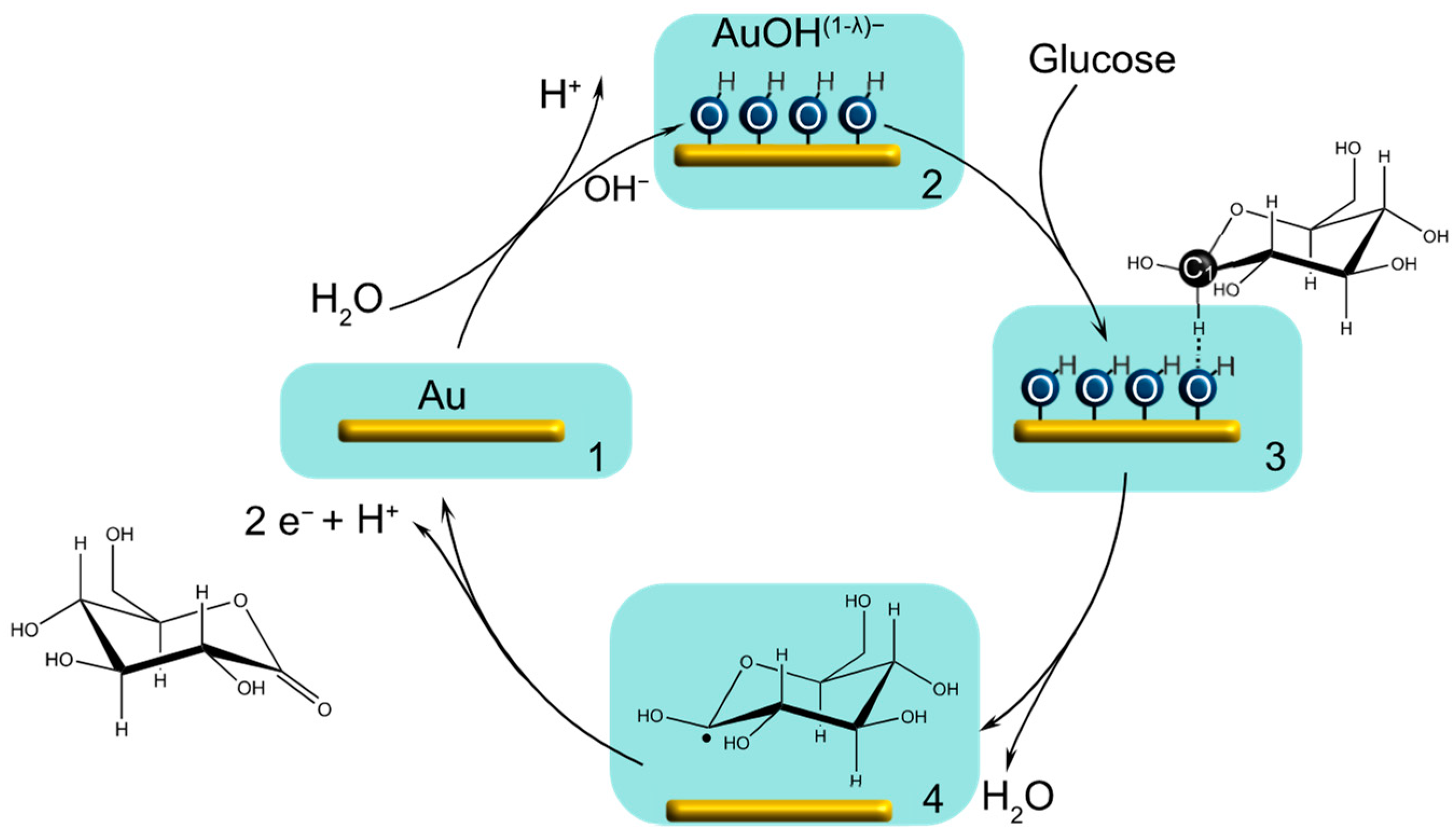
3.2. Gold

At the physiological pH, the adsorption of OH from H2O occurs at potentials from −0.1 to +0.3 V in the absence of glucose and is the first step for DGE; see Figure 5. This phenomenon is attributed to the chemisorption of the OH anion from H2O to form AuOHads [45]. The adsorption of glucose takes place on the AuOHads layer, followed by the oxidation of the weakly bound hydrogen atom at the carbon or the hydrogen of the hydroxyl group. DGE occurs through the interaction between the hemiacetal group and (OH)ads. The hydrogen bound to the C1 carbon atom is the first to oxidize; in this step, one electron is transferred to the electrode. Then, gluconolactone is generated, which is hydrolyzed to form gluconic acid, increasing the registered current [20,46,47]. The radical species is then oxidized to gluconolactone, generating another electron and transferring it to the electrode. In PBS, gluconolactone is hydrolyzed to form sodium gluconate, which has been reported by several investigations [48].
Furthermore, the DGE reaction is sensitively dependent on the crystallographic orientation of the Au single-crystal surfaces. At the most negative potential, DGE occurs on the Au (110) surface but has smaller peak currents than the other two low-index surfaces. Two peaks are observed for Au (110), one peak and a pre-peak for Au (111), and only one prominent peak for Au (110) [47]. In the same way, Karra et al. demonstrated that the morphology of gold nanoparticles (AuNPs) is essential to support a higher surface density of incipient gold oxide [49]. In addition, the mechanism of DGE for AuNPs was explained through the use of the IHOAM model [49].

3.3. Metallic Ions

Electrochemical reactions catalyzed by transition metal complexes have been described for alcohol, water, olefins, small molecules, and carbohydrates, mainly due to their advantages, such as robustness, stability, and low cost [50]. DGE can be catalyzed by the active species generated from electrodes modified with transition metal complex, resulting in novel configurations of NEGS. In this detection, the interaction between glucose and redox couples of transition metals (e.g., Cu, Pd, Ru, Co, and Ag) is used to detect and quantify the glucose levels.

3.4. Phenylboronic Acid and Derivates for Glucose Detection

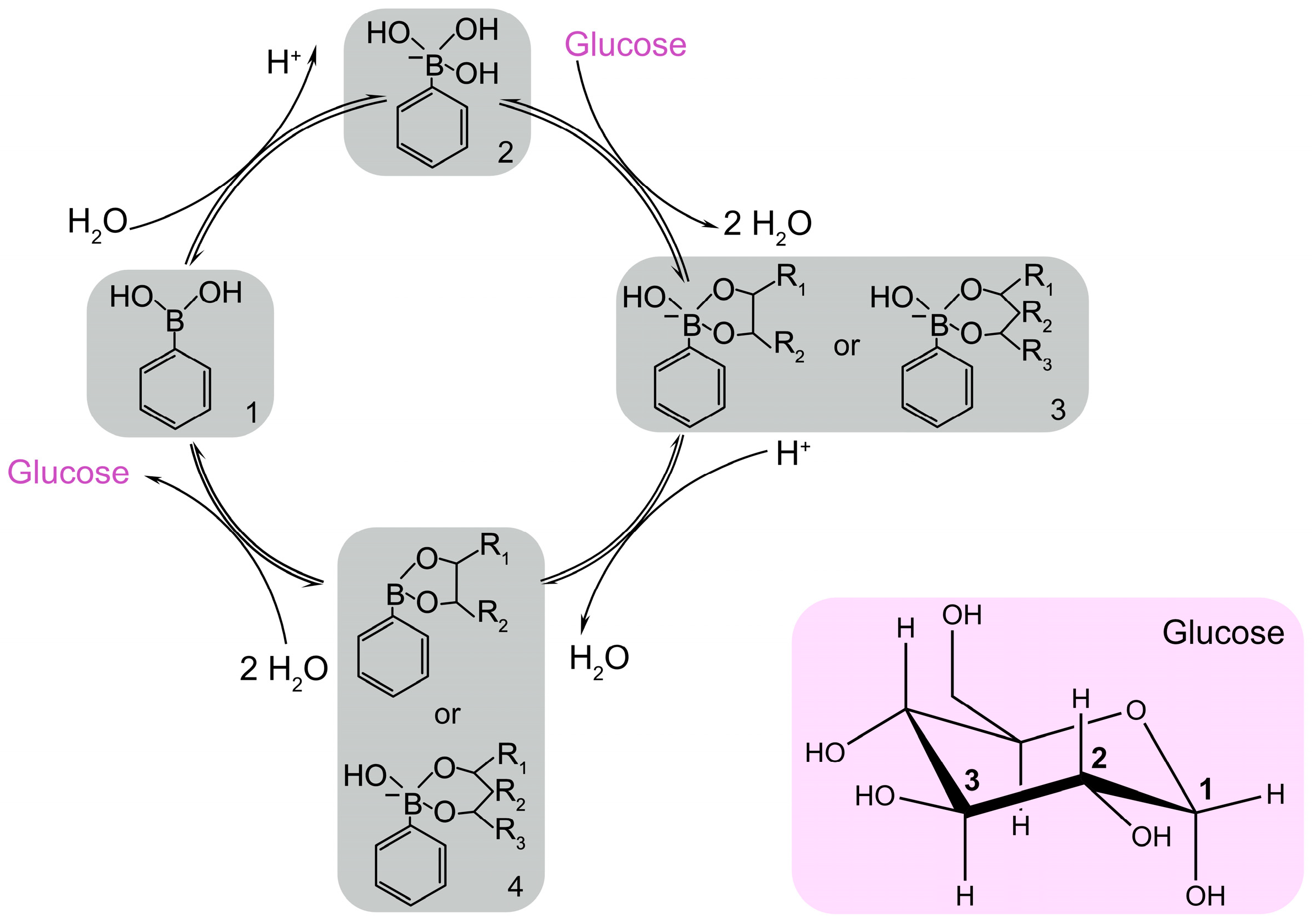
Phenylboronic acid (PBA) and its derivates are compounds that contain a central boron atom bonded to organic substituents (R groups) and at least one hydroxyl group [51]. The substituents are phenyl groups attached to a boronic functional group, and in its derivates compounds, the boronic functional group can be modified or substituted to enhance the reactivity, solubility, or specificity. The affinity of boronic acid compounds towards saccharides (e.g., glucose) has been largely studied mainly for fluorescent sensors [52,53]. However, PBA and its derivates are gaining popularity for glucose detection, since the interaction can also be electrochemically monitored in neutral media. PBA binds selectively to 1,2 and 1,3 diols to form reversible covalent bonds between the boron atom and the hydroxyl groups on such molecules; see Figure 6. This binding results in the formation of a boronate ester, a stable complex between boronic acid and glucose. To obtain PBA-modified electrode surfaces, strategies such as self-assembly on Au electrodes [54] or the electropolymerization of aromatic compounds with a boronic acid moiety can be implemented, leading to selective glucose layers [55].
However, PBA is electrochemically inactive, and glucose capture is usually detected by changes in electrochemical properties using redox-active compounds, e.g., ferrocene. One advantage of using PBA derivatives is that the interaction with glucose is reversible; thus, when the glucose levels change, the boronate ester can easily break, releasing the glucose molecules and allowing the sensor to respond dynamically to glucose concentration changes. The main disadvantage of this molecular interaction is the selectivity of PBA towards carbohydrates, catechol, fluoride, or dopamine. Nonetheless, the selectivity and specificity of PBA-based sensors can be further enhanced by modifying the boronic acid structure or incorporating additional recognition elements to target glucose specifically to help minimize interference from other diol-containing compounds.

3.5. New Strategies to Improve the Efficiency of the Sensing Mechanism

Recently, several pathways have emerged that promote the formation of OH at a neutral pH; see Table 4. Furthermore, the purpose is to increase DGE and, therefore, the efficiency of the sensing mechanism. Based on the working electrode, these can be categorized into pretreatment and in-site generation. In the same way, it can be classified from an electrochemical point of view as the surface modification and generation of OH using water or other substances contained in electrolytes.

4. Nanomaterials for Enzyme-Free Glucose Sensors Working at Neutral pH for Single Measurements

4.1. Platinum and Their Hybrid Nanocomposites

Platinum electrodes in early research showed drawbacks when applied to NEGS. These challenges have been addressed year by year utilizing various strategies. For example, the formation of Pt nanostructures, including mesoporous Pt surfaces [63], highly ordered Pt nanotube arrays [64], 3D ordered microporous Pt templates [65], Pt/carbon material [66,67], and Pt-based alloys [44,68,69,70], is introduced to enhance the sensitivity of non-enzymatic glucose sensors. The main focus of the research is the development of bimetallic or alloy nanoparticles with Pt.
The electrocatalytic activity of Pt-based materials is highly dependent on their morphologies, including dimensionality, surface, geometrical shape, geometrical area, and microstructure; due to their morphology, they can effectively tune the distributions of electrons and energy on Pt-atom surfaces. Yuan et al. [64] demonstrated that the electrode roughness factor positively impacts selectivity for glucose detection. Nevertheless, Chou et al. [71] manifested that the selectivity mainly originates from the morphology of the Pt electrode. In addition, the faradaic current enhances in relation to the effective electrode area, which improves selectively to glucose over interferences. Niu et al. [69] reported on the utilization of two-step electroplating to design and synthesize Pt cubes supported in porous Cu foam. Electrochemical test results indicate that the Pt-based cubic catalyst significantly improves catalytic activity towards DGE in the presence of Cl.
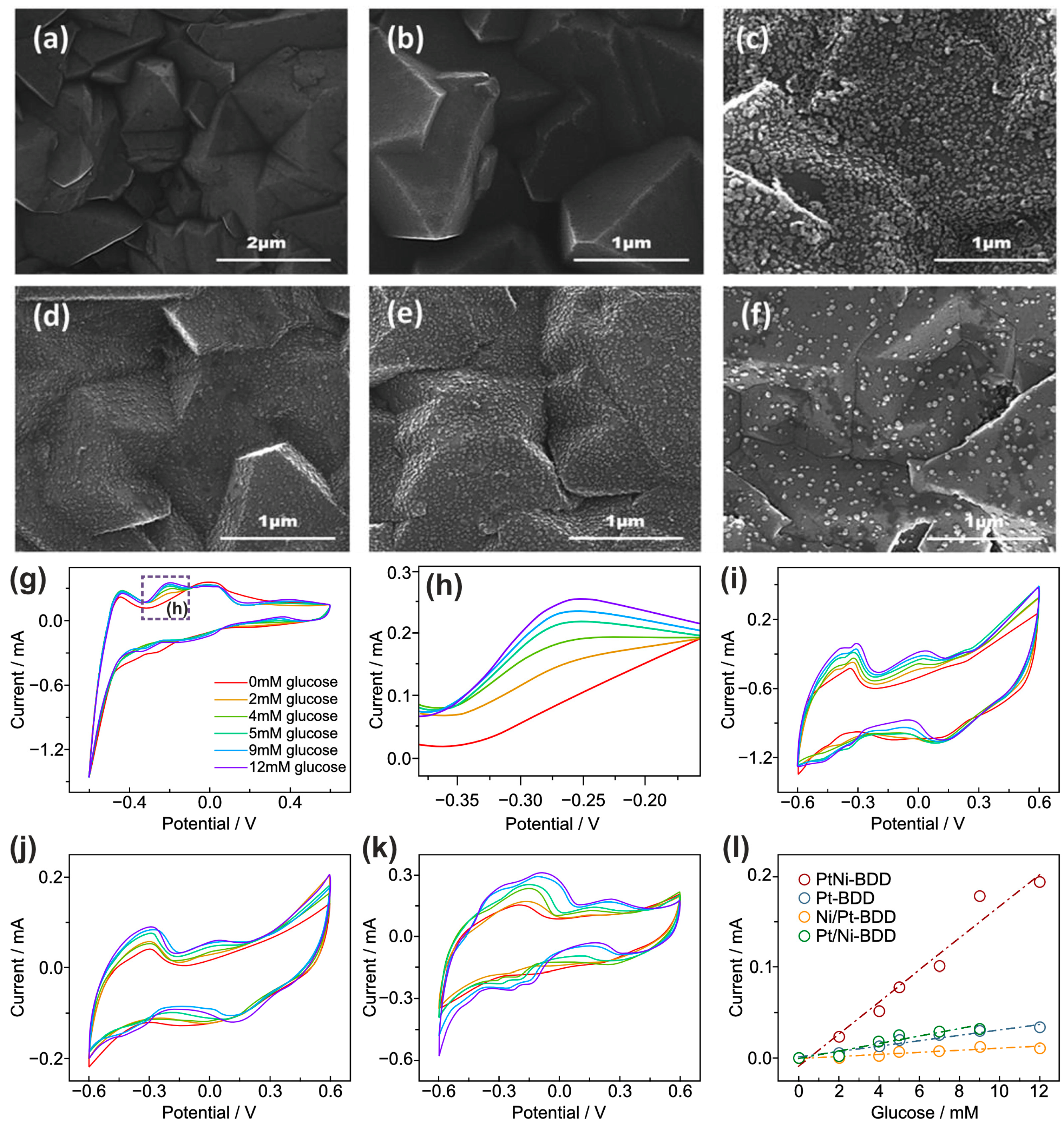
Zhu et al. [68] deposited Pt and Ni nanomaterials on the surface of boron-doped diamond (BDD) film employing the pulse electrodeposition technique; Figure 7a–f. The sensitivity and limit of detection (LOD) of the five manufactured electrodes significantly differ, depending on the order in which the element is deposited on the BDD film. The electrodes were PtNi-BDD (mixture of 5 mM NiSO4 and 1 mM K2(PtCl4)), Pt/Ni-BDD (depositing Pt onto the BDD surface and then Ni), Ni/Pt-BDD(depositing Ni on the BDD film and, afterward, Pt), Ni-BDD, and Pt-BDD. However, after adding 2 mM glucose, the Ni-BDD electrode has a poor catalytic property. Their findings reveal that the PtNi-BDD electrode prepared by co-deposition (bimetals) has excellent electrochemical performance for glucose oxidation. In addition, the co-deposition method improves the sensitivity of the BDD electrode due to an increase in the electron density of Pt caused by the input of Ni into the Pt lattice. Finally, the evaluation of the PtNi-BDD electrode through consecutive CV scans for 2400 s in 0.01 M PBS solution (pH = 7.4) and containing 2 mM glucose registered an increase of 19% regarding the initial current.
The use of carbon materials, including nanotubes and graphene, with Pt has been widely discussed [66,72]. The increase in surface area, therefore, improves the electroactivity of Pt. Wu et al. [73] synthesized dispersed Pt nanoparticles (PtNPs) supported by polydopamine (pDA)-modified N-enriched mesoporous carbon nanorods. In addition, the carbon source and carbon template were cellulose nanocrystals due to the high specific ratio rod shape. The fabricated electrode displays two different linear current responses: the first at low glucose concentrations from 0.01 mM to 2 mM and the second at high concentrations from 2 mM to 30 mM. Wu et al. [66] synthesized defective graphene nanosheet (dGN)-supported and dispersed PtNPs, obtaining a NEGS with a sensitivity of 27.28 μA mM−1 cm−2, a LOD of 0.06 µM, and a linear range from 0.5 to 9 mM, specifically for Pt/dGN600. In addition, they determined the point vacancy for this system, resulting in 5–8–5 defects and 5–9 defects.
Chinnadayyala et al. [74] investigated the fabrication of a NEGS based on a porous Pt-black-modified Au microneedle electrode array to measure the glucose levels in artificial interstitial fluid. The porous material increased the sensitivity of the sensor in correlation with the surface area. After 16 days of operation in PBS, the NEGS demonstrated adequate storage stability with a 3.5% loss of the initial response. AA (0.5 mM), lactic acid (LA) (0.1 mM), mannose (Man) (0.5 mM), Gal (0.5 mM), AC (0.1 mM), fructose (0.5 mM), Cl from NaCl (0.5 M), UA (0.2 mM), and urea (0.4 mM) were studied as interferences. A 10–15-fold higher concentration than the normal physiological level was used.
The advances regarding platinum-based materials for NEGS are reported in Table 5. The revision in the literature revealed that platinum NEGS have demonstrated significant improvements in sensitivity and selectivity through various strategies, such as mesoporous surfaces, Pt-based alloys, or the introduction of composites with carbon nanomaterials. The morphological characteristics of Pt, including dimensionality, surface geometry, and microstructure or roughness, played a key role in the enhanced sensor performance. However, these NEGS still present complex and costly procedures that can hinder large-scale production and practical implementations. Moreover, Pt-based NEGS need to ensure selectivity towards a full range of interferences, such as those presented in Table 3, to avoid inaccurate readings. Although stability data were collected from some of the collected references, the drawbacks in long-term stability due to surface fouling or poisoning can be problematic and need to be fully addressed during sensor design and characterization.

4.2. Gold and Their Hybrid Nanocomposites

Gold exhibits an extraordinarily high rate of DGE in neutral media due to the participation of oxygenated species of the solvent in the electrooxidation reactions [87,88]. Au is widely used as a working electrode for glucose sensors; however, due to passivation by oxidation products or the reaction with Cl in neutral solutions, it has limitations and a low response to DGE. Several strategies have been developed year after year to address the drawbacks it exhibits during DGE. Different Au nanostructures have been employed to manufacture NEGS, increasing their intrinsic properties, including an increased surface area; specific activity; rapid mass transport; and high electrocatalytic activity, e.g., nanoporous Au films (NPGF) [89], dendrite-type Au nanostructures [90], and AuNPs [91,92].
For example, Xia et al. [89] fabricated a three-dimensional NPGF on an Au electrode surface that exhibited high sensitivity (232 μA mM−1 cm−2) towards glucose sensing relative to other porous Au electrodes and worked effectively against interference, including AA and UA. The prepared NPGF electrode showed a wide linear range from 10 µM to 11 mM with a sensitivity of 66.0 μA mM−1 cm−2 in a solution containing highly concentrated Cl. The results demonstrate that NPGF is sensitive to detecting glucose under physiological conditions containing Cl [89]. Shu’s group obtained dendrite-like gold nanostructure-modified GCE showing a wide linear range from 0.1 mM to 25 mM with high sensitivity, a LOD of up to 0.05 mM for glucose, and excellent stability [90].
Moreover, Ismail et al. [91] reported the development of a NEGS based on AuNPs and graphene oxide nanoribbons (GONR) as a functional support matrix. GONR act as pillars with oxygenated functional groups that form noncovalent interactions with the active sites of AuNPs to promote reaction kinetics, showing a broad linear calibration curve between 0.0005 mM and 10 mM and adequate sensitivity. Notwithstanding, poisoning and the strong inhibition of DGE by Cl were observed. To counteract this ion poisoning, they used a protective membrane made of 0.2% polypyrrole and 0.05% Nafion [91]. Likewise, Branagan et al. [92] supported AuNPs on carbon nanotubes functionalized with carboxylic acid groups. Thus, at pH 7, these groups dissociate, forming anionic groups on the surface that repel anionic interferences, such as urate and ascorbate [92]. The sensor shows a sensitivity of 2.77 ± 0.14 mA/mM, a LOD of 4.1 µM, and a linear region up to 20.0 mM. The sensor showed excellent selectivity in the presence of AA, Gal, and Fru, and the coating of a Nafion film eliminated the interference with UA [92].
Gold electrodes with a 3D structure, e.g., an Au micropillar array [93], vertically Au nanotubes arrays (AuNTAs) [94], and Au nanowire arrays [94], have also been explored. The main objective was to increase the surface area in correlation with glucose selectivity. The NEGS, based on an Au micropillar array electrode and fabricated by Prehn’s group using photolithography and electroplating techniques, reported a larger electroactive area that improved glucose electrooxidation. The fabricated sensor has high repeatability, reproducibility, sensitivity (13.2 μA mM−1), and a LOD of 60 µM [93]. Likewise, Tian et al. [94] synthesized vertically aligned AuNTAs and nanowire arrays on alumina oxide templates via galvanostatic deposition. The sensor produced higher amperometry currents at pH 7.2 and exhibited a linear range extending from glucose concentrations of 5 µM to 16.4 mM, sensitivity of 44.2 μA mM−1 cm−2, and LOD of 2.1 µM.
In contrast, Nikolaev et al. [95] employed a different methodology than chemical deposition, which has some drawbacks, e.g., purity, long synthesis time, high cost, and low reproducibility. They utilized the directed electrochemical nanowire assembly method, which involves the electrodeposition of metal under the action of an applied high-frequency alternating voltage and small direct electrochemical synthesis for the amperometric detection of glucose in a neutral medium. The Au nanowires showed a concentration range of 1 × 10⁻⁴ to 5 × 10⁻³ M. This sensor showed sensitivity for glucose detection on the Au nanowires of 3.7 × 10⁴ A M−1 m−2, and the LOD was 3.3 × 10⁻⁵ M [95].
The challenge of detecting glucose with compounds containing Au electrodes in neutral solutions (physiological pH) has led many researchers to continue studying various morphologies or combinations of materials. Various reports have revealed the synergistic effect of bimetallic compounds with other materials, increasing detection performance [96]. Alloys or bimetallic nanoparticles with controlled structures and composition have many advantages due to their strength, usually tunning the surface plasmon band and improving the stability and diffusion of the nanoparticles [97,98]. Consider, as an example, by electrodeposition, Nguyen et al. synthesized ruthenium-Au alloy nanoparticles (Au-RuNPs) [46]. They evaluated the oxidation of glucose in neutral media, observing a large anodic peak at 0.2 V, suggesting that the electrooxidation of glucose with Au-RuNPs carries out a multistep reaction. In a previous work, Au-RuNPs exhibited high electroactivity toward DGE [99]. Shim et al. [100] demonstrated that the synthesis of bimetallic layers of Pt/Au in structured core–shell NPs improved the catalytic performance of these materials individually towards glucose oxidation. This material exhibits more abundant active sites and showed improved sensitivity, stability, and selectivity towards glucose detection at neutral pH due to the synergistic effects of the combination of Au and Pt; see Figure 8. The sensor has a LOD of 445.7 nM.
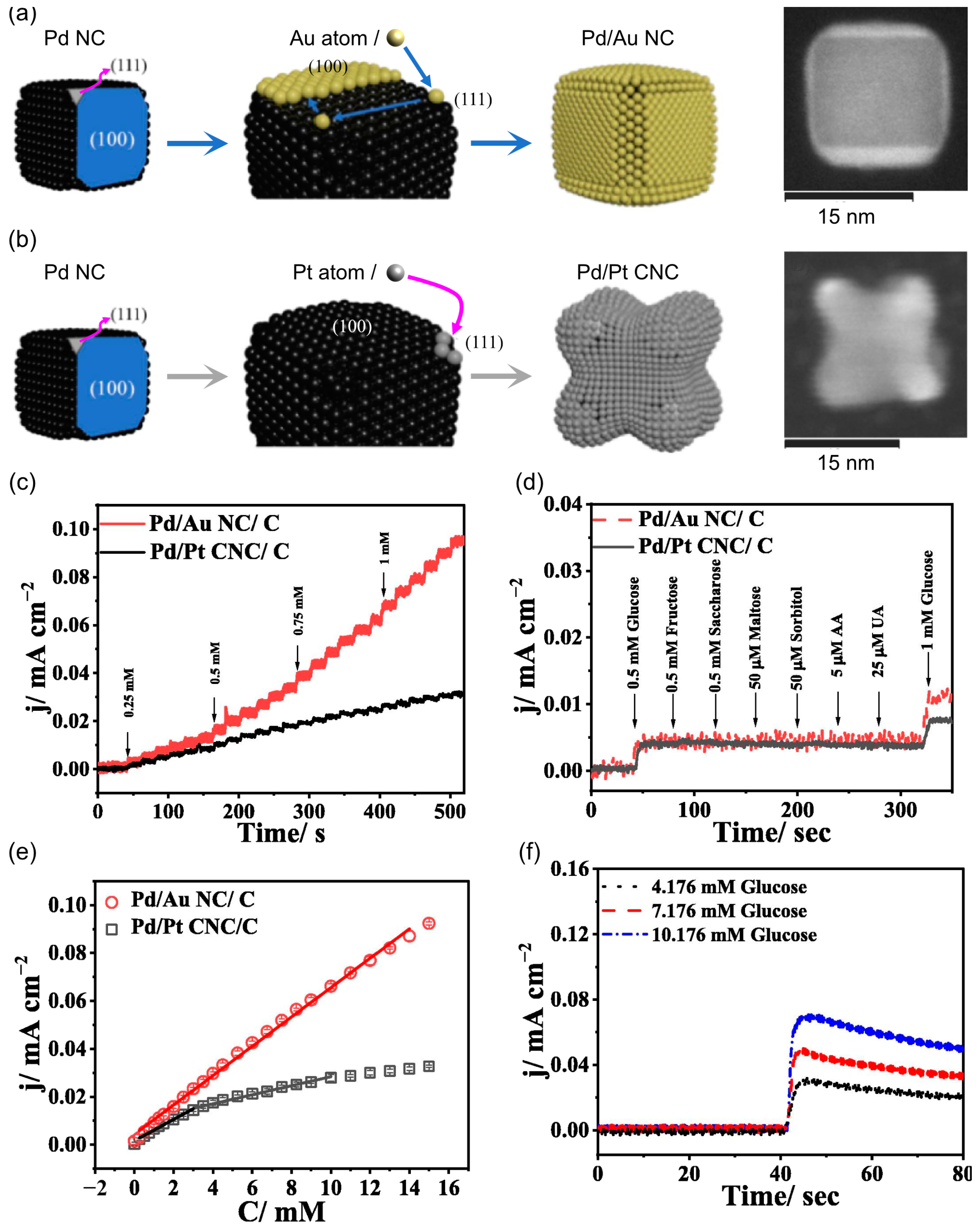
Core–shell structures have been found in other research to enhance the performance of electrocatalysts in sensors by matching the tunable electron density of the shell metal with the core metal. Wang et al. [48] deposited Au and Pt atoms on the basal surface of Pd nanocubes (PdNC); see Figure 9a,b. The palladium/gold nanocube (Pd/Au NC) exhibited a wide linear range from 0.25 to 14 mM and a high sensitivity of 13.56 μA mM−1 cm−2, assessed with real blood samples from 3 to 8 mM.
The reviewed literature regarding Au-based NEGS in neutral pH can be consulted in Table 6. Although the development of these NEGS has shown significant advances, several challenges remain. Under optimized conditions, Au exhibited high DGE rates in neutral media, but the practical applications of such electrodes can be limited by passivation from oxidation products and reaction with Cl ions, reducing the response towards glucose. Strategies, including morphology modification, protective membranes, and functionalized supports, have been introduced to mitigate ion poisoning and interferences. Continued research focusing on the morphology of nanostructures and the synergistic combination of materials is essential for developing more efficient and reliable NEGS. These efforts aim to optimize sensitivity, stability, and LOD, paving the way for a more robust practical implementation of NEGS at physiological conditions.

4.3. Additional Transition Metals (Ag, Co, Cu, Ni, Ru, and Pd) and Their Nanocomposites

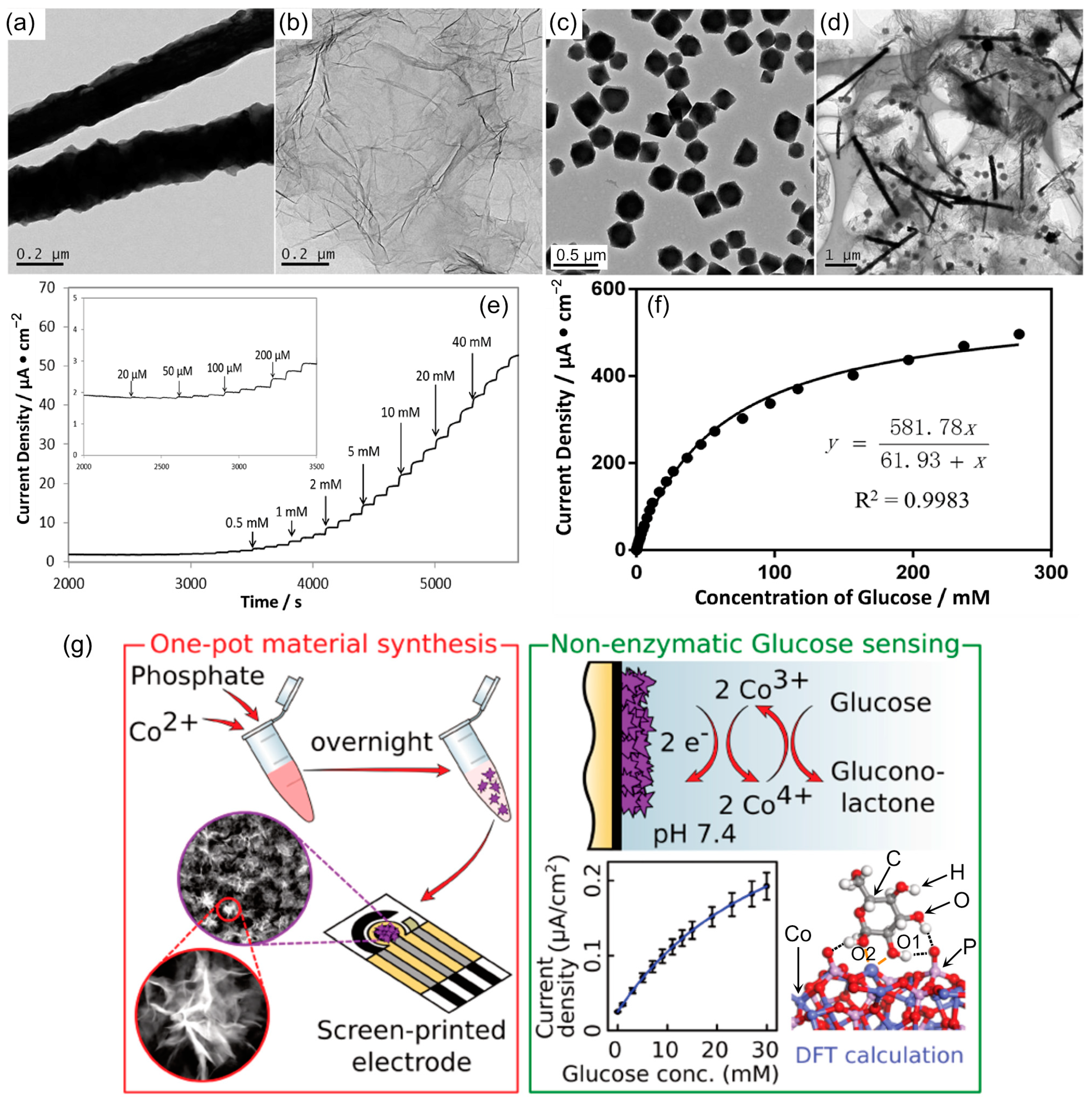
The transition metals explored for DGE at physiological conditions (other than Pt and Au) include Pd, Ag, Ru, Co, and Cu. The substrate design, typically GCE, SPCE, or fluorine-doped tin oxide modified with nanostructured transition metals, exhibiting morphologies with a highly accessible surface area and high index facets, is preferable. In this sense, hybrid hierarchical Co3(PO4)2 nanoflowers were synthesized with a simple, low-cost method for subsequently modifying a SPCE in order to act as a catalytic material for glucose oxidation through the Co4+/Co3+ couple [102]. The SPCE/Co3(PO4)2 nanoflowers sensor showed a calibration working range from 1 to 30 mM at 0.65 V and a LOD of 0.3 mM. However, prone to fouling and a lowering sensitivity were observed when evaluated in solutions with higher human serum contents attributed to the deactivation of catalytic sites.
Similar to Pt and Au, to increase the number of catalytic sites for DGE, the electrode can be modified with nanometric support to enhance the electroactive surface area of the detector. Then, the active material is deposited on the modified electrode, allowing a higher number of interaction sites than those initially available in the unmodified electrode. Most modifiers include carbon-based nanoparticles such as graphene [103], carbon nanotubes [104], and hierarchical structures, e.g., metallic organic frameworks (MOFs) [105]. Following this approach, a nanocomposite of copper nanowires (CuNWs), MOF, and graphene oxide (GO) was prepared by ultrasound mixing. The nanocomposite was applied to modify a GCE and detect glucose amperometrically in the range from 0.02 to 26.6 mM in neutral PBS at 0.3 V [105]; see Figure 10. Additionally, Nafion was cast over the CuNWs/MOF/GO sensor to stabilize the material, resulting in good selectivity against other carbohydrates and common serum interferents. When evaluated in serum samples, the obtained recovery rates correlate well with the glucose content predicted by a commercial method.
Although the advances are limited compared to dominant Pt- and Au-based NEGS, the exploration of transition metals such as Pd, Ag, Ru, Co, and Cu for DGE under physiological conditions has shown promising results, with various innovative designs aiming at enhanced catalytic effects. The complete literature reviewed regarding this issue can be consulted in Table 7. Nevertheless, these sensors face challenges such as a reduced response at DGE and low sensitivity when evaluated in biological samples. The fouling and deactivation of catalytic sites when performing in biological complex samples compromise the long-term stability, and further research is needed to develop effective DGE with these materials.

4.4. Phenylboronic Acid and Its Derivates

Several studies have employed methods to investigate the interaction between PBA and glucose at neutral pH. For example, the electropolymerization of azure in the presence of HAuCl4 and 4-marcaptophenyl boronic acid was performed in 0.1 M PBS at pH 6.5 using a GCE as the support to create selective layers for glucose capture. The characterization revealed the growth of polymeric polyazure A film over the GCE with 4-marcaptophenyl boronic assembled over AuNPs [54]. Cysteamine was used for blockage, and the ferro/ferricyanide redox probe revealed the surface blockage due to glucose capture. The subsequent decrease of the current correlated well with the higher glucose concentration, resulting in a working range of 0.01 to 10 μM and a LOD of 4 nM; see Figure 11a. Due to this limited working range, the analyzed human sample was diluted in PBS before the analysis. In a different approach (Figure 11b,c), a metallic oxide Ta2O5 electrode was modified with mesoporous silica (MPSi) containing a PBA molecule; the design was intended to combine the high surface area of MPSi while avoiding the non-specific absorption of proteins in metal electrodes by choosing a MO substrate [113]. The surface potential change produced during the PBA–glucose interaction was reflected as a negative voltage shift correlated with the glucose concentration after fitting with the Langmuir adsorption isotherm in a working range of 0–20 mM. The improved performance was attributed to the local environment due to amino groups in MPSi that helped PBA molecules dissociate. Although the results in the real sample analysis were inhibited by absorbed proteins and competitive diol binding with PBA of the serum components, the Ta2O5/MPSi-PBA sensor still exhibited the promise to differentiate between the hypoglycemic and hyperglycemic levels. Table 8 illustrates PBA and derivates non-enzymatic sensors at neutral pH.

5. Materials for Non-Enzymatic Glucose Detection at Physiological pH in Wearable and Flexible Systems

For continuous monitoring, wearable technology integrating flexible electrochemical sensors is the natural evolution of traditional, rigid, bulky meters [23]. Frequent glucose monitoring is a key factor that traditional blood glucose meters fail to cover, as they allow only single measurements that can lose track of glucose fluctuations during the day. Commercial CGM are revolutionizing the way diabetes care is handled. However, this could be only one option among potential wearable sensing possibilities, since glucose can be detected in non-invasive body fluids such as saliva, tears, and sweat [17,23]. The analysis of these biofluids has been adapted to wearable glucose systems, mainly based on enzymatic detection, due to the alkaline environment needed in NEGS, which prevents their direct use in the human body. Notwithstanding, several wearable sensors have already been proposed combining the most efficient materials for physiological glucose detection, such as Pt and Au, with flexible substrates such as carbon cloth (CC) [61], flexible SPCE [58], stainless-steel microneedle arrays [74], or graphene-like materials [117]. The technological evolution has allowed combining chemical detection in the form of wristbands, patches, or sweatbands with miniaturized readers for total on-body measurement systems using custom apps for recording data via Wi-Fi or Bluetooth communication.
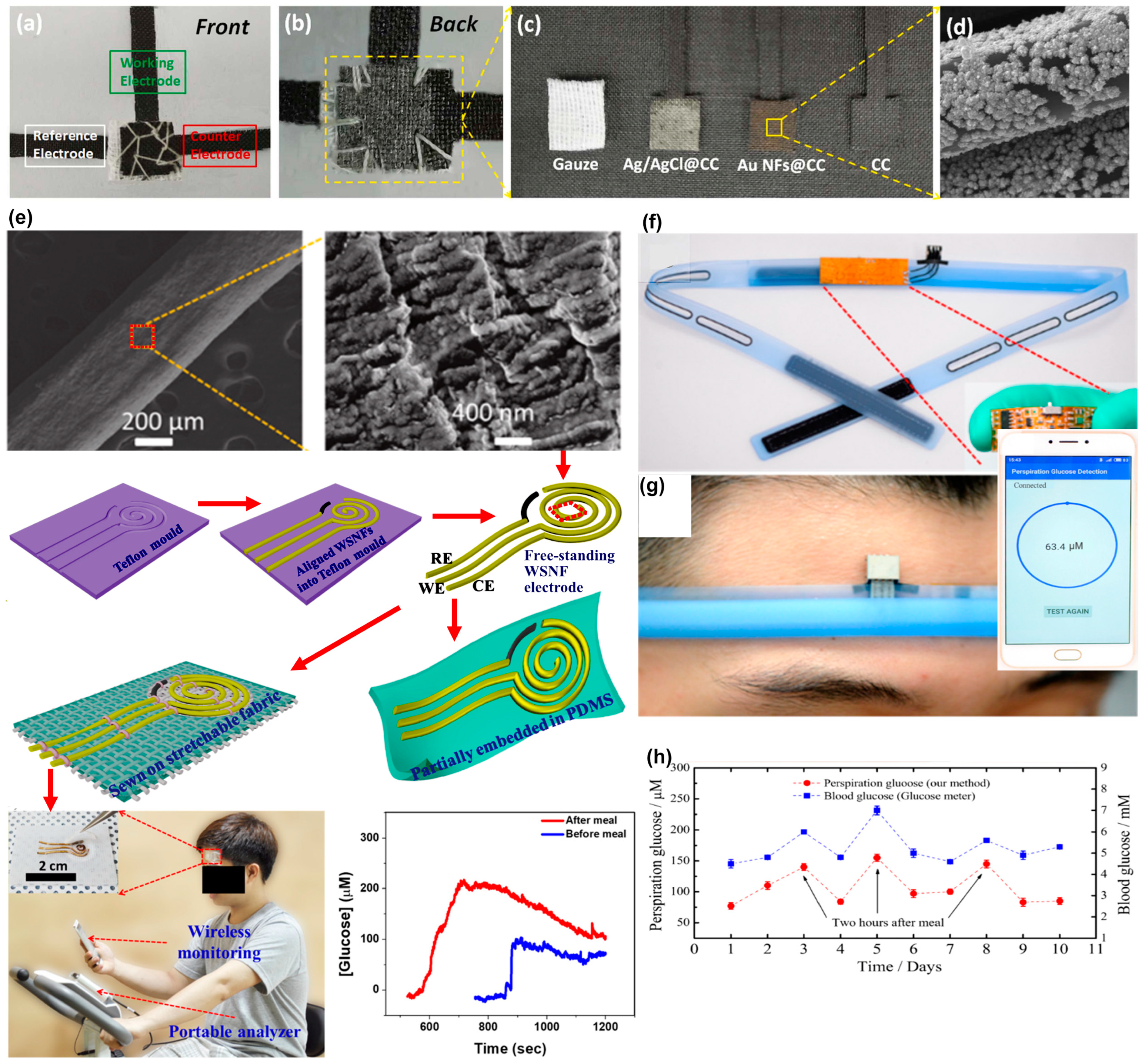
Similar to their handheld counterparts, the wearable non-enzymatic systems at the physiological pH manage the local increment of the pH or the addition of OH anions to facilitate DGE, as discussed in Section 3. When it comes to Au-based sensors, the formation of Au(OH)ads has been reported following strategies such as surface functionalization, electrochemical pretreatment, or chemical pretreatment in either an acidic or alkaline medium. For example, a flexible sensor for active sweat absorption and easy integration with clothes was proposed for glucose non-invasive detection. CC was used as an electrochemical base for WE, CE, and RE three-electrode construction. The active sensing was performed by gold nanoflowers (AuNF), while RE was modified with Ag/AgCl [60]. A gauze functions as a space layer for electrodes and as an active sweat collection mechanism. To create the flexible AuNF-CC sensor, all electrodes were stacked with gauze as a spacer between electrodes and sewn together (Figure 12a–c). Then, the optimized WE decorated AuNF (Figure 12d) was activated to produce AuOHads and promote glucose oxidation in PBS at pH 7.4.
Similarly, a patch biosensor for wearable glucose self-monitoring was assembled with reduced graphene oxide-polyurethane (rGO-PU)- fibers as flexible support for Au nanowrinkles; see Figure 12e. The elastomeric rGO-PU composite was manufactured as a three-electrode system and was transferred to stretchable textiles to create a conformal patch for continuous monitoring [62]. To promote DGE at neutral media, the oxygenated groups in the rGO surface acted synergistically with Au, producing Au(OH)ads on the WE surface, as revealed during cyclic voltammetry studies.
Transition metals other than Pt and Au have proved effective, e.g., Co, Pb, and Cu, due to their redox states, which can significantly enhance when combined with hierarchical structures like MOF. A chemical system for continuous monitoring was manufactured in a sweatband (Figure 12f–h), using a screen-printed sensor chip as a base [58]. The working electrode was modified using palladium nanoparticles (Pd NPs) encapsulated in a cobalt-based zeolitic imidazole framework (Co-ZIF-67). In this work, the additional OH resulted from a water-splitting reaction for the subsequent detection of glucose at lower potentials through the formation of [Co(III)(mim)2(OH)]n/[Co(IV)-(mim)2(OH)2]n over the sensor surface. Additionally, the Pd NPs/Co-ZIF-67 could be regenerated through anodic potential and provided continuous information. The complete revision regarding wearable systems for NEGS can be consulted in Table 9.

6. Prevailing Drawbacks and Challenges Associated with Non-Enzymatic Detection at Neutral pH and Further Perspectives

The development of new materials has enabled significant progress in the area of non-enzymatic glucose sensors. From the reviewed literature, it is safe to affirm that only a minimal part focuses on solving detection in physiological conditions. However, a notable increase in the number of research items was noted compared to previous reviews covering the same topic. From the consulted literature, it was evident that Pt, Au, and their hybrid composites were the most studied and promising nanomaterials for DGE at neutral pH. It is possible to highlight the bimetallic and complex nanostructures (e.g., core–shell) due to the fact that they do not interfere with glucose measurement and electrode poisoning. In addition, the sensitivity is increased because of the synergistic effect of the individual nature of the starting metals. PBA molecular interaction also showed promising results.
Despite research on neutral pH, there is still a lack of optimization on important subjects such as long-term electrode stability and improving performance when testing on real samples. As referred to in Section 2, a meaningful number of interference species, along with an appropriate report of sensor accuracy, is needed if the materials are intended for real clinical analysis. Moreover, poisoning is still an open challenge, although significant progress has been presented with rational designed nanomaterials. In addition, the majority of the presented devices need to further adjust the sample prior to analysis, usually by diluting the sample. So far, a further desirable direction for the oncoming work would be to evaluate the proposed nanomaterials by considering conditions that resemble the intended physiological operation rather than the favorable alkaline medium. The materials already showing promising results in DGE should be analyzed considering their long-term stability, similarly to the evaluation presented for their enzymatic counterpart.
Therefore, the presented works are mainly in the early stages of Technology Readiness Levels (TRLs), usually levels 1 to 3. Commercially available NEGS devices are scarce for conditions at neutral pH or electrolytes under physiological conditions. For instance, the UXN Company (Seoul, Republic of Korea) has developed several patents in recent years with a focus on NEGS (US 11,751,781 B2), interferers (ascorbic acid and acetaminophen, US 11,744,493 B2), and the Pt porous layer manufacturing method (US 10,687,746 B2), among others. Some universities have also developed prototypes, including the University of Colombo (LK, US 2023/0072912 A1) and Central South University (Changsha, CN; US 2023/0184710 A1). The working electrode developed by the latest NEGS patent application is a metal-modified porous BDD. Based on the patent review, a trend was observed in methods of manufacturing layers, where each one has a function, and together, they make up the working electrode. Although limited, these works encourage further research tackling the issues of DGE at physiological conditions to move closer to practical clinical applications.

7. Conclusions

In the present review, we summarize the current strategies for DGE at physiological conditions and highlight the design based on transition metals, mainly Pt and Au. Additionally, Pd, Ag, Cu, and Ni have been evaluated with an acceptable analytical performance. Molecular interaction between glucose and PBA was demonstrated at neutral pH, although it still needs to enhance the selectivity towards glucose. Although CGM based on interstitial fluid analysis were scarce, wearable devices for non-enzymatic, non-invasive detection has been proposed for sweat analysis, paving the way for potential painless continuous glucose monitoring. Challenges to still face for the further development of functional glucose NEGS include poisoning resistance against chloride ions, a higher current density during detection, and evaluations of real samples without an added pretreatment.

Author Contributions

Conceptualization, V.O., E.P.A.M., R.B.D. and A.V.R.; investigation, V.O., E.P.A.M., R.B.D. and A.V.R.; writing—original draft preparation, V.O., E.P.A.M., R.B.D. and A.V.R.; writing—review and editing, V.O., E.P.A.M., R.B.D. and A.V.R.; visualization, A.V.R.; supervision, R.B.D. and A.V.R. All authors have read and agreed to the published version of the manuscript.

Funding

This work was funded by Centro de Investigación en Materiales Avanzados (CIMAV) through intern grant 26018.

Institutional Review Board Statement

Not applicable.

Informed Consent Statement

Not applicable.

Data Availability Statement

Not applicable.

Conflicts of Interest

The authors declare no conflicts of interest.

References

  1. Li, R.; Shen, M.; Yang, Q.; Fairley, C.K.; Chai, Z.; McIntyre, R.; Ong, J.J.; Liu, H.; Lu, P.; Hu, W.; et al. Global Diabetes Prevalence in COVID-19 Patients and Contribution to COVID-19–Related Severity and Mortality: A Systematic Review and Meta-Analysis. Diabetes Care 2023, 46, 890–897. [Google Scholar] [CrossRef] [PubMed]
  2. International Diabetes Federation. IDF Diabetes Atlas, 10th ed.; IDF: Brussels, Belgium, 2021; p. 7. [Google Scholar]
  3. International Diabetes Federation. IDF Diabetes Atlas, 3rd ed.; IDF: Brussels, Belgium, 2006; pp. 192–193. [Google Scholar]
  4. Freckmann, G.; Pleus, S.; Grady, M.; Setford, S.; Levy, B. Measures of Accuracy for Continuous Glucose Monitoring and Blood Glucose Monitoring Devices. J. Diabetes Sci. Technol. 2019, 13, 575–583. [Google Scholar] [CrossRef] [PubMed]
  5. Pullano, S.A.; Greco, M.; Bianco, M.G.; Foti, D.; Brunetti, A.; Fiorillo, A.S. Glucose Biosensors in Clinical Practice: Principles, Limits and Perspectives of Currently Used Devices. Theranostics 2022, 12, 493–511. [Google Scholar] [CrossRef]
  6. Mian, Z.; Hermayer, K.L.; Jenkins, A. Continuous Glucose Monitoring: Review of an Innovation in Diabetes Management. Am. J. Med. Sci. 2019, 358, 332–339. [Google Scholar] [CrossRef] [PubMed]
  7. Erbach, M.; Freckmann, G.; Hinzmann, R.; Kulzer, B.; Ziegler, R.; Heinemann, L.; Schnell, O. Interferences and Limitations in Blood Glucose Self-Testing: An Overview of the Current Knowledge. J. Diabetes Sci. Technol. 2016, 10, 1161–1168. [Google Scholar] [CrossRef]
  8. Reghunath, R.; Devi, K.; Singh, K.K. Recent Advances in Graphene Based Electrochemical Glucose Sensor. Nano-Struct. Nano-Objects 2021, 26, 100750. [Google Scholar] [CrossRef]
  9. Sehit, E.; Altintas, Z. Significance of Nanomaterials in Electrochemical Glucose Sensors: An Updated Review (2016–2020). Biosens. Bioelectron. 2020, 159, 112165. [Google Scholar] [CrossRef]
  10. Wei, M.; Qiao, Y.; Zhao, H.; Liang, J.; Li, T.; Luo, Y.; Lu, S.; Shi, X.; Lu, W.; Sun, X. Electrochemical Non-Enzymatic Glucose Sensors: Recent Progress and Perspectives. Chem. Commun. 2020, 56, 14553–14569. [Google Scholar] [CrossRef]
  11. Osuna, V.; Vega-Rios, A.; Zaragoza-Contreras, E.A.; Estrada-Moreno, I.A.; Dominguez, R.B. Progress of Polyaniline Glucose Sensors for Diabetes Mellitus Management Utilizing Enzymatic and Non-Enzymatic Detection. Biosensors 2022, 12, 137. [Google Scholar] [CrossRef]
  12. Niu, X.H.; Shi, L.B.; Zhao, H.L.; Lan, M.B. Advanced Strategies for Improving the Analytical Performance of Pt-Based Nonenzymatic Electrochemical Glucose Sensors: A Minireview. Anal. Methods 2016, 8, 1755–1764. [Google Scholar] [CrossRef]
  13. Franceschini, F.; Taurino, I. Nickel-Based Catalysts for Non-Enzymatic Electrochemical Sensing of Glucose: A Review. Phys. Med. 2022, 14, 100054. [Google Scholar] [CrossRef]
  14. Zhu, H.; Li, L.; Zhou, W.; Shao, Z.; Chen, X. Advances in Non-Enzymatic Glucose Sensors Based on Metal Oxides. J. Mater. Chem. B 2016, 4, 7333–7349. [Google Scholar] [CrossRef]
  15. Marioli, J.M.; Kuwana, T. Electrochemical Characterization of Carbohydrate Oxidation at Copper Electrodes. Electrochem. Acta 1992, 37, 1187–1197. [Google Scholar] [CrossRef]
  16. Evans, W.L.; Nicoll, W.D.; Strouse, G.C.; Waring, C.E. The Mechanism of Carbohydrate Oxidation. IX. The Action of Copper Acetate Solutions on Glucose, Fructose and Galactose. J. Am. Chem. Soc. 1928, 50, 2267–2285. [Google Scholar] [CrossRef]
  17. Witkowska Nery, E.; Kundys, M.; Jeleń, P.S.; Jönsson-Niedziółka, M.; Nery, E.W.; Kundys, M.; Jeleń, P.S.; Jönsson-Niedziólka, M. Electrochemical Glucose Sensing: Is There Still Room for Improvement? Anal. Chem. 2016, 88, 11271–11282. [Google Scholar] [CrossRef] [PubMed]
  18. Adeel, M.; Rahman, M.M.; Caligiuri, I.; Canzonieri, V.; Rizzolio, F.; Daniele, S. Recent Advances of Electrochemical and Optical Enzyme-Free Glucose Sensors Operating at Physiological Conditions. Biosens. Bioelectron. 2020, 165, 112331. [Google Scholar] [CrossRef] [PubMed]
  19. Vassilyev, Y.B.; Khazova, O.A.; Nikolaeva, N.N. Kinetics and Mechanism of Glucose Electrooxidation on Different Electrode-Catalysts. Part I. Adsorption and Oxidation on Platinum. J. Electroanal. Chem. 1985, 196, 105–125. [Google Scholar] [CrossRef]
  20. Vassilyev, Y.B.; Khazova, O.A.; Nikolaeva, N.N. Kinetics and Mechanism of Glucose Electrooxidation on Different Electrode-Catalysts. Part II. Effect of the Nature of the Electrode and the Electrooxidation Mechanism. J. Electroanal. Chem. 1985, 196, 127–144. [Google Scholar] [CrossRef]
  21. Gorski, W.; Kennedy, R.T. Electrocatalyst for Non-Enzymatic Oxidation of Glucose in Neutral Saline Solutions. J. Electroanal. Chem. 1997, 424, 43–48. [Google Scholar] [CrossRef]
  22. Castro Luna, A.M.; de Mele, M.F.L.; Arvia, A.J. The Electro-Oxidation of Glucose on Microcolumnar Gold Electrodes in Different Neutral Solutions. J. Electroanal. Chem. 1992, 323, 149–162. [Google Scholar] [CrossRef]
  23. Saha, T.; Del Caño, R.; Mahato, K.; De la Paz, E.; Chen, C.; Ding, S.; Yin, L.; Wang, J. Wearable Electrochemical Glucose Sensors in Diabetes Management: A Comprehensive Review. Chem. Rev. 2023, 123, 7854–7889. [Google Scholar] [CrossRef] [PubMed]
  24. North East London Pharmacy and Medicines Optimisation Team. North East London Blood Glucose Test Strips Guideline; North East London Pharmacy and Medicines Optimisation Team: London, UK, 2023. [Google Scholar]
  25. Shah, V.N.; Laffel, L.M.; Wadwa, R.P.; Garg, S.K. Performance of a Factory-Calibrated Real-Time Continuos Glucose Monitoring System Utilizing an Automated Sensor Aplicator. Diabetes Technol. Ther. 2018, 20, 428–433. [Google Scholar] [CrossRef]
  26. Tonyushkina, K.; Nichols, J.H. Glucose Meters: A Review of Technical Challenges to Obtaining Accurate Results. J. Diabetes Sci. Technol. 2009, 3, 971–980. [Google Scholar] [CrossRef]
  27. Parkes, J.L.; Slatin, S.L.; Pardo, S.; Ginsberg, B.H. A New Consensus Error Grid to Evaluate the Clinical Significance of Inaccuracies in the Measurement of Blood Glucose. Diabetes Care 2000, 23, 1143–1148. [Google Scholar] [CrossRef]
  28. ISO 15197:2015; In Vitro Diagnostic Test Systems-Requirements for Blood-Glucose Monitoring Systems for Self-Testing in Managing Diabetes Mellitus. International Organization for Standardization: Geneva, Switzerland, 2015.
  29. FDA-2013-D-1445; Blood Glucose Monitoring Test Systems for Prescription Point-of-Care Use—Guidance for Industry and Food and Drug Administration Staff. U.S. Department of Health and Human Services, Food and Drug Administration, Center for Devices and Radiological Health: Rockville, MD, USA, 2020.
  30. FDA-2013-D-1446; Self-Monitoring Blood Glucose Test Systems for Over-the-Counter Use Guidance for Industry and Food and Drug Administration Staff. U.S. Department of Health and Human Services, Food and Drug Administration, Center for Devices and Radiological Health: Rockville, MD, USA, 2016.
  31. CLSI POCT12-A3; Point-of-Care Blood Glucose Testing in Acute and Chronic Care Facilities; Approved Guideline—Third Edition. Clinical and Laboratory Standards Institute: Wayne, PA, USA, 2013.
  32. Argyle, M.D.; Bartholomew, C.H. Heterogeneous Catalyst Deactivation and Regeneration: A Review. Catalysts 2015, 5, 145–269. [Google Scholar] [CrossRef]
  33. Ayed, D.; Laubender, E.; Souiri, M.; Yurchenko, O.; Marmouch, H.; Urban, G.; Othmane, A. Carbon Nanotubes Supported Ru-Au Nanoparticles with Core-Shell Structure for Glucose Detection with High Resistance against Chloride Poisoning. J. Electrochem. Soc. 2017, 164, B767–B775. [Google Scholar] [CrossRef]
  34. Mello, G.A.B.; Cheuquepán, W.; Briega-Martos, V.; Feliu, J.M. Glucose Electro-Oxidation on Pt(100) in Phosphate Buffer Solution (PH 7): A Mechanistic Study. Electrochim. Acta 2020, 354, 136765. [Google Scholar] [CrossRef]
  35. Naikoo, G.A.; Arshad, F.; Hassan, I.U.; Omar, F.B.; Tambuwala, M.M.; Mustaqeem, M.; Saleh, T.A. Trends in Bimetallic Nanomaterials and Methods for Fourth-Generation Glucose Sensors. TrAC Trends Anal. Chem. 2023, 162, 117042. [Google Scholar] [CrossRef]
  36. Kang, K.N.; Kim, S.I.; Yoon, J.C.; Kim, J.; Cahoon, C.; Jang, J.H. Bi-Functional 3D-NiCu-Double Hydroxide@Partially Etched 3D-NiCu Catalysts for Non-Enzymatic Glucose Detection and the Hydrogen Evolution Reaction. ACS Appl. Mater. Interfaces 2022, 14, 33013–33023. [Google Scholar] [CrossRef]
  37. Ghanam, A.; Haddour, N.; Mohammadi, H.; Amine, A.; Sabac, A.; Buret, F. A Membrane-Less Glucose/O2 Non-Enzymatic Fuel Cell Based on Bimetallic Pd–Au Nanostructure Anode and Air-Breathing Cathode: Towards Micro-Power Applications at Neutral PH. Biosens. Bioelectron. 2022, 210, 114335. [Google Scholar] [CrossRef]
  38. Ataei Kachouei, M.; Shahrokhian, S.; Ezzati, M. Bimetallic CoZn-MOFs Easily Derived from CoZn-LDHs, as a Suitable Platform in Fabrication of a Non-Enzymatic Electrochemical Sensor for Detecting Glucose in Human Fluids. Sens. Actuators B Chem. 2021, 344, 130254. [Google Scholar] [CrossRef]
  39. Pletcher, D. Electrocatalysis: Present and Future. J. Appl. Electrochem. 1984, 14, 403–415. [Google Scholar] [CrossRef]
  40. Burke, L.D. Premonolayer Oxidation and Its Role in Electrocatalysis. Pergamoa Electrochim. Acta 1994, 39, 1841–1848. [Google Scholar] [CrossRef]
  41. Ernst, S.; Heitbaum, J.; Hamann, C.H. The Electrooxidation of Glucose in Phosphate Buffer Solutions Part I. Reactivity and Kinetics below 350 MV/RHE. J. Electroanal. Chem. 1979, 100, 173–183. [Google Scholar] [CrossRef]
  42. Ernst, S.; Heitbaum, J.; Hamann, C.H. The Electrooxidation of Glucose in Phosphate Buffer Solutions: Kinetics and Reaction Mechanism. Phys. Chem. 1980, 84, 50–55. [Google Scholar] [CrossRef]
  43. Singh, B.; Laffir, F.; MxCormac, T.; Dempsey, E. PtAu/C Based Bimetallic Nanocomposites for Non-Enzymatic Electrochemical Glucose Detection. Sens. Actuators B Chem. 2010, 150, 80–92. [Google Scholar] [CrossRef]
  44. Wu, Y.S.; Wu, Z.W.; Lee, C.L. Concave Pd Core/Island Pt Shell Nanoparticles: Synthesis and Their Promising Activities toward Neutral Glucose Oxidation. Sens. Actuators B Chem. 2019, 281, 1–7. [Google Scholar] [CrossRef]
  45. Adžic, R.R.; Hsiao, M.W.; Yeager, E.B. Electrochemical Oxidation of Glucose on Single Crystal Gold Surfaces. J. Electroanal. Chem. 1989, 260, 475–485. [Google Scholar] [CrossRef]
  46. Nguyen, T.N.H.; Jin, X.; Nolan, J.K.; Xu, J.; Le, K.V.H.; Lam, S.; Wang, Y.; Alam, M.A.; Lee, H. Printable Nonenzymatic Glucose Biosensors Using Carbon Nanotube-PtNP Nanocomposites Modified with AuRu for Improved Selectivity. ACS Biomater. Sci. Eng. 2020, 6, 5315–5325. [Google Scholar] [CrossRef]
  47. Hsiao, M.W.; Adžic, R.R.; Yeager, E.B. Electrochemical Oxidation of Glucose on Single Crystal and Polycrystalline Gold Surfaces in Phosphate Buffer. J. Electrochem. Soc. 1996, 143, 759–767. [Google Scholar] [CrossRef]
  48. Wang, S.S.; Wu, C.W.; Chen, P.Y.; Lee, C.L. Preferential Deposition of Gold and Platinum Atom on Palladium Nanocube as Catalysts for Oxidizing Glucose in the Phosphate-Buffered Solution. J. Electroanal. Chem. 2023, 930, 117142. [Google Scholar] [CrossRef]
  49. Karra, S.; Wooten, M.; Griffith, W.; Gorski, W. Morphology of Gold Nanoparticles and Electrocatalysis of Glucose Oxidation. Electrochim. Acta 2016, 218, 8–14. [Google Scholar] [CrossRef]
  50. Cheung, K.C.; Wong, W.L.; Ma, D.L.; Lai, T.S.; Wong, K.Y. Transition Metal Complexes as Electrocatalysts-Development and Applications in Electro-Oxidation Reactions. Coord. Chem. Rev. 2007, 251, 2367–2385. [Google Scholar] [CrossRef]
  51. Hall, D.G. Strucuture, Properties, and Preparation of Boronic Acid Derivatives. Overview of Their Reactions and Applications. In Boronic Acids: Preparation and Applications in Organic Synthesis and Medicine, 1st ed.; Wiley-VCH: Weinheim, Germany, 2005; pp. 1–20. [Google Scholar]
  52. Egawa, Y.; Seki, T.; Takahashi, S.; Anzai, J.I. Electrochemical and Optical Sugar Sensors Based on Phenylboronic Acid and Its Derivatives. Mater. Sci. Eng. C 2011, 31, 1257–1264. [Google Scholar] [CrossRef]
  53. Anzai, J. Recent Progress in Electrochemical Biosensors Based on Phenylboronic Acid and Derivatives. Mater. Sci. Eng. C 2016, 67, 737–746. [Google Scholar] [CrossRef]
  54. Wang, W.; Kong, L.; Zhu, J.; Tan, L. One-Pot Preparation of Conductive Composite Containing Boronic Acid Derivative for Non-Enzymatic Glucose Detection. J. Colloid Interface Sci. 2017, 498, 1–8. [Google Scholar] [CrossRef] [PubMed]
  55. Cambre, J.N.; Sumerlin, B.S. Biomedical Aplications of Boronic Acid Polymers. Polymer 2011, 52, 4361–4643. [Google Scholar] [CrossRef]
  56. Zhu, X.; Ju, Y.; Chen, J.; Liu, D.; Liu, H. Nonenzymatic Wearable Sensor for Electrochemical Analysis of Perspiration Glucose. ACS Sens. 2018, 3, 1135–1141. [Google Scholar] [CrossRef] [PubMed]
  57. Berni, A.; Amine, A.; García-Guzmán, J.J.; Cubillana-Aguilera, L.; Palacios-Santander, J.M. Feather-like Gold Nanostructures Anchored onto 3D Mesoporous Laser-Scribed Graphene: A Highly Sensitive Platform for Enzymeless Glucose Electrochemical Detection in Neutral Media. Biosensors 2023, 13, 678. [Google Scholar] [CrossRef]
  58. Zhu, X.; Yuan, S.; Ju, Y.; Yang, J.; Zhao, C.; Liu, H. Water Splitting-Assisted Electrocatalytic Oxidation of Glucose with a Metal-Organic Framework for Wearable Nonenzymatic Perspiration Sensing. Anal. Chem. 2019, 91, 10764–10771. [Google Scholar] [CrossRef]
  59. Kim, Y.J.; Chinnadayyala, S.R.; Le, H.T.N.; Cho, S. Sensitive Electrochemical Non-Enzymatic Detection of Glucose Based on Wireless Data Transmission. Sensors 2022, 22, 2787. [Google Scholar] [CrossRef] [PubMed]
  60. Zhao, Z.; Wang, T.; Li, K.; Long, D.; Zhao, J.; Zhu, F.; Gong, W. A Flexible Nonenzymatic Sweat Glucose Sensor Based on Au Nanoflowers Coated Carbon Cloth. Sens. Actuators B Chem. 2023, 388, 133798. [Google Scholar] [CrossRef]
  61. Peng, Q.; Zhang, Y.; Yang, S.; Yuwen, T.; Liu, Y.; Fan, J.; Zang, G. Glucose Determination Behaviour of Gold Microspheres-Electrodeposited Carbon Cloth Flexible Electrodes in Neutral Media. Anal. Chim. Acta 2021, 1159, 338442. [Google Scholar] [CrossRef] [PubMed]
  62. Toi, P.T.; Trung, T.Q.; Dang, T.M.L.; Bae, C.W.; Lee, N.E. Highly Electrocatalytic, Durable, and Stretchable Nanohybrid Fiber for On-Body Sweat Glucose Detection. ACS Appl. Mater. Interfaces 2019, 11, 10707–10717. [Google Scholar] [CrossRef] [PubMed]
  63. Park, S.; Chung, T.D.; Kim, H.C. Nonenzymatic Glucose Detection Using Mesoporous Platinum. Anal. Chem. 2003, 75, 3046–3049. [Google Scholar] [CrossRef] [PubMed]
  64. Yuan, J.; Wang, K.; Xia, X. Highly Ordered Platinum-Nanotubule Arrays for Amperometric Glucose Sensing. Adv. Funct. Mater. 2005, 15, 803–809. [Google Scholar] [CrossRef]
  65. Song, Y.-Y.; Zhang, D.; Gao, W.; Xia, X.-H. Nonenzymatic Glucose Detection by Using a Three-Dimensionally Ordered, Macroporous Platinum Template. Chem. Eur. J. 2005, 11, 2177–2182. [Google Scholar] [CrossRef] [PubMed]
  66. Wu, Y.S.; Wang, T.P.; Chen, P.Y.; Lee, C.L. Vacant Graphene Nanosheet-Supported Platinum Nanoparticles as Catalysts for Neutral Glucose Oxidation Reaction. Appl. Surf. Sci. 2022, 578, 152060. [Google Scholar] [CrossRef]
  67. Singh, B.; Dempsey, E.; Dickinson, C.; Laffir, F. Inside/Outside Pt Nanoparticles Decoration of Functionalised Carbon Nanofibers (Pt19.2/f-CNF80.8) for Sensitive Non-Enzymatic Electrochemical Glucose Detection. Analyst 2012, 137, 1639. [Google Scholar] [CrossRef]
  68. Zhu, R.; Zhao, Z.; Cao, J.; Li, H.; Ma, L.; Zhou, K.; Yu, Z.; Wei, Q. Effect of Pt-Ni Deposition Sequence on the Bimetal-Modified Boron-Doped Diamond on Catalytic Performance for Glucose Oxidation in Neutral Media. J. Electroanal. Chem. 2022, 907, 116084. [Google Scholar] [CrossRef]
  69. Niu, X.; Lan, M.; Zhao, H.; Chen, C. Well-Dispersed Pt Cubes on Porous Cu Foam: High-Performance Catalysts for the Electrochemical Oxidation of Glucose in Neutral Media. Chem. Eur. J. 2013, 19, 9534–9541. [Google Scholar] [CrossRef] [PubMed]
  70. He, B.; Hong, L.; Lu, J.; Hu, J.; Yang, Y.; Yuan, J.; Niu, L. A Novel Amperometric Glucose Sensor Based on PtIr Nanoparticles Uniformly Dispersed on Carbon Nanotubes. Electrochim. Acta 2013, 91, 353–360. [Google Scholar] [CrossRef]
  71. Chou, C.H.; Chen, J.C.; Tai, C.C.; Sun, I.W.; Zen, J.M. A Nonenzymatic Glucose Sensor Using Nanoporous Platinum Electrodes Prepared by Electrochemical Alloying/Dealloying in a Water-Insessitive Zinc Chloride-1-Ethyl-3-Methylimidazolium Chloride Ionic Liquid. Electroanalysis 2008, 20, 771–775. [Google Scholar] [CrossRef]
  72. Cui, H.F.; Ye, J.S.; Liu, X.; Zhang, W.D.; Sheu, F.S. Pt-Pb Alloy Nanoparticle/Carbon Nanotube Nanocomposite: A Strong Electrocatalyst for Glucose Oxidation. Nanotechnology 2006, 17, 2334–2339. [Google Scholar] [CrossRef]
  73. Wu, X.; Baig, M.; Milligan, B.; Jiang, G.; Tam, K.C.; Amiri, A. Highly Dispersed Pt Supported by N-Doped Mesoporous Carbon for Ultrasensitive Non-Enzymatic. J. Bionanosci. 2017, 11, 522–530. [Google Scholar] [CrossRef]
  74. Chinnadayyala, S.R.; Cho, S. Porous Platinum Black-Coated Minimally Invasive Microneedles for Non-Enzymatic Continuous Glucose Monitoring in Interstitial Fluid. Nanomaterials 2021, 11, 37. [Google Scholar] [CrossRef] [PubMed]
  75. Wang, R.; Liang, X.; Liu, H.; Cui, L.; Zhang, X.; Liu, C. Non-Enzymatic Electrochemical Glucose Sensor Based on Monodispersed Stone-like PtNi Alloy Nanoparticles. Microchim. Acta 2018, 185, 339. [Google Scholar] [CrossRef] [PubMed]
  76. Yang, J.; Liang, X.; Cui, L.; Liu, H.; Xie, J.; Liu, W. A Novel Non-Enzymatic Glucose Sensor Based on Pt3Ru1 Alloy Nanoparticles with High Density of Surface Defects. Biosens. Bioelectron. 2016, 80, 171–174. [Google Scholar] [CrossRef] [PubMed]
  77. Hu, Y.; Niu, X.; Zhao, H.; Tang, J.; Lan, M. Enzyme-Free Amperometric Detection of Glucose on Platinum-Replaced Porous Copper Frameworks. Electrochim. Acta 2015, 165, 383–389. [Google Scholar] [CrossRef]
  78. Chang, G.; Shu, H.; Huang, Q.; Oyama, M.; Ji, K.; Liu, X.; He, Y. Synthesis of Highly Dispersed Pt Nanoclusters Anchored Graphene Composites and Their Application for Non-Enzymatic Glucose Sensing. Electrochim. Acta 2015, 157, 149–157. [Google Scholar] [CrossRef]
  79. Singh, B.; Dempsey, E.; Laffir, F. Carbon Nanochips and Nanotubes Decorated PtAuPd-Based Nanocomposites for GlucoseSensing: Role of Support Material and Efficient Pt Utilization. Sens. Actuators B Chem. 2014, 205, 401–410. [Google Scholar] [CrossRef]
  80. Badhulika, S.; Paul, R.K.; Rajesh; Terse, T.; Mulchandani, A. Nonenzymatic Glucose Sensor Based on Platinum Nanoflowers Decorated Multiwalled Carbon Nanotubes-Graphene Hybrid Electrode. Electroanalysis 2014, 26, 103–108. [Google Scholar] [CrossRef]
  81. Zhao, L.; Wu, G.; Cai, Z.; Zhao, T.; Yao, Q.; Chen, X. Ultrasensitive Non-Enzymatic Glucose Sensing at near-Neutral PH Values via Anodic Stripping Voltammetry Using a Glassy Carbon Electrode Modified with Pt3Pd Nanoparticles and Reduced Graphene Oxide. Microchim. Acta 2015, 182, 2055–2060. [Google Scholar] [CrossRef]
  82. Zhang, H.; Xu, X.; Yin, Y.; Wu, P.; Cai, C. Nonenzymatic Electrochemical Detection of Glucose Based on Pd1Pt3–Graphene Nanomaterials. J. Electroanal. Chem. 2013, 690, 19–24. [Google Scholar] [CrossRef]
  83. Rathod, D.; Dickinson, C.; Egan, D.; Dempsey, E. Platinum Nanoparticle Decoration of Carbon Materials with Applications in Non-Enzymatic Glucose Sensing. Sens. Actuators B Chem. 2010, 143, 547–554. [Google Scholar] [CrossRef]
  84. Qiu, H.; Huang, X. Effects of Pt Decoration on the Electrocatalytic Activity of Nanoporous Gold Electrode toward Glucose and Its Potential Application for Constructing a Nonenzymatic Glucose Sensor. J. Electroanal. Chem. 2010, 643, 39–45. [Google Scholar] [CrossRef]
  85. Xiao, F.; Zhao, F.; Mei, D.; Mo, Z.; Zeng, B. Nonenzymatic Glucose Sensor Based on Ultrasonic-Electrodeposition of Bimetallic PtM (M = Ru, Pd and Au) Nanoparticles on Carbon Nanotubes-Ionic Liquid Composite Film. Biosens. Bioelectron. 2009, 24, 3481–3486. [Google Scholar] [CrossRef] [PubMed]
  86. Wang, J.; Thomas, D.F.; Chen, A. Nonenzymatic Electrochemical Glucose Sensor Based on Nanoporous PtPb Networks. Anal. Chem. 2008, 80, 997–1004. [Google Scholar] [CrossRef] [PubMed]
  87. Pasta, M.; La Mantia, F.; Cui, Y. Mechanism of Glucose Electrochemical Oxidation on Gold Surface. Electrochim. Acta 2010, 55, 5561–5568. [Google Scholar] [CrossRef]
  88. Ocón, P.; Alonso, C.; Celdrán, R.; González-Velasco, J. Study of the Electrooxidation of N-Propanol on an Au Electrode in Basic Medium. J. Electroanal. Chem. 1986, 206, 179–196. [Google Scholar] [CrossRef]
  89. Xia, Y.; Huang, W.; Zheng, J.; Niu, Z.; Li, Z. Nonenzymatic Amperometric Response of Glucose on a Nanoporous Gold Film Electrode Fabricated by a Rapid and Simple Electrochemical Method. Biosens. Bioelectron. 2011, 26, 3555–3561. [Google Scholar] [CrossRef] [PubMed]
  90. Shu, H.; Cao, L.; Chang, G.; He, H.; Zhang, Y.; He, Y. Direct Electrodeposition of Gold Nanostructures onto Glassy Carbon Electrodes for Non-Enzymatic Detection of Glucose. Electrochim. Acta 2014, 132, 524–532. [Google Scholar] [CrossRef]
  91. Ismail, N.S.; Le, Q.H.; Yoshikawa, H.; Saito, M.; Tamiya, E. Development of Non-Enzymatic Electrochemical Glucose Sensor Based on Graphene Oxide Nanoribbon—Gold Nanoparticle Hybrid. Electrochim. Acta 2014, 146, 98–105. [Google Scholar] [CrossRef]
  92. Branagan, D.; Breslin, C.B. Electrochemical Detection of Glucose at Physiological PH Using Gold Nanoparticles Deposited on Carbon Nanotubes. Sens. Actuators B Chem. 2019, 282, 490–499. [Google Scholar] [CrossRef]
  93. Prehn, R.; Cortina-Puig, M.; Muñoz, F.X. A Non-Enzymatic Glucose Sensor Based on the Use of Gold Micropillar Array Electrodes. J. Electrochem. Soc. 2012, 159, F134–F139. [Google Scholar] [CrossRef]
  94. Tian, T.; Dong, J.; Xu, J. Direct Electrodeposition of Highly Ordered Gold Nanotube Arrays for Use in Non-Enzymatic Amperometric Sensing of Glucose. Microchim. Acta 2016, 183, 1925–1932. [Google Scholar] [CrossRef]
  95. Nikolaev, K.G.; Ermakov, S.S.; Offenhäusser, A.; Mourzina, Y. Nonenzymatic Determination of Glucose on Electrodes Prepared by Directed Electrochemical Nanowire Assembly (DENA). J. Anal. Chem. 2017, 72, 371–374. [Google Scholar] [CrossRef]
  96. Rajeev, R.; Datta, R.; Varghese, A.; Sudhakar, Y.N.; George, L. Recent Advances in Bimetallic Based Nanostructures: Synthesis and Electrochemical Sensing Applications. Microchem. J. 2021, 163, 105910. [Google Scholar] [CrossRef]
  97. Sharma, G.; Kumar, A.; Sharma, S.; Naushad, M.; Prakash Dwivedi, R.; ALOthman, Z.A.; Mola, G.T. Novel Development of Nanoparticles to Bimetallic Nanoparticles and Their Composites: A Review. J. King Saud. Univ. Sci. 2019, 31, 257–269. [Google Scholar] [CrossRef]
  98. Liu, Y.; Ding, Y.; Zhang, Y.; Lei, Y. Pt-Au Nanocorals, Pt Nanofibers and Au Microparticles Prepared by Electrospinning and Calcination for Nonenzymatic Glucose Sensing in Neutral and Alkaline Environment. Sens. Actuators B Chem. 2012, 171–172, 954–961. [Google Scholar] [CrossRef]
  99. Si, P.; Huang, Y.; Wang, T.; Ma, J. Nanomaterials for Electrochemical Non-Enzymatic Glucose Biosensors. RSC Adv. 2013, 3, 3487. [Google Scholar] [CrossRef]
  100. Shim, K.; Lee, W.C.; Park, M.S.; Shahabuddin, M.; Yamauchi, Y.; Hossain, M.S.A.; Shim, Y.B.; Kim, J.H. Au Decorated Core-Shell Structured Au@Pt for the Glucose Oxidation Reaction. Sens. Actuators B Chem. 2019, 278, 88–96. [Google Scholar] [CrossRef]
  101. Cho, S.; Kang, C. Nonenzymatic Glucose Detection with Good Selectivity against Ascorbic Acid on a Highly Porous Gold Electrode Subjected to Amalgamation Treatment. Electroanalysis 2007, 19, 2315–2320. [Google Scholar] [CrossRef]
  102. Tomanin, P.P.; Cherepanov, P.V.; Besford, Q.A.; Christofferson, A.J.; Amodio, A.; McConville, C.F.; Yarovsky, I.; Caruso, F.; Cavalieri, F. Cobalt Phosphate Nanostructures for Non-Enzymatic Glucose Sensing at Physiological PH. ACS Appl. Mater. Interfaces 2018, 10, 42786–42795. [Google Scholar] [CrossRef] [PubMed]
  103. Batool, R.; Akhtar, M.A.; Hayat, A.; Han, D.; Niu, L.; Ahmad, M.A.; Nawaz, M.H. A Nanocomposite Prepared from Magnetite Nanoparticles, Polyaniline and Carboxy-Modified Graphene Oxide for Non-Enzymatic Sensing of Glucose. Microchim. Acta 2019, 186, 2–11. [Google Scholar] [CrossRef] [PubMed]
  104. Meng, L.; Jin, J.; Yang, G.; Lu, T.; Zhang, H.; Cai, C. Nonenzymatic Electrochemical Detection of Glucose Based on Palladium−Single-Walled Carbon Nanotube Hybrid Nanostructures. Anal. Chem. 2009, 81, 7271–7280. [Google Scholar] [CrossRef] [PubMed]
  105. Zang, G.; Hao, W.; Li, X.; Huang, S.; Gan, J.; Luo, Z.; Zhang, Y. Copper Nanowires-MOFs-Graphene Oxide Hybrid Nanocomposite Targeting Glucose Electro-Oxidation in Neutral Medium. Electrochim. Acta 2018, 277, 176–184. [Google Scholar] [CrossRef]
  106. Narayanan, J.S.; Anjalidevi, C.; Dharuman, V. Nonenzymatic Glucose Sensing at Ruthenium Dioxide-Poly(Vinyl Chloride)-Nafion Composite Electrode. J. Solid. State Electrochem. 2013, 17, 937–947. [Google Scholar] [CrossRef]
  107. Chen, Y.; Liu, L.; Wang, M.; Wang, C.; Hu, X.; Wang, G. Self-Made Non-Enzymatic Silver Electrode from Recordable CDs for Fast Detection of Glucose in Blood. Sens. Actuators B Chem. 2013, 177, 555–561. [Google Scholar] [CrossRef]
  108. Barman, K.; Jasimuddin, S. Non-Enzymatic Electrochemical Sensing of Glucose and Hydrogen Peroxide Using a Bis(Acetylacetonato)Oxovanadium(iv) Complex Modified Gold Electrode. RSC Adv. 2016, 6, 20800–20806. [Google Scholar] [CrossRef]
  109. Goodnight, L.; Butler, D.; Xia, T.; Ebrahimi, A. Non-Enzymatic Detection of Glucose in Neutral Solution Using PBS-Treated Electrodeposited Copper-Nickel Electrodes. Biosensors 2021, 11, 409. [Google Scholar] [CrossRef] [PubMed]
  110. Fatema, K.N.; Zhu, L.; Cho, K.Y.; Jung, C.H.; Ullah, K.; Oh, W.C. Non-Enzymatic Sensing of Glucose with High Specificity and Sensitivity Based on High Surface Area Mesoporous BiZnSbV-G-SiO2. J. Mater. Sci. Mater. Electron. 2021, 32, 8330–8346. [Google Scholar] [CrossRef]
  111. Chalil Oglou, R.; Ulusoy Ghobadi, T.G.; Ozbay, E.; Karadas, F. Electrodeposited Cobalt Hexacyanoferrate Electrode as a Non-Enzymatic Glucose Sensor under Neutral Conditions. Anal. Chim. Acta 2021, 1188, 4–10. [Google Scholar] [CrossRef] [PubMed]
  112. Hashemi, S.A.; Mousavi, S.M.; Bahrani, S.; Omidifar, N.; Arjmand, M.; Ramakrishna, S.; Hagfeldt, A.; Lankarani, K.B.; Chiang, W.H. Decorated Graphene Oxide Flakes with Integrated Complex of 8-Hydroxyquinoline/NiO toward Accurate Detection of Glucose at Physiological Conditions. J. Electroanal. Chem. 2021, 893, 115303. [Google Scholar] [CrossRef]
  113. Kajisa, T.; Hosoyamada, S. Mesoporous Silica-Based Metal Oxide Electrode for a Nonenzymatic Glucose Sensor at a Physiological PH. Langmuir 2021, 37, 13559–13566. [Google Scholar] [CrossRef] [PubMed]
  114. Li, J.; Wang, Z.; Li, P.; Zong, N.; Li, F. A Sensitive Non-Enzyme Sensing Platform for Glucose Based on Boronic Acid-Diol Binding. Sens. Actuators B Chem. 2012, 161, 832–837. [Google Scholar] [CrossRef]
  115. Muthuchamy, N.; Gopalan, A.; Lee, K.P. Highly Selective Non-Enzymatic Electrochemical Sensor Based on a Titanium Dioxide Nanowire-Poly(3-Aminophenyl Boronic Acid)-Gold Nanoparticle Ternary Nanocomposite. RSC Adv. 2018, 8, 2138–2147. [Google Scholar] [CrossRef]
  116. Uner-Bahar, D.; Isildak, A.I. Determination of Glucose Using Flow Injection Analysis and Borate Selective Electrode. Int. J. Electrochem. Sci. 2020, 15, 12724. [Google Scholar] [CrossRef]
  117. Tiwari, C.; Jha, S.S.; Kumar, R.; Chhabra, M.; Malhotra, B.D.; Dixit, A. Exfoliated Graphite Carbon Paper-Based Flexible Nonenzymatic Glucose Sensor. Mater. Sci. Eng. B 2022, 285, 115931. [Google Scholar] [CrossRef]
  118. Chalil Oglou, R.; Ulusoy Ghobadi, T.G.; Ozbay, E.; Karadas, F. Selective Glucose Sensing under Physiological PH with Flexible and Binder-Free Prussian Blue Coated Carbon Cloth Electrodes. Chem. Electr. Chem. 2022, 9, 1355. [Google Scholar] [CrossRef]
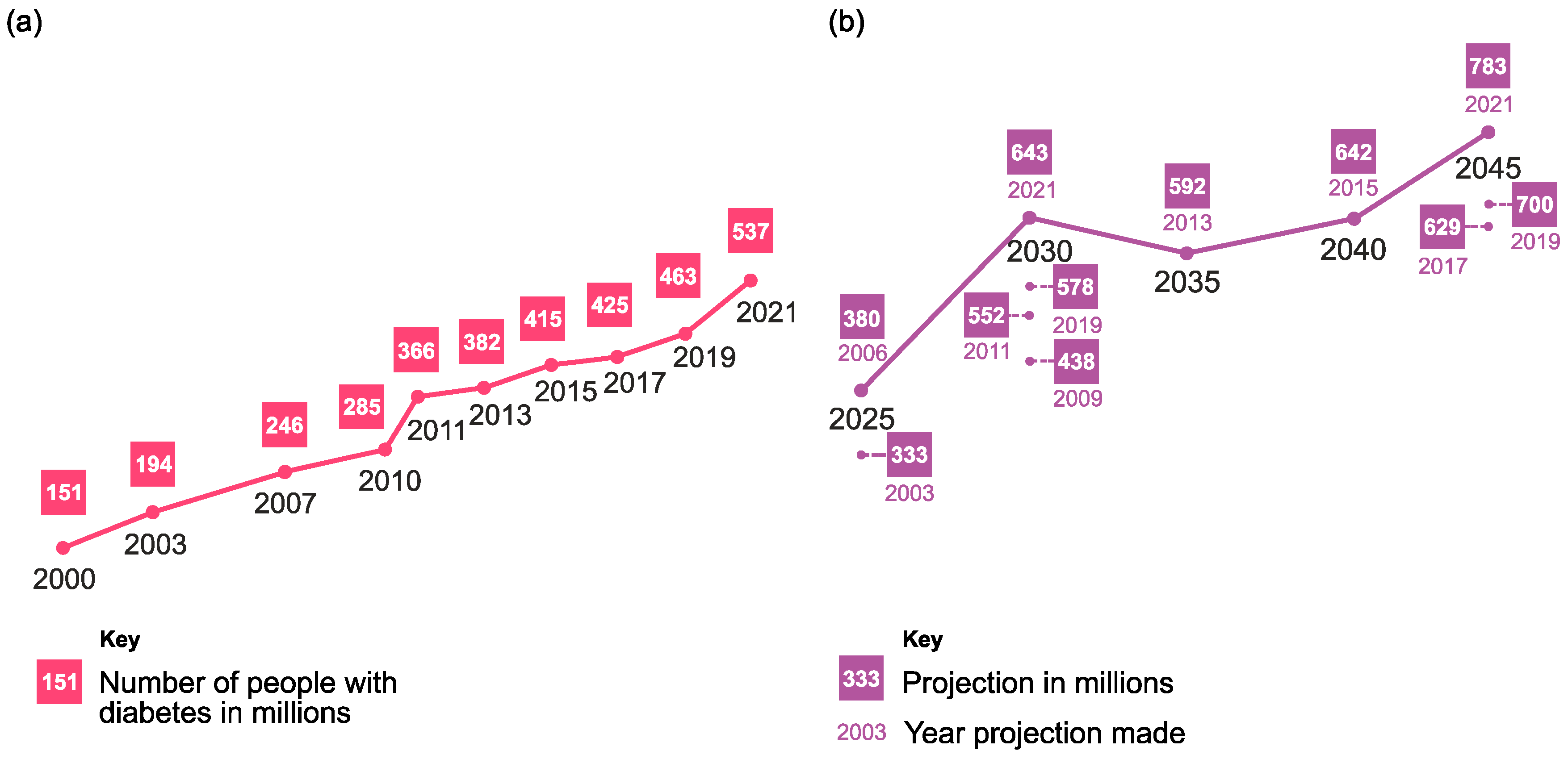
Figure 1. (a) Global prevalence of diabetes in the 20–79 year age group (millions) from 2000 to 2021. (b) Projections of the global prevalence of diabetes in the 20–79 year age group (millions). Reprinted from [2] according to data from the International Diabetes Federation (IDF), under a Creative Commons Attribution 4.0 International License.
Chemosensors 12 00159 g001
Figure 2. Publication trend of scientific papers in the field of electrochemical glucose detection from 1981 to 2023 using Scopus (keywords: glucose non-enzymatic sensor, electrochemical, and neutral pH); blue bars indicate the works in non-enzymatic detection with alkaline electrolytes, the pink bars represent the published works using enzymatic detection, and the green bars include the reported non-enzymatic glucose sensors at physiological conditions.
Chemosensors 12 00159 g002
Figure 3. Diagram of the effect of (a) interference with the glucose measurement, and (b) poisoning on the electrode.
Chemosensors 12 00159 g003
Figure 4. Mechanism of glucose electrooxidation using the Pt electrode according to Ernst and Heitbaum [41,42]. (a) Hydrogen region. (b) Double-layer region.
Chemosensors 12 00159 g004
Figure 5. Mechanism of DGE (direct glucose electrooxidation) using the Au electrode. 1. Au work electrode; 2. Adsorption of OH on surface ((OH)ads); 3. Interaction between the hemiacetal group of glucose and (OH)ads; 4. Gluconolactone generation.
Chemosensors 12 00159 g005
Figure 6. Mechanism of interaction between glucose and phenylboronic acid (PBA) at neutral pH. 1. Phenylboronic acid (PBA) or its derivates; 2. Conformational changes to sp3 hybridized boronates in aqueous media; 3. Reversible covalent bonds between the boron atom and the hydroxyl groups (1,2 and 1,3 diols) of the glucose; 4. Formation of a boronate ester, a stable complex between PBA and glucose.
Chemosensors 12 00159 g006
Figure 7. SEM micrographs of (a) Boron-doped diamond (BDD), (b) Ni-BDD, (c) Pt-BDD, (d) PtNi-BDD, (e) Pt/Ni-BDD, and (f) Ni/Pt BDD. CVs response of (g) PtNi-BDD, (h) Magnification of the oxidation peaks at different glucose concentrations of PtNi-BDD (g), (i) Pt-BDD, (j) Ni/Pt-BDD, and (k) Pt/Ni-BDD analyzed under different glucose concentrations (2, 4, 5, 9, and 12 mM) at a pH of 7.4 (0.01 M PBS). (l) Linear calibration relations of the current response vs. glucose concentration [68]. Reprinted (adapted) with permission from [68] Copyright © 2023 with permission from Elsevier.
Chemosensors 12 00159 g007
Figure 8. (a) Amperometric response to a wide range of glucose concentrations in PBS [0.1 M]. (b) Calibration plot of sensor Au@Pt/Au/Nafion at +0.35 V (Ag/AgCl). (c) Amperometric response for the interference effects of other bio-compounds on the sensor probe. (d) Stability test for 27 days, and the test conditions were PBS [0.1 M] containing 1.0 mM glucose (Note: the electrode was stored at room temperature in a dry state when not in use). Reprinted (adapted) with permission from [100] Copyright © 2023 with permission from Elsevier.
Chemosensors 12 00159 g008
Figure 9. (a) Scheme of Pd/Au NC synthesis and its HAADF-STEM images. (b) Scheme of Pd/Pt concave nanocube (CNC) synthesis and its HAADF-STEM images; (c) amperometric responses from 0.25 mM to 1 mM; (d) interference studies; (e) calibration curves; (f) real sample experiments. Reprinted (adapted) with permission from [48] Copyright © 2023 with permission from Elsevier.
Chemosensors 12 00159 g009
Figure 10. TEM micrographs of the CuNWs/MOF/GO sensor: (a) CuNWs, (b) GO, (c) MOF, and (d) a fully assembled nanocomposite; (e) the amperometric I vs. t curve obtained at 0.3 V with the assembled CuNWs/MOF/GO sensor working in 0.1 M PBS, and (f) the calibration curve. Reprinted (adapted) with permission from [105] Copyright © 2018 with permission from Elsevier; (g) the preparation of the Co3(PO4)2 nanoflowers/SPCE sensor and its response towards glucose oxidation in 0.01 M PBS at a pH of 7.4. Reprinted (adapted) with permission from [102] Copyright © 2018 with permission from American Chemical Society.
Chemosensors 12 00159 g010
Figure 11. (a) The assembly of the GCE/PAA-Au-NPs-MPBA sensor with the blockage of the surface after glucose capture. Reprinted (adapted) with permission from [54] Copyright © 2017 with permission from Elsevier; (b) synthesis of mesoporous silica adding PBA for surface functionalization; (c) set up for recording the interaction of glucose with the Ta2O5/MPSi-PBA sensor and the corresponding response after the glucose addition compared to the minimal response at unmodified PBA surfaces. (d) Reprinted (adapted) with permission from [113] Copyright © 2021 with permission from American Chemical Society.
Chemosensors 12 00159 g011
Figure 12. Fabrication of a carbon cloth electrode modified with AuNF showing the nanostructures deposed on the textile fibers: the fabrication of the CC-AuNFs with space gauze in between electrodes (ad), and a SEM image of active AuNF over the CC fibers. Reprinted (adapted) with permission from [60] Copyright © 2023 with permission from Elsevier; (e) fabrication of the patch-like rGO/PU-Au wearable sensor showing the molding of the base electrode and the subsequent surface modifications (FE-SEM micrographs of rGO/PU-Au); the inset photograph showed the on-body recording obtained by the rGO/PU-Au sweat glucose detection after and before a meal. Reprinted (adapted) with permission from [62] Copyright © 2019 with permission from American Chemical Society; (f) the integration of the PdNPs/Co-ZIF-67 sensor in a wearable sweatband with a miniaturized reader; (g) on-body detection and recording of results in a smartphone app; (h) recorded trend of the PdNPs/Co-ZIF-67 wearable sensor for sweat glucose compared to blood glucose. Reprinted (adapted) with permission from [58] Copyright © 2019 with permission from American Chemical Society.
Chemosensors 12 00159 g012
Table 1. List of glucose self-monitoring devices and their analyzed samples.
FormatDetectionAnalyzed SampleSample
[µL]
Linear Range
[mM]
Reference
BGMTInvasiveCapillary blood0.3 to 0.71.1–33.3[24]
CGMMinimally invasiveInterstitial fluid--2.2–22.2[25]
Table 2. List of the standards applied to different devices.
DeviceSampleConcentration [mg/dL]StandardRef.
Point of Care testVenous whole blood5 intervals between 30 and 400FDA-2013-D-1445 (2020)[29]
Over the counter, Blood glucose monitoring test systems Venous whole blood5 intervals between 30 and 400FDA-2013-D-1446 (2016)[30]
Blood glucose monitoring test systemsVenous whole blood5 intervals between 30 and 400ISO 15197 (2015)[28]
Point of Care test CLSI POCT12-A3 (2013)[31]
Table 3. List of substances classified as interference and their concentrations according to the FDA [29,30].
InterferenceRecommended Test
Concentration (mg/dL)
InterferenceRecommended Test
Concentration (mg/dL)
Acetaminophen (AC)20Ibuprofen (IBU)50
Ascorbic acid (AA)6L-3,4-dihydroxyphenylalanine
(L-Dopa)
0.75
Conjugated Bilirubin (CB)50Maltose (Mlt)480
Unconjugated Bilirubin (UB)40Mannitol (Man)1800
Cholesterol (CH)500Methyldopa (Mdp)2
Creatinine (CR)15Salicylic acid (SA)60
Dopamine (DA)0.09Sodium (Na)180 (mmol/L)
Ethylenediaminetetraacetic
acid (EDTA) *
0.1Tolbutamide (TA)72
Galactose (Gal)60Tolazamide (Tol)9
Gentisic acid (GA)1.8Triglycerides (TG)1500
Reduced glutathione (GSH)4.6Uric acid (UA)23.5
Hemoglobin (Hb)1000Xylose (Xyl)600
Heparin * (Hep)300 (IU/dL)Sugar Alcohols (sorbitol, xylitol,
lactitol, isomalt, maltitol)
0.09
* The inclusion of EDTA and heparin in this table refers to their use as therapeutic substances and not as anticoagulants for sample preparation.
Table 4. Strategies for adding OH on the electrode microregion to facilitate DGE.
StrategyRef.
Electrochemical pretreatment with high anodic or cathodic potential[56,57]
Water-splitting electrocatalytic reaction[58,59]
Chemical pretreatment of Au in an alkaline solution[60]
Chemical pretreatment of carbon in an acidic solution[61]
Surface functionalization[62]
Table 5. Platinum non-enzymatic detection at physiological conditions (NEDPC).
ElectrodeMaterialElectrolytepHTechniqueE
[V]
LR (mM)S
μM mM−1 cm−2
LOD
[μM]
InterferenceReal
Sample
Stability [Days]Ref.
AuPt/dGN600PB 0.1 M7.4AMP0.05 10.5–927.280.06Fru, Suc, Mlt, Sor, AA, UA Human serum [66]
Pt/dGN4000.5–9.523.140.08--25 cycles
Pt/dGN2001.5–98.060.07--
Pt/dGNech1.5–8.56.210.12--
GCEPt5Ni1PBS [1 × 10⁻⁵] + NaCl 0.9%7.4AMP+0.1 20.5–4040.170.35AA, UA, Fru Human serum30 [75]
GCEPd@Pt CINPsPBS [0.1 M] AMP−0.1 11–8.515.140.82Fru, Suc, Mlt, SorHuman serum25 cycles[44]
GCEPtNPs/pDA-NCNRPBS 1X [10 mM] CAMP+0.28 30.01–27.650.01AA, UA -[73]
PBS 1X [10 mM] +0.28 32–3081.240.01
GCEPt3Ru1PBS [0.01 M]7.4AMP+0.05 20–431.30.3UA, AA, Fru 30[76]
SPCE Pt-replaced Cu foamsPBS [0.1 M]7.4AMP+0.4 11–119.62385AA, UA, Fru, AP --[77]
GCEPtNCs/graphene (PVP)PBS [0.1 M]7.4AMP0.05 41–251.2130AA, UA, AP 20[78]
GCEPtAuPd/f-CaNCPBS [0.01 M]7.4AMP0.430–1011.242.9---[79]
GCEPtNFs/MWCNTs/graphenePBS [0.1 M]7.4AMP+0.4 11–711.06387AA, UA -[80]
GCDPt2Ir1/MWCNTPBS [0.2 M]7.4AMP0.1 40.1–12060.5AA, UA, AP, CR, CH -[70]
GCDPt3Pt1/graphenePBS [0.05 M] + NaCl [0.1 M]7.4AMP−0.45 2(30 μM–3 mM)(1.52 μM mM−1) ---AA, UA, AP >14[81]
GCDPd1Pt3/graphenePBS [0.1 M]7.4AMP+0.1 21–23---5AA, UA, DOPAC -[82]
SPCEPt/CuPBS [0.1 M] + KCl [0.15 M]7.4CAMP0.5 1 7.7 AA, UA, DA, AP [69]
CAMP0 1 5.9 -
CAMP−0.4 1 6.7
GCEPt/Au/CPBS [0.01 M]7.4AMP0.3 30–104720UA, AA, AC, DA, Xyl, Mlt, Gal, Fru -[43]
GCEPt-MWCNTPBS [0.1 M] AMP0.55 32–201.10 AA, UA, Fru, Suc, Xyl, Gal [83]
Pt-AC 1.07 20 cycles
Pt-CNF0.52
Gold diskNanoporous Au-Pt (24%)PBS [0.1 M]7.4AMP+0.35 20.5–10145.70.6AA, UA, AP 30[84]
GCEPtRu(1:1)-MWNT-IL/GCEPBS7.4AMP−0.1 21510.60.05AA, UA, Fru, AP >50[85]
Pt wire (1 cm, r = 0.1 cm)PtZn alloyPBS [0.1 M]7.4 0.4 10–10291 AA, AP -[71]
Ti platesPtPb NetworksPBS [0.1 M] + NaCl [0.15 M]7.4AMP0.4 40–1610.8 AA, UA, AP -[86]
Au (111)Pt-NTAEs (ca. 40 nm thickness and length ca. 3 μm)PBS [0.05 M] + KCl (0.1 M)7.4AMP0.4 22–14 0.11.0AA, UA, AP -[64]
Pt rodPt mesoporous electrodepositionPBS 0.1 M + NaCl 0.15 M7.4AMP0.4 30–109.6 AA, AP -[63]
E: potential; LR: linear range; S: sensitivity; LOD: limit of detection; GCE: glassy carbon electrode; SPCE: screen-printed carbon electrode; GCD: glassy carbon disk; PB: phosphate-buffered; PBS: phosphate-buffered saline; dGN: defective graphene nanosheets; CINPs: core/island nanoparticles; PtNPs: platinum nanoparticles; pDA-NCNR: polydopamine-modified mesoporous carbon nanorods; PtNCs: Pt nanoclusters; CaNC: carbon nanochips; Pt NFs: platinum nanoflowers; MWCNT: multi-walled carbon nanotubes; Pt-AC: platinum-activated carbon; Pt-CNF: platinum carbon nanofibers; MWNT: multi-walled carbon nanotubes; Pt-NTAEs: platinum-nanotubules array electrodes; AMP: amperometry; CAMP: chronoamperometry; Fru: fructose; Suc: sucrose; Sor: sorbitol; DOPAC: 3,4-dihydroxyphenilacetic acid; Gal: galactose; 1: Ag/AgCl 3 M KCl; 2: SCE; 3: Ag/AgCl; 4: Ag/AgCl, KCl-saturated.
Table 6. Gold non-enzymatic detection at physiological conditions.
ElectrodeMaterialElectrolytepHTechniqueE
[V]
LR (mM)S
μM mM−1 cm−2
LOD
[μM]
InterferentsReal
Sample
Stability
[Days]
Ref.
AuAu amalgamationPB [0.1 M]7.0AMP0.25 40–10322UA, AP, AA [101]
AuNPGFPBS [0.1 M]7.4AMP0.2 20.001–11668.7AA, UA 20 cycles[89]
AuAuNPs/GONR/CSPBS [0.1 M]7.4AMP0.2 40.0005–1057.10.5UA, AP, AA [91]
AuDGNPBS [0.1 M]7.4AMP0.15 30.1–25190.750UA, AP, AA [90]
AuAu NanowirePBS [0.1 M]7.2AMP0.35 10.0004–0.0050.0003733-- [95]
AuAuNTAs/Au NWAsPBS [0.1 M]7.2AMP0.25 20.005–16.444.22.1AA, DA, UA, Fru, Suc [94]
GCEIrregular AuNPsPBS [0.1 M]7.4AMP0.3 40.2–11066100---[49]
AuAuNPs/MWCNTsPBS [0.1 M]7.4AMP0.2 30.1–252.77 4.1AA, Gal, Fru, UA 14[92]
AuPt-Au nanocoralPB7AMP0.4 1222.128AA, UA [98]
AuAu@Pt NPs
Au@PtNPs
PBS7.4AMP+0.35 30.0005–0.01, 0.01–10.0 445.7AA, AP, UA, DABlood [100]
AuPd/AuNCPBS [0.1 M]7.4AMP0.25 20.25–14 13.56Fru, Suc, Mlt, Sor, UA, AASerum [48]
AuAuRu/CNT -PtNPPBS [0.01 M]7.4AMP0.2 31–100.234768UA, AP, AA 21[46]
NPGF: nanoporous gold film; AuNPs: gold nanoparticles; GONR: graphene oxide nanoribbon; CS: carbon sheet; DGN: dendrite-like gold nanostructure; Au NTAs: gold nanotubes array; Au NWAs: gold nanowires array; Pd/Au NC: palladium/gold nanocubes; AuRu/CNT-PtNP: Au and Ru nanoparticles on the surface of a carbon-nanotube-based platinum–nanoparticle hybrid; 1: Ag/AgCl 3 M KCl; 2: SCE; 3: Ag/AgCl; 4: Ag/AgCl 3 M NaCl.
Table 7. Additional transition metals for non-enzymatic detection at physiological conditions.
ElectrodeMaterialElectrolytepHTechniqueE [V]LR
[mM]
S
μM mM−1 cm−2
LOD [μM]InterferencesReal
Sample
Stability [Days]Ref.
GCEPd/SWCNT0.1 M PBS/
0.15 M NaCl
7.4AMP−0.35 20.5–171600.2AA, UA, AP, DOPAC Human blood (diluted)30[104]
PtRuO2/PVC/
Nafion
0.01 M PBS/
10 mM NaCl/
2.7 mM KCl
7.4AMP0.45 30.1–107, 0.1–61.8----AA, DA, UA, Cat, Fru, Suc, Man, Lac, Gal serum--[106]
--Ag from CD0.1 M PBS6.5AMP−0.5 30.5–13--35UA, AAHuman blood 120[107]
Au/PATP[VO(acac)2]0.1 M PBS7.0AMP0.65 10.001–0.5 --0.1AA, UA, L-dopa, L-cys, Na+, K+, Clblood20 [108]
GCECuNWs/MOF/GO/Nafion0.1 M PBS7.4AMP0.3 10.02–26.6 7.727Lac, Fru, Suc, Mlt, Xyl, and satisfactory anti-interference performance to AP, AA, UA serum30 [105]
SPCECo3(PO4)2 CPN0.01 M PBS7.4AMP0.65 31–307.90 nA/mM300Lac, Gal, AA, DAserum--[102]
SPCEFunctionalized GO/Fe3O4)/PANIPBS7.0CV--5 × 10−5–5 --0.01CH, UA, AA, DA, Fru, Mlt, Suc, CGN serum16 days[103]
--PBS pretreated Ni-Cu NPs0.1 M Na2SO46.4CV0.2 35 × 10−6–20 5.474.2 × 10−3 AA, UA artificial saliva7 days[109]
Ni foamBiZnSbV-G- SiO2 (BZSVGS)0.1 M PBS7.4CV0.2 36 × 10−5–0.001--0.06Lac, Gal, AA, Starch, Fru, NaCl, KCl, and Ureaurine--[110]
FTOCoFe Prussian Blue composite0.1 M PBS/
0.1 M
KCl
7.4AMP1.15 10.1–8.2 mM18.6967Suc, Fru, AA, UA----[111]
GCEGO-NiO-8H-NHSPBS7.4AMP0.55 32 × 10−3–0.08/0.08–5 712.50.041Urea, AA, Mlt, Lac, Suc, Fruplasma30 days[112]
Au/PATP: 4-(pyridine-4′-amido)thiophenol (PATP) monolayer-modified gold electrode; FTO: fluorine-doped tin oxide; Pd/SWCNT: palladium nanoparticles-single-walled carbon nanotube; RuO2/PVC/Nafion: ruthenium dioxide–poly(vinyl chloride)–Nafion composite; CD: compact disc; [VO(acac)2]: bis(acetylacetonato)oxovanadium(IV) complex; CuNWs: copper nanowires; MOF: metal organic framework; GO: graphene oxide; CPN: cobalt phosphate nanostructure; PANI: polyaniline; Ni-Cu NPs: nickel-copper nanoparticles; BiZnSbV-G-SiO2: mesoporous quaternary composite BiZnSbV-graphene oxide-mesoporous silicon dioxide; 8H: hydroxyquinoline; NHS: N-hydroxysuccinimide; CV: cyclic voltammetry; Cat: catechol; Lac: lactose; L-cys: L-cysteine; CGN: carrageenan; 1: Ag/AgCl 3 M KCl; 2: SCE; 3: Ag/AgCl.
Table 8. PBA and derivates non-enzymatic detection at physiological conditions.
ElectrodeMaterialElectrolytepHTechniqueE [V]LR [mM]S
μM mM−1 cm−2
LOD
[μM]
InterferencesReal SampleStability
[Days]
Ref
AuAu-PB/
pDA/AuNPs/
MPBA
0.1 M
KNO3
7.0DPV−0.1–0.4 11 × 10−4–1.35 × 10⁻²--0.05 Fru, Gal, Man, UA, AA, Pro, Ala, MA serum30[114]
GCEPAA-AuNPs/MPBA0.1 M
PBS
6.5CV−0.1–0.5 11 × 10−5–0.01--4 × 10⁻³DA, AA, UAserum30[54]
GCETiO2NW/
PAPBA/
AuNPs
0.1 M
PBS
7.0DPV0–0.7 20.5–1166.89.3 AA, UA, DA, Fru, Lac, Suc, Man,serum--[115]
--CNT--SB PBS7.4POT 0.1–100--100 ------[116]
Ta2O5PBA-MPSiPBS7.4POT 0–20------serum--[113]
Au-PB: Prussian blue-gold nanoparticles; pDA: polydopamine; AuNPs: gold nanoparticles; MPBA: 4-marcaptophenyl boronic acid; PAA: poly(azure A); TiO2NW: titanium dioxide nanowire; PAPBA: poly(3-aminophenyl boronic acid); CNT-SB: carbon nanotube-silver borate; PBA-MPSi: phenylboronic acid-mesoporous silica; DPV: differential pulse voltammetry; POT: potentiometric; Man: mannose; Pro: proline; Ala: alanine; MA: 1-malic acid. 1: SCE; 2: Ag/AgCl.
Table 9. Wearable and flexible non-enzymatic sensors for glucose detection at physiological conditions.
ElectrodeMaterialTypeElectrolytepHTechniqueE
[V]
LR [mM] S
μM mM−1 cm−2
LOD
[μM]
InterferenceReal
Sample
Stability
[Days]
Ref.
--Au/Nafionwristband0.1 M
PBS
5.0MPS0.2 10.3–1.1 114 µA/mM/cm215AA, UA, LA, Gluhuman sweat--[56]
SPCEPd NPs encapsulated in a Co-ZIF- 67 sweatband0.1 M
PBS
7.4PAD0.6 20.01–1 --2.0AA, UA, LA, Glu, AP human sweat60
(closed)
[58]
--rGO-PU/Au nanowrinklespatch0.1 M
PBS
7.4CV0.2 30.001–1 140 µA/mM/cm20.5AA, UA, LA, NaCl human
sweat
--[62]
CCcotton-like
Au microspheres
flexible electrode0.1 M PBS/
0.1 M NaCl
7.4AMP0.3 40.001–0.114
0.114–21.6
25.39/20.609 µA/mM/cm20.78Urea, AcOH, LA, Xyl, Mlt, Suc, Fru, NaClhuman sweat, human
blood
35[61]
MNEAAu/
porous Pt black/
Nafion
patch10X PBS7.4AMP0.12 31–30 1.792 µA/mM/cm27.2AA, LA, Gal, Man, AP, Fru, NaCl, UA, Urea ISF16[74]
CCPB/
CoFe-NO
NPs
flexible electrode0.1 M PBS/
0.5 M KCl
7.0AMP1.15 50.1–6.5 145.43 µA/mM/cm228Suc, Lac, NaCl, AA, UA --15[118]
MNEAAu/porous Pt black/
Nafion
patch10X PBS7.4AMP0.12 61–10/
15–30
445.75/165.83 µA/mM/cm2268AA, UA, LA, Urea, AP, DA, Man, Fru, Gal, NaCl ISF16[59]
ExGCP --flexible electrode0.01 M
PBS
--CV−0.8 to 0.8 32–85.93 μA/mMcm−2812----7[117]
carbon clothAu NFpatchPBS7.4AMP0.35 20.008–463.9 μA/mMcm−25.18LA, Urea, AA, NaCl, UA human
sweat
--[60]
LIG Au dendrite/
Nafion
flexible electrode0.1 M
PBS
7.4AMP0.2 20.5–201.06 μA/mM210AA, UA, AP, Suc, Lac, Fru human
serum
20[57]
MNEA: microneedle electrode array; ExGCP: exfoliated graphite carbon paper; LIG: laser-induced graphene; Pd NPs: palladium nanoparticles; Co-ZIF-67: cobalt-based zeolitic imidazole framework; rGO-PU: reduced graphene oxide-polyurethane; PB/CoFe-NO NPs: Prussian blue/cobalt-nitroprusside nanoparticles; Au NF: gold nanoflowers; MPS: multipotential steps; PAD: pulsed amperometric detection; Glu: glutamic acid; LA: lactic Acid; NaCl: sodium chloride; AcOH: acetic acid; 1: partial oxidized polypyrrole pseudoreference. 2: Ag/AgCl pseudorefence; 3: Ag/AgCl; 4: Ag/AgCl 3 M KCl; 5: Ag/AgCl, KCl-saturated; 6: AgCl microneedle.
Disclaimer/Publisher’s Note: The statements, opinions and data contained in all publications are solely those of the individual author(s) and contributor(s) and not of MDPI and/or the editor(s). MDPI and/or the editor(s) disclaim responsibility for any injury to people or property resulting from any ideas, methods, instructions or products referred to in the content.