Promising Solutions to Address the Non-Specific Adsorption in Biosensors Based on Coupled Electrochemical-Surface Plasmon Resonance Detection

 ,
,  ,
, 
 , ,
, , 

Abstract
1. Introduction
2. NSA in Biosensors
2.1. Contribution of NSA to Biosensor Signal

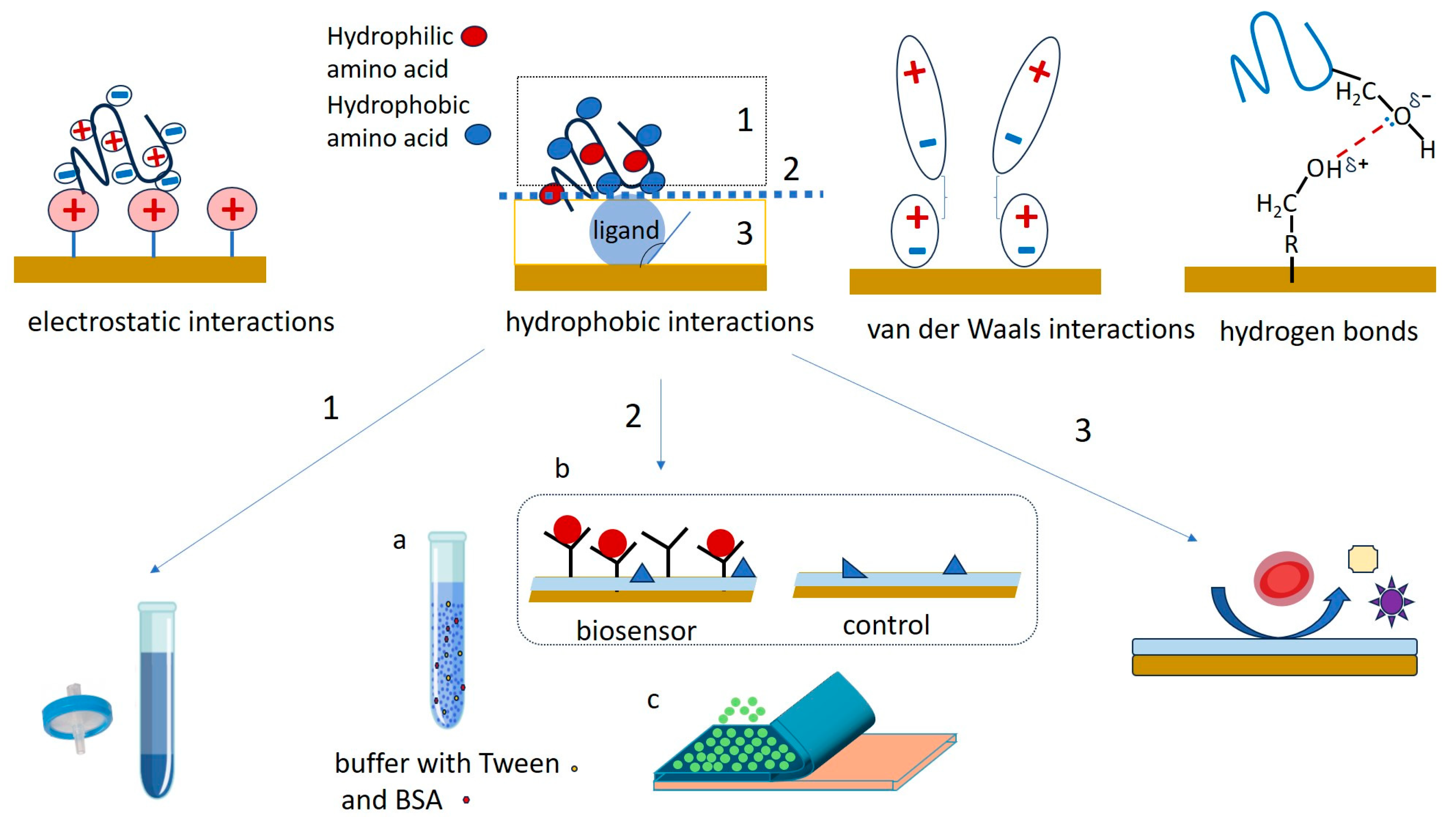
2.2. Mechanisms of NSA and Ways to Counteract It
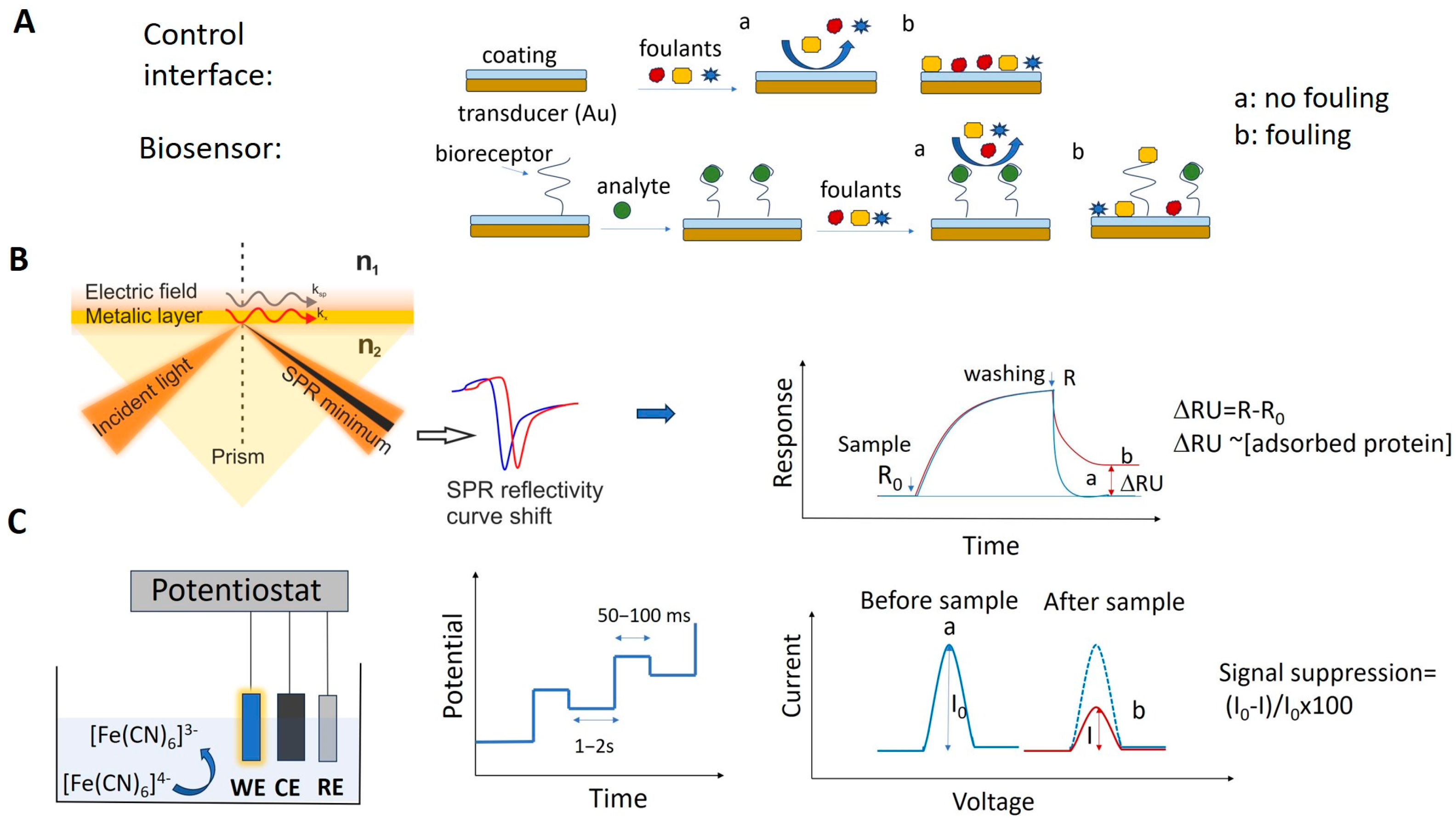
2.3. Methods Used to Evaluate NSA
2.4. NSA-Resistant Coatings
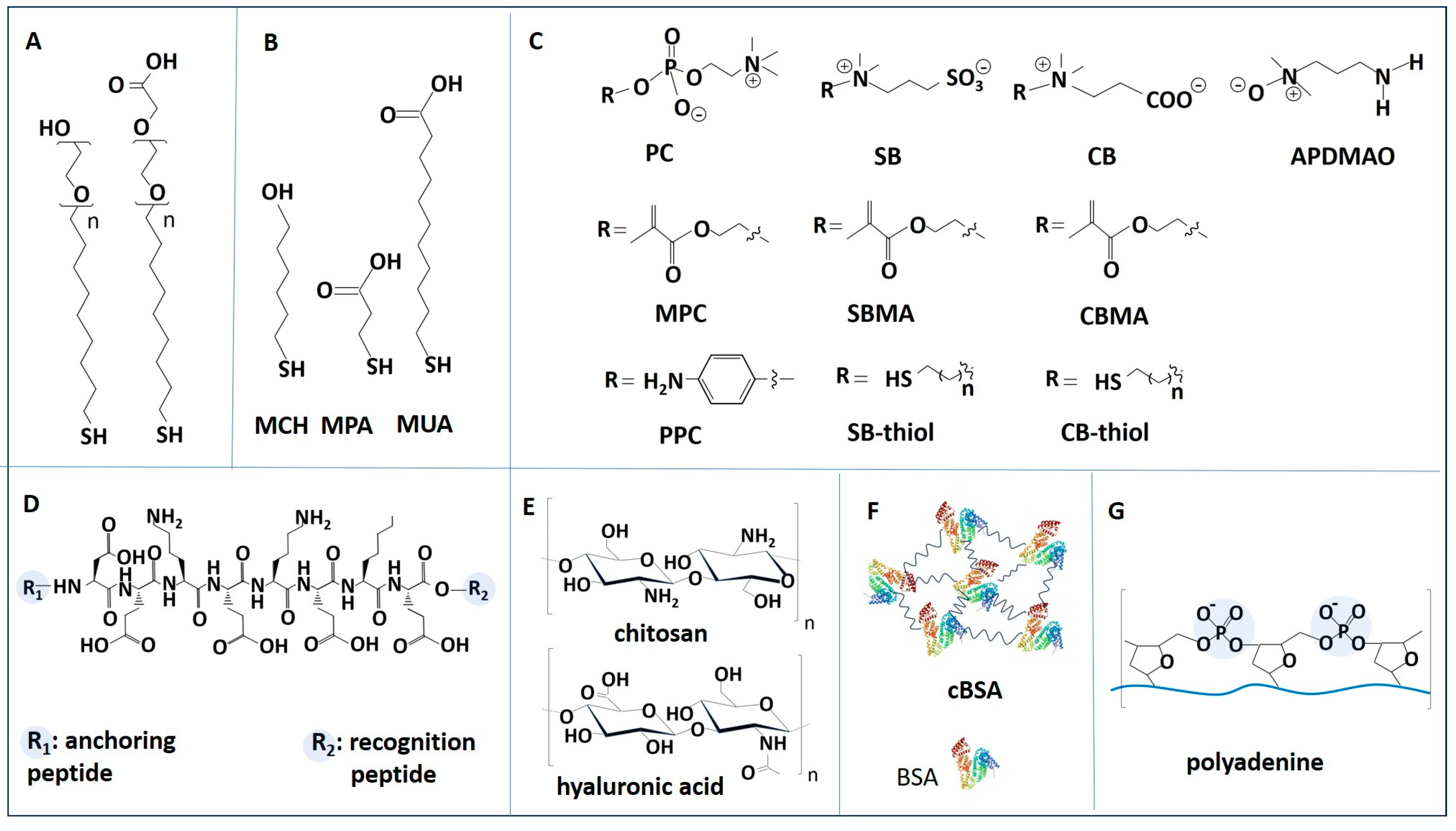
2.4.1. Overview of Fouling-Resistant Materials
2.4.2. PEG and OEG Derivatives
2.4.3. Alkanethiols
2.4.4. Zwitterionic Materials
2.4.5. Antifouling Peptides
2.4.6. Polysaccharides
2.4.7. BSA
2.4.8. DNA-Based Coatings
2.4.9. Biomimetic and Nature-Inspired Materials
2.5. Experimental Protocols for Studying NSA in Biosensors
3. Solutions for Minimizing NSA in Electrochemical Biosensors
3.1. General Strategies to Address Fouling in EC Biosensors
3.2. Folding-Based Biosensors for Measurements in Undiluted Samples
3.3. Antifouling Coatings Used in Electrochemical Biosensors
3.3.1. Applications of Alkanethiol SAMs in Low Fouling Electrochemical Biosensors
3.3.2. Applications of BSA-Based Passivating Films in Low Fouling Electrochemical Biosensors
3.3.3. Applications of Zwitterionic Materials in Low Fouling Electrochemical Biosensors
3.3.4. Applications of Antifouling Peptides in Low Fouling Electrochemical Biosensors
3.3.5. Applications of Other Antifouling Coatings in Low Fouling Electrochemical Biosensors
3.4. Challenges in the Development of Low Fouling Electrochemical Biosensors
4. Solutions to Minimize NSA in SPR Biosensors
4.1. Surface Sensitivity in SPR Biosensing
4.2. Smart Layers in the Design of SPR Surfaces
4.2.1. Applications of DNA-Based Coatings (Thiol-Ended DNA Tetrahedrons and Polyadenine) in Low Fouling SPR Biosensors
4.2.2. Applications of Zwitterionic Compounds in Low Fouling SPR Biosensors
4.2.3. Applications of Other Antifouling Coatings in Low Fouling SPR Biosensors
4.3. SPR Sensors to Work in Undiluted Food Samples
5. Solutions for Minimizing NSA in Coupled EC-SPR Biosensors
5.1. Coupled EC-SPR Biosensors
5.2. Minimizing NSA in Coupled EC-SPR Biosensors
5.2.1. NSA in EC-SPR Biosensors Exploiting Non-Faradaic Processes
5.2.2. NSA in EC-SPR Biosensors Exploiting Faradaic Processes
5.3. Challenges in Low Fouling EC-SPR Biosensors
6. Conclusions and Perspectives
Author Contributions
Funding
Institutional Review Board Statement
Informed Consent Statement
Data Availability Statement
Conflicts of Interest
References
- Campuzano, S.; Pedrero, M.; Barderas, R.; Pingarrón, J.M. Breaking barriers in electrochemical biosensing using bioinspired peptide and phage probes. Anal. Bioanal. Chem. 2024, 416, 7225–7247. [Google Scholar] [CrossRef] [PubMed]
- Campuzano, S.; Pedrero, M.; Gamella, M.; Serafín, V.; Yáñez-Sedeño, P.; Pingarrón, J.M. Beyond Sensitive and Selective Electrochemical Biosensors: Towards Continuous, Real-Time, Antibiofouling and Calibration-Free Devices. Sensors 2020, 20, 3376. [Google Scholar] [CrossRef]
- Jiang, C.; Wang, G.; Hein, R.; Liu, N.; Luo, X.; Davis, J.J. Antifouling Strategies for Selective In Vitro and In Vivo Sensing. Chem. Rev. 2020, 120, 3852–3889. [Google Scholar] [CrossRef]
- Gu, Y.; Li, Y.; Wu, Q.; Wu, Z.; Sun, L.; Shang, Y.; Zhuang, Y.; Fan, X.; Yi, L.; Wang, S. Chemical antifouling strategies in sensors for food analysis: A review. Compr. Rev. Food Sci. Food Saf. 2023, 22, 4074–4106. [Google Scholar] [CrossRef]
- Lichtenberg, J.Y.; Ling, Y.; Kim, S. Non-Specific Adsorption Reduction Methods in Biosensing. Sensors 2019, 19, 2488. [Google Scholar] [CrossRef] [PubMed]
- Mahmoudpour, M.; Jouyban, A.; Soleymani, J.; Rahimi, M. Rational design of smart nano-platforms based on antifouling-nanomaterials toward multifunctional bioanalysis. Adv. Colloid Interface Sci. 2022, 302, 102637. [Google Scholar] [CrossRef]
- Contreras-Naranjo, J.E.; Aguilar, O. Suppressing Non-Specific Binding of Proteins onto Electrode Surfaces in the Development of Electrochemical Immunosensors. Biosensors 2019, 9, 15. [Google Scholar] [CrossRef] [PubMed]
- Li, S.; Zhang, H.; Zhu, M.; Kuang, Z.; Li, X.; Xu, F.; Miao, S.; Zhang, Z.; Lou, X.; Li, H.; et al. Electrochemical Biosensors for Whole Blood Analysis: Recent Progress, Challenges, and Future Perspectives. Chem. Rev. 2023, 123, 7953–8039. [Google Scholar] [CrossRef] [PubMed]
- Russo, M.J.; Han, M.; Desroches, P.E.; Manasa, C.S.; Dennaoui, J.; Quigley, A.F.; Kapsa, R.M.I.; Moulton, S.E.; Guijt, R.M.; Greene, G.W.; et al. Antifouling Strategies for Electrochemical Biosensing: Mechanisms and Performance toward Point of Care Based Diagnostic Applications. ACS Sens. 2021, 6, 1482–1507. [Google Scholar] [CrossRef]
- Saxena, S.; Sen, P.; Soleymani, L.; Hoare, T. Anti-Fouling Polymer or Peptide-Modified Electrochemical Biosensors for Improved Biosensing in Complex Media. Adv. Sens. Res. 2024, 3, 2300170. [Google Scholar] [CrossRef]
- Song, Z.; Han, R.; Yu, K.; Li, R.; Luo, X. Antifouling strategies for electrochemical sensing in complex biological media. Microchim. Acta 2024, 191, 138. [Google Scholar] [CrossRef] [PubMed]
- Szunerits, S.; Pagneux, Q.; M’Barek, Y.B.; Vassal, S.; Boukherroub, R. Do not let electrode fouling be the enemy of bioanalysis. Bioelectrochemistry 2023, 153, 108479. [Google Scholar] [CrossRef] [PubMed]
- Zhou, L.; Li, X.; Zhu, B.; Su, B. An Overview of Antifouling Strategies for Electrochemical Analysis. Electroanalysis 2022, 34, 966–975. [Google Scholar] [CrossRef]
- Zhou, J.; Wang, Y.; Zhang, G.-J. State-of-the-art strategies of surface plasmon resonance biosensors in clinical analysis: A comprehensive review. Coord. Chem. Rev. 2024, 520, 216149. [Google Scholar] [CrossRef]
- Qu, J.-H.; Dillen, A.; Saeys, W.; Lammertyn, J.; Spasic, D. Advancements in SPR biosensing technology: An overview of recent trends in smart layers design, multiplexing concepts, continuous monitoring and in vivo sensing. Anal. Chim. Acta 2020, 1104, 10–27. [Google Scholar] [CrossRef] [PubMed]
- D’Agata, R.; Bellassai, N.; Spoto, G. Exploiting the design of surface plasmon resonance interfaces for better diagnostics: A perspective review. Talanta 2024, 266, 125033. [Google Scholar] [CrossRef]
- Jungnickel, R.; Balasubramanian, K. Electrochemistry-coupled surface plasmon resonance on 2D materials for analysis at solid–liquid interfaces. Curr. Opin. Electrochem. 2025, 50, 101634. [Google Scholar] [CrossRef]
- Hemmerová, E.; Homola, J. Combining plasmonic and electrochemical biosensing methods. Biosens. Bioelectron. 2024, 251, 116098. [Google Scholar] [CrossRef]
- Geilfuss, D.; Boukherroub, R.; Dostalek, J.; Knoll, W.; Masson, J.-F.; Baeumner, A.; Szunerits, S. Can classical surface plasmon resonance advance via the coupling to other analytical approaches? Front. Anal. Sci. 2022, 2, 1091869. [Google Scholar] [CrossRef]
- Ribeiro, J.A.; Sales, M.G.F.; Pereira, C.M. Electrochemistry combined-surface plasmon resonance biosensors: A review. TrAC Trends Anal. Chem. 2022, 157, 116766. [Google Scholar] [CrossRef]
- Watkins, Z.; Karajic, A.; Young, T.; White, R.; Heikenfeld, J. Week-Long Operation of Electrochemical Aptamer Sensors: New Insights into Self-Assembled Monolayer Degradation Mechanisms and Solutions for Stability in Serum at Body Temperature. ACS Sens. 2023, 8, 1119–1131. [Google Scholar] [CrossRef] [PubMed]
- Downs, A.M.; Plaxco, K.W. Real-Time, In Vivo Molecular Monitoring Using Electrochemical Aptamer Based Sensors: Opportunities and Challenges. ACS Sens. 2022, 7, 2823–2832. [Google Scholar] [CrossRef] [PubMed]
- Shaver, A.; Arroyo-Currás, N. The challenge of long-term stability for nucleic acid-based electrochemical sensors. Curr. Opin. Electrochem. 2022, 32, 100902. [Google Scholar] [CrossRef] [PubMed]
- Leung, K.K.; Downs, A.M.; Ortega, G.; Kurnik, M.; Plaxco, K.W. Elucidating the Mechanisms Underlying the Signal Drift of Electrochemical Aptamer-Based Sensors in Whole Blood. ACS Sens. 2021, 6, 3340–3347. [Google Scholar] [CrossRef]
- Polonschii, C.; David, S.; Gaspar, S.; Gheorghiu, M.; Rosu-Hamzescu, M.; Gheorghiu, E. Complementarity of EIS and SPR to Reveal Specific and Nonspecific Binding When Interrogating a Model Bioaffinity Sensor; Perspective Offered by Plasmonic Based EIS. Anal. Chem. 2014, 86, 8553–8562. [Google Scholar] [CrossRef]
- Ruiz-Valdepeñas Montiel, V.; Sempionatto, J.R.; Esteban-Fernández de Ávila, B.; Whitworth, A.; Campuzano, S.; Pingarrón, J.M.; Wang, J. Delayed Sensor Activation Based on Transient Coatings: Biofouling Protection in Complex Biofluids. J. Am. Chem. Soc. 2018, 140, 14050–14053. [Google Scholar] [CrossRef]
- Phillips, K.S.; Han, J.H.; Cheng, Q. Development of a “Membrane Cloaking” Method for Amperometric Enzyme Immunoassay and Surface Plasmon Resonance Analysis of Proteins in Serum Samples. Anal. Chem. 2007, 79, 899–907. [Google Scholar] [CrossRef]
- Jeon, S.I.; Lee, J.H.; Andrade, J.D.; De Gennes, P.G. Protein—Surface interactions in the presence of polyethylene oxide: I. Simplified theory. J. Colloid Interface Sci. 1991, 142, 149–158. [Google Scholar] [CrossRef]
- Liu, B.; Liu, X.; Shi, S.; Huang, R.; Su, R.; Qi, W.; He, Z. Design and mechanisms of antifouling materials for surface plasmon resonance sensors. Acta Biomater. 2016, 40, 100–118. [Google Scholar] [CrossRef]
- Topor, C.-V.; Puiu, M.; Bala, C. Strategies for Surface Design in Surface Plasmon Resonance (SPR) Sensing. Biosensors 2023, 13, 465. [Google Scholar] [CrossRef]
- Nangare, S.N.; Patil, P.O. Affinity-Based Nanoarchitectured Biotransducer for Sensitivity Enhancement of Surface Plasmon Resonance Sensors for In Vitro Diagnosis: A Review. ACS Biomater. Sci. Eng. 2021, 7, 2–30. [Google Scholar] [CrossRef]
- David, S.; Gheorghiu, M.; Daakour, S.; Munteanu, R.-E.; Polonschii, C.; Gáspár, S.; Barboiu, M.; Gheorghiu, E. Real Time SPR Assessment of the Structural Changes of Adaptive Dynamic Constitutional Frameworks as a New Route for Sensing. Materials 2022, 15, 483. [Google Scholar] [CrossRef] [PubMed]
- Kurt, H.; Pishva, P.; Pehlivan, Z.S.; Arsoy, E.G.; Saleem, Q.; Bayazıt, M.K.; Yüce, M. Nanoplasmonic biosensors: Theory, structure, design, and review of recent applications. Anal. Chim. Acta 2021, 1185, 338842. [Google Scholar] [CrossRef]
- Wang, H.; Wang, T.; Yuan, X.; Wang, Y.; Yue, X.; Wang, L.; Zhang, J.; Wang, J. Plasmonic Nanostructure Biosensors: A Review. Sensors 2023, 23, 8156. [Google Scholar] [CrossRef]
- Chen, S.; Cao, Z.; Jiang, S. Ultra-low fouling peptide surfaces derived from natural amino acids. Biomaterials 2009, 30, 5892–5896. [Google Scholar] [CrossRef] [PubMed]
- Mauriz, E. Low-Fouling Substrates for Plasmonic Sensing of Circulating Biomarkers in Biological Fluids. Biosensors 2020, 10, 63. [Google Scholar] [CrossRef] [PubMed]
- Nurrohman, D.T.; Chiu, N.-F.; Hsiao, Y.-S.; Lai, Y.-J.; Nanda, H.S. Advances in Nanoplasmonic Biosensors: Optimizing Performance for Exosome Detection Applications. Biosensors 2024, 14, 307. [Google Scholar] [CrossRef]
- Lísalová, H.; Brynda, E.; Houska, M.; Víšová, I.; Mrkvová, K.; Song, X.C.; Gedeonová, E.; Surman, F.; Riedel, T.; Pop-Georgievski, O.; et al. Ultralow-Fouling Behavior of Biorecognition Coatings Based on Carboxy-Functional Brushes of Zwitterionic Homo- and Copolymers in Blood Plasma: Functionalization Matters. Anal. Chem. 2017, 89, 3524–3531. [Google Scholar] [CrossRef]
- Wang, Y.-M.; Kálosi, A.; Halahovets, Y.; Romanenko, I.; Slabý, J.; Homola, J.; Svoboda, J.; de los Santos Pereira, A.; Pop-Georgievski, O. Grafting density and antifouling properties of poly[N-(2-hydroxypropyl) methacrylamide] brushes prepared by “grafting to” and “grafting from”. Polym. Chem. 2022, 13, 3815–3826. [Google Scholar] [CrossRef]
- Blaszykowski, C.; Sheikh, S.; Thompson, M. A survey of state-of-the-art surface chemistries to minimize fouling from human and animal biofluids. Biomater. Sci. 2015, 3, 1335–1370. [Google Scholar] [CrossRef]
- Du, Q.; Wang, W.; Zeng, X.; Luo, X. Antifouling zwitterionic peptide hydrogel based electrochemical biosensor for reliable detection of prostate specific antigen in human serum. Anal. Chim. Acta 2023, 1239, 340674. [Google Scholar] [CrossRef] [PubMed]
- Wu, Q.; Zhang, C.; Yi, L.; Fan, X.; Gu, Y.; Wang, S. Biomimetic phosphorylcholine-based antifouling electrochemical biosensor for tetracycline analysis in milk. Sens. Actuators B Chem. 2024, 419, 136411. [Google Scholar] [CrossRef]
- Song, Z.; Li, R.; Yang, X.; Zhang, Z.; Luo, X. Functional DNA-peptide conjugates with enhanced antifouling capabilities for electrochemical detection of proteins in complex human serum. Sens. Actuators B Chem. 2022, 367, 132110. [Google Scholar] [CrossRef]
- Chen, M.; Song, Z.; Yang, X.; Song, Z.; Luo, X. Antifouling peptides combined with recognizing DNA probes for ultralow fouling electrochemical detection of cancer biomarkers in human bodily fluids. Biosens. Bioelectron. 2022, 206, 114162. [Google Scholar] [CrossRef]
- Zhou, H.; Yang, X.; Shi, M.; Yu, X.; Luo, X. Antifouling electrochemical biosensors based on mussel-inspired poly(norepinephrine) and functional peptides for the detection of extracellular signal-regulated kinase 2 in serum. Sens. Actuators B Chem. 2025, 425, 136964. [Google Scholar] [CrossRef]
- Hedayati, M.; Marruecos, D.F.; Krapf, D.; Kaar, J.L.; Kipper, M.J. Protein adsorption measurements on low fouling and ultralow fouling surfaces: A critical comparison of surface characterization techniques. Acta Biomater. 2020, 102, 169–180. [Google Scholar] [CrossRef]
- Rodahl, M.; Kasemo, B. A simple setup to simultaneously measure the resonant frequency and the absolute dissipation factor of a quartz crystal microbalance. Rev. Sci. Instrum. 1996, 67, 3238–3241. [Google Scholar] [CrossRef]
- Regev, C.; Jiang, Z.; Kasher, R.; Miller, Y. Critical surface density of zwitterionic polymer chains affect antifouling properties. Appl. Surf. Sci. 2022, 596, 153652. [Google Scholar] [CrossRef]
- Xia, Y.; Adibnia, V.; Huang, R.; Murschel, F.; Faivre, J.; Xie, G.; Olszewski, M.; De Crescenzo, G.; Qi, W.; He, Z.; et al. Biomimetic Bottlebrush Polymer Coatings for Fabrication of Ultralow Fouling Surfaces. Angew. Chem. Int. Ed. 2019, 58, 1308–1314. [Google Scholar] [CrossRef]
- Forinová, M.; Pilipenco, A.; Lynn, N.S.; Obořilová, R.; Šimečková, H.; Vrabcová, M.; Spasovová, M.; Jack, R.; Horák, P.; Houska, M.; et al. A reusable QCM biosensor with stable antifouling nano-coating for on-site reagent-free rapid detection of E. coli O157:H7 in food products. Food Control 2024, 165, 110695. [Google Scholar] [CrossRef]
- Spagnolo, S.; Davoudian, K.; De La Franier, B.; Hianik, T.; Thompson, M. Staphylococcus aureus Detection in Milk Using a Thickness Shear Mode Acoustic Aptasensor with an Antifouling Probe Linker. Biosensors 2023, 13, 614. [Google Scholar] [CrossRef]
- Vaisocherová, H.; Ševců, V.; Adam, P.; Špačková, B.; Hegnerová, K.; de los Santos Pereira, A.; Rodriguez-Emmenegger, C.; Riedel, T.; Houska, M.; Brynda, E.; et al. Functionalized ultra-low fouling carboxy- and hydroxy-functional surface platforms: Functionalization capacity, biorecognition capability and resistance to fouling from undiluted biological media. Biosens. Bioelectron. 2014, 51, 150–157. [Google Scholar] [CrossRef] [PubMed]
- Wu, J.; Lin, W.; Wang, Z.; Chen, S.; Chang, Y. Investigation of the Hydration of Nonfouling Material Poly(sulfobetaine methacrylate) by Low-Field Nuclear Magnetic Resonance. Langmuir 2012, 28, 7436–7441. [Google Scholar] [CrossRef]
- Vrabcová, M.; Spasovová, M.; Houska, M.; Mrkvová, K.; Lynn, N.S.; Fekete, L.; Romanyuk, O.; Dejneka, A.; Vaisocherová-Lísalová, H. Long-term stability of antifouling poly(carboxybetaine acrylamide) brush coatings. Prog. Org. Coat. 2024, 188, 108187. [Google Scholar] [CrossRef]
- Han, M.; Hlaing, M.M.; Stoddart, P.R.; Greene, G.W. Self-Assembled Lubricin (PRG-4)-Based Biomimetic Surface-Enhanced Raman Scattering Sensor for Direct Droplet Detection of Melamine in Undiluted Milk. Biosensors 2024, 14, 591. [Google Scholar] [CrossRef] [PubMed]
- Leng, C.; Sun, S.; Zhang, K.; Jiang, S.; Chen, Z. Molecular level studies on interfacial hydration of zwitterionic and other antifouling polymers in situ. Acta Biomater. 2016, 40, 6–15. [Google Scholar] [CrossRef]
- Breault-Turcot, J.; Chaurand, P.; Masson, J.-F. Unravelling Nonspecific Adsorption of Complex Protein Mixture on Surfaces with SPR and MS. Anal. Chem. 2014, 86, 9612–9619. [Google Scholar] [CrossRef]
- Li, C.; Xia, Y.; Liu, C.; Huang, R.; Qi, W.; He, Z.; Su, R. Lubricin-Inspired Loop Zwitterionic Peptide for Fabrication of Superior Antifouling Surfaces. ACS Appl. Mater. Interfaces 2021, 13, 41978–41986. [Google Scholar] [CrossRef]
- Sun, S.; Chen, J. Recent Advances in Hydrogel-Based Biosensors for Cancer Detection. ACS Appl. Mater. Interfaces 2024, 16, 46988–47002. [Google Scholar] [CrossRef]
- Zhang, C.; Huang, H.; Wang, X.; Zhang, Y.; Sun, W.; Liu, Q.; Zhou, X.; Xu, W.; Luo, Y.; Huang, K.; et al. Zwitterions modified biosensors improve detection performance in complex food matrices. Trends Food Sci. Technol. 2024, 145, 104374. [Google Scholar] [CrossRef]
- Wang, N.; Zhang, R.; Liu, K.; Zhang, Y.; Shi, X.; Sand, W.; Hou, B. Application of nanomaterials in antifouling: A review. Nano Mater. Sci. 2024, 6, 672–700. [Google Scholar] [CrossRef]
- Wu, J.; Zhao, C.; Lin, W.; Hu, R.; Wang, Q.; Chen, H.; Li, L.; Chen, S.; Zheng, J. Binding characteristics between polyethylene glycol (PEG) and proteins in aqueous solution. J. Mater. Chem. B 2014, 2, 2983–2992. [Google Scholar] [CrossRef]
- Zheng, J.; Li, L.; Tsao, H.-K.; Sheng, Y.-J.; Chen, S.; Jiang, S. Strong Repulsive Forces between Protein and Oligo (Ethylene Glycol) Self-Assembled Monolayers: A Molecular Simulation Study. Biophys. J. 2005, 89, 158–166. [Google Scholar] [CrossRef]
- Shaver, A.; Curtis, S.D.; Arroyo-Currás, N. Alkanethiol Monolayer End Groups Affect the Long-Term Operational Stability and Signaling of Electrochemical, Aptamer-Based Sensors in Biological Fluids. ACS Appl. Mater. Interfaces 2020, 12, 11214–11223. [Google Scholar] [CrossRef] [PubMed]
- Ge, D.; Wang, X.; Williams, K.; Levicky, R. Thermostable DNA Immobilization and Temperature Effects on Surface Hybridization. Langmuir 2012, 28, 8446–8455. [Google Scholar] [CrossRef]
- Phares, N.; White, R.J.; Plaxco, K.W. Improving the Stability and Sensing of Electrochemical Biosensors by Employing Trithiol-Anchoring Groups in a Six-Carbon Self-Assembled Monolayer. Anal. Chem. 2009, 81, 1095–1100. [Google Scholar] [CrossRef] [PubMed]
- Ding, Y.; Zhang, M.; Ding, M.; Ji, X.; Song, X.; Ding, C. Ultrasensitive Electrochemical Biosensor Based on Efficient PDA-APDMAO Antifouling Interface and Dual-Signal Ratio Strategy for Trace Detection of Alpha-Fetoprotein in Human Serum. Anal. Chem. 2024, 96, 14108–14115. [Google Scholar] [CrossRef]
- Li, B.; Jain, P.; Ma, J.; Smith, J.K.; Yuan, Z.; Hung, H.-C.; He, Y.; Lin, X.; Wu, K.; Pfaendtner, J.; et al. Trimethylamine N-oxide–derived zwitterionic polymers: A new class of ultralow fouling bioinspired materials. Sci. Adv. 2019, 5, eaaw9562. [Google Scholar] [CrossRef]
- Li, Y.; Pu, X.; Niu, M.; Fan, X.; Ding, Y.; Ma, W.; Gu, Y. Engineering an antifouling electrochemical aptasensor based on a designed zwitterionic peptide for tetracycline detection in milk. Food Control 2023, 153, 109929. [Google Scholar] [CrossRef]
- Li, Y.; Pu, X.; Ding, Y.; Yi, L.; Yang, Y.; Gu, Y.; Wang, S. An antifouling electrochemical sensor based on a U-shaped four-in-one peptide and poly(3,4-ethylenedioxythiophene) for vancomycin detection in fresh goat milk. Food Chem. 2025, 463, 141056. [Google Scholar] [CrossRef]
- Hui, N.; Wang, J.; Wang, D.; Wang, P.; Luo, X.; Lv, S. An ultrasensitive biosensor for prostate specific antigen detection in complex serum based on functional signal amplifier and designed peptides with both antifouling and recognizing capabilities. Biosens. Bioelectron. 2022, 200, 113921. [Google Scholar] [CrossRef]
- Li, Y.; Pu, X.; Ge, K.; Yi, L.; Ren, D.; Gu, Y.; Wang, S. Engineering an Antifouling Electrochemical Sensing Platform Based on an All-in-One Peptide and a Hierarchical β-Bi2O3–Au Microsphere for Vancomycin Detection in Food. J. Agric. Food Chem. 2023, 71, 19866–19878. [Google Scholar] [CrossRef] [PubMed]
- Pu, X.; Hu, Y.; Niu, M.; Liu, H.; Li, C.; Ma, W.; Gu, Y. An antifouling electrochemical aptasensor based on a Y-shaped peptide for tetracycline detection in milk. J. Food Compos. Anal. 2024, 132, 106349. [Google Scholar] [CrossRef]
- Song, Z.; Ma, Y.; Chen, M.; Ambrosi, A.; Ding, C.; Luo, X. Electrochemical Biosensor with Enhanced Antifouling Capability for COVID-19 Nucleic Acid Detection in Complex Biological Media. Anal. Chem. 2021, 93, 5963–5971. [Google Scholar] [CrossRef]
- Han, R.; Hou, W.; Li, Y.; Chen, M.; Ding, C.; Luo, X. The design of anti-fouling and anti-hydrolysis cyclic peptides for accurate electrochemical antigen testing in human blood. Sens. Diagn. 2023, 2, 382–389. [Google Scholar] [CrossRef]
- Yang, X.; Chen, M.; Zhang, Z.; Li, Y.; Wang, P.; Luo, X.; Lv, S. Alpha-aminoisobutyric acid incorporated peptides for the construction of electrochemical biosensors with high stability and low fouling in serum. Anal. Chim. Acta 2023, 1238, 340646. [Google Scholar] [CrossRef] [PubMed]
- Zhao, S.; Qiao, X.; Chen, M.; Li, Y.; Wang, X.; Xu, Z.; Wu, Y.; Luo, X. d-Amino Acid-Based Antifouling Peptides for the Construction of Electrochemical Biosensors Capable of Assaying Proteins in Serum with Enhanced Stability. ACS Sens. 2022, 7, 1740–1746. [Google Scholar] [CrossRef] [PubMed]
- Gentilucci, L.; Marco, R.; Cerisoli, L. Chemical Modifications Designed to Improve Peptide Stability: Incorporation of Non-Natural Amino Acids, Pseudo-Peptide Bonds, and Cyclization. Curr. Pharm. Des. 2010, 16, 3185–3203. [Google Scholar] [CrossRef]
- Song, Z.; Li, Y.; Li, R.; Fan, G.-C.; Luo, X. Robust Electrochemical Biosensors Based on Antifouling Peptide Nanoparticles for Protein Quantification in Complex Biofluids. ACS Sens. 2024, 9, 1525–1532. [Google Scholar] [CrossRef]
- Liu, X.; Huang, R.; Su, R.; Qi, W.; Wang, L.; He, Z. Grafting Hyaluronic Acid onto Gold Surface to Achieve Low Protein Fouling in Surface Plasmon Resonance Biosensors. ACS Appl. Mater. Interfaces 2014, 6, 13034–13042. [Google Scholar] [CrossRef]
- Polonschii, C.; David, S.; Tombelli, S.; Mascini, M.; Gheorghiu, M. A novel low-cost and easy to develop functionalization platform. Case study: Aptamer-based detection of thrombin by surface plasmon resonance. Talanta 2010, 80, 2157–2164. [Google Scholar] [CrossRef] [PubMed]
- Lee, J.-C.; Kim, S.Y.; Song, J.; Jang, H.; Kim, M.; Kim, H.; Choi, S.Q.; Kim, S.; Jolly, P.; Kang, T.; et al. Micrometer-thick and porous nanocomposite coating for electrochemical sensors with exceptional antifouling and electroconducting properties. Nat. Commun. 2024, 15, 711. [Google Scholar] [CrossRef]
- Sabaté del Río, J.; Henry, O.Y.F.; Jolly, P.; Ingber, D.E. An antifouling coating that enables affinity-based electrochemical biosensing in complex biological fluids. Nat. Nanotechnol. 2019, 14, 1143–1149. [Google Scholar] [CrossRef] [PubMed]
- Zupančič, U.; Jolly, P.; Estrela, P.; Moschou, D.; Ingber, D.E. Electrochemical Biosensors: Graphene Enabled Low-Noise Surface Chemistry for Multiplexed Sepsis Biomarker Detection in Whole Blood (Adv. Funct. Mater. 16/2021). Adv. Funct. Mater. 2021, 31, 2170107. [Google Scholar] [CrossRef]
- Bharti, A.M.; Rakesh Kumar, R.K.; Chuang, C.-H.; Shaikh, M.O. Universal nanocomposite coating with antifouling and redox capabilities for electrochemical affinity biosensing in complex biological fluids. Nanoscale Horiz. 2024, 9, 843–852. [Google Scholar] [CrossRef]
- Yu, X.; Meng, W.; Li, Y.; Luo, X. A low-fouling electrochemical biosensor based on BSA hydrogel doped with carbon black for the detection of cortisol in human serum. Anal. Chim. Acta 2024, 1307, 342645. [Google Scholar] [CrossRef] [PubMed]
- Wu, Q.; Hou, Q.; Wang, P.; Ding, C.; Lv, S. Antifouling Electrochemiluminescence Biosensor Based on Bovine Serum Albumin Hydrogel for the Accurate Detection of p53 Gene in Human Serum. ACS Appl. Mater. Interfaces 2023, 15, 44322–44330. [Google Scholar] [CrossRef]
- Kimura-Suda, H.; Petrovykh, D.Y.; Tarlov, M.J.; Whitman, L.J. Base-Dependent Competitive Adsorption of Single-Stranded DNA on Gold. J. Am. Chem. Soc. 2003, 125, 9014–9015. [Google Scholar] [CrossRef]
- Opdahl, A.; Petrovykh, D.Y.; Kimura-Suda, H.; Tarlov, M.J.; Whitman, L.J. Independent control of grafting density and conformation of single-stranded DNA brushes. Proc. Natl. Acad. Sci. USA 2007, 104, 9–14. [Google Scholar] [CrossRef]
- Schreiner, S.M.; Shudy, D.F.; Hatch, A.L.; Opdahl, A.; Whitman, L.J.; Petrovykh, D.Y. Controlled and Efficient Hybridization Achieved with DNA Probes Immobilized Solely through Preferential DNA-Substrate Interactions. Anal. Chem. 2010, 82, 2803–2810. [Google Scholar] [CrossRef]
- Li, H.; Dauphin-Ducharme, P.; Arroyo-Currás, N.; Tran, C.H.; Vieira, P.A.; Li, S.; Shin, C.; Somerson, J.; Kippin, T.E.; Plaxco, K.W. A Biomimetic Phosphatidylcholine-Terminated Monolayer Greatly Improves the In Vivo Performance of Electrochemical Aptamer-Based Sensors. Angew. Chem. Int. Ed. 2017, 56, 7492–7495. [Google Scholar] [CrossRef] [PubMed]
- Huang, Y.; Wu, H.; Xie, N.; Zhang, X.; Zou, Z.; Deng, M.; Cheng, W.; Guo, X.; Ding, S.; Guo, B. Conductive Antifouling Sensing Coating: A Bionic Design Inspired by Natural Cell Membrane. Adv. Healthc. Mater. 2023, 12, 2202790. [Google Scholar] [CrossRef] [PubMed]
- Li, Y.; Han, R.; Feng, J.; Li, J.; Luo, X. Phospholipid Bilayer Integrated with Multifunctional Peptide for Ultralow-Fouling Electrochemical Detection of HER2 in Human Serum. Anal. Chem. 2024, 96, 531–537. [Google Scholar] [CrossRef] [PubMed]
- Wang, G.; Han, R.; Li, Q.; Han, Y.; Luo, X. Electrochemical Biosensors Capable of Detecting Biomarkers in Human Serum with Unique Long-Term Antifouling Abilities Based on Designed Multifunctional Peptides. Anal. Chem. 2020, 92, 7186–7193. [Google Scholar] [CrossRef]
- Abedi-Firoozjah, R.; Alizadeh-Sani, M.; Zare, L.; Rostami, O.; Azimi Salim, S.; Assadpour, E.; Azizi-Lalabadi, M.; Zhang, F.; Lin, X.; Jafari, S.M. State-of-the-art nanosensors and kits for the detection of antibiotic residues in milk and dairy products. Adv. Colloid Interface Sci. 2024, 328, 103164. [Google Scholar] [CrossRef]
- Matharu, Z.; Daggumati, P.; Wang, L.; Dorofeeva, T.S.; Li, Z.; Seker, E. Nanoporous-Gold-Based Electrode Morphology Libraries for Investigating Structure–Property Relationships in Nucleic Acid Based Electrochemical Biosensors. ACS Appl. Mater. Interfaces 2017, 9, 12959–12966. [Google Scholar] [CrossRef]
- Ferguson, B.S.; Hoggarth, D.A.; Maliniak, D.; Ploense, K.; White, R.J.; Woodward, N.; Hsieh, K.; Bonham, A.J.; Eisenstein, M.; Kippin, T.E.; et al. Real-Time, Aptamer-Based Tracking of Circulating Therapeutic Agents in Living Animals. Sci. Transl. Med. 2013, 5, 213ra165. [Google Scholar] [CrossRef]
- Patel, J.; Radhakrishnan, L.; Zhao, B.; Uppalapati, B.; Daniels, R.C.; Ward, K.R.; Collinson, M.M. Electrochemical Properties of Nanostructured Porous Gold Electrodes in Biofouling Solutions. Anal. Chem. 2013, 85, 11610–11618. [Google Scholar] [CrossRef]
- Osman, E.; Sakib, S.; Maclachlan, R.; Saxena, S.; Akhlaghi, A.A.; Adhikari, B.R.; Zhang, Z.; Li, Y.; Soleymani, L. A Comparison of DNA–DNA Hybridization Kinetics in Complex Media on Planar and Nanostructured Electrodes. ACS Sens. 2024, 9, 4599–4607. [Google Scholar] [CrossRef]
- Arroyo-Currás, N.; Somerson, J.; Vieira, P.A.; Ploense, K.L.; Kippin, T.E.; Plaxco, K.W. Real-time measurement of small molecules directly in awake, ambulatory animals. Proc. Natl. Acad. Sci. USA 2017, 114, 645–650. [Google Scholar] [CrossRef]
- Uçar, A.; González-Fernández, E.; Staderini, M.; Murray, A.F.; Mount, A.R.; Bradley, M. pH-Activated Dissolvable Polymeric Coatings to Reduce Biofouling on Electrochemical Sensors. J. Funct. Biomater. 2023, 14, 329. [Google Scholar] [CrossRef] [PubMed]
- Ruiz-Valdepeñas Montiel, V.; Sempionatto, J.R.; Campuzano, S.; Pingarrón, J.M.; Esteban Fernández de Ávila, B.; Wang, J. Direct electrochemical biosensing in gastrointestinal fluids. Anal. Bioanal. Chem. 2019, 411, 4597–4604. [Google Scholar] [CrossRef] [PubMed]
- Li, H.; Somerson, J.; Xia, F.; Plaxco, K.W. Electrochemical DNA-Based Sensors for Molecular Quality Control: Continuous, Real-Time Melamine Detection in Flowing Whole Milk. Anal. Chem. 2018, 90, 10641–10645. [Google Scholar] [CrossRef]
- Li, H.; Dauphin-Ducharme, P.; Ortega, G.; Plaxco, K.W. Calibration-Free Electrochemical Biosensors Supporting Accurate Molecular Measurements Directly in Undiluted Whole Blood. J. Am. Chem. Soc. 2017, 139, 11207–11213. [Google Scholar] [CrossRef]
- Idili, A.; Parolo, C.; Ortega, G.; Plaxco, K.W. Calibration-Free Measurement of Phenylalanine Levels in the Blood Using an Electrochemical Aptamer-Based Sensor Suitable for Point-of-Care Applications. ACS Sens. 2019, 4, 3227–3233. [Google Scholar] [CrossRef]
- Liu, Y.; Liu, Y.; Matharu, Z.; Rahimian, A.; Revzin, A. Detecting multiple cell-secreted cytokines from the same aptamer-functionalized electrode. Biosens. Bioelectron. 2015, 64, 43–50. [Google Scholar] [CrossRef] [PubMed]
- Li, H.; Li, S.; Dai, J.; Li, C.; Zhu, M.; Li, H.; Lou, X.; Xia, F.; Plaxco, K.W. High frequency, calibration-free molecular measurements in situ in the living body. Chem. Sci. 2019, 10, 10843–10848. [Google Scholar] [CrossRef]
- Chan, D.; Chien, J.-C.; Axpe, E.; Blankemeier, L.; Baker, S.W.; Swaminathan, S.; Piunova, V.A.; Zubarev, D.Y.; Maikawa, C.L.; Grosskopf, A.K.; et al. Combinatorial Polyacrylamide Hydrogels for Preventing Biofouling on Implantable Biosensors. Adv. Mater. 2022, 34, 2109764. [Google Scholar] [CrossRef]
- Santos-Cancel, M.; White, R.J. Collagen Membranes with Ribonuclease Inhibitors for Long-Term Stability of Electrochemical Aptamer-Based Sensors Employing RNA. Anal. Chem. 2017, 89, 5598–5604. [Google Scholar] [CrossRef]
- Pellitero, M.A.; Curtis, S.D.; Arroyo-Currás, N. Interrogation of Electrochemical Aptamer-Based Sensors via Peak-to-Peak Separation in Cyclic Voltammetry Improves the Temporal Stability and Batch-to-Batch Variability in Biological Fluids. ACS Sens. 2021, 6, 1199–1207. [Google Scholar] [CrossRef]
- Kourti, D.; Geka, G.; Nemtsov, L.; Ahmadi, S.; Economou, A.; Thompson, M. Electrochemical Aptasensor with Antifouling Properties for Label-Free Detection of Oxytetracycline. Sensors 2024, 24, 5488. [Google Scholar] [CrossRef] [PubMed]
- Saxena, S.; Lu, Y.; Zhang, Z.; Li, Y.; Soleymani, L.; Hoare, T. Zwitter-repel: An anti-fouling coating promoting electrochemical biosensing in biological fluids. Chem. Eng. J. 2024, 495, 153522. [Google Scholar] [CrossRef]
- Huang, S.; Tang, R.; Zhang, T.; Zhao, J.; Jiang, Z.; Wang, Q. Anti-fouling poly adenine coating combined with highly specific CD20 epitope mimetic peptide for rituximab detection in clinical patients’ plasma. Biosens. Bioelectron. 2021, 171, 112678. [Google Scholar] [CrossRef]
- Capatina, D.; Lupoi, T.; Feier, B.; Blidar, A.; Hosu, O.; Tertis, M.; Olah, D.; Cristea, C.; Oprean, R. Label-Free Electrochemical Aptasensor for the Detection of the 3-O-C12-HSL Quorum-Sensing Molecule in Pseudomonas aeruginosa. Biosensors 2022, 12, 440. [Google Scholar] [CrossRef]
- Jambrec, D.; Conzuelo, F.; Zhao, B.; Schuhmann, W. Potential-pulse assisted thiol chemisorption minimizes non-specific adsorptions in DNA assays. Electrochim. Acta 2018, 276, 233–239. [Google Scholar] [CrossRef]
- Bhadra, P.; Siu, S.W.I. Effect of Concentration, Chain Length, Hydrophobicity, and an External Electric Field on the Growth of Mixed Alkanethiol Self-Assembled Monolayers: A Molecular Dynamics Study. Langmuir 2021, 37, 1913–1924. [Google Scholar] [CrossRef] [PubMed]
- Li, S.; Wang, Y.; Zhang, Z.; Wang, Y.; Li, H.; Xia, F. Exploring End-Group Effect of Alkanethiol Self-Assembled Monolayers on Electrochemical Aptamer-Based Sensors in Biological Fluids. Anal. Chem. 2021, 93, 5849–5855. [Google Scholar] [CrossRef]
- Gerson, J.; Erdal, M.K.; McDonough, M.H.; Ploense, K.L.; Dauphin-Ducharme, P.; Honeywell, K.M.; Leung, K.K.; Arroyo-Curras, N.; Gibson, J.M.; Emmons, N.A.; et al. High-precision monitoring of and feedback control over drug concentrations in the brains of freely moving rats. Sci. Adv. 2023, 9, eadg3254. [Google Scholar] [CrossRef]
- Swensen, J.S.; Xiao, Y.; Ferguson, B.S.; Lubin, A.A.; Lai, R.Y.; Heeger, A.J.; Plaxco, K.W.; Soh, H.T. Continuous, Real-Time Monitoring of Cocaine in Undiluted Blood Serum via a Microfluidic, Electrochemical Aptamer-Based Sensor. J. Am. Chem. Soc. 2009, 131, 4262–4266. [Google Scholar] [CrossRef]
- Li, R.; Yu, K.; Fan, G.-C.; Song, Z.; Luo, X. Polyethyleneimine cross-linked BSA with enhanced antifouling capability for electrochemical detection of cancer antigen 125 in complex biofluids. Sens. Actuators B Chem. 2024, 408, 135520. [Google Scholar] [CrossRef]
- Pillai, R.G.; Azyat, K.; Chan, N.W.C.; Jemere, A.B. Rapid assembly of mixed thiols for toll-like receptor-based electrochemical pathogen sensing. Anal. Methods 2024, 16, 7021–7032. [Google Scholar] [CrossRef]
- Kilic, T.; Gessner, I.; Cho, Y.K.; Jeong, N.; Quintana, J.; Weissleder, R.; Lee, H. Zwitterionic Polymer Electroplating Facilitates the Preparation of Electrode Surfaces for Biosensing. Adv. Mater. 2022, 34, 2107892. [Google Scholar] [CrossRef]
- Goda, T.; Miyahara, Y. Electrodeposition of Zwitterionic PEDOT Films for Conducting and Antifouling Surfaces. Langmuir 2019, 35, 1126–1133. [Google Scholar] [CrossRef] [PubMed]
- Chen, H.; Wang, Z.; Song, Z.-L.; Fan, G.-C.; Luo, X. Click Conjugation of Zwitterionic Peptide with DNA Strand: An Efficient Antifouling Strategy for Versatile Photoelectrochemical Aptasensor. Anal. Chem. 2024, 96, 3233–3242. [Google Scholar] [CrossRef] [PubMed]
- Song, Z.; Chen, M.; Ding, C.; Luo, X. Designed Three-in-One Peptides with Anchoring, Antifouling, and Recognizing Capabilities for Highly Sensitive and Low-Fouling Electrochemical Sensing in Complex Biological Media. Anal. Chem. 2020, 92, 5795–5802. [Google Scholar] [CrossRef]
- Li, Y.; Wei, Z.; Guo, S.; Zhan, Y.; Fan, G.-C.; Luo, X. Design of U-shaped peptides with long-lasting antifouling efficacy: Toward a feasible electrochemical aptasensor for robust detection in human serum. Anal. Chim. Acta 2024, 1318, 342953. [Google Scholar] [CrossRef] [PubMed]
- Li, Y.; Han, R.; Yu, X.; Jiang, L.; Luo, X. A low-fouling electrochemical biosensor based on multifunction branched peptides with antifouling, antibacterial and recognizing sequences for protein detection in saliva. Sens. Actuators B Chem. 2024, 405, 135322. [Google Scholar] [CrossRef]
- Han, R.; Li, Y.; Zhang, Y.; Wang, P.; Ding, C.; Luo, X.; Lv, S. An electrochemical biosensor with enhanced antifouling properties enabled by peptide self-assembly via robust Pt-S interactions. Sens. Actuators B Chem. 2024, 418, 136321. [Google Scholar] [CrossRef]
- Shi, M.; Li, Y.; Wang, W.; Han, R.; Luo, X. A Super-Antifouling Electrochemical Biosensor for Protein Detection in Complex Biofluids Based on PEGylated Multifunctional Peptide. ACS Sens. 2024, 9, 2956–2963. [Google Scholar] [CrossRef]
- Xu, Y.; Wang, X.; Ding, C.; Luo, X. Ratiometric Antifouling Electrochemical Biosensors Based on Multifunctional Peptides and MXene Loaded with Au Nanoparticles and Methylene Blue. ACS Appl. Mater. Interfaces 2021, 13, 20388–20396. [Google Scholar] [CrossRef]
- Wang, W.; Han, R.; Chen, M.; Luo, X. Antifouling Peptide Hydrogel Based Electrochemical Biosensors for Highly Sensitive Detection of Cancer Biomarker HER2 in Human Serum. Anal. Chem. 2021, 93, 7355–7361. [Google Scholar] [CrossRef]
- Olaru, A.; Gheorghiu, M.; David, S.; Polonschii, C.; Gheorghiu, E. Quality assessment of SPR sensor chips; case study on L1 chips. Biosens. Bioelectron. 2013, 45, 77–81. [Google Scholar] [CrossRef]
- Olaru, A.; Gheorghiu, M.; David, S.; Wohland, T.; Gheorghiu, E. Assessment of the Multiphase Interaction between a Membrane Disrupting Peptide and a Lipid Membrane. J. Phys. Chem. B 2009, 113, 14369–14380. [Google Scholar] [CrossRef] [PubMed]
- Rumi, R.B.; Paul, A.K.; Alyami, S.A.; Moni, M.A. Multi-Disease Detection Using a Prism-Based Surface Plasmon Resonance Sensor: A TMM and FEM Approach. IEEE Trans. NanoBioscience 2024, 23, 51–62. [Google Scholar] [CrossRef]
- Srivastava, S.; Singh, S.; Mishra, A.C.; Lohia, P.; Dwivedi, D.K. Numerical Study of Titanium Dioxide and MXene Nanomaterial-Based Surface Plasmon Resonance Biosensor for Virus SARS-CoV-2 Detection. Plasmonics 2023, 18, 1477–1488. [Google Scholar] [CrossRef] [PubMed]
- Chou, Y.-N.; Sun, F.; Hung, H.-C.; Jain, P.; Sinclair, A.; Zhang, P.; Bai, T.; Chang, Y.; Wen, T.-C.; Yu, Q.; et al. Ultra-low fouling and high antibody loading zwitterionic hydrogel coatings for sensing and detection in complex media. Acta Biomater. 2016, 40, 31–37. [Google Scholar] [CrossRef] [PubMed]
- Nie, W.; Wang, Q.; Zou, L.; Zheng, Y.; Liu, X.; Yang, X.; Wang, K. Low-Fouling Surface Plasmon Resonance Sensor for Highly Sensitive Detection of MicroRNA in a Complex Matrix Based on the DNA Tetrahedron. Anal. Chem. 2018, 90, 12584–12591. [Google Scholar] [CrossRef] [PubMed]
- Huertas, C.S.; Soler, M.; Estevez, M.C.; Lechuga, L.M. One-Step Immobilization of Antibodies and DNA on Gold Sensor Surfaces via a Poly-Adenine Oligonucleotide Approach. Anal. Chem. 2020, 92, 12596–12604. [Google Scholar] [CrossRef]
- Bellassai, N.; D’Agata, R.; Spoto, G. Plasmonic aptasensor with antifouling dual-functional surface layer for lysozyme detection in food. Anal. Chim. Acta 2023, 1283, 341979. [Google Scholar] [CrossRef]
- Bellassai, N.; D’Agata, R.; Marti, A.; Rozzi, A.; Volpi, S.; Allegretti, M.; Corradini, R.; Giacomini, P.; Huskens, J.; Spoto, G. Detection of Tumor DNA in Human Plasma with a Functional PLL-Based Surface Layer and Plasmonic Biosensing. ACS Sens. 2021, 6, 2307–2319. [Google Scholar] [CrossRef]
- Malinick, A.S.; Stuart, D.D.; Lambert, A.S.; Cheng, Q. Surface plasmon resonance imaging (SPRi) in combination with machine learning for microarray analysis of multiple sclerosis biomarkers in whole serum. Biosens. Bioelectron. X 2022, 10, 100127. [Google Scholar] [CrossRef]
- D’Agata, R.; Bellassai, N.; Giuffrida, M.C.; Aura, A.M.; Petri, C.; Kögler, P.; Vecchio, G.; Jonas, U.; Spoto, G. A new ultralow fouling surface for the analysis of human plasma samples with surface plasmon resonance. Talanta 2021, 221, 121483. [Google Scholar] [CrossRef]
- Wang, F.; Zhang, H.; Yu, B.; Wang, S.; Shen, Y.; Cong, H. Review of the research on anti-protein fouling coatings materials. Prog. Org. Coat. 2020, 147, 105860. [Google Scholar] [CrossRef]
- Choi, Y.; Tran, H.-V.; Lee, T.R. Self-Assembled Monolayer Coatings on Gold and Silica Surfaces for Antifouling Applications: A Review. Coatings 2022, 12, 1462. [Google Scholar] [CrossRef]
- Emilsson, G.; Schoch, R.L.; Feuz, L.; Höök, F.; Lim, R.Y.H.; Dahlin, A.B. Strongly Stretched Protein Resistant Poly(ethylene glycol) Brushes Prepared by Grafting-To. ACS Appl. Mater. Interfaces 2015, 7, 7505–7515. [Google Scholar] [CrossRef]
- Sun, R.; Ma, H.; Wang, H.; Wang, Y.; Zhang, L.; Zhang, X.; Sun, H.; Zheng, H.; Guo, J.; Guo, N.; et al. A multifunctional nanoplatform for dual-readout visual detection and real-time inactivation of Staphylococcus aureus with nanozymes and Au nanorods. Food Biosci. 2024, 60, 104453. [Google Scholar] [CrossRef]
- Ribeiro, B.V.; Cordeiro, T.A.R.; Oliveira e Freitas, G.R.; Ferreira, L.F.; Franco, D.L. Biosensors for the detection of respiratory viruses: A review. Talanta Open 2020, 2, 100007. [Google Scholar] [CrossRef]
- Víšová, I.; Houska, M.; Vaisocherová-Lísalová, H. Biorecognition antifouling coatings in complex biological fluids: A review of functionalization aspects. Analyst 2022, 147, 2597–2614. [Google Scholar] [CrossRef]
- Vaisocherová, H.; Yang, W.; Zhang, Z.; Cao, Z.; Cheng, G.; Piliarik, M.; Homola, J.; Jiang, S. Ultralow Fouling and Functionalizable Surface Chemistry Based on a Zwitterionic Polymer Enabling Sensitive and Specific Protein Detection in Undiluted Blood Plasma. Anal. Chem. 2008, 80, 7894–7901. [Google Scholar] [CrossRef]
- Su, H.; Li, S.; Kerman, K. Novel thiolated-PEG linker molecule for biosensor development on gold surfaces. Biosens. Bioelectron. 2019, 141, 111477. [Google Scholar] [CrossRef]
- Li, Q.; Wen, C.; Yang, J.; Zhou, X.; Zhu, Y.; Zheng, J.; Cheng, G.; Bai, J.; Xu, T.; Ji, J.; et al. Zwitterionic Biomaterials. Chem. Rev. 2022, 122, 17073–17154. [Google Scholar] [CrossRef]
- Zhang, D.; Ren, B.; Zhang, Y.; Liu, Y.; Chen, H.; Xiao, S.; Chang, Y.; Yang, J.; Zheng, J. Micro- and macroscopically structured zwitterionic polymers with ultralow fouling property. J. Colloid Interface Sci. 2020, 578, 242–253. [Google Scholar] [CrossRef]
- Vaisocherová, H.; Šípová, H.; Víšová, I.; Bocková, M.; Špringer, T.; Laura Ermini, M.; Song, X.; Krejčík, Z.; Chrastinová, L.; Pastva, O.; et al. Rapid and sensitive detection of multiple microRNAs in cell lysate by low-fouling surface plasmon resonance biosensor. Biosens. Bioelectron. 2015, 70, 226–231. [Google Scholar] [CrossRef] [PubMed]
- McKeating, K.S.; Hinman, S.S.; Rais, N.A.; Zhou, Z.; Cheng, Q. Antifouling Lipid Membranes over Protein A for Orientation-Controlled Immunosensing in Undiluted Serum and Plasma. ACS Sens. 2019, 4, 1774–1782. [Google Scholar] [CrossRef]
- Stuart, D.D.; Pike, C.D.; Malinick, A.S.; Cheng, Q. Characterization of a Charged Biomimetic Lipid Membrane for Unique Antifouling Effects against Clinically Relevant Matrices in Biosensing. ACS Appl. Mater. Interfaces 2024, 16, 56438–56447. [Google Scholar] [CrossRef] [PubMed]
- Giarola, J.F.; Estevez, M.C.; Lechuga, L.M. Plasmonic biosensors: Towards fully operative detection platforms for biomedical application and its potential for the diagnosis of autoimmune diseases. TrAC Trends Anal. Chem. 2024, 176, 117763. [Google Scholar] [CrossRef]
- Bellassai, N.; D’Agata, R.; Giordani, E.; Ziccheddu, G.; Corradini, R.; Spoto, G. A novel method for detecting genetic biomarkers in blood-based liquid biopsies using surface plasmon resonance imaging and magnetic beads shows promise in cancer diagnosis and monitoring. Talanta 2025, 286, 127543. [Google Scholar] [CrossRef] [PubMed]
- Vaisocherová-Lísalová, H.; Surman, F.; Víšová, I.; Vala, M.; Špringer, T.; Ermini, M.L.; Šípová, H.; Šedivák, P.; Houska, M.; Riedel, T.; et al. Copolymer Brush-Based Ultralow-Fouling Biorecognition Surface Platform for Food Safety. Anal. Chem. 2016, 88, 10533–10539. [Google Scholar] [CrossRef]
- Vaisocherová-Lísalová, H.; Víšová, I.; Ermini, M.L.; Špringer, T.; Song, X.C.; Mrázek, J.; Lamačová, J.; Scott Lynn, N.; Šedivák, P.; Homola, J. Low-fouling surface plasmon resonance biosensor for multi-step detection of foodborne bacterial pathogens in complex food samples. Biosens. Bioelectron. 2016, 80, 84–90. [Google Scholar] [CrossRef]
- Víšová, I.; Vrabcová, M.; Forinová, M.; Zhigunová, Y.; Mironov, V.; Houska, M.; Bittrich, E.; Eichhorn, K.-J.; Hashim, H.; Schovánek, P.; et al. Surface Preconditioning Influences the Antifouling Capabilities of Zwitterionic and Nonionic Polymer Brushes. Langmuir 2020, 36, 8485–8493. [Google Scholar] [CrossRef]
- Hsu, C.-Y.; Saleh, R.O.; Pallathadka, H.; Kumar, A.; Mansouri, S.; Bhupathi, P.; Jasim Ali, S.H.; Al-Mashhadani, Z.I.; Alzubaidi, L.H.; Hizam, M.M. Advances in electrochemical-optical dual-mode biosensors for detection of environmental pathogens. Anal. Methods 2024, 16, 1306–1322. [Google Scholar] [CrossRef] [PubMed]
- Zhao, L.; Wu, D.; Xiao, S.; Yin, Y.; Li, L.; Wang, J.; Wu, Y.; Qiu, Y.; Dong, Y. Dual-mode aptasensors with cross validation capacity for reliability enhancement and analytical assurance. TrAC Trends Anal. Chem. 2024, 177, 117755. [Google Scholar] [CrossRef]
- Li, T.; Zhang, J.; Bu, P.; Wu, H.; Guo, J.; Guo, J. Multi-modal nanoprobe-enabled biosensing platforms: A critical review. Nanoscale 2024, 16, 3784–3816. [Google Scholar] [CrossRef]
- Liu, S.; Shi, J.; Lin, Y.; Luo, H.; Wu, Y.; Yan, J.; Tan, X.; Huang, K.-J. A sandwich-type dual-mode biosensor based on graphdiyne and DNA nanoframework for ultra-sensitive detection of CD142 gene. Biosens. Bioelectron. 2024, 248, 115962. [Google Scholar] [CrossRef] [PubMed]
- Dong, H.; Zhao, L.; Wang, T.; Chen, Y.; Hao, W.; Zhang, Z.; Hao, Y.; Zhang, C.; Wei, X.; Zhang, Y.; et al. Dual-Mode Ratiometric Electrochemical and Turn-On Fluorescent Detection of Butyrylcholinesterase Utilizing a Single Probe for the Diagnosis of Alzheimer’s Disease. Anal. Chem. 2023, 95, 8340–8347. [Google Scholar] [CrossRef]
- Polonschii, C.; Gheorghiu, M.; David, S.; Gaspar, S.; Melinte, S.; Majeed, H.; Kandel, M.; Popescu, G.; Gheorghiu, E. High-resolution impedance mapping using electrically activated quantitative phase imaging. Light: Sci. Appl. 2021, 10, 20. [Google Scholar] [CrossRef]
- Ma, S.-C.; Gupta, R.; Ondevilla, N.A.P.; Barman, K.; Lee, L.-Y.; Chang, H.-C.; Huang, J.-J. Voltage-modulated surface plasmon resonance biosensors integrated with gold nanohole arrays. Biomed. Opt. Express 2023, 14, 182–193. [Google Scholar] [CrossRef]
- Blidar, A.; Feier, B.; Tertis, M.; Galatus, R.; Cristea, C. Electrochemical surface plasmon resonance (EC-SPR) aptasensor for ampicillin detection. Anal. Bioanal. Chem. 2019, 411, 1053–1065. [Google Scholar] [CrossRef]
- Smeazzetto, S.; Sacconi, A.; Schwan, A.L.; Margheri, G.; Tadini-Buoninsegni, F. Binding of a Monoclonal Antibody to the Phospholamban Cytoplasmic Domain Interferes with the Channel Activity of Phospholamban Reconstituted in a Tethered Bilayer Lipid Membrane. Langmuir 2014, 30, 10384–10388. [Google Scholar] [CrossRef]
- Wu, C.; Rehman, F.u.; Li, J.; Ye, J.; Zhang, Y.; Su, M.; Jiang, H.; Wang, X. Real-Time Evaluation of Live Cancer Cells by an in Situ Surface Plasmon Resonance and Electrochemical Study. ACS Appl. Mater. Interfaces 2015, 7, 24848–24854. [Google Scholar] [CrossRef]
- Michaelis, S.; Wegener, J.; Robelek, R. Label-free monitoring of cell-based assays: Combining impedance analysis with SPR for multiparametric cell profiling. Biosens. Bioelectron. 2013, 49, 63–70. [Google Scholar] [CrossRef]
- Gheorghiu, M.; David, S.; Polonschii, C.; Olaru, A.; Gaspar, S.; Bajenaru, O.; Popescu, B.O.; Gheorghiu, E. Label free sensing platform for amyloid fibrils effect on living cells. Biosens. Bioelectron. 2014, 52, 89–97. [Google Scholar] [CrossRef] [PubMed]
- Wang, S.; Boussaad, S.; Wong, S.; Tao, N.J. High-sensitivity Stark spectroscopy obtained by surface plasmon resonance measurement. Anal. Chem. 2000, 72, 4003–4008. [Google Scholar] [CrossRef]
- Garland, J.E.; Assiongbon, K.A.; Pettit, C.M.; Roy, D. Surface plasmon resonance transients at an electrochemical interface: Time resolved measurements using a bicell photodiode. Anal. Chim. Acta 2003, 475, 47–58. [Google Scholar] [CrossRef]
- Foley, K.J.; Shan, X.; Tao, N.J. Surface Impedance Imaging Technique. Anal. Chem. 2008, 80, 5146–5151. [Google Scholar] [CrossRef] [PubMed]
- Wang, S.; Huang, X.; Shan, X.; Foley, K.J.; Tao, N. Electrochemical Surface Plasmon Resonance: Basic Formalism and Experimental Validation. Anal. Chem. 2010, 82, 935–941. [Google Scholar] [CrossRef]
- Patskovsky, S.; Latendresse, V.; Dallaire, A.M.; Doré-Mathieu, L.; Meunier, M. Combined surface plasmon resonance and impedance spectroscopy systems for biosensing. Analyst 2014, 139, 596–602. [Google Scholar] [CrossRef]
- Chen, T.; Xin, J.; Chang, S.J.; Chen, C.-J.; Liu, J.-T. Surface Plasmon Resonance (SPR) Combined Technology: A Powerful Tool for Investigating Interface Phenomena. Adv. Mater. Interfaces 2023, 10, 2202202. [Google Scholar] [CrossRef]
- Chieng, A.; Chiang, M.; Triloges, K.; Chang, M.; Wang, Y. Recent progress in the studies of electrochemical interfaces by surface plasmon resonance spectroscopy and microscopy. Curr. Opin. Electrochem. 2019, 13, 94–99. [Google Scholar] [CrossRef]
- Lazar, J.; Schnelting, C.; Slavcheva, E.; Schnakenberg, U. Hampering of the Stability of Gold Electrodes by Ferri-/Ferrocyanide Redox Couple Electrolytes during Electrochemical Impedance Spectroscopy. Anal. Chem. 2016, 88, 682–687. [Google Scholar] [CrossRef]
- Dijksma, M.; Boukamp, B.A.; Kamp, B.; van Bennekom, W.P. Effect of Hexacyanoferrate(II/III) on Self-Assembled Monolayers of Thioctic Acid and 11-Mercaptoundecanoic Acid on Gold. Langmuir 2002, 18, 3105–3112. [Google Scholar] [CrossRef]
- Ribeiro, J.A.; Sales, M.G.F.; Pereira, C.M. Electrochemistry-assisted surface plasmon resonance detection of miRNA-145 at femtomolar level. Sens. Actuators B Chem. 2020, 316, 128129. [Google Scholar] [CrossRef]
- Liang, W.; Wang, S.; Festa, F.; Wiktor, P.; Wang, W.; Magee, M.; LaBaer, J.; Tao, N. Measurement of Small Molecule Binding Kinetics on a Protein Microarray by Plasmonic-Based Electrochemical Impedance Imaging. Anal. Chem. 2014, 86, 9860–9865. [Google Scholar] [CrossRef] [PubMed]
- Patskovsky, S.; Dallaire, A.-M.; Blanchard-Dionne, A.-P.; Vallée-Bélisle, A.; Meunier, M. Electrochemical structure-switching sensing using nanoplasmonic devices. Ann. Phys. 2015, 527, 806–813. [Google Scholar] [CrossRef]
- Dallaire, A.-M.; Patskovsky, S.; Vallée-Bélisle, A.; Meunier, M. Electrochemical plasmonic sensing system for highly selective multiplexed detection of biomolecules based on redox nanoswitches. Biosens. Bioelectron. 2015, 71, 75–81. [Google Scholar] [CrossRef] [PubMed]
- Lu, J.; Wang, W.; Wang, S.; Shan, X.; Li, J.; Tao, N. Plasmonic-Based Electrochemical Impedance Spectroscopy: Application to Molecular Binding. Anal. Chem. 2012, 84, 327–333. [Google Scholar] [CrossRef]
- Lu, J.; Li, J. Monitoring DNA conformation and charge regulations by plasmonic-based electrochemical impedance platform. Electrochem. Commun. 2014, 45, 5–8. [Google Scholar] [CrossRef]
- Ribeiro, J.A.; Sales, M.G.F.; Pereira, C.M. Electrochemistry-Assisted Surface Plasmon Resonance Biosensor for Detection of CA 15–3. Anal. Chem. 2021, 93, 7815–7824. [Google Scholar] [CrossRef]
- Liu, X.-W.; Yang, Y.; Wang, W.; Wang, S.; Gao, M.; Wu, J.; Tao, N. Plasmonic-Based Electrochemical Impedance Imaging of Electrical Activities in Single Cells. Angew. Chem. Int. Ed. 2017, 56, 8855–8859. [Google Scholar] [CrossRef]
- Lu, J.; Yang, Y.; Wang, W.; Li, J.; Tao, N.; Wang, S. Label-Free Imaging of Histamine Mediated G Protein-Coupled Receptors Activation in Live Cells. Anal. Chem. 2016, 88, 11498–11503. [Google Scholar] [CrossRef]
- Lu, J.; Li, J. Label-Free Imaging of Dynamic and Transient Calcium Signaling in Single Cells. Angew. Chem. Int. Ed. 2015, 54, 13576–13580. [Google Scholar] [CrossRef] [PubMed]
- Qatamin, A.H.; Ghithan, J.H.; Moreno, M.; Nunn, B.M.; Jones, K.B.; Zamborini, F.P.; Keynton, R.S.; O’Toole, M.G.; Mendes, S.B. Detection of influenza virus by electrochemical surface plasmon resonance under potential modulation. Appl. Opt. 2019, 58, 2839–2844. [Google Scholar] [CrossRef] [PubMed]
- Lenyk, B.; Figueroa-Miranda, G.; Pavlushko, I.; Lo, Y.; Tanner, J.A.; Offenhäusser, A.; Mayer, D. Dual-Transducer Malaria Aptasensor Combining Electrochemical Impedance and Surface Plasmon Polariton Detection on Gold Nanohole Arrays. ChemElectroChem 2020, 7, 4594–4600. [Google Scholar] [CrossRef]
- Chung, J.; Billante, A.; Flatebo, C.; Leung, K.K.; Gerson, J.; Emmons, N.; Kippin, T.E.; Sepunaru, L.; Plaxco, K.W. Effects of storage conditions on the performance of an electrochemical aptamer-based sensor. Sens. Diagn. 2024, 3, 1044–1050. [Google Scholar] [CrossRef] [PubMed]
- Mishi, R.D.; Stokes, M.A.; Campbell, C.A.; Plaxco, K.W.; Stocker, S.L. Real-Time Monitoring of Antibiotics in the Critically Ill Using Biosensors. Antibiotics 2023, 12, 1478. [Google Scholar] [CrossRef]


| Analyte (Method) | Biosensor Configuration | Antifouling Layer | Sample Preparation | Antifouling Performance | Biosensor Performance and Comparison with Standard Methods | Ref. |
|---|---|---|---|---|---|---|
| Vancomycin (DPV) | Peptide/PEDOT:PSS/GCE | U-shaped four-in-one peptide | Goat milk diluted to 1% | Signal suppression: -2.71% (10 mg/mL BSA, Mb, Lys, 30 min incubation) -0.7% (1% milk) to 6.23% (25% milk), for 30 min incubation -8.25% (1% milk, 4 h) -Resistance to E. coli for 2 h | LR: 0.05–10 μg/mL DL: 2.06 ng/mL Recovery from 3 spiked samples: 101.3–105.3% Verified by a commercial kit | [70] |
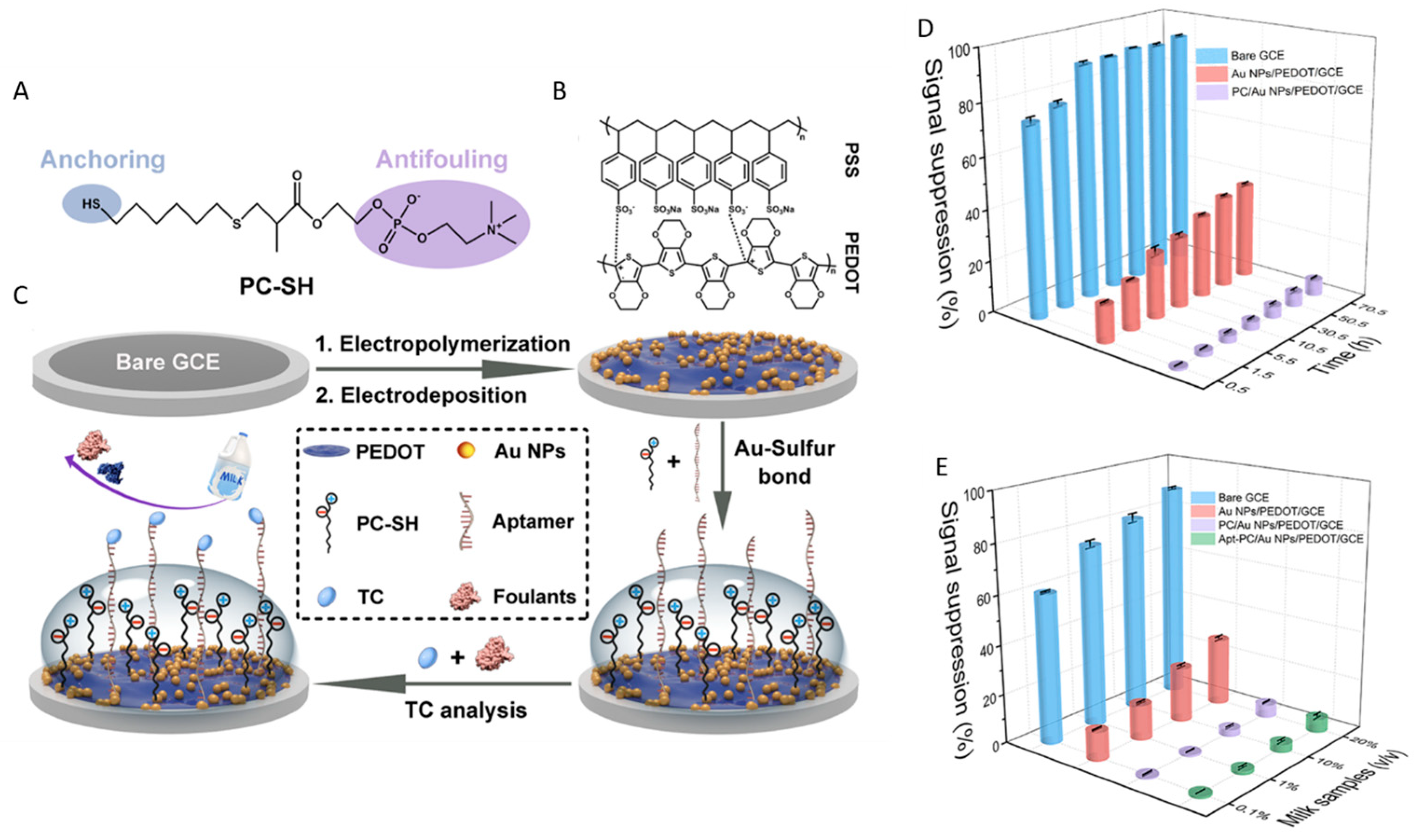
| Tetracycline (DPV) | Apt/PC-SH/Au NPs/PEDOT-PSS/GCE DPV | PC-SH | Milk diluted to 1% | Signal suppression: -CTRL, 30 min incubation with 10 mg/mL proteins:3.7% (BSA), 4.5% (Lys)and 6.2% (Hb) -CTRL, 20% milk, 30 min: 5.3% Biosensor, 1% milk, 70.5 h: 6.1% | Recovery: 96.9–107.6% LR: 0.05–100 ng mL−1 LOD: 8.8 pg mL−1 Agreement with HPLC (3 samples) | [42] |
| Oxytetracycline (DPV) | APT/α-lipoic acid/AuSPE | α-lipoic acid | Milk prepared from low-fat milk powder (1 mg/mL, centrifuged) | Similar sensitivities in milk and buffer | LR: 25–500 ng/mL DL: 14 ng/mL (buffer) DL: 10 ng/mL (milk) | [111] |
| IgG (DPV) | PolyA15-polyT5-Pep/AuNPs/PEDOT/GCE | PolyA15-polyT5-Pep | Human serum diluted at least 1000 times | Signal suppression: -Biosensor, undiluted serum: 8.5% (30 min) to 17% * (6 h) | Recovery 101.6–107.0% LR: 0.1 ng/mL−10 µg/mL DL: 0.037 ng/mL Agreement with immunoturbidimetry (5 samples) | [43] |
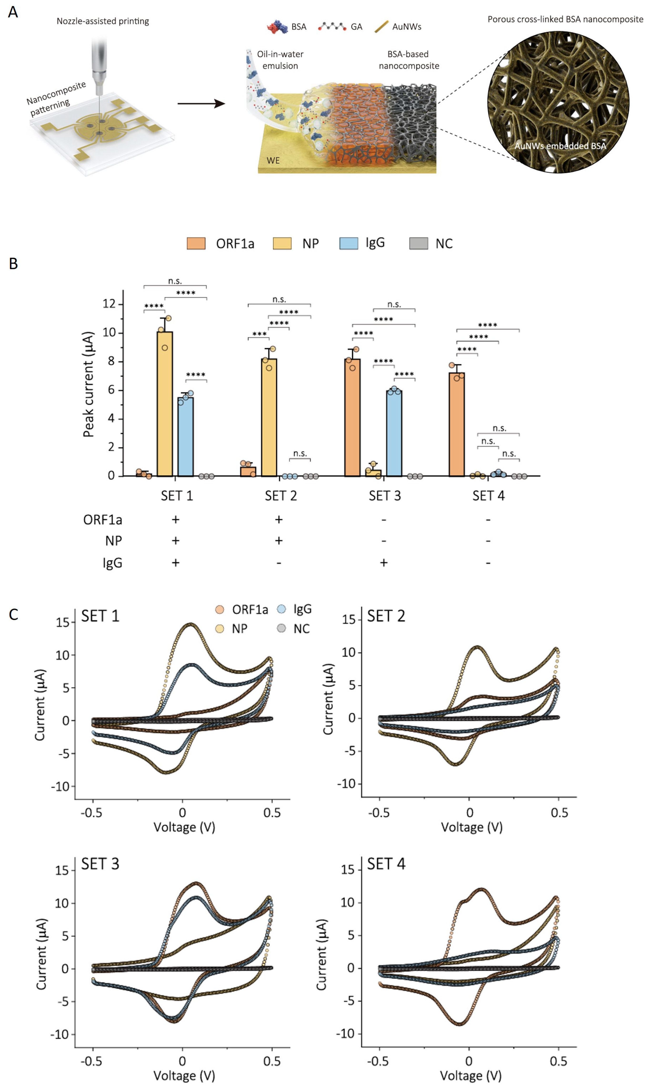
| SARS-CoV-2 ORF1a gene, NP and Ig G antibody (CV) | Array of electrodes coated with cBSA-AuNW; detection with SA-HRP, TMB (1) biotin-ssDNA/cPNA (ORF1a gene); (2) biotin-Ab2/NP/Ab1 (NP); (3) Ab2-biotin/IgG/S1 (IgG) | cBSA-AuNW | Individual sensors: RT-RPA, CRISPR-CAS12a mix (ORF1a) Dilution of NPS to 20% and of serum to 10% with 2.5% non-fat dry milk (for NP and IgG) Multiplexed detection: undiluted NPS spiked with IgG-containing serum (NP and IgG) and RT-RPA/CRISPR mix (ORF1a) | Changes of 1.8% to 2.7% in the EC signal after 1 month storage in serum, NPS and 1% BSA | DL: 0.22 copies/µL (ORF1a) DL: 1.9 pg/mL (NP) DL: 20.4 pg/mL (IgG) 60 NPS samples, 53 serum samples (for individual sensors); comparison with RT-qPCR and ELISA 4 sets of serum-spiked NPS (for multiplexed detection) | [82] |
| Interleukin 6 (CV) | SA-HRP/Ab/Il-6/Ab/cBSA-AuNW/Au | cBSA-AuNW | Unprocessed, undiluted human plasma | ~7% loss of sensitivity after 1 month in 1% BSA, human plasma or human serum -<12% decrease in current density after coating with cBSA-AuNW | DL: 23 pg/mL Stability of >1 month in unprocessed human plasma at 4 °C | [83] |
| Vancomycin, cortisol, L-phenylalanine (SWV) | E-AB/Au with mixed layer of MB-labeled aptamer and PC, MCH or MCO with/without coating with a hydrogel | PC-SH MCH MCO Zwitterionic polysulfobetaine hydrogel | Undiluted bovine serum | Stability during 7 days of operation: MCO > MCH Stability upon repetitive electrochemical scanning: PC > MCH | After 3 days in undiluted bovine serum at 37 °C the biosensors maintain their initial responses but the apparent affinity for the target analyte decreases (from Kd = 263 ± 74 µM to 546 ± 252 µM, for MCH-coated and from Kd = 361 ± 156 µM to 882 ± 262 µM for MCO-coated biosensors) | [21] |
| HER2 (DPV) | Pep/DSPE-PEG/sodium alginate doped PEDOT/GCE | Phospholipid bilayer and branched multifunctional peptide | Undiluted serum | Signal suppression: <6% after 2 h incubation at RT in undiluted blood, sweat, saliva, and milk, 20 mg/mL proteins (HSA, BSA, Mb), and small molecules (UA, DA, 5-HT) −17.7% (biosensor) and 18.4% CTRL) after 20 days in undiluted human serum | LR: 1.0 pg/mL−1.0 μg/mL DL: 0.24 pg/mL Agreement with ELISA (7 samples) | [93] |
| Doxorubicin, kanamycin (SWV) | MCH/MB-aptamer/Au PC/MB-aptamer/Au MCU/MB-aptamer/Au | PC-SH or MCH (doxorubicin) PC-SH or MCU (kanamycin) | Whole blood | Current drift in flowing blood over 12 h: <10% (PC-SH coated biosensors) compared to ~70% (MCH coated biosensors) and 50% (MCU-coated sensors) | In vitro measurements of doxorubicin and kanamycin in flowing blood Real-time continuous measurement of doxorubicin (Dox) in living animals | [91] |
| Kanamycin (CV, in vitro and SWV in vivo) | F50–C50 hydrogel /MCH/MB-aptamer/Au | MCH SAM and F50-C50 polyacrylamide hydrogel | -50% fetal bovine serum -Platelet-rich rat plasma -Undiluted human blood -Whole rat blood | -Reduced platelet adhesion compared to control PEG-coated or bare Au, after 3 days at RT, in stationary conditions or after 12 days in flowing blood −33.8 ± 4.9% decrease in signal over 200 min (hydrogel coated biosensor in vivo) and 64.2 ± 16.1% (PEG-coated biosensor) | In vitro and in vivo evaluation of F50-C50 hydrogel Forced degradation studies Real-time monitoring of kanamycin in living rats | [108] |
| RBD (DPV) | ACE2/cPep/AuNP/PEDOT/GCE | Cyclic peptide | Dilution to 25% blood | CTRL: -Very low adsorption after 1 day in E.coli 106 cfu/mL -Signal suppression: 3.1% (in 25% blood) to 6.1% (100% blood), 30 min incubation -9.76% (20 days, 25% blood) Biosensor: 85% of initial signal after 20 days in 25% blood | Recovery 95.1– 96.1% from spiked 25% blood LR: 0.1 pg/mL−100 ng/mL DL: 0.45 pg/mL Compared to ELISA (6 samples) | [75] |
| CA125 (DPV) | DNA aptamer-Pep P2-biotin/SA/biotin-Pep P1/AuNP/PEDOT/GCE | Antifouling peptide | Undiluted serum | Signal suppression: -15% after 30 min in undiluted human serum -<10% after 30 min in 100% sweat or 100% urine -9.6% after 48 h in 20% human serum | LR: 0.01–1000 U/mL DL: 0.003 U/mL Compared to ECL (5 samples) | [44] |
| MB-labeled DNA and SARS-CoV-2 pseudo virus (SWV and EIS) | SH-aptamer/NH2-aptamer/Zwitter-repel/Au electrode | Zwitter-repel polymers DMAPS75-Ald15-MAA-SH10 and DMAPS90-MAA-SH10 | Unprocessed human plasma; unfiltered 50% saliva | Change in the peak current after 1 h incubation with 10 mg/mL HSA: 5% (DMAPS75-Ald15-MAA-SH10) and 10% (DMAPS90-MAA-SH10) | DL: 21 nM MB-labeled DNA, in serum, by SWV (DMAPS75-Ald15-MAA-SH10) DL: 104 cp/ mL of SARS-CoV-2 pseudovirus in saliva by EIS (DMAPS90-MAA-SH10) Recoveries of 95.1–100.1% MB-labeled DNA spiked in undiluted human plasma (4 samples) | [112] |
| ERK2 (DPV) | Pep/AuNPs/PNE/PEDOT/GCE | Multifunctional peptide + PNE | Not specified | -Signal suppression after 30 min incubation in 100% serum: 7% (biosensor) and 14% (control); -No fluorescence after 30 min in 0.2 mg/mL FITC-BSA; <10% decrease in the initial signal after 26 days in 20% human serum | LR: 10 pg/mL–10 µg/mL DL3.97 pg/mL Agreement with ELISA (5 serum samples) | [45] |
| Rituximab (EIS) | AuNP-Ab/Rituximab/CN14 peptide/polyA20/Au | PolyA20 | Human plasma diluted to 10% | Lowest variation in the impedance after exposure to 10% plasma, compared to MCH and BSA coatings | LR: 0.1–50 µg/mL DL: 35.26 ng/mL Recovery: 99.1–108.3% agreement to ELISA (8 clinical plasma samples) | [113] |
| PSA (DPV) | Ab/Zwitterionic Pep hydrogel/AuNPs/PEDOT/GCE | Zwitterionic peptide hydrogel | Not specified | Signal suppression: <5% after 30 min in 20% human serum; <10% after 30 min in 100% human serum or 10 mg/mL LYZ, BSA, Mb; <10% after 5 h in 20% human serum | LR: 0.1–100 ng/mL LOD: 5.6 pg/mL Agreement with ELISA (5 serum samples) | [41] |
| Analyte | Biosensor Configuration | Antifouling Layer | Sample | Investigation of Antifouling Performance | Biosensor Performance | Reference |
|---|---|---|---|---|---|---|
| TSH | Ab/hydrogel/Au | Zwitterionic carboxybetaine hydrogel | Undiluted blood | Γ(adsorption levels) < 5 ng/cm2 from undiluted serum | 693 ng/cm2 loading capacity with TSH antibodies | [136] |
| miRNA | MCH/ssDNA-DNA tetrahedrons/Au; amplification (catalytic growth of AuNP) MCH/Capture DNA/Au | -DNA tetrahedrons/MCH -MCH | Serum and cancer cell lysates | After exposure to undiluted serum and plasma, 9.85 × 10⁸ red cell/mL, 5% whole blood, cell lysate, HSA (1 mg/mL and 48 mg/mL): Γ < 8 ng/cm2 (DNA tetrahedron/MCH) Γ = 41.5–150.3 ng/cm2 (capture DNA/MCH) | DL: 0.8 fM Agreement with qRT-PCR (3 samples) | [137] |
| miRNA | Copolymer coating/DNA probe | Copolymer of CBMAA and HPMAA, with 15% CBMAA. | Blood plasma | Better than simple CBMAA or OEG SAM | miRNA detection in whole blood plasma | [38] |
| BSA | antiBSA/HA/Au | HA | Undiluted milk Juice | Γ = 17 ng/cm2 (cow milk) Γ = 2.5 ng/cm2 (soybean milk) Γ ~ 60 ng/cm2 (undiluted 100% serum) | Loading capacity: 780 ng/cm2 of antiBSA | [80] |
| E. coli | Polymer brushes/Ab; AT-SAM/Ab | pCBAA pHEMA AT-SAM with carboxyl end groups, | Undiluted milk | pCBAA has the lowest Γ | DL: 6 × 10⁴ cells/mL for E. coli in undiluted milk | [52] |
| CRP and Fas 567 gene mRNA | CRP: Ab/polyT26-poliA15 /Au; physically adsorbed Ab/Au; Ab/MHDA-MCU/Au FAS567: polyA-DNA; NH2-DNA/MCU-MDHA/Au SH-DNA/Au | polyadenine | Undiluted serum | CRP: ΔR = 3.5% (Ab/ polyT26-poliA15) 4.3% * (Ab/MHDA-MCU) 7.1% * (physically adsorbed Ab) Fas 567: ΔR = 2.4% (polyA-DNA), 6% (SH-DNA and NH2-DNA/MHDA-MCU) CRP: | CRP: DL—2 ng/mL (oriented polyA); 11 ng/mL (non-oriented polyA); 7 ng/mL (covalent binding) 72 ng/mL (physical adsorption). Fas567: DL557 pM (polyA-DNA) 698 pM (MCU-MDHA, NH2-DNA) 863 pM (SH-DNA) | [138] |
| Lysozyme | Dual functional polymer PLL-mal-CEEEEE-APTA LYS | PLL-mal-CEEEE | Spiked milk, diluted to 10% Serum diluted to 10% | Biosensor: Γ = 26 ± 3 ng/cm2 (1 µg/mL Lys) 68.3 ± 0.2 ng/cm2 (10 µg/mL Lys) Γ = 7.9 ± 0.4 ng/cm2 (1.0 μg/mL BSA), 9.8 ± 0.4 ng/cm2 at 10.0 μg/mL Γ = 9 ± 4 ng cm2 (1.0 μg/mL Cyt C); 38 ± 5 ng cm2 (10.0 μg/mL CytC) Γ = 9 ± 1 ng/cm2 (1.0 μg/mL Mb), =9.0 ±0.8 ng/cm2 (10.0 μg/mL Mb) CTRL with control aptamer instead of Lys aptamer; CTRL—Γ = 5 ± 2 ng/cm2 (1.0 μg/mL Lys), 31 ± 1 ng/cm2 ( 10.0 μg/mL Lys) | DR: 0.13–20.0 μg/ mL DL: 0.04 μg/mL QL: 0.13 μg/mL | [139] |
| Wild-type, KRAS p.G12D- or KRAS p.G13D-mutatedDNA | SPRI, PLL-mal (26%)-PNA-CEEEEE polymer/Au, AuNP amplification | PLL-mal-CEEEEE | Plasma centrifuged at 10,800× g 10 min at 4° and diluted to 10% | 30 min in 10% human plasma, 10 min washing: Γ = 46 ± 34 ng/cm2 (PLL-mal(26%)-PNA-CEEEE); 213 ± 53 ng/cm2 (PLL-mal(26%)/EEEEE); 381 ± 31ng/cm2 (PLL-mal(26%)-PNA) 535 ± 17 ng/cm2(PLL) | DR (G12D-mutated DNA): 1.45−20.0 ng/mL RDL = 1.45 ng/mL | [140] |
| Anti-GT1b, anti-GM1, and anti-GA1 | SPRI, ganglioside coated array Ganglioside (GT1b, GM1 or GA1)/PFTDS/SiO2 (1–3 nm)/Au | Ganglioside/PFTDS | Undiluted serum | No specific fouling studies | LR: 1–100 ng/mL DL: 4.5 ng/mL (anti-GT1b), 5.6 ng/mL (anti-GM1), 6.6 ng/mL (anti-GA1) | [141] |
| Arginase 1 | Ab2/Arginase1Ab1/PPCB/AuB | PPCB | Blood plasma, undiluted and diluted 1:5 and 1:10 | Γ = 2.4 ng/cm2 after 30 min in human plasma diluted 1:10 | Detection of 12.5 and 50 nM Arginase 1 | [142] |
| Analyte | Analytical Method, Biosensor Configuration and Setup | Antifouling Layer | Investigation of Antifouling Performance | Operational Conditions | Biosensor Performance and Comparison with Standard Methods | Reference |
|---|---|---|---|---|---|---|
| DNA1: rpoB gene of Mycobacterium tuberculosis, DNA2: associated with E. coli * | EC-SPR WE: Au/Stem loop cDNA-MB/MCH | MCH | -Control MCH-coated interfaces -Tests with mismatched DNA | 1). AC amplitude: 50mV. Frequency: 100 Hz. plus Linear sweep from 0 to −450 mv 2). Cyclic voltammetry: −450 mV–0 V. 10 mV/s 3). EIS: DC:−275 mV, AC:50 mV, frequency 100 Hz | 1). DL: 5 nM (DNA1) and 10 nM (DNA2) Buffer and whole blood | [185] |
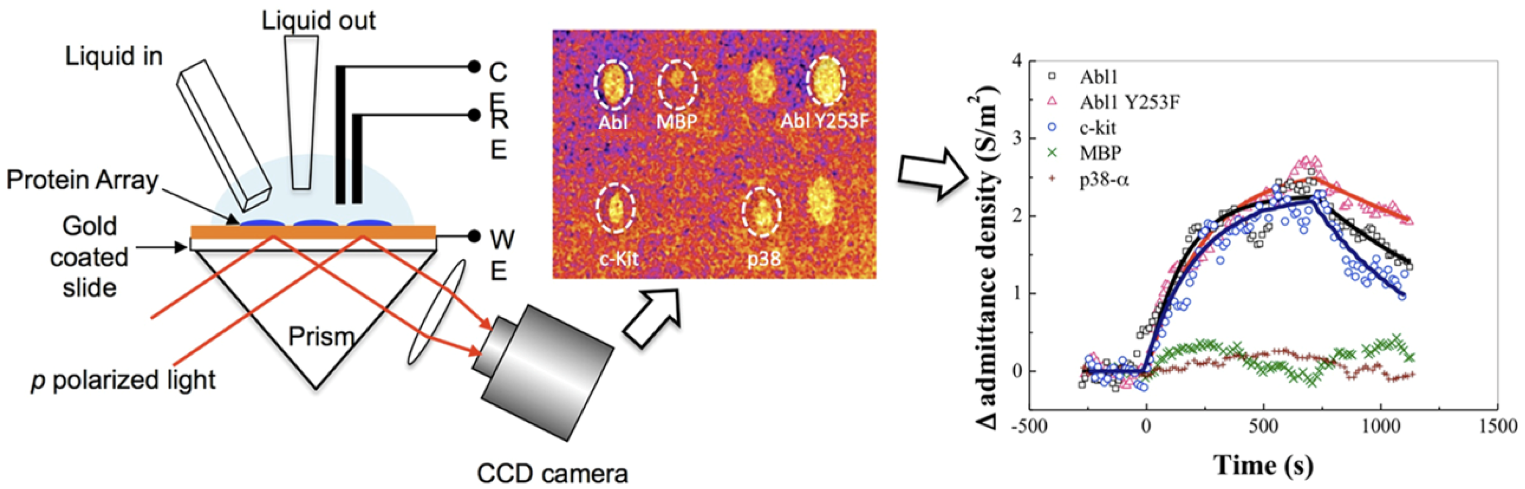
| Imatinib, SB202190 drugs | Protein microarray. P-EIM WE: Au/diSH-PEG-COOH:diSH-PEG-OH/protein | diSHPEG-COOH: diSH-PEG-OH | Positive and negative control proteins | AC amplitude: 250 mV. Frequency:10 Hz. 2.5 mM Tris buffer pH 7.5 + 1 mM MgCl2 | Binding affinities to proteins: Abl, Abl Y253F, p38-α, myelin basic protein, C-kit | [183] |
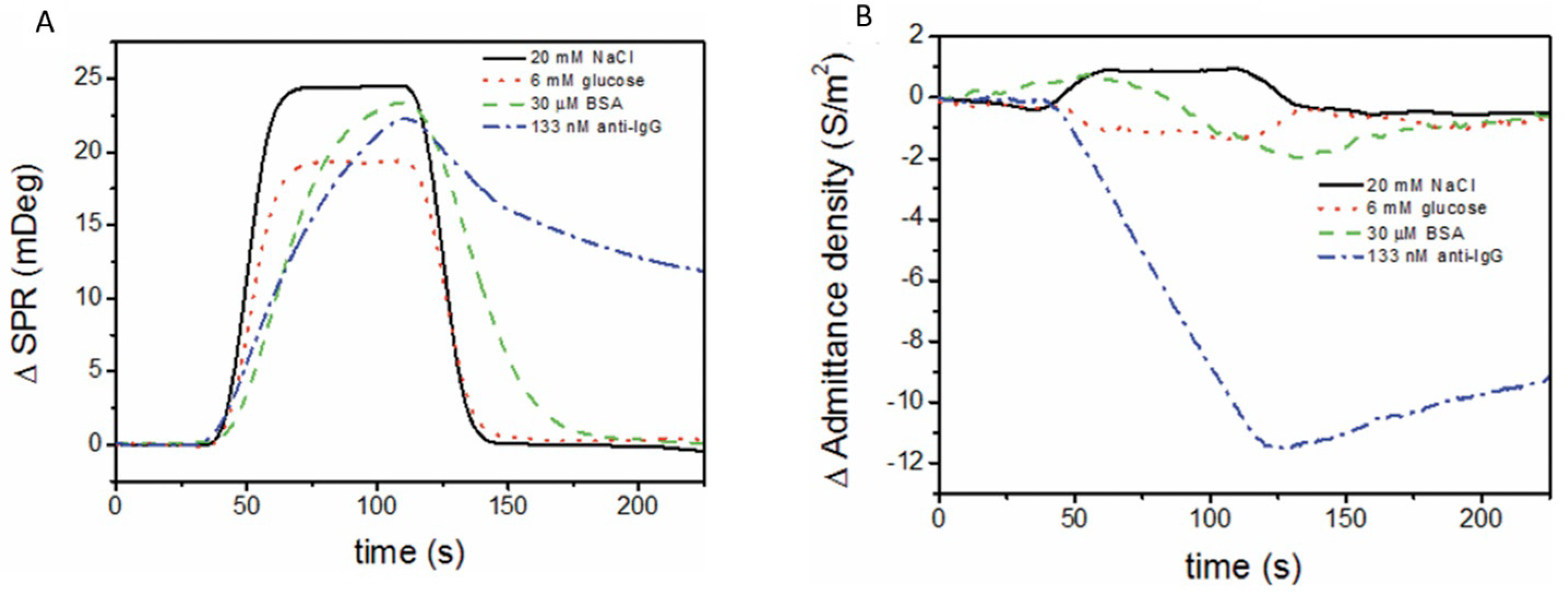
| AntiIgG | P-EIS WE: Au/MHA:MPO/IgG | MHA/MPO | BSA, NaCl, glucose | AC amplitude: 200 mV. DC bias: 120 mV. Frequency: 100 Hz, PBS | Anti IgG | [186] |
| Anti IgG | P-EIS WE: Au/cBSA/IgG | cBSA | Biotin, fetal serum diluted 1:100, biotin mixed with antiIgG | DC bias: 120 mV. AC amplitude: 200 mV. Frequency: 3.5 Hz; HBS buffer pH 7.4 with 0.005% P20 | Anti IgG | [25] |
| K+ | P-EIS WE: DNA/MCH | MCH | Na+, Mg2+ | AC amplitude: 400 mV. Frequency: 100 Hz; 10 mM Tris pH 7.4 with 250 mM NaCl | K+-induced G quadruplex assembly and pH effect on DNA charge | [187] |
| Anti IgG | SPR with in situ EC detection PDT/MPO /IgG, PC:DOPC cloaking membrane /HRP-anti IgG; TMB added in solution | Removable «cloaking» membrane over PDT/MPO | Tests with PDT/MPO control surfaces -SPR test with nanoparticle-conjugated anti-rabbit IgG spiked in donkey serum with/without cloaking membrane | 0.175 V (vs Ag/AgCl | Anti IgG DL < 5 fM | [27] |
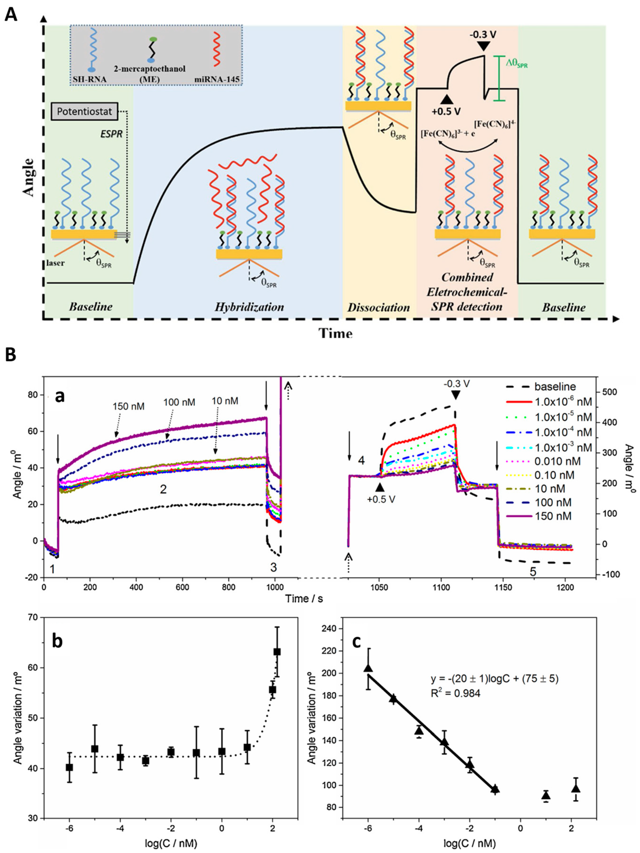
| miRNA-145 | EC-SPR Au/RNA/MCH(ME) Variation of the SPR angle after electrochemical deposition/desorption of [Fe(CN)6]3−/4− as redox probe | MCH or ME serum filtered through 10 kDa membrane and diluted 15 times | Hybridization buffer, non-complementary miRNA, Fe(CN)6]3−/4− | Electrochemical concentration/desorption of [Fe(CN)6]3−/4−: 1) 60 s deposition at +0.5 V; 2) LSV to −0.3 V at 50 mV/s. 3) 20s at −0.3 V | miRNA-145 in buffer and in synthetic human serum LR: 1 fM–0.1 nM DL: 0.56 fM | [182] |
| CA15−3 | EC-SPR WE: Au/MSA/anti CA 15–3 Variation of the SPR angle after SWV of [Fe(CN)6]3−/4− | MSA, MPA, MUA -Serum diluted 1:100 100 times in PBST BSA -Blocking with diluted serum | Diluted serum | SWV, −0.3 to 0.5 V [Fe(CN)6]3−/4−: | CA15−3 Human serum SPR—DL: 21 UmL−1 (SPR) DL: 0.098 UmL−1 (EC-SPR) LR: 0.10–250 U mL−1 | [188] |
| DNA of rpoB gene from Mycobacterium tuberculosis | EC-SPR WE: nanohole Au array /Stem loop cDNA-MB/MCH | MCH | Control (MCH) | DC potential: −275 mV. AC amplitude: 50 mV. Frequency: 100 Hz | Pathogenic DNA | [184] |
Disclaimer/Publisher’s Note: The statements, opinions and data contained in all publications are solely those of the individual author(s) and contributor(s) and not of MDPI and/or the editor(s). MDPI and/or the editor(s) disclaim responsibility for any injury to people or property resulting from any ideas, methods, instructions or products referred to in the content. |
© 2025 by the authors. Licensee MDPI, Basel, Switzerland. This article is an open access article distributed under the terms and conditions of the Creative Commons Attribution (CC BY) license (https://creativecommons.org/licenses/by/4.0/).
Share and Cite
Vasilescu, A.; Gáspár, S.; Gheorghiu, M.; Polonschii, C.; Banciu, R.M.; David, S.; Gheorghiu, E.; Marty, J.-L. Promising Solutions to Address the Non-Specific Adsorption in Biosensors Based on Coupled Electrochemical-Surface Plasmon Resonance Detection. Chemosensors 2025, 13, 92. https://doi.org/10.3390/chemosensors13030092
Vasilescu A, Gáspár S, Gheorghiu M, Polonschii C, Banciu RM, David S, Gheorghiu E, Marty J-L. Promising Solutions to Address the Non-Specific Adsorption in Biosensors Based on Coupled Electrochemical-Surface Plasmon Resonance Detection. Chemosensors. 2025; 13(3):92. https://doi.org/10.3390/chemosensors13030092
Chicago/Turabian StyleVasilescu, Alina, Szilveszter Gáspár, Mihaela Gheorghiu, Cristina Polonschii, Roberta Maria Banciu, Sorin David, Eugen Gheorghiu, and Jean-Louis Marty. 2025. "Promising Solutions to Address the Non-Specific Adsorption in Biosensors Based on Coupled Electrochemical-Surface Plasmon Resonance Detection" Chemosensors 13, no. 3: 92. https://doi.org/10.3390/chemosensors13030092
APA StyleVasilescu, A., Gáspár, S., Gheorghiu, M., Polonschii, C., Banciu, R. M., David, S., Gheorghiu, E., & Marty, J.-L. (2025). Promising Solutions to Address the Non-Specific Adsorption in Biosensors Based on Coupled Electrochemical-Surface Plasmon Resonance Detection. Chemosensors, 13(3), 92. https://doi.org/10.3390/chemosensors13030092