Review

Luminescent Pyrene-Derivatives for Hg2+ and Explosive Detection

1
National Research Institute of Chinese Medicine, Ministry of Health and Welfare, Taipei 11221, Taiwan
2
Department of Applied Chemistry, National Yang-Ming Chiao Tung University, Hsinchu 300, Taiwan
3
Department of Physics, AMET University, Chennai 603112, Tamil Nadu, India
4
Department of Chemistry, North Eastern Regional Institute of Science & Technology, Nirjuli, Itanagar 791109, Arunachal Pradesh, India
5
Crystal Growth Centre, Anna University, Chennai 600025, Tamil Nadu, India
6
Department of Biotechnology, Graphic Era (Deemed to be University), Dehradun 248002, Uttarakhand, India
7
Department of Chemical Engineering, Faculty of Engineering, Mahidol University, Nakhon Pathom 73170, Thailand
8
Centre for Nanoscience and Technology, MSGET, Pondicherry University (A Central University), Puducherry 605014, Tamil Nadu, India
9
Unit of Biomaterials Research, Department of Orthodontics, Saveetha Institute of Medical and Technical Sciences (SIMATS), Saveetha Dental College and Hospitals, Saveetha University, Chennai 600077, Tamil Nadu, India
*
Author to whom correspondence should be addressed.
Chemosensors 2025, 13(4), 145; https://doi.org/10.3390/chemosensors13040145
Submission received: 17 March 2025 / Revised: 10 April 2025 / Accepted: 11 April 2025 / Published: 14 April 2025

Abstract

:
Mercury and explosives are well-known hazards that affect the environment and threaten society. Mercury generally exists as inorganic mercuric (Hg2+) salts, and its detection via fluorometric response is highly notable. Likewise, mainstream explosives contains a nitro (−NO2) moiety as a functional unit, and numerous reports have quantified them using fluorescence quenching. Among the available literature, there are still noticeable concerns about the environmental and biological applicability of luminescent pyrene derivaives-tunedfluorometric detection of Hg2+ and explosives. In the presence of Hg2+ ions, pyrene derivatives tend to form excimers, which can be tuned to the chelation-enhanced fluorescence (CHEF), photo-induced electron transfer (PET), or fluorescence resonance energy transfer (FRET), etc., to exhibit “Turn-On” or “Turn-Off” fluorescence responses. On the other hand, π-π stacking of emissive pyrene-derivatives may lead to J- or H-type aggregation via self-excimers (Py-Py*), which has been found to be quenched/enhanced by explosive hazards. In fact, −NO2-containing explosives interact with pyrene derivatives, leading to exceptional fluorescence quenching or enhancement. This review details the use of pyrene derivatives toward the sensing of Hg2+ and explosives with demonstrated applications. Further, the design requirements, sensory mechanisms, advantages, limitations, and the future scope of using the reported pyrene derivatives in Hg2+ and explosives sensing are discussed.

1. Introduction

Environmental toxicity related to hazardous metal ions is a global threat that must be resolved with care [1,2,3]. Among common metal hazards, mercury detection is a serious concern requiring environmental remediation. Mercury can enter the environment in three ways: 1. volcano explosions into the air, forest fires, and the weathering of rocks and soils; 2. The burning of industrial fossil fuels, coals, and from industries that produce gold, thermometers, barometers, caustic soda, mercury lamps, etc.; and, finally, 3. ocean evaporation can re-introduce mercury into the environment [4,5,6,7]. The accumulation of mercury in the environment affects the living eco-system by entering into the daily food cycle, thus requiring more scrutiny [8,9,10]. At high concentrations, mercury can cause diseases such as brain damage, Minamata disorders, cognitive and motion disorders, central nervous syndromes, impaired infant development, cardiovascular diseases, anxiety depression, etc. [11,12,13,14,15]. Thus, the U.S. Environmental Protection Agency (EPA) has regulated the levels of mercury allowed in drinking water and fish tissue to 2 ppb (10 nM) and 3 ppm (1.5 µM), respectively [16,17]. Likewise, the allowable mercury concentration in normal soil is fixed at 625 ppb by the Agency for Toxic Substances and Disease Registry (ATSDR) of the U.S. Department of Health and Human Services [18,19]. To overcome the mercury toxicity in the eco-system, several detection and removal tactics were proposed, including organic/polymeric probes, nanoparticle-based colorimetric detection, instrumentation-based quantification, covalent/metal–organic frameworks (COFs/MOFs), and nanomaterials-based fluorescent sensors, etc. [20,21,22,23,24,25,26].
Like mercury’s environmental toxicity, explosive hazards threaten human life and society [27,28,29]. An explosion may cause damage to living systems and properties. The severity of damage caused by an explosion can be ordered as follows: (A) the death of living people, (B) diseases or loss of health, (C) damage to properties such as buildings, and (D) loss of activity, i.e., physical inactivity [30]. Even though explosives are stored in a non-exploding state, they can harm the environment and lead to numerous diseases in living species [31]. This might be due to the atomic composition present in the explosive materials. For example, most explosive materials consist of electron-deficient nitro groups, which destroy the natural pressure in the environment during an explosion [27,28,29,30,31]. However, for security purposes and to restrict terrorism, the use of explosives by the military cannot be avoided. Instead, while explosives are in the hands of terrorists, their rapid detection using suitable strategies is necessary. Therefore, numerous methods, including COFs/MOFs, luminescent nanomaterials, electrochemical detection, colorimetric assay, fluorescent organic probes, carbon dots, fluorescent semiconductor quantum dots, perovskite nanomaterials, etc., were developed for explosive detection with various applications [32,33,34,35,36,37,38,39,40,41]. Among these, the luminescent organic probe-based detection of nitro-explosives seems to be particularly efficient. In general, the strong fluorescence of organic probes is quenched (static/dynamic) by the interactive nitro groups in explosives, which alter the π-π stacking of the probe, or by the supramolecular interactions of explosive materials with the probe [42,43,44]. In the same vein, through similar mechanistic approaches, organic probes for the fluorescent “Turn-On” detection of explosives are also available; however, it should be noted that reports on these approaches are rare [45,46].
In the design of fluorescent organic probes to be applied in Hg2+ and explosives detection, pyrene-based derivatives were established as extraordinary innovations in terms of their fluorescent properties and real-time application [47,48]. In fact, due to the π-π-stacking and highly emissive excimer-forming abilities of pyrene moieties, their derivatives display exceptional fluorescence properties and are applied in optoelectronic devices, sensor bioimaging, etc. [49,50,51]. By tuning the π-π stacking of pyrene derivatives, its aggregation/self-assembly features can be enhanced toward in vitro/in vivo theranostic applications [52]. In this way, numerous reports on the aggregation-induced emission enhancement (AIEE) of pyrene derivatives with certain applications are available [53], allowing researchers to design new pyrene-based probes for Hg2+ and explosive detection. Among them, a few reports reveal π-π stacking variations and H-aggregation (head-to-head) and J-aggregation (head-to-tail) during sensing [54,55,56,57,58,59,60]. The detection of Hg2+ by pyrene-based probes was achieved by either “Turn-On” or “Turn-Off” fluorescence responses by optimizing the solvent and probe’s design. Likewise, several highly emissive pyrene-based probes (in certain solvent conditions) can detect explosives via a “Turn-Off” fluorescence response via π-π stacking and supramolecular interactions. In contrast, only one report on a hybrid pyrene-based probe is available with a “Turn-On” fluorescent response to explosives; thus, this should be noted as a rare case (see Figure 1). Until now, no review has described the use of pyrene-based probes in the selective detection of Hg2+ and explosives with mechanistic details, which allows us to deliver this review.
In this review, the use of luminescent pyrene-based probes to detect Hg2+ ions and nitro-explosives is described with mechanistic evidence. Figure 1 briefly outlines the use of weakly/strongly emissive pyrene derivatives in detecting Hg2+ ions and explosives via photoluminescence (PL) “Turn-On” and “Turn-Off” fluorescence responses directed towards applications. Finally, requirements for the probe’s design, merits, limitations, and perspective points are debated for the readers.

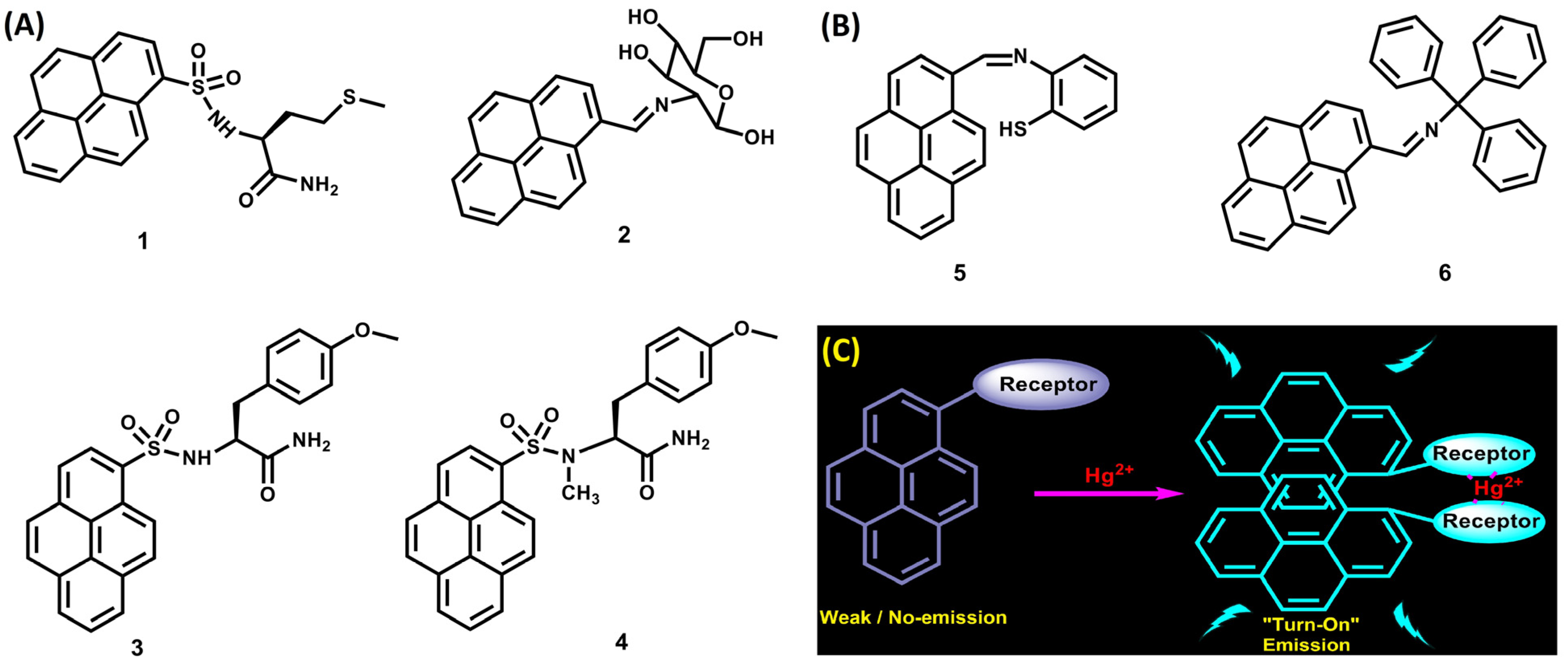
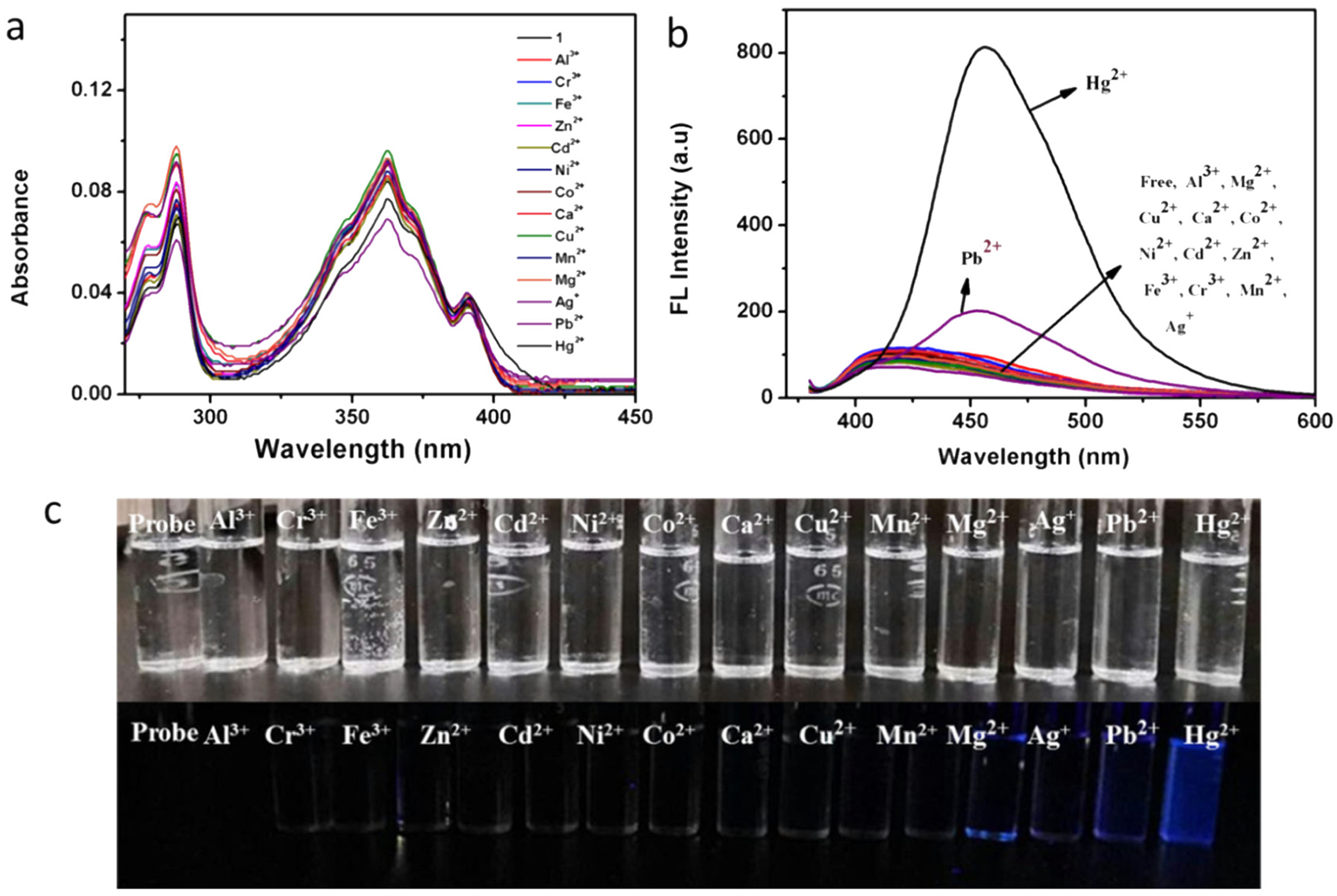
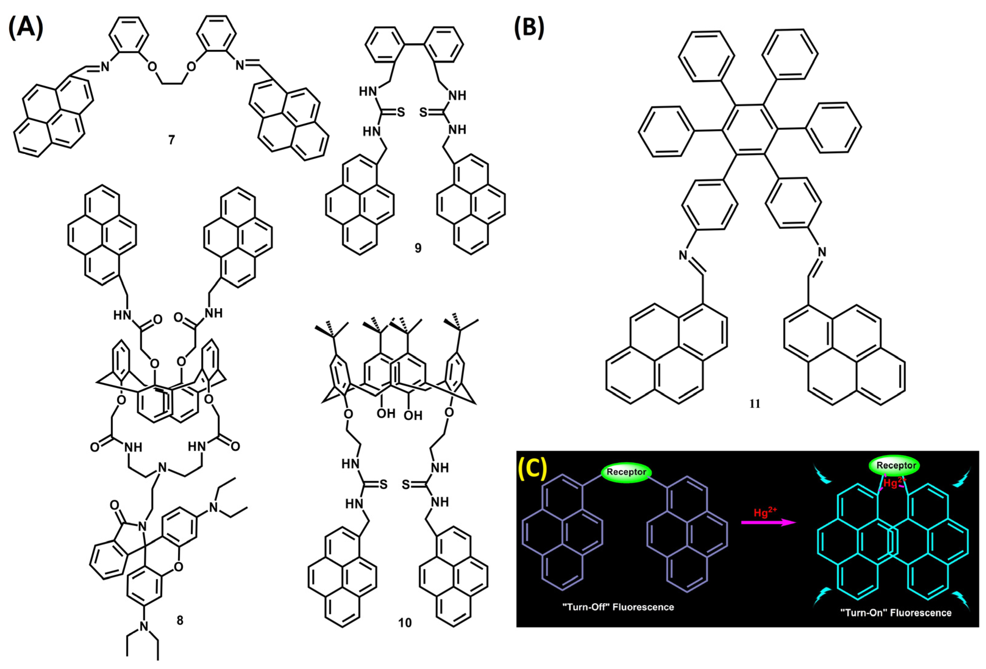
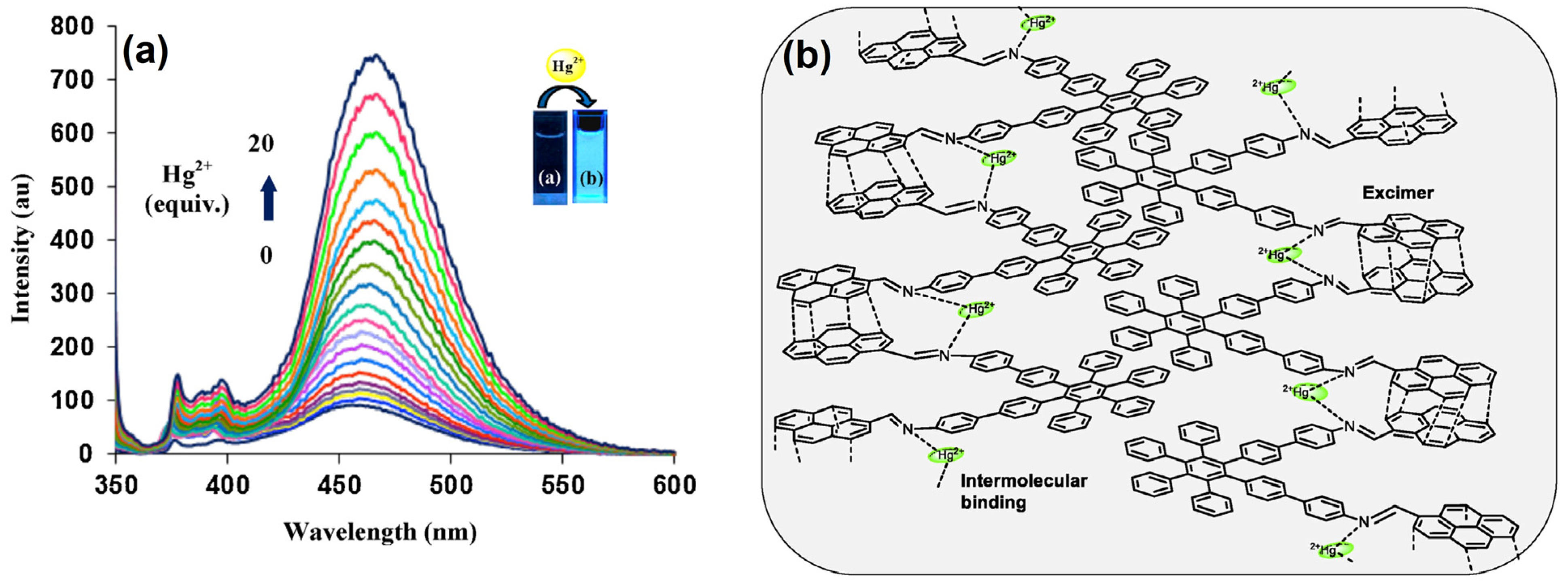
2. Pyrene Derivatives for Hg2+ Detection via PL for “Turn On” Responses

2.1. Excimer Facilitated Hg2+ Detection

Yang et al. developed a simple pyrene and amino acid-conjugated probe 1 (See Figure 2A) via multi-step synthesis and employed it for PL “Turn-On” detection via excimer formation [61]. Probe 1 undergoes 2:1 excimer formation to induce the PL “Turn-On” response. In the presence of Hg2+, the excimer emission at 480 nm was enhanced with a simultaneous decrease at 383 nm. Linear regression was witnessed between 0 and 750 nM with an LOD of 57.2 nM (nM = nanomole (10−9 M)). The association constant (Ka) of 1 to Hg2+ was estimated as 7.84 × 10¹² M−2 (M = Mole), which defines the probe’s high sensitivity to Hg2+ ions. Though this work looks impressive, it lacks real-world applications. Further, the design and development of such probes require careful optimization of all the intermediate synthetic steps and critical analysis. However, this work has driven researchers to invent numerous sensory probes, as detailed below.
Areti et al. synthesized the pyrene–glucose conjugate 2 (See Figure 2A) using a simple Schiff base condensation between glucosamine and 1-pyrenecarboxaldehyde and engaged it in the ratiometric PL detection of Hg2+ ions [62]. The emission at 398 nm was quenched with a 32-fold enhancement at 500 nm. The formation of a 2-Hg2+-2 excimer complex was clarified from the mass data with a proposed LOD of 45 ± 5 nM. Further, the excimer complex displayed reversibility with tetrabutylammonium fluoride (TBAF), which extended up to five cycles. This work looks impressive in terms of the mechanism and LOD. However, it lacks linear regression and real-time demonstration.
Following on from the previous report [61], Thirupathi and co-workers developed two similar pyrene and amino acid conjugates, 3 and 4 (see Figure 2A), with slight modifications and employed them in the discrimination of Hg2+ ions via 2:1 excimer formation [63]. Both probes have a linearity between 0 and 500 nM with LODs of 22.2 nM (3) and 44 nM (4). Further, the association constants of 3-Hg2+ and 4-Hg2+ were estimated as 5.72 × 10¹³ M−2 and 1.15 × 10¹³ M−2, respectively. Since this work was a follow-up work and lacks real applications, it cannot be labelled as a unique report. Shellaiah et al. described the use of the aggregation-induced emission (AIE) active pyrene-based probe 5 (See Figure 2B) for the PL “Turn-On” detection of Hg2+ ions via 2:1 excimer complex formation and a photoinduced electron transfer (PET) mechanism [64]. Probe 5 was synthesized by refluxing 1-pyrenecarboxaldehyde and 2-Aminothiol in ethanol and show “AIE” in the presence of water (H2O). The linear regression of 5 to Hg2+ was estimated as 0–60 µM (µM = micromole (10−6 M)) with an LOD of 2.82 µM and a Ka value of 7.36 × 10⁴ M−1. The sensitivity of 5 to Hg2+ and the 2:1 complex was defined by time-resolved photoluminescence (TRPL), fluorescence quantum yield (ΦF; varied from 0.035 to 0.289), and mass data. HeLa cellular imaging interrogations attested to the Hg2+ detection. Later, the authors reported the OTFT-based detection of Hg2+ with a nanomolar LOD using probe 5 [47]. However, additional studies are still mandatory due to the solution’s LOD at the micromolar level. Using a Schiff base reaction, Wu et al. constructed the “AIE” active probe 6 (See Figure 2B) and demonstrated its Hg2+ detection via 2:1 excimer and PET-tuned PL “Turn-On” response [65]. Figure 2C schematically defines the 2:1 excimer complex formation driven by pyrene probes 16. With the incremental addition of water, 6 displays an “AIE” and detects Hg2+ in H2O:DMF (v:v = 2:3, PBS buffer, pH 7.0; PBS = phosphate-buffered saline, and DMF = dimethylformamide). As seen in Figure 3a–c, in the presence of Hg2+, the emission of 6 at 450 nm was enhanced compared to that of other ions. The linear detection range of Hg2+ lies between 0 and 20 µM with an LOD of 0.42 µM and a Ka value of 3.62 × 10⁴ M−1. Spiked real water recoveries were found to be between 89.0 and 94.5%. HeLa cellular imaging studies supported this research, hence it can be noted as a unique work. However, in-depth studies on energy band gaps, ΦF, and π-π stacking models are still in demand.
Similar to 2:1 complex-tuned excimer formation, a few pyrene-based conjugates display sensitivity to Hg2+ ions via 1:1 and 1:2 (probe to metal) complex formation. Figure 4A,B represent the structures of the pyrene-based probes described in this review. The 1:1 excimer complex construction during Hg2+ binding is schematically depicted in Figure 3c. Zhou et al. synthesized the azadiene–pyrene probe 7 and utilized it to sense Hg2+ via a 1:1 excimer mechanism [66]. The one pot-synthesized Schiff base probe showed a PL “Turn-On” response at 462 nm at 365 nm excitation in HEPES-CH3CN (80:20, v/v; HEPES = 4-(2-hydroxyethyl)-1-piperazineethanesulfonic acid and CH3CN = Acetonitrile). The fluorescence emission enhancement was visualized between 0.1 and 10 µM. From nonlinear curve fitting, the Ka value was established as 4.32 × 10⁵ M−1 with a LOD of 0.2 µM. The 1:1 excimer complex was recognized from 1H NMR (NMR = Nuclear Magnetic Resonance) titration. This work appears to be preliminary, not in line with the current state of the art, and requires further investigation to assess the real-world utility of this strategy. Lee et al. developed a series of pyrene-based calix[4]arene compounds; among them, probe 8 displays Hg2+ selectivity via 1:2 (probe to Hg2+) complex formation, disturbance of the initial pyrene–pyrene excimer, and a fluorescence resonance energy transfer (FRET) mechanism [67]. Here, the pyrene excimer emission at 470 nm was diminished with ratiometric PL enhancement at 576 nm (at 343 nm excitation). An FRET between pyrene and rhodamine was inhibited due to the excimer formation, which became a “Turn-On” response during Hg2+ binding via the rhodamine ring opening. The ratiometric response (I576/I470) starts from 5 µM onwards, which is labelled as an LOD. This work was carried out in CHCl3/CH3CN (50:50, v/v; CHCl3 = chloroform), which raises the question of its actual applicability. Wang and co-workers constructed the pyrene–thiourea conjugate probe 9 to detect Hg2+ ions via a 1:1 complex-tuned excimer formation [68]. The monomer emission of 9 at 381 nm diminished while binding with Hg2+ with an enhanced excimer peak at 490 nm (22-fold). In this study, the linear regression lies between 0 and 1 µM, with an LOD of 0.74 µM and a Ka value of 1.34 × 10⁴ M−1. Cellular imaging, C. elegans fluorescence images, and Zebra fish imaging interrogations defined the in vitro and in vivo applications of 9. This is an impressive report, but it should be extended with details of material applications.
Rodr’ıguez-Lavado et al. produced the calix[4]arene 10, consisting of pyrene and thiourea conjugation for detecting Hg2+ and Ag+ ions [69]. The monomer emission at 377 and 395 nm decreased with ratiometric PL enhancement at 477 nm due to the metal complex stimulating excimer formation. The linear regression of 10 to Hg2+ ranged between 0 and 1 µM with an LOD of 8.11 nM. The apparent association constant “Kapp” value for 10-Hg2+ was estimated to be 10.64. Though the application of logic gate supports this research, this probe shows more sensitivity to Ag+ ions. Thus, this can be considered a supplementary report, and its applicability is still questionable. Pramanik et al. used the hexaphenylbenzene–pyrene derivative 11 to detect Hg2+ via 1:1 a complex-tuned excimer formation [70]. Probe 11 shows an “AIE” with an increasing H2O percentile. Next, during the Hg2+ detection, the emission of 11F = 0.076; at 456 nm, λex = 342 nm) displayed a 10-fold enhancement at 468 nm (ΦF = 0.74). The linear detection of Hg2+ lies between 0 and 100 µM with an LOD of 4.5 nM. Figure 5a,b show the 1:1 (11-Hg2+) complex-tuned excimer formation and emission enhancement. The 11-Hg2+ was reversible by adding ethylenediamine tetraacetic acid (EDTA) and iodide (I) anions. A supramolecular assembly has been witnessed when using 11-Hg2+ to detect picric acid, as described in Section 4.1. This report used the strip method, but it requires further development for real-world applications.
Tu¨may et al. developed the pyrene–cyclotriphosphazene derivatives ‘1214’ and engaged them in the quantification of Hg2+ via 1:1, 1:2, and 1:3 complex-induced excimers and the “CHEF” mechanism [71]. Figure 6a,b display the structure of 1214 and their 1:1, 1:2, and 1:3 complex-promoted excimer formation.
The linear regression of 12-Hg2+, 13-Hg2+, and 14-Hg2+ ensembles at 450 nm (λex = 365 nm) in ACN (ACN = CH3CN) were estimated as 0–50 µM, 0–25 µM, and 0–20 µM with LODs of 0.223 µM, 0.114 µM, and 0.050 µM, correspondingly. Subsequently, the ΦF values of 12, 13, and 14 (0.10, 0.12, and 0.15) were also enhanced for 12-Hg2+, 13-Hg2+, and 14-Hg2+ (0.74, 0.76, and 0.79). The Job’s plot and 1H-NMR titrations confirm the 1:1, 1:2, and 1:3 complexes of 12-Hg2+, 13-Hg2+, and 14-Hg2+. HeLa cellular imaging investigations attested to the applicability of this report. However, the synthetic complications may raise questions about the cost effectiveness of this research. Using CH3CN for sensor applications could still be evaluated using the appropriate aquatic solvents.

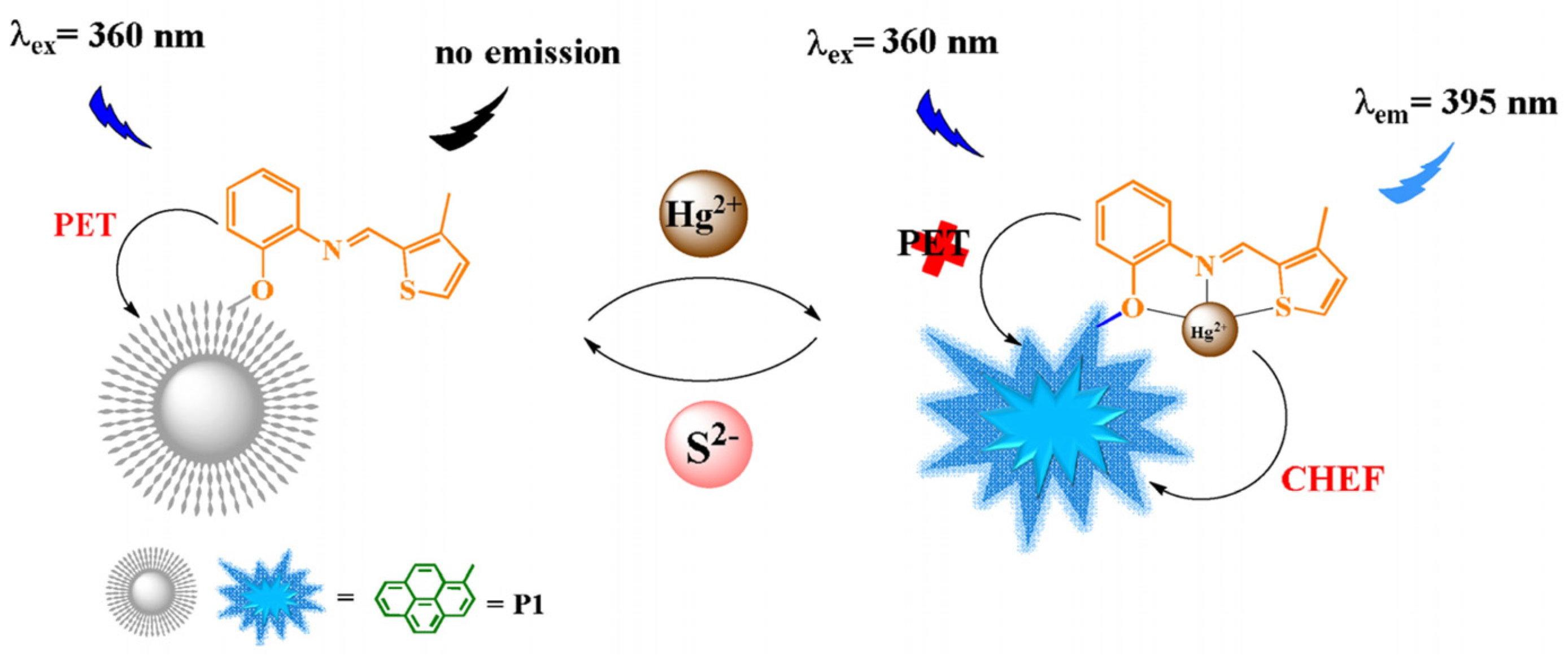
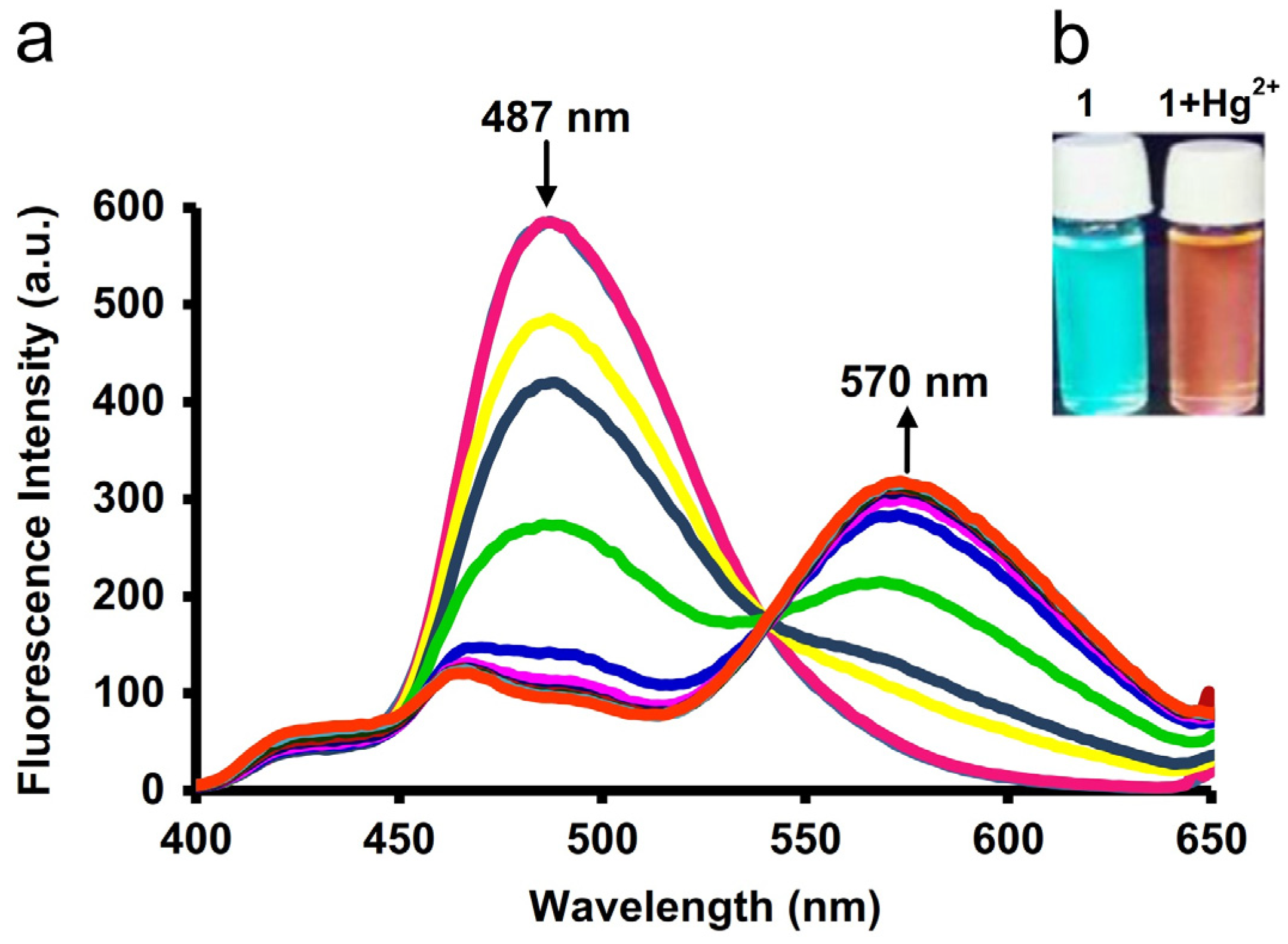
2.2. CHEF-Tuned Hg2+ Detection

A pyrene–NBD (NBD = 7-nitrobenzo-2-oxa-1,3-diazolyl) cyclam probe 15, pyrene–thiosemicarbazone derivative 16, pyrene-conjugated Schiff base derivatives 1719, phenanthro[4,5-fgh]pyrido[2,3-b]quinoxaline 20, and twisted (pyrene)2–pyridine Schiff base derivative 21 were reported for the “CHEF”-promoted PL “Turn-On” sensing of Hg2+ ions [72,73,74,75,76,77,78]. Figure 7 shows the structures of analogues 1521.
Probe 15 displays an Off–On PL response to Hg2+ and an On–Off PL response to Cu2+ at 538 nm [72]. The estimated LOD of Hg2+ by 15 was 7.9 µM with a Ka value of 2.5 × 10⁴ M from nonlinear curve fitting. This preliminary report supports several sensory reports but lacks a discussion of interference, practical applications, discrimination between Cu2+ and Hg2+, and mechanisms. Pyrene–thiosemicarbazone derivative 16 witnesses a PL “Turn-On” response to Hg2+ via the “CHEF” mechanism [73]. The semilogarithmic plot defined its LOD as 3.98 µM. This report lacks the use of interferences, applications, and in-depth discussion. Thus, the findings are still in question. The pyrene-based Schiff base 17 shows 12-fold PL enhancement (at 472 nm; λex = 365 nm) in the presence of Hg2+ ions via a 1:1 “CHEF” complex [74]. The 17-Hg2+ base has a linearity from 0.1 to 10 µM with an LOD of 22 nM and a Ka value of 1.02 × 10⁴ M−1. Candida albicans cellular imaging clarified the Hg2+ sensor. However, this work is lacking in interference studies; thus, additional interrogations are still mandatory. A “CHEF”-promoted PL “Turn-On” detection of Hg2+ was described using pyrene-based Schiff base probe 18 [75]. Upon adding Hg2+, probe 18 exhibits a PL enhancement at 395 nm (λex = 345 nm) in water–DMSO (1:2 v/v, pH:8.0; DMSO = Dimethylsulfoxide). This might be attributed to the “CHEF” mechanism, as seen in Figure 8.
The linear range lies between 0.008 and 38 μM with an LOD of 8.32 nM and a Ka value of 1.05 × 10⁵ M−1 (1:1 complex). The ΦF value of 18-Hg2+ was estimated to be 0.71 (70-fold enhancement) and shows reversibility in the presence of S2− ions (See Figure 8). Notably, the sensing ability of 18 was well defined by its application on test papers, food, and environmental samples. Thus, this can be considered an impressive work. Bai et al. used the pyrene-based Schiff base 19 to measure Hg2+ ions [76]. At 470 nm excitation, 19-Hg2+ in HEPES/ACN (10 mM, pH = 7.4, 3:7 (v/v)) revealed a PL “Turn-On” peak at 607 nm through a 1:2 “CHEF”-complex. The linear regression ranged from 0 to 21 µM with an estimated LOD of 36 nM. The 19-Hg2+ shows reversibility while adding I via the formation of HgI2. This work lacks real-world applications (except preliminary test strip studies); hence, further investigations are essential. During the sensing of Hg2+ ions, a ratiometric fluorescence shift was witnessed by the pyrene-based probe 20 [77]. As explored in Figure 9, the initial emission peak at 487 nm red-shifted to 570 nm in the presence of Hg2+, which might be assigned to excimer, “CHEF”, and Intramolecular charge transfer (ICT) mechanisms.
The LOD of Hg2+ by 20 was stated to be 1.31 µM, and 20-Hg2+ (1:1 complex) was found to be reversible with cysteine. Test strips were used to demonstrate its applicability. However, sensor studies in tetrahydrofuran (THF) may affect real applications. Thus, this could be counted as a supplementary report. Twisted (pyrene)2–pyridine Schiff base 21F = 0.31; λex = 368 nm and λem = 408 and 430 nm) shows selectivity to both Hg2+ and Pb2+ ions via 1:1 “CHEF” complexation [78]. In the presence of Hg2+, the emission at 460 nm is enhanced, with a ΦF value of 0.75. The limit of detection (LOD) for Hg2+ was established at 12.0 μM, demonstrating considerable sensitivity. This work lacks information on the discrimination between Hg2+ and Pb2+, hence it can be counted as a supplementary report.

2.3. Pyrene Conjugates for Reaction-Based Detection of Hg2+

Pyrene-attached rhodamine conjugates (2224), pyrene–hydrazone 25, and pyrene–bissulfanes (2628) were described for the reaction-based detection of Hg2+ ions [79,80,81,82,83]. Figure 10 displays the structures of probes 2228.
Pyrene–rhodamine 6G conjugate 22 undergoes a ring-opening reaction with Hg2+ ions, which induces FRET between the pyrene and rhodamine units to trigger an emission “Turn-On” at 550 nm [79]. Probe 22 at 0.5 µM in ACN displays excellent selectivity for Hg2+ and is reversible with EDTA. However, the emission enhancement of 22-Hg2+ was saturated at 34 µM. The proposed binding stoichiometry is 1:1 and 2:1, but further clarification and evidence are needed. This work emphasizes FRET but lacks linear regression analysis and real applications; hence, it is considered as a supplementary report. Two pyrene–rhodamine B probes (23 and 24) were synthesized and applied in a reaction-based Hg2+ sensing application (in ethanol: water (1:1)) [80]. For Hg2+, probe 23 exhibited fluorescence enhancement at 452 and 576 nm (λex = 365 nm) with an estimated LOD of 19.1 µM and a quantification limit (LOQ) of 63.7 µM. In contrast, probe 24 shows an excellent response to Hg2+ ions at 582 nm (λex = 365 nm) with an LOD and LOQ of 0.43 and 1.45 µM, respectively. Both probes show a greater sensitivity to Hg2+ due to the inhibition of initial FRET. The mass data also confirms 1:1 stoichiometry and the formation of Rhodamine B, thus clarifying the reaction-based mechanism. Figure 11a,b schematically represent the sensing mechanism of 23-Hg2+ and 24-Hg2+. This work must be updated using real sample applications.
Refluxing the 1-pyrenecarboxaldehyde and hydrazine afforded probe 25, which undergoes hydrolysis to detect Hg2+ ions in CH3CN–CH2Cl2 (1:1, v/v; CH2Cl2 = dichloromethane) [81]. The Hg2+ induces the hydrolysis of 25 to yield 1-pyrenecarboxaldehyde at 440 nm, confirmed through NMR titration. It has been stated that 0.5 equivalent of Hg2+ was found to be enough to cause the hydrolysis. This work lacks details on linear regression, LOD, and real applications. Thus, further investigations are still mandatory for to support this technique.
Due to the high affinity of Hg2+ for S, pyrene-based derivatives with bisethylsulfane (26) and thioacetal units consisting of carboxyl and hydroxyl groups (27 and 28) were designed as chemodosimetric probes for the detection of Hg2+ ions [82,83]. The bisethylsulfane units of probe 26F = 0.01) interact with Hg2+ to afford the chemodosimetric response at 455 nm (ΦF = 0.27; λex = 365 nm) [82]. This might be attributed to the formation of 1-pyrenecarboxaldehyde, as confirmed by NMR titration. A stable response was seen in the pH range of 4–10. A linear PL response was witnessed from 0 to 30 µM with an LOD of 55 nM. This work was carried out using RAW 264.7 cellular and zebra imaging studies; thus, it can be regarded as innovative research. However, further investigations in environmental samples still need to be performed. By using a similar interactive reaction-based mechanism, the probes 27 and 28 in ethanol/PBS (pH 7.4; 2:1 (v/v)) revealed PL “Turn-On” responses at 457 nm (λex = 395 nm) [83]. Figure 12a,b depict the chemodosimetric sensory response of 28 to Hg2+ ions.
The stoichiometric coordination of 27-Hg2+ and 28-Hg2+ was established at 1:1 and 1:2 ratios, respectively. The linear regression of 27-Hg2+ and 28-Hg2+ was found to be 0–3.5 µM and 0–12 µM with corresponding LODs of 1.49 nM and 1.03 nM. Probes 27 and 28 show an excellent sensitivity to Hg2+ in a direct buffer solution, with linearity ranging from 0–0.2 µM to 0–0.25 µM, and impressive limits of detection (LODs) of 1.74 nM and 1.53 nM. The in vitro imaging ability of 27 and 28 for Hg2+ was defined in A549 cell lines. In addition, real water analysis showed >90% recovery with RSD (RSD = Relative Standard Deviation) values < 1%. This work is an impressive one, and should be extended toward commercialization. Table 1 summarizes the pyrene-based probes utilized in the PL “Turn-On” detection of Hg2+, their linear ranges, LODs, and applications.

2.4. Critical Analysis of PL “Turn-On” Detection of Hg2+

Pyrene-based conjugates deliver PL “Turn-On” responses to Hg2+ via excimer formation, PET, CHEF, and the chemodosimeter mechanism [61,62,63,64,65,66,67,68,69,70,71,72,73,74,75,76,77,78,79,80,81,82,83], as discussed in the previous sections. Those reports still have certain limitations, as follows: (A) in the case of the excimer complex-tuned mechanism, the reports are still lacking a discussion of the PET and FRET mechanisms [61,62,63,64,65,66,67,68,69,70,71], which require critical in-depth interrogations; (B) for pyrene–rhodamine conjugate probes [67,79,80], the FRET mechanism was inconsistently recorded, which must be addressed via critical examinations; (C) reports describing the “CHEF” mechanism [72,73,74,75,76,77,78] are lacking theoretical support, which requires critical inquiries using DFT (Density Functional Theory); (D) the majority of pyrene-based probes [61,62,63,64,65,66,67,68,69,70,71,72,73,74,75,76,77,78,79,80,81,82,83] lack information on “AIE” and solvent effects on real-time sensory responses, and this requires critical analysis to address aquatic sensor studies; (E) the extensive stability of probes and their proposed sensory complexes under temperature, pressure, diverse solvent conditions, and distinct environmental conditions require integrative critical examinations.

3. Pyrene Derivatives for PL “Turn-Off” Detection of Hg2+

Pyrene derivatives with initial emissive properties undergo PL quenching while interacting with Hg2+ ions. Figure 13 represents the pyrene derivatives that detect Hg2+ via a 1:1 complex-tuned PL “Turn-Off” response [84,85,86,87,88].
Wu’s research group conjugated pyrene and ribose through triazole linkage to afford probes 29 and 30 and applied them to quantify Hg2+ ions [84]. These probes possess a strong emission at 478 nm (λex = 343 nm), and while titrating with metal ions in CH2Cl2/CH3OH (8:2, v/v; CH3OH = Methanol), with both showing a high sensitivity to Hg2+ via PL “Turn-Off” response. Probes 29 and 30 form a 1:1 ligand-to-metal complex via triazole units, inhibiting pyrene excimer formation, as shown in Figure 14. From Stern–Volmer quenching plots, the LODs of Hg2+ by 29 and 30 were estimated as 10 µM and 15 µM with Ka values of 1.73 × 10⁵ M−1 and 4.44 × 10⁵ M−1. The DFT and NMR titrations support the proposed binding mechanism. This work lacks suitable solvents and applications, which require further research. Following a similar 1:1 complexation and binding mechanism, three pyrene-based probes (3133) with triazole units were developed to detect Hg2+ ions [85]. In the presence of Hg2+, probes 31, 32, and 33 display fluorescence quenching at 474 nm (λex = 343 nm) via the 1:1 binding of triazole, which inhibits the pyrene–pyrene* (Py-Py*) excimer formation. The LODs of 31-Hg2+, 32-Hg2+, and 33-Hg2+ were defined as 1.74, 2.42, and 3.83 µM with Ka values of 1.68 × 10³ M−1, 1.57 × 10³ M−1, and 1.52 × 10³ M−1, respectively. Imaging of 31-Hg2+, 32-Hg2+, and 33-Hg2+ was performed on RAW 264.7 cell lines. This work still requires real sample analysis.
Banerjee and co-workers proposed using the methionine–pyrene hybrid probe 34 for detecting Hg2+ ions via a 1:1 complex-mediated PL “Turn-Off” response [86]. Due to the excimer formation, the hybrid Schiff base probe 34 holds the emission at 455 nm (λex = 360 nm) in methanol/water (2:1, v/v). Upon adding Hg2+ to 34 in methanol/water (2:1, v/v), the emission at 455 nm was quenched linearly between 0 and 40 nM, with a LOD 0.14 nM and Ka value of 7.5630 × 10⁴ M−1. The ΦF values of 34 and 34-Hg2+ were calculated as 0.1206 and 0.0757. This work impresses the researchers by delivering NMR titrations, computational calculations, and industrial waste water applications. A pyrene-based Schiff base probe 35F = 0.07) was marked for the PL “Turn-Off” detection of Hg2+ through 1:1 complex formation [87]. Probe 35 shows linear quenching at 450 nm (λex = 348 nm) in ACN/water (1:1, v/v; pH 7.2) from 100 nM to 2.5 µM with an estimated LOD of 0.35 nM and Ka value of 9.08 × 10⁵ M−1. Cellular imaging and real water analysis define the uniqueness of this research, but the low ΦF value of the probe’s emission restricts its further exploration. Pyrene-tied imidazole derivative 36 witnessed the PL “Turn-Off” response through 1:1 complexation [88]. The emission at 469 nm in buffered CH3CN (2:1 v/v, pH 5.0) was linearly quenched from 0 to 100 µM with a projected LOD and LOQ of 98 nM and 0.295 µM. The NMR titrations and mass analysis clarify 1:1 complexation, and real sample analysis justifies its applicability. This work requires a detailed discussion on DFT and recovery studies. Figure 15 shows the pyrene-based probes 3741 detecting the Hg2+ through PL “Turn-Off” response employing 1:2 complexation, chemodosimeter, and solvent control [89,90,91,92]. Among BINOL-pyrene derivatives (37 and 38), 37 shows selectivity to Ag+ and Hg2+ via 1:1 and 1:2 complexes [89]. For 37-Hg2+, the Ka value was established as 4.332×10⁸ M−2. However, this work can be counted as a supplementary report due to the possible chances of Ag+ interference. Similarly, a solvent-tuned PL “Turn-Off” detection of Hg2+ in H2O: CH3OH (7:3 v/v) was proposed by probe 39 with a LOD of 2.8 nM [90]. However, the sensing of Hg2+ seems to be greatly affected by Cu2+ ions, thus noted as a supplementary report.
The pyrene and boronic acid-containing probe 40 undergoes self-assembly in the presence of Hg2+ to reveal a PL “Turn-Off” response at 387 nm (λex = 387 nm) [91]. The HgCl2 replaces the boronic acid in 40 to afford a different pyrene moiety with HgCl, which induces self-assembly to deliver the PL quenching. This chemodosimetric probe 40 shows linear quenching of Hg2+ from 0 to 3 µM with an LOD of 6.6 nM and a Stern–Volmer quenching constant (KSV) value of KSV = 1.8 × 10⁶ M−1. Real water- and soil-based test strip interrogations revealed the exclusive features of this research. However, this work must be supported with nano-instrumental inquiries. The pyrene-based probe 41 shows selectivity for Hg2+ with a KSV value of 2.9 × 10⁴ M−1 through 1:1 complex formation. However, probe 41 also shows a selectivity for Cu2+ and Pb2+; thus, it should be regarded as a supplementary report and requires additional research.

Critical Analysis of PL “Turn-Off” Detection of Hg2+

Though pyrene derivatives 2941 show PL “Turn-Off” responses to Hg2+ [84,85,86,87,88,89,90,91,92], reports showing this have critics, as noted here: (A) the majority of reports do not clarify the information regarding the “AIE” effect of the probe in the presence of water and the “AIE” effect on the sensitivity of Hg2+ also requires critical investigation; (B) the excimer emission of the probe stated in 35 is still in question considering its ΦF value [87], and its demonstrated Hg2+ sensing ability in ACN–water (1:1) requires critical evaluation. This is due to the feasible “AIE” effect induced by pyrene derivatives; (C) many reports lack theoretical support for the probe’s π-π stacking and proposed Hg2+ sensing capabilities, which should be critically evaluated; (D) the sensing ability of probes 3739 and 41 [89,90,92] does not equal the detection capability of Hg2+ due to the effects of interference and solvents, which requires additional critical assessment.

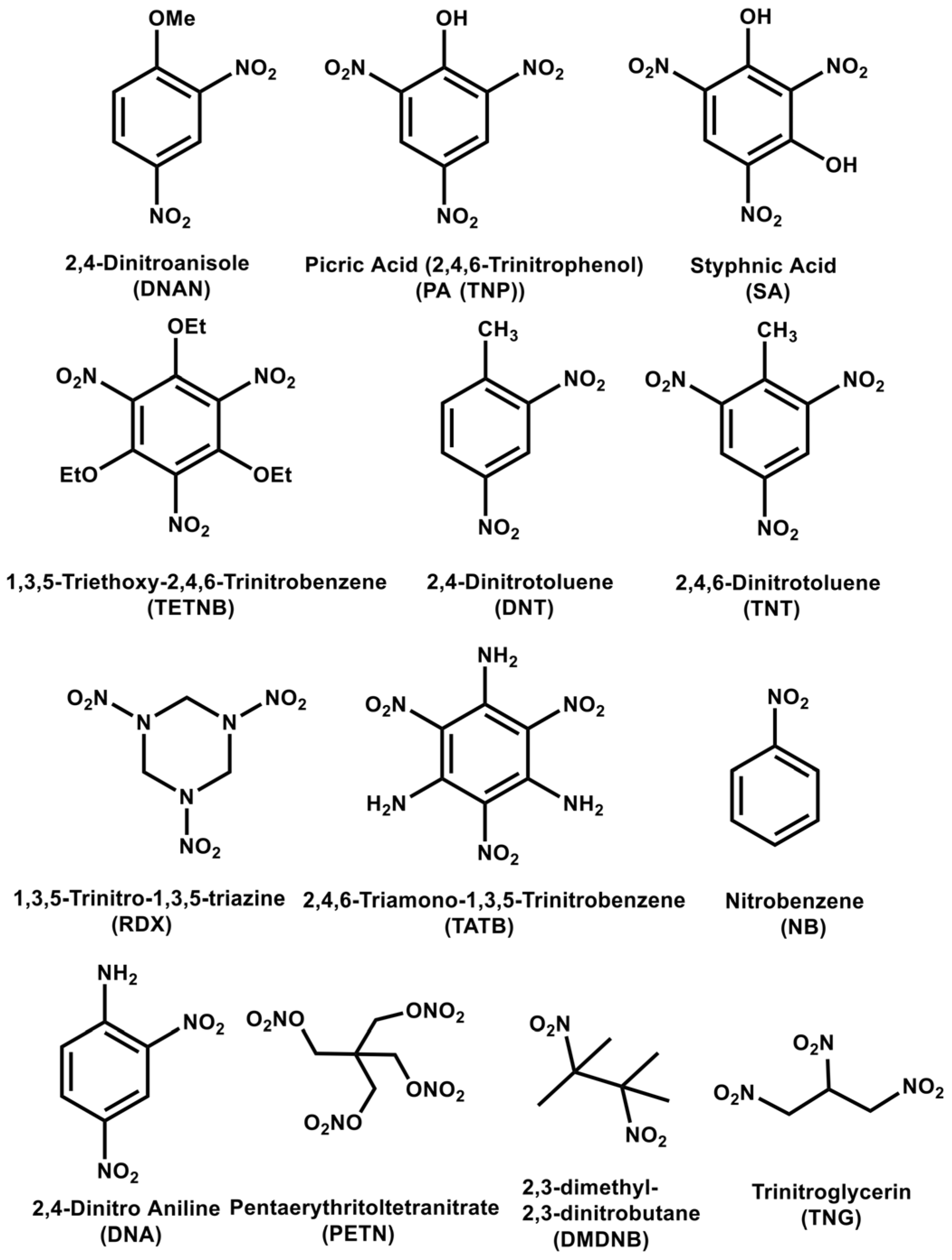
4. Pyrene Derivatives for Explosive Detection

This section describes the detection capability of pyrene derivatives for types of explosives. There are numerous explosive materials known throughout the world. The names of common explosives and their denotations are as follows: 2,4-Dinitro Anisole (DNAN), Picric Acid (2,4,6-Trinitro Phenol) (PA (TNP)), Styphnic Acid (SA), 1,3,5-Triethoxy-2,4,6-Trinitro Benzene (TETNB), 2,4-Dinitro Toluene (DNT), 2,4,6-Dinitro Toluene (TNT), 1,3,5-Trinitro-1,3,5-triazine (RDX), Nitrobenzene (NB), 2,4-Dinitro Aniline (DNA), Pentaerythritoltetranitrate (PETN), 2,3-dimethyl-2,3-dinitrobutane (DMDNB), and Trinitroglycerin (TNG). Figure 16 shows the structures of the explosives mentioned above.

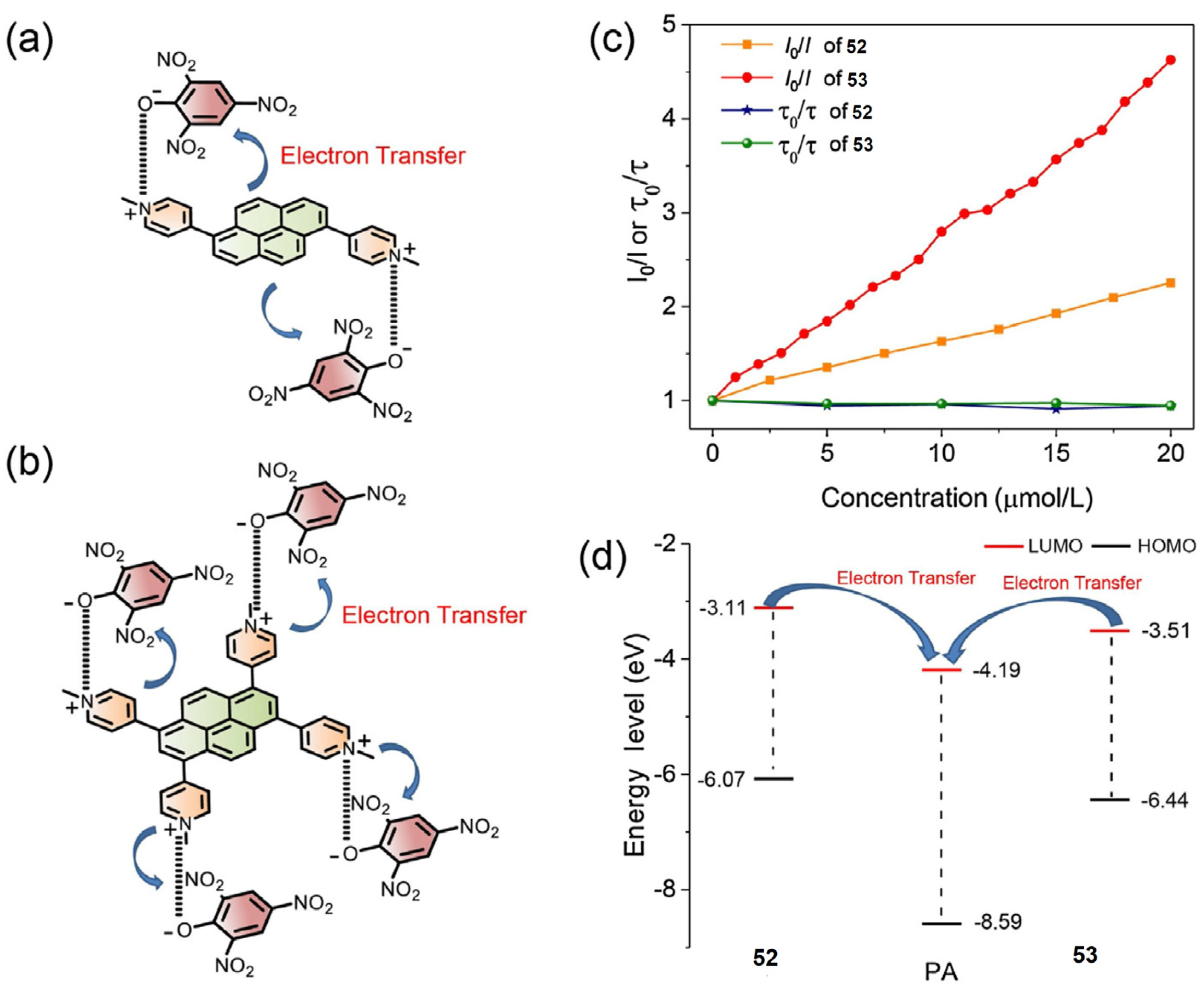
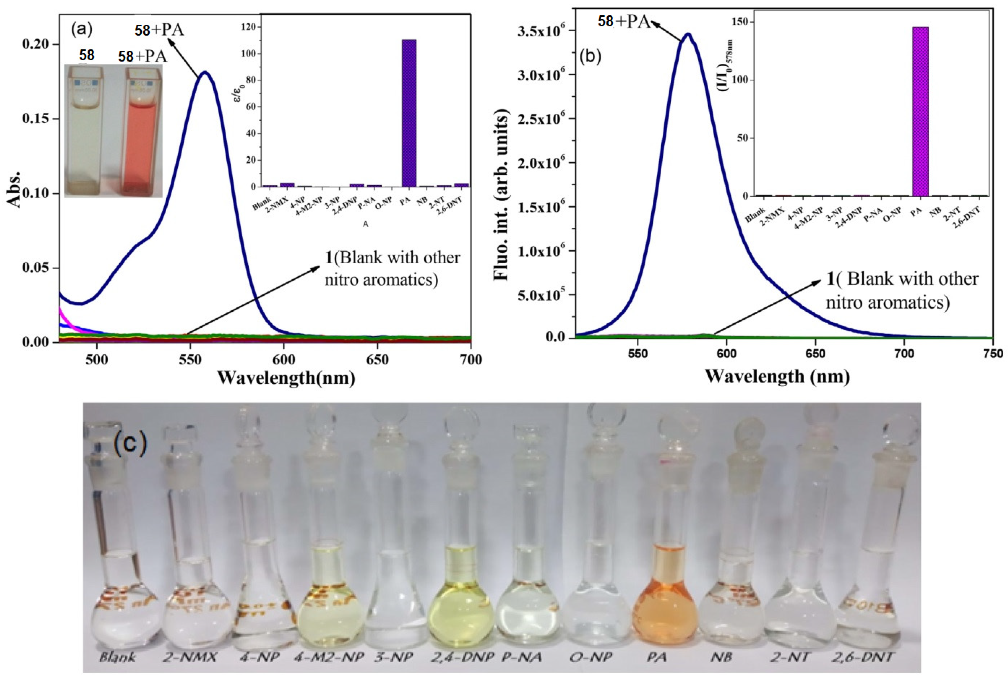
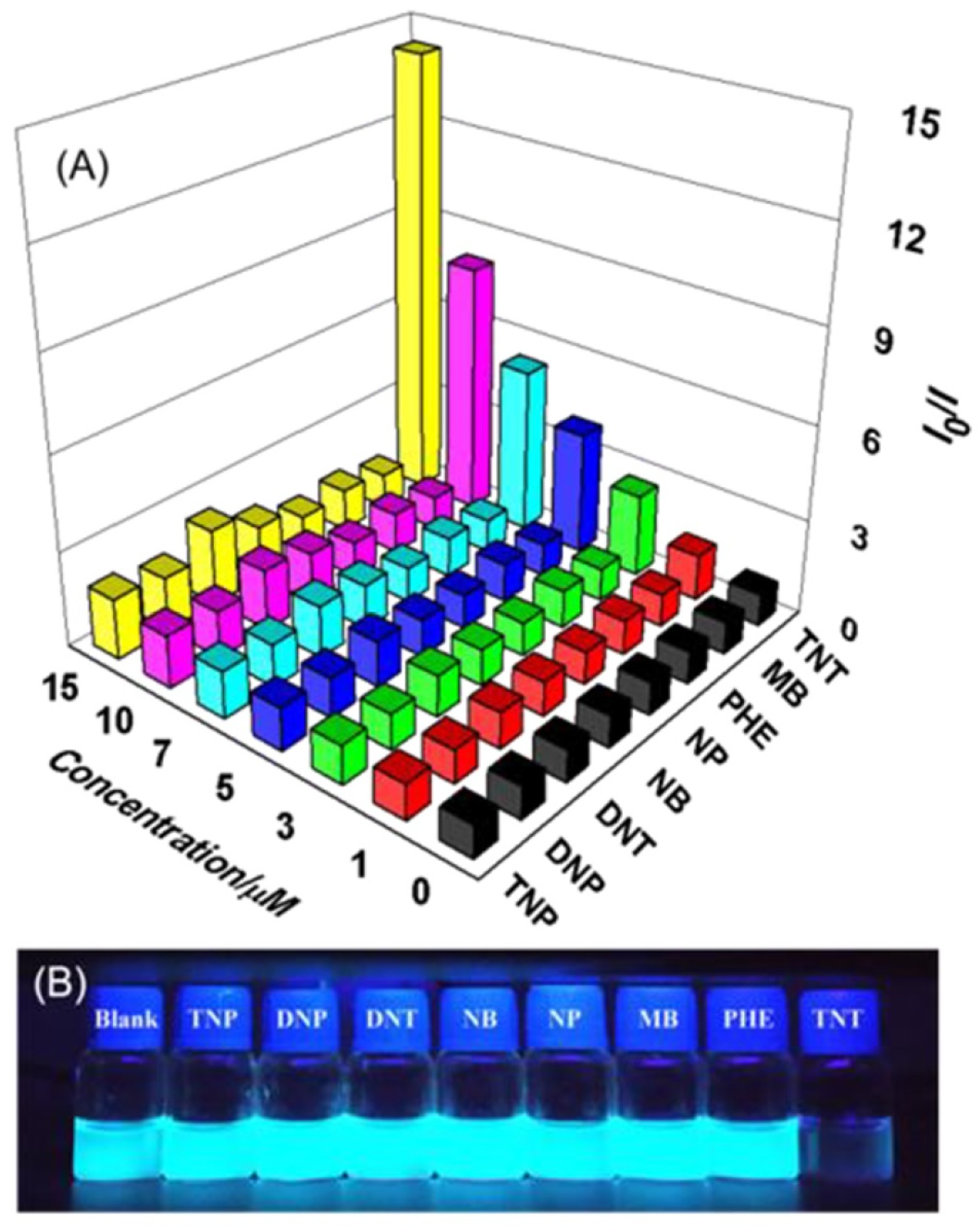
4.1. Pyrene Derivatives for Picric Acid (PA) Detection

Pyrene derivatives 4258 were used in the detection of PA via PL “Turn-Off” and ratiometric PL “Turn-On” responses [93,94,95,96,97,98,99,100,101,102,103,104,105,106,107]. Figure 17A–C display the structures of pyrene derivatives 4258. A few of them have a certain selectivity for other explosives [93,94,96,97,100,103,105]. However, those reports still possess a higher PL quenching percentile, which allows us to include them in this section. Apart from the above probes, the emissive pyrene–mercury complex probe 11-Hg2+ undergoes quenching with PA [70], as detailed below.
Due to the energy transfer between self-assembled 11-Hg2+ and PA, the PL emission at 550 nm (λex = 342 nm in H2O/ethanol (1:1, v/v) with HEPES, pH = 7.05) was quenched with a KSV value of 0.55 × 10⁵ M−1 and LOD of 35 nM. Paper strip and DFT studies were used to describe PA detection and its proposed sensing mechanism. This is a unique report describing the selectivity for Hg2+ and PA. Probe 42 showed a quenching efficiency of 70% (PA), 60% (TNT), and 46% (NB) in the solution state [93]. But when exposed to vapours, the quenching efficiency was reversed in the order of NB > TNT > PA. Due to the interference effect and lack of real-time detection, this report was regarded as a supplementary report for PA quantification. The electrospinning of pyrene over polyethersulfone afforded Py-PES, which shows a static PL quenching sensitivity in the order of PA > TNT > DNT > RDX [94]. From linear plots (0−1 μM), the KSV values of PA, TNT, DNT, and RDX were established as 1.263 × 10⁶ M−1, 1.80 × 10⁵ M−1, 7.52 × 10⁴ M−1, and 3.26 × 10⁴ M−1, respectively, with corresponding LODs of 23, 160, 400, and 980 nM. This work demonstrates the application of this probe in real water samples but does not provide details for discrimination between PA, TNT, DNT, and RDX. This work requires additional critical investigations.
Probe 43 was reported as a PA sensor via a PL “Turn-Off” response (λem = 402 nm; λex = 345 nm in 10 mM HEPES) through π-π interactions and a charge transfer mechanism [95]. The quenching efficiency of PA was found to be 80% with an LOD of 23.2 nM and KSV value of 1.16 µM−1. Similar to PA, Nitrophenol (NP) and Dinitrophenol (DNP) also show PL quenching with KSV values of 0.087 µM−1 and 0.31 µM−1, respectively. The strip method defines the detection of PA. However, this work can be further tuned by describing the “AIE” effect of the probe followed by PA detection. Deshmukh and co-workers developed the pyrene-based probe 44 for the selective detection of Cu2+ and PA [96]. The probe undergoes “AIE” and becomes emissive at 60% water fraction; the emissive probe 44 was used to detect Cu2+ and PA via a PL “Turn-Off” response. Between 0 and 39.6 μM, the PL emission at 498 nm (λex = 343 nm) was linearly quenched with an estimated LOD of 155 nM. The detection mechanism was assigned to π-π stacking and charge transfer (intercalation). This work lacks quenching (static/dynamic) and real application studies. Thus, additional research is mandatory. Push–pull pyrene compounds 4547 were developed with a nitroaromatic sensing ability [97]. However, 4547 show greater sensitivity to PA with defined LODs of 0.18, 528, and 0.29 µM compared to all nitroaromatics. The KSV values of 4547 for PA were quantified as 5.5 × 10⁶ M−1, 7.6 × 10⁴ M−1, and 3.5 × 10⁶ M−1, correspondingly. Due to the interference effect of the remaining nitroaromatics and the lack of applicability, this work is regarded as a supplementary report.
The pyrene–imidazole conjugate probe 48 was proposed for the PL “Turn-Off” detection of PA via a 1:1 guest–host stoichiometric complex and a resonance energy transfer (RET) mechanism [98]. In the presence of PA, the fluorescence of 48F = 0.126) quenches due to complex-tuned charge transfer and RET. The PLQY value of 48-PA was established as 0.02 with an LOD of 2.5 µM and a KSV of 8.47 × 10⁴ M−1. The high selectivity of PA by 48 was supported by DFT and single crystal X-ray diffraction (XRD) investigations. This work must be boosted with additional research on real-time applications. At 80% water content, probe 49F = 0.001) drives the formation of emissive “AIE” (ΦF = 0.79; λem = 476 nm; λex = 390 nm), which undergoes static PL quenching with PA [99]. Between 0 and 250 µM of PA, the emission (of “AIE”) was quenched to create a nonlinear curve, and the KSV value was recorded as 1.4 ± 0.02 × 10⁴ M−1. Due to π-π stacking and charge transfer, the “AIE” emission of 49 was quenched in the presence of PA. This work was applied in test strips. However, more clarification through LOD and interference studies is required. Compound 50 undergoes static and dynamic quenching with PA and 2-nitro-p-cresol (NPC) with estimated KSV values of 1.4 × 10⁵ M−1 and 1.1 × 10³ M−1 [100]. This work is classified as a supplementary report because it lacks interference studies and real-world applications.
Kathiravan and co-workers constructed pyrene-based probe 51F = 0.19) for the selective sensing of PA via the PL “Turn-Off” response [101]. PA and probe 51 underwent intramolecular hydrogen bonding (H-bonding) to reveal emission quenching at 530 nm with a recorded LOD of 63 nM. A smartphone-attached portable fluorimeter model and aqueous PA detection validates this report. This is innovative research. Pyrene-attached conjugates 52 and 53F = 0.64 and 0.95) display fluorescence quenching at 513 nm and 482 nm via electrostatic interaction-tuned charge transfer [102]. After the PA interaction, the PLQY of 52 and 53 reaches >1% and 15% with KSV values of 1.75 × 10⁴ M−1 and 6.04 × 10⁴ M−1, respectively. Among all nitroaromatics, PA displays a high quenching efficiency with 52 and 53. Due to the extended conjugation, probe 53 shows greater quenching of PA than 52. Figure 18a–d demonstrate the charge transfer between 52 and 53 to PA. This work requires additional information on LOD and real-world applications.
“AIE” active nanoaggregates were developed using compound 54 by adding 90% water and were then utilized for PA detection [103]. At 0% water fraction, probe 54 in THF possesses a ΦF value of 0.01, which is enhanced to 0.546 for a 90% water fraction (λem = 469 nm; λem = 375 nm). The PA, DNP, and Trinitrobenzene (TNB) show quenching efficiency with KSV values of 1.37 × 10⁴ M−1, 2.903 × 10³ M−1, and 1.732 × 10³ M−1. In actuality, PA shows a greater quenching efficiency than other analytes. From TRPL, cyclic voltammetry (CV), and time-correlated single photon counting (TCSPC), FRET was assigned as the mechanism. Evaluation of the LODs, real applications, and discrimination between PA and DNP requires extended interrogations. The 1:1 guest–host stoichiometric complex-induced PL “Turn-Off” quantification of PA was illustrated by probe 55F = 0.007) [104]. A red-shifted PL quenching of 55 from 425 nm to 462 nm was witnessed during the titration with PA. The K SV of 55-PA was calculated to be 2.29 × 10⁷ M−1 and its LOD of was 19 nM. DFT investigations revealed the band gaps of 55 and 55-PA as 3.04 eV and 2.77 eV (eV = electron volts), which suggest a feasible charge transfer mechanism. The strip method detection of PA at different temperatures (288 K, 298 K, and 308 K) attests to its applicability. This probe is also applied in the PL “Turn-On” detection of Cu2+, standing out as being unique. However, for PA detection, extended inquiries on interferences are still mandatory.
Pyrene-methylimidazolium chloride (56; ΦF = 0.266 in water) was reported in the PL “Turn-Off” detection of PA via a charge transfer mechanism [105]. Due to the n-π* and π-π* transition between PA to 56, the PL emission around 374, 393, and 415 nm (λex = 342 nm) was quenched with a Ka value of 1.69 × 10⁵ M−1 and a LOD of 50 μM. The ΦF value drops down to 0.003 during the sensing of PA. Though this work shows a good sensitivity of 56 in real water analysis and compares the results with anthracene derivatives, other phenolic compounds may affect the sensitivity in different pH conditions. Extended investigations on the “AIE” of 56 should address this issue. One-pot-synthesized pyrene compound 57 displays PL quenching of PA in ACN–water (1:2, v/v) with a Ka value of 2.13 × 10⁴ M−1 [106]. Due to the π-π interaction, the PL emission at 470 nm (λex = 310 nm) was quenched linearly between 0 and 180 µM with an estimated LOD of 0.814 µM. This work was applied to smartphone-assisted quantification and invisible ink. More data on DFT are needed to justify the mechanism.
Pyrene–rhodamine conjugate 58F < 0.001) was reported for the ratiometric PL “Turn-On” detection of PA via PET inhibition, ring opening, and FRET initiation [107]. As shown in Figure 19, probe 58 reveals both colorimetric and fluorometric responses for PA.
The PL emission of pyrene at 428 nm was quenched along with the emergence of rhodamine peak at 578 nm. From mass analysis, the stoichiometry of 58-PA (ΦF = 0.44) was established as 1:2. The Ka value of 58-PA is approximately 1011 M−1, with a limit of detection (LOD) of 13.8 ± 0.2 nM, demonstrating both its potency and sensitivity. Probe 58 initially possesses a PET between the amino group and pyrene, which is inhibited via ring opening and PA-induced FRET between rhodamine and pyrene. None of the nitro-aromatics show an interfering response, but acetate anion reverses the PA-induced sensor signal. Spiked soil analysis demonstrates the applicability of this work. This is a unique, stand-out ratiometric sensor for PA.

Critical Analysis of Detection of PA

Pyrene conjugate-based detection of PA [93,94,95,96,97,98,99,100,101,102,103,104,105,106,107] requires the following critical points to be taken into account: (A) Many of the reports [93,94,96,97,100,103,105] do not answer ‘how to avoid the interference effect?’, and discussions on the discrimination between the interferences are not valid enough; (B) The charge transfer mechanism, FRET, and the intramolecular H-bonding proposed for the detection of PA require supportive experimental and theoretical evidence. This requires multiple critical inquiries, which are lacking in many reports; (C) None of the reports provide guidelines for developing application models (except test strips and spiked water) that directly detect the PA. This issue must be critically optimized for innovative designs; (D) So far, only one ratiometric PL “Turn-On” sensor for PA quantification is available, which is insufficient to justify such designs. Thus, researchers must critically evaluate similar designs for PA discrimination; (E) The majority of “AIE” active probes for PA are lacking nano-level morphological and crystalline analyses, which should be critically examined further.

4.2. Pyrene Derivatives for Trinitrotoluene (TNT) Detection

In this section, the TNT-sensing ability of pyrene derivatives 5976 is discussed in detail [108,109,110]. Figure 20A,B show the structures of compounds 5967 engaged in TNT detection [108,109,110,111,112,113,114,115,116]. A pyrene excimer formation over the mesoporous thin films was described for the sensing of TNT via the PL quenching response [108]. After 5 min exposure of the film to nitroaromatics, the quenching efficiency at 470 nm (λex = 340 nm) was in the order of TNT (>80%) > DNT (>60%) > NB (>60%). The π-π stacking between pyrene and nitroaromatic analytes (TNT, DNT, and NB) and the mesoporous structure of thin films play a vital role in achieving the higher PL quenching efficacy. This work also defines the reusability of films for extended cycles of TNT detection. This is an innovative work. However, extensive research is mandatory for validation of this technique.
Venkatramaiah and co-workers reported the phosphonated pyrene probe 59em = 515 nm; λex = 330 nm) with a high sensitivity towards TNT via π-π stacking and effective intramolecular H-bonding [109]. The probe shows a 91% quenching efficiency for TNT with a KSV value of 8.4 × 10⁴ M−1. Similarly, it holds a 74% quenching efficiency for PA with a KSV value of 3.6 × 10⁴ M−1. Probe 59 shows PL quenching sensitivity in the order TNT > TNP > DNP > DNT. This study confirms the feasibility TNT sensing with a thin film and its reusability while pointing out important interference effects. Later on, Han’s research group carried out an in-depth mechanistic investigation on 59-TNT [110]. The theoretical investigation suggests the involvement of the PET mechanism along with the energy level crossing between the charge transfer state (CT) and the local excited state (LE). These findings could help upcoming researchers create novel designs for TNT detection. Probe 60 detects TNT and RDX via a fluorescence quenching response [111]. The fluorescence at 481 nm (λex = 345 nm in cetyl trimethylammonium bromide (CTAB)) was quenched with TNT and RDX with LODs of 8.8 nM and 1.5 µM, respectively. The Ka values were 1.0 × 10⁶ M−1 and 6.8 × 10⁴ M−1. This work must be investigated further with additional interferences such as PA, DNP, NP, etc. Also, this work lacks real-world applications, thus it is regarded as an additional study.
Pyrene–carboxylic acid and polyethylene glycols were condensed to form ester probes 61 and 62, which were then engaged in sensing TNT and PETN [112]. Probe 61 is more sensitive to TNT than probe 62. The ΦF values lie between 35 and 62%, and upon interaction with TNT and PETN, the KSV values were appraised in the order of 104 M−1 with LODs at µM levels. However, this work appears to be preliminary without experimental sensor details, short interference studies, and real applications. 12-pyren-1-yldodecanoic acid (63) was employed in the selective detection of TNT via the PL “Turn-Off” response [113]. Probe 63 displays a higher sensitivity to TNT than other types of explosives, as shown in Figure 21.
The emission at 482 nm (λex = 365 nm) was quenched linearly between 0.001−15 μM with an appraised LOD of 5 nM and a KSV value of 6.87 × 10⁵ M−1. The effectiveness of this work was illustrated by a soil extract analysis, which estimates the LOD at 10 nM. It has been concluded that the charge transfer between pyrene excimers and TNT afford the 90% PL quenching response. This is a standout work and can be extended for commercial device development.
The use of bispyrenylalkane 64 was proposed for the sensing of TNT [114]. The PL emission of 64 was quenched in the presence of TNT with a quantified LOD of 0.6 µM and a KSV value of 4.67 × 10⁵ M−1. This is a preliminary work without details of interferences, applications, and mechanisms. Similarly, compounds 65 and 66 were utilized to detect DNT and TNT [115]. The KSV values were at 103 M−1, and the DFT studies supported the charge transfer. Due to a lack of interference and real applications, this work can be considered as a supplementary report on the TNT sensor. Hao and co-workers synthesized probe 67 and validated its “AIE” and TNT sensing ability [116]. When adding the TNT, the PL emission at 505 nm (λex =376 nm, in water) was quenched linearly between 0 and 3 µM with a calculated LOD of 4.24 nM and KSV value of 6.2 × 10⁵ M−1. The KSV value of TNT was higher than PA (5.24 × 10⁴ M−1; LOD = 56.3 nM). Notably, the PL quenching efficiency reaches 96.18% with 67-TNT. Using the naked eye, the strip method detects the TNT even at 10 nM concentration. Based on TRPL and DFT studies, the PET mechanism was assigned as the sensor response. Apart from the unique performance, this work still needs to be extended to interference and real-time applications. Yeşilot’s research group developed a series of dendritic pyrene-functionalized cyclotriphosphazene-based dye molecules 6874 (see Figure 22A,B) for the detection of TNT and other explosives [117,118].
The ΦF values of 6874 were reported to be >0.5 and underwent static PL quenching in the presence of TNT, DNT, NP, and NB. Due to the π-π interaction and charge transfer between nitroaromatics (NACs) and 6874, the fluorescence emission was quenched rapidly. These reports focused on explaining the photophysical and thermal properties of 6874 and lacked an in-depth discussion of NAC sensors. In addition, the unspecific selectivity and sensitivity of TNT and insufficient information on real applications make them supplementary reports.
A ring-opening metathesis polymer (ROMP) with pyrene substitution (75; see Figure 23A) was designed and used in the identification of TNT [119]. In water, the polymer 75 forms a nano-sized micelle structure with emission bands at 376 and 395 nm (λex = 340 nm). The PL emission was quenched linearly for TNT in the 0.1–30 µM range with a quantified LOD and LOQ of 64 nM and 190 nM, respectively. Interestingly, TNT shows a 72% quenching efficiency compared to other interferences (quenching efficiencies at 22 -35%). The estimated KSV value of 75-TNT was 1.5 × 10⁵ M−1, which attests to the probe’s effectiveness. This work requires protracted investigations for applications. A pyrene-attached polystyrene nanofibrous copolymer 76 was developed via the electrospinning technique and applied in the sensing of TNT [120]. The fluorescent polymeric nanofibrous membrane shows a PL “Turn-On” response to TNT at 1 mM compared to other analytes, as shown in Figure 23B. The quenching efficiency was linear between 5 nM and 1 mM with a maximum of 99% at 1 mM TNT. The established LOD of 5 nM for TNT and time-dependent investigations supported this report. However, extra interrogations of the mechanism, interference, and applicability are mandatory.

Critical Scrutiny of Detection of TNT

Many reported pyrene derivatives for TNT sensing lack proper validation and need critical evaluation. (A) The sensitivity of the probes to TNT was possibly affected by the presence of other NACs, which requires perilous investigations to discriminate between them; (B) probes 65, 66, and 6874 were able to detect the TNT along with other NACs [115,117,118], thus selectivity of them towards TNT is questionable. This requires additional critical inquiries; (C) many reports on TNT detection lack an interference effect, KSV values, quenching efficiency, detailed mechanistic investigations, and validated real applications. This could lead to the question, “Why the need for such jumbled research?”, which could be assessed by more critical studies; (D) few reports on pyrene derivatives on nanostructure formation for TNT detection have been delivered without assessing the nano-level variation during the sensing studies. Upcoming researchers could critically evaluate these points further.

4.3. Pyrene Derivatives for RDX and Multi-Explosive Discovery

Similar to compounds 65, 66, and 6874, pyrene derivatives 20 and 7786 (see Figure 24) were effective in multi-NAC detection using fluorescence quenching [121,122,123].
Compound 20 (in THF; λex = 330 nm and λem = 487 nm) displays PL quenching responses to 2,4-dinitroaniline (2,4-DNA), p-nitroaniline (p-NA), p-nitrophenol (p-NP), PA, and 2,4-DNP of 99%, 89%, 75%, 55%, and 31%, respectively, with KSV values at the 105 M−1 level [121]. The LODs for 2,4-DNA, p-NA, p-NP, PA, and 2,4-DNP were estimated to be 0.27, 0.21, 0.43, 0.52, and 0.71 µM, respectively. The PL quenching was attributed to the PET and electron transfer mechanism. Further, 2,4-DNA follows static and dynamic quenching, whereas 2,4-DNP follows the static quenching phenomena seen with 20. The strip method shows the research’s relevance. We need to confirm if 2,4-DNP is present at significant levels and how other analytes affect quenching. Kovalev and co-workers developed a series of pyrene derivatives ‘7782F = 0.07–0.78)’ to quantify 2,4-DNT and 2,4,6-TNT in aqueous solvent [122]. Among them, 81 and 82F = 0.76 and 0.78; λem = 380 nm) show exceptional quenching response to 2,4-DNT and 2,4,6-TNT with an LOD of 1 (for both) and KSV values at 104 M−1 level. The PET process, π-π interaction, and false static quenching mechanism were assigned as mechanisms due to the “sphere of action”. The strip method validates applications. This work must be boosted by additional research on discriminative quantification and real sample analysis. The pyrene-based iptycenes probes 83–86 were reported for sensing RDX, TNT, and DNT via the PL “Turn-Off” response [123]. Probe 83 shows a higher response at an order of RDX>TNT>DNT with KSV values at the 103 M−1 level. The cavity of iptycenes provides a better binding site for the RDX moiety than others. Interfering NACs can affect this work, so it is included as a supplementary report.
1-Hydroxy pyrene-based derivatives 8790 (See Figure 25) were used for sensing RDX, PETN, DMDNB, and TNG via the PET-driven PL quenching response [124]. The ΦF values of 8790 were appraised between 0.23 and 0.98 and possess KSV values at 105 M−1 for those explosives. For RDX, the PL emission bands at 384 and 403 nm (λex = 345 nm) were quenched linearly between 0 and 1 µM, with an LOD of 67.5 nM. Dynamic Light Scattering (DLS), DFT, and TRPL investigations validated the nano size of the probes and quenching mechanism. Due to insufficient information on interferences and applications, this research is considered supplementary. Mosca and co-workers described the PET-induced PL “Turn-On” detection of RDX by pyrene-based probes 9193 (See Figure 25) [125]. RDX undergoes base-promoted hydrolysis to afford formaldehyde (HCHO) and Dinitrogen oxide (N2O). The interaction of HCHO with 9193 is responsible for the PL “Turn-On” response. However, in the presence of TNT, DNT, and DMDNB, the PL “Turn-On” response was quenched with appraised KSV values within the 103 to 104 M−1 range. Intramolecular H-bonding between −NH and the explosives and FRET mechanism was proposed for the PL quenching induced by explosives other than RDX. Later on, this mechanism was excluded by DFT and time-dependent Density Functional Theory (TDDFT) investigations by Lu’s research unit [126]. In this model, PET and π-π interactions are assigned as mechanisms. This is unique research showing a PL “Turn-On” response to RDX. But, additional inquiries are mandatory to exclude the quenching effect of interfering explosives and real applications.
As seen in Figure 25, pyrene-conjugated organosilicon polymers ‘9496’ were developed for the excimer–monomer dual emissive “Turn-Off” detection of TNT and TNP [127]. Polymer probe 94 (at 417 nm and 524 nm; λex = 350 nm) shows a high response to TNT and TNP. From theoretical studies, π-π interactions and intramolecular H-bonding were assigned as mechanisms. Details of interference and real-time applications are missing from this report. A polypseudorotaxane (λex = 345 and λem = 480 nm) was formed from a copolymer and pyrene-ended ammonium salt via a guest–host interaction and reported as a multi-stimuli responsive probe [128]. As depicted in Figure 26, the cavities prevailing in the polymer play a vital role in interacting with bases, anions, temperature, and explosives. The LODs for NACs are at the µM level. Due to the lack of interference, detailed mechanistic studies, and real applications, this work is counted as a supplementary one.

Critical Views on RDX and Multi-Explosive Encounters

Reports on pyrene-based compounds for RDX and multi-explosive detection are lacking in the literature. (A) RDX detection by pyrene conjugates was ultimately affected by the presence of other moderate explosives. This is not enough to convince society and researchers. Thus, in-depth critical analysis is mandatory for unique design and development; (B) Pyrene-derivatives for multi-NACs detection are not convincing enough to address explosive threats, which should be critically evaluated for their specific sensitivity; (C) Exposure of RDX and multi-explosive vapours to pyrene-based derivatives may lead to decomposition of probe’s structure in the solid state. Researchers should address this issue; (D) On what basis are the multistep synthetic polymers being synthesized and explored for RDX and multi-explosive sensing? None show a specific selectivity, have an interference effect, or have confirmed applications, and lack they mechanistic studies; (E) Exploration of polypseudorotaxane to NACs may lead to new research directions. However, due to the involvement of multi-synthetic steps to afford low-yield polypseudorotaxane, how could it be cost-effective research for developing countries?

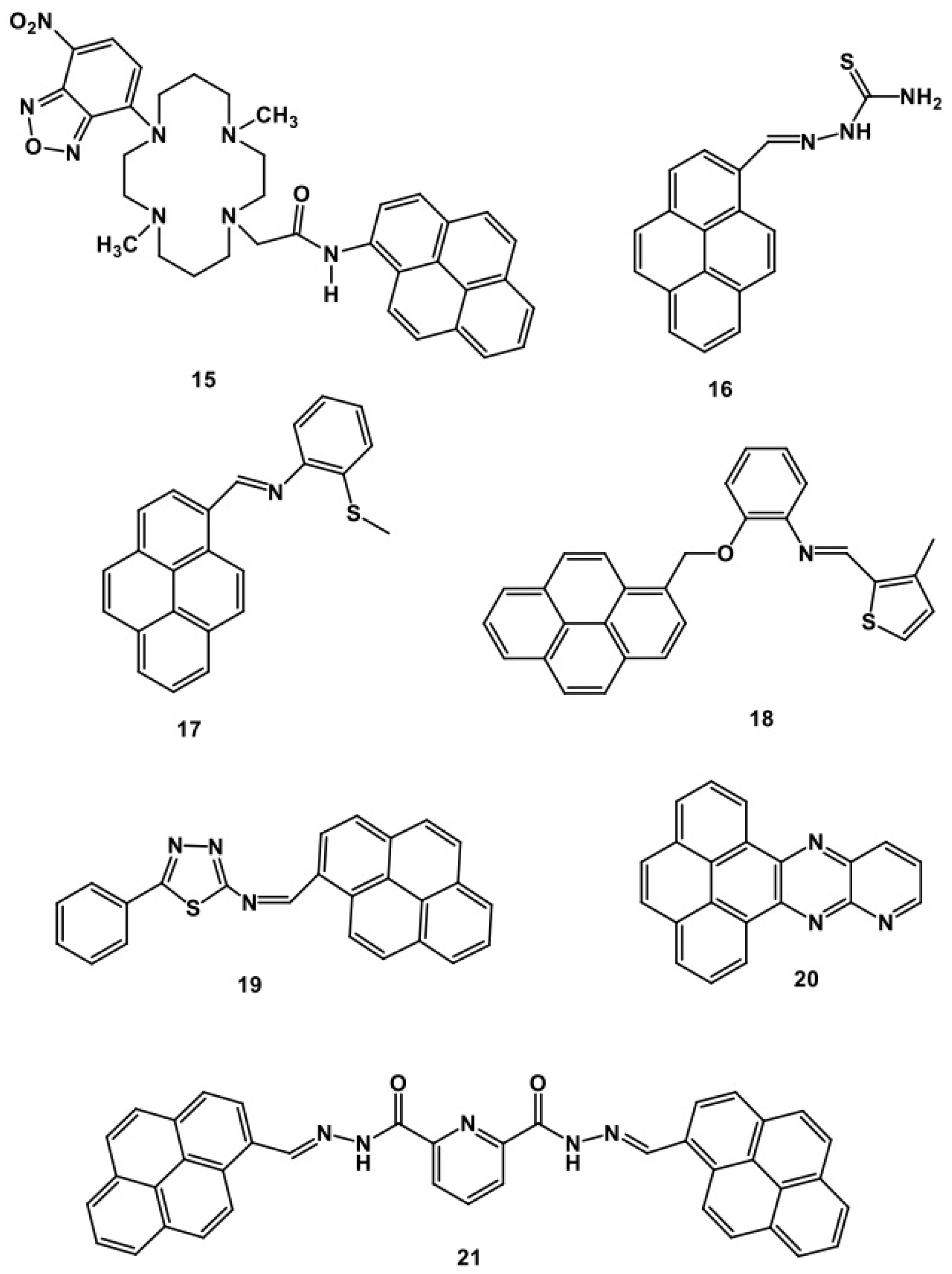
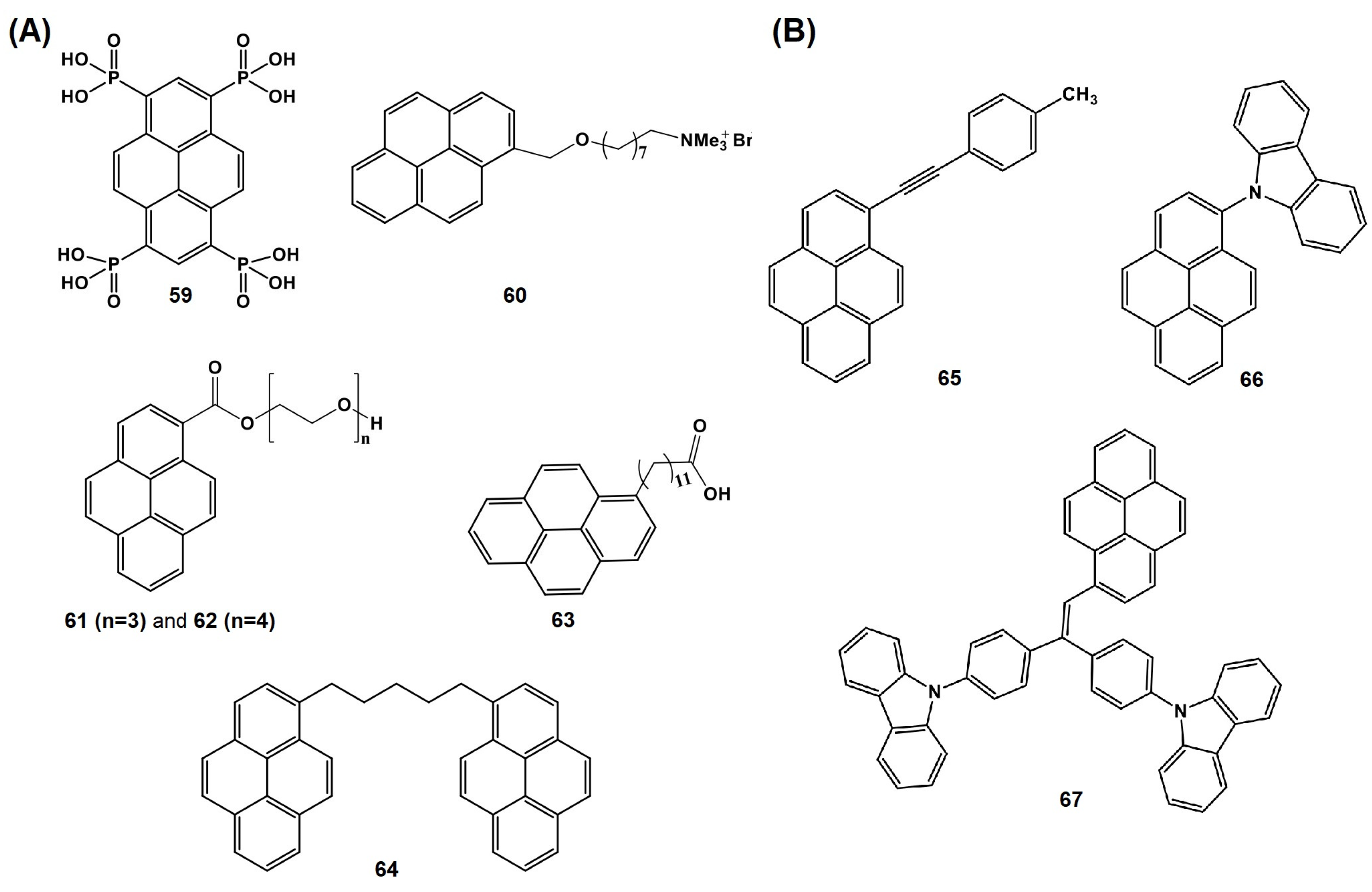
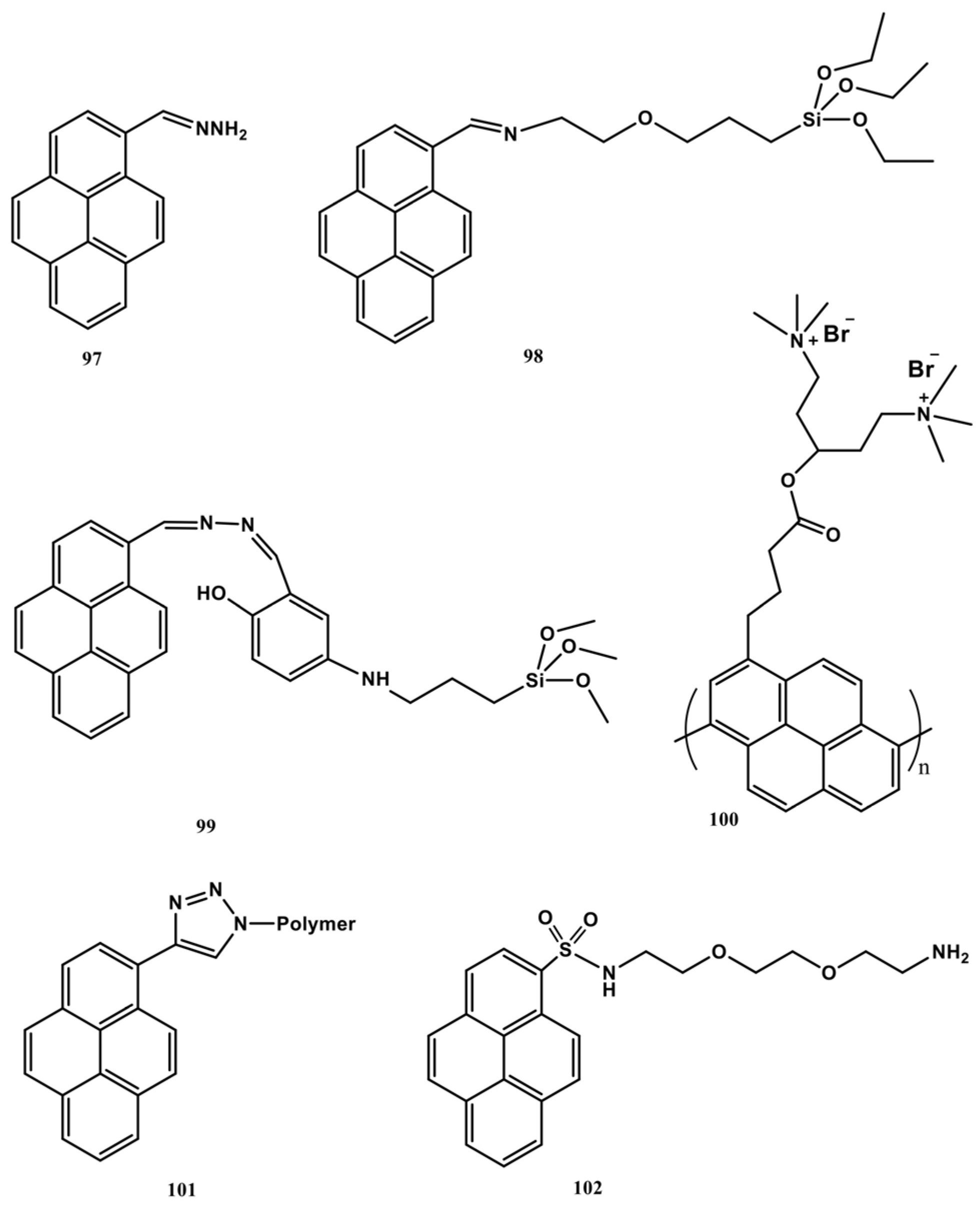
5. Pyrene-Conjugated Hybrid Probes for Hg2+ and Explosive Detection
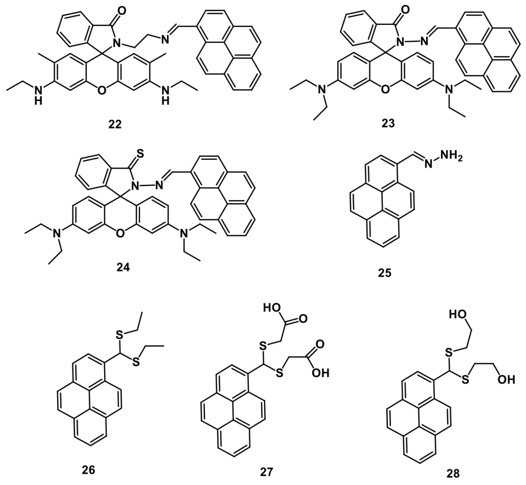
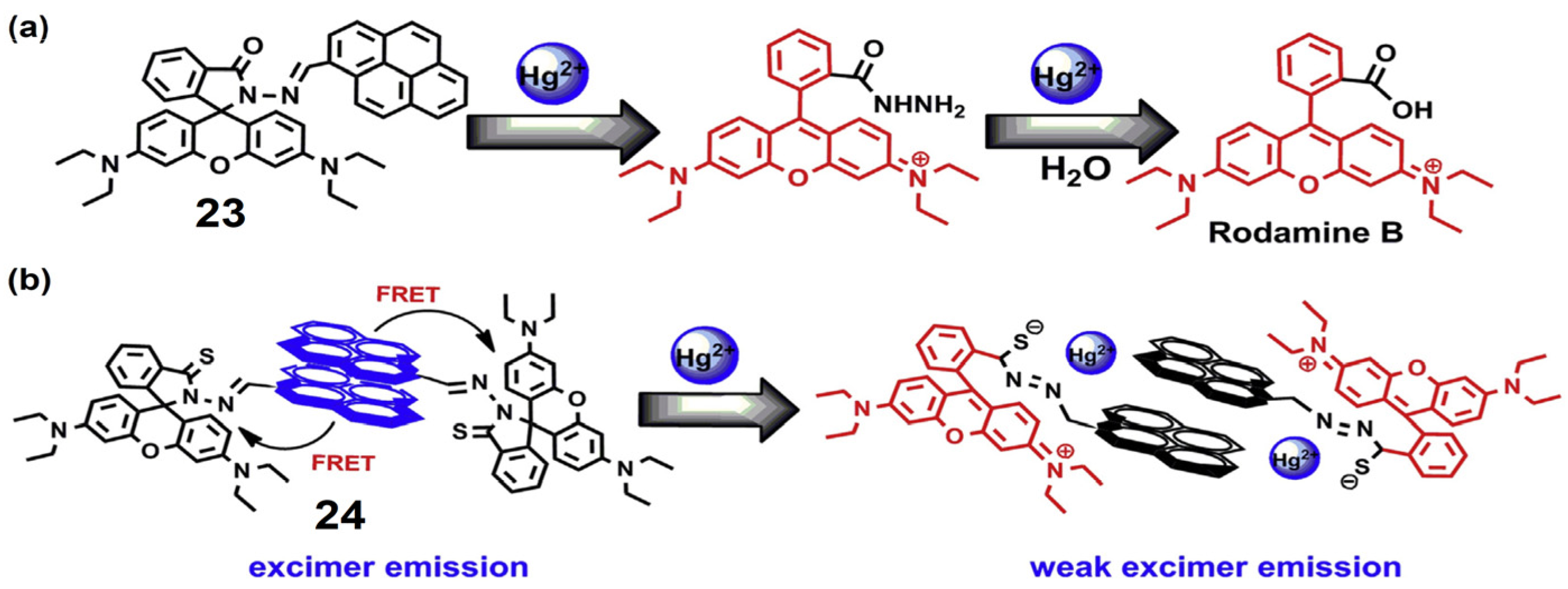
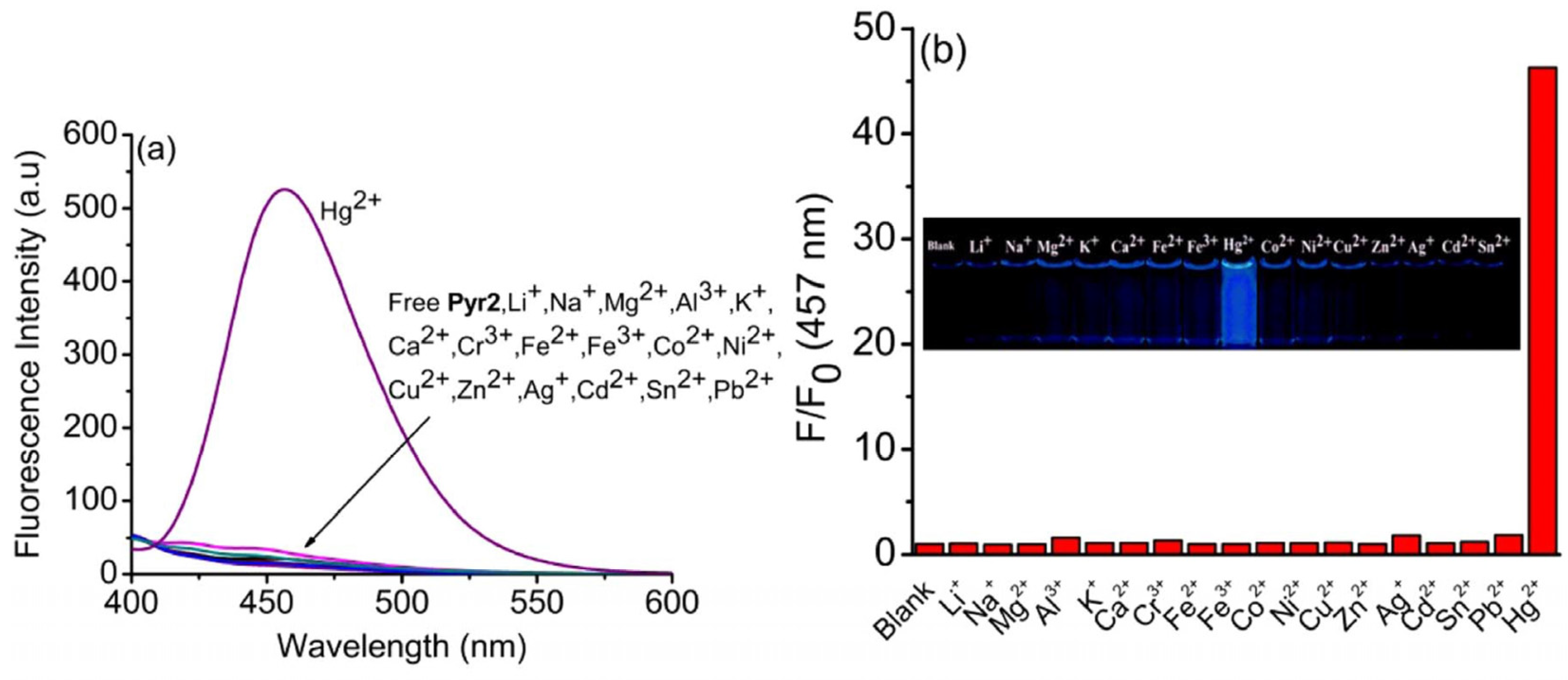
5.1. Hg2+ Sensors

This section explores the pyrene units conjugated with nanostructural materials for Hg2+ discriminations [129,130,131,132,133,134,135]. The structures of pyrene derivatives ‘97102’ utilized for developing the hybrid Hg2+ sensors are shown in Figure 27. The pyrene Schiff base 97 was attached to hybrid magnetic silica nanoparticles (Fe3O4@SiO2 NPs) to afford Py–Si–Fe3O4@SiO2 NPs and applied to detect Hg2+ via PL “Turn-On” response [129]. The hybrid system shows linear PL enhancement (at 450 nm; λex = 363 nm) from 4 to 40 µM with an LOD of 55 ± 15 nM. This hybrid probe shows >95% recoveries in real water and serum samples, hence it has been noted as a good innovation. However, additional information on the mechanism is required for validation.
A silica gel hybrid sensor was developed using compound 98 to detect Cu2+ and Hg2+ ions via a PL “Turn-On” response [130]. The hybrid sensor shows 10-fold PL enhancement to Hg2+ at 506 nm (λex = 365 nm) than Cu2+ ions (1.5-fold). Linear regression of Hg2+ ranged from 0 to 20 µM with an appraised LOD of 3.4 pM (pM = Picomole; 10−12 M) and Ka value of 2.5 × 10⁵ M−2. The 2:1 stoichiometric complex-induced excimer formation was assigned as the mechanism. HeLa cellular imaging, the test strip method, and real water recovery studies validated this work. The recoveries were found to be >95%. This is good work, but the discrimination between Cu2+ and Hg2+ must be investigated further. Pyrene derivative 99 was conjugated with magnetic core–shell fibrous silica (CoFe2O4/SiO2/KCC-1) to afford a “Py-CoFe2O4/SiO2/KCC-1” hybrid model that could be applied in Hg2+ quantification [131]. The hybrid system has linear sensitivity to Hg2+ from 1 to 20 µM with an LOD of 4.1 nM. Notably, the sensor shows >99% recoveries in environmental water samples and also possesses reversibility with EDTA (up to six cycles). Based on the available results, this can be considered as a good innovation.
Cationic oligopyrene 100 was complexed with oligothymine to afford a OHPBDB/poly(dT) complex, which reveals a PL “Turn-Off” selectivity towards Hg2+ [132]. The PL emission at 465 nm (λex = 340 nm) was quenched via thymine–Hg2+–thymine (T-Hg2+-T) base pair formation. The LOD of H g2+ by PL and using the naked eye are expressed as 5 nM and 5 µM. This is innovative work, but it must be explored further for extended its applications. A cryogel consisting of polyacrylamide and pyrene derivative 101 was expressed to discriminate Hg2+ ions [133]. This hybrid system detects Hg2+ with an estimated LOD of 10 nM. However, extended investigations on interference studies, mechanisms, and environmental applications are still needed. Coupling of 1-pyrenecarboxylic acid and NH2@SiO2 NPs leads to Pyr-NH2@SiO2 NPs, which detects Hg2+ ions via the PL “Turn-Off” response [134]. The Brunauer–Emmett–Teller (BET) surface area of the hybrid sensor was found to be 44.5 m2 g−1. The sensor shows PL quenching linearity from 0 to 25 µM with a LOD of 50 nM and a KSV value of 1.14 × 10⁵ M−1. This work still needs to be investigated to assess static or dynamic quenching and applicability. The reaction between 102 and mesoporous silica-functionalized epoxy-terminated organosilanes forms the hybrid sensor (MSN@Py-EOA) for PL “Turn-Off” detection of Hg2+ ions [135]. The BET surface area of MSN@Py-EOA was estimated to be 164.41 m2 g−1 with a 2.2 nm pore size. Linear regression of MSN@Py-EOA-Hg2+ (at 498 nm) was exhibited between 1 and 20 µM with an LOD of 0.62 µM and a KSV value of 4.0 × 10⁴ M−1. The NMR titration validates the feasible 1:1 complexation. This work falls short in real applications.

5.2. Explosive Sensors

Similar to Hg2+ sensing, hybrid models with pyrene units were effectively employed to detect NACs [136,137,138,139,140,141,142,143,144,145,146,147]. The pyrene derivatives 103110 (see Figure 28) were used to construct the hybrid models for the sensing of NACs.
Pyrene-functionalized ruthenium nanoparticles [136], 104-linked 2-bromoethyl methacrylate polymer (PBEMA) [138], 105-functionalized self-assembled monolayer (SAM) film [139], 106-functionalized fluorescent film [140], 107-coupled mesoporous silica nanomaterials [141], and 109-attached mesoporous silica microspheres [143] were demonstrated for multi-NAC detection with LODs between nM to µM and their KSV values are lies between 103–105 M−1. The 1-pyreneiodide coupled polyvinylpyrrolidone-functionalized gold nanoclusters (Au-PVP) (or) CTAB-stabilized gold nanorods (Au-CTAB) also follow a similar PL quenching trend for multi-NACs with a high KSV value of 3.88 × 10⁶ M−1 and an LOD of 10 nM [145]. Next, polyacrylonitrile nanofibers coupled with pyrene units (Py-PAN NFM) were also used for the detection of TNT and DNT [146]. This work seems impressive in terms of the KSV (105–106 M−1), LODs (at nM), and real water analysis. However, these reports reveal similar mechanistic aspects and lack interference studies and real applications, as stated in Section 4.1 and Section 4.3; thus, additional details are not provided here.
The compound 103-assembled glass substrate detects the PA with high sensitivity via a PL “Turn-Off” response at 500 nm (λex = 353 nm) [137]. The substrate follows the PL quenching trend of PA > TNT > DNT > NB. The estimated LOD of PA is 10 nM with a KSV value of 5.1 × 10⁴ M−1. The π-π interaction between 103 and PA was assigned as the mechanism. This work defines the reversible use of film by using ethanol exposure. In-depth mechanistic investigations may further validate this technique. The copper-complexed pyrene-conjugate 108F = 0.51) shows a high sensitivity to PA (>95% quenching efficiency at 360 µM) [142]. The PL emission at 387 nm and 407 nm (λex = 342 nm) was quenched between 0 and 360 µM with an LOD of 1.2 µM. The PL quenching was assigned to electron/energy transfer and the involvement of static and dynamic quenching phenomena. Preliminary soil tests attest to the applicability of the complex. However, extended investigations for interference and mechanistic aspects are still needed.
A mesoporous silica nanomaterial generated from pyrene derivative 110 was used for the discovery of the PL “Turn-On” by TNT and Tetryl [144]. The mesoporous support was loaded with a tris(2,20-bipyridyl)ruthenium(II) chloride ([Ru(bipy)3]Cl2) complex and then functionalized with 3-(azido-propyl)triethoxysilane, which undergoes a click reaction with 110 to afford the hybrid sensor model. In the presence of TNT and Tetryl, the PL intensity at 625 nm (λex = 453 nm) was enhanced due to the complexation between the electron donor pyrene unit and the electron-withdrawing benzene rings of NACs. This is innovative research, but additional details on sensitivity, discrimination between TNT and Tetryl, applications, and effects (pH, time, and interference) are missing. Pyrene-functionalized mesoporous Si NPs (pMSNs) were utilized to selectively identify TNT via the PL “Turn-Off” response [147]. Herein, cetyltrimethylammonium micelles, pyrene, and tetraethyl orthosilicate (TEOS) were self-assembled to form the hybrid sensor with emission located at 475 nm (λex = 340 nm). The emission quenched linearly (for TNT) from 10 nM to 1 µM with an established LOD of 12 nM. Incredibly, the quenching efficiency of pMSNs reached 98.6% for TNT at a 1 µM concentration, which is higher than all other NACs. This innovative research requires additional support from detailed mechanistic and real-application studies.

5.3. Critical Opinions on Hybrid Sensors for Hg2+ and Explosives

Hybrid probes constructed by coupling with pyrene derivatives hold certain criticisms and require extended investigations. (A) Most of the hybrid models derived from pyrene derivatives were possibly affected by the proposed protocols, and their stability is still in question. This requires careful optimization; (B) The sensitivity of hybrid probes to Hg2+ and specific NAC cannot be judged before analytical interrogations. Thus, while choosing the nanomaterial and probe structure, critical thinking is necessary; (C) Most of the pyrene derivatives already show a high selectivity to Hg2+ and specific NACs in the solution state, so why is there a need for a hybrid design for sensing? This requires critical evaluation; (D) Many pyrene-based hybrid sensors are designed for multi-explosive detection. This begs the question: “How do these developments benefit the research community?” Future researchers must tackle this directly; (E) Hybrid sensory materials for Hg2+ and NAC quantification lack evidence for mechanistic analysis and proposed applications. This requires in-depth critical investigations.

6. Design Requirements, Advantages, and Limitations

6.1. Design Requirements

To sense Hg2+ and specific NAC, pyrene derivatives with the following requirements may be helpful.
  • The probe for Hg2+ must hold hetero atoms or functional groups such as nitrogen/imine (N/−C=N), sulphur/thiols (S/−SH), oxygen/hydroxyl (O/−OH), carboxylic acid (−COOH), etc. This could lead to strong coordination with Hg2+ to deliver either PL enhancement or a quenching response.
  • The pyrene-based probe for Hg2+ must hold exceptional fluorescent ΦF values in two ways: (i) For the PL “Turn-On” sensor, the probe must possess a low ΦF value with weak or no-emission; (ii) For the PL “Turn-Off” sensor, the probe must possess a higher ΦF value with superior emission. This could be achieved by the inclusion of heteroatoms, functional units, Schiff base imine (−C=N) bonds, and extended conjugation, etc. [148,149,150,151], which initiates either PET/FRET On or Off, “CHEF”, and chemodosimetric approaches mechanisms.
  • For the chemodosimetric and ratiometric detection of Hg2+, the probe must be designed with highly reactive groups like sulphur/thiols (S/−SH) and a suitable acceptor moiety such as rhodamine, Fluorinated Boron-Dipyrromethene (BODIPY), etc.
  • To detect the NACs via the PL “Turn-Off” response, the designed pyrene-based probe must have extended conjugation, “AIE” characteristics, and exceptional π-π stacking. On the other hand, for the PL “Turn-On” response, the probe must be designed with H-bonding and electron/charge transfer units that initiate the emission enhancement.
  • To design the pyrene-based hybrid sensor, suitable nanostructured materials must be selected to attach with the proposed pyrene conjugate to afford a weak/highly emissive unique structured material for Hg2+/NACs detection. However, before synthesis, it is important to ensure the hybrid system has a particular affinity for either Hg2+ or NACs.

6.2. Advantages

Reported pyrene-conjugated probes for Hg2+ and NACs detection have advantages, as noted below.
  • Most of the probes that detect Hg2+ ions hold unique luminescent features to afford PL “Turn-On” or “Turn-Off” responses with exceptional changes in ΦF values and can be applied for in vitro/in vivo imaging and real sample analysis. This is advantageous over nanomaterials such as quantum dots (QDs), carbon dots (CDs), nanoparticles, 2D-materials, etc. [152,153,154,155].
  • The sensitivity of pyrene-based probes to Hg2+ is comparable and reliable to other known tactics such as electrochemical, colorimetric, chemiluminescence, etc. [154,156,157,158]. This is an added advantage of using pyrene derivatives in Hg2+ quantification.
  • The distinct PL response achieved during the ratiometric Hg2+ sensor may be helpful to quantify the Hg2+ via dual/multi-channel response, which is an advantage over common probes.
  • Most pyrene-based probes for NACs show incredible fluorescence quenching, and many of them are applied in real-time detection that can be extended for commercial device fabrication. This is a reliable advantage over other sensory materials [159,160].
  • Hybrid pyrene–mesoporous conjugates show specific affinity to Hg2+/NACs by affording a large surface area to deliver greater PL responses. This could be advantageous and reliable compared to other hybrid systems.

6.3. Limitations

Though pyrene derivatives have many advantages over other methods and sensors, they also have certain limitations, as noted below.
  • Pyrene is a light-sensitive moiety; hence, the stability and durability of pyrene-based probes are still in question. This limits the long-term use of the pyrene-based probes for Hg2+ and NACs detection.
  • Most of the pyrene-based probes for Hg2+ and NACs are operable in organic solvent or semi-aqueous media, and few probes show “AIE” features in an aqueous environment. This could limit the easy operation and real-time performance of sensors.
  • Many reports on the use of pyrene derivatives for Hg2+ discrimination lack precise mechanistic evidence and extended applications. This raises doubts about the reliability of the probes and limits further investigations.
  • Recently reported pyrene-based probes for NAC detection still lack evidence of their interference effect, mechanistic evidence, and details of real applications [161,162,163,164]. This could restrict the emergence of novel pyrene-based designs.
  • The few reports on pyrene-conjugated hybrid systems for Hg2+ and their multi-NAC sensing ability limit upcoming researchers investigating such a direction. In addition, the test-strip method using probes reported for NAC detection is described without proper protocols. This limits the real applications.

7. Conclusions and Perspectives

This comprehensive review detailed the use of pyrene derivatives 1110 for Hg2+ and nitroaromatic explosive detection. The py-py* excimer-tuned “Turn-On” detection, using the “CHEF” chemodosimetric approaches, and the PL “Turn-Off” detection of pyrene derivatives 141 are discussed with mechanism and their applications. Subsequently, pyrene derivatives 4296 applied in the detection of diverse NACs are described alongside their mechanisms and real applications. Rare examples of the pyrene-conjugated hybrid sensors 97110 for Hg2+ and NACs are described in detail. At the end of each section, critical thoughts and queries are offered. Ultimately, the design requirements, advantages, and limitations of the pyrene-based probes for Hg2+ and nitroaromatic explosive detection are explored for future researchers. Addressing the following perspective points may help the society to remove hazardous Hg2+ and NACs: (A) Though numerous reports are available on pyrene-based probes for Hg2+, a “state-of-the-art” procedure is still missing; this could be addressed with great care and optimization; (B) To date, pyrene-based probes are rarely reported for the ratiometric sensing of Hg2+, which should be extended with novel acceptor dyes and molecules; (C) The conjugation of pyrene derivatives with 0–2D-materials such as Graphene, Mxenes, perovskites, QDs, MOFs, etc., may enhance the sensitivity to Hg2+ and NACs; (D) Numerous reports are available on the utilization of pyrene-conjugated probes for the PL “Turn-Off” detection of NACs. But, none of these methods are standardized for commercialization, and this could be a unique research direction; (E) The design and development of novel pyrene-based probes for the PL “Turn-On” detection of specific NAC is still in demand, and this could be focused on by upcoming researchers; (F) Developing pyrene probes with longer alkyl and alkoxy chains may help to detect Hg2+ and NACs via gelation studies [165]. This could be an exclusive direction; (G) The development of p- or n-type organic thin-film transistor (OTFT) might be advanced by using pyrene-conjugated probes for Hg2+ and NACs detection [47]; (H) The design of pyrene derivatives to detect specific NAC in the presence other NACs is important and ground-breaking research, which upcoming researchers could focus on.
Apart from the above open questions, pyrene derivatives are still noted as unique candidates for the discrimination of Hg2+ and NACs. Innovations from current and upcoming researchers might address all of the issues described and deliver ground-breaking outputs.

Author Contributions

Literature collection, organization, writing—original draft preparation, M.S. (Muthaiah Shellaiah); writing—review and editing, and project administration, K.-W.S.; writing—review and editing K.A.; writing—review and editing A.M.; writing—review and editing V.V.; writing—review and editing M.B.; writing—review and editing M.S. (Mani Sivakumar); writing—review and editing E.M.; writing—review and editing K.K.; writing—review and editing W.-T.L. All authors have read and agreed to the published version of the manuscript.

Funding

This research was supported by the Ministry of Science and Technology of Taiwan and the SPROUT project of NYCU under contract No. MOST 111–2112-M-A49-031-, MOST 111-2811-M-A49-528, and 112W84100.

Institutional Review Board Statement

Not applicable.

Informed Consent Statement

Not applicable.

Data Availability Statement

Not applicable.

Conflicts of Interest

The authors declare no conflicts of interest.

References

  1. Balali-Mood, M.; Naseri, K.; Tahergorabi, Z.; Khazdair, M.R.; Sadeghi, M. Toxic Mechanisms of Five Heavy Metals: Mercury, Lead, Chromium, Cadmium, and Arsenic. Front. Pharmacol. 2021, 12, 643972. [Google Scholar] [CrossRef]
  2. Mitra, S.; Chakraborty, A.J.; Tareq, A.M.; Emran, T.B.; Nainu, F.; Khusro, A.; Idris, A.M.; Khandaker, M.U.; Osman, H.; Alhumaydhi, F.A.; et al. Impact of Heavy Metals on the Environment and Human Health: Novel Therapeutic Insights to Counter the Toxicity. J. King Saud Univ. Sci. 2022, 34, 101865. [Google Scholar] [CrossRef]
  3. Briffa, J.; Sinagra, E.; Blundell, R. Heavy Metal Pollution in the Environment and Their Toxicological Effects on Humans. Heliyon 2020, 6, e04691. [Google Scholar] [CrossRef] [PubMed]
  4. Gworek, B.; Dmuchowski, W.; Baczewska-Dąbrowska, A.H. Mercury in the Terrestrial Environment: A Review. Environ. Sci. Eur. 2020, 32, 128. [Google Scholar] [CrossRef]
  5. Wang, L.; Hou, D.; Cao, Y.; Ok, Y.S.; Tack, F.M.G.; Rinklebe, J.; O’Connor, D. Remediation of Mercury Contaminated Soil, Water, and Air: A Review of Emerging Materials and Innovative Technologies. Environ. Inter. 2020, 134, 105281. [Google Scholar] [CrossRef] [PubMed]
  6. Gworek, B.; Dmuchowski, W.; Baczewska, A.H.; Brągoszewska, P.; Bemowska-Kałabun, O.; Wrzosek-Jakubowska, J. Air Contamination by Mercury, Emissions and Transformations—A Review. Water Air Soil Pollut. 2017, 228, 123. [Google Scholar] [CrossRef]
  7. Gworek, B.; Bemowska-Kałabun, O.; Kijeńska, M.; Wrzosek-Jakubowska, J. Mercury in Marine and Oceanic Waters—A Review. Water Air Soil Pollut. 2016, 227, 371. [Google Scholar] [CrossRef]
  8. Basu, N.; Abass, K.; Dietz, R.; Krümmel, E.; Rautio, A.; Weihe, P. The Impact of Mercury Contamination on Human Health in the Arctic: A State of the Science Review. Sci. Total Environ. 2022, 831, 154793. [Google Scholar] [CrossRef]
  9. Al-Sulaiti, M.M.; Soubra, L.; Al-Ghouti, M.A. The Causes and Effects of Mercury and Methylmercury Contamination in the Marine Environment: A Review. Curr. Pollut. Rep. 2022, 8, 249–272. [Google Scholar] [CrossRef]
  10. Mergler, D. Ecosystem Approaches to Mercury and Human Health: A Way Toward the Future. Ambio 2021, 50, 527–531. [Google Scholar] [CrossRef]
  11. Kollur, S.P.; Shivamallu, C.; Prasad, S.K.; Veerapur, R.; Patil, S.S.; Cull, C.A.; Coetzee, J.F.; Amachawadi, R.G. Recent Advances on the Development of Chemosensors for the Detection of Mercury Toxicity: A Review. Separations 2021, 8, 192. [Google Scholar] [CrossRef]
  12. Genchi, G.; Sinicropi, M.S.; Carocci, A.; Lauria, G.; Catalano, A. Mercury Exposure and Heart Diseases. Int. J. Environ. Res. Public Health 2017, 14, 74. [Google Scholar] [CrossRef]
  13. Feng, X.; Li, P.; Fu, X.; Wang, X.; Zhang, H.; Lin, C.-J. Mercury Pollution in China: Implications on the Implementation of the Minamata Convention. Environ. Sci. Process. Impacts 2022, 24, 634–648. [Google Scholar] [CrossRef]
  14. Santos-Sacramento, L.; Arrifano, G.P.; Lopes-Araújo, A.; Augusto-Oliveira, M.; Albuquerque-Santos, R.; Takeda, P.Y.; Souza-Monteiro, J.R.; Macchi, B.M.; do Nascimento, J.L.M.; Lima, R.R.; et al. Human Neurotoxicity of Mercury in the Amazon: A Scoping Review with Insights and Critical Considerations. Ecotoxicol. Environ. Saf. 2021, 208, 111686. [Google Scholar] [CrossRef] [PubMed]
  15. Bjørklund, G.; Dadar, M.; Mutter, J.; Aaseth, J. The Toxicology of Mercury: Current Research and Emerging Trends. Environ. Res. 2017, 159, 545–554. [Google Scholar] [CrossRef]
  16. Gauthama, B.U.; Narayana, B.; Sarojini, B.K.; Suresh, N.K.; Sangappa, Y.; Kudva, A.K.; Satyanarayana, G.; Raghu, S.V. Colorimetric “off–on” Fluorescent Probe for Selective Detection of Toxic Hg2+ Based on Rhodamine and Its Application for In-Vivo Bioimaging. Microchem. J. 2021, 166, 106233. [Google Scholar] [CrossRef]
  17. Zhu, N.; Xu, J.; Ma, Q.; Gang, Y.; Li, L.; Liu, S.; Liu, S.; Wang, G. Rhodamine-Based Fluorescent Probe for Highly Selective Determination of Hg2+. ACS Omega 2022, 7, 29236–29245. [Google Scholar] [CrossRef] [PubMed]
  18. Małuszyński, M.J.; Małuszyńska, I. The Mercury Content in the Upper Layers of Soils in the Selected Area of the Masovian Landscape Park. Sustainability 2022, 14, 405. [Google Scholar] [CrossRef]
  19. Busairi, N.; Syahir, A. Recent Advances in Mercury Detection: Towards Enabling a Sensitive and Rapid Point-of-Check Measurement. J. Toxicol. Risk Assess. 2018, 4, 1–10. [Google Scholar]
  20. Shuai, H.; Xiang, C.; Qian, L.; Bin, F.; Xiaohui, L.; Jipeng, D.; Chang, Z.; Jiahui, L.; Wenbin, Z. Fluorescent Sensors for Detection of Mercury: From Small Molecules to Nanoprobes. Dye. Pig. 2021, 187, 109125. [Google Scholar] [CrossRef]
  21. Sharma, R.; Lee, H.-I. Recent Advances in Polymeric Chemosensors for the Detection and Removal of Mercury Ions in Complex Aqueous Media. J. Macromol. Sci. A 2022, 59, 389–402. [Google Scholar] [CrossRef]
  22. Zarlaida, F.; Adlim, M. Gold and Silver Nanoparticles and Indicator Dyes as Active Agents in Colorimetric Spot and Strip Tests for Mercury(II) Ions: A Review. Microchim. Acta 2017, 184, 45–58. [Google Scholar] [CrossRef]
  23. Lim, J.W.; Kim, T.-Y.; Woo, M.-A. Trends in Sensor Development Toward Next-Generation Point-of-Care Testing for Mercury. Biosens. Bioelectron. 2021, 183, 113228. [Google Scholar] [CrossRef] [PubMed]
  24. He, Y.; Wang, X.; Wang, K.; Wang, L. A Triarylamine-Based Fluorescent Covalent Organic Framework for Efficient Detection and Removal of Mercury(II) ion. Dye. Pig. 2020, 173, 10788. [Google Scholar] [CrossRef]
  25. Shellaiah, M.; Sun, K.-W. Progress in Metal-Organic Frameworks Facilitated Mercury Detection and Removal. Chemosensors 2021, 9, 101. [Google Scholar] [CrossRef]
  26. Ding, Y.; Wang, S.; Li, J.; Chen, L. Nanomaterial-Based Optical Sensors for Mercury Ions. TrAC Trends Anal. Chem. 2016, 82, 175–190. [Google Scholar] [CrossRef]
  27. Zhang, W.; Tang, Y.; Shi, A.; Bao, L.; Shen, Y.; Shen, R.; Ye, Y. Recent Developments in Spectroscopic Techniques for the Detection of Explosives. Materials 2018, 11, 1364. [Google Scholar] [CrossRef]
  28. Klapec, D.J.; Czarnopys, G.; Pannuto, J. Interpol Review of Detection and Characterization of Explosives and Explosives Residues 2016–2019. Forensic Sci. Inter. Synergy 2020, 2, 670–700. [Google Scholar]
  29. Sun, X.; Wang, Y.; Lei, Y. Fluorescence Based Explosive Detection: From Mechanisms to Sensory Materials. Chem. Soc. Rev. 2015, 44, 8019–8061. [Google Scholar] [CrossRef]
  30. Ye, M.; Ma, H.; Ni, Q. Research on a Sudden Explosion and its Environmental Impact. IOP Conf. Ser. Mater. Sci. Eng. 2017, 275, 12022. [Google Scholar] [CrossRef]
  31. Panz, K.; Miksch, K.; Sójka, T. Synergetic Toxic Effect of an Explosive Material Mixture in Soil. Bull. Environ. Contamin. Toxicol. 2013, 91, 555–559. [Google Scholar] [CrossRef] [PubMed]
  32. Qian, Y.; Li, J.; Ji, M.; Li, J.; Ma, D.; Liu, A.; Zhao, Y.; Yang, C. Fluorescent Covalent Organic Frameworks: A Promising Material Platform for Explosive Sensing. Front. Chem. 2022, 10, 943813. [Google Scholar] [CrossRef] [PubMed]
  33. Hu, Z.; Deibert, B.J.; Li, J. Luminescent Metal–Organic Frameworks for Chemical Sensing and Explosive Detection. Chem. Soc. Rev. 2014, 43, 5815–5840. [Google Scholar] [CrossRef]
  34. Ma, Y.; Wang, S.; Wang, L. Nanomaterials for luminescence detection of nitroaromatic explosives. TrAC Trends Anal. Chem. 2015, 65, 13–21. [Google Scholar] [CrossRef]
  35. Yu, H.A.; DeTata, D.A.; Lewis, S.W.; Silvester, D.S. Recent Developments in the Electrochemical Detection of Explosives: Towards Field-Deployable Devices for Forensic Science. TrAC Trends Anal. Chem. 2017, 97, 374–384. [Google Scholar] [CrossRef]
  36. Arshad, A.; Wang, H.; Bai, X.; Jiang, R.; Xu, S.; Wang, L. Colorimetric Paper Sensor for Sensitive Detection of Explosive Nitroaromatics Based on Au@Ag Nanoparticles. Spectrochim. Acta A 2019, 206, 16–22. [Google Scholar] [CrossRef]
  37. Das, P.; Mandal, S.K. Understanding the effect of an amino group on the selective and ultrafast detection of TNP in water using fluorescent organic probes. J. Mater. Chem. C 2018, 6, 3288–3297. [Google Scholar] [CrossRef]
  38. Verhagen, A.; Kelarakis, A. Carbon Dots for Forensic Applications: A Critical Review. Nanomaterials 2020, 10, 1535. [Google Scholar] [CrossRef]
  39. Maluleke, R.; Oluwafemi, O.S. Synthetic Approaches, Modification Strategies and the Application of Quantum Dots in the Sensing of Priority Pollutants. Appl. Sci. 2021, 11, 11580. [Google Scholar] [CrossRef]
  40. Mitri, F.; De Iacovo, A.; De Santis, S.; Giansante, C.; Sotgiu, G.; Colace, L. Chemiresistive Device for the Detection of Nitroaromatic Explosives Based on Colloidal PbS Quantum Dots. ACS Appl. Electron. Mater. 2021, 3, 3234–3239. [Google Scholar] [CrossRef]
  41. Shellaiah, M.; Sun, K.W.; Thirumalaivasan, N.; Bhushan, M.; Murugan, A. Sensing Utilities of Cesium Lead Halide Perovskites and Composites: A Comprehensive Review. Sensors 2024, 24, 2504. [Google Scholar] [CrossRef] [PubMed]
  42. Ouyang, T.; Guo, X.; Cui, Q.; Zhang, W.; Dong, W.; Fei, T. Conjugated Polymer Nanoparticles Based on Anthracene and Tetraphenylethene for Nitroaromatics Detection in Aqueous Phase. Chemosensors 2022, 10, 366. [Google Scholar] [CrossRef]
  43. Tanwar, A.S.; Parui, R.; Garai, R.; Chanu, M.A.; Iyer, P.K. Dual “Static and Dynamic” Fluorescence Quenching Mechanisms Based Detection of TNT via a Cationic Conjugated Polymer. ACS Meas. Sci. Au 2022, 2, 23–30. [Google Scholar] [CrossRef] [PubMed]
  44. Shellaiah, M.; Awasthi, K.; Chandran, S.; Aazaad, B.; Sun, K.W.; Ohta, N.; Wu, S.-P.; Lin, M.-C. Methylammonium Tin Tribromide Quantum Dots for Heavy Metal Ion Detection and Cellular Imaging. ACS Appl. Nano Mater. 2022, 5, 2859–2874. [Google Scholar] [CrossRef]
  45. Germain, M.E.; Knapp, M.J. Optical Explosives Detection: From Color Changes to Fluorescence Turn-On. Chem. Soc. Rev. 2009, 38, 2543–2555. [Google Scholar] [CrossRef]
  46. Lee, J.Y.; Root, H.D.; Ali, R.; An, W.; Lynch, V.M.; Bähring, S.; Kim, I.S.; Sessler, J.L.; Park, J.S. Ratiometric Turn-On Fluorophore Displacement Ensembles for Nitroaromatic Explosives Detection. J. Am. Chem. Soc. 2020, 142, 19579–19587. [Google Scholar] [CrossRef]
  47. Rullyani, C.; Shellaiah, M.; Ramesh, M.; Lin, H.-C.; Chu, C.-W. Pyrene-SH Functionalized OTFT for Detection of Hg2+ Ions in Aquatic Environments. Org. Electron. 2019, 69, 275–280. [Google Scholar] [CrossRef]
  48. Manandhar, E.; Wallace, K.J. Host–Guest Chemistry of Pyrene-Based Molecular Receptors. Inorg. Chim. Acta 2012, 381, 15–43. [Google Scholar] [CrossRef]
  49. Gong, Y.; Zhan, X.; Li, Q.; Li, Z. Progress of Pyrene-Based Organic Semiconductor in Organic Field Effect Transistors. Sci. China Chem. 2016, 59, 1623–1631. [Google Scholar] [CrossRef]
  50. Shellaiah, M.; Wu, Y.-H.; Singh, A.; Ramakrishnam Raju, M.V.; Lin, H.-C. Novel Pyrene- and Anthracene-Based Schiff Base Derivatives as Cu2+ and Fe3+ Fluorescence Turn-on Sensors and for Aggregation Induced Emissions. J. Mater. Chem. A 2013, 1, 1310–1318. [Google Scholar] [CrossRef]
  51. Shellaiah, M.; Chen, Y.-T.; Thirumalaivasan, N.; Aazaad, B.; Awasthi, K.; Sun, K.W.; Wu, S.-P.; Lin, M.-C.; Ohta, N. Pyrene-Based AIEE Active Nanoprobe for Zn2+ and Tyrosine Detection Demonstrated by DFT, Bioimaging, and Organic Thin-Film Transistor. ACS Appl. Mater. Interfaces 2021, 13, 28610–28626. [Google Scholar] [CrossRef] [PubMed]
  52. Shellaiah, M.; Sun, K.-W. Pyrene-Based AIE Active Materials for Bioimaging and Theranostics Applications. Biosensors 2022, 12, 550. [Google Scholar] [CrossRef]
  53. Shellaiah, M.; Simon, T.; Srinivasadesikan, V.; Lin, C.-M.; Sun, K.W.; Ko, F.-H.; Lin, M.-C.; Lin, H.-C. Novel Pyrene Containing Monomeric and Dimeric Supramolecular AIEE Active Nano-Probes Utilized in Selective “Off–On” Trivalent Metal and Highly Acidic pH Sensing with Live Cell Applications. J. Mater. Chem. C 2016, 4, 2056–2071. [Google Scholar] [CrossRef]
  54. Zhao, Q.; Lai, H.; Chen, H.; Li, H.; He, F. H- and J-Aggregation Inspiring Efficient Solar Conversion. J. Mater. Chem. A 2021, 9, 1119–1126. [Google Scholar] [CrossRef]
  55. Heo, J.; Murale, D.P.; Yoon, H.Y.; Arun, V.; Choi, S.; Kim, E.; Lee, J.-S.; Kim, S. Recent trends in molecular aggregates: An exploration of biomedicine. Aggregate 2022, 3, e159. [Google Scholar] [CrossRef]
  56. Fang, H.-P.; Shellaiah, M.; Singh, A.; Raju, M.V.R.; Wu, Y.-H.; Lin, H.-C. Naked Eye and Fluorescent Detections of Hg2+ Ions and Cysteine via J-Aggregation and Deaggregation of a Perylene Bisimide Derivative. Sens. Actuators B 2014, 194, 229–237. [Google Scholar] [CrossRef]
  57. Shellaiah, M.; Ramakrishna Raju, M.V.; Singh, A.; Lin, H.-C.; Wei, K.-H.; Lin, H.-C. Synthesis of Novel Platinum Complex Core as A Selective Ag+ Sensor and Its H-Bonded Tetrads Self-Assembled with Triarylamine Dendrimers for Electron/Energy Transfers. J. Mater. Chem. A 2014, 2, 17463–17476. [Google Scholar] [CrossRef]
  58. Shellaiah, M.; Thirumalaivasan, N.; Aazaad, B.; Awasthi, K.; Sun, K.W.; Wu, S.-P.; Lin, M.-C.; Ohta, N. An AIEE Active Anthracene-Based Nanoprobe for Zn2+ and Tyrosine Detection Validated by Bioimaging Studies. Chemosensors 2022, 10, 381. [Google Scholar] [CrossRef]
  59. Más-Montoya, M.; Janssen, R.A.J. The Effect of H- and J-Aggregation on the Photophysical and Photovoltaic Properties of Small Thiophene–Pyridine–DPP Molecules for Bulk-Heterojunction Solar Cells. Adv. Funct. Mater. 2017, 27, 1605779. [Google Scholar] [CrossRef]
  60. Zhao, Q.; Yu, X.; Xie, Z.; Liu, J.; Han, Y. Optimizing H-/J-Type Aggregation and Vertical Phase Separation to Improve Photovoltaic Efficiency of Small Molecule Solar Cells by Adding a Macromolecule Additive. ACS Appl. Energy Mater. 2018, 1, 6338–6344. [Google Scholar] [CrossRef]
  61. Yang, M.-H.; Thirupathi, P.; Lee, K.-H. Selective and Sensitive Ratiometric Detection of Hg(II) Ions Using a Simple Amino Acid Based Sensor. Org. Lett. 2011, 13, 5028–5031. [Google Scholar] [CrossRef] [PubMed]
  62. Areti, S.; Hinge, V.K.; Rao, C.P. Pyrenyl-imino-C2-Glucosyl Conjugate: Synthesis, Characterization, and Ratiometric and Reversible OFF–ON Receptor for Hg2+. Carbohydr. Res. 2014, 399, 64–69. [Google Scholar] [CrossRef]
  63. Thirupathi, P.; Saritha, P.; Lee, K.-H. Ratiometric Fluorescence Chemosensor Based on Tyrosine Derivatives for Monitoring Mercury Ions in Aqueous Solutions. Org. Biomol. Chem. 2014, 12, 7100–7109. [Google Scholar] [CrossRef] [PubMed]
  64. Shellaiah, M.; Rajan, Y.C.; Balu, P.; Murugan, A. A Pyrene-Based Schiff Base Probe for Selective Fluorescence Turn-On Detection of Hg2+ Ions with Live Cell Application. New J. Chem. 2015, 39, 2523–2531. [Google Scholar] [CrossRef]
  65. Wu, Y.; Wen, X.; Fan, Z. An AIE Active Pyrene-Based Fluorescent Probe for Selective Sensing Hg2+ and Imaging in Live Cells. Spectrochim. Acta A 2019, 223, 117315. [Google Scholar] [CrossRef]
  66. Zhou, Y.; Zhu, C.-Y.; Gao, X.-S.; You, X.-Y.; Yao, C. Hg2+-Selective Ratiometric and “Off−On” Chemosensor Based on the Azadiene−Pyrene Derivative. Org. Lett. 2010, 12, 2566–2569. [Google Scholar] [CrossRef]
  67. Lee, Y.H.; Lee, M.H.; Zhang, J.F.; Kim, J.S. Pyrene Excimer-Based Calix[4]arene FRET Chemosensor for Mercury(II). J. Org. Chem. 2010, 75, 7159–7165. [Google Scholar] [CrossRef]
  68. Wang, G.K.; Mi, Q.L.; Zhao, L.Y.; Hu, J.J.; Guo, L.E.; Zou, X.J.; Liu, B.; Xie, X.G.; Zhang, J.F.; Zhao, Q.H.; et al. A Pyrene Derivative for Hg2+-Selective Fluorescent Sensing and Its Application in In Vivo Imaging. Chem. Asian J. 2014, 9, 744–748. [Google Scholar] [CrossRef] [PubMed]
  69. Rodríguez-Lavado, J.; Lorente, A.; Flores, E.; Ochoa, A.; Godoy, F.; Jaque, P.; Saitz, C. Elucidating Sensing Mechanisms of A Pyrene Excimer-Based Calix[4]arene for Ratiometric Detection of Hg(ii) and Ag(i) and Cemosensor Behaviour as INHIBITION or IMPLICATION Logic Gates. RSC Adv. 2020, 10, 21963–21973. [Google Scholar] [CrossRef]
  70. Pramanik, S.; Bhalla, V.; Kumar, M. Mercury Assisted Fluorescent Supramolecular Assembly of Hexaphenylbenzene Derivative for Femtogram Detection of Picric Acid. Anal. Chim. Acta 2013, 793, 99–106. [Google Scholar] [CrossRef]
  71. Tümay, S.O.; Uslu, A.; Ardıç Alidağı, H.; Kazan, H.H.; Bayraktar, C.; Yolaçan, T.; Durmuş, M.; Yeşilot, S. A Systematic Series of Fluorescence Chemosensors with Multiple Binding sites for Hg(ii) Based on Pyrenyl-Functionalized Cyclotriphosphazenes and Their Application in Live Cell Imaging. New J. Chem. 2018, 42, 14219–14228. [Google Scholar] [CrossRef]
  72. Kim, S.H.; Kim, J.S.; Park, S.M.; Chang, S.-K. Hg2+-Selective OFF−ON and Cu2+-Selective ON−OFF Type Fluoroionophore Based upon Cyclam. Org. Lett. 2006, 8, 371–374. [Google Scholar] [CrossRef]
  73. Wang, X.M.; Yan, H.; Feng, X.L.; Chen, Y. 1-Pyrenecarboxaldehyde Thiosemicarbazone: A Novel Fluorescent Molecular Sensor Towards Mercury (II) Ion. Chin. Chem. Lett. 2010, 21, 1124–1128. [Google Scholar] [CrossRef]
  74. Sivaraman, G.; Anand, T.; Chellappa, D. Development of A Pyrene Based “Turn On” Fluorescent Chemosensor for Hg2+. RSC Adv. 2012, 2, 10605–10609. [Google Scholar] [CrossRef]
  75. Tümay, S.O.; Yeşilot, S. Highly Selective “Turn-On” Fluorescence Determination of Mercury Ion in Food and Environmental Samples Through Novel Anthracene and Pyrene Appended Schiff Bases. J. Photochem. Photobiol. A 2021, 407, 113093. [Google Scholar] [CrossRef]
  76. Bai, C.-B.; Xu, P.; Zhang, J.; Qiao, R.; Chen, M.-Y.; Mei, M.-Y.; Wei, B.; Wang, C.; Zhang, L.; Chen, S.-S. Long-Wavelength Fluorescent Chemosensors for Hg2+ based on Pyrene. ACS Omega 2019, 4, 14621–14625. [Google Scholar] [CrossRef] [PubMed]
  77. Singla, P.; Kaur, P.; Singh, K. A Fluorescent Probe for the Detection of Hg2+: Shift From “On-State A” to “On-State B”. Talanta 2014, 130, 571–576. [Google Scholar] [CrossRef] [PubMed]
  78. Muthukumar, V.; KulathuIyer, S. Twisted Pyrene with Perfect Hetero Atomic Cavity Optical Sensor for Hg22+ and Pb2+. Inorg. Chem. Commun. 2020, 121, 108187. [Google Scholar] [CrossRef]
  79. Ahamed, B.N.; Ghosh, P. An Integrated System of Pyrene and Rhodamine-6G for Selective Colorimetric and Fluorometric Sensing of Mercury(II). Inorg. Chim. Acta 2011, 372, 100–107. [Google Scholar] [CrossRef]
  80. Chu, K.-H.; Zhou, Y.; Fang, Y.; Wang, L.-H.; Li, J.-Y.; Yao, C. Rhodamine–Pyrene Conjugated Chemosensors for Ratiometric Detection of Hg2+ Ions: Different Sensing Behavior Between a Spirolactone and a Sirothiolactone. Dye. Pig. 2013, 98, 339–346. [Google Scholar] [CrossRef]
  81. Bist, A.; Cho, S.J.; Ahmed, N. A Simple Pyrene-Based Highly Sensitive Turn-On Fluorescent Chemodosimeter for Hg2+. J. Incl. Phenom. Macrocycl. Chem. 2013, 77, 75–81. [Google Scholar] [CrossRef]
  82. Liu, Y.-Y.; Naha, S.; Thirumalaivasan, N.; Velmathi, S.; Wu, S.-P. A Novel Nanomolar Highly Selective Fluorescent Probe for Imaging Mercury (II) in Living cells and Zebrafish. Sens. Actuators B 2018, 277, 673–678. [Google Scholar] [CrossRef]
  83. Gao, Y.; Ma, T.; Ou, Z.; Cai, W.; Yang, G.; Li, Y.; Xu, M.; Li, Q. Highly Sensitive and Selective Turn-On Fluorescent Chemosensors for Hg2+ Based on Thioacetal Modified Pyrene. Talanta 2018, 178, 663–669. [Google Scholar] [CrossRef] [PubMed]
  84. Chen, K.-H.; Lu, C.-Y.; Cheng, H.-J.; Chen, S.-J.; Hu, C.-H.; Wu, A.-T. A Pyrenyl-Appended Triazole-Based Ribose as A Fluorescent Sensor for Hg2+ Ion. Carbohyd. Res. 2010, 345, 2557–2561. [Google Scholar] [CrossRef] [PubMed]
  85. Wang, H.-F.; Wu, S.-P. Highly Selective Fluorescent Sensors for Mercury(II) Ions and Their Applications in Living Cell Imaging. Tetrahedron 2013, 69, 1965–1969. [Google Scholar] [CrossRef]
  86. Banerjee, A.; Karak, D.; Sahana, A.; Guha, S.; Lohar, S.; Das, D. Methionine–Pyrene Hybrid-Based Fluorescent Probe for Trace Level Detection and Estimation of Hg(II) in Aqueous Environmental Samples: Experimental and Computational Studies. J. Hazard. Mater. 2011, 186, 738–744. [Google Scholar] [CrossRef]
  87. Rani, B.K.; John, S.A. Fluorogenic Mercury Ion Sensor Based on Pyrene-Amino Mercapto Thiadiazole Unit. J. Hazard. Mater. 2018, 343, 98–106. [Google Scholar] [CrossRef]
  88. Harsha, K.G.; Appalanaidu, E.; Chereddy, N.R.; Baggi, T.R.; Rao, V.J. Pyrene Tethered Imidazole Derivative for the Qualitative and Quantitative Detection of Mercury Present in Various Matrices. Sens. Actuators B 2018, 256, 528–534. [Google Scholar] [CrossRef]
  89. Liu, X.; Yang, X.; Fu, Y.; Zhu, C.; Cheng, Y. Novel Fluorescent Sensor for Ag+ and Hg2+ Based on the BINOL-Pyrene Derivative via Click Reaction. Tetrahedron 2011, 67, 3181–3186. [Google Scholar] [CrossRef]
  90. Puangsamlee, T.; Tachapermpon, Y.; Kammalun, P.; Sukrat, K.; Wainiphithapong, C.; Sirirak, J.; Wanichacheva, N. Solvent Control Bifunctional Fluorescence Probe for Selective Detection of Cu2+ and Hg2+ Via the Excimer of Pyrenylacetamide Subunits. J. Lumin. 2018, 196, 227–235. [Google Scholar] [CrossRef]
  91. Lee, S.W.; Lee, S.Y.; Lee, S.H. Self-assembly of Pyrene Boronic Acid-Based Chemodosimeters for Highly Efficient Mercury(II) Ion Detection. Tetrahed. Lett. 2019, 60, 151048. [Google Scholar] [CrossRef]
  92. Merz, V.; Merz, J.; Kirchner, M.; Lenhart, J.; Marder, T.B.; Krueger, A. Pyrene-Based “Turn-Off” Probe with Broad Detection Range for Cu2+, Pb2+ and Hg2+ Ions. Chem. Eur. J. 2021, 27, 8118–8126. [Google Scholar] [CrossRef] [PubMed]
  93. Shaligram, S.; Wadgaonkar, P.P.; Kharul, U.K. Fluorescent Polymeric Ionic Liquids for the Detection of Nitroaromatic Explosives. J. Mater. Chem. A 2014, 2, 13983–13989. [Google Scholar] [CrossRef]
  94. Sun, X.; Liu, Y.; Shaw, G.; Carrier, A.; Dey, S.; Zhao, J.; Lei, Y. Fundamental Study of Electrospun Pyrene–Polyethersulfone Nanofibers Using Mixed Solvents for Sensitive and Selective Explosives Detection in Aqueous Solution. ACS Appl. Mater. Interfaces 2015, 7, 13189–13197. [Google Scholar] [CrossRef] [PubMed]
  95. Liang, H.; Yao, Z.; Ge, W.; Qiao, Y.; Zhang, L.; Cao, Z.; Wu, H.-C. Selective and Sensitive Detection of Picric Acid Based on A Water-Soluble Fluorescent Probe. RSC Adv. 2016, 6, 38328–38331. [Google Scholar] [CrossRef]
  96. Deshmukh, S.C.; Rana, S.; Shinde, S.V.; Dhara, B.; Ballav, N.; Talukdar, P. Selective Sensing of Metal Ions and Nitro Explosives by Efficient Switching of Excimer-to-Monomer Emission of an Amphiphilic Pyrene Derivative. ACS Omega 2016, 1, 371–377. [Google Scholar] [CrossRef]
  97. Verbitskiy, E.V.; Baranova, A.A.; Lugovik, K.I.; Khokhlov, K.O.; Chuvashov, R.D.; Dinastiya, E.M.; Rusinov, G.L.; Chupakhin, O.N.; Charushin, V.N. Linear and V-Shaped Push–Pull Systems on A Base of Pyrimidine Scaffold with A Pyrene-Donative Fragment for Detection of Nitroaromatic Compounds. J. Iran. Chem. Soc. 2018, 15, 787–797. [Google Scholar] [CrossRef]
  98. Sodkhomkhum, R.; Masik, M.; Watchasit, S.; Suksai, C.; Boonmak, J.; Youngme, S.; Wanichacheva, N.; Ervithayasuporn, V. Imidazolylmethylpyrene Sensor for Dual Optical Detection of Explosive Chemical: 2,4,6-Trinitrophenol. Sens. Actuators B 2017, 245, 665–673. [Google Scholar] [CrossRef]
  99. Islam, A.S.M.; Sasmal, M.; Maiti, D.; Dutta, A.; Show, B.; Ali, M. Design of a Pyrene Scaffold Multifunctional Material: Real-Time Turn-On Chemosensor for Nitric Oxide, AIEE Behavior, and Detection of TNP Explosive. ACS Omega 2018, 3, 10306–10316. [Google Scholar] [CrossRef]
  100. Hu, Y.; Joung, J.F.; Jeong, J.-E.; Jeong, Y.; Woo, H.Y.; She, Y.; Park, S.; Yoon, J. 2-(Benzothiazol-2-yl)pyren-1-ol, A New Excited State Intramolecular Proton Transfer-Based Fluorescent Sensor for Nitroaromatic Compounds. Sens. Actuators B 2019, 280, 298–305. [Google Scholar] [CrossRef]
  101. Kathiravan, A.; Gowri, A.; Khamrang, T.; Kumar, M.D.; Dhenadhayalan, N.; Lin, K.-C.; Velusamy, M.; Jaccob, M. Pyrene-Based Chemosensor for Picric Acid—Fundamentals to Smartphone Device Design. Anal. Chem. 2019, 91, 13244–13250. [Google Scholar] [CrossRef] [PubMed]
  102. Yan, N.; Song, J.; Wang, F.; Kan, L.; Song, J.; Wang, W.; Ma, W.; Zhang, W.; He, G. Pyrenoviologen-Based Fluorescent Sensor for Detection of Picric Acid in Aqueous Solution. Chin. Chem. Lett. 2019, 30, 1984–1988. [Google Scholar] [CrossRef]
  103. Panigrahi, A.; Sahu, B.P.; Mandani, S.; Nayak, D.; Giri, S.; Sarma, T.K. AIE Active Fluorescent Oorganic Nanoaggregates for Selective Detection of Phenolic-Nitroaromatic Explosives and Cell Imaging. J. Photochem. Photobiol. A 2019, 374, 194–205. [Google Scholar] [CrossRef]
  104. Kundu, B.K.; Pragti; Reena; Mobin, S.M.; Mukhopadhyay, S. Mechanistic and Thermodynamic Aspects of A Pyrene-Based Fluorescent Probe to Detect Picric Acid. New J. Chem. 2019, 43, 11483–11492. [Google Scholar] [CrossRef]
  105. Pherkkhuntod, C.; Ervithayasuporn, V.; Chanmungkalakul, S.; Wang, C.; Liu, X.; Harding, D.J.; Kiatkamjornwong, S. Water-Soluble Polyaromatic-Based Imidazolium for Detecting Picric Acid: Pyrene vs. Anthracene. Sens. Actuators B 2021, 330, 129287. [Google Scholar] [CrossRef]
  106. Jindal, G.; Kaur, N. Fluorimetric Quantification of Picric Acid in Aqueous Medium Via Smartphone and Invisible Ink Applications Using Pyrene Based Sensor. Inorg. Chem. Commun. 2022, 140, 109481. [Google Scholar] [CrossRef]
  107. Charan Behera, K.; Mallick, D.; Narayan Patra, B.; Bag, B. A Pyrene-Rhodamine FRET Couple as A Chemosensor for Selective Detection of Picric Acid. Spectrochim. Acta A 2022, 271, 120934. [Google Scholar] [CrossRef]
  108. Beyazkilic, P.; Yildirim, A.; Bayindir, M. Formation of Pyrene Excimers in Mesoporous Ormosil Thin Films for Visual Detection of Nitro-explosives. ACS Appl. Mater. Interfaces 2014, 6, 4997–5004. [Google Scholar] [CrossRef]
  109. Venkatramaiah, N.; Firmino, A.D.G.; Almeida Paz, F.A.; Tomé, J.P.C. Fast Detection of Nitroaromatics Using Phosphonate Pyrene Motifs as Dual Chemosensors. Chem. Commun. 2014, 50, 9683–9686. [Google Scholar] [CrossRef]
  110. Lu, M.; Zhou, P.; Li, Z.; Liu, J.; Yang, Y.; Han, K. New Insights Into the Sensing Mechanism of A Phosphonate Pyrene Chemosensor for TNT. Phys. Chem. Chem. Phys. 2018, 20, 19539–19545. [Google Scholar] [CrossRef]
  111. Hong, J.-H.; Choi, J.-H.; Cho, D.-G. Simple Pyrene Derivatives as Fluorescence Sensors for TNT and RDX in Micelles. Bull. Korean Chem. Soc. 2014, 35, 3158–3162. [Google Scholar] [CrossRef]
  112. Sadieva, L.K.; Taniya, O.S.; Kovalev, I.S.; Kopchuk, D.S.; Zyryanov, G.V.; Rusinov, V.L.; Chupakhin, O.N. Pyrene-1-Carboxylic Acid Polyethylene Glycol Esters: Synthesis and Photophysical Studies. Russ. Chem. Bull. 2021, 70, 1174–1179. [Google Scholar] [CrossRef]
  113. Sun, Y.; Wu, Y.; Yu, C.; Zhang, L.; Song, G.; Yao, Z. Self-Assembly of Nanoscale Induced Excimers of 12-Pyren-1-yldodecanoic Acid for TNT Detection. ACS Appl. Nano Mater. 2019, 2, 3453–3458. [Google Scholar] [CrossRef]
  114. Kovalev, I.S.; Sadieva, L.K.; Taniya, O.S.; Yurk, V.M.; Minin, A.S.; Kopchuk, D.S.; Zyryanov, G.V.; Charushin, V.N.; Chupakhin, O.N. Bispyrenylalkane Chemosensor for the Naked-eye Detection of Nitro-explosives. Chim. Techno Acta 2021, 8, 20218209. [Google Scholar] [CrossRef]
  115. Sriyab, S.; Jorn-Iat, K.; Prompinit, P.; Wolschann, P.; Hannongbua, S.; Suramitr, S. Photophysical Properties of 1-Pyrene-based Derivatives for Nitroaromatic Explosives Detection: Experimental and Theoretical Studies. J. Lumin. 2018, 203, 492–499. [Google Scholar] [CrossRef]
  116. Hao, H.; Ye, Z.; Dai, H.; Liu, C.; Yi, A.; Xu, B.; Shi, G.; Su, S.; Azad, F.; Chi, Z. Pyrenyl-Based Aggregation-Induced Emission Luminogen for Highly Sensitive and Selective Detection of 2,4,6-Trinitrotoluene in Water. ChemistrySelect 2021, 6, 12182–12187. [Google Scholar] [CrossRef]
  117. Alidağı, H.A.; Tümay, S.O.; Şenocak, A.; Yeşilot, S. Pyrene Functionalized Cyclotriphosphazene-based Dyes: Synthesis, Intramolecular Excimer Formation, and Fluorescence Receptor for the Detection of Nitro-aromatic Compounds. Dye. Pig. 2018, 153, 172–181. [Google Scholar] [CrossRef]
  118. Ardic Alidagi, H.; Tümay, S.O.; Şenocak, A.; Çiftbudak, Ö.F.; Çoşut, B.; Yeşilot, S. Constitutional Isomers of Dendrimer-like Pyrene Substituted Cyclotriphosphazenes: Synthesis, Theoretical Calculations, and Use as Fluorescence Receptors for the Detection of Explosive Nitroaromatics. New J. Chem. 2019, 43, 16738–16747. [Google Scholar] [CrossRef]
  119. Öztürk, B.Ö.; Şehitoğlu, S.K. Pyrene Substituted Amphiphilic ROMP Polymers as Nano-Sized Fluorescence Sensors for Detection of TNT in Water. Polymer 2019, 183, 121868. [Google Scholar] [CrossRef]
  120. Senthamizhan, A.; Celebioglu, A.; Bayir, S.; Gorur, M.; Doganci, E.; Yilmaz, F.; Uyar, T. Highly Fluorescent Pyrene-Functional Polystyrene Copolymer Nanofibers for Enhanced Sensing Performance of TNT. ACS Appl. Mater. Interfaces 2015, 7, 21038–21046. [Google Scholar] [CrossRef]
  121. Singla, P.; Kaur, P.; Singh, K. Discrimination in Excimer Emission Quenching of Pyrene by Nitroaromatics. Tetrahed. Lett. 2015, 56, 2311–2314. [Google Scholar] [CrossRef]
  122. Kovalev, I.S.; Taniya, O.S.; Slovesnova, N.V.; Kim, G.A.; Santra, S.; Zyryanov, G.V.; Kopchuk, D.S.; Majee, A.; Charushin, V.N.; Chupakhin, O.N. Fluorescent Detection of 2,4-DNT and 2,4,6-TNT in Aqueous Media by Using Simple Water-Soluble Pyrene Derivatives. Chem. Asian J. 2016, 11, 775–781. [Google Scholar] [CrossRef] [PubMed]
  123. Khasanov, A.F.; Kopchuk, D.S.; Kovalev, I.S.; Taniya, O.S.; Giri, K.; Slepukhin, P.A.; Santra, S.; Rahman, M.; Majee, A.; Charushin, V.N.; et al. Extended Cavity Pyrene-based Iptycenes for the Turn-Off Fluorescence Detection of RDX and Common Nitroaromatic Explosives. New J. Chem. 2017, 41, 2309–2320. [Google Scholar] [CrossRef]
  124. Kovalev, I.S.; Taniya, O.S.; Kopchuk, D.S.; Giri, K.; Mukherjee, A.; Santra, S.; Majee, A.; Rahman, M.; Zyryanov, G.V.; Bakulev, V.A.; et al. 1-Hydroxypyrene-based Micelle-Forming Sensors for the Visual Detection of RDX/TNG/PETN-based Bomb Plots in Water. New J. Chem. 2018, 42, 19864–19871. [Google Scholar] [CrossRef]
  125. Mosca, L.; Karimi Behzad, S.; Anzenbacher, P., Jr. Small-Molecule Turn-On Fluorescent Probes for RDX. J. Am. Chem. Soc. 2015, 137, 7967–7969. [Google Scholar] [CrossRef]
  126. Lu, M.; Zhou, P.; Ma, Y.; Tang, Z.; Yang, Y.; Han, K. Reconsideration of the Detection and Fluorescence Mechanism of a Pyrene-Based Chemosensor for TNT. J. Phys. Chem. A 2018, 122, 1400–1405. [Google Scholar] [CrossRef]
  127. Gou, Z.; Wang, A.; Tian, M.; Zuo, Y. Pyrene-Based Monomer-Excimer Dual Response Organosilicon Polymer for the Selective Detection of 2,4,6-Trinitrotoluene (TNT) and 2,4,6-Trinitrophenol (TNP). Mater. Chem. Front. 2022, 6, 607–612. [Google Scholar] [CrossRef]
  128. Wang, S.; Yang, Y.; Shi, X.; Liu, L.; Chang, W.; Li, J. Multiple Stimuli-Responsiveness Fluorescent Probe Derived from Cyclopolymers and Pyrene-Ended Ammonium Salts. ACS Appl. Polym. Mater. 2020, 2, 2246–2251. [Google Scholar] [CrossRef]
  129. Chen, L.; Zheng, B.; Guo, Y.; Du, J.; Xiao, D.; Bo, L. A Highly Sensitive and Selective Turn-On Fluorogenic and Colorimetric Sensor Based on Pyrene-Functionalized Magnetic Nanoparticles for Hg2+ Detection and Cell Imaging. Talanta 2013, 117, 338–344. [Google Scholar] [CrossRef]
  130. Razi, S.S.; Ali, R.; Srivastava, P.; Misra, A. Smart Excimer Fluorescence Probe for Visual Detection, Cell Imaging and Extraction of Hg2+. RSC Adv. 2015, 5, 79538–79547. [Google Scholar] [CrossRef]
  131. Radhakrishnan, K.; Panneerselvam, P.; Ravikumar, A.; Morad, N. Magnetic Core-Shell Fibrous Silica Functionalized with Pyrene Derivative for Highly Sensitive and Selective Detection of Hg (II) Ion. J. Dispers. Sci. Technol. 2019, 40, 1368–1377. [Google Scholar] [CrossRef]
  132. Chen, Y.; Bai, H.; Hong, W.; Shi, G. Fluorescence Detection of Mercury Ions in Aqueous Media with the Complex of A Cationic Oligopyrene Derivative and Oligothymine. Analyst 2009, 134, 2081–2086. [Google Scholar] [CrossRef] [PubMed]
  133. Sahin, Z.M.; Alimli, D.; Tonta, M.M.; Kose, M.E.; Yilmaz, F. Highly Sensitive and Reusable Mercury (II) Sensor Based on Fluorescence Quenching of Pyrene Moiety in Polyacrylamide-Based Cryogel. Sens. Actuators B 2017, 242, 362–368. [Google Scholar] [CrossRef]
  134. Ali, S.; Mansha, M.; Baig, N.; Khan, S.A. Cost-Effective and Selective Fluorescent Chemosensor (Pyr-NH@SiO2 NPs) for Mercury Detection in Seawater. Nanomaterials 2022, 12, 1249. [Google Scholar] [CrossRef] [PubMed]
  135. Wang, Z.; Gao, Z.; Qiao, M.; Peng, J.; Ding, L. Pyrene-Functionalized Mesoporous Silica as A Fluorescent Nanosensor for Selective Detection of Hg2+ in Aqueous Solution. Colloids Surf. A 2022, 637, 128269. [Google Scholar] [CrossRef]
  136. Chen, W.; Zuckerman, N.B.; Konopelski, J.P.; Chen, S. Pyrene-Functionalized Ruthenium Nanoparticles as Effective Chemosensors for Nitroaromatic Derivatives. Anal. Chem. 2010, 82, 461–465. [Google Scholar] [CrossRef]
  137. Du, H.; He, G.; Liu, T.; Ding, L.; Fang, Y. Preparation of Pyrene-Functionalized Fluorescent Film with A Benzene Ring in Spacer and Sensitive Detection to Picric Acid in Aqueous Phase. J. Photochem. Photobiol. A 2011, 217, 356–362. [Google Scholar] [CrossRef]
  138. Turhan, H.; Tukenmez, E.; Karagoz, B.; Bicak, N. Highly Fluorescent Sensing of Nitroaromatic Explosives in Aqueous Media Using Pyrene-Linked PBEMA Microspheres. Talanta 2018, 179, 107–114. [Google Scholar] [CrossRef]
  139. Zhang, S.; Ding, L.; Lü, F.; Liu, T.; Fang, Y. Fluorescent Film Sensors Based on SAMs of Pyrene Derivatives for Detecting Nitroaromatics in Aqueous Solutions. Spectrochim. Acta A 2012, 97, 31–37. [Google Scholar] [CrossRef]
  140. Liu, T.; Ding, L.; Zhao, K.; Wang, W.; Fang, Y. Single-Layer Assembly of Pyrene End-Capped Terthiophene and Its Sensing Performances to Nitroaromatic Explosives. J. Mater. Chem. 2012, 22, 1069–1077. [Google Scholar] [CrossRef]
  141. Sarkar, K.; Salinas, Y.; Campos, I.; Martínez-Máñez, R.; Marcos, M.D.; Sancenón, F.; Amorós, P. Organic–Inorganic Hybrid Mesoporous Materials as Regenerable Sensing Systems for the Recognition of Nitroaromatic Explosives. ChemPlusChem 2013, 78, 684–694. [Google Scholar] [CrossRef]
  142. Reddy, K.L.; Kumar, A.M.; Dhir, A.; Krishnan, V. Selective and Sensitive Fluorescent Detection of Picric Acid by New Pyrene and Anthracene Based Copper Complexes. J. Fluoresc. 2016, 26, 2041–2046. [Google Scholar] [CrossRef] [PubMed]
  143. Zhang, X.; Zhu, D.; Fu, Y.; He, Q.; Cao, H.; Li, W.; Cheng, J. Enhanced Fluorescence of Functionalized Silica Microsphere Based on Whispering Gallery Mode for Nitrate Ester Explosives and Hexogen Vapour Detection. J. Mater. Chem. C 2017, 5, 2114–2122. [Google Scholar] [CrossRef]
  144. Salinas, Y.; Agostini, A.; Pérez-Esteve, É.; Martínez-Máñez, R.; Sancenón, F.; Dolores Marcos, M.; Soto, J.; Costero, A.M.; Gil, S.; Parra, M.; et al. Fluorogenic Detection of Tetryl and TNT Explosives Using Nanoscopic-Capped Mesoporous Hybrid Materials. J. Mater. Chem. A 2013, 1, 3561–3564. [Google Scholar] [CrossRef]
  145. Thakarda, J.; Agrawal, B.; Anil, D.; Jana, A.; Maity, P. Detection of Trace-Level Nitroaromatic Explosives by 1-Pyreneiodide-Ligated Luminescent Gold Nanostructures and Their Forensic Applications. Langmuir 2020, 36, 15442–15449. [Google Scholar] [CrossRef] [PubMed]
  146. Cao, J.; Zhang, H.; Liang, S.; Xu, Q. Rapid and Sensitive Fluorescence Sensing Detection of Nitroaromatic Compounds in Water Samples Based on Pyrene Functionalized Nanofibers Mat Prepared via Green Approach. Microchem. J. 2021, 165, 106175. [Google Scholar] [CrossRef]
  147. Beyazkilic, P.; Yildirim, A.; Bayindir, M. Nanoconfinement of Pyrene in Mesostructured Silica Nanoparticles for Trace Detection of TNT in the Aqueous Phase. Nanoscale 2014, 6, 15203–15209. [Google Scholar] [CrossRef]
  148. Chethanakumar; Budri, M.; Gudasi, K.B.; Vadavi, R.S.; Bhat, S.S. Luminescent Pyrene-based Schiff base Receptor for Hazardous Mercury(II) Detection Demonstrated by Cell Imaging and Test Strip. J. Fluoresc. 2023, 33, 539–551. [Google Scholar] [CrossRef]
  149. Yang, X.-L.; Hu, D.-Y.; Chen, Q.; Li, L.; Li, P.-X.; Ren, S.-B.; Bertuzzo, M.; Chen, K.; Han, D.-M.; Zhou, X.-H.; et al. A Pyrene-Cored Conjugated Microporous Polycarbazole for Sensitive and Selective Detection of Hazardous Explosives. Inorg. Chem. Commun. 2019, 107, 107453. [Google Scholar] [CrossRef]
  150. Xue, Z.-M.; Zhang, X.-T.; Zhao, W.-X.; Yang, G.; Wang, C.-Z.; Chen, S.-H.; Lin, H.-T.; Elsegood, M.R.J.; Yamato, T. Extended π-Conjugated Pyrene-Based Chemosensors for Highly Sensitive and Selective Detection of Nitro-Contaminants. Microchem. J. 2025, 209, 112699. [Google Scholar] [CrossRef]
  151. Gawas, P.P.; Selvaraj, K.; Pamanji, R.; Selvin, J.; Nutalapati, V. Highly Sensitive Fluorescence Turn-OFF and Reversible Chemical Sensor for Hg2+ Ion Based on Pyrene Appended 2-Thiohydantoin. Chemosphere 2024, 352, 141470. [Google Scholar] [CrossRef]
  152. Shellaiah, M.; Manikandan, E.; Sun, K.W.; Venkatachalam, V. Luminescent Probes/Conjugates Derived from Quantum Dots, Nanoparticles, and Nanoclusters for Cyanide (CN−) Detection. Nano Exp. 2024, 5, 42001. [Google Scholar] [CrossRef]
  153. Shellaiah, M.; Sun, K.W. Review on Carbon Dot-Based Fluorescent Detection of Biothiols. Biosensors 2023, 13, 335. [Google Scholar] [CrossRef] [PubMed]
  154. Shellaiah, M.; Sun, K.-W. Review on Anti-Aggregation-Enabled Colorimetric Sensing Applications of Gold and Silver Nanoparticles. Chemosensors 2022, 10, 536. [Google Scholar] [CrossRef]
  155. Anh, N.T.N.; Chowdhury, A.D.; Doong, R.-A. Highly Sensitive and Selective Detection of Mercury Ions using N, S-Codoped Graphene Quantum Dots and Its Paper Strip Based Sensing Application in Wastewater. Sens. Actuators B 2017, 252, 1169–1178. [Google Scholar] [CrossRef]
  156. Shellaiah, M.; Sun, K.W. Diamond-Based Electrodes for Detection of Metal Ions and Anions. Nanomaterials 2022, 12, 64. [Google Scholar] [CrossRef] [PubMed]
  157. Hou, Y.; Chen, Y.; Guo, X.; Liu, W.; Zhang, L.; Lv, C.; Xu, Y.; Jin, Y.; Li, B. Aggregation-Induced Chemiluminescence System for Sensitive Detection of Mercury Ions. Anal. Bioanal. Chem. 2021, 413, 625–633. [Google Scholar] [CrossRef]
  158. Wu, S.; Yang, Y.; Cheng, Y.; Wang, S.; Zhou, Z.; Zhang, P.; Zhu, X.; Wang, B.; Zhang, H.; Xie, S.; et al. Fluorogenic Detection of Mercury Ion in Aqueous Environment Using Hydrogel-Based AIE Sensing Films. Aggregate 2023, 4, e287. [Google Scholar] [CrossRef]
  159. To, K.C.; Ben-Jaber, S.; Parkin, I.P. Recent Developments in the Field of Explosive Trace Detection. ACS Nano 2020, 14, 10804–10833. [Google Scholar] [CrossRef]
  160. Ni, J.-S.; Lu, G.-H. Natural Protoberberine Alkaloid–Montmorillonite Nanocomposite Powders with AIE Features for Visualizing High-Resolution Latent Fingerprints. Spectrochim. Acta A 2023, 300, 122908. [Google Scholar] [CrossRef]
  161. Syiemlieh, C.; Narayanan, M.; Velusamy, M.; Kathiravan, A. Pyrene Based AIE-Active Probe for Selective Detection of Picric Acid Through the Inner Filter Effect Channel in Aqueous Medium. J. Mol. Liq. 2024, 407, 125125. [Google Scholar] [CrossRef]
  162. Cui, F.; Xie, Z.; Yang, R.; Zhang, Y.; Liu, Y.; Zheng, H.; Han, X. Aggregation-Induced Emission Enhancement (AIEE) Active Bispyrene-Based Fluorescent Probe: “Turn-Off” Fluorescence for the Detection of Nitroaromatics. Spectrochim. Acta A 2024, 314, 124222. [Google Scholar] [CrossRef] [PubMed]
  163. Jamuna, K.; Govindharaj, P.; Krishnan, A.; Savitha Devi, N.; Sebastian, A.T.; Selvapalam, N.; Mukherjee, M.; Data, P.; Gayathri, S.; Sivakumar, S.; et al. Development of Pyrene Embedded Luminophore via π-linker: Room Temperature Phosphorescence (RTP) and Sensing towards Nitroaromatics (NACs). ChemPhotoChem 2024, 8, e202400046. [Google Scholar] [CrossRef]
  164. Shepeleva, I.I.; Birin, K.P.; Polivanovskaia, D.A.; Martynov, A.G.; Shokurov, A.V.; Tsivadze, A.Y.; Selektor, S.L.; Gorbunova, Y.G. Unusual ‘Turn-on’ Ratiometric Response of Fluorescent Porphyrin-Pyrene Dyads to the Nitroaromatic Compounds. Chemosensors 2023, 11, 43. [Google Scholar] [CrossRef]
  165. Liu, L.-N.; Tao, H.; Chen, G.; Chen, Y.; Cao, Q.-Y. An Amphiphilic Pyrene-Based Probe for Multiple Channel Sensing of Mercury Ions. J. Luminesc. 2018, 203, 189–194. [Google Scholar] [CrossRef]
Figure 1. Schematic representation of weakly/strongly emissive pyrene derivatives for detecting Hg2+ ions and explosives via PL “Turn-On” and “Turn-Off” fluorescence responses and feasible aggregations during analyte detection.
Chemosensors 13 00145 g001
Figure 2. (A,B) Structures of pyrene derivatives utilized in Hg2+ detection via 2:1 (probe to Hg2+) excimer formation and (C) schematic representation of 2:1 (probe to Hg2+) excimer formation.
Chemosensors 13 00145 g002
Figure 3. (a) UV–vis absorption spectra and (b) fluorescence spectra, λex = 363 nm; (c) Photographs of 1 with 10.0 equiv. Metal ions in H2O/DMF (‘v/v’= 2:3, PBS buffer, pH 7.0) solution under daylight and UV lamp (365 nm) (reproduced with permission from ref. [65]).
Chemosensors 13 00145 g003
Figure 4. (A,B) Structures of pyrene derivatives utilized in Hg2+ detection via 1:1 (probe to Hg2+) excimer formation and (C) schematic representation of 1:1 (probe to Hg2+) excimer formation.
Chemosensors 13 00145 g004
Figure 5. (a) Change in fluorescence spectra of compound 11 (5 µM) with the addition of Hg2+ ions (20 equiv.) in H2O/EtOH (1:1, v/v; EtOH = Ethanol) buffered with HEPES, pH = 7.05, inset showing the normalized fluorescence intensity; (a) before and (b) after the addition of Hg2+ ions, λex = 342 nm and (b) schematic presentation of the formation of pyrene appended fibrous aggregate formation of 11-Hg2+ ensemble in H2O/EtOH (1:1, v/v) buffered with HEPES, pH = 7.05 (Permission opted from ref. [70]).
Chemosensors 13 00145 g005
Figure 6. (a) Structures of 1214 and (b) schematic representation of 1:1, 1:2, and 1:3 complexes of 12-Hg2+, 13-Hg2+, and 14-Hg2+ ensembles.
Chemosensors 13 00145 g006
Figure 7. Structures of 1521 engaged in “CHEF”-based Hg2+ detection.
Chemosensors 13 00145 g007
Figure 8. The proposed binding mechanism for 18 + Hg2+ (reproduced with permission from ref. [75]).
Chemosensors 13 00145 g008
Figure 9. (a) Changes in the emission spectra of 20 (5 × 10⁻⁶ M, in THF) upon addition of varying concentrations of Hg2+ (2.8 × 10⁻⁶ − 3.42 × 10⁻⁵ M, in H2O), (THF: H2O = 99:1, v/v); (b) naked-eye emission colour change upon addition of Hg2+ (reproduced with permission from ref. [77]).
Chemosensors 13 00145 g009
Figure 10. Structures of 22–28 engaged in reaction-based Hg2+ detection.
Chemosensors 13 00145 g010
Figure 11. (a,b) Proposed mechanism for 23 and 24 with Hg2+, demonstrating Hg2+ coordination and hydrolysis-mediated rhodamine B generation (Permission opted from ref. [80]).
Chemosensors 13 00145 g011
Figure 12. (a) Emission spectra of 28 (10 μM) and (b) fluorescence enhancement (F/F0) of 28 in ethanol/PBS (2:1, v/v, pH 7.4) in the absence or presence of 1.0 mM of Li+, Na+, K+, Mg2+, Ca2+, 40 μM of the other cations and 20 μM of Hg2+ respectively (λex = 365 nm). Inset: fluorescence photograph of 28 (10 μM) in the absence or presence of various metal ions under a portable UV lamp at 365 nm (reproduced with permission from ref. [83]).
Chemosensors 13 00145 g012
Figure 13. Structures of 2936 engaged in PL “Turn-Off” detection of Hg2+ via 1:1 complexation.
Chemosensors 13 00145 g013
Figure 14. Proposed binding mechanism for 29-Hg2+ and 30-Hg2+ (reprinted with permission from ref. [84]).
Chemosensors 13 00145 g014
Figure 15. Structures of 3741 engaged in PL “Turn-Off” detection of Hg2+ via 1:1 complex, 1:2 complex, solvent control, and chemodosimeter approaches.
Chemosensors 13 00145 g015
Figure 16. Structures of explosives and their designations.
Chemosensors 13 00145 g016
Figure 17. (AC) Structures of pyrene derivatives 4258 employed in PA detection.
Chemosensors 13 00145 g017
Figure 18. Schematic representation of the electron transfer mechanism for the quenching of the fluorescence of (a) 52 and (b) 53 by PA. (c,d) Decay Profile and energy levels of HOMO (p) and LUMO (p*) for orbitals of 52, 53, and PA showing favourable electron transfer from 52 and 53 to the photo-excited state of PA (reproduced with permission from ref. [102]).
Chemosensors 13 00145 g018
Figure 19. (a) Absorption and (b) fluorescence signalling responses of 58 alone and following the addition of various nitro-aromatics in acetone–water (1:1 v/v) medium, (inset) corresponding extent of absorption and fluorescence enhancements, Experimental conditions: Abs: [58] = 10 µM; [PA] = 50 µM, Fluo.; [58] = 1 µM, λex = 500 nm; excitation and emission band-pass: 5 nm. (c) Photograph of solutions of 58 (10 µM) in the presence of various nitro-aromatic analytes ([Nitroaromatics] = 50 µM) at similar solvent condition (reproduced with permission from ref. [107]).
Chemosensors 13 00145 g019
Figure 20. (A,B) Structures of pyrene derivatives 5967 employed in TNT detection.
Chemosensors 13 00145 g020
Figure 21. (A) Fluorescence responses of 63 assemblies upon the addition of different concentrations of various nitro compounds as indicated. (B) Photographs of 63 assemblies in the presence of various nitro compounds. Conditions: λex = 365 nm; [63] = 10 μM; [all analytes] = 10 μM. (Reproduced with permission from ref. [113].)
Chemosensors 13 00145 g021
Figure 22. (A,B) Structures of pyrene-functionalized cyclotriphosphazene derivatives 6874 employed in the detection of TNT and other explosives.
Chemosensors 13 00145 g022
Figure 23. (A) Structures of polymers 75 and 76 and (B) selective sensing performance of 76 upon exposure to other nitro-aromatic compounds and toxic metal ions. Concentrations of DNT, NP, and TNT are fixed at 1 mM, and all other metal ions are fixed at 20 ppm. DNT decreases the fluorescence intensity considerably, and NP at a moderate level, TNT is found to quench the entire fluorescence of 76, and all other metal ions have no effect. (Reproduced with permission from ref. [120]).
Chemosensors 13 00145 g023
Figure 24. Structures of pyrene derivatives 7786 employed in RDX and multi-explosive detection.
Chemosensors 13 00145 g024
Figure 25. Structures of pyrene derivatives 8796 employed in RDX and multi-explosive detection.
Chemosensors 13 00145 g025
Figure 26. (a) Fabrication of the proposed fluorescent probe through the host–guest interaction between cyclopolymers and pyrene-ended ammonium salts in CHCl3. (b) Possible mechanism of the fluorescent polypseudorotaxane responding to different analytes (reproduced with permission from ref. [128]).
Chemosensors 13 00145 g026
Figure 27. Structures of pyrene derivatives 97102 employed to construct the hybrid Hg2+ sensors.
Chemosensors 13 00145 g027
Figure 28. Structures of pyrene derivatives 103110 employed to construct the hybrid explosive sensors.
Chemosensors 13 00145 g028
Table 1. Summary of pyrene-based derivatives illustrated for PL “Turn-On” detection of Hg2+ ions.
Probe Number Detection Conditions (Solvent; pH)Linear RegressionDetection Limit (LOD)Assigned MechanismApplicationsRef
1H2O/DMF (98:2, v/v, 10 mM HEPES; pH 7.4)0–750 nM57.2 nM2:1 (1-Hg2+) excimer formationNA[61]
2Ethanol; NANA45 ± 5 nM2:1 (2-Hg2+) excimer formationNA[62]
3 and 4H2O–DMSO, 95:5, v/v, 10 mM HEPES; pH 7.40–500 nM (for both)22.2 nM (3) and 44 nM (4)2:1 (3-Hg2+ and 4-Hg2+) excimer formationNA[63]
5DMSO/H2O (v/v = 7/3); pH 7.00–60 µM2.82 µM2:1 (5-Hg2+) excimer formation and PETCellular Imaging[64]
6H2O/DMF (v/v = 2:3, PBS buffer); pH 7.00–20 µM0.42 µM2:1 (6-Hg2+) excimer formation and PETReal water and cellular Imaging[65]
7HEPES-CH3CN (80:20, v/v); pH 7.2NA0.2 µM1:1 (7-Hg2+) excimer formationNA[66]
8CHCl3/CH3CN (1:1, v/v); NANA5 µMFRET “On”NA[67]
9DMSO/HEPES buffer (20Mm; 9:1 v/v); pH 7.4 0–1 µM0.74 µM1:1 (9-Hg2+) excimer formationCellular, C-elegans, and Zebra imaging[68]
10CH3CN/DMSO (99:1); NA0–1 µM8.11 nM1:1 (10-Hg2+) excimer formationLogic gate applications[69]
11H2O/EtOH (1:1, v/v) buffered with HEPES; pH = 7.050–100 µM4.5 nM1:1 (11-Hg2+) excimer formationTest Strip Method[70]
12, 13, and 14CH3CN; NA0–50 µM, 0–25 µM, and 0–20 µM, respectively 0.223 µM, 0.114 µM, and 0.050 µM, respectively1:1, 1:2, and 1:3 (12-Hg2+, 13-Hg2+, and 14-Hg2+) excimer formation and “CHEF”Cellular Imaging[71]
15H2O-CH3CN (10:90, v/v; pH 4.8NA7.9 µM“CHEF”NA[72]
16CH3CN; NANA3.98 µM“CHEF”NA[73]
17H2O–CH3CN (30:70, v/v); NA0.1–10 µM22 nM“CHEF”Cellular imaging[74]
18H2O:DMSO (1:2 v/v); pH:8.00.008–38 μM8.32 nM“CHEF”Test paper, food, and environmental samples[75]
19HEPES buffer (10 mM)/CH3CN (30:70, v/v); pH = 7.40–21 µM36 nM“CHEF”Test strips and silica gel plate[76]
20THF:H2O (99:1, v/v); NANA1.31 µMExcimer, “CHEF”, and ICTTest strips [77]
21THF; NANA12 µM“CHEF”NA[78]
22CH3CN; NANANAFRET and ChemodosimeterNA[79]
23 and 24Ethanol/H2O (1:1, v/v); pH 7.00–300 µM and 0 –4 µM19.1 µM and 0.43 µMFRET and ChemodosimeterWater samples[80]
25CH3CN–CH2Cl2 (1:1, v/v); NANANAChemodosimeterNA[81]
26H2O-CH3CN (v/v = 50/50); NA0–30 µM55 nMChemodosimeterCellular and Zebra imaging[82]
27 and 28Ethanol/PBS buffer (2:1, v/v); pH 7.40–3.5 µM and
0–12 µM
1.49 nM and 1.03 nMChemodosimeter and ICTCellular imaging and real water samples[83]
NA = not available; µM = micromole (10−6 M); nM = nanomole (10−9 M).
Disclaimer/Publisher’s Note: The statements, opinions and data contained in all publications are solely those of the individual author(s) and contributor(s) and not of MDPI and/or the editor(s). MDPI and/or the editor(s) disclaim responsibility for any injury to people or property resulting from any ideas, methods, instructions or products referred to in the content.

