Abstract
An electrostatic potential well may be applied to trap and manipulate charged micro- and nanoparticles. An electrostatic potential well obtained from a certain charge distribution may be used to mimic the electrostatic interactions among biomolecules in live biosystems. In this study, we present a simulation study on the trapping performance of dipole clusters, which are arranged in 10 nm-sized, pentagon-shaped structures in a saline solution. The influence of electrostatic energy, entropy, and van der Waals interaction on the trapping performance of these nanostructures is then systematically calculated. The results show that the electrostatic potential well system demonstrated a moderate trapping capability, which could be enhanced using van der Waals interactions. The entropy significantly contributes to the trapping capability. This study offers some ideas for developing practical biomimetic electrostatic tweezers and nanorobots working in an ionic solution.
1. Introduction
Micro-electro-mechanical systems (MEMSs) [1] and nano-electro-mechanical systems (NEMSs) [2,3,4] are the most extensively investigated devices at the micro- and nano-scales. Among them, many types of techniques, such as laser tweezers [5,6], optical electric potential wells [7,8,9], magnetic electric potential wells [10,11,12], dielectrophoresis traps [13,14,15], Paul traps [16,17], ion traps [18], and electric potential wells [19,20], have been developed in the previous few decades to trap and manipulate the micro- and nanoparticles. The electromagnetic fields and interactions were directly applied as the working forces in these mechanisms.
Recently, the development of electric potential wells and tweezers using electrostatic interactions has attracted considerable attention. For example, naturally formed three-dimensional (3D) dynamic electrostatic traps were observed via transmission electron microscopy (TEM) [21,22]. The interactions among neutral or charged nanoparticles in an ionic solution attracted attention in a recent study of soft materials [23,24]. Krishnan et al. constructed a fluidic device and observed the effective trapping of nanoparticles with electrostatic charges that were naturally distributed in nanosized pits and grooves [23]. At a much larger scale, a prototype device with a trap dimension of 30–50 μm was used to demonstrate the basic functions of trapping, releasing, and manipulating of charged microparticles in deionized water [25]. These studies represent solid steps towards realizing nanosized devices with the full functionality of electrostatic tweezers, which might demonstrate promising potential in nanoscience and bioscience applications. Ultimately, nanobiorobots, which can work in a much efficient manner than natural cells, may replace the blood cells in certain patients. However, the fabrication of nanodevices, such as electrostatic nanotweezers and nanobiological robots, remains a tough technical challenge [26,27,28,29]. However, the expectation exists that, in an ionic solution environment, complicated interactions other than electrostatic forces may occur at the nanoscale, e.g., thermal energy, van der Waals forces, and entropy may affect the interplay among nanoparticles.
Similar working mechanisms based on complicated interactions apart from electrostatic forces can be observed at the nanoscale in nature. Living cells contain many different types of biomacromolecules such as enzymes and proteins. Usually, a biomacromolecule often maintains its electric neutrality but it exhibits a non-uniform distribution of local charge [30,31], and a specific electric charge distribution can be observed around the macromolecular group or site, resulting in remarkably changed local electric fields at various sites in terms of the magnitude and direction on a single protein or enzyme [32,33,34]. Thus, this protein or enzyme may appear as active in an electrolyte solution in terms of biochemical reactions at the micro- and nano-scales [35,36]. Among the four fundamental forces of nature, i.e., nuclear force, weak nuclear force, electromagnetic force, and gravitational force, electromagnetic interactions dominate at the molecular and subcellular scales [37]. Therefore, various phenomena related to electromagnetic interactions such as the Coulomb electrostatic force, Lorenz force, and van de Waals interactions are among the basic physical mechanisms at play in micro-nanoscience and technology, as well as in biosciences. Due to the absence of complicating factors applied in current artificial electric potential wells (e.g., lasers, magnetic fields, and high frequency electric fields) in natural biosystems, electrostatic interactions, a type of electromagnetic interaction, play the most important role in biochemical reactions, such as protein folding [38], specific binding of antibodies and antigens [39,40], binding of a virus to certain proteins (e.g., the Covid-19 virus to Ace2) [41,42], and enzyme reactions [32,43].
Recently, several researchers have investigated the roles of electrostatic energy and entropy or van der Waals force in the interactions among nanostructures such as the binding of nanoparticles or nanowires, protein folding, and flow viscosity in nanofluids [44,45,46]. However, the collective impact of these factors on the binding and release among biomolecules have been rarely reported. For example, Luca et al. simulated the pH-mediated interplay of electrostatic forces, van der Waals interactions, and entropy in the binding and release between a polymeric carrier and protein drugs [47], which demonstrated that the trapping/binding properties of a nanoscale electrostatic well may be complemented by other entropic forces, and mediated by van der Waals interactions and pH. The biochemical processes induced by the electrostatic interactions and the effects of entropy and van der Waals forces, should be understood for fabricating nanodevices.
Constructing a nanodevice at the subcellular scale and observing the activities of biomacromolecules within a living cell is difficult. In this study, using computational simulations based on a simple pentagon nanostructure model we examine the electric potential distribution of certain arrangements of discrete charges to demonstrate the basic concept by which a nanoscale electrostatic well could be formed in a saline solution. Furthermore, we discuss the trapping performance of the electrostatic well under the interplay of electrostatic energy and local entropy, van der Waals interactions, and the change of trapping performance with the distance and angle. This work attempts to mimic and simulate the electrostatic interactions occurring among biomolecules in live biosystems to assess the effects of various factors, including the entropy and van der Waals interactions.
2. Materials and Methods
To mimic the electrostatic characteristics of biomacromolecules such as enzymes and proteins in living cells, we considered the following processes as the basic elements in our simulation model: (1) Electric neutrality was maintained but we considered a non-uniform distribution of local charges, and (2) we considered a simplified electric dipole consisting of a pair of charges with opposite signs.
One electric dipole exhibited a polarized electric nature, but it was insufficient to create a local electrostatic potential well. In this model, a similar electrical distribution of biological molecules is assumed, i.e., individual molecules are electrically neutral, whereas local groups are always charged based on the unbalanced charge distribution. During the simulation, dipole clusters, each of which comprised five identical electric dipoles, were set with different construction patterns to mimic the local electrostatic potential distribution and other physical properties of various functionalized biomacromolecules. Although the considered model is simple, we believe it will present certain basic flavours of how local charge distribution, entropy, and van der Waals interactions contribute to the electrostatic potential. In our study, we considered the feasibility and stability of forming an electrostatic potential well at the nanoscale.
Using COMSOL 5.4, 3D electrostatic potential distributions resulting from clusters of nanodipoles with different construction patterns were simulated under the only physical condition of the electrostatic field module (ES). The home-made program was primarily used in MATLAB 2014a to simulate and calculate the changes of electrostatic energy, entropic contribution, van der Waals interaction, and their comprehensive impact on the trapping performance of electrical potential wells, characterized as free energy here. The complete simulation environment was set in a solution of 0.15 mol/L NaCl in water, i.e., the same as the saline solution present in a live biosystem. In each particular construction pattern, more specific settings of geometrical and electrical parameters in simulations were presented right in front of the corresponding results.
3. Results and Discussion
3.1. Charge Distribution in the Electrostatic Potential
To set up a comparison framework for the simulated results, we first verified the characteristics of an electrostatic potential well formed by continuous charge distribution in a double-ring shape. Note that such a continuous distribution of electric charges does not appear in nature. The diameters of the inner and outer rings were 4.0 and 8.0 nm, respectively, and the width of the rings was 2.0 nm. Then, two types of charge distributions were applied to this structure. For the first configuration, the inner and outer rings were charged with the same charge, 100-unit charges, but opposite signs: Positive for outer ring and negative for inner ring. Thus, the whole system was electrically neutral. For the second configuration, the two rings demonstrated the same geometric shape as the first arrangement; however, they were oppositely charged with the same density in which the inner ring exhibited a quantity of 100 electrons. The electrostatic potentials obtained from the specific charge distribution in the 0.15 mol/L NaCl solution are graphically shown in Figure 1a,b.
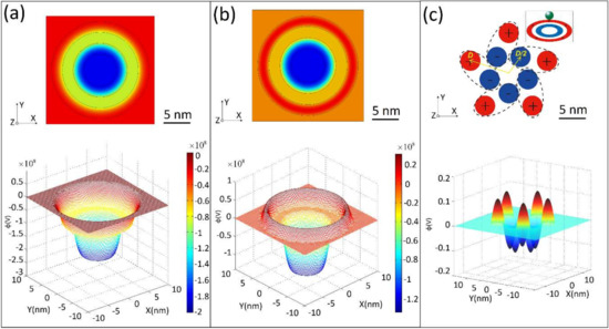
Figure 1.
Electric potential distributions were generated by different charge distributions at the nanoscale. (a) Continuous charge distribution in a double-ring structure, where the inner ring (negative charged) and outer ring (positive charged) exhibit the same charge quantity of 100 charges. (b) Continuous charge distribution of the same double-ring structure, where both rings exhibit the charge density but opposite sign: Positive for outer ring and negative for inner ring. (c) Discrete charge distribution of five dipoles arranged in a nano-pentagon shape.
Figure 1 shows that the depth of the potential well of the first configuration (the same charge quantity) is deeper than that of the second configuration (the same charge density). This provides a clue for designing electrostatic wells with a limited number of discrete charges: The first charge configuration could result in a considerably effective trapping/binding performance, and maintain electrically neutral meanwhile, which is considered to be quite similar to the actual situation in case of proteins or enzymes.
Next, we designed a special pentagon nanostructure with five dipoles as a simple but representative example of a local charge distribution resembling that in biomacromolecules. As shown in Figure 1c, this structure can be considered as a pentagram consisting of 10 charged nanospheres, five positively charged and five negatively charged, where each sphere exhibits an absolute charge of Q = 20 e. Note that each nanosphere exhibits a diameter of 4.0 nm. The distance from the center of the pentagon to the center of each outer nanosphere was defined as D (highlighted in yellow in Figure 1c), where each of the five inner nanospheres were located at a distance of D/2 to the center. As the whole nanostructure was electrically neutral, it can be considered as a cluster of five dipoles, as marked by the dashed ovals. The lower diagram in Figure 1c shows a typical distribution of the electrostatic potential of the pentagon nanostructure with D = 8.0 nm.
3.2. Effect of the Energy on the Trapping Performance of Nanoscale Electric Potential Trapping Wells in a Saline Solution
At the nanometer scale, the thermodynamic effects, van der Waals interactions, and screen effects play non-negligible roles in the trapping performance of electric potential wells. Considering that 0.15 mol/L NaCl solutions could be approximately simplified as an implicit solvent because of the lower valence of Na+ and Cl−, our numerical calculation was based on the nonlinear Poisson–Boltzmann equation [48]:
where is the potential, is the vacuum permittivity, is the relative dielectric constant of water (considered as 80 in this study), is the Boltzmann constant, kBT is the thermal quantum energy at temperature T, and is the charge number of the individual ions, i.e., zi = 1 for Na+ and zi = −1 for Cl− [49]. In the saline solution, the concentrations of Na+ and Cl− were considerably higher than those of H+ and OH−; thus, the latter was considered to be negligible. Therefore, the net charge density in the solution was . At locations far away from the nanostructure, the potential of the bulk solution was considered to be 0. The ion concentration was then set to be a constant (0.15 mol/L) for both Na+ and Cl−, and . The ion concentration near the nanostructure, , was then calculated from a Boltzmann distribution . For each nanosphere, the charges were assumed to be uniformly distributed with a density of , which resulted into within the nanosphere.
The equilibrium state of a trapping well system is assumed to correspond to the minimum free energy. The change of the Helmholtz free energy, ΔF, for the whole system is , which comprises an electrostatic energy term () and an entropy contribution (−TΔS) [50,51], where and:
The values of Uel, −TΔS, and ΔF of the whole system were reported to be very sensitive to D. At smaller D values, e.g., D = 7.2 nm, Uel, −TΔS, and ΔF were calculated to be 177.0, 35.8, and 231.3 kBT, respectively. When D increased from 7.2 to 10.4 nm, Uel increases but both ΔF and −TΔS decreased. The scenarios corresponding to the abovementioned values of D is plotted in Figure 2a, which shows that at D > 8 nm, the charging rate of all three terms reduced. Therefore, in the following calculations, we fix D at 8.0 nm to study the remaining properties of the system.
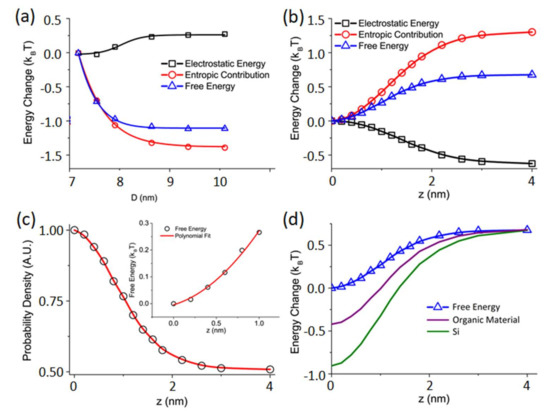
Figure 2.
Dependence of the electrostatic energy, entropic contribution, and free energy on the other related factors for the nanoscale pentagon electric potential well in the saline solution. (a) Relation between the energy change and the diameter, D, along the z = 0 plane. (b) Dependence of the energy change at varied heights, z, above the structure plane. (c) Probability density as a function of the height, z. (d) The influence of different materials on the energy change.
Then, we examined the trapping performance of the pentagon nanostructure. Figure 2b shows the calculated energy depth for 3 m diameter trapping nanospheres with a uniform charge of +20 e. As shown in Figure 1c, the pentagon nanostructure is located at the center of the X-Y plane, and the particle is located at (0, 0, z). At z = 0, Uel, −TΔS, and ΔF were calculated to be 203.3, 39.1, and 242.4 kBT, respectively. When z increased, Uel decreased; however, −TΔS increased, and ΔF continued to increase. The best-fitting curve for ΔF shown in blue clearly shows an effective energy well, where the trapping depth is ~0.65 kBT when z lies between (−4, + 4 nm); thus, a localized shallow trap can be characterized.
We can obtain the probability density distribution from the energy terms using the general correlation , as shown in Figure 2c. In the trapping well, the charged target nanoparticle is supposed to experience a force, T, towards the center, which can be determined by . Moreover, in the vicinity of z = 0, the system can be considered as a harmonic oscillator [28]. This configuration results in a second-order free energy, , and an elastic coefficient of . The data for are then plotted in Figure 2c.
Under this condition, the absolute value of −TΔS was always reported to be larger than that of Uel. This is attributed to a strong shielding effect because of a high numerical density of ions in the system, where the Debye length resulting from a charge screen effect was in the order of 1 nm. This led to a weakened effective electrostatic field at locations a few nanometers away from the pentagon nanostructure [48]. Furthermore, the large number of ions in the solution resulted in a larger reduction of entropy because the random distribution of ions in the bulk solution turned into a relatively regular distribution near the pentagon nanostructure.
The Van der Waals interaction is another important factor that may affect the performance of the trapping well [27]. To calculate the influence of the van der Waals effect, the 4 nm diameter nanospheres and the target nanoparticle were assumed to be composed of a material with a known dielectric constant. Here, two fixed materials were used to obtain the results: Silicon and an organic material. The additional van der Waals energy was then calculated using Hamaker’s equation [52]:
where r is the distance between the two spheres, and and are the respective radii of the two spheres. The Hamaker constant, A, for silicon and the organic material were set at and , respectively [24]. The effect of the van der Waals interaction was reported to be negligible when z was ≥4 nm. However, when z approached 0, the effect became stronger, and it enhanced the depth of the trap. This effect was reported to be sensitive to the materials chosen for the trap and particle. The system composed of Si demonstrated a deeper trapping depth than that composed of the organic material. The results are plotted in Figure 2d, which indicate that different choices of device materials and different compositions of the target nanoparticles may result in a remarkable change in the trapping performance of artificial electrostatics nanotraps.
The trapping depth shown in Figure 2d ranges from 1 to 1.5 kBT, depending on the choice of the construction material. This result may lead to a moderate trapping performance. Moreover, certain fluctuations in the surrounding environment, such as the Brownian motion induced by thermal energy, may cause the trapping effect to be unstable. Another effect to be mentioned is the Stern layer at the solid-liquid interface, which was not included in the present simulations for simplification, but which may weaken the performance of the trap [27].
Clearly, the electric potential distribution resulting from groups of biomacromolecules could be dramatically influenced by different charge distributions. To mimic the phase change of biomacromolecules, we simulated the electric potential distributions from eight cases where each cluster of five dipoles demonstrated the same geometric shape (Figure 1c) but a different charge polarity orientation, as shown in Figure 3a. Figure 3b shows the corresponding potential distributions along the plane of the pentagon nanostructure. This simulated result provides a reference basis for the local charge design of nanorobots.
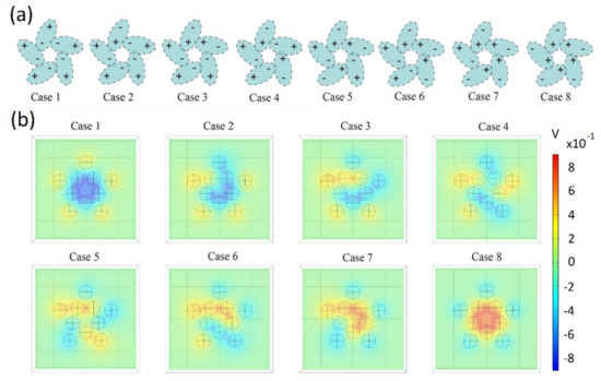
Figure 3.
(a) Eight different charge distribution cases on the pentagon-shaped dipole arrangement. (b) Simulated results of the corresponding electrostatic potential distributions.
3.3. Trapping Capacity between Two-Pentagon Nanostructures
Understanding the electrical interaction among the above pentagon nanostructures is interesting, e.g., whether they are capable of trapping each other. However, it is hard to discuss, due to various combinations between these eight nanostructures. Here, only a simple configuration was selected for our calculations to obtain a flavor of the energy terms of a two-pentagon system.
In this simple case, the two pentagons were parallel to each other along the X-Y plane with one center located at (0, 0, 0) and the other at (0, 0, z0). We considered z0 = 5 nm, and the latter pentagon was rotated at angle (θ) in the X-Y plane compared to the other pentagon. For such a five-fold rotational symmetry, the energy terms of F(θ) were calculated from θ = 0° to θ = 72° for each of the eight different configurations (Figure 4a).
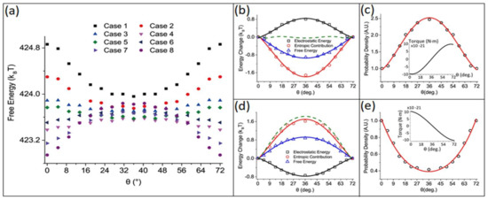
Figure 4.
Energy change between two relative pentagon nanostructures as a function of the rotation angle, θ. (a) Dependence of the free energy on θ for the eight different cases shown in Figure 3a. In (b,c), the calculated results of the energy change and probability density as a function of the rotation angle in Case 1 are shown, whereas in (d,e), the corresponding values for Case 8 are presented.
As shown in Figure 4b,d, two sets of typical results exist for the configurations of Case 1 and Case 8 as shown in Figure 3a, respectively. In Case 1, the system was stable at θ = 36°; however, in Case 8, the stable location was located at θ = 0°. For both cases, the angle-dependent probability density distribution, f(θ), and torque, τ(θ), are shown in Figure 4c,e, respectively. Researchers reported that the van der Waals interactions could influence the stability of the two-pentagon systems. The contributions of the van der Waals interaction for each case are presented by a dashed green line in Figure 4b,d, respectively, where constant A was considered as . In this condition, the van der Waals interaction enhanced the angular stability in Case 1 but weakened it in Case 8.
For all different configurations of the two-pentagon combinations, researchers reported that the total system energy was always lowered by 10 kBT, which was more than the sum of energy when the two pentagons were located far away (i.e., at infinity) from each other. This indicates an aggregation trend of these nanostructures. Moreover, as expected, the results demonstrate that because of the same size and geometric shape, the charge distribution of neutral nanostructures can influence the interactions between them.
Our work demonstrates that the distance z and relative angle θ influence the energy changes of these factors, and hence the final trapping performance of a nanoscale potential well. Understanding the comprehensive impact of various factors, including the electrostatic Coulomb potential, entropy, van der Waals interactions, and hydrogen bonding, on the trapping, binding, and releasing between nanostructures will be very helpful for guiding the development of nanorobot preparation, protein design, macromolecular detection, as well as other related fields.
4. Conclusions
Using our simplified model, we briefly studied the contributions of electrostatic energy, change of entropy, and van der Waals interactions on the trapping performance of a 10 nm, pentagon-shaped nanotrap (i.e., an electric potential well) for a charged nanoparticle in a saline solution. Our semiquantitative results demonstrate that under certain configurations, the system exhibited a moderate trapping effect with a well depth of 1.0–1.5 kBT. Compared to the thermal quantum energy, kBT, such a trap was very unstable. However, van der Waals interactions between the trap and the target particle were reported to enhance the trapping capability. The contribution of the entropy term was reported to play a remarkable mediation role in the trapping performance. Although the considered model was somewhat simple, the results of this work offer certain ideas for designing and constructing artificial micro- and nano-electrostatic potential tweezers, and in particular, for constructing and operating nanorobots working in the wet environment of a biosystem. These results may shed some light on understanding the working mechanisms of biomacromolecules such as antibodies, enzymes, and ion channels.
Author Contributions
Conceptualization, J.X. and S.X.; methodology and software, J.G., Z.L. and F.W.; validation, F.W.; formal analysis, J.G. and J.X.; writing—original draft preparation, Z.L. and J.X.; writing—review and editing, S.X.; funding acquisition, S.X. and J.X. All authors have read and agreed to the published version of the manuscript.
Funding
This research was funded by the National Key R&D Program of China (2016YFA0200802N and 2017YFA0701302) and the Fundamental Research Funds of Shandong University (2018GN030 and 2019GSF111053).
Acknowledgments
We thank Jianming Xue for his valuable advice and Sanjin Xu for his help in the revision.
Conflicts of Interest
The authors declare no conflict of interest.
References
- Cao, B.-Y.; Sun, J.; Chen, M.; Guo, Z.-Y. Molecular Momentum Transport at Fluid-Solid Interfaces in MEMS/NEMS: A Review. Int. J. Mol. Sci. 2009, 10, 4638–4706. [Google Scholar] [CrossRef] [PubMed]
- Sikder, U.; Usai, G.; Yen, T.-T.; Horace-Herron, K.; Hutin, L.; Liu, T.-J.K. Back-End-of-Line Nano-Electro-Mechanical Switches for Reconfigurable Interconnects. IEEE Electron Device Lett. 2020, 41, 625–628. [Google Scholar] [CrossRef]
- Maity, R.; Maity, N.P.; Baishya, S. An Efficient Model of Nanoelectromechanical Systems Based Ultrasonic Sensor With Fringing Field Effects. IEEE Sens. J. 2020, 20, 1746–1753. [Google Scholar] [CrossRef]
- Mendes, R.G.; Wrobel, P.S.; Bachmatiuk, A.; Sun, J.; Gemming, T.; Liu, Z.; Ruemmeli, M.H. Carbon Nanostructures as a Multi-Functional Platform for Sensing Applications. Chemosensors 2018, 6, 60. [Google Scholar] [CrossRef]
- Qiu, S.; Weng, Y.; Li, Y.; Chen, Y.; Pan, Y.; Liu, J.; Lin, W.; Chen, X.; Li, M.; Lin, T.; et al. Raman profile alterations of irradiated human nasopharyngeal cancer cells detected with laser tweezer Raman spectroscopy. RSC Adv. 2020, 10, 14368–14373. [Google Scholar] [CrossRef]
- Padgett, M.; Bowman, R. Tweezers with a twist. Nat. Photonics 2011, 5, 343–348. [Google Scholar] [CrossRef]
- Anderegg, L.; Cheuk, L.W.; Bao, Y.; Burchesky, S.; Doyle, J.M. An Optical Tweezer Array of Ultracold Molecules. Science 2019, 365, 1156–1158. [Google Scholar] [CrossRef]
- Norcia, M.A.; Young, A.W.; Eckner, W.J.; Oelker, E.; Kaufman, A.M. Seconds-scale coherence in a tweezer-array optical clock. arXiv 2019, arXiv:1904.10934. [Google Scholar] [CrossRef]
- Barredo, D.; De, L.S.; Lienhard, V.; Lahaye, T.; Browaeys, A. An atom-by-atom assembler of defect-free arbitrary two-dimensional atomic arrays. Science 2016, 354, 1021–1023. [Google Scholar] [CrossRef]
- Bai, Y.; Lu, Y.; Wang, K.; Cheng, Z.; Qu, Y.; Qiu, S.; Zhou, L.; Wu, Z.; Liu, H.; Zhao, J.; et al. Rapid Isolation and Multiplexed Detection of Exosome Tumor Markers Via Queued Beads Combined with Quantum Dots in a Microarray. Nano-Micro Lett. 2019, 11. [Google Scholar] [CrossRef]
- Xiao, Z.; Kim, H.; Min, J.K. Design, Implementation, and Analysis of a 3-D Magnetic Tweezer System With High Magnetic Field Gradient. IEEE Trans. Instrum. Meas. 2019, 68, 680–687. [Google Scholar]
- Ostrofet, E.; Papini, F.S.; Dulin, D. Correction-free force calibration for magnetic tweezers experiments. Sci. Rep. 2018, 8, 17811. [Google Scholar] [CrossRef] [PubMed]
- Yao, J.; Zhu, G.; Zhao, T.; Takei, M. Microfluidic device embedding electrodes for dielectrophoretic manipulation of cells—A review. Electrophoresis 2019, 40, 1166–1177. [Google Scholar] [CrossRef] [PubMed]
- Viefhues, M.; Eichhorn, R. DNA dielectrophoresis: Theory and applications a review. Electrophoresis 2017, 38, 1483–1506. [Google Scholar] [CrossRef]
- Liang, H.; Peng, Z.; Wu, J.; Chuang, H.S.; Wang, W. On-demand dielectrophoretic immobilization and high-resolution imaging of C. elegans in microfluids. Sens. Actuators B Chem. 2017, 259, 703–708. [Google Scholar]
- Sohani, M.; Ebrahimi, A.M. Time-dependent magnetic field effects on the stability regions of a Paul trap. Eur. Phys. J. Plus 2016, 131, 1–7. [Google Scholar] [CrossRef]
- Li, H.X.; Zhang, Y.; He, S.G.; Tong, X. Determination of the geometric parameters kappa(z) and kappa(r) of a linear Paul trap. Chin. J. Phys. 2019, 60, 61–67. [Google Scholar] [CrossRef]
- Figgatt, C.; Ostrander, A.; Linke, N.M.; Landsman, K.A.; Zhu, D.; Maslov, D.; Monroe, C. Parallel Entangling Operations on a Universal Ion Trap Quantum Computer. Nature 2018, 572, 368–372. [Google Scholar] [CrossRef]
- Oleshko, V.P.; Howe, J.M. Are electron tweezers possible? Ultramicroscopy 2011, 111, 1599–1606. [Google Scholar] [CrossRef]
- Angel Castellanos-Reyes, J.; Castrejon-Figueroa, J.; Maciel-Escudero, C.; Reyes-Coronado, A. Electronic tweezers for magnesium oxide nanoparticles. Mater. Today-Proc. 2019, 13, 341–348. [Google Scholar] [CrossRef]
- Sheng-Yong, X.; Wei-Qiang, S.; Meng, Z.; Jian, X.; Lian-Mao, P. Transmission electron microscope observation of a freestanding nanocrystal in a Coulomb potential well. Nanoscale 2010, 2, 248–253. [Google Scholar]
- Zheng, H.; Mirsaidov, U.M.; Wang, L.W.; Matsudaira, P. Electron beam manipulation of nanoparticles. Nano Lett. 2012, 12, 5644–5648. [Google Scholar] [CrossRef] [PubMed]
- Madhavi, K.; Nassiredin, M.; Philipp, K.; Vahid, S. Geometry-induced electrostatic trapping of nanometric objects in a fluid. Nature 2010, 467, 692–695. [Google Scholar]
- Madhavi, K. Electrostatic free energy for a confined nanoscale object in a fluid. J. Chem. Phys. 2013, 138, 114906. [Google Scholar]
- Xu, J.; Lei, Z.; Guo, J.; Huang, J.; Wang, W.; Reibetanz, U.; Xu, S. Trapping and Driving Individual Charged Micro-particles in Fluid with an Electrostatic Device. Nano-Micro Lett. 2016, 8, 270–281. [Google Scholar] [CrossRef] [PubMed]
- Pethig, R. Review Article-Dielectrophoresis: Status of the theory, technology, and applications. Biomicrofluidics 2010, 4. [Google Scholar] [CrossRef]
- Zheng, L.; Brody, J.P.; Burke, P.J. Electronic manipulation of DNA, proteins, and nanoparticles for potential circuit assembly. Biosens. Bioelectron. 2005, 20, 606–619. [Google Scholar] [CrossRef]
- Yang, L.X.; Zhang, G.C.; Fan, N.; Guo, J.; Peng, B. Minimum damping profile of micro/nano-robot and as the carrier for drug delivery: Theory study. J. Phys. Conf. Ser. 2019, 1209, 012019. [Google Scholar] [CrossRef]
- Halder, A.; Sun, Y. Biocompatible propulsion for biomedical micro/nano robotics. Biosens. Bioelectron. 2019, 139. [Google Scholar] [CrossRef]
- Malinska, M.; Jarzembska, K.N.; Goral, A.M.; Kutner, A.; Wozniak, K.; Dominiak, P.M. Sunitinib: From charge-density studies to interaction with proteins. Acta Cryst. Sect. D-Struct. Biol. 2014, 70, 1257–1270. [Google Scholar] [CrossRef]
- Berg, B.V.D.; Clemons, W.M.; Collinson, I.; Modis, Y.; Hartmann, E.; Harrison, S.C.; Rapoport, T.A. X-ray structure of a protein-conducting channel. Nature 2003, 427, 36–44. [Google Scholar] [CrossRef]
- Fried, S.D.; Boxer, S.G. Response to Comments on “Extreme electric fields power catalysis in the active site of ketosteroid isomerase“. Science 2014, 346, 1510–1514. [Google Scholar] [CrossRef]
- Jeronimo, L.; Ram Prasad, B.; Zhen, T.; Chu Arieh, W. Methyltransferases do not work by compression, cratic, or desolvation effects, but by electrostatic preorganization. Proteins Struct. Funct. Bioinform. 2015, 83, 318–330. [Google Scholar]
- Suydam, I.T.; Snow, C.D.; Pande, V.S.; Boxer, S.G. Electric fields at the active site of an enzyme: Direct comparison of experiment with theory. Science 2006, 313, 200–204. [Google Scholar] [CrossRef] [PubMed]
- Benkovic, S.J.; Sharon, H.S. A perspective on enzyme catalysis. Science 2003, 301, 1196–1202. [Google Scholar] [CrossRef] [PubMed]
- Irina, A.; Vassylyeva, M.N.; Dmitri, S.; Vladimir, S.; Anna, P.; Noriyuki, I.; Naohiro, M.; Soichi, W.; Tahirov, T.H.; Vassylyev, D.G. Allosteric modulation of the RNA polymerase catalytic reaction is an essential component of transcription control by rifamycins. Cell 2005, 122, 351–363. [Google Scholar]
- Nelso, P.; Doniach, S. Biological Physics: Energy, Information, Life, 3rd ed.; W.H. Freeman and Company: New York, NY, USA, 2013. [Google Scholar]
- Duan, L.L.; Mei, Y.; Zhang, D.; Zhang, Q.G.; Zhang, J.Z.H. Folding of a helix at room temperature is critically aided by electrostatic polarization of intraprotein hydrogen bonds. J. Am. Chem. Soc. 2010, 132, 11159. [Google Scholar] [CrossRef]
- Cho, A.E.; Victor, G.; Berne, B.J.; Richard, F. Importance of accurate charges in molecular docking: Quantum mechanical/molecular mechanical (QM/MM) approach. J. Comput. Chem. 2005, 26, 915. [Google Scholar] [CrossRef]
- Frauke, G.T.; Schwarzl, S.M.; Annick, D.; Stefan, F.; Smith, J.C. Protein/ligand binding free energies calculated with quantum mechanics/molecular mechanics. J. Phys. Chem. B 2005, 109, 10474. [Google Scholar]
- Yi, C.; Sun, X.; Ye, J.; Ding, L.; Liu, M.; Yang, Z.; Lu, X.; Zhang, Y.; Ma, L.; Gu, W.; et al. Key residues of the receptor binding motif in the spike protein of SARS-CoV-2 that interact with ACE2 and neutralizing antibodies. Cell. Mol. Immunol. 2020. [Google Scholar] [CrossRef]
- Hussain, M.; Jabeen, N.; Raza, F.; Shabbir, S.; Baig, A.A.; Amanullah, A.; Aziz, B. Structural variations in human ACE2 may influence its binding with SARS-CoV-2 spike protein. J. Med Virol. 2020, 1–7. [Google Scholar] [CrossRef] [PubMed]
- Gascon, J.A.; Batista, V.S. QM/MM study of energy storage and molecular rearrangements due to the primary event in vision. Biophys. J. 2004, 87, 2931–2941. [Google Scholar] [CrossRef] [PubMed]
- Abrar, M.N.; Sagheer, M.; Hussain, S. Entropy generation during peristaltically flowing nanofluid in an axisymmetric channel with flexible walls. Phys. Scr. 2020, 95, 035206. [Google Scholar] [CrossRef]
- Dimova, M.; Devedjiev, Y.D. Protein crystal lattices are dynamic assemblies: The role of conformational entropy in the protein condensed phase. IUCrJ 2018, 5, 130–140. [Google Scholar] [CrossRef] [PubMed]
- Roudbari, M.A.; Jorshari, T.D.; Arani, A.G.; Lu, C.; Rabczuk, T. Transient responses of two mutually interacting single-walled boron nitride nanotubes induced by a moving nanoparticle. Eur. J. Mech. A-Solids 2020, 82, 103978. [Google Scholar] [CrossRef]
- De Luca, S.; Chen, F.; Seal, P.; Stenzel, M.H.; Smith, S.C. Binding and Release between Polymeric Carrier and Protein Drug: pH-Mediated Interplay of Coulomb Forces, Hydrogen Bonding, van der Waals Interactions, and Entropy. Biomacromolecules 2017, 18, 3665–3677. [Google Scholar] [CrossRef]
- Munday, D.L. Surfaces, Interfaces and Colloids—Principles and Applications, 2nd ed.; Myers, D., Ed.; Wiley-VCH: New York, NY, USA, 1999. [Google Scholar]
- Evans, D.F.; Wennerström, H. The Colloidal Domain: Where Physics, Chemistry, Biology, and Technology Meet; VCH Publishers: New York, NY, USA, 1999. [Google Scholar]
- Stigter, D. Evaluation of the counterion condensation theory of polyelectrolytes. Biophys. J. 1995, 69, 380. [Google Scholar] [CrossRef]
- Theodoor, J.; Overbeek, G. The role of energy and entropy in the electrical double layer. Colloids Surf. 1990, 51, 61–75. [Google Scholar] [CrossRef]
- Hamaker, H.C. The London—van der Waals attraction between spherical particles. Physica 1937, 4, 1058–1072. [Google Scholar] [CrossRef]
© 2020 by the authors. Licensee MDPI, Basel, Switzerland. This article is an open access article distributed under the terms and conditions of the Creative Commons Attribution (CC BY) license (http://creativecommons.org/licenses/by/4.0/).