Article

Polymer Thin Film Promotes Tumor Spheroid Formation via JAK2-STAT3 Signaling Primed by Fibronectin-Integrin α5 and Sustained by LMO2-LDB1 Complex

1
Department of Biotechnology, College of Life Science and Biotechnology, Korea University, Seoul 02841, Korea
2
Department of Biological Sciences, Korea Advanced Institute of Science and Technology (KAIST), Daejeon 34141, Korea
*
Authors to whom correspondence should be addressed.
These authors contributed equally to this work.
Biomedicines 2022, 10(11), 2684; https://doi.org/10.3390/biomedicines10112684
Submission received: 6 October 2022 / Revised: 18 October 2022 / Accepted: 19 October 2022 / Published: 24 October 2022
(This article belongs to the Collection Recent Advances in Cancer Stem Cells)

Abstract

:
Cancer stem-like cells (CSCs) are considered promising targets for anti-cancer therapy owing to their role in tumor progression. Extensive research is, therefore, being carried out on CSCs to identify potential targets for anti-cancer therapy. However, this requires the availability of patient-derived CSCs ex vivo, which remains restricted due to the low availability and diversity of CSCs. To address this limitation, a functional polymer thin-film (PTF) platform was invented to induce the transformation of cancer cells into tumorigenic spheroids. In this study, we demonstrated the functionality of a new PTF, polymer X, using a streamlined production process. Polymer X induced the formation of tumor spheroids with properties of CSCs, as revealed through the upregulated expression of CSC-related genes. Signal transducer and activator of transcription 3 (STAT3) phosphorylation in the cancer cells cultured on polymer X was upregulated by the fibronectin-integrin α5-Janus kinase 2 (JAK2) axis and maintained by the cytosolic LMO2/LBD1 complex. In addition, STAT3 signaling was critical in spheroid formation on polymer X. Our PTF platform allows the efficient generation of tumor spheroids from cancer cells, thereby overcoming the existing limitations of cancer research.

1. Introduction

With recent advances in cancer biology, significant improvements have been made in clinical diagnosis and cancer treatment. However, successful anti-cancer treatments remain restricted due to cancer recurrence, metastasis, and cell resistance towards therapeutics [1,2,3]. Recent studies have suggested that cancer stem-like cells (CSCs) are a subpopulation of cancer cells responsible for posing challenges to the complete surgical resection of tumors [4,5]. CSCs share several molecular and biological characteristics with normal stem cells [4,5]. However, multiple signaling pathways mediating the survival, proliferation, and differentiation of normal stem cells are aberrantly regulated in CSCs [4,6]. Notch, Wnt, Hedgehog, nuclear factor-κB (NF-κB), Janus kinase/signal transducers and activators of transcription (JAK/STAT), and tumor growth factor-β (TGF-β) signaling pathways play crucial roles in maintaining the population and characteristics of CSCs [4,6].
To identify potential targets for cancer therapy, the isolation and characterization of CSCs in primary tumors of patients are crucial. Thus, methods to isolate CSCs from primary tumors using cell surface markers or specific cell culture conditions have recently been developed [7,8,9]. For example, cellular surface markers such as CD24, CD44, CD133, epithelial cell adhesion molecule (EpCAM), leucine-rich-repeat-containing G-protein-coupled receptor 5 (LGR5), and CD117 have been used to isolate CSCs from cancer cells using fluorescence-activated cell sorting (FACS) and magnetic cell sorting [10,11,12,13]. In addition, serum-free culture conditions supplemented with various combinations of growth factors have been applied to enrich or maintain isolated CSCs from patients diagnosed with cancer [14,15,16].
Despite the current efforts, the use of patient-derived CSCs involves several limitations. First, the majority of human cancers harbor rare populations of CSCs (<1–2%) [17,18,19,20]. Second, CSC markers identified to date have not been fully established. Third, only a few CSCs express the identified CSC markers [21].
A functional polymer thin-film (PTF) platform was, therefore, developed to overcome these limitations [22]. Poly (2,4,6,8-tetravinyl-2,4,6,8-tetramethyl cyclotetrasiloxane), designated as pV4D4, induced the transformation of cancer cells to tumorigenic and drug-resistant CSCs [22]. Non-CSCs can dedifferentiate and acquire stem cell-like properties owing to their plasticity [23,24]. Several cellular signaling pathways, such as Wnt, TGF-β, and JAK-STAT, can drive cancer cell dedifferentiation and enhance stem cell-like properties [25,26,27].
In this study, we evaluated the functionality of a modified version of a previous PTF platform, pV4D4, which had issues with mass production. This version was designated as polymer X. In addition, we identified the cell-signaling mechanisms underlying tumor spheroid formation under polymer X culture conditions.

2. Materials and Methods

2.1. Cell Lines and Culture Condition

The human ovarian cancer cell line SKOV3 was purchased from the Korean Cell Line Bank. The cells were cultured and maintained in the Roswell Park Memorial Institute-1640 (RPMI-1640) medium (Gibco, Grand Island, NY, USA) supplemented with 10% fetal bovine serum (FBS) (HyClone, Logan, UT, USA), 1% penicillin/streptomycin (HyClone), and 25 mM HEPES (Gibco).
Human glioblastoma (GBM) cell lines A1207, LN18, LN229, T98G, and U87MG and the human cervical cancer cell line HeLa were purchased from the American Type Culture Collection (ATCC). These cell lines were cultured and maintained in Dulbecco’s modified Eagle’s medium (DMEM) with high glucose (4500 mg/L) (HyClone), supplemented with 10% FBS, 1% penicillin/streptomycin, 2 mM L-glutamine, and 50 µg/mL gentamicin (Tocris, Bristol, UK). All cells were incubated and maintained at 37 °C in a humidified 5% CO2 chamber.

2.2. Spheroid Formation on pV4D4 and Polymer X and Treatment of Inhibitors

Cells were seeded at a density of 2 × 10⁶ cells (100 mm dish) or 0.6 × 10⁶ cells (60 mm dish) on pV4D4-coated plates or polymer X-coated plates. RPMI-1640 or DMEM, as appropriate, containing 10% Knockout Serum Replacement (SR) (Gibco) 1% penicillin/streptomycin and 25 mM HEPES, was used as culture medium. For optimum spheroid growth, the culture medium was replaced every 2 days. Spheroids were dissociated with an accutase solution (Sigma-Aldrich, St. Louis, MO, USA) every 4 days to prevent cell death at the spheroid core.
The focal adhesion kinase (FAK) inhibitor 14, Y15 (7.5 μM) (Cat. #3414, Tocris) was used to inhibit integrin-mediated activation of FAK. JAK inhibitor I (1 μM) (Cat. #420099, Calbiochem, Ct, San Diego, CA, USA) and AG-490 (200 μM) (Cat. #0414, Tocris) was used to prevent the phosphorylation of JAK. Nifuroxazide (50–100 μM) (cat. #481984, Calbiochem) and S3I-201 (50 μM) (Cat. #S1155, Selleckchem, Houston, TX, USA) were used to prevent phosphorylation of STAT3.

2.3. Plasmids

For the reporter assay, transcription factor response elements (22 × TAT3 response element: 5′-TGCTTCCCGAACGT-3′, 5 × CSL response element: 5′-CCGTGGGAA-3′, 4 × Smad binding element: 5′-CAGACA-3′, 4 × TCF/LEF1 response element: 5′-ATCAAAGG-3′, 2 × NF-κB response element: 5′-GGGACTTTCCGCTGGGGACTTTCC-3′, 8 × GLI response element: 5′-GAACACCCA-3′) were cloned into the pLL-mCMV-EGFP-PGK-Blast vector.

2.4. Lentiviral Transduction and Gene Transfections

For lentivirus preparation, each reporter gene vector was transfected with second-generation lentiviral packaging plasmids, ∆8.9 and VSV-G, using the LipoJet transfection reagent (SignaGen Laboratories, Rockville, MD, USA) into an immortalized human kidney cell line, HEK293FT cells (ATCC). The pLL-mCMV-EGFP-PGK-Blast vector was used as a control. The transformed lentiviruses were harvested after 48 h post-transfection, incubated with the Lenti-X concentrator (Clontech Laboratories, Mountain View, CA, USA), and centrifuged. The cells were then infected with the transformed lentiviruses for 24 h in the presence of 6 μg/mL hexadimethrine bromide (Sigma-Aldrich) for 24 h.
Small-interfering RNAs (siRNAs) targeting ITGA5, LDB1, and LMO2 were purchased from Sigma Aldrich. siRNAs targeting FN1, JAK1, and JAK2 were designed using the siDirect version 2.0 software [28]. The ScreenFect™ A transfection reagent (Wako Pure Chemical, Osaka, Japan) was used for cellular transfection, according to the manufacturer’s instructions. The siRNA sequences are listed in Table S1.

2.5. Establishment of Reporter Gene-Introduced Cell Lines

SKOV3 cells were infected with lentiviruses containing each reporter gene vector and selected using blasticidin (Cayman Chemical, Ann Arbor, MI, USA). The cells were harvested and washed twice with 1× phosphate buffer saline (PBS). Cells were re-suspended in 1× PBS containing 1% FBS and analyzed using a flow cytometry system (BD FACSAria). Green fluorescent protein (GFP)-negative cells were sorted and used for subsequent experiments.

2.6. Immunocytochemistry/Immunofluorescence (ICC/IF)

Eight-day-cultured SKOV3 spheroids of 100~200 μm were fixed with 4% paraformaldehyde (PFA) (Sigma-Aldrich) for 30 min at room temperature and incubated in 15% sucrose followed by 30% sucrose in PBS overnight at 4 °C. Thereafter, spheroid samples were frozen embedded in the OCT compound (Leica, Wetzlar, Germany) in cryomolds and stored at −80 °C until use. Sample blocks were sliced into 10-μm-thick sections via the Cryostat (Leica) instrument. After rinsing with distilled water and blocking with 3% BSA/1× PBS, they were stained with rabbit anti-human fibronectin primary antibody (ab2413; Abcam) overnight at 4 °C, followed by Alexa Fluor® 647 donkey anti-rabbit secondary antibody (ab150075; Abcam) for 1 h at room temperature. The sections were counterstained with Hoechst 33342 for 10 min and mounted with Mounting Medium (Dako, Santa Clara, CA, USA) for confocal (Carl Zeiss, Oberkochen, Germany) or fluorescent microscopy (Nikon, Minato City, Tokyo, Japan). Captured images were further processed and analyzed using ImageJ (https://imagej.nih.gov accessed on 8 June 2021), and average fluorescence (FL) intensity data were calculated for 5 spheroids and normalized to that of the ultra-low attachment (ULA) (Corning, Corning, NY, USA) spheroid control group.

2.7. RNA Extraction and Quantitative RT-PCR

Total RNA was extracted from cells using the QIAzol lysis reagent (Qiagen, Hilden, Germany) according to the manufacturer’s instructions. RNase-free and DNase-treated RNA (1 µg) were used as a template to synthesize cDNA using the RevertAid First-Strand cDNA Synthesis Kit (Thermo Fisher Scientific, Waltham, MA, USA). Quantitative Reverse Transcription-PCR (qRT-PCR) analysis was performed on an iCycler IQ real-time detection system (Bio-Rad, Hercules, CA, USA) using IQ Supermix with SYBR Green (TaKaRa Bio, Shiga, Japan). Gene expression was quantified using the standard2−∆∆Ct method as described previously [22,29]. The expression levels of the target genes were normalized to those of 18S rRNA. Primers used for qRT-PCR amplification are listed in Table S2.

2.8. Western Blot Analysis

For Western blots, whole-cell extracts were prepared using the radioimmunoprecipitation assay (RIPA) lysis buffer (LPS solution, Daejeon, Korea) comprised of 150 mM NaCl, 1% NP-40, 0.1% SDS, and 50 mM Tris (pH 7.4) containing 1 mM NaF, 1 mM Na3VO4, 1 mM ß-glycerophosphate, 2.5 mM sodium pyrophosphate, and protease inhibitor cocktail (Roche, Basel, Switzerland). Proteins were quantified using the Bradford assay reagent (Bio-Rad) according to the manufacturer’s instructions. Proteins (20–30 μg) were separated on 8–12% sodium dodecyl sulfate polyacrylamide gel electrophoresis (SDS-PAGE) and immunoblotting was performed as described above. The primary antibodies used were as follows: Anti-pSTAT3 (Y705) (1:500, 9145S; Cell Signaling Technology, Danvers, MA, USA), anti-STAT3 (1:1000, 4904S; Cell Signaling Technology), anti-fibronectin (1:500, ab6328; Abcam, Cambridge, UK), anti-integrin α5 (1:1000, ab150361; Abcam), anti-JAK1 (1:500, 50996S; Cell Signaling Technology), anti-JAK2 (1:1000, 3230S; Cell Signaling Technology), anti-GP130 (1:2000, ab226346; Abcam), anti-LDB1 (1:1000, ab96799; Abcam), anti-LMO2 (1:500, NB110-78626SS; Novus Biologicals, Littleton, CO, USA), and anti-α-tubulin (1:10,000, T6199; Sigma Aldrich). α-Tubulin was used as a loading control.

2.9. Reporter Gene Assay

GFP-negative cells were used for the reporter gene assay. Cells were seeded at a density of 6.0 × 10⁵ cells (6-well plate) or 2.0 × 10⁶ cells (100 mm plate) and allowed to form spheroids using the method described earlier. After 8 days of culture, the intensity of the GFP signal was measured using IncuCyte Zoom (Sartorius, Version; 2016 B).

2.10. In Silico Analysis

To investigate the differentially regulated cellular pathways between cells cultured on the tissue culture plate (TCP) and polymer X-coated plate, gene set enrichment analysis (GSEA) was performed using the MsigDB Hallmark gene signature (https://www.broadinstitute.org/gsea accessed on 15 February 2020) [30]. ClueGO, a Cytoscape plugin for the visualization of functionally organized gene ontology/pathway networks, was used to determine the enriched biological terms in spheroids cultured on polymer X-coated plates [31]. The GO analysis was repeated on a published expression dataset (GSE213872).

2.11. Quantification and Statistical Analysis

All experiments were performed at a minimum of three independent replicates. Data were analyzed using a two-tailed Student’s t-test and presented as mean ± standard error of the mean (SEM). The level of statistical significance stated in the text was based on the p-values. a: p < 0.05; b: p < 0.01; and c: p < 0.001 were considered statistically significant.

3. Results

3.1. Polymer X Allowed Formation of Uniform Spheroids and Activated Cellular STAT3 Signaling

To verify the utility of polymer X for the enrichment of tumor spheroids (Figure 1A), we investigated the functionality of polymer X employing an established cancer cell line (Figure 1B). The human ovarian cancer cell line SKOV3 was seeded on a tissue culture plate (TCP) or a polymer X-coated plate. After 8 days of culture, SKOV3 cells formed tumor spheroids on polymer X without additional supplementary factors, but not on TCP (Figure 1B).
CSCs can self-renew and form spheres in serum-free culture. Sphere-forming ability is one of the indicators of the stem cell-like properties of cancer cells [32,33,34]. Thus, we verified the expression of phenotypic markers of CSCs on spheroids cultured on TCP and polymer X-coated plates (Figure 1C). The cellular mRNA expression of LGR5, CD44, sex-determining region Y-box 2 (SOX2), NANOG, Aldehyde dehydrogenase 1 (ALDH1A1), CD117, and EpCAM was upregulated in polymer X-cultured spheroids relative to TCP-cultured cells. Thus, cells culturing on polymer X induced the formation of tumor spheroids, which expressed CSC-associated genes.
To investigate the signaling pathways that contributed to CSC-associated marker upregulation in polymer X-induced spheroids, we constructed a reporter gene assay system (Figure 1D). Reporter system vectors contain promoter elements responsible for Notch, Wnt, Hedgehog, NF-κB, STAT3, and TGF-β signaling pathways. The activation of each signaling pathway was identified using GFP expression. Reporter system-introduced SKOV3 cells were cultured on polymer X for 8 days (Figure 1E). Interestingly, the GFP signal was observed in the tumor spheroids introduced with the STAT3 reporter system. In addition, GSEA showed that STAT3 signaling was more enriched in spheroids cultured on polymer X as compared to those cultured on TCP (Figure 1F).
STAT3 has two important phosphorylation sites, Tyr705 phosphorylated by JAK, and Ser727 phosphorylated by serine/threonine kinase [35]. Phosphorylation of Tyr705 is critical for the nuclear translocation of STAT3, therefore regulating STAT3 signal activation. Therefore, we performed Western blotting to investigate the phosphorylation status of STAT3 in cells cultured on polymer X including previously used pV4D4 (Figure 1G, Figure S1. During spheroid culturing on polymer X, the cells were harvested every 24 h for a total culturing period of 8 days to observe STAT3 phosphorylation (pY705-STAT3; p-STAT3) over time. An upregulation of p-STAT3 was observed at 24 h of culture. Additionally, mRNA expression of target genes of the STAT3 signaling pathway, interleukin-6 (IL-6), early growth response protein 1 (EGR1), p21-activated kinase 2 (PAK2), and prostaglandin-endoperoxide synthase 1 (PTGS1) was analyzed (Figure 1H). Upregulation in the cellular expression of EGR1 and PTGS1 was observed, which was in agreement with the activation of the protein STAT3. Collectively, polymer X induced tumor spheroid formation ability and STAT3 signaling activation in the candidate cancer cells.

3.2. Initial Activation of STAT3 Signaling Was Induced by Fibronectin-JAK2 Axis

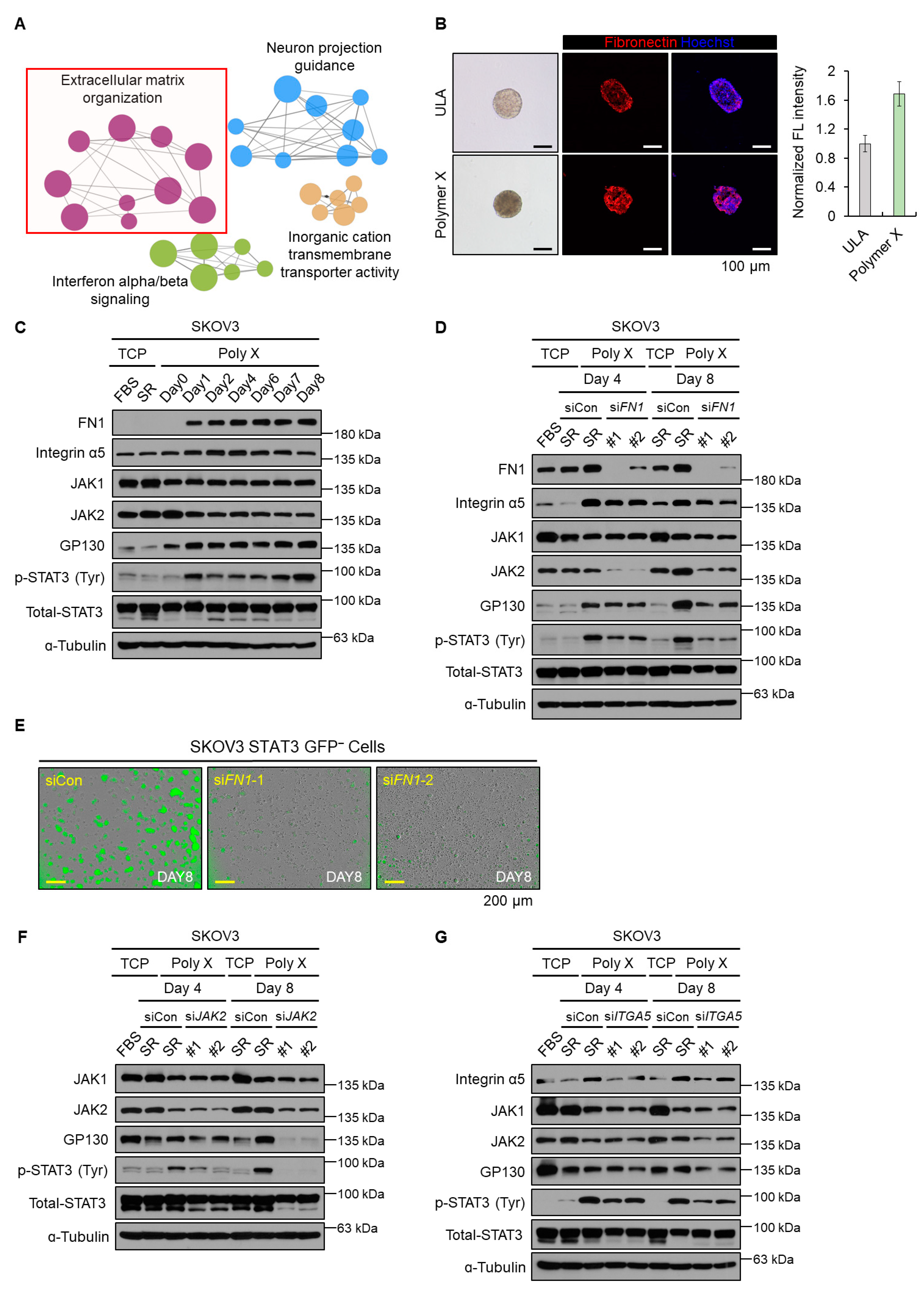
IL-6 is a well-known canonical STAT3 signaling inducer [35]. Interestingly, while STAT3 activation was observed for cells cultured on polymer X, the mRNA expression of IL-6 in these cells decreased (Figure 1H). Therefore, to identify alternative mechanisms inducing STAT3 activation in these cells, gene ontology (GO) analysis using the Cytoscape software add-on ClueGO plugin was conducted on differentially expressed genes (DEGs) that were upregulated in polymer X-cultured spheroids and the results were compared to those for the TCP-cultured cells (Figure 2A). The extracellular matrix (ECM)-related ontology was enriched in the tumor spheroids obtained using polymer X. This observation was in agreement with the findings of a previous study (Figure 2A) [22]. Compared to the tumor spheroids cultured on an ultra-low attachment (ULA) plate, a large amount of ECM was enriched inside those cultured on polymer X (Figure 2B). Additionally, immunofluorescence (IF) staining revealed high expression of fibronectin 1 (FN1) on the tumor spheroids cultured on polymer X (Figure 2B).
Fibronectin has been reported to mediate STAT3 activation [36,37]. Therefore, we cultured cells on polymer X and investigated the levels of cellular proteins associated with the fibronectin and STAT3-signaling pathway (Figure 2C). The protein level of FN1 in these cells increased from day 1 of THE culture and this increase was maintained for 8 days. siRNA-mediated FN1 knockdown during the culture period caused a reduction in the levels of STAT3-related proteins on days 4 and 8 (Figure 2D). In addition, based on the fluorescence intensity measured using the IncuCyte system, FN1 knockdown compromised the spheroid forming ability of the cells cultured on polymer X. Accordingly, the intensity of GFP indicated that the STAT3 reporter activity also reduced (Figure 2E).
Fibronectin binds to integrin and activates focal adhesion kinase (FAK) and SRC [38,39]. As a result, FAK and SRC mediate the outside-in signal transduction [40]. To verify whether the phosphorylation of STAT3 was mediated by the activation of FAK-SRC-mediated signaling, FAK inhibitor 14 was added during the polymer X culture condition, and the levels of STAT3-related proteins were analyzed (Figure S2A). The results showed that while the level of p-SRC decreased, that of p-STAT3 remained unchanged. We therefore hypothesized that canonical integrin-FAK signaling was not a mediator of STAT3 activation in cells cultured on polymer X [36,37].
Additionally, although the levels of STAT3-related proteins revealed maximum reduction on day 8 of the culture, the JAK2 level decreased significantly on day 4 (Figure 2C). JAK transmits an upstream ligand-receptor signal to STAT3 by phosphorylating the Tyr705 residue of STAT3. Several previous studies showed that STAT3 can be activated by integrin via JAK2 [36,41]. To determine whether phosphorylation of STAT3 can be mediated via activation of the fibronectin-JAK2 axis, we knocked down JAK2 using siRNA and analyzed the levels of STAT3-related proteins. The levels of STAT3-related proteins decreased on day 4 of the culture. This observation was in agreement with that recorded for FN1 knockdown in cells (Figure 2D). Additionally, the results displayed that the levels of STAT3-related proteins and tumor spheroid formation decreased when cultured on polymer X supplemented with JAK inhibitor I (Figure S2B,C). In contrast to JAK2, JAK1 is not affected by fibronectin but is related to the STAT3 signaling pathway. To evaluate the effect of JAK1 on the activation of STAT3-related proteins, we knocked down JAK1 using siRNA (Figure S2D). As a result, the level of STAT3-related proteins decreased to some extent but did not show any significant changes compared to that observed as a result of the JAK2 knockdown. Therefore, we concluded that early phosphorylation of STAT3 could be mediated by fibronectin-activated JAK2.
In addition, as fibronectin generally regulates cells through integrin receptors, we examined whether integrin signaling was involved in the activation of JAK2 via fibronectin [42,43]. Since integrin α5β1 is a major receptor of fibronectin, we knocked down integrin subunit alpha 5 (ITGA5) in the SKOV3 cells using siRNA and identified the levels of STAT3-related proteins. The levels of all STAT3-related proteins, including JAK2, decreased when compared to cells transfected with non-target siRNA (Figure 2G). Therefore, we concluded that the tumor spheroids cultured on polymer X were activated by the fibronectin-integrin-JAK2-STAT3 signaling axis.

3.3. Long-Term Activation of STAT3 Signaling via LMO2-LDB1 Complex

In our study, polymer X-cultured cells displayed fibronectin expression that increased from day 1 with the phosphorylation of STAT3, whereas the expression levels of JAK2 were increased from day 1, but gradually decreased over time.
Therefore, we hypothesized that there may be other factors that render p-STAT3 continuously active. According to our previous study, the phosphorylation of STAT3 can be induced by the cytoplasmic LMO2-LDB1 complex [44]. For cells cultured on polymer X, the levels of LMO2 and LDB1 gradually increased from the time of culture until day 8 (Figure 3A).
Unlike the observations recorded post-FN1 knockdown, the knockdown of LMO2 did not cause a reduction in the levels of STAT3-related proteins on day 4 of the culture. This observation was accompanied by an insignificant change in the LMO2 level on day 4 of culture. In contrast, the level of LMO2 significantly increased on day 8. Hence, it was not surprising to observe a significant decrease in the levels of STAT3-related proteins on day 8 as a result of LMO2 knockdown (Figure 3B). Similar results were observed on LDB1 knockdown (Figure 3C). In addition, LMO2 or LDB1 knockdown caused significant reductions in GFP intensity and the spheroid-forming ability of the cells (Figure 3D). Taken together, these results indicated that STAT3 activation and tumor spheroid formation were sustained by LMO2 and LDB1.

3.4. Polymer X-Induced Tumor Spheroids Acquired Cancer Stem-Like Properties via STAT3 Signaling

To examine the significance of upstream genes involved in STAT3 activation regulating properties of tumor spheroids, we knocked down FN1, ITGA5, JAK1, JAK2, LDB1, and LMO2 from the cells. As a result, the mRNA expression of the CSC-associated genes was significantly decreased (Figure 4A). Since early STAT3 activation was regulated by fibronectin-JAK2, and its long-term activation was maintained by the cytoplasmic LMO2-LDB1 complex, phosphorylation of STAT3 on the cells cultured on polymer X might play an important role in regulating cancer stem-like properties in these cells.
We also performed direct STAT3 inhibition to examine its effects on the CSC properties of the candidate cells. The results showed that tumor spheroid formation and the expression of CSC-related genes decreased, even when cultured on polymer X supplemented with inhibitors. We confirmed that various p-STAT3 inhibitors decreased the levels of p-STAT3 in the cells cultured on polymer X (Figure 4B,C), therefore reducing the spheroid-forming ability of these cells (Figure 4D). In addition, the mRNA expression of both STAT3 target genes and CSC-related genes notably decreased (Figure 4E,F).
Moreover, when tumor spheroids cultured on polymer X were transferred to TCP, the levels of STAT3-related proteins, in particular, LMO2 and LDB1, decreased on day 2 of the transfer (Figure 4G). Subsequently, the mRNA expression of CSC-associated markers also markedly decreased (Figure 4H). Collectively, these results indicated that the STAT3 signaling pathway played an important role in the activation of cancer stemness in cancer cells grown on polymer X.

3.5. General Application of Polymer X Using Various Cancer Cell Lines

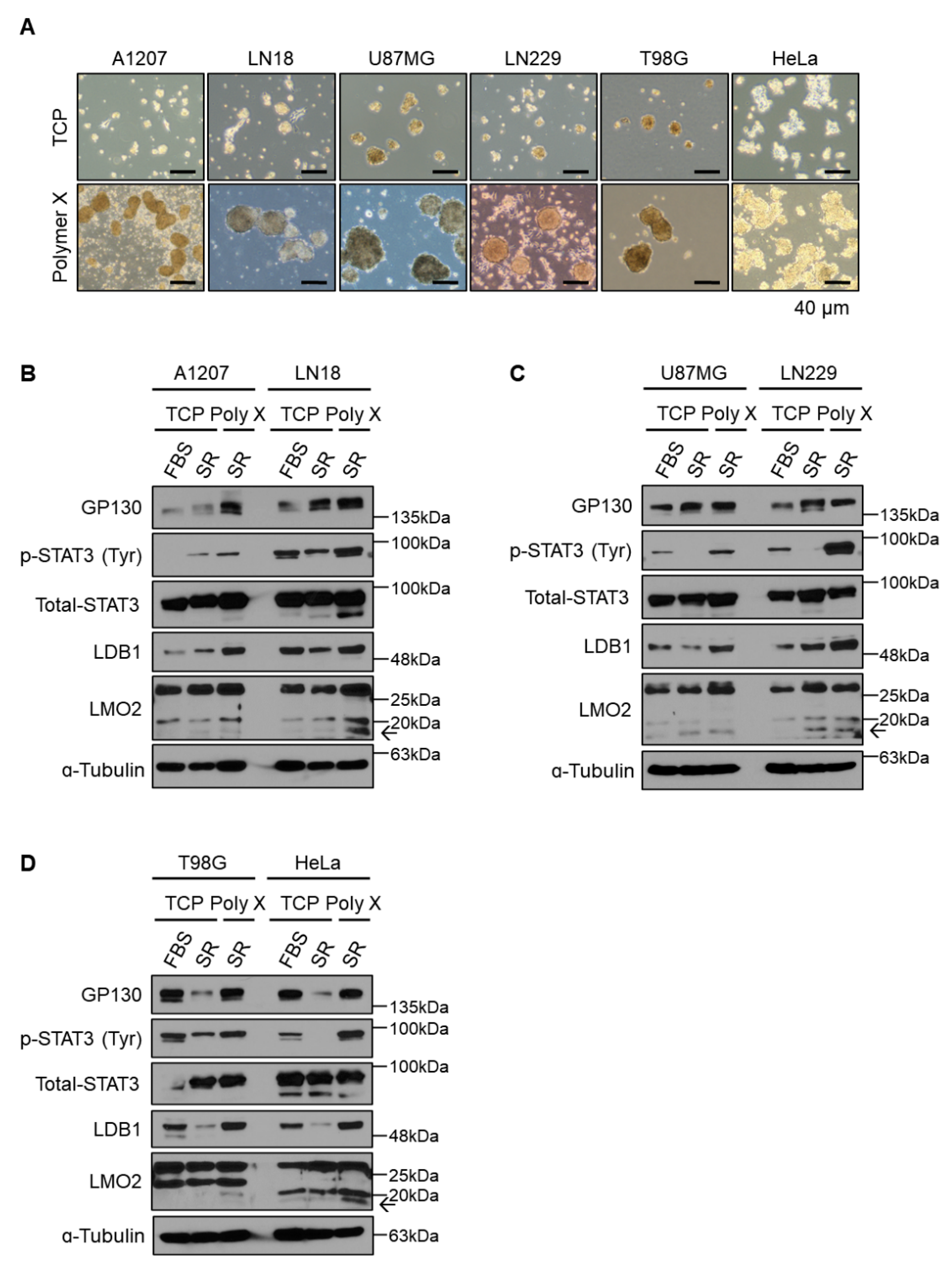
To determine the application of polymer X to other cancer cell lines, human GBM cell lines A1207, LN18, U87MG, LN229, and T98G and the human cervical cancer cell line HeLa were cultured on polymer X and tested for their tumor spheroid formation ability. Similar to the SKOV3 cells, polymer X-cultured GBM cells and HeLa cells formed better spheroids as compared to those cultured on TCP (Figure 5A). In addition, we attempted to determine whether the formation of tumor spheroids was mediated by the activated STAT3 signaling pathway in the cells cultured on polymer X. In line with SKOV3 ovarian cancer cells, STAT3 signaling was also activated in tumor spheroids formed by the various GBM cells and HeLa cells cultured on polymer X (Figure 5B–D). Therefore, the formation of tumor spheroids and STAT3 signaling activation may be a general phenomenon in polymer X-based cell cultures.
Taken together, culturing established cancer cell lines on polymer X would be of great help in the study of cancer biology. In addition, it allows the use of tumor spheroids that exhibit cellular physiological conditions similar to those present in tumors in vivo.

4. Discussion

In this study, we elucidated that when cultured on the newly revised PTF platform, polymer X, the STAT3 signaling pathway was activated to obtain CSC-like properties in tumor cells. In particular, the STAT3 signaling pathway, which was activated through the fibronectin-JAK2 axis, was continuously maintained by the cytoplasmic LMO2-LDB1 complex when cultured on polymer X.
CSCs have been identified as one of the predominant causes of inducing therapeutic resistance in cancer cells. The importance of cells with cancer stem-like properties has been, therefore, constantly emerging among researchers [5,45,46,47]. Despite the significance of CSCs, the low availability of patient-derived CSC populations makes it difficult to apply them to research on cancer biology and the discovery of therapeutic targets [5,45]. In addition, the commonly used TCP enriched with various supplements is not sufficient to maintain the original characteristics of CSCs derived from patients [48,49]. To overcome these limitations, in this study, we proposed PTF-coated plates that can be utilized as a useful platform to obtain cancer stem-like populations. These plates provide an efficient and convenient platform to re-acquire cancer stem-like characteristics from differentiated cancer cells without any additional modulation.
Although our study failed to extensively investigate the mechanism of STAT3 activation via the fibronectin-JAK2 signaling pathway during the early time point, we considered that one plausible reason for STAT3 activation might be due to cellular stress during low cell attachment to the surface of polymer X. Another possible cause might be mechanotransduction, which includes the concept that enriched ECM molecules can be converted to biochemical signals that provoke adaptive transcriptional and other cellular responses [50,51]. Fibronectin is one of the most common ECM components and is a well-known regulator of mechanotransduction signaling due to the conformational flexibility of its complex [52,53,54]. Cancer cells are more likely to sense and respond to their physical contact with ECM and the neighboring cells, which improves the cell cycle, epithelial-hepatic metastasis, and cellular motility [52,55]. Changes in the enrichment of ECM proteins modulate the physical environment and forces exerted on the cell and are transmitted to the nucleus, eliciting biochemical and transcriptional responses in cells [50,51,56]. Interestingly, in our study, fibronectin was highly enriched in tumor spheroids obtained by culturing cells on polymer X (Figure 2B). It is noteworthy that when cultured on polymer X, the cells did not attach very well to the bottom surface, similar to a ULA plate. In our previous study, tumor spheroid formation was compared between pV4D4 and ULA [22]. Both plates did not allow efficient cell attachment to the bottom of the plate. However, compared to ULA, PTF-coated plates provided enriched ECM, therefore allowing cancer cells to form uniformly sized tumor spheroids. These results were consistent with the results of the RNA sequencing analysis reported in one of our previous studies [22].
We also demonstrated the role of the activated STAT3 signaling pathway in mediating tumor spheroid formation by cells cultured on polymer X. Many studies have shown that differentiated cancer cells can acquire cancer stem-like features by de-differentiation via various genetic alterations and disruption of their epigenetic status [25,26,57,58]. Thus, the phenotypic changes in the tumor spheroids obtained by culturing differentiated cancer cells, including established cancer cell lines on polymer X, may explain the serial de-differentiation processes. Research on the biological and molecular mechanisms of the cellular acquisition of stem-like properties is essential to understanding the phenotypic changes induced by polymer X. While adequate information on additional phenotypic changes in tumor spheroids obtained from the actual polymer X is also required, our results provide preliminary mechanistic insights into phenotypic changes in cancer cells induced by polymer X.
According to our previous study, the cytoplasmic LMO2-LDB1 complex enhanced STAT3 activity to maintain CSC characteristics in cancer cells [44]. However, culturing cells on polymer X cannot clearly explain how the protein level of LMO2 gradually increased with time, or how the STAT3 signaling pathway was maintained by the LMO2-LDB1 complex. In our study, the knockdown of JAK2 and ITGA5, including fibronectin (upstream factors involved in the initial tumor spheroid formation) mediated significant reductions in the levels of LMO2 and LDB1 (Figure S3A–C). This finding might be attributed to a feedback loop system, which can be described in two ways: A biological phenomenon in which the output either amplifies (positive feedback) or inhibits (negative feedback) the system [59,60]. Accordingly, we assumed that after p-STAT3 signaling was activated, the cytoplasmic LMO2-LDB1 complex was formed as a result of the increased level of LMO2 through positive feedback to maintain the cancer stem-like properties of the cancer cells. Taken together, we revealed that tumor spheroids obtained using our newly designed polymer X culture platform acquired cancer stem-like characteristics as revealed through the activated STAT3 signaling pathway, which was initially mediated via the fibronectin-JAK2 axis and the subsequent maintenance of the increased level of p-STAT3 by the LMO2-LDB1 complex.
In conclusion, polymer X can be used as a platform for identifying and targeting various characteristics of CSCs and establishing potential multi-directional anti-cancer therapeutic approaches. Therefore, further studies on the application of polymer X for culturing not only established cancer cell lines but also patient-derived cancer cells would shed light on advances in cancer biology.

Supplementary Materials

The following supporting information can be downloaded at: https://www.mdpi.com/article/10.3390/biomedicines10112684/s1, Figure S1. Expression of GP130-JAK-STAT3 signaling proteins in the SKOV3 cells grown under polymer X, pV4D4, or TCP culture conditions supplemented with FBS or SR. Figure S2. Activation of STAT3 phosphorylation by JAK2 rather than JAK1 in the SKOV3 cells grown on polymer X. Figure S3. Expression of STAT3 signaling and LMO2/LDB1 proteins in the FN-, JAK2-, or Integrin ɑ5-depleted SKOV3 cells grown under polymer X or TCP culture conditions. Table S1. The sequences of siRNAs. Table S2. The list of primers for qRT-PCR amplification.

Author Contributions

S.S., N.H. and J.S. designed and performed all the experiments. S.S. performed the in vitro knockdown analysis and imaging analysis. N.H. performed the in vitro experiments and drew the graphical illustrations. S.S. and N.H. constructed the plasmid vectors for the experiments. Y.C. and D.L. performed the bioinformatics analysis. S.S. and N.H. established the cell lines for the experiments. J.S., D.K. and S.J. provided technical support. S.S., N.H. and H.K. wrote the manuscript. S.J. and H.K. conceived, designed, and supervised the project. All authors have read and agreed to the published version of the manuscript.

Funding

This study was supported by grants from the Samsung Research Funding Center of Samsung Electronics (SRFC-MA1501-51) and the National Research Foundation of Korea (NRF) funded by the Ministry of Education (2020R1A2C2099668).

Institutional Review Board Statement

Not applicable.

Informed Consent Statement

Not applicable.

Data Availability Statement

RNA sequencing data are available at the National Center for Biotechnology Information Gene Expression Omnibus (GEO) data repository with the accession code (GSE213872). Any other relevant data are available from the lead contact upon request.

Acknowledgments

We thank all members of the Cancer Growth Regulation Lab for supportive discussions and technical assistance. Moreover, we thank all members who participated in the Samsung Research Funding Center of Samsung Electronics (SRFC-MA1501-51) project.

Conflicts of Interest

The authors declare no conflict of interest.

References

  1. Zugazagoitia, J.; Guedes, C.; Ponce, S.; Ferrer, I.; Molina-Pinelo, S.; Paz-Ares, L. Current Challenges in Cancer Treatment. Clin. Ther. 2016, 38, 1551–1566. [Google Scholar] [CrossRef] [PubMed] [Green Version]
  2. Liu, M.; Yang, J.; Xu, B.; Zhang, X. Tumor metastasis: Mechanistic insights and therapeutic interventions. MedComm 2021, 2, 587–617. [Google Scholar] [CrossRef] [PubMed]
  3. Labrie, M.; Brugge, J.S.; Mills, G.B.; Zervantonakis, I.K. Therapy resistance: Opportunities created by adaptive responses to targeted therapies in cancer. Nat. Rev. Cancer 2022, 22, 323–339. [Google Scholar] [CrossRef] [PubMed]
  4. Yang, L.; Shi, P.; Zhao, G.; Xu, J.; Peng, W.; Zhang, J.; Zhang, G.; Wang, X.; Dong, Z.; Chen, F.; et al. Targeting cancer stem cell pathways for cancer therapy. Signal Transduct. Target. Ther. 2020, 5, 8. [Google Scholar] [CrossRef] [Green Version]
  5. Atashzar, M.R.; Baharlou, R.; Karami, J.; Abdollahi, H.; Rezaei, R.; Pourramezan, F.; Zoljalali Moghaddam, S.H. Cancer stem cells: A review from origin to therapeutic implications. J. Cell. Physiol. 2020, 235, 790–803. [Google Scholar] [CrossRef]
  6. Matsui, W.H. Cancer stem cell signaling pathways. Medicine 2016, 95, S8–S19. [Google Scholar] [CrossRef]
  7. Duan, J.J.; Qiu, W.; Xu, S.L.; Wang, B.; Ye, X.Z.; Ping, Y.F.; Zhang, X.; Bian, X.W.; Yu, S.C. Strategies for isolating and enriching cancer stem cells: Well begun is half done. Stem Cells Dev. 2013, 22, 2221–2239. [Google Scholar] [CrossRef] [Green Version]
  8. Masciale, V.; Grisendi, G.; Banchelli, F.; D’Amico, R.; Maiorana, A.; Sighinolfi, P.; Stefani, A.; Morandi, U.; Dominici, M.; Aramini, B. Isolation and Identification of Cancer Stem-Like Cells in Adenocarcinoma and Squamous Cell Carcinoma of the Lung: A Pilot Study. Front. Oncol. 2019, 9, 1394. [Google Scholar] [CrossRef] [Green Version]
  9. Zhang, Y.; An, J.; Liu, M.; Li, N.; Wang, W.; Yao, H.; Li, N.; Yang, X.; Sun, Y.; Xu, N.; et al. Efficient isolation, culture, purification, and stem cell expression profiles of primary tumor cells derived from uterine cervical squamous cell carcinoma. Am. J. Reprod. Immunol. 2020, 84, e13251. [Google Scholar] [CrossRef]
  10. Ooki, A.; VandenBussche, C.J.; Kates, M.; Hahn, N.M.; Matoso, A.; McConkey, D.J.; Bivalacqua, T.J.; Hoque, M.O. CD24 regulates cancer stem cell (CSC)-like traits and a panel of CSC-related molecules serves as a non-invasive urinary biomarker for the detection of bladder cancer. Br. J. Cancer 2018, 119, 961–970. [Google Scholar] [CrossRef]
  11. Tsunekuni, K.; Konno, M.; Haraguchi, N.; Koseki, J.; Asai, A.; Matsuoka, K.; Kobunai, T.; Takechi, T.; Doki, Y.; Mori, M.; et al. CD44/CD133-Positive Colorectal Cancer Stem Cells are Sensitive to Trifluridine Exposure. Sci. Rep. 2019, 9, 14861. [Google Scholar] [CrossRef] [PubMed] [Green Version]
  12. Leng, Z.; Xia, Q.; Chen, J.; Li, Y.; Xu, J.; Zhao, E.; Zheng, H.; Ai, W.; Dong, J. Lgr5+CD44+EpCAM+ Strictly Defines Cancer Stem Cells in Human Colorectal Cancer. Cell Physiol. Biochem. 2018, 46, 860–872. [Google Scholar] [CrossRef]
  13. Chen, J.; Wang, J.; Chen, D.; Yang, J.; Yang, C.; Zhang, Y.; Zhang, H.; Dou, J. Evaluation of characteristics of CD44+CD117+ ovarian cancer stem cells in three dimensional basement membrane extract scaffold versus two dimensional monocultures. BMC Cell Biol. 2013, 14, 7. [Google Scholar] [CrossRef] [Green Version]
  14. Fang, D.D.; Kim, Y.J.; Lee, C.N.; Aggarwal, S.; McKinnon, K.; Mesmer, D.; Norton, J.; Birse, C.E.; He, T.; Ruben, S.M.; et al. Expansion of CD133+ colon cancer cultures retaining stem cell properties to enable cancer stem cell target discovery. Br. J. Cancer 2010, 102, 1265–1275. [Google Scholar] [CrossRef] [Green Version]
  15. Zhang, G.; Zhang, S.; Ren, J.; Yao, C.; Zhao, Z.; Qi, X.; Zhang, X.; Wang, S.; Li, L. Salinomycin may inhibit the cancer stem-like populations with increased chemoradioresistance that nasopharyngeal cancer tumorspheres contain. Oncol. Lett. 2018, 16, 2495–2500. [Google Scholar] [CrossRef] [Green Version]
  16. Serra, A.T.; Serra, M.; Silva, A.C.; Brckalo, T.; Seshire, A.; Brito, C.; Wolf, M.; Alves, P.M. Scalable Culture Strategies for the Expansion of Patient-Derived Cancer Stem Cell Lines. Stem Cells Int. 2019, 2019, 8347595. [Google Scholar] [CrossRef] [PubMed] [Green Version]
  17. Schatton, T.; Murphy, G.F.; Frank, N.Y.; Yamaura, K.; Waaga-Gasser, A.M.; Gasser, M.; Zhan, Q.; Jordan, S.; Duncan, L.M.; Weishaupt, C.; et al. Identification of cells initiating human melanomas. Nature 2008, 451, 345–349. [Google Scholar] [CrossRef] [PubMed] [Green Version]
  18. Li, C.; Heidt, D.G.; Dalerba, P.; Burant, C.F.; Zhang, L.; Adsay, V.; Wicha, M.; Clarke, M.F.; Simeone, D.M. Identification of pancreatic cancer stem cells. Cancer Res. 2007, 67, 1030–1037. [Google Scholar] [CrossRef] [PubMed] [Green Version]
  19. Al-Hajj, M.; Wicha, M.S.; Benito-Hernandez, A.; Morrison, S.J.; Clarke, M.F. Prospective identification of tumorigenic breast cancer cells. Proc. Natl. Acad. Sci. USA 2003, 100, 3983–3988. [Google Scholar] [CrossRef] [Green Version]
  20. Hope, K.J.; Jin, L.; Dick, J.E. Acute myeloid leukemia originates from a hierarchy of leukemic stem cell classes that differ in self-renewal capacity. Nat. Immunol. 2004, 5, 738–743. [Google Scholar] [CrossRef]
  21. Hanahan, D.; Weinberg, R.A. Hallmarks of cancer: The next generation. Cell 2011, 144, 646–674. [Google Scholar] [CrossRef] [PubMed] [Green Version]
  22. Choi, M.; Yu, S.J.; Choi, Y.; Lee, H.R.; Lee, E.; Lee, E.; Lee, Y.; Song, J.; Son, J.G.; Lee, T.G.; et al. Polymer Thin Film-Induced Tumor Spheroids Acquire Cancer Stem Cell-like Properties. Cancer Res. 2018, 78, 6890–6902. [Google Scholar] [CrossRef] [PubMed] [Green Version]
  23. Friedmann-Morvinski, D.; Verma, I.M. Dedifferentiation and reprogramming: Origins of cancer stem cells. EMBO Rep. 2014, 15, 244–253. [Google Scholar] [CrossRef] [Green Version]
  24. Carvalho, J. Cell Reversal From a Differentiated to a Stem-Like State at Cancer Initiation. Front. Oncol. 2020, 10, 541. [Google Scholar] [CrossRef] [Green Version]
  25. Hu, Y.B.; Yan, C.; Mu, L.; Mi, Y.L.; Zhao, H.; Hu, H.; Li, X.L.; Tao, D.D.; Wu, Y.Q.; Gong, J.P.; et al. Exosomal Wnt-induced dedifferentiation of colorectal cancer cells contributes to chemotherapy resistance. Oncogene 2019, 38, 1951–1965. [Google Scholar] [CrossRef] [PubMed] [Green Version]
  26. Nakano, M.; Kikushige, Y.; Miyawaki, K.; Kunisaki, Y.; Mizuno, S.; Takenaka, K.; Tamura, S.; Okumura, Y.; Ito, M.; Ariyama, H.; et al. Dedifferentiation process driven by TGF-beta signaling enhances stem cell properties in human colorectal cancer. Oncogene 2019, 38, 780–793. [Google Scholar] [CrossRef] [PubMed]
  27. Lee, J.H.; Choi, S.I.; Kim, R.K.; Cho, E.W.; Kim, I.G. Tescalcin/c-Src/IGF1Rβ-mediated STAT3 activation enhances cancer stemness and radioresistant properties through ALDH1. Sci. Rep. 2018, 8, 10711. [Google Scholar] [CrossRef] [Green Version]
  28. Naito, Y.; Yoshimura, J.; Morishita, S.; Ui-Tei, K. siDirect 2.0: Updated software for designing functional siRNA with reduced seed-dependent off-target effect. BMC Bioinform. 2009, 10, 392. [Google Scholar] [CrossRef] [Green Version]
  29. Livak, K.J.; Schmittgen, T.D. Analysis of relative gene expression data using real-time quantitative PCR and the 2 Δ Δ C T Method. Methods 2001, 25, 402–408. [Google Scholar] [CrossRef]
  30. Liberzon, A.; Birger, C.; Thorvaldsdóttir, H.; Ghandi, M.; Mesirov, J.P.; Tamayo, P. The Molecular Signatures Database (MSigDB) hallmark gene set collection. Cell Syst. 2015, 1, 417–425. [Google Scholar] [CrossRef]
  31. Bindea, G.; Mlecnik, B.; Hackl, H.; Charoentong, P.; Tosolini, M.; Kirilovsky, A.; Fridman, W.H.; Pagès, F.; Trajanoski, Z.; Galon, J. ClueGO: A Cytoscape plug-in to decipher functionally grouped gene ontology and pathway annotation networks. Bioinformatics 2009, 25, 1091–1093. [Google Scholar] [CrossRef] [Green Version]
  32. Joshi, R.K.; Megha, S.; Basu, U.; Rahman, M.H.; Kav, N.N. Genome Wide Identification and Functional Prediction of Long Non-Coding RNAs Responsive to Sclerotinia sclerotiorum Infection in Brassica napus. PLoS ONE 2016, 11, e0158784. [Google Scholar] [CrossRef] [PubMed] [Green Version]
  33. Jin, F.; Miao, Y.; Xu, P.; Qiu, X. IL-8 regulates the stemness properties of cancer stem cells in the small-cell lung cancer cell line H446. Onco Targets Ther. 2018, 11, 5723–5731. [Google Scholar] [CrossRef] [PubMed] [Green Version]
  34. Young, M.J.; Wu, Y.H.; Chiu, W.T.; Weng, T.Y.; Huang, Y.F.; Chou, C.Y. All-trans retinoic acid downregulates ALDH1-mediated stemness and inhibits tumour formation in ovarian cancer cells. Carcinogenesis 2015, 36, 498–507. [Google Scholar] [CrossRef] [PubMed] [Green Version]
  35. Yu, H.; Lee, H.; Herrmann, A.; Buettner, R.; Jove, R. Revisiting STAT3 signalling in cancer: New and unexpected biological functions. Nat. Rev. Cancer 2014, 14, 736–746. [Google Scholar] [CrossRef] [PubMed]
  36. Balanis, N.; Wendt, M.K.; Schiemann, B.J.; Wang, Z.; Schiemann, W.P.; Carlin, C.R. Epithelial to mesenchymal transition promotes breast cancer progression via a fibronectin-dependent STAT3 signaling pathway. J. Biol. Chem. 2013, 288, 17954–17967. [Google Scholar] [CrossRef] [Green Version]
  37. Wendt, M.K.; Balanis, N.; Carlin, C.R.; Schiemann, W.P. STAT3 and epithelial-mesenchymal transitions in carcinomas. Jakstat 2014, 3, e28975. [Google Scholar] [CrossRef] [Green Version]
  38. Meng, X.N.; Jin, Y.; Yu, Y.; Bai, J.; Liu, G.Y.; Zhu, J.; Zhao, Y.Z.; Wang, Z.; Chen, F.; Lee, K.Y.; et al. Characterisation of fibronectin-mediated FAK signalling pathways in lung cancer cell migration and invasion. Br. J. Cancer 2009, 101, 327–334. [Google Scholar] [CrossRef] [Green Version]
  39. Kim, N.G.; Gumbiner, B.M. Adhesion to fibronectin regulates Hippo signaling via the FAK-Src-PI3K pathway. J. Cell Biol. 2015, 210, 503–515. [Google Scholar] [CrossRef] [Green Version]
  40. Su, C.Y.; Li, J.Q.; Zhang, L.L.; Wang, H.; Wang, F.H.; Tao, Y.W.; Wang, Y.Q.; Guo, Q.R.; Li, J.J.; Liu, Y.; et al. The Biological Functions and Clinical Applications of Integrins in Cancers. Front. Pharmacol. 2020, 11, 579068. [Google Scholar] [CrossRef]
  41. Behera, R.; Kumar, V.; Lohite, K.; Karnik, S.; Kundu, G.C. Activation of JAK2/STAT3 signaling by osteopontin promotes tumor growth in human breast cancer cells. Carcinogenesis 2010, 31, 192–200. [Google Scholar] [CrossRef] [Green Version]
  42. Gu, J.; Taniguchi, N. Regulation of integrin functions by N-glycans. GlycoconJ. J. 2004, 21, 9–15. [Google Scholar] [CrossRef] [PubMed]
  43. Schaffner, F.; Ray, A.M.; Dontenwill, M. Integrin α5β1, the Fibronectin Receptor, as a Pertinent Therapeutic Target in Solid Tumors. Cancers 2013, 5, 27–47. [Google Scholar] [CrossRef] [PubMed] [Green Version]
  44. Park, C.G.; Choi, S.H.; Lee, S.Y.; Eun, K.; Park, M.G.; Jang, J.; Jeong, H.J.; Kim, S.J.; Jeong, S.; Lee, K.; et al. Cytoplasmic LMO2-LDB1 Complex Activates STAT3 Signaling through Interaction with gp130-JAK in Glioma Stem Cells. Cells 2022, 11, 2031. [Google Scholar] [CrossRef] [PubMed]
  45. Batlle, E.; Clevers, H. Cancer stem cells revisited. Nat. Med. 2017, 23, 1124–1134. [Google Scholar] [CrossRef]
  46. Prasetyanti, P.R.; Medema, J.P. Intra-tumor heterogeneity from a cancer stem cell perspective. Mol. Cancer 2017, 16, 41. [Google Scholar] [CrossRef] [Green Version]
  47. Bayik, D.; Lathia, J.D. Cancer stem cell-immune cell crosstalk in tumour progression. Nat. Rev. Cancer 2021, 21, 526–536. [Google Scholar] [CrossRef]
  48. Lee, J.; Kotliarova, S.; Kotliarov, Y.; Li, A.; Su, Q.; Donin, N.M.; Pastorino, S.; Purow, B.W.; Christopher, N.; Zhang, W.; et al. Tumor stem cells derived from glioblastomas cultured in bFGF and EGF more closely mirror the phenotype and genotype of primary tumors than do serum-cultured cell lines. Cancer Cell 2006, 9, 391–403. [Google Scholar] [CrossRef] [Green Version]
  49. Ben-David, U.; Siranosian, B.; Ha, G.; Tang, H.; Oren, Y.; Hinohara, K.; Strathdee, C.A.; Dempster, J.; Lyons, N.J.; Burns, R.; et al. Genetic and transcriptional evolution alters cancer cell line drug response. Nature 2018, 560, 325–330. [Google Scholar] [CrossRef]
  50. Paluch, E.K.; Nelson, C.M.; Biais, N.; Fabry, B.; Moeller, J.; Pruitt, B.L.; Wollnik, C.; Kudryasheva, G.; Rehfeldt, F.; Federle, W. Mechanotransduction: Use the force(s). BMC Biol. 2015, 13, 47. [Google Scholar] [CrossRef]
  51. Wagh, K.; Ishikawa, M.; Garcia, D.A.; Stavreva, D.A.; Upadhyaya, A.; Hager, G.L. Mechanical Regulation of Transcription: Recent Advances. Trends Cell Biol. 2021, 31, 457–472. [Google Scholar] [CrossRef]
  52. Wang, K.; Seo, B.R.; Fischbach, C.; Gourdon, D. Fibronectin Mechanobiology Regulates Tumorigenesis. Cell. Mol. Bioeng. 2016, 9, 1–11. [Google Scholar] [CrossRef] [Green Version]
  53. Erickson, H.P.; Carrell, N.; McDonagh, J. Fibronectin molecule visualized in electron microscopy: A long, thin, flexible strand. J. Cell Biol. 1981, 91, 673–678. [Google Scholar] [CrossRef] [PubMed] [Green Version]
  54. Bradshaw, M.J.; Smith, M.L. Multiscale relationships between fibronectin structure and functional properties. Acta Biomater. 2014, 10, 1524–1531. [Google Scholar] [CrossRef] [PubMed]
  55. Broders-Bondon, F.; Nguyen Ho-Bouldoires, T.H.; Fernandez-Sanchez, M.E.; Farge, E. Mechanotransduction in tumor progression: The dark side of the force. J. Cell Biol. 2018, 217, 1571–1587. [Google Scholar] [CrossRef] [PubMed] [Green Version]
  56. Martino, F.; Perestrelo, A.R.; Vinarský, V.; Pagliari, S.; Forte, G. Cellular Mechanotransduction: From Tension to Function. Front Physiol. 2018, 9, 824. [Google Scholar] [CrossRef] [Green Version]
  57. Eun, K.; Ham, S.W.; Kim, H. Cancer stem cell heterogeneity: Origin and new perspectives on CSC targeting. BMB Rep. 2017, 50, 117–125. [Google Scholar] [CrossRef] [PubMed]
  58. Granados, K.; Poelchen, J.; Novak, D.; Utikal, J. Cellular Reprogramming-A Model for Melanoma Cellular Plasticity. Int. J. Mol. Sci. 2020, 21, 8274. [Google Scholar] [CrossRef]
  59. Cinquin, O.; Demongeot, J. Positive and negative feedback: Striking a balance between necessary antagonists. J. Theor. Biol. 2002, 216, 229–241. [Google Scholar] [CrossRef] [Green Version]
  60. Wang, L.S.; Li, N.X.; Chen, J.J.; Zhang, X.P.; Liu, F.; Wang, W. Modulation of dynamic modes by interplay between positive and negative feedback loops in gene regulatory networks. Phys Rev. E 2018, 97, 042412. [Google Scholar] [CrossRef]
Figure 1. Acquisition of sphere formation ability and stemness signaling in the SKOV3 cells grown under polymer X culture condition. (A) Schematic illustration indicating the formation of tumor spheroid on polymer X. (B) Morphologies of SKOV3 cancer cells when cultured on TCP or polymer X for 8 days (d). Scale bar, 40 μm. (C) The mRNA levels of each indicated gene compared between cells cultured on TCP versus polymer X, as determined by real-time PCR. Data are expressed as mean ± SEM. Two-tailed Student’s t-test was used to analyze the statistical significance between each group (n = 3 for each group). b: p < 0.01; c: p < 0.001. (D) A schematic diagram showing GFP-negative population in cells cultured on polymer X and sorted by establishing a reporter system. (E) Image obtained by culturing each GFP-negative cell line transduced with each reporter system vector as indicated on polymer X for 8 days using IncuCyte system. Scale bar, 200 μm. (F) Gene set enrichment analysis (GSEA) displayed JAK-STAT3 signaling signature enrichment in SKOV3 cancer cells cultured on polymer X. (G) Cell lysates from SKOV3 cancer cells cultured on TCP or polymer X by date were immunoblotted with antibodies specific to pY705-STAT3 (p-STAT3), total STAT3, and α-tubulin. (H) The mRNA levels of the STAT3 target genes compared between cells cultured on TCP versus polymer X, as determined by real-time PCR. Data are expressed as mean ± SEM. Two-tailed Student’s t-test was used to analyze the statistical significance between each group (n = 3 for each group). c: p < 0.001.
Biomedicines 10 02684 g001
Figure 2. Initial activation of STAT3 signaling was induced by fibronectin-JAK2 axis. (A) Functional network analysis of Gene Ontology (GO). Significantly enriched GO terms were visualized using the Cytoscape software add-on ClueGO plugin. The Benjamini–Hochberg false discovery rate was used to analyze statistical significance. Each node represents a significantly enriched GO term. (B) Image obtained by SKOV3 cancer cells cultured on polymer X or ULA for 8 days, and representative image displaying IF representing fibronectin expression in 8 days of SKOV3-derived tumor spheroids cultured on polymer X or ULA. Scale bar, 100 μm. (C) Cell lysates from SKOV3 cancer cells cultured on polymer X by date were immunoblotted with antibodies specific to FN1, integrin ɑ5, JAK1, JAK2, GP130, p-STAT3, total STAT3, and α-tubulin. (D) Cell lysates from SKOV3 cancer cells transfected with either non-target siRNA or siFN1 cultured on polymer X on days 4 and 8 were immunoblotted with antibodies specific to FN1, integrin ɑ5, JAK1, JAK2, GP130, p-STAT3, total STAT3, and α-tubulin. (E) Image obtained by culturing STAT3 GFP-negative cell line transfected with either non-target siRNA or siFN1, cultured on polymer X for 8 days using IncuCyte system. Scale bar, 200 μm. (F) Cell lysates from SKOV3 cancer cells transfected with either non-target siRNA or siJAK2, cultured on polymer X on days 4 and 8, were immunoblotted with antibodies specific to JAK1, JAK2, GP130, p-STAT3, total STAT3, and α-tubulin. (G) Cell lysates from SKOV3 cancer cells transfected with either non-target siRNA or siITGA5, cultured on TCP or polymer X on days 4 and 8, were immunoblotted with antibodies specific to integrin ɑ5, JAK1, JAK2, GP130, p-STAT3, total STAT3, and α-tubulin.
Biomedicines 10 02684 g002
Figure 3. Long-term activation of STAT3 signaling via LMO2-LDB1 complex. (A) Cell lysates from SKOV3 cancer cells cultured on polymer X by date were immunoblotted with antibodies specific to JAK1, JAK2, GP130, p-STAT3, total STAT3, LMO2, LDB1, and α-tubulin. (B) Cell lysates from SKOV3 cancer cells transfected with either non-target siRNA or siLMO2, cultured on TCP or polymer X on days 4 and 8, were immunoblotted with antibodies specific to JAK1, JAK2, GP130, p-STAT3, total STAT3, LMO2, LDB1, and α-tubulin. (C) Cell lysates from SKOV3 cancer cells transfected with either non-target siRNA or siLDB1, cultured on TCP or polymer X on days 4 and 8, were immunoblotted with antibodies specific to JAK1, JAK2, GP130, p-STAT3, total STAT3, LMO2, LDB1, and α-tubulin. (D) Image obtained by culturing STAT3 GFP-negative cell line transfected with either non-target siRNA or siLMO2 andsiLDB1, cultured on polymer X for 8 days using IncuCyte system. Scale bar, 200 μm. The arrow means LMO2.
Biomedicines 10 02684 g003
Figure 4. Polymer X induced-tumor spheroids acquired cancer stem-like properties via STAT3 signaling. (A) A heatmap showing the mRNA levels of stemness markers obtained by knocking down each gene as displayed and performing real-time PCR. (B) Cell lysates from SKOV3 cancer cells treated with the STAT3 inhibitors, AG490 and nifuroxazide (NFZ), cultured on TCP or polymer X were immunoblotted with antibodies specific to JAK1, JAK2, GP130, p-STAT3, total STAT3, and α-tubulin. (C) Cell lysates from SKOV3 cancer cells treated with the STAT3 inhibitor, S3I-201, cultured on TCP or polymer X were immunoblotted with antibodies specific to p-STAT3, total STAT3, LDB1, LMO2, and ɑ-tubulin. (D) Images of tumor spheroids cultured on polymer X while administering STAT3 inhibitors. Scale bar, 40 μm. (E) The mRNA levels of STAT3 target genes in SKOV3 cancer cells treated with S3I-201 or vehicle were determined by real-time PCR. Data are expressed as mean ± SEM. Two-tailed Student’s t-test was used to analyze the statistical significance between each group (n = 3 for each group). a: p < 0.05; b: p < 0.01.(F) The mRNA levels of each indicated gene in SKOV3 cancer cells treated with S3I-201 or vehicle were determined by real-time PCR. Data are expressed as mean ± SEM. Two-tailed Student’s t-test was used to analyze the statistical significance between each group (n = 3 for each group). a: p < 0.05; b: p < 0.01; and c: p < 0.001. (G) Cell lysates from SKOV3 cancer cells cultured on TCP and polymer X, and those cultured on polymer X and transferred to the TCP on days 2 and 4 were immunoblotted with antibodies specific to JAK1, JAK2, GP130, p-STAT3, total STAT3, LMO2, LDB1, and α-tubulin. (H) mRNA levels of each indicated gene in SKOV3 cancer cells cultured on TCP and polymer X, and those cultured on polymer X and transferred to the TCP on days 2 and 4 were determined by real-time PCR. Data are expressed as mean ± SEM. Two-tailed Student’s t-test was used to analyze the statistical significance between each group (n = 3 for each group). a: p < 0.05; b: p < 0.01; and c: p < 0.001. The arrow means LMO2.
Biomedicines 10 02684 g004
Figure 5. General application of polymer X using various cancer cell lines. (A) Images of tumor spheroids cultured in each indicated cell line on TCP or polymer X for 8 days. Scale bar, 40 μm. (B) Cell lysates from A1207 and LN18 GBM cells cultured on TCP or polymer X for 8 days were immunoblotted with antibodies specific to GP130, p-STAT3, total STAT3, LMO2, LDB1, and α-tubulin. (C) Cell lysates from U87MG and LN229 GBM cells cultured on TCP or polymer X for 8 days were immunoblotted with antibodies specific to GP130, p-STAT3, total STAT3, LMO2, LDB1, and α-tubulin. (D) Cell lysates from T98G GBM cells and HeLa cells cultured on TCP or polymer X for 8 days were immunoblotted with antibodies specific to GP130, p-STAT3, total STAT3, LMO2, LDB1, and ɑ-tubulin. The arrow means LMO2.
Biomedicines 10 02684 g005
Publisher’s Note: MDPI stays neutral with regard to jurisdictional claims in published maps and institutional affiliations.