Review

Axonal Regeneration: Underlying Molecular Mechanisms and Potential Therapeutic Targets

1
Neurochemicalbiology and Genetics Laboratory (NGL), Department of Physiology, Faculty of Life Sciences, Government College University, Faisalabad 38000, Pakistan
2
Department of Physiology, Sargodha Medical College, Sargodha 40100, Pakistan
3
Department of Zoology, Faculty of Life Sciences, Government College University, Faisalabad 38000, Pakistan
4
Department of Food Sciences, Government College University, Faisalabad 38000, Pakistan
5
Department of Biochemistry, Sargodha Medical College, Sargodha 40100, Pakistan
6
Department of Zoology, Faculty of Chemistry and Life Sciences, Government College University, Lahore 54000, Pakistan
7
Department of Pharmaceutics, Faculty of Pharmaceutical Sciences, Government College University, Faisalabad 38000, Pakistan
8
Center for Precision Medicine, School of Medicine and School of Biomedical Sciences, Huaqiao University, Xiamen 361021, China
9
Department of Physiology, School of Medicine, Clinical Omics Institute, Kyungpook National University, Daegu 41944, Republic of Korea
*
Author to whom correspondence should be addressed.
Biomedicines 2022, 10(12), 3186; https://doi.org/10.3390/biomedicines10123186
Submission received: 23 August 2022 / Revised: 21 November 2022 / Accepted: 1 December 2022 / Published: 8 December 2022
(This article belongs to the Special Issue Axonal Pathology: From Cellular Mechanism to Therapy Targets)

Abstract

:
Axons in the peripheral nervous system have the ability to repair themselves after damage, whereas axons in the central nervous system are unable to do so. A common and important characteristic of damage to the spinal cord, brain, and peripheral nerves is the disruption of axonal regrowth. Interestingly, intrinsic growth factors play a significant role in the axonal regeneration of injured nerves. Various factors such as proteomic profile, microtubule stability, ribosomal location, and signalling pathways mark a line between the central and peripheral axons’ capacity for self-renewal. Unfortunately, glial scar development, myelin-associated inhibitor molecules, lack of neurotrophic factors, and inflammatory reactions are among the factors that restrict axonal regeneration. Molecular pathways such as cAMP, MAPK, JAK/STAT, ATF3/CREB, BMP/SMAD, AKT/mTORC1/p70S6K, PI3K/AKT, GSK-3β/CLASP, BDNF/Trk, Ras/ERK, integrin/FAK, RhoA/ROCK/LIMK, and POSTN/integrin are activated after nerve injury and are considered significant players in axonal regeneration. In addition to the aforementioned pathways, growth factors, microRNAs, and astrocytes are also commendable participants in regeneration. In this review, we discuss the detailed mechanism of each pathway along with key players that can be potentially valuable targets to help achieve quick axonal healing. We also identify the prospective targets that could help close knowledge gaps in the molecular pathways underlying regeneration and shed light on the creation of more powerful strategies to encourage axonal regeneration after nervous system injury.

1. Introduction

Axonal loss is one of the most common and severe symptoms of brain and spinal cord injuries. Axonal injury is triggered by various neurotoxins and neurological illnesses, which result in the loss of neural connections and the failure of axonal regeneration [1,2]. The capacity of a neuron to regenerate, fortunately, plays a vital role in the healing of severed axons. The rate of axonal regeneration is affected by several factors, including the intrinsic growth capacity of neurons, the rate of protein synthesis, cytoskeletal organization, rapid or slow clearance of myelin debris from a damaged nerve, along with the optimal growth factor supplies [3,4].
Due to the self-repairing ability of peripheral neurons and the quick reactivation of intrinsic growth programs, axons in the peripheral nervous system (PNS) can regenerate spontaneously after damage. The distal part of an axon is separated from the neuronal cell body after injury, resulting in Wallerian degeneration, which causes disintegration and fragmentation of the axon. The clearance of debris by glial cells, particularly macrophages, is a key component of PNS axonal regeneration and prevents the formation of scars that obstruct the process of axonal regeneration. This leads to the active regeneration of the proximal stump of the axon and successful target reinnervation. Immediately after a peripheral nerve injury, cytoskeletal modifications trigger cascades of events that promote axonal outgrowth and growth cone formation. Later, injury activates transcription factors that further turn on pro-regenerative transcriptional programs and facilitate axon rejuvenation [5,6]. However, in the adult central nervous system (CNS), axonal abrasion causes a reduction in the regeneration ability of axons due to a lack of intrinsic growth factors [7]. Even in CNS injury, various signalling pathways, including mTOR-S6, cAMP, and JAK-STAT3, are activated and enhance the process of regeneration in stark contrast to peripheral nerve injury [4,8].
Apart from the regenerative potential of both peripheral and central axons, researchers are now working to understand the molecular pathways that activate the regenerative program for axonal regeneration in mammalian PNS and CNS [9]. Identification and modulation of these pathways may give therapeutic approaches to improve neuronal functional recovery after axonal damage.

2. CNS vs. PNS Regeneration

Abortive and unsuccessful axonal regeneration in the CNS is partially ascribed to the altered proteomic profile of the CNS, particularly the involvement of the extracellular milieu around the damaged axons. Upon injury, oligodendrocyte precursor cells, reactive astrocytes, and reactive macrophages migrate toward the lesion site and release myelin-associated inhibitors, chondroitin sulfate proteoglycan (CSPG), and cytokines that create an inhibitory environment for CNS axons growth. Further, the lesion contributes to forming trabeculated or cystic cavities, due to which axons lose their attachment to the lesion site for growth. In the PNS, robust axonal regeneration occurs after injury since none of the inhibitory molecules mentioned above is present [10].
Microtubule stabilization is the second distinction between central and peripheral axons. After an injury, most CNS axons pull back and allow minimal sprouting of axons only for a short distance. Dystrophic and swollen endings of sprouted axons fail to start the stabilization of microtubules in the growth cones due to their exposure to an inhibitory environment of CNS. According to previous research, retraction bulbs of the CNS exhibit disorderly microtubules in contrast to the organized bundles of microtubules found in growth cones of the PNS [11]. The absence of ribosomes and mRNAs in CNS axons is a third mechanism in limiting regeneration. Successful regeneration of peripheral nerves has been intimately linked to mRNA localization and protein synthesis. At the site of a peripheral injury, ribosomes actively translate proteins that serve as signalling molecules that return messages to the soma and act as building blocks for the development of new growth cones [12,13]. Lastly, the activation of intrinsic cellular pathways differs between central and peripheral neurons [14].
Regeneration in the PNS shows that peripheral axotomy commences a program of changes in gene expression with upregulation of a collection of molecules called regeneration-associated genes (RAGs), whereas cutting axons in the CNS results in little to no upregulation of RAGs in those neurons. Injured peripheral neurons orchestrate the RAG response with the help of transcription factors. The first RAG transcription factor (RAG-TF) to be discovered was C-Jun. Later, it was shown that additional TFs, including ATF3, Sox 11, KLF7, and STAT3, can only partially promote axon development [15].

3. Barriers to Regeneration

Regeneration and plasticity are strictly monitored by maintaining a balance between growth-promoting and growth-inhibiting factors in the injured mammalian nervous system. Interestingly, both intrinsic and extrinsic factors influence the spontaneous regeneration of neurons. Axonal regeneration at the site of the contusion is severely constrained by the accumulation of growth-inhibiting molecules (semaphorins, netrins, ephrins), deficiency of neurotrophic factors, inflammatory responses, glial scar formation, inhibitory molecules of extracellular matrix (CSPG), and myelin-associated inhibitors (MAIs), such as Neurite outgrowth inhibitor (NOGO), Oligodendrocyte myelin glycoprotein (OMGP), and myelin-associated glycoprotein (MAG). These MAIs bind to the NgR1, Lingo1, Troy, or p75 receptors through the Nogo-66 terminus and are transported to the location of the lesion, where they change the CNS’s capacity for regeneration.
Despite lots of inhibitory molecules, some spontaneous regeneration can occur following spinal cord lesions due to the presence of regenerative molecules. Additionally, fostering remyelination and the creation of synapses close to the location of the lesion may be the most remarkable ways to encourage injured axons to grow over long distances [2,10,16].

4. Epigenetic Modifications and Axonal Regeneration

Epigenetic modifiers influence the transcriptional activity of DNA or DNA-associated protein complex by mediating the acetylation/deacetylation of histone protein residues that act as the primary mechanism for axonal regeneration. Acetylation of histone by histone acetyltransferases (HAT) with the help of RAGs allows the opening of the chromatin for the initiation of regeneration. Histone deacetylases (HDACs), on the other hand, contribute to the deacetylation of histone lysine residues, which shuts chromatin expression and decreases RAG expression on chromatin. Modifications in histone acetylation, in particular, help control RAG expression. Histone acetylation at the promoters of specific RAGs rises after peripheral axon contusion but not after central axon laceration. The HAT protein PCAF modulates damage response in peripheral neurons by increasing acetylation. By increasing histone acetylation at specific RAG promoters, overexpression of HAT-PCAF and p300 encourages the healing of spinal cord nerve injuries. TRP53 forms a complex with HATs-PCAF and CBP/p300 in neurons, increasing promoter accessibility to selectively activate multiple RAGs’ expression, including that of Coro1b, Gap-43, and Rab13, and triggering the intrinsic outgrowth program. TRP53, PCAF, or the CBP/p300 complex were shown to be necessary for neurite outgrowth, but TRP53 was only required for axonal regeneration following facial nerve axotomy [17,18,19].

5. Molecular Pathways and Therapeutic Targets

Several signalling pathways are attributed to axonal regeneration upon injury or axotomy. Aberration in these pathways contributes to poor axonal growth by disrupting transcriptional machinery. The central regulators that coordinate the regenerative program for intrinsic development are described in detail in this review.

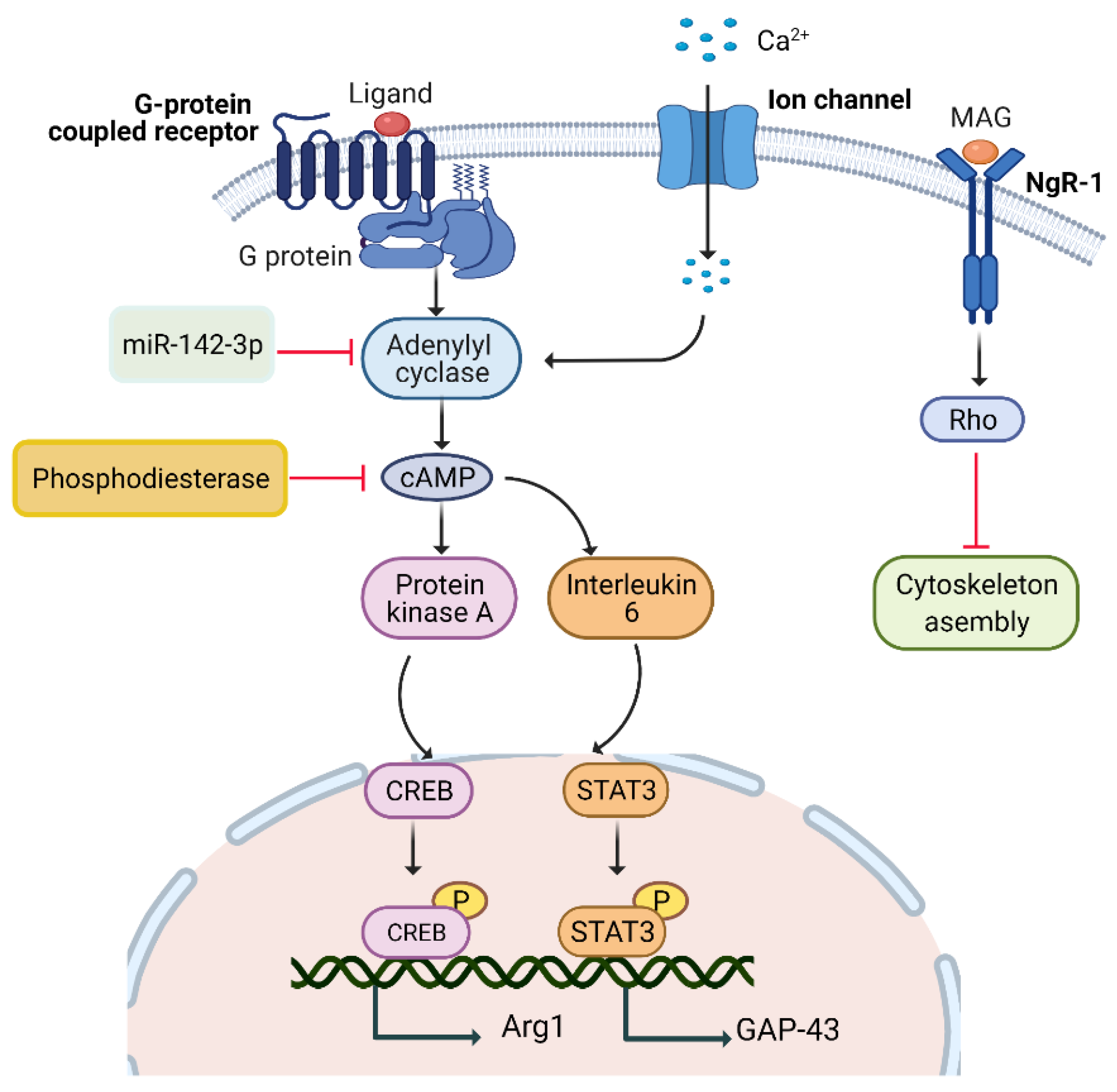
5.1. Rapid Injury Signals

5.1.1. cAMP Pathway

Cyclic adenosine monophosphate (cAMP) is a secondary messenger essential for maintaining a neuron’s growth. It is critical for successful axonal regeneration, even in a non-permissive environment. It also controls axon repulsion or attraction in response to environmental cues. It has been seen that in vivo injection of dibutyryl cAMP (db-cAMP) in dorsal root ganglion (DRG) leads to potentiated regeneration of central axons even in the presence of MAIs, thus mimicking conditioning lesions [20]. Another study on zebrafish exhibits an increase in the growth of neurons after induction of exogenous cAMP [21]. Many studies show that cAMP signalling stimulates multiple downstream pathways that drive neurite outgrowth after damage [22].

5.1.2. Calcium/cAMP Pathway

Calcium (Ca2+) is the most important factor in regulating cAMP expression following an injury. EG-19 unit (α-subunit) of voltage-gated calcium channels (VGCCs) and internal storage vesicles are two primary sources of the subsequent release of Ca2+. After laser axotomy in the Caenorhabditis elegans (C. elegans) model, Ca2+ increases the adenylate cyclase (AC) activity [23,24], triggering cAMP production, which ultimately accelerates the development of growth cones and strengthens the link between damaged axon fragments and the formation of synaptic branches [25].

5.1.3. IL-6/cAMP Pathway

After nerve injury, inflammatory stimulation causes significant axonal regeneration and neuroprotection. Expression of cytokine mediators such as interleukin 6 (IL-6), ciliary neurotrophic factor (CNTF), and leukemia inhibitory factor (LIF) was found to be upregulated in response to injury. Knockout studies have explored the regeneration-promoting effects of these inflammatory molecules. In the adult nervous system, IL-6 is found only in a limited amount in normal situations, but its level rises under injured circumstances and possesses growth-promoting properties. Moreover, the IL-6 knockout study directly indicates the key role of IL-6 in enhancing the regeneration of dorsal column axons in conditioning lesions. cAMP upregulation after injury directly mediates the release of IL-6 and induces activation of downstream pathways for STAT3 and, ultimately Gap-43 [26,27,28].

5.1.4. PKA/cAMP Pathway

Following injury, cAMP is found to upregulate protein kinase A (PKA) formation, which further promotes the assemblage of cytoskeletal filaments and axonal growth via inhibiting MAG/RhoA-GTPase interaction [4]. On the other hand, it phosphorylates the basic leucine zipper domain (bZip) factor, cAMP response element-binding protein (CREB), which upregulates arginase-1 (Arg-1). Upregulation of Arg-1 enhances polyamine production, which helps to overcome MAG-induced myelin inhibition and stimulates the development of axons [29].

5.1.5. miR-142-3p/AC9/cAMP Pathway

Micro RNAs (miRNAs), non-coding RNAs, regulate gene expression by incorporating them at the 3′ untranslated sites of target mRNA. miR-142-3p plays a key function in regulating the conversion of adenosine triphosphate (ATP) to cAMP by targeting AC9-mRNA (Figure 1). In the sciatic nerve lesion model, miR-142-3p downregulation increased cAMP levels after 9 hrs. As a result, miR-142-3p downregulation promotes AC9 expression, which then raises cAMP levels and improves the regeneration of sensory neurons [30,31].

5.1.6. Pharmacological Agents

Pharmacological treatment approaches can raise cAMP levels in neurons, allowing them to repair more effectively. Intracellular cAMP may be boosted by activating AC using forskolin, which further speeds up the sciatic nerve regeneration. Several neuronal subtypes, including DRG, cerebellar, cortical, and hippocampal neurons, utilize db-cAMP, a non-hydrolyzable cAMP analogue, to overcome MAG and to increase CREB phosphorylation. Brain-derived neurotrophic factor (BDNF) and db-cAMP boost the Arg-1 expression and antagonize MAG inhibition.
Moreover, repulsive turning reactions in Xenopus (African clawed frog) growth cones are induced by microscopic gradients of the chemo-repellant semaphorin III/D, which may be changed to an attraction on the addition of the cAMP analogue Sp-cAMPS. Surprisingly, Sp-cAMPS therapy boosts calcium levels further, converting MAG-mediated repulsion to attraction to regulate growth cone-turning reactions. The major enzyme responsible for cAMP hydrolysis, phosphodiesterase 4 (PDE4), increases intracellular cAMP levels. Rolipram and roflumilast reduce PDE4 activity to increase cAMP expression [32]. Sorafenib increases sensory conduction function recovery after dorsal column damage by downregulating miR-142-3p [33].

5.2. Delayed Injury Signals

5.2.1. DLK-JNK MAPK Pathway

Mammalian zipper protein kinase/dual leucine zipper kinase (DLK) is a mitogen-activated protein kinase kinase kinase (MAPKKK). It plays a central role in neural development, apoptosis, and axonal regeneration in the CNS and PNS [34]. This kinase can further activate cJUN N-terminal kinases (JNK) and p38 MAPK. DLK is a critical component of the sciatic nerve and is required for sensory axon regeneration as well as the activation of regenerative pathways in injured neurons’ cell bodies (Figure 2). The failure of cJUN activation in the DLK knockout mouse model indicates that it is crucial for reorganizing microtubules in the growth cone after axotomy.
A scaffolding protein called JNK-interacting protein 3 (JIP3) connects DLK and JNK-associated axon retrograde trafficking of pSTAT3/JNK toward the injury site [35,36,37]. Hence, DLK is an essential component and target for initiating a pro-regenerative program following injury [38].

5.2.2. Pharmacological Agents

Sarm1, a TIR-domain protein, which functions as a NADase to break down the key metabolite NAD+, is a significant contributor to Wallerian degeneration. The NAD+ biosynthesis enzyme, NMNAT2, hampers Sarm1’s function. MAPK inhibition reduces degeneration triggered by ectopic Sarm1 activation in DRG explants. DLK and NMNAT2 are linked because both are regulated by the PHR ubiquitin ligase. CEP-1347, a pan-mixed lineage kinase (MLK) inhibitor, acts as a key upstream regulator of JNK activation in neurons [39]. Furthermore, palmitoylation regulates DLK binding to JIPs [37], which might affect how they interact with motor proteins. It also facilitates DLK’s interaction with the axonal survival factor NMNAT2 [40].

5.3. Transcriptional Factors’ Mediated Pathways

5.3.1. JAK/STAT Pathway

The signal transducer and activator of transcription 3 (STAT3) acts as a ‘jump-start’ for controlling axonal development and remodelling in the PNS and CNS. IL-6, CNTF, LIF, phosphatase and tensin homolog (PTEN), and the suppressor of cytokine signalling 3 (SOCS3) all have a role in STAT3 activation via the Gp130 receptor. It is generally expressed in axons and becomes phosphorylated locally after damage. It then is reassembled in DRG cell bodies, where it promotes axon rejuvenation. Neuropoietic cytokines are produced in response to injury, and Janus kinase 2 (JAK-2) may be activated, which is required for stable STAT3 phosphorylation. DLK deficiency, a key component of the DLK/JNK/MAPK pathway, can prevent p-STAT3 from accumulating in DRG, suggesting that it is required for pSTAT3 translocation [4]. Blockade of PTEN and SOCS3, the small proline-rich protein 1a (Sprr1a), and the cell cycle inhibitor p21/Cip1/Waf1 are therapeutical targets to enhance the expression of STAT3. However, SOCS3 and PTEN blockage are more favorable and common target sites to overcome STAT3 inhibition for axon regeneration [41].

5.3.2. ATF3/CREB Pathway

Normal levels of activating transcription factor 3 (ATF3) expression are low, but PNS laceration increases ATF3 expression in DRG neurons. It has other names, such as LRG-21, TI-241, CRG-5 (in mice), LRF-1 (in rats), and ATF3 (in humans). It belongs to the CREB family, and ATF/CREB can form heterodimers with other bzip proteins such as AP-1 (activator protein-1 transcription factor) family members, including JUN, Fos, and C/EBPs (CCAATenhancer-binding proteins), JUNB, JUND, ATF2, and cJUN. Notably, ATF3 is a stress-stimulated factor and is activated when it binds to ATF/CRE site [42,43]. Different pathways have been found to activate ATF3 expression in response to injury or stress. JNK/SAPK (c-Jun N-terminal kinases/ stress-activated kinase) controls the expression of ATF3 upon peripheral injury and further regulates the expression of different bZip genes to enhance axonal outgrowth [44,45].
After nerve injury, ATF3 activates and targets RAGs, including cJUN, Hsp27, and Cap-23, which is functionally related to Gap-43, and Sprr1a to induce axonal outgrowth. Similarly, it also mediates the activation of AP-1, which further stimulates cJUN activation through JUN N-terminal kinases (JNKs) signalling. c-JUN controls the expression of galanin and CD44, and α7β1 integrin to promote axonal regeneration [4]. Thus, ATF3 acts as a point of convergence for different regulators, signals, and transcriptional pathways, suggesting that ATF3 upregulation after an injury is a strong candidate for axon regeneration. Additionally, ATF3 expression correlates with the intrinsic growth capacity of central axons following injury. Sox11 modulates ATF3 expression positively and acts as an upstream target to enhance ATF3 levels and promote axonal growth [46,47].

5.3.3. BMP/SMAD Pathway

The bone morphogenic proteins (BMPs), members of the transforming growth factor (TGF) ligands, are found near the tips of axons and play a role in controlling actin dynamics during dendritic creation and synaptic stability [48,49]. SMADs are involved in neural differentiation, neuronal circuit mapping, and synaptic maturation. Only peripheral branch axotomy (not central branch) increases SMAD1 expression in adult DRG neurons [50,51].
Following peripheral axotomy or contusion, BMP production increases. It was seen that the BMP receptor mediates signalling through SMAD 1 and 4 (Figure 3). Particularly, SMAD 1 plays a chief role in neurite outgrowth. Intra-ganglionic injection of BMP translocates the phosphorylated SMAD to the nucleus and hence modulates its expression. SMAD1/4 recruits other DNA regulatory activators, such as CBP and p300 and activates Gap-43 to enhance axonal growth [52]. Blockade of BMP in preconditioned or naive DRG neurons inhibits axonal growth, indicating that BMP is necessary for the activation of SMAD. BMP/SMAD1 activation not only increases the growth of dorsal columns in vivo but also promotes intrinsic growth of DRG neurons in in vitro studies using an AAV-based approach. These shreds of data suggest that BMP and SMAD activation work together to promote axonal regeneration. Taken together, BMP is an upstream target of SMAD activation that mediates growth programs in response to injury [52,53,54].

5.3.4. Pharmacological Agents

4-Methylhistamine dihydrochloride (4-MeH), a specific JAK agonist, significantly increases axon growth and remodelling of DRG neurons [55].

5.4. Neurotrophins

During development and in the aftermath of injury, neurotrophins, a small family of well-conserved neuropeptides, aid in maintaining the survival of neurons. Peripheral targets such as skin and muscles exhibit baseline expression of neuronal trophic growth factors and are crucial for functional synapse re-establishment following injury. BDNF, NGF, and NT3/4/5 are the most well-known members of this family. NGF is the most researched of all neurotrophins [56,57]. Tyrosine kinase receptors (TrkA, TrkB, TrkC) and low-affinity p75NTR receptors interact with members of this family. Phospholipase C-γ (PLC-γ), phosphatidylinositol-3-kinase (PI3K), and Ras pathways are activated when neurotrophins bind to Trk receptors to mediate gene expression (Figure 4) [58].

5.4.1. Ras/ERK Pathway

Nerve growth factor (NGF), the first-ever reported neurotrophin, helps maintain phenotypes, promotes PNS development, and ensures the functional unity of cholinergic neurons in the CNS (for detailed review, see [59]). It binds to the Trk receptor (Tropomyosin receptor kinase) and activates Ras/ERK signalling. Activated Ras stimulates Raf (serine/threonine kinase), MEK (MAP/ERK kinase), and, lastly, ERK (extracellular signal-regulated kinase). ERK belongs to the MAPK (mitogen-associated protein kinase) family and plays an important role in neurite outgrowth. Robust and rapid activation of ERK following nerve injury in Schwann cells (SCs) has been monitored at the injury site. A higher level of ERK has been seen in both infectious and inherited peripheral neuropathies models. It also plays a chief role in neurological healing and remyelination of neurons. Ras/ERK signalling enhances axon growth and activates anti-apoptotic protein Bcl-2 via CREB to promote axonal survival [8,60,61]. Hence, the upregulation of ERK is the leading candidate for the Ras/ERK pathway [62].

5.4.2. PI3K/AKT Pathway

The PI3K/AKT pathway is an important survival mechanism that controls signalling, differentiation, and axonal growth. The interaction of NGF and Trk receptors activates PI3K via the Ras and Gab-1 adaptor proteins, allowing PI3K phosphorylation and activation of downstream molecules such as AKT and ERK1/2.
Activated AKT reduces Fas ligand (FasL) expression by suppressing the pro-apoptotic transcription factor Bad (inhibitor of Bcl-2 anti-apoptotic protein), and Forkhead. Phosphorylated AKT, on the other side, promotes the assemblage of cytoskeletal filaments and axonal outgrowth [63,64]. It was seen in PC12 rat pheochromocytoma cells that PI3K activates Rac1, which translocates RhoA from the nucleus to the cytoplasm, forming a complex with GDP dissociation inhibitors (GDIs), rendering it unable to halt axon renewal [65]. Knockout studies of PTEN demonstrate accelerated outgrowth in both in vitro and in vivo models of sensory neurons. The inhibition of PTEN acts as a therapeutic target to increase neurite growth, suggesting that upregulation of AKT is required for healing damaged nerve responses [66,67,68].

5.4.3. Dock/BDNF Pathway

Rho-GTPase family members (Rac-1, RhoA, and Cdc42) regulate the actin cytoskeletal organization. All members of this family are inactivated in GDP-bound form while activated in the form of GTP. GTPase activating proteins (GAP) and guanine nucleotide exchange factors (GEFs) regulate Rho activation and inactivation by converting it into a GDP-bound form and stimulating nucleotide release, respectively [69].
The Rho-GEF family (Rho-guanine nucleotide exchange factors) includes Dock1 (Dock180)-related proteins, although Dock 1 has no PH/DH (pleckstrin homology/Dbl-homology) domains but exhibits the DHR-1/DHR-2 evolutionary proteins region. Dock (1–4) has conserved several amino acid regions in their DHR-2 domains that are essential for GEF activity. Moreover, in vivo and in vitro studies have proved Dock-3 as a critical component in neuron maintenance. It participates in axonal growth via downstream BDNF-TrkB signalling and is essential in CNS developmental processes such as synaptic pruning [70].
Furthermore, Dock-3 forms a complex with the WAVE-1 and Fyn-SH3 domain at the cell membrane level. In this notion, it is clear that Dock-3 expression is directly linked to WAVE-1 expression to promote the regeneration of injured optic nerves. BDNF expression is linked to Rac-1 activation, which in turn fosters neurite development. Dock-3 also promotes WAVE protein recruitment and hinders the long-term activation of WAVE/Rac1 for actin polymerization (Figure 5). Taken together, Dock upregulation has been shown to cure various glaucoma conditions. Interestingly, BDNF and Dock-3 have a synergistic relationship upstream of Rac-1 and WAVE proteins, indicating BDNF as a possible therapeutic target for the upregulation of Dock-3 and their associated mediators to enhance axonal outgrowth [71,72,73]. A list of intrinsic regulators is shown in Table 1.

5.4.4. Pharmacological Agents

At nanomolar doses, bisperoxovanadium compounds (bpVs) selectively inhibit PTEN [74,75]. Interferon-β enhances NGF protein; thus, it can initiate NGF/Trk signalling in damaged axons to ameliorate functional recovery [76,77].
BRD4, a BET bromodomain reader of acetyl-lysine histones, is an essential component that enhances BDNF synthesis and memory after HDAC inhibition. HDAC2 and HDAC3 knockdown increased BDNF-mRNA expression but not the expression of other HDACs, whereas BRD4 knockdown blocked these effects. The BDNF promoter-specific targeting of BRD4 by the dCas9-BRD4 locus enhanced BDNF-mRNA. RGFP966, a pharmacological inhibitor of HDAC3, was suppressed by JQ1, which increased BDNF expression and BRD4 binding to the BDNF promoter (an inhibitor of BRD4). Following RGFP966 treatment, H4K5ac and H4K8ac alterations, as well as H4K5ac enrichment at the BDNF promoter, were enhanced according to documented epigenetic targets of BRD4 and HDAC3 [78].
Table 1. Intrinsic regulators for axonal regeneration under injury condition.
RegulatorStudied inAxonal RegenerationStudied by
Regeneration-associated transcription factors
STAT3Dorsal root ganglion (DRG) neuronsBiomedicines 10 03186 i001[79]
cJUNDRG neuronsBiomedicines 10 03186 i002[80,81,82]
KLF4Retinal ganglion cells (RGC) neuronsBiomedicines 10 03186 i003[4,83]
KLF6RGC neuronsBiomedicines 10 03186 i004[11,84,85]
KLF7Corticospinal tract (CST) neuronsBiomedicines 10 03186 i005[11,84,85]
KLF9RGC neuronsBiomedicines 10 03186 i006[86]
SMAD1DRG neuronsBiomedicines 10 03186 i007[87]
ATF3DRG neurons*
Biomedicines 10 03186 i008
[46,88]
CREBDRG neuronsBiomedicines 10 03186 i009[89]
AP-1DRG neuronsBiomedicines 10 03186 i010[90]
Intrinsic growth-mediating molecules
cAMPDRG neuronsBiomedicines 10 03186 i011[9,22,28,91]
mTORCST neurons
RGC neurons
DRG neurons
Biomedicines 10 03186 i012[92,93,94,95]
PTENCST neurons
RGC neurons
Biomedicines 10 03186 i013[96,97]
SOCS3RGC neuronsBiomedicines 10 03186 i014[98,99]
TSC1 (hamartin)RGC neuronsBiomedicines 10 03186 i015[100,101]
TSC2 (tuberin)RGC neuronsBiomedicines 10 03186 i016[102,103]
GSK-3βDRG neuronsBiomedicines 10 03186 i017[104]
LIFSensory neurons **Biomedicines 10 03186 i018[15]
IL6Sensory neurons,
Schwann cells
Biomedicines 10 03186 i019[105,106]
CNTFDistal nerve stump of DRG neuronsBiomedicines 10 03186 i020[106,107,108]
IGF-1RGC neuronsBiomedicines 10 03186 i021[109]
PI3KDRG neuronsBiomedicines 10 03186 i022[110]
ERKDRG neuronsBiomedicines 10 03186 i023[111,112,113]
FAKRGC neuronsBiomedicines 10 03186 i024[114]
DLKDRG neuronsBiomedicines 10 03186 i025[115,116]
Regeneration-associated genes (RAGs)
GalaninDRG neuronsBiomedicines 10 03186 i026[117,118]
IntegrinDRG neuronsBiomedicines 10 03186 i027[119]
Gap-43DRG neuronsBiomedicines 10 03186 i028[19,120]
Cap23DRG neuronsBiomedicines 10 03186 i029[121]
Hsp27DRG neuronsBiomedicines 10 03186 i030[121,122,123]
P21/cip1/Waf1DRG neuronsBiomedicines 10 03186 i031[41]
Sprr1aDRG neuronsBiomedicines 10 03186 i032[4]
Crmp2Motor neuronsBiomedicines 10 03186 i033[124]
Map1bGrowth coneBiomedicines 10 03186 i034[125]
Spry2Sensory axonsBiomedicines 10 03186 i035[126,127]
Pdcd4Spinal cord neuronsBiomedicines 10 03186 i036[128]
Dickkopf1DRG neuronsBiomedicines 10 03186 i037[129]
Rho GTPases
Rho-ADRG neuronsBiomedicines 10 03186 i038[130]
Rac1RGC neuronsBiomedicines 10 03186 i039[131]
ROCKRGC neuronsBiomedicines 10 03186 i040[132]
miRNAs
miR-142-3pDRG neuronsBiomedicines 10 03186 i041[133]
miR-21DRG neuronsBiomedicines 10 03186 i042[134]
miRNA-431DRG neuronsBiomedicines 10 03186 i043[135]
CNS inhibitors
NogoRGC neuronsBiomedicines 10 03186 i044[136]
MAGCentral nervous system (CNS) neuronsBiomedicines 10 03186 i045[137]
OmgpCNS neuronsBiomedicines 10 03186 i046[137]
CSPGsCNS neuronsBiomedicines 10 03186 i047[15]
SemaphorinsCNS neuronsBiomedicines 10 03186 i048[138]
* In PNS lesion, not in CNS lesion. ** Schwann cells upregulate it after injury even though it is not typically present in the nervous system.

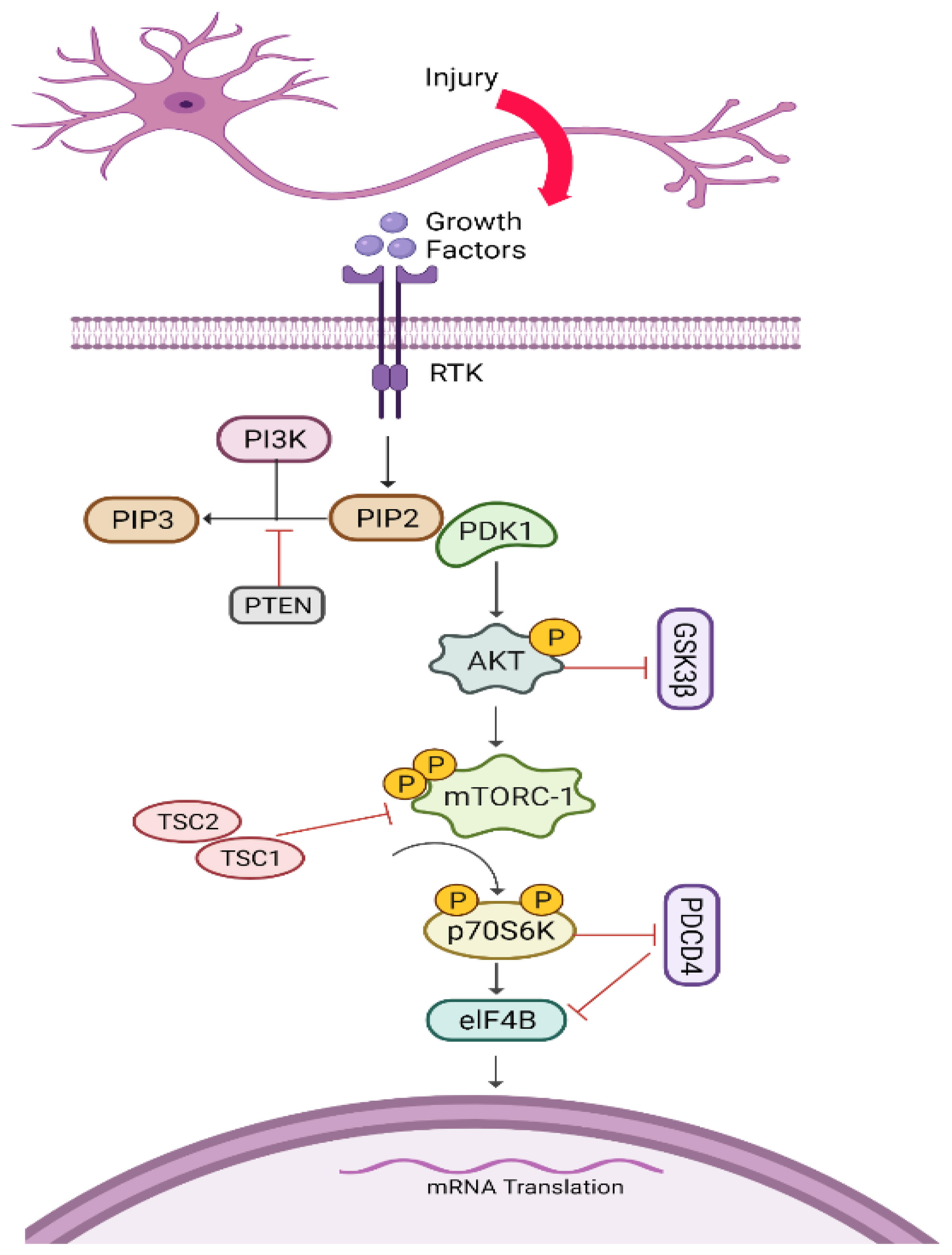
5.5. AKT/mTORC1/p70S6K Pathway

The mammalian target of rapamycin (mTOR) is a 268 kDa protein and serine-threonine kinase that controls cell growth and regeneration via balancing nutrient availability. It is also known as RAFT, RAPT, and FRAP. When the PI3K/AKT pathway activates mTORC1, it stimulates the expression of p70S6K, which limits muscle atrophy and promotes axonal regeneration. Through the AKT/mTORC1/p70S6K pathway, mTOR can influence the regeneration potential of growth cones in adult corticospinal neurons (Figure 6) (for details, please see [139,140]).
It was seen that mTOR increases axon regeneration in the PNS through the deletion of negative regulators, including PTEN, hamartin (TSC1), and tuberin (TSC2) [4,57]. Interestingly, TSC2 inhibits axon growth by inactivating Rheb (Ras homolog enriched in the brain), which is a GTP-binding protein. Typically, activated Rheb acts as a direct activator of AKT-mediated mTORC1 signalling as mentioned by in vivo study of the ischemic spinal cord injury model [141]. Indeed, the balance of inhibitors and activators determines the pace of growth; it is necessary to target these inhibitors in order to offer the optimum regenerative environment for damaged neurons. PTEN knockdown or long-term inhibition, on the other hand, has been linked to neoplasia, including malignant glial tumors and hamartomas, according to prior research [63,100,142]. TSC-1 and TSC-2, which stimulates axonal growth, can be inhibited by activated AKT. Conclusively, these shreds of evidence suggested that the upregulation of AKT, as opposed to PTEN or TSC1 and TSC-2, is a more useful and appealing therapeutic target due to fewer adverse effects [139]. It is suggested that there is an AKT-independent route for axonal regeneration because PTEN deletion results in considerably more potent axonal regeneration, whether combined with AKT overexpression or GSK3β deletion [143]. Blockage of Pdcd4 has also been shown to be an important technique in preventing muscle atrophy by boosting elF4B-induced mRNA and thus assisting axonal regeneration [144].

Pharmacological Agents

Sirolimus and everolimus, two mTOR inhibitors, have the potential to deliver tailored treatment. In the United States and Europe, Everolimus is the first mTOR inhibitor to be licensed as a therapy option [145].

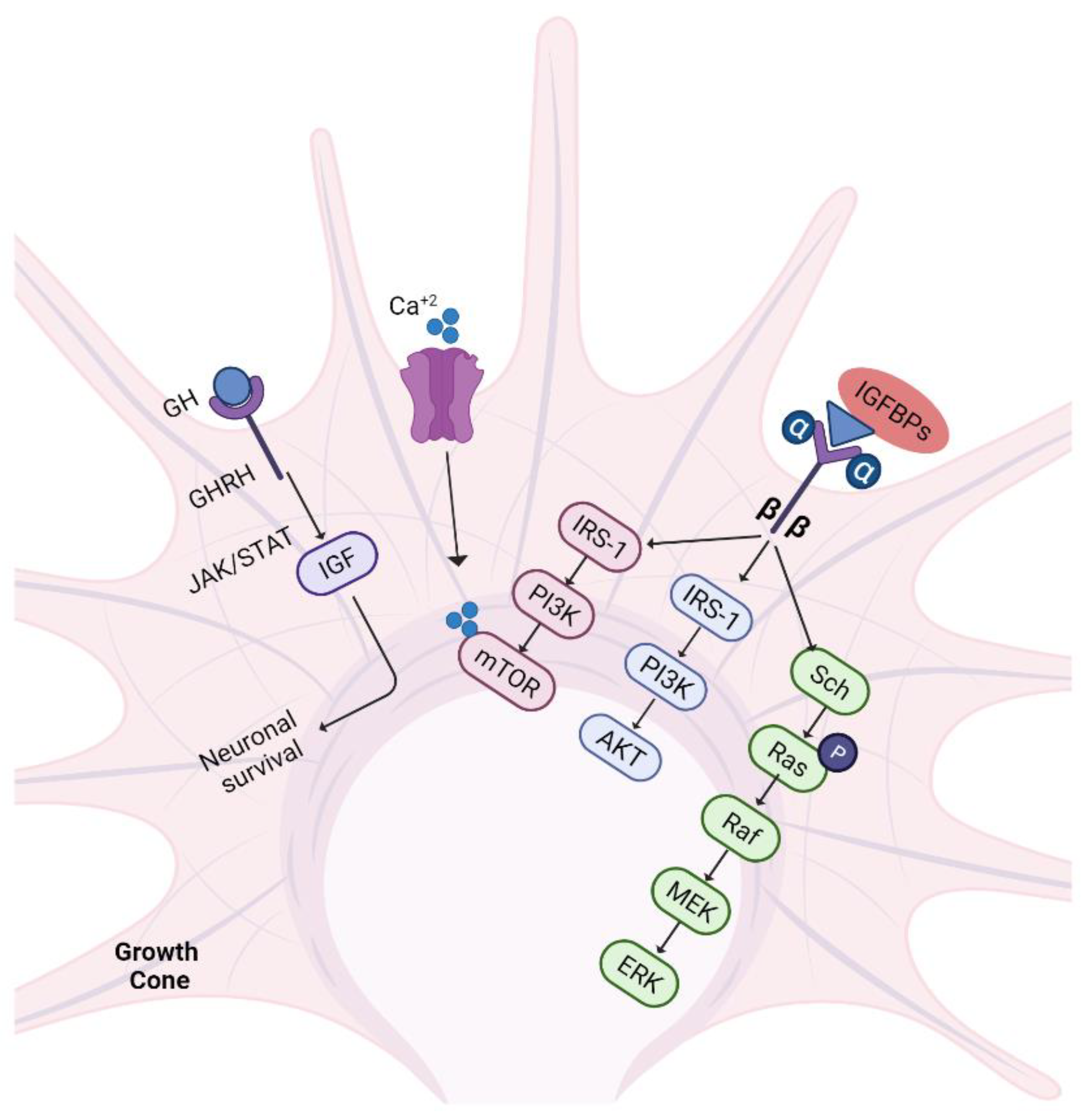
5.6. IGF-1/GH Pathway

Hypothalamic growth hormone-releasing hormones (GHRHs) act on the pituitary gland to maintain the release of growth hormone (GH). These hormones have therapeutic potential due to their role in accelerating axonal regeneration and maintaining denervated muscle and SCs before reinnervation. It works by releasing insulin-like growth factor-1 (IGF-1) into the bloodstream, which is synthesized mainly in the liver and also in peripheral tissues (for a detailed study, see [146,147]). IGF-1 is involved in the regeneration and survival of CNS neurons and the stimulation of neurite outgrowth [148,149]. In addition, IGF-1 mediates axon elongation and is also involved in developing neurons’ survival, proliferation, and synaptogenesis [150].
After nerve trauma, IGF-1 production is upregulated locally, which in turn upregulates the expression of Gap-43 and focal adhesion molecules via the PI3K pathway. Taken together with these changes, IGF-1 promotes motility of the neurite growth cone and prevents neuronal apoptosis. Overexpressed IGF-1 also upregulates the mTOR/AKT pathway, MuRF1, and MADbx to counteract the denervation-induced muscle atrophy [151,152]. IGF-1’s supportive role in peripheral nerve injury (PNI) recovery in animal models is strongly supported by research. IGF-1 can slow the pace of muscle atrophy, promote axonal development, and reduce SC apoptosis [153].
IGF binding proteins (IGFBPs) facilitate the binding of IGF molecules with the IGF-1R (tyrosine kinase receptor) and function by activating numerous pathways such as PI3K/AKT, mTOR, and MEK/ERK (Figure 7). The interplay of IGF/IGFBP/IGF-1R causes calcium influx and PI3K phosphorylation, as well as mTOR activation to promote axon growth. Additionally, receptor activation causes phosphorylation of receptor tyrosine residues, which activates Ras GTPases and their molecules to activate Raf, MEK, and eventually ERK. Rho-GTPase, Cdc42, and glycogen synthase kinase 3 beta (GSK-3β) are downstream targets of IGF-1 and play a role in stabilising microtubule polymerization. Similarly, GH exerts its action by activating the JAK/STAT pathway to enhance neuronal survival and outgrowth [147,154]. In a mouse model of nerve damage, GH reduces the negative consequences of muscle denervation and improves not just functional recovery but also motor axon reinnervation. These findings suggested that increasing GH levels might be a potential therapy option for improving motor function recovery [155].

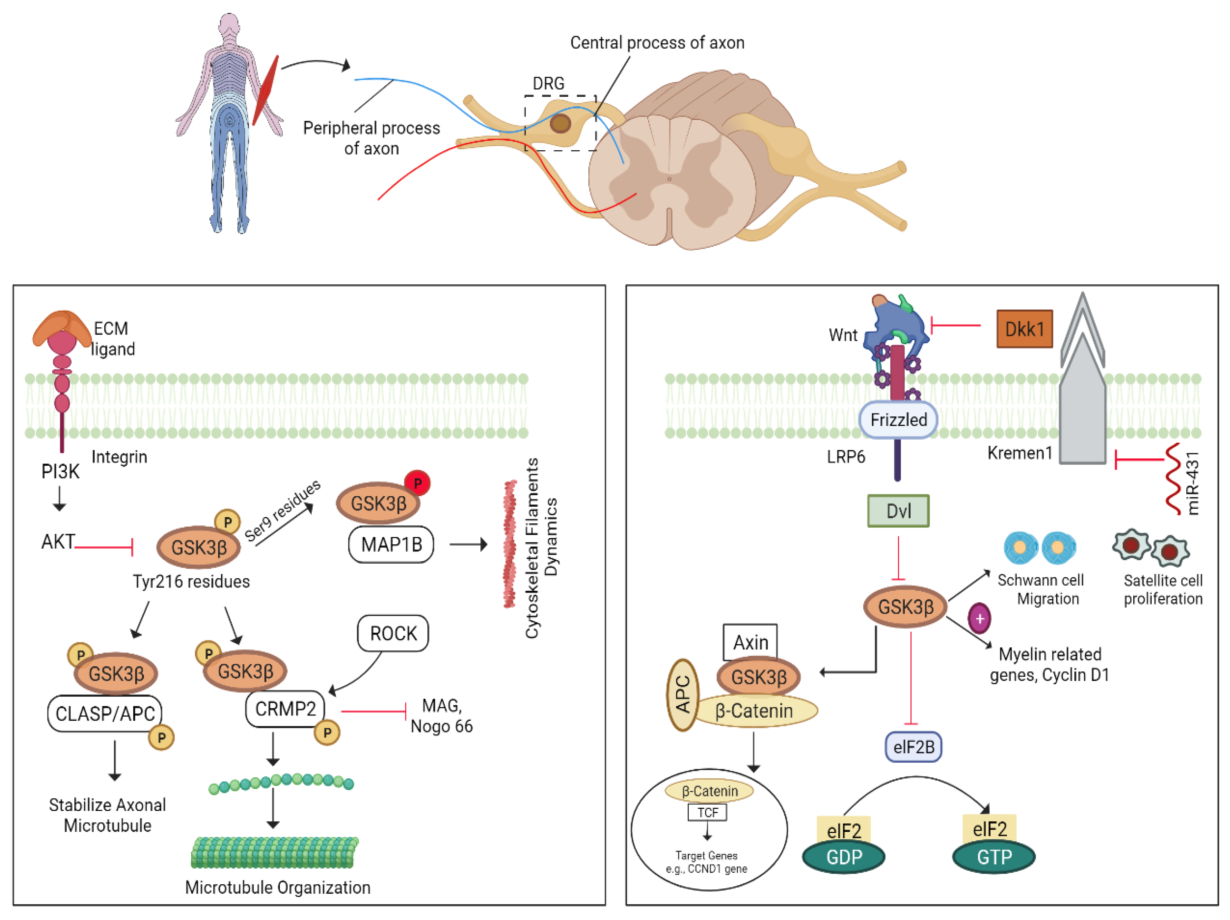
5.7. GSK3β–CLASP/APC Pathway

GSK-3β, a serine-threonine kinase, can phosphorylate several proteins, including microtubule-associated proteins (tau and MAP1B), collapsin response mediator protein 2 (CRMP2), and cytoplasmic linker-associated protein 2 (CLASP2), when activated. GSK3β activity can be justified by mediating their phosphorylating sites [156]. It is normally phosphorylated at its Tyr216 residues of the kinase domain to become active when it is at rest, whereas phosphorylation of Ser9 residues is responsible for the inactivity of GSK3β [157,158]. Integrin-mediated inhibition of GSK3β can stimulate the CLASP/APC pathway where CLASP2 (a cytoplasmic linker protein) plays a role in axonal microtubule stability for potential axonal regeneration [104,159]. CRMP2, another substrate of GSK3β, promotes microtubule organization and enhances axon elongation by binding to tubulin heterodimers. This binding is reduced upon phosphorylation of GSK3β. GSK3β can stimulate CRMP2 phosphorylation at the Thr514 residue, while ROCK phosphorylates CRMP2 at the Thr555 residue. ROCK-mediated CRMP2 phosphorylation can downstream Nogo-66 and MAG thereby counteracting myelin inhibition. So, CRMP2 is a central therapeutic target for reversing the inhibitory effect of myelin inhibitors for axon regeneration [159]. In addition, protein phosphatase 2A dephosphorylates CRMP2 and promotes axon outgrowth [160], whereas CRMP2 is active only when GSK3β is inactive. Furthermore, activated GSK3β can mediate the expression of MAP1B to regulate the growth of cytoskeletal filaments [91,158]. The pCRMP2 improves axonal regeneration after GSK3β knockout following optic nerve damage. GSK3β mediates axonal regeneration by targeting specific substrates, suggesting that it might be a therapeutic target to improve regeneration [161].

5.7.1. GSK-3β/Wnt Pathway

Wnt ligands, also known as glycoproteins, have an important and diversified function in cell growth, development, and various human disorders [162]. Wnt-mRNA expression is not detectable in the intact spinal cord, whereas Wnt 1, 4, and 5a immediately surround the lesion site after spinal cord contusion. These molecules also increase Wnt-Ryk signalling, which inhibits motor neuron renewal [163]. Wnt ligands target particular receptors to execute their activities; for example, Wnt-4/Ryk ligand interaction is found to inhibit sensory neuron regeneration, whereas Wnt-LRP5/6 interaction is found to promote neurite outgrowth [164]. Consistent with this interaction, downregulation of Wnt-Ryk signalling is an attractive therapeutical approach to promote the plasticity of cortico-spinal motor neurons and robust axonal growth following injury [163,165,166].
Wnt ligands can block GSK-3β activity and promote re-myelination of facial and sciatic nerves through Wnt/β-catenin signalling. In addition, Wnt-GSK3β interaction has been shown to enhance axonal regeneration as much as neurotrophins [161,167]. GSK-3β inhibition can stimulate the migration of SCs, MAG, myelin-related genes, cyclin-D1, nicotinic-acetylcholine receptors, and muscle gene myogenin to promote axon regeneration by reducing muscle degeneration. This suggests that blocking GSK3β or using inhibitors to promote myelination and nerve regeneration is a valuable option [168]. Moreover, earlier research has shown that upregulation of the Wnt/β-catenin pathway improves axonal regeneration after damage [139,169,170,171,172].

5.7.2. miRNA-431/Kremen/Wnt Pathway

Kremen (1/2), a transmembrane protein, has a kringle domain and acts as a receptor for Dickkopf1 (Dkk1) [129]. Dkk1 and Kremen-1 form a complex that further inhibits Wnt signalling (Figure 8. By blocking Kremen 1, overexpressed miR-431 can significantly suppress the expression of other genes, such as Braf and Zkscan 1, thus promoting axonal growth [129,135].

6. Other Injury Signals

6.1. Regeneration by Astrocytes/Inflammatory Mediators

In mammals, the inflammatory response occurs instantaneously after a CNS contusion and plays an important role in providing restorative output. Microglia are the inflammatory mediators of the CNS and present the first-line defensive approach against trauma. After activation, they undergo morphological modifications such as ramification into amoeboids. They begin to proliferate and migrate toward the injury site, thereby initiating the production of pro-inflammatory and anti-inflammatory markers such as cytokines [173,174]. Moreover, macrophages and neutrophils are recruited to the injured area from the periphery. Together with microglia/macrophages, reactive astrocytes also participate in the organization of regenerating-inhibiting glial damage [91,175,176].
Upon optic nerve contusion, microglial cells of the retina reactivate themselves and stimulate the release of blood-borne macrophages and neutrophils, which secrete inflammatory mediators such as oncomodulin (Ocm). Conversely, inflammatory stimulation provokes reactive astrocytes to promote axon regeneration and neuroprotection via mTOR and JAK/STAT3 signalling pathways. SOCS3 acts as an inhibitor of the JAK/STAT3 pathway, whereas PTEN is the inhibitor of the mTOR pathway. Deletion of SOCS3 and PTEN may prove a valuable therapeutic target to enhance axonal regeneration. Notably, SOCS3 expression can be opposed by cAMP level; hence, an elevated level of cAMP can enhance neurite regeneration (Figure 9). Additionally, overexpression of Ras homolog regulator (Rhen1), which is enriched in the brain, can boost mTOR activity and, thereby, RGC axonal regeneration [176].

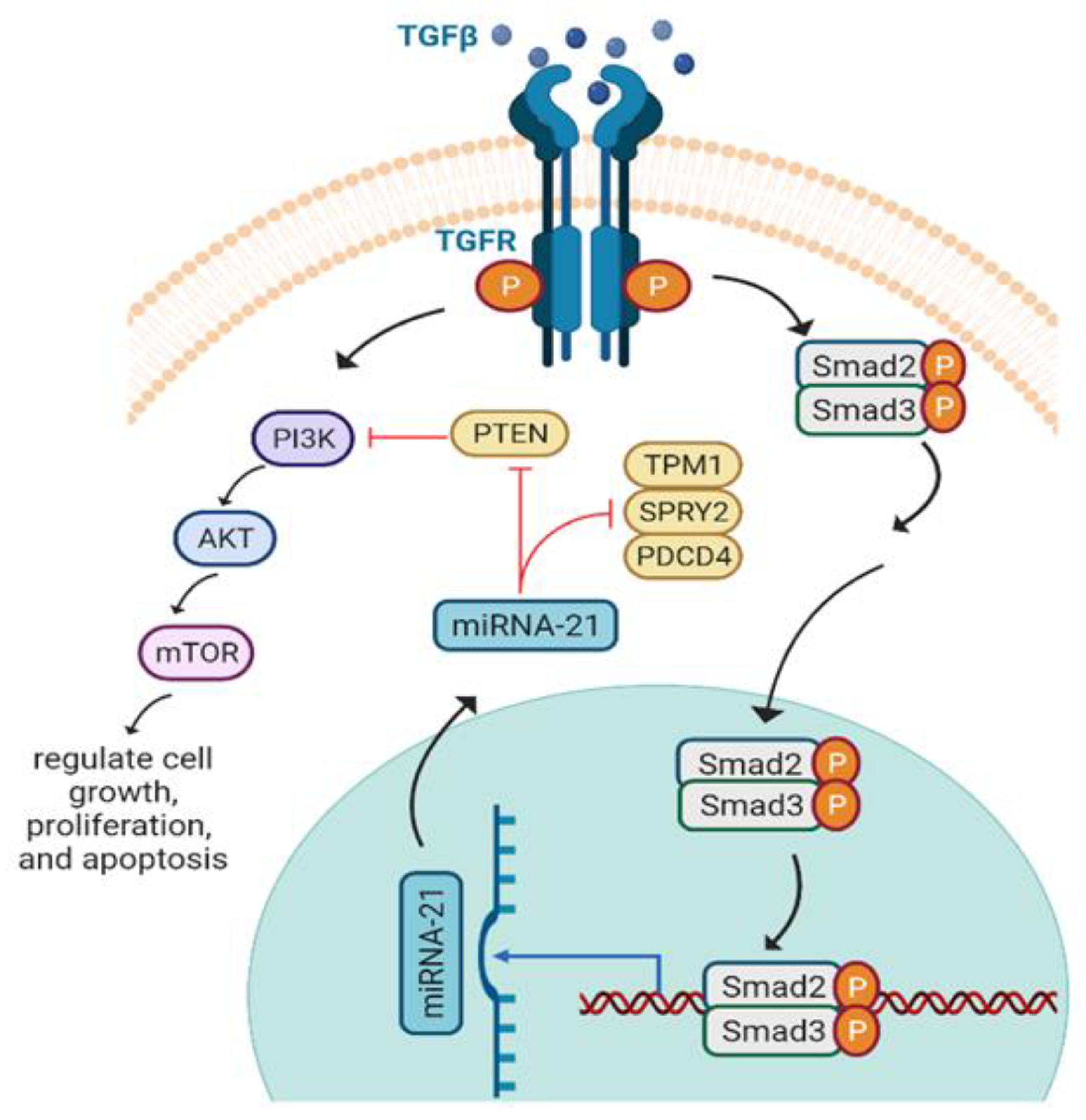
6.2. miRNA-21 and Axon Regeneration

Micro RNAs (miRNAs) are encoded endogenously to regulate gene expression by inhibiting or increasing the degradation of the translational protein. Due to their role in neuronal development, regeneration, and degeneration, miRNAs have been explored as a therapeutic tool for various diseases [177,178,179,180,181]. miRNA-21 (miR-21) can enhance the intrinsic growth capacity of damaged neurons after nerve denervation [134]. PTEN, Pdcd4, Spry2, and Tpm1 are the targets of miR-21, through which it mediates neurite outgrowth [182]. Different studies revealed that miR-21 regulates Spry2 in cardiomyocytes [183] and PTEN in hepatocellular carcinoma [184]. TGF-β1 mediates miR-21 proliferative and apoptotic activity by targeting the PI3K/AKT/mTOR pathway and promotes astrocytes activation after spinal cord injury (SCI) both in vitro and in vivo. Moreover, TGF-β1 activates SMAD2/3 and upregulates miR-21 to prevent astrocytic scar formation, which is the leading cause of hindrance of axonal regeneration (Figure 10). Herein, the upregulation of miR-21 and downregulation of PTEN are promising therapeutic targets to regulate neural regeneration [182]. Moreover, it was seen that miR-21 promotes axonal growth and maintains the number of SCs by interacting with TIMP3, EPHA4, and TGFβ1 during nerve repair [185].

6.3. Integrin/FAK Pathway

The integrin receptor family is vital for developing tissues, nervous system formation, immune responses, and regeneration of axons. There are two subunits in integrin receptors: alpha and beta. In mammals, a total of eighteen α and 8 β-subunits have been identified [186,187,188].
Activated integrins specifically bind to particular extracellular matrix ligands (ECM) and stimulate signalling to regulate the actin cytoskeleton [189]. Tenascin-C is a ligand for integrins present in the CNS that can be regulated by reactive astrocytes. Tenascin-C interacts with an α9β1-integrin receptor in neurons to achieve axon regeneration in CNS (for details, see [190,191]). It was found that α9 delivery (viral vector-mediated) in DRG causes the localization of integrins in axons; therefore, it induces neurite growth [119,192,193,194].
Once integrin is activated, it interacts with tenascin C and stimulates FAK, which further activates downstream molecules, including PI3K, AKT, Src kinase, and RhoA. The lesion site is also enriched with molecules such as NogoA, CSPGs, MAG, and semaphorins class III (Sema3s). These molecules inhibit integrin signalling, and block phosphorylation of FAK to prevent axon regeneration via binding to their respective receptors to mediate axonal guidance and growth cone dynamics (Figure 11). An appropriate elevated integrin expression can serve as a therapeutic approach for CNS axonal regeneration. To overcome integrin inactivation, PTEN and GSK-3β blockage are considered as the target sites to elevate integrin enrichment at the injury site in promoting axonal growth [195,196,197].
It has been noted that in C. elegans, the RhoA GTPase-ROCK pathway stimulates MLC-4 phosphorylation to enhance axon regeneration. Axonal injury activates TLN-1/talin via the cAMP-Epac-Rap GTPase cascade, which results in integrin inside-out activation and promotes axonal regeneration by activating the RhoA signalling pathway. Src-1 activates EPHX-1/ephexin RhoGEF in this pathway after integrin activation by phosphorylating the Tyr-568 residue in the autoinhibitory domain. These findings show that the Src-RhoGEF-RhoA axis controls axon regeneration by following an integrin signalling network [198].

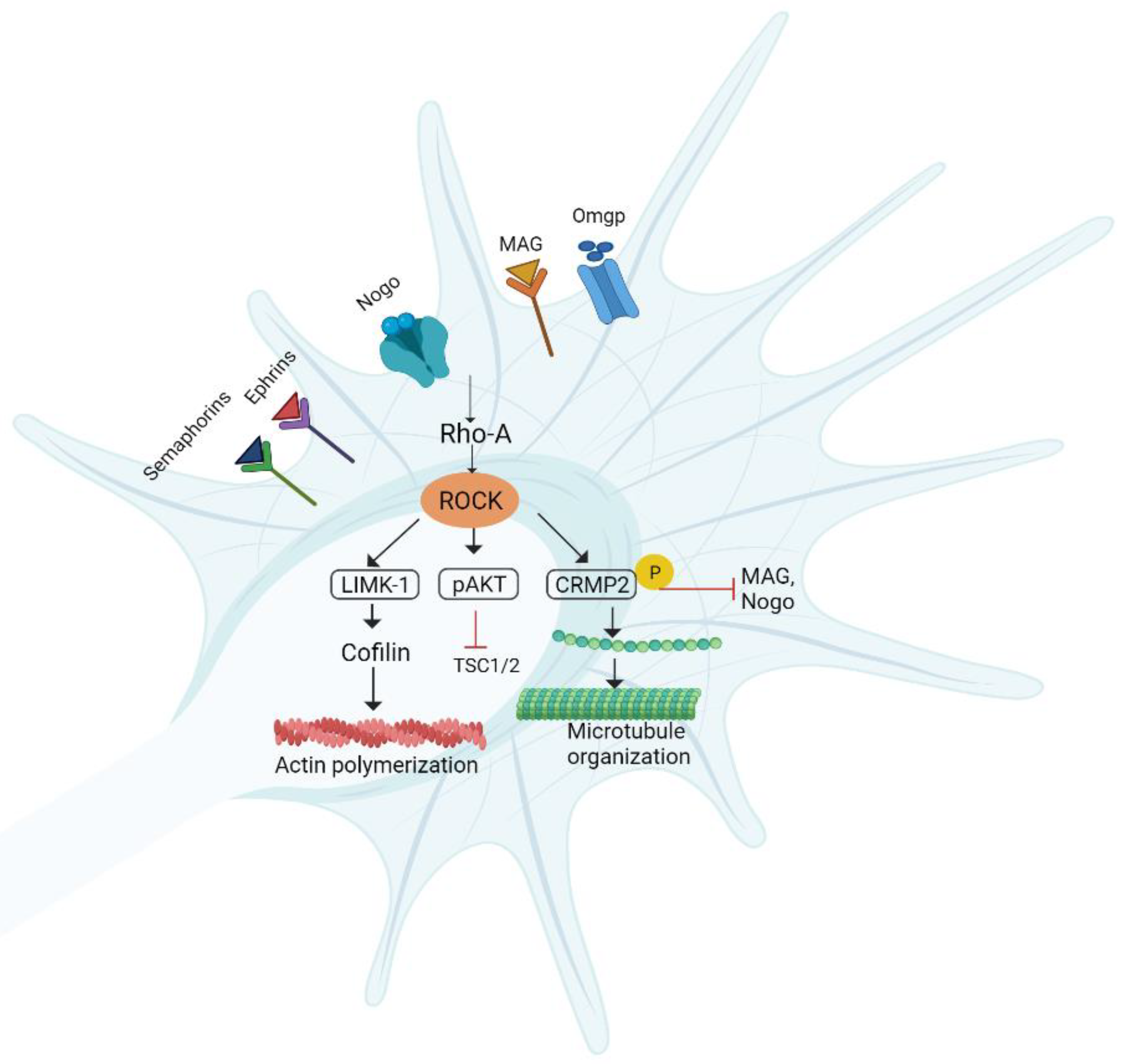
6.4. RhoA/ROCK/LIMK Pathway

The RhoA/Rho kinase pathway has a role in the regulation of inflammatory responses and the production of cytokines, including interleukin 2 (IL-2), interleukin 1 beta (IL-1β), tumor necrosis factor-α (TNF-α), and CXC chemokines [199]. Rho-GTPases allow the interaction of extracellular ligands with the growth cone and modulate neurite outgrowth [200]. RhoA-GTPases and their downstream effectors, including ROCK, along with MAGs and chondroitin sulfate, activate PNI [201,202]. ROCK negatively affects the regeneration of axons by modulating the growth cone dynamics [132]. ROCK1 and ROCK2 are two isoforms of ROCK. ROCK1 mostly exists in non-neuronal tissues, while ROCK2 is found in the spinal cord and brain [203,204].
Guanine nucleotide exchange factors (GEFs), GDP dissociation inhibitors (GDIs), and GTPase activating proteins (GAPs) play a crucial role in regulating the phosphorylation of Rho-GDP into Rho-GTP. ROCK also inactivates MLCP (myosin light chain phosphatase), which dephosphorylates the myosin light chain. ROCK-induced MLCP inactivation enhances myosin’s phosphorylation that causes growth cone collapse and axon growth inhibition [205]. LIM domain kinase (LIMK) is considered a downstream target of ROCK2. LIMK activation results in a collapse in growth cone stability. This effect is achieved after cofilin phosphorylation. Upon phosphorylation, cofilin becomes inactivated and further incapable of organizing actin filaments. Nogo/Lingo-1/p75NTR or ephrin/semaphorin receptors are responsible for the activation of ROCK by binding with myelin-based inhibitors such as NOGO, MAG, OMGP, semaphorins, and ephrins. ROCK activation not only plays a role in the regulation of axon guidance but is also important in promoting regenerative machinery in mammalian CNS. Apart from this, ROCK has deleterious effects on cell survival by activating Fas or ROCK homolog PTEN (Figure 12). ROCK inhibition looks to be a promising treatment option for a variety of neurodegenerative illnesses in this regard. CRMP2 has also been discovered to increase microtubule assembly, which is important for axon polarity and outgrowth. ROCK2 can phosphorylate and activate CRMP2, hence having growth-promoting action by regulating growth cone morphology [203,206,207].

Pharmacological Agents

In an optic nerve damage model, blocking the RhoA/ROCK pathway with Y-27362 dramatically enhances axonal sprouting and locomotor functional recovery [208]. An experiment examined the effects of Y-27632 on peripheral motor and sensory neurons that had developed in the presence of CSPGs that hindered axonal extension. In vitro, motor neurons showed a more favorable response to Y-27632 than that of sensory neurons in a non-growth-permissive environment. These variations were related to different RhoA expressions and activation patterns in sensory and motor axons. After systemic treatment of high doses of Y-27632, the regeneration of motor axons was significantly enhanced in vivo, in contrast to the regeneration of sensory fibers. The findings demonstrate that the RhoA/ROCK pathway differentially influences both sensory and motor axon regeneration, with motor neurons responding more quickly than sensory neurons under a growth-inhibitory environment.
In addition, the regeneration of axons was investigated in symptomatic and asymptomatic SOD1G93A animals after the sciatic nerve was crushed. Axonal regeneration was severely hampered when symptomatic SOD1G93A animals were compared to presymptomatic SOD1G93A mice and wild types. Treatment with Y-27632 enhanced motor axon functional and morphological measurements following the sciatic crush in all examined situations. Although Y-27632 therapy enhanced neuromuscular junction axonal reinnervation, it did not increase the lifespan of symptomatic SOD1G93A animals [209]. To alleviate neurofunctional impairments, bFGF might inhibit RhoA by activating Ras-related C3-botulinum toxin substrate 1 (Rac1). Thus, inhibiting RhoA/ROCK activation in case ofneurological illnesses is critical for neuroprotection and neurogenesis [210].

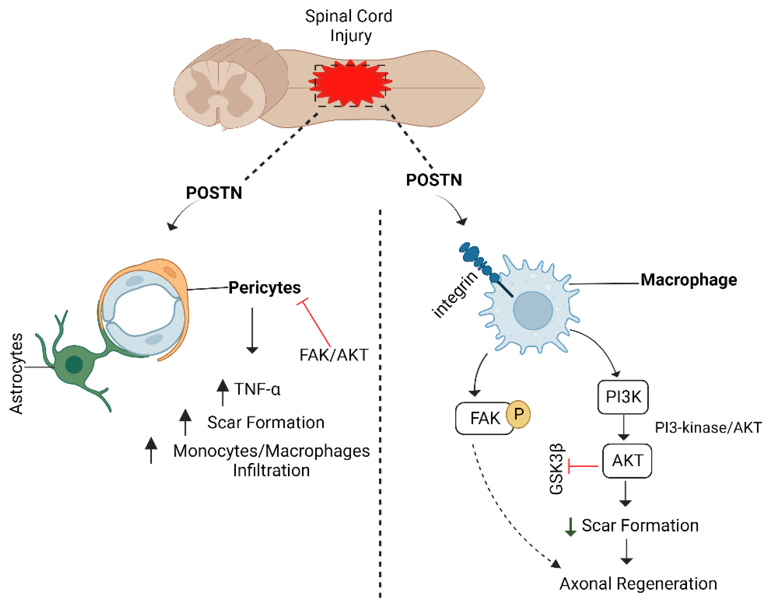
6.5. POSTN/Integrin Pathway and Axon Regeneration

Periostin (POSTN), an extracellular matrix protein, belongs to the fasciclin family [211]. It has been linked to the regeneration of astrocytes. During the early stages of development, it is expressed in various tissues, including the CNS. Its expression rises in adult tissues after acute injury, including shattered bone infarcted myocardial tissues, and during wound healing [212,213]. A recent study shows that POSTN stimulates neurite outgrowth by activating AKT and FAK [214].
Similarly, POSTN is involved in cell proliferation, survival, migration, and adhesion by interacting with integrins α5/β3 and α5/β1, and it also promotes downstream activation of PI3K, FAK, and AKT [211,215]. Moreover, POSTN can bind to other extracellular matrix proteins such as tenascin C, fibronectin, and collagen 1 [216]. Through the FAK/AKT signalling pathway, the POSTN/integrin axis is activated in the injured spinal cord and promotes macrophage migration toward the injury site. It inhibits pericyte proliferation to minimize scar formation, which is thought to be the principal hindrance to CNS axonal regeneration [217]. The ability of POSTN to induce neuronal development is further enhanced when the FAK pathway is activated (Figure 13). As a result, upregulating FAK/AKT is a therapeutic strategy for preventing pericyte proliferation [205,218].

7. Conclusions

Both central and peripheral axons are very delicate and highly sensitive structures, and even mild stress or pressure can injure them. Axonal injury can be caused by neurotoxins, traffic accidents, acute lacerations, and neurological disorders resulting in the loss of functions. Failure of axonal regeneration can lead to lifelong disability. Peripheral nerves can regenerate in the presence of a supportive environment provided by SCs. Another important factor is the fast clearance of cellular debris in the distal part through the involvement of macrophages that facilitate the regeneration. On the other hand, in case of injury to CNS, oligodendrocytes fail to provide the supportive environment required for regeneration; instead, they secrete inhibitory molecules. Central nerves cannot regenerate since they do not have enough intrinsic growth factors. On the other hand, excessive stress on peripheral nerves may irreversibly damage axons and inhibit axon regrowth. After an injury, several signalling molecules are activated and travel toward the damaged area, potentially speeding up the regeneration process. Among them, signalling pathways such as cAMP, JAK/STAT, BDNF, ERK, PI3K/AKT, mTORC1, integrin/FAK, and Rho/ROCK are common and are thought to initiate the process of regeneration. Furthermore, astrocytes, inflammatory cytokines, and microRNAs all have a role in growth cone stability. Upregulation of cAMP, STAT3, AKT, BDNF, integrin, miR-21, miR-431, and downregulation or blocking of GSK-3β, PTEN, SOCS3, TSC1, TSC2, miR-142-3p, Pdcd4, Spry2, Tpm1, and myelin-associated inhibitors can alter the fate of regenerating axon and therefore are considered as potential therapeutic targets to restore the function of injured axons. However, when it comes to the execution of therapeutic approaches for functional axon recovery, a valid and affordable clinical approach is still nonexistent.

Author Contributions

R.A., H.A., A.R., M.S.J., C.R., A.I., I.U.K., F.S., T.I., G.H., S.A.M. and T.S. have equally contributed to the conceptualization, literature analysis, and review drafting. Funding acquisition: H.S.H.; supervision: G.H.; project administration: G.H., T.S. and H.S.H. All authors have read and agreed to the published version of the manuscript.

Funding

We are thankful for participating families and the Higher Education Commission (HEC) of Pakistan for providing financial support to conduct this study (Grant No.7612/Punjab/NRPU/R&D/HEC/2017 to G. H).

Institutional Review Board Statement

Not applicable.

Informed Consent Statement

Not applicable.

Acknowledgments

The listed author(s) also express gratitude to their representative institutes and universities for providing access to the literature.

Conflicts of Interest

The authors declare no conflict of interest.

References

  1. Lingor, P.; Koch, J.C.; Tönges, L.; Bähr, M. Axonal Degeneration as a Therapeutic Target in the CNS. Cell Tissue Res. 2012, 349, 289–311. [Google Scholar] [CrossRef] [PubMed] [Green Version]
  2. Sutherland, T.C.; Geoffroy, C.G. The Influence of Neuron-Extrinsic Factors and Aging on Injury Progression and Axonal Repair in the Central Nervous System. Front. Cell Dev. Biol. 2020, 8, 190. [Google Scholar] [CrossRef] [PubMed]
  3. Farah, M.H.; Pan, B.H.; Hoffman, P.N.; Ferraris, D.; Tsukamoto, T.; Nguyen, T.; Wong, P.C.; Price, D.L.; Slusher, B.S.; Griffin, J.W. Reduced BACE1 Activity Enhances Clearance of Myelin Debris and Regeneration of Axons in the Injured Peripheral Nervous System. J. Neurosci. 2011, 31, 5744–5754. [Google Scholar] [CrossRef] [PubMed] [Green Version]
  4. Mahar, M.; Cavalli, V. Intrinsic Mechanisms of Neuronal Axon Regeneration. Nat. Rev. Neurosci. 2018, 19, 323–337. [Google Scholar] [CrossRef] [PubMed]
  5. Hussain, G.; Wang, J.; Rasul, A.; Anwar, H.; Qasim, M.; Zafar, S.; Aziz, N.; Razzaq, A.; Hussain, R.; de Aguilar, J.L.G.; et al. Current Status of Therapeutic Approaches against Peripheral Nerve Injuries: A Detailed Story from Injury to Recovery. Int. J. Biol. Sci. 2020, 16, 116–134. [Google Scholar] [CrossRef] [PubMed] [Green Version]
  6. Zafar, S.; Rasul, A.; Iqbal, J.; Anwar, H.; Imran, A.; Jabeen, F.; Shabbir, A.; Akram, R.; Maqbool, J.; Sajid, F.; et al. Calotropis Procera (Leaves) Supplementation Exerts Curative Effects on Promoting Functional Recovery in a Mouse Model of Peripheral Nerve Injury. Food Sci. Nutr. 2021, 9, 5016–5027. [Google Scholar] [CrossRef] [PubMed]
  7. Kaplan, A.; Morquette, B.; Kroner, A.; Leong, S.Y.; Madwar, C.; Sanz, R.; Banerjee, S.L.; Antel, J.; Bisson, N.; David, S.; et al. Small-Molecule Stabilization of 14-3-3 Protein-Protein Interactions Stimulates Axon Regeneration. Neuron 2017, 93, 1082–1093. [Google Scholar] [CrossRef] [Green Version]
  8. Chan, K.M.; Gordon, T.; Zochodne, D.W.; Power, H.A. Improving Peripheral Nerve Regeneration: From Molecular Mechanisms to Potential Therapeutic Targets. Exp. Neurol. 2014, 261, 826–835. [Google Scholar] [CrossRef]
  9. Jara, J.S.; Agger, S.; Hollis, E.R. Functional Electrical Stimulation and the Modulation of the Axon Regeneration Program. Front. Cell Dev. Biol. 2020, 8, 736. [Google Scholar] [CrossRef]
  10. Nagappan, P.G.; Chen, H.; Wang, D.Y. Neuroregeneration and Plasticity: A Review of the Physiological Mechanisms for Achieving Functional Recovery Postinjury. Mil. Med. Res. 2020, 7, 30. [Google Scholar] [CrossRef]
  11. He, Z.; Jin, Y. Intrinsic Control of Axon Regeneration. Neuron 2016, 90, 437–451. [Google Scholar] [CrossRef] [PubMed] [Green Version]
  12. Jung, H.; Yoon, B.C.; Holt, C.E. Axonal MRNA Localization and Local Protein Synthesis in Nervous System Assembly, Maintenance and Repair. Nat. Rev. Neurosci. 2012, 13, 308. [Google Scholar] [CrossRef] [Green Version]
  13. Cooke, P.; Janowitz, H.; Dougherty, S.E. Neuronal Redevelopment and the Regeneration of Neuromodulatory Axons in the Adult Mammalian Central Nervous System. Front. Cell. Neurosci. 2022, 16, 183. [Google Scholar] [CrossRef] [PubMed]
  14. Van Niekerk, E.A.; Tuszynski, M.H.; Lu, P.; Dulin, J.N. Molecular and Cellular Mechanisms of Axonal Regeneration after Spinal Cord Injury. Mol. Cell. Proteom. 2016, 15, 394–408. [Google Scholar] [CrossRef] [PubMed] [Green Version]
  15. Fawcett, J.W. The Struggle to Make CNS Axons Regenerate: Why Has It Been so Difficult? Neurochem. Res. 2020, 45, 144–158. [Google Scholar] [CrossRef] [Green Version]
  16. Mironets, E.; Wu, D.; Tom, V.J. Manipulating Extrinsic and Intrinsic Obstacles to Axonal Regeneration after Spinal Cord Injury. Neural Regen. Res. 2016, 11, 224. [Google Scholar] [CrossRef]
  17. Wahane, S.; Halawani, D.; Zhou, X.; Zou, H. Epigenetic Regulation of Axon Regeneration and Glial Activation in Injury Responses. Front. Genet. 2019, 10, 640. [Google Scholar] [CrossRef]
  18. Ma, T.C.; Willis, D.E. What Makes a RAG Regeneration Associated? Front. Mol. Neurosci. 2015, 8, 43. [Google Scholar] [CrossRef] [Green Version]
  19. Shin, J.E.; Cho, Y. Epigenetic Regulation of Axon Regeneration after Neural Injury. Mol. Cells 2017, 40, 10–16. [Google Scholar] [CrossRef] [Green Version]
  20. Yan, K.; Gao, L.N.; Cui, Y.L.; Zhang, Y.; Zhou, X. The Cyclic AMP Signaling Pathway: Exploring Targets for Successful Drug Discovery (Review). Mol. Med. Rep. 2016, 13, 3715. [Google Scholar] [CrossRef]
  21. Ifegwu, O.C.; Awale, G.; Rajpura, K.; Lo, K.W.H.; Laurencin, C.T. Harnessing CAMP Signaling in Musculoskeletal Regenerative Engineering. Drug Discov. Today 2017, 22, 1027. [Google Scholar] [CrossRef] [PubMed]
  22. Blesch, A.; Lu, P.; Tsukada, S.; Alto, L.T.; Roet, K.; Coppola, G.; Geschwind, D.; Tuszynski, M.H. Conditioning lesions before or after spinal cord injury recruit broad genetic mechanisms that sustain axonal regeneration: Superiority to camp-mediated effects. Exp. Neurol. 2012, 235, 162–173. [Google Scholar] [CrossRef] [PubMed] [Green Version]
  23. Cui, W.; Wu, H.; Yu, X.; Song, T.; Xu, X.; Xu, F. The Calcium Channel A2δ1 Subunit: Interactional Targets in Primary Sensory Neurons and Role in Neuropathic Pain. Front. Cell. Neurosci. 2021, 15, 397. [Google Scholar] [CrossRef]
  24. Steuer Costa, W.; Yu, S.-c.; Liewald, J.F.; Gottschalk, A. Fast CAMP Modulation of Neurotransmission via Neuropeptide Signals and Vesicle Loading. Curr. Biol. 2017, 27, 495–507. [Google Scholar] [CrossRef] [PubMed] [Green Version]
  25. Qiu, L.; LeBel, R.P.; Storm, D.R.; Chen, X. Type 3 Adenylyl Cyclase: A Key Enzyme Mediating the CAMP Signaling in Neuronal Cilia. Int. J. Physiol. Pathophysiol. Pharmacol. 2016, 8, 95. [Google Scholar] [PubMed]
  26. Kummer, K.K.; Zeidler, M.; Kalpachidou, T.; Kress, M. Role of IL-6 in the Regulation of Neuronal Development, Survival and Function. Cytokine 2021, 144, 155582. [Google Scholar] [CrossRef]
  27. Knott, E.P.; Assi, M.; Pearse, D.D. Cyclic AMP Signaling: A Molecular Determinant of Peripheral Nerve Regeneration. Biomed Res. Int. 2014, 2014, 651625. [Google Scholar] [CrossRef]
  28. Niemi, J.P.; Defrancesco-Oranburg, T.; Cox, A.; Lindborg, J.A.; Echevarria, F.D.; McCluskey, J.; Simmons, D.D.; Zigmond, R.E. The Conditioning Lesion Response in Dorsal Root Ganglion Neurons Is Inhibited in Oncomodulin Knock-Out Mice. eNeuro 2022, 9, 1–13. [Google Scholar] [CrossRef]
  29. Li, Z.H.; Cui, D.; Qiu, C.J.; Song, X.J. Cyclic Nucleotide Signaling in Sensory Neuron Hyperexcitability and Chronic Pain after Nerve Injury. Neurobiol. Pain 2019, 6, 100028. [Google Scholar] [CrossRef]
  30. Li, X.; Wang, S.; Yang, X.; Chu, H. MiR-142-3p Targets AC9 to Regulate Sciatic Nerve Injury-Induced Neuropathic Pain by Regulating the CAMP/AMPK Signalling Pathway. Int. J. Mol. Med. 2021, 47, 561. [Google Scholar] [CrossRef]
  31. Gao, J.; Gu, J.; Pan, X.; Gan, X.; Ju, Z.; Zhang, S.; Xia, Y.; Lu, L.; Wang, X. Blockade of MiR-142-3p Promotes Anti-Apoptotic and Suppressive Function by Inducing KDM6A-Mediated H3K27me3 Demethylation in Induced Regulatory T Cells. Cell Death Dis. 2019, 10, 332. [Google Scholar] [CrossRef] [PubMed] [Green Version]
  32. Moradi, K.; Golbakhsh, M.; Haghighi, F.; Afshari, K. International Immunopharmacology Inhibition of Phosphodiesterase IV Enzyme Improves Locomotor and Sensory Complications of Spinal Cord Injury via Altering Microglial Activity: Introduction of Ro Fl Umilast as an Alternative Therapy. Int. Immunopharmacol. 2020, 86, 106743. [Google Scholar] [CrossRef] [PubMed]
  33. Wang, T.; Li, B.; Wang, Z.; Wang, X.; Xia, Z.; Ning, G.; Wang, X.; Yuan, X.; Feng, S.; Chen, X. Sorafenib Promotes Sensory Conduction Function Recovery via MiR-142-3p/AC9/CAMP Axis Post Dorsal Column Injury. Neuropharmacology 2019, 148, 347–357. [Google Scholar] [CrossRef] [PubMed]
  34. Watkins, T.A.; Wang, B.; Huntwork-Rodriguez, S.; Yang, J.; Jiang, Z.; Eastham-Anderson, J.; Modrusan, Z.; Kaminker, J.S.; Tessier-Lavigne, M.; Lewcock, J.W. DLK Initiates a Transcriptional Program That Couples Apoptotic and Regenerative Responses to Axonal Injury. Proc. Natl. Acad. Sci. USA 2013, 110, 4039–4044. [Google Scholar] [CrossRef] [PubMed] [Green Version]
  35. Valakh, V.; Frey, E.; Babetto, E.; Walker, L.J.; DiAntonio, A. Cytoskeletal Disruption Activates the DLK/JNK Pathway, Which Promotes Axonal Regeneration and Mimics a Preconditioning Injury. Neurobiol. Dis. 2015, 77, 13–25. [Google Scholar] [CrossRef] [PubMed] [Green Version]
  36. Shin, J.E.; Ha, H.; Kim, Y.K.; Cho, Y.; DiAntonio, A. DLK Regulates a Distinctive Transcriptional Regeneration Program after Peripheral Nerve Injury. Neurobiol. Dis. 2019, 127, 178. [Google Scholar] [CrossRef]
  37. Holland, S.M.; Collura, K.M.; Ketschek, A.; Noma, K.; Ferguson, T.A.; Jin, Y. Palmitoylation Controls DLK Localization, Interactions and Activity to Ensure Effective Axonal Injury Signaling. Proc. Natl. Acad. Sci. USA 2016, 113, 763–768. [Google Scholar] [CrossRef] [Green Version]
  38. Schellino, R.; Boido, M.; Vercelli, A. JNK Signaling Pathway Involvement in Spinal Cord Neuron Development and Death. Cells 2019, 8, 1576. [Google Scholar] [CrossRef] [Green Version]
  39. Asghari Adib, E.; Smithson, L.J.; Collins, C.A. An Axonal Stress Response Pathway: Degenerative and Regenerative Signaling by DLK. Curr. Opin. Neurobiol. 2018, 53, 110–119. [Google Scholar] [CrossRef]
  40. Summers, D.W.; Milbrandt, J.; Diantonio, A. Palmitoylation Enables MAPK-Dependent Proteostasis of Axon Survival Factors. Proc. Natl. Acad. Sci. USA 2018, 115, E8746–E8754. [Google Scholar] [CrossRef]
  41. Fischer, D. Hyper-IL-6: A Potent and Efficacious Stimulator of RGC Regeneration. Eye 2016, 31, 173–178. [Google Scholar] [CrossRef] [PubMed] [Green Version]
  42. Kole, C.; Brommer, B.; Nakaya, N.; Sengupta, M.; Bonet-Ponce, L.; Zhao, T.; Wang, C.; Li, W.; He, Z.; Tomarev, S. Activating Transcription Factor 3 (ATF3) Protects Retinal Ganglion Cells and Promotes Functional Preservation After Optic Nerve Crush. Invest. Ophthalmol. Vis. Sci. 2020, 61, 31. [Google Scholar] [CrossRef] [PubMed] [Green Version]
  43. Gey, M.; Wanner, R.; Schilling, C.; Pedro, M.T.; Sinske, D.; Knöll, B. Atf3 Mutant Mice Show Reduced Axon Regeneration and Impaired Regeneration-Associated Gene Induction after Peripheral Nerve Injury. Open Biol. 2016, 6, 160091. [Google Scholar] [CrossRef] [PubMed] [Green Version]
  44. Scheib, J.; Höke, A. Advances in Peripheral Nerve Regeneration. Nat. Rev. Neurol. 2013, 9, 668–676. [Google Scholar] [CrossRef]
  45. Ousman, S.S.; Frederick, A.; Lim, E.M.F. Chaperone Proteins in the Central Nervous System and Peripheral Nervous System after Nerve Injury. Front. Neurosci. 2017, 11, 79. [Google Scholar] [CrossRef] [Green Version]
  46. Cheng, Y.C.; Snavely, A.; Barrett, L.B.; Zhang, X.; Herman, C.; Frost, D.J.; Riva, P.; Tochitsky, I.; Kawaguchi, R.; Singh, B.; et al. Topoisomerase I Inhibition and Peripheral Nerve Injury Induce DNA Breaks and ATF3-Associated Axon Regeneration in Sensory Neurons. Cell Rep. 2021, 36, 109666. [Google Scholar] [CrossRef]
  47. Perry, R.B.T.; Hezroni, H.; Goldrich, M.J.; Ulitsky, I. Regulation of Neuroregeneration by Long Noncoding RNAs. Mol. Cell 2018, 72, 553–567.e5. [Google Scholar] [CrossRef] [Green Version]
  48. Higashi, T.; Tanaka, S.; Iida, T.; Okabe, S. Synapse Elimination Triggered by BMP4 Exocytosis and Presynaptic BMP Receptor Activation. Cell Rep. 2018, 22, 919–929. [Google Scholar] [CrossRef] [Green Version]
  49. Podkowa, M.; Zhao, X.; Chow, C.-W.; Coffey, E.T.; Davis, R.J.; Attisano, L. Microtubule Stabilization by Bone Morphogenetic Protein Receptor-Mediated Scaffolding of c-Jun N-Terminal Kinase Promotes Dendrite Formation. Mol. Cell. Biol. 2010, 30, 2241–2250. [Google Scholar] [CrossRef] [Green Version]
  50. Zou, H.; Ho, C.; Wong, K.; Tessier-Lavigne, M. Axotomy-Induced Smad1 Activation Promotes Axonal Growth in Adult Sensory Neurons. J. Neurosci. 2009, 29, 7116–7123. [Google Scholar] [CrossRef]
  51. Parikh, P.; Hao, Y.; Hosseinkhani, M.; Patil, S.B.; Huntley, G.W.; Tessier-Lavigne, M.; Zou, H. Regeneration of Axons in Injured Spinal Cord by Activation of Bone Morphogenetic Protein/Smad1 Signaling Pathway in Adult Neurons. Proc. Natl. Acad. Sci. USA 2011, 108, 99–107. [Google Scholar] [CrossRef] [PubMed] [Green Version]
  52. Tedeschi, A. Tuning the Orchestra: Transcriptional Pathways Controlling Axon Regeneration. Front. Mol. Neurosci. 2012, 4, 60. [Google Scholar] [CrossRef] [PubMed] [Green Version]
  53. Hollis, E.R.; Zou, Y. Expression of the Wnt Signaling System in Central Nervous System Axon Guidance and Regeneration. Front. Mol. Neurosci. 2012, 5, 5. [Google Scholar] [CrossRef] [PubMed] [Green Version]
  54. Zhong, J.; Zou, H. BMP Signaling in Axon Regeneration. Curr. Opin. Neurobiol. 2014, 27, 127–134. [Google Scholar] [CrossRef] [PubMed] [Green Version]
  55. Elsaeidi, F.; Bemben, M.A.; Zhao, X.-F.; Goldman, D. Jak/Stat Signaling Stimulates Zebrafish Optic Nerve Regeneration and Overcomes the Inhibitory Actions of Socs3 and Sfpq. J. Neurosci. 2014, 34, 2632. [Google Scholar] [CrossRef] [Green Version]
  56. Stanga, S.; Boido, M.; Kienlen-Campard, P. How to Build and to Protect the Neuromuscular Junction: The Role of the Glial Cell Line-Derived Neurotrophic Factor. Int. J. Mol. Sci. 2020, 22, 136. [Google Scholar] [CrossRef]
  57. Allodi, I.; Udina, E.; Navarro, X. Specificity of Peripheral Nerve Regeneration: Interactions at the Axon Level. Prog. Neurobiol. 2012, 98, 16–37. [Google Scholar] [CrossRef]
  58. Bo, X.; Wu, D.; Yeh, J.; Zhang, Y. Gene Therapy Approaches for Neuroprotection and Axonal Regeneration after Spinal Cord and Spinal Root Injury. Curr. Gene Ther. 2011, 11, 101–115. [Google Scholar] [CrossRef]
  59. Aloe, L.; Rocco, M.L.; Bianchi, P.; Manni, L. Nerve Growth Factor: From the Early Discoveries to the Potential Clinical Use. J. Transl. Med. 2012, 10, 239. [Google Scholar] [CrossRef] [Green Version]
  60. Napoli, I.; Noon, L.A.; Ribeiro, S.; Kerai, A.P.; Parrinello, S.; Rosenberg, L.H.; Collins, M.J.; Harrisingh, M.C.; White, I.J.; Woodhoo, A.; et al. A Central Role for the ERK-Signaling Pathway in Controlling Schwann Cell Plasticity and Peripheral Nerve Regeneration In Vivo. Neuron 2012, 73, 729–742. [Google Scholar] [CrossRef]
  61. Duraikannu, A.; Krishnan, A.; Chandrasekhar, A.; Zochodne, D.W. Beyond Trophic Factors: Exploiting the Intrinsic Regenerative Properties of Adult Neurons. Front. Cell. Neurosci. 2019, 13, 128. [Google Scholar] [CrossRef] [PubMed] [Green Version]
  62. Liu, T.; Cao, F.J.; Xu, D.D.; Xu, Y.Q.; Feng, S.Q. Upregulated Ras/Raf/ERK1/2 Signaling Pathway: A New Hope in the Repair of Spinal Cord Injury. Neural Regen. Res. 2015, 10, 792–796. [Google Scholar] [CrossRef] [PubMed]
  63. Christie, K.J.; Webber, C.A.; Martinez, J.A.; Singh, B.; Zochodne, D.W. PTEN Inhibition to Facilitate Intrinsic Regenerative Outgrowth of Adult Peripheral Axons. J. Neurosci. 2010, 30, 9306–9315. [Google Scholar] [CrossRef]
  64. Nieuwenhuis, B.; Barber, A.C.; Evans, R.S.; Pearson, C.S.; Fuchs, J.; MacQueen, A.R.; van Erp, S.; Haenzi, B.; Hulshof, L.A.; Osborne, A.; et al. PI 3-Kinase Delta Enhances Axonal PIP3 to Support Axon Regeneration in the Adult CNS. EMBO Mol. Med. 2020, 12, e11674. [Google Scholar] [CrossRef]
  65. Auer, M.; Schweigreiter, R.; Hausott, B.; Thongrong, S.; Höltje, M.; Just, I.; Bandtlow, C.; Klimaschewski, L. Rho-Independent Stimulation of Axon Outgrowth and Activation of the ERK and AKT Signaling Pathways by C3 Transferase in Sensory Neurons. Front. Cell. Neurosci. 2012, 6, 43. [Google Scholar] [CrossRef] [Green Version]
  66. Li, S.; Overman, J.J.; Katsman, D.; Kozlov, S.V.; Donnelly, C.J.; Twiss, J.L.; Giger, R.J.; Coppola, G.; Geschwind, D.H.; Carmichael, S.T. An Age-Related Sprouting Transcriptome Provides Molecular Control of Axonal Sprouting after Stroke. Nat. Neurosci. 2010, 13, 1496–1506. [Google Scholar] [CrossRef] [PubMed] [Green Version]
  67. Christie, K.J.; Zochodne, D. Peripheral Axon Regrowth: New Molecular Approaches. Neuroscience 2013, 240, 310–324. [Google Scholar] [CrossRef] [PubMed]
  68. Skaper, S.D. The Neurotrophin Family of Neurotrophic Factors: An Overview. Methods Mol. Biol. 2012, 1–12. [Google Scholar] [CrossRef]
  69. Phuyal, S.; Farhan, H. Multifaceted Rho GTPase Signaling at the Endomembranes. Front. Cell Dev. Biol. 2019, 7, 127. [Google Scholar] [CrossRef] [Green Version]
  70. Namekata, K.; Kimura, A.; Kawamura, K.; Harada, C.; Harada, T. Dock GEFs and Their Therapeutic Potential: Neuroprotection and Axon Regeneration. Prog. Retin. Eye Res. 2014, 43, 1–16. [Google Scholar] [CrossRef]
  71. Namekata, K.; Harada, C.; Taya, C.; Guo, X.; Kimura, H.; Parada, L.F.; Harada, T. Dock3 Induces Axonal Outgrowth by Stimulating Membrane Recruitment of the WAVE Complex. Proc. Natl. Acad. Sci. USA 2010, 107, 7586–7591. [Google Scholar] [CrossRef] [PubMed] [Green Version]
  72. Kimura, A.; Namekata, K.; Guo, X.; Harada, C.; Harada, T. Neuroprotection, Growth Factors and BDNF-TrkB Signalling in Retinal Degeneration. Int. J. Mol. Sci. 2016, 17, 1584. [Google Scholar] [CrossRef] [Green Version]
  73. Bai, Y.; Xiang, X.; Liang, C.; Shi, L. Regulating Rac in the Nervous System: Molecular Function and Disease Implication of Rac GEFs and GAPs. Biomed Res. Int. 2015, 2015. [Google Scholar] [CrossRef] [PubMed] [Green Version]
  74. Walker, C.L.; Xu, X.M. PTEN Inhibitor Bisperoxovanadium Protects Oligodendrocytes and Myelin and Prevents Neuronal Atrophy in Adult Rats Following Cervical Hemicontusive Spinal Cord Injury. Neurosci. Lett. 2014, 573, 64–68. [Google Scholar] [CrossRef] [PubMed] [Green Version]
  75. Wang, J.; Tierney, L.; Mann, R.; Lonsway, T.; Walker, C.L. Bisperoxovanadium Promotes Motor Neuron Survival and Neuromuscular Innervation in Amyotrophic Lateral Sclerosis. Mol. Brain 2021, 14, 155. [Google Scholar] [CrossRef] [PubMed]
  76. de Souza Aarão, T.L.; de Sousa, J.R.; Falcão, A.S.C.; Falcão, L.F.M.; Quaresma, J.A.S. Nerve Growth Factor and Pathogenesis of Leprosy: Review and Update. Front. Immunol. 2018, 9, 939. [Google Scholar] [CrossRef]
  77. Biernacki, K.; Antel, J.P.; Blain, M.; Narayanan, S.; Arnold, D.L.; Prat, A. Interferon Beta Promotes Nerve Growth Factor Secretion Early in the Course of Multiple Sclerosis. Arch. Neurol. 2005, 62, 563–568. [Google Scholar] [CrossRef] [Green Version]
  78. Sartor, G.C.; Malvezzi, A.M.; Kumar, A.; Andrade, N.S.; Wiedner, H.J.; Vilca, S.J.; Janczura, K.J.; Bagheri, A.; Al-Ali, H.; Powell, S.K.; et al. Enhancement of BDNF Expression and Memory by HDAC Inhibition Requires BET Bromodomain Reader Proteins. J Neurosci. 2019, 39, 612–626. [Google Scholar] [CrossRef] [Green Version]
  79. Bareyre, F.M.; Garzorz, N.; Lang, C.; Misgeld, T.; Büning, H.; Kerschensteiner, M. In Vivo Imaging Reveals a Phase-Specific Role of Stat3 during Central and Peripheral Nervous System Axon Regeneration. Proc. Natl. Acad. Sci. USA 2011, 108, 6282–6287. [Google Scholar] [CrossRef] [Green Version]
  80. Ruff, C.A.; Staak, N.; Patodia, S.; Kaswich, M.; Rocha-Ferreira, E.; Da Costa, C.; Brecht, S.; Makwana, M.; Fontana, X.; Hristova, M.; et al. Neuronal C-Jun Is Required for Successful Axonal Regeneration, but the Effects of Phosphorylation of Its N-Terminus Are Moderate. J. Neurochem. 2012, 121, 607–618. [Google Scholar] [CrossRef]
  81. Simpson, M.T.; Venkatesh, I.; Callif, B.L.; Thiel, L.K.; Winsor, K.N.; Wang, Z.; Kramer, A.A.; Lerch, J.K.; Repair, S.C.; Ohio, T. The Tumor Suppressor HHEX Inhibits Axon Growth When Prematurely Expressed in Developing Central Nervous System Neurons. Mol Cell Neurosci. 2015, 68, 272–283. [Google Scholar] [CrossRef] [PubMed] [Green Version]
  82. Chandran, V.; Coppola, G.; Nawabi, H.; Omura, T.; Huebner, E.A.; Zhang, A.; Costigan, M.; Yekkirala, A.; Barrett, L.; Blesch, A.; et al. A Systems-Level Analysis of the Peripheral Nerve Intrinsic Axonal Growth Program. Neuron 2016, 89, 956–970. [Google Scholar] [CrossRef] [PubMed] [Green Version]
  83. Xu, J.H.; Qin, X.Z.; Zhang, H.N.; Ma, Y.X.; Qi, S.B.; Zhang, H.C.; Ma, J.J.; Fu, X.Y.; Xie, J.L.; Saijilafu. Deletion of Krüppel-like Factor-4 Promotes Axonal Regeneration in Mammals. Neural Regen. Res. 2021, 16, 166–171. [Google Scholar] [CrossRef] [PubMed]
  84. Blackmore, M.G.; Moore, D.L.; Smith, R.P.; Goldberg, J.L.; Bixby, J.L.; Lemmon, V.P. High Content Screening of Cortical Neurons Identifies Novel Regulators of Axon Growth. Mol. Cell. Neurosci. 2010, 44, 43–54. [Google Scholar] [CrossRef] [PubMed] [Green Version]
  85. Moore, D.L.; Blackmore, M.G.; Hu, Y.; Kaestner, K.H.; Bixby, J.L.; Lemmon, V.P.; Goldberg, J.L. KLF Family Members Regulate Intrinsic Axon Regeneration Ability. Science 2009, 326, 298–301. [Google Scholar] [CrossRef] [Green Version]
  86. Apara, X.A.; Galvao, X.J.; Wang, Y.; Blackmore, M.; Trillo, X.A.; Iwao, X.K.; Brown, D.P.; Fernandes, K.A.; Huang, X.A.; Nguyen, T.; et al. KLF9 and JNK3 Interact to Suppress Axon Regeneration in the Adult CNS. J. Neurosci. 2017, 37, 9632–9644. [Google Scholar] [CrossRef] [Green Version]
  87. Finelli, M.J.; Wong, J.K.; Zou, H. Epigenetic Regulation of Sensory Axon Regeneration after Spinal Cord Injury. J. Neurosci. 2013, 33, 19664–19676. [Google Scholar] [CrossRef] [Green Version]
  88. Lee, B.; Lee, J.; Jeon, Y.; Jang, E.; Oh, Y.; Kim, H.; Kwon, M.; Shin, J.E.; Cho, Y. Promoting Axon Regeneration by Enhancing the Non-Coding Function of the Injury-Responsive Coding Gene Gpr151. bioRxiv 2021. [Google Scholar] [CrossRef]
  89. Tao, T.; Wei, M.Y.; Guo, X.W.; Zhang, J.; Yang, L.Y.; Zheng, H. Modulating CAMP Responsive Element Binding Protein 1 Attenuates Functional and Behavioural Deficits in Rat Model of Neuropathic Pain. Eur. Rev. Med. Pharmacol. Sci. 2019, 23, 2602–2611. [Google Scholar] [CrossRef]
  90. Ma, T.C.; Barco, A.; Ratan, R.R.; Willis, D.E. CAMP-Responsive Element-Binding Protein (CREB) and CAMP Co-Regulate Activator Protein 1 (AP1)-Dependent Regeneration-Associated Gene Expression and Neurite Growth. J. Biol. Chem. 2014, 289, 32914–32925. [Google Scholar] [CrossRef]
  91. Fischer, D.; Leibinger, M. Promoting Optic Nerve Regeneration. Prog. Retin. Eye Res. 2012, 31, 688–701. [Google Scholar] [CrossRef] [PubMed]
  92. Berry, M.; Ahmed, Z.; Morgan-Warren, P.; Fulton, D.; Logan, A. Prospects for MTOR-Mediated Functional Repair after Central Nervous System Trauma. Neurobiol. Dis. 2016, 85, 99–110. [Google Scholar] [CrossRef] [PubMed] [Green Version]
  93. Liu, L.; Das, S.; Losert, W.; Parent, C.A. mTORC2 regulates neutrophil chemotaxis in a camp- and rhoa-dependent fashion. Dev. Cell 2010, 19, 845–857. [Google Scholar] [CrossRef] [PubMed] [Green Version]
  94. Wu, D.; Jin, Y.; Shapiro, T.M.; Hinduja, A.; Baas, P.W.; Tom, V.J. Chronic Neuronal Activation Increases Dynamic Microtubules to Enhance Functional Axon Regeneration after Dorsal Root Crush Injury. Nat. Commun. 2020, 11, 6131. [Google Scholar] [CrossRef]
  95. Chen, W.; Lu, N.; Ding, Y.; Wang, Y.; Chan, L.T.; Wang, X.; Gao, X.; Jiang, S.; Liu, K. Rapamycin-Resistant MTOR Activity Is Required for Sensory Axon Regeneration Induced by a Conditioning Lesion. eNeuro 2017, 3, 1–17. [Google Scholar] [CrossRef] [Green Version]
  96. Liu, K.; Lu, Y.; Lee, J.K.; Samara, R.; Willenberg, R.; Sears-Kraxberger, I.; Tedeschi, A.; Park, K.K.; Jin, D.; Cai, B.; et al. PTEN Deletion Enhances the Regenerative Ability of Adult Corticospinal Neurons. Nat. Neurosci. 2010, 13, 1075–1081. [Google Scholar] [CrossRef] [Green Version]
  97. Duan, X.; Qiao, M.; Bei, F.; Kim, I.-J.; He, Z.; Sanes, J.R. Subtype-specific regeneration of retinal ganglion cells following axotomy: Effects of osteopontin and mtor signaling. Neuron 2015, 85, 1244–1256. [Google Scholar] [CrossRef] [Green Version]
  98. Smith, P.D.; Sun, F.; Park, K.K.; Cai, B.; Wang, C.; Martinez-carrasco, I.; Connolly, L.; He, Z. SOCS3 Deletion Promotes Optic Nerve Regeneration in Vivo. Neuron 2009, 64, 617–623. [Google Scholar] [CrossRef] [Green Version]
  99. Sun, F.; Park, K.K.; Belin, S.; Wang, D.; Lu, T.; Chen, G.; Yeung, C.; Feng, G.; Yankner, B.A.; He, Z.; et al. Sustained Axon Regeneration Induced by Co-Deletion of PTEN and SOCS3. Nature 2011, 480, 372–375. [Google Scholar] [CrossRef] [Green Version]
  100. Park, K.K.; Liu, K.; Hu, Y.; Smith, P.D.; Wang, C.; Cai, B.; Xu, B.; Connolly, L.; Kramvis, I.; Sahin, M.; et al. Promoting Axon Regeneration in the Adult CNS by Modulation of the PTEN/MTOR Pathway. Science 2008, 323, 963–966. [Google Scholar] [CrossRef]
  101. Yang, G.; Sau, C.; Lai, W.; Cichon, J.; Li, W. The MTORC1 Effectors S6K1 and 4E-BP Play Different Roles in CNS Axon Regeneration. Nat. Community 2015, 5, 5416. [Google Scholar] [CrossRef] [PubMed] [Green Version]
  102. Doron-Mandel, E.; Fainzilber, M.; Terenzio, M. Growth Control Mechanisms in Neuronal Regeneration. FEBS Lett. 2015, 589, 1669–1677. [Google Scholar] [CrossRef] [PubMed] [Green Version]
  103. Laplante, M.; Sabatini, D.M. MTOR Signaling in Growth Control and Disease. Cell 2012, 149, 1–36. [Google Scholar] [CrossRef] [PubMed] [Green Version]
  104. Saijilafu; Zhang, B.Y.; Zhou, F.Q. Signaling Pathways That Regulate Axon Regeneration. Neurosci. Bull. 2013, 29, 411–420. [Google Scholar] [CrossRef] [Green Version]
  105. O’Donovan, K.J. Intrinsic Axonal Growth and the Drive for Regeneration. Front. Neurosci. 2016, 10, 486. [Google Scholar] [CrossRef] [Green Version]
  106. Hu, Z.; Deng, N.; Liu, K.; Zhou, N.; Sun, Y.; Zeng, W. CNTF-STAT3-IL-6 Axis Mediates Neuroinflammatory Cascade across Schwann Cell-Neuron-Microglia. Cell Rep. 2020, 31, 107657. [Google Scholar] [CrossRef]
  107. Homs, J.; Ariza, L.; Pagès, G.; Udina, E.; Navarro, X.; Chillón, M.; Bosch, A. Schwann Cell Targeting via Intrasciatic Injection of AAV8 as Gene Therapy Strategy for Peripheral Nerve Regeneration. Gene Ther. 2011, 18, 622–630. [Google Scholar] [CrossRef] [Green Version]
  108. Gordon, T. The Role of Neurotrophic Factors in Nerve Regeneration. Neurosurg Focus 2009, 26, E3. [Google Scholar] [CrossRef] [Green Version]
  109. Apel, P.J.; Jun, J.; Callahan, M.; Northam, C.N.; Alton, T.B.; Sonntag, W.E.; Li, Z. Effect of locally delivered igf-1 on nerve regeneration during aging: An experimental study in rats. Muscle Nerve 2010, 41, 335–341. [Google Scholar] [CrossRef] [Green Version]
  110. Saijilafu; Hur, E.M.; Liu, C.M.; Jiao, Z.; Xu, W.L.; Zhou, F.Q. PI3K-GSK3 Signaling Regulates Mammalian Axon Regeneration by Inducing the Expression of Smad1. Nat. Commun. 2013, 4, 2690. [Google Scholar] [CrossRef]
  111. Jin, L.Q.; John, B.H.; Hu, J.; Selzer, M.E. Activated Erk Is an Early Retrograde Signal After Spinal Cord Injury in the Lamprey. Front. Neurosci. 2020, 14, 1166. [Google Scholar] [CrossRef] [PubMed]
  112. Zhong, J. RAFting the Rapids of Axon Regeneration Signaling. Neural Regen. Res. 2015, 10, 341. [Google Scholar] [CrossRef] [PubMed]
  113. Hausott, B.; Klimaschewski, L. Promotion of Peripheral Nerve Regeneration by Stimulation of the Extracellular Signal-Regulated Kinase (ERK) Pathway. Anat. Rec. 2019, 302, 1261–1267. [Google Scholar] [CrossRef] [Green Version]
  114. Onofrio, P.M.D.; Shabanzadeh, A.P.; Choi, B.K.; Mathias, B.; Paulo, D. MMP Inhibition Preserves Integrin Ligation and FAK Activation to Induce Survival and Regeneration in RGCs Following Optic Nerve Damage. Investig. Ophthalmol. Vis. Sci. 2019, 60, 634–649. [Google Scholar] [CrossRef] [Green Version]
  115. Valakh, V.; Walker, L.J.; Skeath, J.B.; Diantonio, A.; Mapkkk, T. Loss of the Spectraplakin Short Stop Activates the DLK Injury Response Pathway in Drosophila. J. Neurosci. 2013, 33, 17863–17873. [Google Scholar] [CrossRef] [Green Version]
  116. Hao, Y.; Frey, E.; Yoon, C.; Wong, H.; Nestorovski, D.; Holzman, L.B.; Giger, R.J.; DiAntonio, A.; Collins, C. An Evolutionarily Conserved Mechanism for CAMP Elicited Axonal Regeneration Involves Direct Activation of the Dual Leucine Zipper Kinase DLK. eLife 2016, 5, e14048. [Google Scholar] [CrossRef]
  117. Xu, X.F.; Zhang, D.D.; Liao, J.C.; Xiao, L.; Wang, Q.; Qiu, W. Galanin and Its Receptor System Promote the Repair of Injured Sciatic Nerves in Diabetic Rats. Neural Regen. Res. 2016, 11, 1517. [Google Scholar] [CrossRef] [PubMed]
  118. Lyu, C.; Lyu, G.W.; Mulder, J.; Uhlén, M.; Cai, X.H.; Hökfelt, T.; Sten Shi, T.J. Expression and Regulation of FRMD6 in Mouse DRG Neurons and Spinal Cord after Nerve Injury. Sci. Rep. 2020, 10, 1880. [Google Scholar] [CrossRef] [Green Version]
  119. Cheah, M.; Andrews, M.R.; Chew, D.J.; Moloney, E.B.; Verhaagen, J.; Fässler, R.; Fawcett, J.W. Expression of an Activated Integrin Promotes Long-Distance Sensory Axon Regeneration in the Spinal Cord. J. Neurosci. 2016, 36, 7283–7297. [Google Scholar] [CrossRef]
  120. Chung, D.; Shum, A.; Caraveo, G. GAP-43 and BASP1 in Axon Regeneration: Implications for the Treatment of Neurodegenerative Diseases. Front. Cell Dev. Biol. 2020, 8, 890. [Google Scholar] [CrossRef]
  121. Ma, C.H.E.; Omura, T.; Cobos, E.J.; Latrémolière, A.; Ghasemlou, N.; Brenner, G.J.; Van Veen, E.; Barrett, L.; Sawada, T.; Gao, F.; et al. Accelerating Axonal Growth Promotes Motor Recovery after Peripheral Nerve Injury in Mice. J. Clin. Investig. 2011, 121, 4332–4347. [Google Scholar] [CrossRef] [Green Version]
  122. Höke, A. Commentaries A (Heat) Shock to the System Promotes Peripheral Nerve Regeneration. J. Clin. Investig. 2011, 121, 4231–4234. [Google Scholar] [CrossRef] [PubMed]
  123. Lerch, J.K.; Alexander, J.K.; Madalena, K.M.; Motti, D.; Quach, T.; Dhamija, A.; Zha, A.; Gensel, J.C.; Marketon, J.W.; Lemmon, V.P.; et al. Stress Increases Peripheral Axon Growth and Regeneration through Glucocorticoid Receptor-Dependent Transcriptional Programs. eNeuro 2017, 4. [Google Scholar] [CrossRef] [Green Version]
  124. Kondo, S.; Takahashi, K.; Kinoshita, Y.; Nagai, J.; Wakatsuki, S.; Araki, T.; Goshima, Y.; Ohshima, T. Genetic Inhibition of CRMP2 Phosphorylation at Serine 522 Promotes Axonal Regeneration after Optic Nerve Injury. Sci. Rep. 2019, 9, 7188. [Google Scholar] [CrossRef] [Green Version]
  125. Ishikawa, Y.; Okada, M.; Honda, A.; Ito, Y.; Tamada, A.; Endo, N.; Igarashi, M. Phosphorylation Sites of Microtubule-Associated Protein 1B (MAP 1B) Are Involved in Axon Growth and Regeneration. Mol. Brain 2019, 12, 93. [Google Scholar] [CrossRef] [PubMed] [Green Version]
  126. Marvaldi, L.; Thongrong, S.; Kozłowska, A.; Irschick, R.; Pritz, C.O.; Bäumer, B.; Ronchi, G.; Geuna, S.; Hausott, B.; Klimaschewski, L. Enhanced Axon Outgrowth and Improved Long-Distance Axon Regeneration in Sprouty2 Deficient Mice. Dev. Neurobiol. 2015, 75, 217–231. [Google Scholar] [CrossRef] [PubMed]
  127. Hausott, B.; Vallant, N.; Auer, M.; Yang, L.; Dai, F.; Brand-saberi, B.; Klimaschewski, L. Sprouty2 Down-Regulation Promotes Axon Growth by Adult Sensory Neurons. Mol. Cell. Neurosci. 2009, 42, 328–340. [Google Scholar] [CrossRef] [PubMed]
  128. Jiang, Y.; Zhao, S.; Ding, Y.; Nong, L.; Li, H.; Gao, G.; Zhou, D.; Xu, N. MicroRNA-21 Promotes Neurite Outgrowth by Regulating PDCD4 in a Rat Model of Spinal Cord Injury. Mol. Med. Rep. 2017, 16, 2522–2528. [Google Scholar] [CrossRef] [Green Version]
  129. Ross, S.P.; Baker, K.E.; Fisher, A.; Hoff, L.; Pak, E.S.; Murashov, A.K. MiRNA-431 Prevents Amyloid-β-Induced Synapse Loss in Neuronal Cell Culture Model of Alzheimer’s Disease by Silencing Kremen1. Front. Cell. Neurosci. 2018, 12, 87. [Google Scholar] [CrossRef] [Green Version]
  130. Zhou, Z.; Peng, X.; Chiang, P.; Kim, J.; Sun, X.; Fink, D.J.; Mata, M. HSV-Mediated Gene Transfer of C3 Transferase Inhibits Rho to Promote Axonal Regeneration. Exp. Neurol. 2012, 237, 126–133. [Google Scholar] [CrossRef]
  131. Lorenzetto, E.; Ettorre, M.; Pontelli, V.; Bolomini-vittori, M.; Bolognin, S.; Zorzan, S.; Laudanna, C.; Buffelli, M. Rac1 Selective Activation Improves Retina Ganglion Cell Survival and Regeneration. PLoS ONE 2013, 8, e64350. [Google Scholar] [CrossRef] [PubMed]
  132. Tönges, L. ROCKing Regeneration: Rho Kinase Inhibition as Molecular Target for Neurorestoration. Front. Mol. Neurosci. 2011, 4, 39. [Google Scholar] [CrossRef] [PubMed] [Green Version]
  133. Zhou, S.; Yu, B.; Qian, T.; Yao, D.; Wang, Y.; Ding, F.; Gu, X. Early Changes of MicroRNAs Expression in the Dorsal Root Ganglia Following Rat Sciatic Nerve Transection. Neurosci. Lett. 2011, 494, 89–93. [Google Scholar] [CrossRef]
  134. Strickland, I.T.; Richards, L.; Holmes, F.E.; Wynick, D.; Uney, J.B.; Wong, L.F. Axotomy-Induced Mir-21 Promotes Axon Growth in Adult Dorsal Root Ganglion Neurons. PLoS ONE 2011, 6, e23423. [Google Scholar] [CrossRef] [PubMed] [Green Version]
  135. Wu, D.; Murashov, A.K. MicroRNA-431 Regulates Axon Regeneration in Mature Sensory Neurons by Targeting the Wnt Antagonist Kremen1. Front. Mol. Neurosci. 2013, 6, 35. [Google Scholar] [CrossRef] [PubMed] [Green Version]
  136. Li, H.; Sun, Z.; Yang, X.; Zhu, L.; Feng, D. Exploring Optic Nerve Axon Regeneration. Curr. Neuropharmacol. 2017, 15, 861–873. [Google Scholar] [CrossRef] [Green Version]
  137. McKerracher, L.; Rosen, K.M. MAG, Myelin and Overcoming Growth Inhibition in the CNS. Front. Mol. Neurosci. 2015, 8, 51. [Google Scholar] [CrossRef] [Green Version]
  138. Giger, R.J.; Hollis, E.R.; Tuszynski, M.H. Guidance Molecules in Axon Regeneration. Cold Spring Harb. Perspect. Biol. 2010, 2, a001867. [Google Scholar] [CrossRef] [Green Version]
  139. Zhao, T.; Qi, Y.; Li, Y.; Xu, K. PI3 Kinase Regulation of Neural Regeneration and Muscle Hypertrophy after Spinal Cord Injury. Mol. Biol. Rep. 2012, 39, 3541–3547. [Google Scholar] [CrossRef]
  140. Dan, H.C.; Antonia, R.J.; Baldwin, A.S.; Dan, H.C.; Antonia, R.J.; Baldwin, A.S. PI3K/AKT Promotes Feedforward MTORC2 Activation through IKKα. Oncotarget 2016, 7, 21064–21075. [Google Scholar] [CrossRef]
  141. Switon, K.; Kotulska, K.; Janusz-Kaminska, A.; Zmorzynska, J.; Jaworski, J. Molecular Neurobiology of MTOR. Neuroscience 2017, 341, 112–153. [Google Scholar] [CrossRef] [PubMed] [Green Version]
  142. Miao, L.; Yang, L.; Huang, H.; Liang, F.; Ling, C.; Hu, Y. MTORC1 Is Necessary but MTORC2 and GSK3β Are Inhibitory for AKT3-Induced Axon Regeneration in the Central Nervous System. eLife 2016, 5, e14908. [Google Scholar] [CrossRef] [PubMed]
  143. Tungsukruthai, S.; Sritularak, B.; Chanvorachote, P. Cycloartobiloxanthone? Nhibits Μigration and Nvasion of Lung Cancer Cells. Anticancer Res. 2017, 37, 6311–6319. [Google Scholar] [CrossRef] [PubMed]
  144. Saxton, R.A.; Sabatini, D. MTOR Signaling in Growth, Metabolism and Disease. Cell 2017, 168, 960–976. [Google Scholar] [CrossRef] [PubMed] [Green Version]
  145. Curatolo, P.; Moavero, R. MTOR Inhibitors in Tuberous Sclerosis Complex. Curr. Neuropharmacol. 2012, 10, 404–415. [Google Scholar] [CrossRef] [PubMed]
  146. Rabinovsky, E.D. The Multifunctional Role of IGF-1 in Peripheral Nerve Regeneration. Neurol. Res. 2004, 26, 204–210. [Google Scholar] [CrossRef]
  147. Tuffaha, S.H.; Singh, P.; Budihardjo, J.D.; Means, K.R.; Higgins, J.P.; Shores, J.T.; Salvatori, R.; Höke, A.; Lee, W.P.A.; Brandacher, G. Therapeutic Augmentation of the Growth Hormone Axis to Improve Outcomes Following Peripheral Nerve Injury. Expert Opin. Ther. Targets 2016, 20, 1259–1265. [Google Scholar] [CrossRef]
  148. Yang, X.; Wei, A.; Liu, Y.; He, G.; Zhou, Z.; Yu, Z. IGF-1 Protects Retinal Ganglion Cells from Hypoxia-Induced Apoptosis by Activating the Erk-1/2 and AKT Pathways. Mol. Vis. 2013, 19, 1901–1912. [Google Scholar]
  149. Dupraz, S.; Grassi, D.; Karnas, D.; Nieto Guil, A.F.; Hicks, D.; Quiroga, S. The Insulin-Like Growth Factor 1 Receptor Is Essential for Axonal Regeneration in Adult Central Nervous System Neurons. PLoS ONE 2013, 8, e54462. [Google Scholar] [CrossRef] [Green Version]
  150. Guo, C.; Cho, K.S.; Li, Y.; Tchedre, K.; Antolik, C.; Ma, J.; Chew, J.; Utheim, T.P.; Huang, X.A.; Yu, H.; et al. IGFBPL1 Regulates Axon Growth through IGF-1-Mediated Signaling Cascades. Sci. Rep. 2018, 8, 2054. [Google Scholar] [CrossRef] [Green Version]
  151. Yoshida, T.; Delafontaine, P. Mechanisms of IGF-1-Mediated Regulation of Skeletal Muscle Hypertrophy and Atrophy. Cells 2020, 9, 1970. [Google Scholar] [CrossRef]
  152. Bianchi, V.E.; Locatelli, V.; Rizzi, L. Neurotrophic and Neuroregenerative Effects of GH/IGF1. Int. J. Mol. Sci. 2017, 18, 2441. [Google Scholar] [CrossRef] [Green Version]
  153. Slavin, B.R.; Sarhane, K.A.; von Guionneau, N.; Hanwright, P.J.; Qiu, C.; Mao, H.Q.; Höke, A.; Tuffaha, S.H. Insulin-Like Growth Factor-1: A Promising Therapeutic Target for Peripheral Nerve Injury. Front. Bioeng. Biotechnol. 2021, 9, 549. [Google Scholar] [CrossRef] [PubMed]
  154. Li, Y.; Zhao, E.; Chen, D.F. Emerging Roles for Insulin-like Growth Factor Binding Protein like Protein 1. Neural Regen. Res. 2019, 14, 258–259. [Google Scholar] [CrossRef] [PubMed]
  155. Lopez, J.; Quan, A.; Budihardjo, J.; Xiang, S.; Wang, H.; Koshy, K.; Cashman, C.; Lee, W.P.A.; Hoke, A.; Tuffaha, S.; et al. Growth Hormone Improves Nerve Regeneration, Muscle Re-Innervation, and Functional Outcomes After Chronic Denervation Injury. Sci. Rep. 2019, 9, 3117. [Google Scholar] [CrossRef] [Green Version]
  156. Liu, C.M.; Hur, E.M.; Zhou, F.Q. Coordinating Gene Expression and Axon Assembly to Control Axon Growth: Potential Role of GSK3 Signaling. Front. Mol. Neurosci. 2012, 5, 3. [Google Scholar] [CrossRef] [PubMed] [Green Version]
  157. Martelli, A.M.; Paganelli, F.; Evangelisti, C.; Chiarini, F.; McCubrey, J.A. Pathobiology and Therapeutic Relevance of GSK-3 in Chronic Hematological Malignancies. Cells 2022, 11, 1812. [Google Scholar] [CrossRef] [PubMed]
  158. Beurel, E.; Grieco, S.F.; Jope, R.S. Glycogen Synthase Kinase-3 (GSK3): Regulation, Actions, and Diseases. Pharmacol. Ther. 2015, 148, 114–131. [Google Scholar] [CrossRef] [PubMed] [Green Version]
  159. Liz, M.A.; Mar, F.M.; Santos, T.E.; Pimentel, H.I.; Marques, A.M.; Morgado, M.M.; Vieira, S.; Sousa, V.F.; Pemble, H.; Wittmann, T.; et al. Neuronal Deletion of GSK3β Increases Microtubule Speed in the Growth Cone and Enhances Axon Regeneration via CRMP-2 and Independently of MAP1B and CLASP2. BMC Biol. 2014, 12, 47. [Google Scholar] [CrossRef] [Green Version]
  160. Xing, H.; Lim, Y.A.; Chong, J.R.; Lee, J.H.; Aarsland, D.; Ballard, C.G.; Francis, P.T.; Chen, C.P.; Lai, M.K.P. Increased Phosphorylation of Collapsin Response Mediator Protein-2 at Thr514 Correlates with β-Amyloid Burden and Synaptic Deficits in Lewy Body Dementias. Mol. Brain 2016, 9, 84. [Google Scholar] [CrossRef] [Green Version]
  161. Leibinger, M.; Andreadaki, A.; Golla, R.; Levin, E.; Hilla, A.M.; Diekmann, H.; Fischer, D. Boosting CNS Axon Regeneration by Harnessing Antagonistic Effects of GSK3 Activity. Proc. Natl. Acad. Sci. USA 2017, 114, E5454–E5463. [Google Scholar] [CrossRef] [PubMed]
  162. Suh, H.I.; Min, J.; Choi, K.H.; Kim, S.W.; Kim, K.S.; Jeon, S.R. Axonal Regeneration Effects of Wnt3a-Secreting Fibroblast Transplantation in Spinal Cord-Injured Rats. Acta Neurochir. 2011, 153, 1003–1010. [Google Scholar] [CrossRef] [PubMed]
  163. Garcia, A.L.; Udeh, A.; Kalahasty, K.; Hackam, A.S. A Growing Field: The Regulation of Axonal Regeneration by Wnt Signaling. Neural Regen. Res. 2018, 13, 43–52. [Google Scholar]
  164. Tassew, N.G.; Charish, J.; Shabanzadeh, A.P.; Luga, V.; Harada, H.; Farhani, N.; D’Onofrio, P.; Choi, B.; Ellabban, A.; Nickerson, P.E.B.; et al. Exosomes Mediate Mobilization of Autocrine Wnt10b to Promote Axonal Regeneration in the Injured CNS. Cell Rep. 2017, 20, 99–111. [Google Scholar] [CrossRef] [PubMed] [Green Version]
  165. Liu, Y.; Wang, X.; Lu, C.C.; Sherman-Kermen, R.; Steward, O.; Xu, X.M.; Zou, Y. Repulsive Wnt Signaling Inhibits Axon Regeneration after CNS Injury. J. Neurosci. 2008, 28, 8376–8382. [Google Scholar] [CrossRef] [PubMed] [Green Version]
  166. Yam, P.T.; Charron, F. Signaling Mechanisms of Non-Conventional Axon Guidance Cues: The Shh, BMP and Wnt Morphogens. Curr. Opin. Neurobiol. 2013, 23, 965–973. [Google Scholar] [CrossRef]
  167. Makoukji, J.; Belle, M.; Meffre, D.; Stassart, R.; Grenier, J.; Shackleford, G.; Fledrich, R.; Fonte, C.; Branchu, J.; Goulard, M.; et al. Lithium Enhances Remyelination of Peripheral Nerves. Proc. Natl. Acad. Sci. USA 2012, 109, 3973–3978. [Google Scholar] [CrossRef] [Green Version]
  168. Weng, J.; Wang, Y.H.; Li, M.; Zhang, D.Y.; Jiang, B.G. GSK3β Inhibitor Promotes Myelination and Mitigates Muscle Atrophy after Peripheral Nerve Injury. Neural Regen. Res. 2018, 13, 324–330. [Google Scholar] [CrossRef]
  169. Zhao, A.; Yang, L.; Ma, K.; Sun, M.; Li, L.; Huang, J.; Li, Y.; Zhang, C.; Li, H.; Fu, X. Overexpression of Cyclin D1 Induces the Reprogramming of Differentiated Epidermal Cells into Stem Cell-like Cells. Cell Cycle 2016, 15, 644–653. [Google Scholar] [CrossRef]
  170. Chen, Y.; Weng, J.; Han, D.; Chen, B.; Ma, M.; Yu, Y.; Li, M.; Liu, Z.; Zhang, P.; Jiang, B. GSK3β Inhibition Accelerates Axon Debris Clearance and New Axon Remyelination. Am. J. Transl. Res. 2016, 8, 5410–5420. [Google Scholar]
  171. Brack, A.S.; Fabienne Murphy-Seiler, J.H.; Deka, J.; Eyckerman, S.; Keller, C.; Aguet, M.; Rando, T.A. BCL9 Is an Essential Component of Canonical Wnt Signaling That Mediates the Differentiation of Myogenic Progenitors during Muscle Regeneration. Dev. Biol. 2009, 335, 93–105. [Google Scholar] [CrossRef] [Green Version]
  172. Otto, A.; Schmidt, C.; Luke, G.; Allen, S.; Valasek, P.; Muntoni, F.; Lawrence-Watt, D.; Patel, K. Canonical Wnt Signalling Induces Satellite-Cell Proliferation during Adult Skeletal Muscle Regeneration. J. Cell Sci. 2008, 121, 2939–2950. [Google Scholar] [CrossRef] [PubMed] [Green Version]
  173. Czeh, M.; Gressens, P.; Kaindl, A.M. The Yin and Yang of Microglia. Dev. Neurosci. 2011, 33, 199–209. [Google Scholar] [CrossRef]
  174. Zheng, R.; Lee, K.; Qi, Z.; Wang, Z.; Xu, Z.; Wu, X.; Mao, Y. Neuroinflammation Following Traumatic Brain Injury: Take It Seriously or Not. Front. Immunol. 2022, 13, 855701. [Google Scholar] [CrossRef]
  175. Liu Lee, D.; Edwards-Faret, G.; Tapia, V.S.; Larraín, J. Spinal Cord Regeneration: Lessons for Mammals from Non-Mammalian Vertebrates. Genesis 2013, 51, 529–544. [Google Scholar] [CrossRef] [PubMed]
  176. Bollaerts, I.; Van Houcke, J.; Andries, L.; De Groef, L.; Moons, L. Neuroinflammation as Fuel for Axonal Regeneration in the Injured Vertebrate Central Nervous System. Mediators Inflamm. 2017, 2017, 9478542. [Google Scholar] [CrossRef] [PubMed] [Green Version]
  177. Sicard, A.; Yvon, M.; Timchenko, T.; Gronenborn, B.; Michalakis, Y.; Gutierrez, S.; Blanc, S. Gene Copy Number Is Differentially Regulated in a Multipartite Virus. Nat. Commun. 2013, 4, 2248. [Google Scholar] [CrossRef] [Green Version]
  178. Roberts, T.C.; Wood, M.J.A. Therapeutic Targeting of Non-Coding RNAs. Essays Biochem. 2013, 54, 127–145. [Google Scholar] [CrossRef] [Green Version]
  179. Mak, H.; Leung, C. MicroRNA-Based Therapeutics for Optic Neuropathy: Opportunities and Challenges. Neural Regen. Res. 2021, 16, 1996–1997. [Google Scholar] [CrossRef]
  180. Jinek, M.; Doudna, J.A. A Three-Dimensional View of the Molecular Machinery of RNA Interference. Nature 2009, 457, 405–412. [Google Scholar] [CrossRef]
  181. Eacker, S.M.; Dawson, T.M.; Dawson, V.L. Understanding MicroRNAs in Neurodegeneration. Nat. Rev. Neurosci. 2009, 10, 837–841. [Google Scholar] [CrossRef] [PubMed]
  182. Liu, R.; Wang, W.; Wang, S.; Xie, W.; Li, H.; Ning, B. MicroRNA-21 Regulates Astrocytic Reaction Post-Acute Phase of Spinal Cord Injury through Modulating TGF-ß Signaling. Aging 2018, 10, 1474–1488. [Google Scholar] [CrossRef] [PubMed]
  183. Abu-Elmagd, M.; Goljanek Whysall, K.; Wheeler, G.; Münsterberg, A. Sprouty2 Mediated Tuning of Signalling Is Essential for Somite Myogenesis. BMC Med. Genom. 2015, 8, S8. [Google Scholar] [CrossRef] [PubMed]
  184. Cao, L.Q.; Yang, X.W.; Chen, Y.B.; Zhang, D.W.; Jiang, X.F.; Xue, P. Exosomal MiR-21 Regulates the TETs/PTENp1/PTEN Pathway to Promote Hepatocellular Carcinoma Growth. Mol. Cancer 2019, 18, 148. [Google Scholar] [CrossRef] [PubMed] [Green Version]
  185. Ning, X.J.; Lu, X.H.; Luo, J.C.; Chen, C.; Gao, Q.; Li, Z.Y.; Wang, H. Molecular Mechanism of MicroRNA-21 Promoting Schwann Cell Proliferation and Axon Regeneration during Injured Nerve Repair. RNA Biol. 2020, 17, 1508–1519. [Google Scholar] [CrossRef]
  186. Eva, R.; Fawcett, J. Integrin Signalling and Traffic during Axon Growth and Regeneration. Curr. Opin. Neurobiol. 2014, 27, 179–185. [Google Scholar] [CrossRef] [PubMed]
  187. Cheah, M.; Andrews, M.R. Integrin Activation: Implications for Axon Regeneration. Cells 2018, 7, 20. [Google Scholar] [CrossRef] [Green Version]
  188. Colognato, H.; Tzvetanova, I.D. Glia Unglued: How Signals from the Extracellular Matrix Regulate the Development of Myelinating Glia. Dev. Neurobiol. 2011, 71, 924–955. [Google Scholar] [CrossRef] [Green Version]
  189. Rognoni, E.; Ruppert, R.; Fässler, R. The Kindlin Family: Functions, Signaling Properties and Implications for Human Disease. J. Cell Sci. 2016, 129, 17–27. [Google Scholar] [CrossRef] [Green Version]
  190. Tucker, R.P.; Chiquet-Ehrismann, R. Tenascin-C: Its Functions as an Integrin Ligand. Int. J. Biochem. Cell Biol. 2015, 65, 165–168. [Google Scholar] [CrossRef]
  191. Reinhard, J.; Roll, L.; Faissner, A. Tenascins in Retinal and Optic Nerve Neurodegeneration. Front. Integr. Neurosci. 2017, 11, 30. [Google Scholar] [CrossRef] [PubMed]
  192. Andrews, M.R.; Czvitkovich, S.; Dassie, E.; Vogelaar, C.F.; Faissner, A.; Blits, B.; Gage, F.H.; Ffrench-Constant, C.; Fawcett, J.W. A9 Integrin Promotes Neurite Outgrowth on Tenascin-C and Enhances Sensory Axon Regeneration. J. Neurosci. 2009, 29, 5546–5557. [Google Scholar] [CrossRef] [PubMed] [Green Version]
  193. Andrews, M.R.; Soleman, S.; Cheah, M.; Tumbarello, D.A.; Mason, M.R.J.; Moloney, E.; Verhaagen, J.; Bensadoun, J.C.; Schneider, B.; Aebischer, P.; et al. Axonal Localization of Integrins in the CNS Is Neuronal Type and Age Dependent. eNeuro 2016, 3, 5546–5557. [Google Scholar] [CrossRef] [PubMed]
  194. Fawcett, J.W. An Integrin Approach to Axon Regeneration. Eye 2017, 31, 206–208. [Google Scholar] [CrossRef]
  195. Goshima, Y.; Sasaki, Y.; Yamashita, N.; Nakamura, F. Class 3 Semaphorins as a Therapeutic Target. Expert Opin. Ther. Targets 2012, 16, 933–944. [Google Scholar] [CrossRef]
  196. Nieuwenhuis, B.; Haenzi, B.; Andrews, M.R.; Verhaagen, J.; Fawcett, J.W. Integrins Promote Axonal Regeneration after Injury of the Nervous System. Biol. Rev. 2018, 93, 1339–1362. [Google Scholar] [CrossRef]
  197. Rao, S.N.R.; Pearse, D.D. Regulating Axonal Responses to Injury: The Intersection between Signaling Pathways Involved in Axon Myelination and the Inhibition of Axon Regeneration. Front. Mol. Neurosci. 2016, 9, 33. [Google Scholar] [CrossRef]
  198. Sakai, Y.; Tsunekawa, M.; Ohta, K.; Shimizu, T.; Pastuhov, S.I.; Hanafusa, H.; Hisamoto, N.; Matsumoto, K. The Integrin Signaling Network Promotes Axon Regeneration via the Src–Ephexin–RhoA GTPase Signaling Axis. J. Neurosci. 2021, 41, 4754–4767. [Google Scholar] [CrossRef]
  199. Wu, X.; Xu, X.M. RhoA/Rho Kinase in Spinal Cord Injury. Neural Regen. Res. 2016, 11, 23–27. [Google Scholar] [CrossRef]
  200. Govek, E.E.; Newey, S.E.; Van Aelst, L. The Role of the Rho GTPases in Neuronal Development. Genes Dev. 2005, 19, 1–49. [Google Scholar] [CrossRef] [Green Version]
  201. Joshi, A.R.; Bobylev, I.; Zhang, G.; Sheikh, K.A.; Lehmann, H.C. Inhibition of Rho-Kinase Differentially Affects Axon Regeneration of Peripheral Motor and Sensory Nerves. Exp. Neurol. 2014, 263, 1–11. [Google Scholar] [CrossRef] [PubMed]
  202. Stern, S.; Hilton, B.J.; Burnside, E.R.; Dupraz, S.; Handley, E.E.; Gonyer, J.M.; Brakebusch, C.; Bradke, F. RhoA Drives Actin Compaction to Restrict Axon Regeneration and Astrocyte Reactivity after CNS Injury. Neuron 2021, 109, 3436–3455.e9. [Google Scholar] [CrossRef] [PubMed]
  203. Koch, J.C.; Tatenhorst, L.; Roser, A.E.; Saal, K.A.; Tönges, L.; Lingor, P. ROCK Inhibition in Models of Neurodegeneration and Its Potential for Clinical Translation. Pharmacol. Ther. 2018, 189, 1–21. [Google Scholar] [CrossRef]
  204. de Sousa, G.R.; Vieira, G.M.; das Chagas, P.F.; Pezuk, J.A.; Brassesco, M.S. Should We Keep Rocking? Portraits from Targeting Rho Kinases in Cancer. Pharmacol. Res. 2020, 160, 105093. [Google Scholar] [CrossRef] [PubMed]
  205. Tan, H.B.; Zhong, Y.S.; Cheng, Y.; Shen, X. Rho/ROCK Pathway and Neural Regeneration: A Potential Therapeutic Target for Central Nervous System and Optic Nerve Damage. Int. J. Ophthalmol. 2011, 4, 652–657. [Google Scholar] [CrossRef]
  206. Koch, J.C.; Tönges, L.; Barski, E.; Michel, U.; Bähr, M.; Lingor, P. ROCK2 Is a Major Regulator of Axonal Degeneration, Neuronal Death and Axonal Regeneration in the CNS. Cell Death Dis. 2014, 5, e1225. [Google Scholar] [CrossRef] [Green Version]
  207. Lu, W.; Wen, J.; Chen, Z. Distinct Roles of ROCK1 and ROCK2 on the Cerebral Ischemia Injury and Subsequently Neurodegenerative Changes. Pharmacology 2020, 105, 3–8. [Google Scholar] [CrossRef]
  208. Oka, M.; Fagan, K.A.; Jones, P.L.; McMurtry, I.F. Therapeutic Potential of RhoA/Rho Kinase Inhibitors in Pulmonary Hypertension. Br. J. Pharmacol. 2008, 155, 444–454. [Google Scholar] [CrossRef] [Green Version]
  209. Joshi, A.R.; Muke, I.; Bobylev, I.; Lehmann, H.C. ROCK Inhibition Improves Axonal Regeneration in a Preclinical Model of Amyotrophic Lateral Sclerosis. J. Comp. Neurol. 2019, 527, 2334–2340. [Google Scholar] [CrossRef]
  210. Li, R.; Li, D.H.; Zhang, H.Y.; Wang, J.; Li, X.K.; Xiao, J. Growth Factors-Based Therapeutic Strategies and Their Underlying Signaling Mechanisms for Peripheral Nerve Regeneration. Acta Pharmacol. Sin. 2020, 41, 1289–1300. [Google Scholar] [CrossRef]
  211. Conway, S.J.; Izuhara, K.; Kudo, Y.; Litvin, J.; Markwald, R.; Ouyang, G.; Arron, J.R.; Holweg, C.T.J.; Kudo, A. The Role of Periostin in Tissue Remodeling across Health and Disease. Cell. Mol. Life Sci. 2014, 71, 1279–1288. [Google Scholar] [CrossRef] [PubMed] [Green Version]
  212. Nishiyama, T.; Kii, I.; Kashima, T.G.; Kikuchi, Y.; Ohazama, A.; Shimazaki, M.; Fukayama, M.; Kudo, A. Delayed Re-Epithelialization in Periostin-Deficient Mice during Cutaneous Wound Healing. PLoS ONE 2011, 6, e18410. [Google Scholar] [CrossRef] [PubMed] [Green Version]
  213. Zhu, L.Q.; Zheng, H.Y.; Peng, C.X.; Liu, D.; Li, H.A.; Wang, Q.; Wang, J.Z. Protein Phosphatase 2A Facilitates Axonogenesis by Dephosphorylating CRMP2. J. Neurosci. 2010, 30, 3839–3848. [Google Scholar] [CrossRef]
  214. Shih, C.H.; Lacagnina, M.; Leuer-Bisciotti, K.; Pröschel, C. Astroglial-Derived Periostin Promotes Axonal Regeneration after Spinal Cord Injury. J. Neurosci. 2014, 34, 2438–2443. [Google Scholar] [CrossRef] [Green Version]
  215. Li, F.Q.; Fowler, K.A.; Neil, J.E.; Colton, C.A.; Vitek, M.P. An Apolipoprotein E-Mimetic Stimulates Axonal Regeneration and Remyelination after Peripheral Nerve Injury. J. Pharmacol. Exp. Ther. 2010, 334, 106–115. [Google Scholar] [CrossRef] [Green Version]
  216. Kii, I.; Nishiyama, T.; Li, M.; Matsumoto, K.I.; Saito, M.; Amizuka, N.; Kudo, A. Incorporation of Tenascin-C into the Extracellular Matrix by Periostin Underlies an Extracellular Meshwork Architecture. J. Biol. Chem. 2010, 285, 2028–2039. [Google Scholar] [CrossRef] [Green Version]
  217. Yokota, K.; Kobayakawa, K.; Saito, T.; Hara, M.; Kijima, K.; Ohkawa, Y.; Harada, A.; Okazaki, K.; Ishihara, K.; Yoshida, S.; et al. Periostin Promotes Scar Formation through the Interaction between Pericytes and Infiltrating Monocytes/Macrophages after Spinal Cord Injury. Am. J. Pathol. 2017, 187, 639–653. [Google Scholar] [CrossRef] [Green Version]
  218. Kawakita, F.; Suzuki, H. Periostin in Cerebrovascular Disease. Neural Regen. Res. 2020, 15, 63–64. [Google Scholar] [CrossRef] [PubMed]
Figure 1. cAMP pathway. Peripheral nerve injury triggers calcium release that upregulates cAMP level via enhancing AC9 expression and regulates three pathways in DRG neurons to promote neurite outgrowth. miR-142-3p, PKA, Rho and phosphodiesterase are the target sites of AC9, Rho, cytoskeletal assembly, and cAMP, respectively, and can be used as therapeutic targets to enhance neurite outgrowth. cAMP: cycline adenosine monophosphate; AC9: adenylyl cyclase 9; DRG: dorsal root ganglion; PKA: protein kinase A; MAG: myelin-associated glycoproteins; NgR1: NOGO receptor; CREB: cAMP response element-binding protein; STAT3: signal transducer and activator of transcription 3; Gap-43: growth-associated proteins; Arg-1: arginase-1.
Biomedicines 10 03186 g001
Figure 2. DLK/JNK/MAPK pathway. DLK acts on JIP3 and MAP2k4/7 to activate JNK-mediated STAT3 and cJUN to enhance axon regeneration. DLK: dual leucine zipper kinase; JIP3: JNK-interacting protein 3; MAP2k4/7: mitogen-activated protein kinase; JNK: c-Jun N terminal kinase; STAT3: signal transducer and activator of transcription 3; Map1b: microtubule-associated proteins; Sprr1a: small proline-rich protein 1A; Gap-43: growth-associated proteins.
Biomedicines 10 03186 g002
Figure 3. Transcriptional factors’ mediated pathways. Following injury, different intrinsic mechanisms are stimulated and mediated by regeneration associated transcriptional factors. First, injury upregulates the expression of IL-6, LIF, and CTNF in DRG neurons that interfere with Gp130 receptors to activate the JAK/STAT pathway to promote axon growth. Similarly, ATF3 increases upon injury and undergoes JNK signaling to activate AP-1 and c-JUN-mediated RAGs to increase axon outgrowth, respectively. Moreover, phosphorylation of SMAD 1 and 4 causes p300 and CBP recruitment and enhances Gap-43 expression to promote axon regeneration. IL-6: interleukin 6; LIF: leukemia inhibitory factor; CTNF: ciliary neurotrophic factor; DRG: dorsal root ganglion; ATF3: activating transcription factor 3; JNK; AP-1: activator protein-1; Gap-43: growth-associated protein; SMAD: protein; p300/CBP: co-activating proteins; BMP: bone morphogenic proteins; BMPR: bone morphogenic proteins receptors; Sprr1a/Hsp27/Gap-43/Cap-23/Galanin/α7βintegrin: regeneration-associated genes.
Biomedicines 10 03186 g003
Figure 4. NGF-mediated pathways. Following NGF binding to tropomyosin, receptor kinase activates Ras/ERK and PI3K/AKT, which subsequently leads to increase neuronal survival and, ultimately, axonal growth. TrkA: tyrosine kinase receptor; NGF: nerve growth factor; GEF: guanine nucleotide exchange factor; SOS/Grb2: adaptor proteins; Frs2: fibroblast growth factor receptor substrate 2; ERK: extracellular signal-regulated kinase; PI3K: phosphatidylinositol-3-kinase; AKT: protein kinase B; DAG: diacyl glycerol; PKC: protein kinase C; IP3: inositol triphosphate; Ras/Raf/MEK: protein kinases; Gab-1: GRB2-associated-binding protein 1.
Biomedicines 10 03186 g004
Figure 5. Dock-mediated BDNF signaling. Upon injury, Dock-3 expression increases at the injury site that recruits WAVE-1 to form a complex with Fyn. This complex interacts with Trk-B receptors to modulate Rac-GDP to Rac-GTP and mediates actin organization for axonal growth. BDNF: brain-derived neurotrophic factor; TrkB: tyrosine kinase receptor; GDP: guanosine diphosphate; GTP: guanosine triphosphate; Dock3/Rac/Fyn/WAVE-1: protein.
Biomedicines 10 03186 g005
Figure 6. AKT/mTORC1/p70S6K pathway. After nerve injury, several growth factors participate to initiate a cascade of reactions for axonal growth. They bind to receptor tyrosine kinase and activate PI3K/AKT-mediated mTORC1, which further phosphorylates S6. pS6 stimulates elG4B to prevent muscle atrophy and thus increases axon regeneration. RTK: receptor tyrosine kinase; PI3K: phosphatidylinositol-3-kinase; PIP3: phosphatidylinositol (3,4,5)-trisphosphate; PIP2: phosphatidylinositol 4,5-bisphosphate; PDK1: pyruvate dehydrogenase kinase 1; PTEN: phosphatase and tensin homolog; AKT: protein kinase B; Gsk3β: glycogen synthase kinase; mTORC-1: mammalian target of rapamycin; TSC1: hamartin; TSC2: tuberin; Pdcd4: programmed cell death 4 gene.
Biomedicines 10 03186 g006
Figure 7. IGF-1/GH pathway. Nerve trauma triggers the release of IGF-1 at the injury site. IGF-1 binds to IGF-1R with the help of binding proteins and increases axonal outgrowth by acting in 3 different ways. Concomitantly, GH also activates IGF by JAK/STAT signalling and promotes neuronal survival. GH: growth hormone; GHRH: growth hormone receptor hormones; JAK/STAT: Janus kinase/signal transducer and activator of transcription; IGF: insulin-like growth factors; IGFBPs: IGF binding proteins; IRS-1: insulin receptor substrate-1; PI3K: phosphatidylinositol-3-kinase; AKT: protein kinase B; mTOR: mammalian target of rapamycin.
Biomedicines 10 03186 g007
Figure 8. GSK3β mediated pathways. GSK3β mediates CLASP/APC signalling and Wnt signalling to stabilize growth cone and axon regeneration. ECM: extracellular matrix proteins; PI3K: phosphatidylinositol-3-kinase; AKT: protein kinase B; GSK3β: glycogen synthase kinase; CLASP/APC: clip-associated proteins/tumor suppressor protein; CRMP2: collapsin response mediator protein 2; Map1b: microtubule-associated proteins; ROCK: Rho-associated kinases; MAG/Nogo; myelin inhibitors; Wnt: protein; Dkk1: Dickkopf-1; Dvl: dishevelled proteins; GDP: guanosine diphosphate; GTP: guanosine triphosphate.
Biomedicines 10 03186 g008
Figure 9. Regeneration by astrocytes/inflammatory mediators. Injury and inflammatory stimuli upon CNS contusion/trauma trigger different pathways and cytokines release. Migration of macrophages and neutrophils toward the injury site, as well as morphological changes of astrocytes promote axonal regeneration. IL-6: interleukin 6; LIF: leukemia inhibitory factor; CTNF: ciliary neurotrophic factor; cAMP: cyclic adenosine monophosphate: PTEN: phosphatase and tensin homolog; SOCS3: suppressor of cytokine signalling 3; Ocm: oncomodulin.
Biomedicines 10 03186 g009
Figure 10. mi-RNA-21 signaling and axonal growth. Following trauma, TGF-β1 binds to particular TGF receptors and stimulates PI3K/AKT/mTOR signalling as well as phosphorylating and translocating SMAD2 and SMAD3 into the nucleus where it modulates miR-21 expression and allows it to move to the cytoplasm and inhibits growth suppressors such as PTEN, Pdcd4, Spry2, and Tpm1 to regulate activation of astrocytes and axonal growth. TGFβ: transforming growth factor; TGFR: transforming growth factor receptors; Smad; PI3K: phosphatidylinositol-3-kinase; PTEN: phosphatase and tensin homolog; AKT: protein kinase B; mTOR: mammalian target of rapamycin; Pdcd4: programmed cell death 4 gene; Tpm1: tropomyosin 1; Spry2: sprouty RTK signalling antagonist 2.
Biomedicines 10 03186 g010
Figure 11. Integrin/FAK pathway. After nerve damage, integrin binds to different extracellular matrix ligands to activate FAK and PI3K/AKT, and Src. At the same time, Nogo and MAG not only bind to their respective receptors but also interfere with integrin to prevent activation of FAK. FAK: focal adhesion kinase; PI3K: phosphatidylinositol-3-kinase; PTEN: phosphatase and tensin homolog; AKT: protein kinase B; Gsk3β: glycogen synthase kinase; MAG: myelin-associated glycoproteins; NgR: Nogo receptor; Rho-A/Src: protein.
Biomedicines 10 03186 g011
Figure 12. RhoA/ROCK/LIMK pathway. Different myelin inhibitors activate RhoA and Rho-associated kinases (ROCK), which in turn activate LIMK-1, cofilin, CRMP2, and AKT to control axonal development. RhoA and ROCK inactivation enhances cytoskeletal actin filament formation and prevents axonal degeneration to boost growth cone stability. TSC1/2 (hamartin–tuberin); AKT: protein kinase B; MAG: myelin-associated glycoproteins; Omgp: oligodendrocyte myelin glycoprotein; CRMP2: collapsin response mediator protein 2; LIMK: LIM domain kinase.
Biomedicines 10 03186 g012
Figure 13. POSTN/integrin pathway. POSTN binds to pericytes and increases scar formation that hinders CNS neurons’ regeneration capacity. On the other side, POSTN binds to integrin receptors and activates PI3K/AKT and FAK signalling. AKT increases monocyte/macrophage migration at the injury site and prevents the formation of scars to increase axon regeneration in CNS neurons. POSTN: periostin; PI3K: phosphatidylinositol-3-kinase; AKT: protein kinase B; FAK: focal adhesion kinase; CNS: central nervous system.
Biomedicines 10 03186 g013
Publisher’s Note: MDPI stays neutral with regard to jurisdictional claims in published maps and institutional affiliations.

